Review

The Use of Autogenous Teeth for Alveolar Ridge Preservation: A Literature Review

1
Clinical Research Unit (CRU), Centro de Investigação Interdisciplinar, Egas Moniz (CiiEM), Egas Moniz–Cooperativa de Ensino Superior, 2829-511 Almada, Portugal
2
Evidence-Based Hub, Clinical Research Unit (CRU), Centro de Investigação Interdisciplinar, Egas Moniz (CiiEM), Egas Moniz–Cooperativa de Ensino Superior, 2829-511 Almada, Portugal
3
Oral and Biomedical Sciences Research Unit, Faculty of Dental Medicine, University of Lisbon, 1649-004 Lisbon, Portugal
*
Author to whom correspondence should be addressed.
Appl. Sci. 2021, 11(4), 1853; https://doi.org/10.3390/app11041853
Submission received: 19 January 2021 / Revised: 15 February 2021 / Accepted: 17 February 2021 / Published: 19 February 2021
(This article belongs to the Special Issue New Techniques, Materials and Technologies in Dentistry)

Abstract

:
Alveolar ridge resorption is a natural consequence of teeth extraction, with unpleasant aesthetic and functional consequences that might compromise a future oral rehabilitation. To minimize the biological consequences of alveolar ridge resorption, several surgical procedures have been designed, the so-called alveolar ridge preservation (ARP) techniques. One important characteristic is the concomitant use of biomaterial in ARP. In the past decade, autogenous teeth as a bone graft material in post-extraction sockets have been proposed with very interesting outcomes, yet with different protocols of preparation. Here we summarize the available evidence on autogenous teeth as a biomaterial in ARP, its different protocols and future directions.

1. Introduction

A tooth is indicated for extraction when it is no longer possible to restore or maintain in acceptable conditions considering its health, function and/or aesthetics [1]. The extraction of a tooth triggers a series of events that further result in the decrease of height and width of the alveolar process, particularly on the buccal side and horizontally [2,3,4,5,6,7]. After extraction, this resorptive event occurs during the first three months of healing until one year, with potential aesthetic and functional consequences for prosthetic rehabilitation [2,8].
Due to the fallouts of alveolar ridge resorption after tooth extraction, a socket-filling procedure is frequently required when dental implants are planned to rehabilitate function, aesthetics and comfort [9]. To this end, alveolar ridge preservation (ARP) in post-extraction sockets is a well described surgical technique able to prevent bone resorption partially but not completely [10,11].
Several graft materials have been advocated in ARP including bone substitutes, such as allografts, xenografts, alloplasts and autografts (i.e., autogenous bone) [4]. Bone graft materials must have three main properties: osteoconduction (the ability to provide scaffold for bone regeneration), osteoinduction (the capacity to recruit primitive, undifferentiated and pluripotent cells that are developed into having a bone-forming capacity) and osteogenesis (presence of cells that promote bone regeneration) [12,13]. Autogenous bone is widely accepted as the gold standard bone graft material as it contemplates all three characteristics [9]. Nonetheless, autogenous bone has limited intra-oral availability, causes high donor site morbidity and presents elevated resorption rates [9,12].
A recently proposed material was autogenous teeth, commonly seen as dental waste after dental extractions [14]. Chemically, dentin is very similar to bone, with an osteoconductive and osteoinductive matrix, and therefore is a viable candidate for bone grafting [15,16]. Autogenous teeth have fair intra-oral availability and may be obtained through standard procedures with low morbidity [17]. Nonetheless, it is important to bear in mind that the amount of dentin graft is dependent on the condition of the discarded teeth [12].
Ever since, several protocols have been proposed for ARP using autogenous teeth as a graft material and, so far, three different methods of dentin processing have been developed: demineralized dentin matrix (DDM), partially demineralized dentin matrix (PDDM) and undemineralized dentin (UDD) (Figure 1) [12,14]. However, these different methods present clinical pros and cons that deserve attention. For this reason, this review summarizes the available evidence on autogenous teeth as graft material, its different types and its applicability in ARP.

2. Alveolar Ridge Preservation in Extraction Sockets

2.1. Biological Effect of a Tooth Extraction

Tooth extraction sets off a series of biological events, with a local inflammatory response and an irreversible structural transformation of the periodontium [3]. In terms of hard tissues, as previously stated, it can be expected a bone resorption, mainly in the first three months, causing both vertical and horizontal changes in the alveolar process [5,10].
These anatomical changes are more buccally and horizontally pronounced [3,4,5,6,7], with an average horizontal reduction of 3.79–3.87 mm and an average vertical reduction on the buccal side of 0.64–1.24 mm [5,7].
Concerning the soft tissues, the socket defect will determine the healing process through secondary intention resulting in cell proliferation, whereas the gingival form mostly depends on the external shape of the alveolar bone [5].
In order to diminish the biological effect of a tooth extraction, an appropriate treatment plan and technique are central [3]. As for the surgical technique, a flapless approach is considered a simple, atraumatic and conservative method, being the usual choice to reduce post-surgery healing period, discomfort and inflammation [4]. This surgical approach is characterized by the nondetachment of the periosteum, preserving the blood supply to the buccal bone, which, as mentioned before, suffers a more pronounced resorption [18].

2.2. Alveolar Ridge Preservation: Concept and Bone Graft Materials Used

The biological effect subsequent to a tooth extraction might have a devastating impact on the rehabilitation treatment, affecting both hard and soft tissues [3]. While bone availability might decrease, which is a key factor in the implant placement, the aesthetic result may also be compromised, by damaging the soft tissue [8,19].
Under the rationale of ARP, filling a socket with grafting materials might reduce alveolar ridge resorption comparing to natural healing via blood clot [19]. Overall, ARP comprises three essential goals: (1) the maintenance of the existing soft and hard tissue envelope; (2) the preservation of a stable alveolar ridge in order to maximize the functional and aesthetic outcome; and (3) the simplification of the treatment procedures following the alveolar ridge regeneration [20].
When considering ridge resorption in this procedure, one must not only analyze the socket graft material and the surgical protocol but also the systemic and local characteristics that can play a role in this clinical procedure [1]. Among the factors that might affect ARP are number of adjacent teeth to be extracted, socket morphology (single versus multirooted teeth), integrity of the socket walls, periodontal biotype (assessing its thickness), smoking status, systemic factors (e.g., bone metabolic disorders, uncontrolled diabetes) and patient compliance [1,3,8].
Regarding the numerous biomaterials used for socket grafting, many approaches have been described in the literature [2,6,21]. Examples of these approaches are: using only bone grafting alone, including autografts (e.g., autogenous bone), allografts (including cortical mineralized freeze-dried bone and cortical demineralized freeze-dried bone) xenografts (derived from bovine bone), alloplasts (including medical-grade calcium sulfate, hydroxyapatite and beta-tricalcium phosphate (β-TCP)) [2,6,21].
Finally, it has been also described the application of a membrane alone (resorbable or non-resorbable), or combined with a grafting material [2,6,21].
Several systematic reviews have addressed ARP effectiveness. While some of these reviews points out to a rather scarce evidence with no significant conclusions [2,19,20], more recent systematic reviews showed more promising results [1,3,4,6,8,10,11,21,22,23]. Comprehensively, there is general consensus that ARP does not avoid completely the inevitable dimensional loss that exists [1,3,4].
While xenogenic or allogenic materials have been associated with better results when compared with alloplastic grafts [1,3,4], others highlight the positive influence of the use of barrier membranes, resorbable and non-resorbable [8,10,21] or the combined use of a bone graft with a resorbable membrane [6]. Nonetheless, other studies advocate that although the benefit of this procedure exists, the evidence available is insufficient to state which method is best in reducing the dimensional changes addressed before [8,11,23].
The reason behind this limited evidence can be explained by the high heterogeneity present in the existing systematic reviews [1,2,3,4,6,8,10,11,19,20,21,22,23]. This heterogeneity is dependent on the broad definition of alveolar ridge regeneration, where many aspects enter the equation. Some examples are: type of graft material used, with or without resorbable or non-resorbable membrane, use of growth factors, with or without raising a flap when extracting the tooth, achieving primary or secondary intention closure, with damaged or intact sockets, multi rooted teeth or single rooted teeth, mandible or maxilla, among other patient related factors mentioned before [3,10,19,21,22].
Another technique that has also been described with promising results is the socket-shield [24]. The hypothesis behind this technique is that by retaining a section of the buccal side of the root during implant placement the extensive bone loss that occurs on the buccal side of the bone will be reduced [24,25,26]. Regarding this technique, some modifications have appeared since it was first presented, showing promising results [25]. However, more high level evidence studies are required to better assess this approach [25,26].

3. Autogenous Teeth as a Bone Graft Material

3.1. Biological Plausibility

In order to understand the use of human teeth as a bone graft material, we must bear in mind the chemical composition of human teeth and alveolar bone. The ratio inorganic/organic/water of the various components of the teeth goes as follows: enamel (95%/0.6%/4%), dentin (70–75%/20%/10%) and cementum (45–55%/50–55%) [12,16]. When comparing with the alveolar bone ratio, (65%/35%/0%), the similarity between bone and especially dentin becomes clear [16].
Considering this potential, researches started looking for the different hard tissues present in teeth. Yeomans and Urist pioneer study on the potential bone-inducing properties of dentin opened up new boundaries on graft materials [27]. Yeomans and Urist firstly reported the bone induction capacity of autogenous demineralized dentin matrix [27]. In the same year, Bang and Urist also referred the similarity between dentin collagenous matrix and bone matrix in terms of osteoinductive capacity [28]. Only in 2009, the Korean Tooth Bank, in Seoul, Korea, developed an autogenous tooth bone graft material, which lead to a significant increase of studies in this field regarding the clinical performance of this material [29].
Given the role and highly percentage of dentin in autogenous tooth [30], several studies have focused on different methods of treating dentin matrix towards the optimization of the procedure clinical effectiveness [14].
In the inorganic component of dentin, X-ray diffraction analysis showed that, unlike enamel hydroxyapatite, dentin hydroxyapatite (which consists of 70% of the dentin in its weight volume) is structured with low-crystalline calcium phosphate, which in turn, allows the osteoclasts to easily decompose this mineral, promoting an effective bone remodeling [15,30]. This property is not only similar to bone tissue, also mainly composed by low-crystalline calcium phosphate, but also essential in alveolar ridge regeneration, ensuring osteoconductive capacity [12,16,29]. Besides hydroxyapatite, there are other three biological calcium phosphates such as: tricalcium phosphate, octacalcium phosphate and amorphous calcium phosphate [31]. All these forms interact with each other, playing a positive role in bone remodeling [15].
In the organic component of the dentin matrix, a dense network of type 1 collagen fibrils represents 90% of its content [12]. The other 10% is formed by the so-called non-collagenous proteins such as: osteocalcin, osteonectin, sialoprotein and phosphoprotein, which are known to be involved in bone calcification [15]. Additionally, growth factors are also present, including bone morphogenetic proteins (BMP), LIM mineralization protein 1, transforming growth factor-β among others [12,14]. Bessho et al. compared the dentin-matrix derived BMP with the bone-matrix derived BMP, concluding that although they are not identical, both induce bone formation [32]. Similarly, Boden et al. demonstrated that LIM mineralization protein 1 is a positive regulator of the osteoblast differentiation [33]. These growth factors, alongside other non-collagenous proteins have a proven osteoinductive capacity [12,15,16,29,32,33,34,35].
One important aspect that can be beneficial in terms of implant placement is the healing period. In the literature, this period usually varies from 4 to 6 months [36,37], although in some cases, dental implants may be placed 2 to 3 months after alveolar ridge preservation [38]. Several authors compared the use of autogenous tooth graft versus a xenograft [17,39,40]. While some studies assess the performance of the implants immediately placed after graft [17,40], one study compared the two grafts after a healing period of 6 months [39]. Regarding this matter, thanks to the reduced resorption rate of the autogenous tooth graft (4 to 6 months) an earlier placement of the implant can be done, reducing the healing period [38,41].

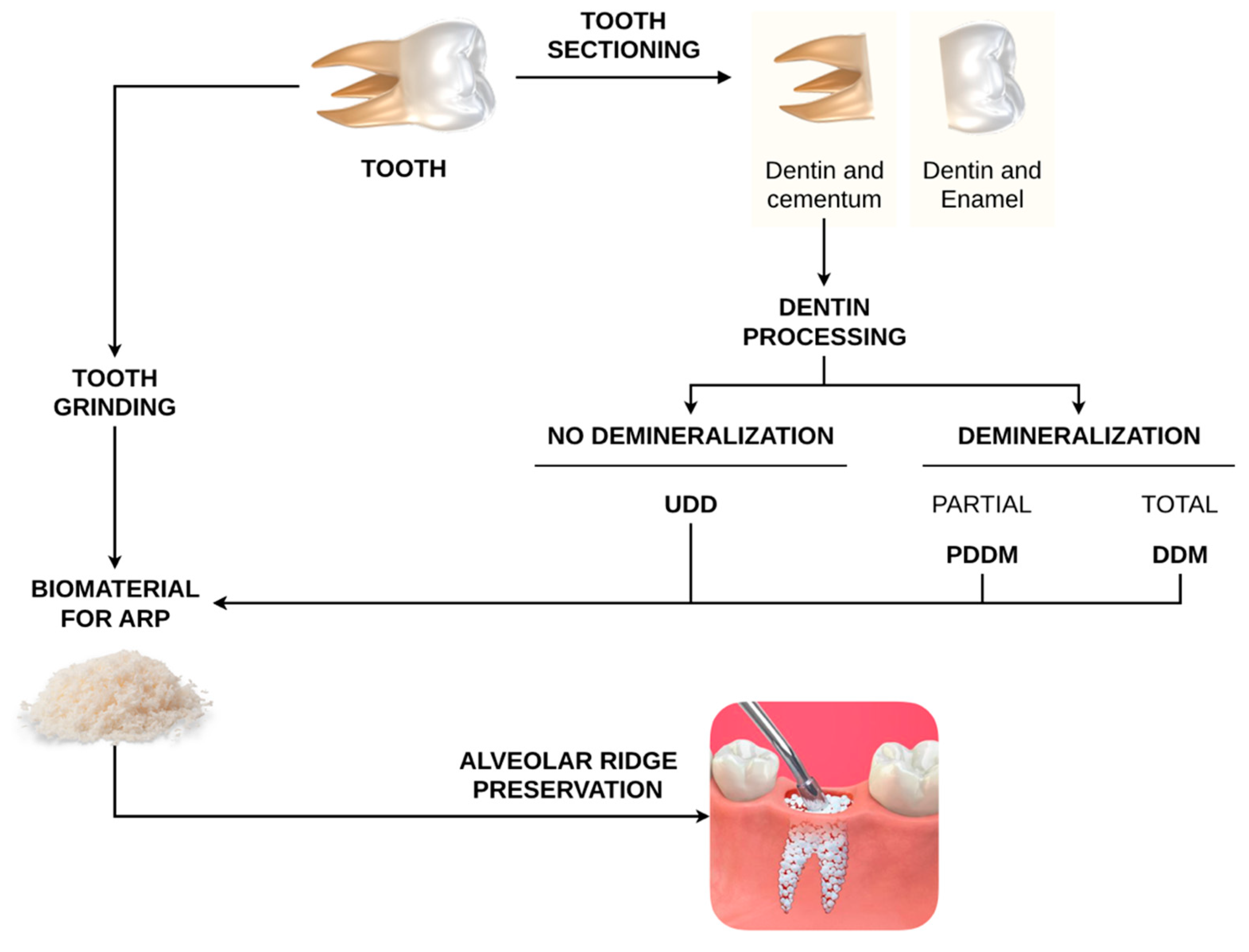
3.2. Dentin Processing

As aforementioned, autogenous teeth can be used as bone graft material with osteoconductive and osteoinductive potential [9,12]. However, several concerns have been addressed regarding the need for any dentin processing prior to bone grafting for the purpose of clinical optimization. Some examples are the extraction of non-collagenous proteins [42], elimination of the organic matrix [14] and finally, one of the most commonly used, dentin preparation by demineralization [12].
The hypothesis of demineralization is that through this procedure, the organic substances (type 1 collagen fibrils, non-collagenous proteins and growth factors) will be more exposed, decreasing the graft crystallinity and increasing its porosity and surface area [16,35,43]. This process releases growth factors and non-collagenous proteins, which in turn, results in an enhanced osteoinductive activity [14].
Although protocols vary from study to study, a general protocol includes tooth extraction, removal of soft tissue, carious lesions and filling materials of any nature [17,44,45,46], sectioning into blocks or particles and finally choosing the degree of demineralization [47]. Among the demineralization agent are ethylenediaminetetraacetic acid (EDTA), phosphoric acid, chloridric acid, nitric acid, hydrogen oxide, ethyl ether and ethyl alcohol [14]. Hence, dentin materials were categorized into 3 categories: demineralized dentin matrix (DDM), partially demineralized dentin matrix (PDDM) and undemineralized dentin (UDD) [12].
While some investigators have reported success when using DDM (or PDDM) [17,37,39,40,44,48,49,50] others prefer using in its undemineralized form [38,51,52,53,54,55,56,57].
Mineralized dentin particles offer a mechanical stability, creating a solid site for implant placement [38,52]. With the use of a mineralized graft, although the osteoinductive properties of dentin may be delayed, the low crystallinity of dentin hydroxyapatite allows the progressive bone remodeling [15,30].
Due to a lack of uniformity and standardization in the literature, it is difficult to determine with certainty which form of graft is advantageous for which clinical indication. Regarding ARP procedures, several authors have reported success when using DDM, PDDM and UDD, indicating that each form can be a viable option [38,40,41]. Nonetheless, some authors suggest an approach patient-based. DDM and PDDM can be indicated when the socket walls have already been resorbed or destroyed due to pathological causes [58]. The exposure of growth factors and non-collagenous proteins, as previously stated, will allow an earlier regeneration [14,38]. The UDM on the other hand, thanks to the mechanical stability inherent to the graft, may allow an earlier placement of dental implants [38].
As previously stated, the amount of biomaterial that the clinician can gather is dependent on the extension of carious lesions and filling materials [17,44,45,46], nonetheless, one possible approach that can overcome this limitation is the extraction of impacted third molars, when it is required a larger amount of biomaterial [51,59].
In order to obtain a demineralized graft, the Korea Tooth Bank, established in Seoul, was one of the first to be available for clinicians [60]. However, due to this time-consuming option, several devices appeared on the market for this purpose.
The VacuaSonic® (Cosmobiomedicare, Seoul, Korea) produces a demineralized graft. This system comes with a powder reagent (DecalSi® DM Powder reagent) and a block reagent (DecalSi® DM Block reagent), giving the clinician a choice, on which form of graft he prefers. According to the manufacturer, the process takes 30 min for powder graft and 2 h for block graft [50].
Another system that can be used is the Smart Dentin Grinder™ (Kometa Bio ltd., Holon, Israel) which is a device that grinds the tooth to particles of 250–1200 µm, according to the manufacturer. Alongside this grinder, comes a disposable griding chamber (single-use) as well as a dentin cleanser (0.5 M NaOH and 30% ethanol (V/V)) which is applied for 5 min and a phosphate buffer saline (PBS) solution with calcium and magnesium with an application time of 1 min, repeating this last step. This device can be used in order to produce a mineralized or partially demineralized graft, which in this last case, a 10% solution of EDTA during 2 min is added [38].
Finally, another system commonly used to produce a demineralized graft is the Tooth Transformer device (TT Tooth Transformer srl. Milan, Italy). This device comes with a tooth grinder and a series of disposable accessories that contacts with the resulting autologous material and liquid responsible for the demineralization. According to Minetti, this process takes approximately 25 min [61]. Regarding prices, while the VacuaSonic® (Cosmobiomedicare, Seoul, Korea) costs around 12365 €, the Smart Dentin Grinder™ (Kometa Bio ltd., Holon, Israel) costs around 1277 € and finally the Tooth Transformer (TT Tooth Transformer srl. Milan, Italy) device has a price of around 2000 €.
Due to the potential of the autogenous tooth as a bone substitute, several clinical applications have appeared in the literature besides ARP procedures [9,12,35].
One study performed lateral alveolar ridge augmentation comparing the use of autogenous tooth roots versus the use of autogenous bone blocks [62]. In this particular study, after 26 weeks of healing, the implants were placed with no significant difference between groups (p > 0.05) in terms of primary implant stability quotient [62].
The use of autogenous tooth graft was also associated with the treatment of grade II and III furcation defects by one study, which compared the clinical and radiologic performance of this graft material with the use of freeze-dried bone allograft [63]. The results of this study point out the potential benefit that autogenous tooth can have as a bone graft material, demonstrating a significant reduction in vertical bone depth, horizontal bone depth as well as radiographically bony defect [63].
Another possible application for autogenous tooth is in sinus floor elevation procedures [64,65]. One particular study compared the use of autogenous tooth versus the bovine-derived xenograft Bio-Oss (Geistlich Pharma AG, Wolhusen, Switzerland) [64]. With a follow up period of 4 months, there was no significant difference between the two groups after a clinical, radiologic and histomorphometry assessment [64]. In another study, Kim et al. performed a micromorphometry and histological evaluation 9 months after sinus bone graft using autogenous tooth [65]. This evaluation concluded that autogenous tooth showed excellent bone healing, proving to be a viable option for this kind of procedure [65].

4. Demineralized Dentin Matrix (DDM)

4.1. Preclinical Studies

Two preclinical studies have confirmed the potential of human DDM placed in extraction sockets as well as the influence that this biomaterial has on proteins and growth factors such as BMP-2, BMP-4 and vascular endothelial growth factor (VEGF) [45,46]. In both studies, there was a common protocol: after the removal of caries lesions, pulp tissues and periodontal ligament, the agent responsible for the demineralization was a 10% EDTA solution with a pH varying from 7.2 to 7.3 [45,46]. After cutting with a cryostat, one study stored the material in a sterile phosphate buffered saline (PBS) with penicillin and streptomycin for decontamination [46].

4.1.1. Histologic and Histomorphometric Outcomes

A histologic and morphometric analysis showed that human DDM integrated newly formed bone after 14 days showing histological features of mature bone, proving its osteoconductivity [45,46].

4.1.2. Immunohistochemistry Outcomes

One interesting aspect regarding these studies results from the immunohistochemistry evaluation. Oliveira et al. found that with the degradation of the human DDM, the number of BMP-2 and BMP-4 immunostained cells increased at day 10, suggesting that this event is key in stimulating cellular differentiation and consequently bone formation [46]. A similar result can be seen in Reis-Filho et al.’s study after a period of 14 and 21 days, where with the human DDM resorption, the expression of VEGF increased, indicating angiogenesis, which in turn accelerates the healing process [45]. Both these results support the evidence of the osteoinductive capacity of the DDM [45,46].

4.2. Clinical Studies

Furthermore, several clinical studies have been published endorsing the use of DDM in ARP despite diverging in terms of the protocol used for dentin processing [17,37,39,40,44,48,49,50,66,67].
The removal of carious lesions, fillings and soft tissues seems shoes an apparent unanimity [17,37,39,44,66], however, while most authors use dentin, enamel and cementum, some eliminate these last two [40], or simply use the root portion of the tooth [37]. Some investigators defend the use of dentin alone due to its osteoconductive and osteoinductive properties being similar to alveolar bone [35,43] and in this way enamel shall be removed because it has high-crystalline calcium phosphate and therefore might complicate the absorption process [15,30]. On the other hand, it is described in some protocols the use of the whole tooth as a bone graft material, combining the chemical properties of dentin with the mechanical advantage that enamel brings, allowing an earlier placement of dental implants [38,52,57].
In the majority of these studies, the demineralization agent was not specified [17,39,44,48,49,50,66], but rather the explanation that the autogenous graft went through a dehydration, defatting, demineralized and lyophilized course [39,66]. Nonetheless, studies often report the use of 70% ethyl alcohol, 0.6 N chloridric acid and 2% nitric acid [37,40,67]. The size of the graft particles varies from 200 to 1000 µm [37,39,40,66].
These studies evaluated the efficacy of this biomaterial in the clinical, radiologic, histologic and morphometric scenario.

4.2.1. Clinical Outcomes

Overall, grafted sites healed without any clinical manifestation of infection, wound dehiscence, or implant failure, in the cases where dental implants were placed [17,44,50]. In these studies, the primary stabilization ranged from 71.8 to 74 implant stability quotient (ISQ) [37,39,49].
Several intervention studies performed an interesting evaluation comparing the clinical, radiologic and histologic efficacy between DDM and a standard xenograft (Bio-Oss (Geistlich Pharma AG, Wolhusen, Switzerland) with excellent and proven efficacy [17,39,40]. Both groups, showed comparable healing process, implant stability and bone formation ration, proving that this biomaterial can be a viable alternative to the xenograft, with the advantage of being autogenous [17,39,40]. Furthermore, from the patient’s perspective, autogenous teeth were associated with low levels of pain and swelling [17].

4.2.2. Radiologic Outcomes

Regarding radiologic outcomes, the studies presented favorable results. The mean density of the graft decreased with time, with the architecture of the DDM becoming increasingly more similar to that of the surrounding bone, suggesting a satisfactory bone healing [40,50,67].

4.2.3. Histologic and Histomorphometric Outcomes

Histologic and histomorphometric analysis showed a good tissue integration with a direct union between the new bone and the graft material, evidencing osteoconductive and osteoinductive properties [39,66]. It was reported a dense lamellar bone formation [48,49,50] associated with connective tissue reach in angiogenesis [37], fulfilling the goal of minimizing the alveolar bone loss in extraction sockets [44]. The follow-up period on which these results were found varied from 3.5 to 6 months [39,44,48,66].
The most important limitations when comparing these studies are the variety of protocols and adjuvant materials used, for instance absorbable [44,50] or non-absorbable membranes [67], or the use of platelet-rich fibrin (PRF) [40].

5. Partially Demineralized Dentin Matrix (PDDM)

Regarding PDDM, only two interventional studies have studied its clinical potential for ARP, one pilot on PDDM associated with platelet-rich plasma [36], and another randomized trial concerning PDDM alone [41].
In terms of protocol for dentin processing, both studies present slight changes. In both studies, the soft tissues, caries and calculus were removed with the teeth being crushed with the auxiliary of different grinders, who generated graft particles varying between 300 and 800 µm [36,41]. The main difference in terms of protocol is the agent used for partial demineralization. On the one side, Minamizato et al. used a 2% HNO3 solution (pH 1.0) for 10 min, followed by an extensive 10 min rinse with 0.1 M Tris-HCl (pH 7.4) [36]. On the other side, Joshi et al. used lactic acid (1N) for a 15–20 min period and later a sterile normal saline solution [41]. The reason behind the choice of an organic acid was, according to Joshi et al., the contact between the residues with human tissues [41].

5.1. Clinical Outcomes

Clinically speaking, the postoperative follow-up occurred uneventfully [36,41]. In the Minamizato trial, dental implants were placed at 4–6 months postoperative with primary stability and insertion torque varying from 25 to 40 N cm [36]. At the time of the second surgery, the implant stability quotient (ISQ) was over 60 in all cases, suggesting a positive osteointegration [36]. One factor that could help the healing process is the demineralization the occurred, enhancing the antimicrobial activity of some dentin components [36]. In the Joshi Trial, although no implants were placed, after a period of 4 months, the authors reported that the sockets grafted with PDDM showed visually less width shrinkage when compared with the sites grafted with β-TCP and non-grafted sites [41].

5.2. Radiologic Outcomes

Radiographic assessment was made by X-ray panoramic and cone beam computed tomography (CBCT). This analysis showed that the radiopacity of the PDDM decreased gradually with the lamina dura around the graft becoming progressively indistinguishable [36]. Comparing the dimensional changes of the alveolar ridge between PDDM and β-TCP, the width and height loss was lower in the PDDM group, with these values being statistically significant [41]. In terms of ridge height, while in the PDDM group there was a reduction of 0.28 ± 0.13 mm, in the β-TCP group there was a reduction of 1.72 ± 0.56 mm and in the control group it was reported a reduction of 2.60 ± 0.88 mm (p < 0.05) [41]. In terms of width, a similar result was achieved, with the control group showing an increased reduction (2.29 ± 0.40 mm), followed by the β-TCP group (1.45 ± 0.40 mm) and finally the PDDM group (0.15 ± 0.08 mm) (p < 0.05) [41].

5.3. Histologic Outcomes

Finally, histologic analysis showed a positive integration of the PDDM at 4 to 6 months postoperative. Histologic specimens of the PDDM group showed newly formed bone in both studies [36,41], with a higher percentage of osteoid formation, when comparing to the β-TCP group [41].
These two studies point out the use of PDDM as a viable option in alveolar ridge regeneration, displaying good clinical, radiologic and histologic outcomes.

6. Undemineralized Dentin Matrix (UDD)

In terms of UDD, a solid number of preclinical and clinical studies have been performed evaluating its efficacy [38,51,52,53,54,55,56,57,68].

6.1. Preclinical Studies

The protocol used for the preparation of this biomaterial was very similar in all of the preclinical studies. The crown portion of the tooth was removed, as well as pulp tissues and periodontal ligament still attached [52,53,68]. This was made by using curettes, ultrasonic devices, hand instruments and specific burs [52,53,68]. In all studies, the preparation was rinsed with a saline solution along with a basic alcohol cleanser [52,53,68]. Finally, either using the Smart Dentin Grinder™ (Kometa Bio ltd., Holon, Israel) [52,53] or a specific grinder [68], the teeth were grinded into particles with diameters over 300 µm and less than 1200 µm [52,53], or between 350 and 500 µm [68].

Histologic and Histomorphometric Outcomes

After assessing the viability of UDD under radiologic, histologic and histomorphometry analysis, some preclinical studies reached opposite conclusions. In 2015, one study, after histologic and histomorphometry analysis, reported that the use of UDD, after 8 weeks did not offered any improvement in bone regeneration, showing in terms of ratio of bone to total area of each probe 170 ± 16 µm3 for the control group (no bone graft material used) and 71 ± 14 µm3 for the UDD group, with a significant difference (p < 0.05) [68].
The opposite was concluded in two other preclinical studies [52,53]. When compared to a healing without any bone graft material, after a 90 days observation period, the added benefit of the UDD was proven [52,53]. In one study, the percentage of newly bone formation was 91.32 ± 0.8% in the UDD group and 65.89 ± 0.6% in the control group (p < 0.05) [52]. In the other study, the percentage of immature bone was 14.2 ± 0.66% in the UDD group and 35.17 ± 0.74% in the control group (p < 0.05) [53].

6.2. Clinical Studies

Equivalently to the studies mentioned before, these clinical studies applied a similar protocol when preparing UDD. Generally speaking, after the teeth extraction, removal of crowns, fillings of any nature, pulp tissues and periodontal ligament, the biomaterial was grinded in order to generate particles with a diameter varying from 300 to 1200 µm [38,51,54,55,56,57,59]. In most of the studies, a basic alcohol cleanser consisting of 0.5 M of NaOH and 30%/20% alcohol as well as a sterile phosphate buffered saline were applied to the samples gathered [38,55,56]. This step is important in order to remove organic debris and also possible bacteria and toxins found in dentine [38,55,56].
When assessing the efficacy of the UDD in these studies, one aspect that is worthy of mention is that in some studies a combination of UDD was used either with platelet-rich fibrin [54] or with leukocyte-platelet-rich fibrin and fibrinogen [57], which can be seen as a drawback when analyzing this biomaterial due to lack of standardization as well as understanding the real influence of the UDD.

6.2.1. Clinical Outcomes

Clinically, the healing process was satisfactory, with no major post-operative complications, with less inflammation and rejection response, one potential limitation of other types of bone grafts [38,57].
One particular split-mouth randomized double-blind study deviated from the usual analysis of this subject. This study used UDD from lower third molar extractions and evaluated clinical outcomes such as: pocket depth, recession, clinical attachment level regarding the lower second molar, as well as patient-related outcomes: pain, healing and swelling [51]. After a 3-month observation period, in terms of pocket depth (control group: 3.43 ± 0.79, UDD group: 2.86 ± 0.9), gingival recession (control group: −2.29 ± 1.25, UDD group: −2.86 ± 0.9) and clinical attachment level (control group: 1.14 ± 1.57, UDD group: 0 ± 0), the differences found were not statistically significant (p > 0.05) [51]. Finally, regarding patient-related outcomes (pain, healing and swelling), similar results were found, with no statistically significant differences found between groups (p > 0.05) [51]. A similar result was found in a split-mouth clinical trial where the use of the lower third molar as a bone substitute resulted in a significant reduction of the pocket depth, mainly in the first 3 months [59]. After a 6-month-period, the bone density found in the test group was greater, with statistically significant difference (p < 0.001) [59].

6.2.2. Radiologic Outcomes

Radiologically, CBCT images showed that alveolar ridge dimensions were preserved in most cases [54,55,57]. In the study by Andrade et al. the vertical and horizontal dimensions of the sockets grafted were preserved, and in some cases increased [57]. One particular study by Pohl et al. performed a retrospective radiographic cone-beam computed tomography in order to better assess the efficacy of this graft in terms of volume stability [54]. Comparing the preoperative and the postoperative (4 months after ARP procedure) dimensions, the reduction in the buccal bone plate thickness at 1 mm, 3 mm and 5 mm bellow the buccal crest was, respectively: −0.87 ± 0.84 mm; −0.60 ± 0.70 mm and −0.41 ± 0.55 mm [54]. Following the same level measurements, the mean ridge width changes were, respectively: 1.38 ± 1.24 mm, 0.82 ± 1.13 mm, and 0.43 ± 0.89 mm [54]. Finally, the authors concluded that the average mid-buccal bone height gain was 1.1% and the mid-lingual height gain was 5.6% [54]. Another study that evaluated through CBCT analysis the alveolar ridge dimensions before and 4 months after the ARP procedure reported a loss of 0.76 mm in the vertical dimension and a loss of 1.1 mm in the horizontal dimension [69].

6.2.3. Histologic and Histomorphometry Outcomes

Regarding histologic and histomorphometry analyses, UDD generated moderate osteoblastic activity, presenting some dentin fragments as well as connective tissue, suggesting a gradual increase in the graft resorption and consequently bone formation [54,56,57]. Additionally, Andrade et al. reported a consecutive increase of the bone percentage (26.3% at 4 months and 66.5% at 6 months), and a decrease on dentin (10.4% at 4 months and 0.9% at 6 months) and connective tissue (63.3% at 4 months and 32.6% at 6 months) in the socket, substantiating a gradual bone formation along with graft resorption [57].

7. Conclusions

Autogenous teeth as a biomaterial for ARP present osteoconductive and osteoinductive properties, which suggests that they can be an equally effective bone substitute. In some cases, autogenous teeth have superior clinical performance when compared with other grafts. According to this literature review, autogenous teeth, in every form of processing, present potential within the clinical, radiologic and histologic outcomes.
Nonetheless, further research, with standardized protocols in terms of patient selection, dentin processing, surgery procedure and comparison with other grafts are essential in order to reach a definitive conclusion about this graft efficacy. Particularly, there is a scarcity of studies comparing the different dentin processing protocols with each other, though the difference between each method may only rely on the advantages and fallouts of the method itself (demineralization vs. non-demineralization) and not in the clinical potential per se.

Author Contributions

Conceptualization, J.C., J.B., and A.S.; validation, A.S.; writing—original draft preparation, J.C., J.B., V.M., and A.S.; writing—review and editing, J.C., J.B., V.M., J.J.M., P.M., G.A. and A.S. All authors have read and agreed to the published version of the manuscript.

Funding

This research received no external funding.

Institutional Review Board Statement

Not applicable.

Informed Consent Statement

Not applicable.

Conflicts of Interest

The authors declare no conflict of interest.

References

  1. Avila-Ortiz, G.; Elangovan, S.; Kramer, K.W.O.; Blanchette, D.; Dawson, D.V. Effect of alveolar ridge preservation after tooth extraction: A systematic review and meta-analysis. J. Dent. Res. 2014, 93, 950–958. [Google Scholar] [CrossRef]
  2. Ten Heggeler, J.M.A.G.; Slot, D.E.; Van Der Weijden, G.A. Effect of socket preservation therapies following tooth extraction in non-molar regions in humans: A systematic review. Clin. Oral Implants Res. 2011, 22, 779–788. [Google Scholar] [CrossRef]
  3. Avila-Ortiz, G.; Chambrone, L.; Vignoletti, F. Effect of alveolar ridge preservation interventions following tooth extraction: A systematic review and meta-analysis. J. Clin. Periodontol. 2019, 46, 195–223. [Google Scholar] [CrossRef] [Green Version]
  4. Jambhekar, S.; Kernen, F.; Bidra, A.S. Clinical and histologic outcomes of socket grafting after flapless tooth extraction: A systematic review of randomized controlled clinical trials. J. Prosthet. Dent. 2015, 113, 371–382. [Google Scholar] [CrossRef]
  5. Tan, W.L.; Wong, T.L.T.; Wong, M.C.M.; Lang, N.P. A systematic review of post-extractional alveolar hard and soft tissue dimensional changes in humans. Clin. Oral Implants Res. 2012, 23, 1–21. [Google Scholar] [CrossRef]
  6. Troiano, G.; Zhurakivska, K.; Lo Muzio, L.; Laino, L.; Cicciù, M.; Lo Russo, L. Combination of Bone Graft and Resorbable Membrane for Alveolar Ridge Preservation: A Systematic Review, Meta-analysis and Trial Sequential Analysis. J. Periodontol. 2017, 89, 46–57. [Google Scholar] [CrossRef]
  7. Van Der Weijden, F.; Dell’Acqua, F.; Slot, D.E. Alveolar bone dimensional changes of post-extraction sockets in humans: A systematic review. J. Clin. Periodontol. 2009, 36, 1048–1058. [Google Scholar] [CrossRef] [PubMed]
  8. Vignoletti, F.; Matesanz, P.; Rodrigo, D.; Figuero, E.; Martin, C.; Sanz, M. Surgical protocols for ridge preservation after tooth extraction: A systematic review. Clin. Oral Implants Res. 2012, 23, 22–38. [Google Scholar] [CrossRef] [PubMed]
  9. Ramanauskaite, A.; Sahin, D.; Sader, R.; Becker, J.; Schwarz, F. Efficacy of autogenous teeth for the reconstruction of alveolar ridge deficiencies: A systematic review. Clin. Oral Investig. 2019, 23, 4263–4287. [Google Scholar] [CrossRef] [PubMed]
  10. Bassir, S.; Alhareky, M.; Wangsrimongkol, B.; Jia, Y.; Karimbux, N. Systematic Review and Meta-Analysis of Hard Tissue Outcomes of Alveolar Ridge Preservation. Int. J. Oral Maxillofac. Implants 2018, 33, 979–994. [Google Scholar] [CrossRef] [PubMed]
  11. Stumbras, A.; Kuliesius, P.; Januzis, G.; Juodzbalys, G. Alveolar Ridge Preservation after Tooth Extraction Using Different Bone Graft Materials and Autologous Platelet Concentrates: A Systematic Review. J. Oral Maxillofac. Res. 2019, 10, 1–15. [Google Scholar] [CrossRef]
  12. Gual-Vaqués, P.; Polis-Yanes, C.; Estrugo-Devesa, A.; Ayuso-Montero, R.; Marí-Roig, A.; López-López, J. Autogenous teeth used for bone grafting: A systematic review. Med. Oral Patol. Oral Cir. Bucal 2018, 23, e112–e119. [Google Scholar] [CrossRef] [PubMed]
  13. Albrektsson, T.; Johansson, C. Osteoinduction, osteoconduction and osseointegration. Eur. Spine J. 2001, 10, S96–S101. [Google Scholar] [CrossRef] [PubMed] [Green Version]
  14. Tabatabaei, F.S.; Tatari, S.; Samadi, R.; Moharamzadeh, K. Different methods of dentin processing for application in bone tissue engineering: A systematic review. J. Biomed. Mat. Res. Part A 2016, 104, 2616–2627. [Google Scholar] [CrossRef] [PubMed]
  15. Kim, Y.-K. Bone graft material using teeth. J. Korean Assoc. Oral Maxillofac. Surg. 2012, 38, 134–138. [Google Scholar] [CrossRef] [Green Version]
  16. Kim, Y.-K.; Lee, J.; Um, I.-W.; Kim, K.-W.; Murata, M.; Akazawa, T.; Mitsugi, M. Tooth-derived bone graft material. J. Korean Assoc. Oral Maxillofac. Surg. 2013, 39, 103. [Google Scholar] [CrossRef] [PubMed] [Green Version]
  17. Wu, D.; Zhou, L.; Lin, J.; Chen, J.; Huang, W.; Chen, Y. Immediate implant placement in anterior teeth with grafting material of autogenous tooth bone vs xenogenic bone. BMC Oral Health 2019, 19, 1–11. [Google Scholar] [CrossRef] [PubMed] [Green Version]
  18. Barone, A.; Borgia, V.; Covani, U.; Ricci, M.; Piattelli, A.; Lezzi, G. Flap versus flapless procedure for ridge preservation in alveolar extraction sockets: A histological evaluation in a randomized clinical trial. Clin. Oral Implants Res. 2015, 26, 806–813. [Google Scholar] [CrossRef]
  19. Barallat, L.; Ruíz-Magaz, V.; Levi, P.A.; Mareque-Bueno, S.; Galindo-Moreno, P.; Nart, J. Histomorphometric results in ridge preservation procedures comparing various graft materials in extraction sockets with nongrafted sockets in humans: A systematic review. Implant Dent. 2014, 23, 539–554. [Google Scholar] [CrossRef]
  20. De Risi, V.; Clementini, M.; Vittorini, G.; Mannocci, A.; De Sanctis, M. Alveolar ridge preservation techniques: A systematic review and meta-analysis of histological and histomorphometrical data. Clin. Oral Implants Res. 2015, 26, 50–68. [Google Scholar] [CrossRef]
  21. Vittorini Orgeas, G.; Clementini, M.; De Risi, V.; De Sanctis, M. Surgical Techniques for Alveolar Socket Preservation: A Systematic Review. Int. J. Oral Maxillofac. Implants 2013, 28, 1049–1061. [Google Scholar] [CrossRef] [Green Version]
  22. Horváth, A.; Mardas, N.; Mezzomo, L.A.; Needleman, I.G.; Donos, N. Alveolar ridge preservation. A systematic review. Clin. Oral Investig. 2013, 17, 341–363. [Google Scholar] [CrossRef]
  23. Chan, H.-L.; Lin, G.-H.; Fu, J.-H.; Wang, H.-L. Alterations in Bone Quality After Socket Preservation with Grafting Materials: A Systematic Review. Int. J. Oral Maxillofac. Implants 2013, 28, 710–720. [Google Scholar] [CrossRef] [PubMed] [Green Version]
  24. Hürzeler, M.B.; Zuhr, O.; Schupbach, P.; Rebele, S.F.; Emmanouilidis, N.; Fickl, S. The socket-shield technique: A proof-of-principle report. J. Clin. Periodontol. 2010, 37, 855–862. [Google Scholar] [CrossRef] [PubMed]
  25. Mourya, A.; Mishra, S.K.; Gaddale, R.; Chowdhary, R. Socket-shield technique for implant placement to stabilize the facial gingival and osseous architecture: A systematic review. J. Investig. Clin. Dent. 2019, 10, e12449. [Google Scholar] [CrossRef]
  26. Gharpure, A.S.; Bhatavadekar, N.B. Current evidence on the socket-shield technique: A systematic review. J. Oral Implantol. 2017, 43, 395–403. [Google Scholar] [CrossRef]
  27. Yeomans, J.D.; Urist, M.R. Bone Induction by Decalcified Dentine Implanted into Oral, Osseous and Muscle Tissues. Arch. Oral Biol. 1967, 12, 999–1008. [Google Scholar] [CrossRef]
  28. Bang, G.; Urist, M.R. Bone Induction in Excavation Chambers in Matrix of Decalcified Dentin. Arch. Surg 1967, 94, 781–789. [Google Scholar] [CrossRef]
  29. Kim, Y.-K.; Lee, J.K.; Kim, K.-W.; Um, I.-W.; Murata, M. Healing Mechanism and Clinical Application of Autogenous Tooth Bone Graft Material; Pignatello, R., Ed.; IntechOpen: London, UK, 2013; Volume 395, ISBN 978-953-51-1051-4. [Google Scholar]
  30. Kim, Y.K.; Kim, S.G.; Oh, J.S.; Jin, S.C.; Son, J.S.; Kim, S.Y.; Lim, S.Y. Analysis of the inorganic component of autogenous tooth bone graft material. J. Nanosci. Nanotechnol. 2011, 11, 7442–7445. [Google Scholar] [CrossRef] [PubMed]
  31. Kim, Y.K.; Kim, S.G.; Bae, J.H.; Um, I.W.; Oh, J.S.; Jeong, K.I. Guided bone regeneration using autogenous tooth bone graft in implant therapy: Case series. Implant Dent. 2014, 23, 138–143. [Google Scholar] [CrossRef]
  32. Bessho, K.; Tanaka, N.; Matsumoto, J.; Tagawa, T.; Murata, M. Human Dentin-matrix-derived Bone Morphogenetic Protein. J. Dent. Res. 1991, 70, 171–175. [Google Scholar] [CrossRef]
  33. Boden, S.D.; Liu, Y.; Hair, G.A.; Helms, J.A.; Hu, D.; Racine, M.; Nanes, M.S.; Titus, L. LMP-1, A LIM-Domain Protein, Mediates BMP-6 Effects on Bone Formation. Endocrinology 1998, 139, 5125–5134. [Google Scholar] [CrossRef]
  34. Um, I.W. Demineralized Dentin Matrix (DDM) As a Carrier for Recombinant Human Bone Morphogenetic Proteins (rhBMP-2). In Advances in Experimental Medicine and Biology; Springer: Berlin, Germany, 2018; Volume 1077, pp. 487–499. ISBN 9789811309472. [Google Scholar]
  35. Um, I.-W.; Young-Kyun Kim, Y.-K.; Mitsugi, M. Demineralized dentin matrix scaffolds for alveolar bone engineering. J. Indian Prosthodont. Soc. 2017, 17, 120–127. [Google Scholar] [CrossRef]
  36. Minamizato, T.; Koga, T.; Takashi, I.; Nakatani, Y.; Umebayashi, M.; Sumita, Y.; Ikeda, T.; Asahina, I. Clinical application of autogenous partially demineralized dentin matrix prepared immediately after extraction for alveolar bone regeneration in implant dentistry: A pilot study. Int. J. Oral Maxillofac. Surg. 2017, 47, 125–132. [Google Scholar] [CrossRef]
  37. Kim, S.Y.; Kim, Y.K.; Park, Y.H.; Park, J.C.; Ku, J.K.; Um, I.W.; Kim, J.Y. Evaluation of the healing potential of demineralized dentin matrix fixed with recombinant human bone morphogenetic protein-2 in bone grafts. Materials 2017, 10, 49. [Google Scholar] [CrossRef] [PubMed] [Green Version]
  38. Binderman, I.; Hallel, G.; Nardy, C.; Yaffe, A.; Sapoznikov, L. A Novel Procedure to Process Extracted Teeth for Immediate Grafting of Autogenous Dentin. JBR J. Interdiscip. Med. Dent. Sci. 2014, 2, 2–6. [Google Scholar] [CrossRef]
  39. Pang, K.-M.; Um, I.-W.; Kim, Y.-K.; Woo, J.-M.; Kim, S.-M.; Lee, J.-H. Autogenous demineralized dentin matrix from extracted tooth for the augmentation of alveolar bone defect: A prospective randomized clinical trial in comparison with anorganic bovine bone. Clin. Oral Implants Res. 2016, 28, 809–815. [Google Scholar] [CrossRef]
  40. Li, P.; Zhu, H.C.; Huang, D.H. Autogenous DDM versus Bio-Oss granules in GBR for immediate implantation in periodontal postextraction sites: A prospective clinical study. Clin. Implant Dent. Relat. Res. 2018, 20, 923–928. [Google Scholar] [CrossRef]
  41. Joshi, C.P.; Dani, N.H.; Khedkar, S.U. Alveolar ridge preservation using autogenous tooth graft versus beta-tricalcium phosphate alloplast: A randomized, controlled, prospective, clinical pilot study. J. Indian Soc. Periodontol. 2016, 20, 429–434. [Google Scholar] [CrossRef] [PubMed]
  42. Huang, B.; Sun, Y.; MacIejewska, I.; Qin, D.; Peng, T.; McIntyre, B.; Wygant, J.; Butler, W.T.; Qin, C. Distribution of SIBLING proteins in the organic and inorganic phases of rat dentin and bone. Eur. J. Oral Sci. 2008, 116, 104–112. [Google Scholar] [CrossRef] [Green Version]
  43. Park, M.; Mah, Y.J.; Kim, D.H.; Kim, E.S.; Park, E.J. Demineralized deciduous tooth as a source of bone graft material: Its biological and physicochemical characteristics. Oral Surg. Oral Med. Oral Pathol. Oral Radiol. 2015, 120, 307–314. [Google Scholar] [CrossRef]
  44. Minetti, E.; Giacometti, E.; Gambardella, U.; Contessi, M.; Ballini, A.; Marenzi, G.; Celko, M.; Mastrangelo, F. Alveolar socket preservation with different autologous graft materials: Preliminary results of a multicenter pilot study in human. Materials 2020, 13, 1153. [Google Scholar] [CrossRef] [PubMed] [Green Version]
  45. Reis-Filho, C.R.; Silva, E.R.; Martins, A.B.; Pessoa, F.F.; Gomes, P.V.N.; De Araújo, M.S.C.; Miziara, M.N.; Alves, J.B. Demineralised human dentine matrix stimulates the expression of VEGF and accelerates the bone repair in tooth sockets of rats. Arch. Oral Biol. 2012, 57, 469–476. [Google Scholar] [CrossRef]
  46. De Oliveira, G.S.; Miziara, M.N.; Silva, E.R.D.; Ferreira, E.L.; Biulchi, A.P.F.; Alves, J.B. Enhanced bone formation during healing process of tooth sockets filled with demineralized human dentine matrix. Aust. Dent. J. 2013, 58, 326–332. [Google Scholar] [CrossRef]
  47. Gharpure, A.S.; Bhatavadekar, N.B. Clinical efficacy of tooth-bone graft: A systematic review and risk of bias analysis of randomized control trials and observational studies. Implant Dent. 2017, 26, 119–134. [Google Scholar] [CrossRef] [PubMed]
  48. Chung, J.-H.; Lee, J.-H. Study of bone healing pattern in extraction socket after application of demineralized dentin matrix material. J. Korean Assoc. Oral Maxillofac. Surg. 2011, 37, 365–374. [Google Scholar] [CrossRef]
  49. Park, S.-M.; Um, I.-W.; Kim, Y.-K.; Kim, K.-W. Clinical application of auto-tooth bone graft material. J. Korean Assoc. Oral Maxillofac. Surg. 2012, 38, 1–8. [Google Scholar] [CrossRef] [Green Version]
  50. Kim, E.-S. Autogenous fresh demineralized tooth graft prepared at chairside for dental implant. Maxillofac. Plast. Reconstr. Surg. 2015, 37, 4–9. [Google Scholar] [CrossRef] [Green Version]
  51. Nadershah, M.; Zahid, T.M. Use of Autogenous Dentin Graft in Mandibular Third Molar Extraction Sockets: A Split-Mouth Randomized Double-Blind Study. Int. J. Pharm. Res. Allied Sci. 2019, 8, 73–79. [Google Scholar]
  52. Calvo-Guirado, J.L.; Cegarra Del Pino, P.; Sapoznikov, L.; Delgado Ruiz, R.A.; Fernández-Domínguez, M.; Gehrke, S.A. A new procedure for processing extracted teeth for immediate grafting in post-extraction sockets. An experimental study in American Fox Hound dogs. Ann. Anat. 2018, 217, 14–23. [Google Scholar] [CrossRef]
  53. Calvo-Guirado, J.L.; Maté-Sánchez De Val, J.E.; Ramos-Oltra, M.L.; Martínez, C.P.A.; Ramírez-Fernández, M.P.; Maiquez-Gosálvez, M.; Gehrke, S.A.; Fernández-Domínguez, M.; Romanos, G.E.; Delgado-Ruiz, R.A. The use of tooth particles as a biomaterial in post-extraction sockets. Experimental study in dogs. Dent. J. 2018, 6, 12. [Google Scholar] [CrossRef] [Green Version]
  54. Pohl, S.; Binderman, I.; Tomac, J. Maintenance of alveolar ridge dimensions utilizing an extracted tooth dentin particulate autograft and platelet-rich fibrin: A retrospective radiographic cone-beam computed tomography study. Materials 2020, 13, 1083. [Google Scholar] [CrossRef] [Green Version]
  55. Dwivedi, A.; Kour, M. A neoteric procedure for alveolar ridge preservation using autogenous fresh mineralized tooth graft prepared at chair side. J. Oral Biol. Craniofac. Res. 2020, 10, 535–541. [Google Scholar] [CrossRef]
  56. Del Canto-Díaz, A.; De Elío-Oliveros, J.; Del Canto-Díaz, M.; Alobera-Gracia, M.A.; Del Canto-Pingarrón, M.; Martínez-González, J.M. Use of autologous tooth-derived graft material in the post-extraction dental socket. Pilot study. Med. Oral Patol. Oral Cir. Bucal 2019, 24, e53–e60. [Google Scholar] [CrossRef] [PubMed]
  57. Andrade, C.; Camino, J.; Nally, M.; Quirynen, M.; Martínez, B.; Pinto, N. Combining autologous particulate dentin, L-PRF, and fibrinogen to create a matrix for predictable ridge preservation: A pilot clinical study. Clin. Oral Investig. 2019, 24, 1151–1160. [Google Scholar] [CrossRef] [PubMed]
  58. Kim, Y.; Pang, K.; Yun, P.; Leem, D.; Um, I. Long-term follow-up of autogenous tooth bone graft blocks with dental implants. Clin. Case Rep. 2016, 5, 108–118. [Google Scholar] [CrossRef] [Green Version]
  59. Sánchez-Labrador, L.; Martín-Ares, M.; Ortega-Aranegui, R.; López-Quiles, J.; Martínez-González, J.M. Autogenous dentin graft in bone defects after lower third molar extraction: A split-mouth clinical trial. Materials 2020, 13, 3090. [Google Scholar] [CrossRef] [PubMed]
  60. Kim, Y.; System, B.; Kim, Y.; Um, I.; Murata, M. Clinical Report Tooth Bank System for Bone Regeneration-Safety Report. J. Hard Tissue Biol. 2014, 23, 371–376. [Google Scholar] [CrossRef] [Green Version]
  61. Minetti, E.; Berardini, M.; Trisi, P. A New Tooth Processing Apparatus Allowing to Obtain Dentin Grafts for Bone Augmentation: The Tooth Transformer. Open Dent. J. 2019, 13, 6–14. [Google Scholar] [CrossRef]
  62. Schwarz, F.; Hazar, D.; Becker, K.; Parvini, P.; Sader, R.; Becker, J. Short-term outcomes of staged lateral alveolar ridge augmentation using autogenous tooth roots. A prospective controlled clinical study. J. Clin. Periodontol. 2019, 46, 969–976. [Google Scholar] [CrossRef]
  63. Reddy, G.V.; Abhinav, A.; Malgikar, S.; Bhagyashree, C.; Babu, P.R.; Reddy, G.J.; Sagar, S.V. Clinical and Radiographic Evaluation of Autogenous Dentin Graft and Demineralized Freeze-Dried Bone Allograft with Chorion Membrane in the Treatment of Grade II and III Furcation Defects: A Randomized Controlled Trial. Indian J. Dent. Sci. 2019, 11, 10–13. [Google Scholar] [CrossRef]
  64. Jun, S.H.; Ahn, J.S.; Lee, J.I.; Ahn, K.J.; Yun, P.Y.; Kim, Y.K. A prospective study on the effectiveness of newly developed autogenous tooth bone graft material for sinus bone graft procedure. J. Adv. Prosthodont. 2014, 6, 528–538. [Google Scholar] [CrossRef] [Green Version]
  65. Kim, Y.-K.; Jun, S.-H.; Um, I.-W.; Kim, S. Evaluation of the Healing Process of Autogenous Tooth Bone Graft Material Nine Months after Sinus Bone Graft: Micromorphometric and Histological Evaluation. J. Korean Assoc. Maxillofac. Plast. Reconstr. Surg. 2013, 35, 310–315. [Google Scholar] [CrossRef] [Green Version]
  66. Kim, Y.K.; Kim, S.G.; Byeon, J.H.; Lee, H.J.; Um, I.U.; Lim, S.C.; Kim, S.Y. Development of a novel bone grafting material using autogenous teeth. Oral Surg. Oral Med. Oral Pathol. Oral Radiol. Endodontol. 2010, 109, 496–503. [Google Scholar] [CrossRef]
  67. Gomes, M.F.; De Abreu, P.P.; Cantarelli Morosolli, A.R.; Araújo, M.M.; Vilela Goulart, M.D.G. Densitometric analysis of the autogenous demineralized dentin matrix on the dental socket wound healing process in humans. Braz. Oral Res. 2006, 20, 324–330. [Google Scholar] [CrossRef] [PubMed] [Green Version]
  68. Kadkhodazadeh, M.; Ghasemianpour, M.; Soltanian, N.; Sultanian, G.R.; Ahmadpour, S.; Amid, R. Effects of fresh mineralized dentin and cementum on socket healing: A preliminary study in dogs. J. Korean Assoc. Oral Maxillofac. Surg. 2015, 41, 119–123. [Google Scholar] [CrossRef] [PubMed] [Green Version]
  69. Valdec, S.; Pasic, P.; Soltermann, A.; Thoma, D.; Stadlinger, B.; Rücker, M. Alveolar ridge preservation with autologous particulated dentin—A case series. Int. J. Implant Dent. 2017, 3, 1–9. [Google Scholar] [CrossRef] [PubMed] [Green Version]
Figure 1. Schematic diagram explaining different dentin processing approaches. UDD—undemineralized dentin matrix; PDDM—partially demineralized dentin matrix; DDM—demineralized dentin matrix).
Applsci 11 01853 g001
Publisher’s Note: MDPI stays neutral with regard to jurisdictional claims in published maps and institutional affiliations.

