Article

AFM Study of the Influence of Glycerol Flow on Horseradish Peroxidase near the in/out Linear Sections of a Coil

1
Institute of Biomedical Chemistry, 119121 Moscow, Russia
2
Foundation of Perspective Technologies and Novations, 115682 Moscow, Russia
*
Author to whom correspondence should be addressed.
Appl. Sci. 2021, 11(4), 1723; https://doi.org/10.3390/app11041723
Submission received: 14 January 2021 / Revised: 8 February 2021 / Accepted: 10 February 2021 / Published: 15 February 2021

Abstract

:

Featured Application

The effect of an electromagnetic field, induced upon the motion of glycerol through polymeric pipes, on the aggregation state and functional activity of enzymes should be taken into account in the development of novel highly sensitive biosensor systems intended for studying structural and functional features of enzymes. The effect observed herein should also be considered in studying hemodynamics in human body.

Abstract

Flow-based coiled systems, through which a heat transfer fluid (such as glycerol) is pumped, are widely used for thermal stabilization of bioreactors and biosensor cuvettes and cells. Previously, using horseradish peroxidase (HRP) as a model protein, we have demonstrated that the incubation of a protein solution in a flow-based system over coiled pipe with flowing glycerol leads to a change in the adsorption properties of the protein macromolecules. Herein, we have studied the effect of the glycerol flow on the properties of HRP, the solution of which was placed differently: i.e., near either the inflow or the outflow linear sections of the pipe, while the coiled section of the pipe was shielded with a grounded metallic cover. Atomic force microscopy (AFM) has been employed in order to visualize the HRP protein macromolecules adsorbed from its solution onto the mica substrate surface. The quantity of adsorbed protein was estimated based on the AFM data. The enzymatic activity of HRP was estimated by spectrophotometry. We demonstrate that a change in the properties of HRP enzyme was observed after the incubation of its solution near the inflow/outflow linear sections of the pipe with flowing glycerol. Namely, after the incubation of HRP solution near the inflow section, a decrease in the protein adsorption onto mica was observed, but its enzymatic activity remained unchanged in comparison to the control sample. In another case, when the HRP solution was incubated near the outflow section, an increased protein adsorption was observed, while the enzyme exhibited considerably lower activity.

1. Introduction

In bioreactors and bioanalytical devices, a thermal stabilization of measuring cells and/or reaction vessels is often required to provide optimal process conditions. For this purpose, biosensors and bioreactors can be equipped with flow-based systems with circulating heat transfer fluid [1], such as water, ethylene glycol, glycerol, etc. Glycerol and glycerol-containing solutions can be employed for thermal stabilization [2], since their use allows one to vary the process temperature over a broad range from −43.5 °C [3] to 17 °C [4]. Coiled construction of flow-based systems is a popular technical solution realized in bioanalytical equipment. To achieve optimal heat transfer, the reactor, cuvette, etc. is usually placed within the coil with circulating heat transfer fluid. Previously, we demonstrated that the incubation of a protein solution over the coiled pipe with flowing liquid leads to a change in adsorption properties of the protein macromolecules [5]. We discussed that the flow of a non-aqueous liquid (such as glycerol) generates an electric charge [5,6]. This charge induces an electric field, which, in turn, can affect proteins’ properties.
Herein, we have studied the effect of glycerol flow on the properties of horseradish peroxidase (HRP) model enzyme protein, the solution of which was placed near either the inflow or the outflow linear sections of a flow-based system. In order to eliminate possible interference from the electromagnetic field generated in the coiled section of the system, the coil has been shielded with a grounded metallic cover.
The use of HRP protein in our experiments was justified in our previous papers [5,7,8]. Briefly, this protein was studied in much detail, and this is why it is often employed as a model in studies of enzymes, including peroxidases which play important roles in living systems [9]. In this way, for instance, myeloperoxidase participates in atherogenesis in humans [10]. HRP, in turn, participates in the oxidation of many organic and inorganic compounds by hydrogen peroxide [11]. HRP represents a 40- to 44-kDa [12,13] heme-containing enzyme glycoprotein [14], containing 18 to 27% carbohydrate residues [13,15,16].
In order to investigate the effect of the glycerol flow on the adsorption properties of HRP, atomic force microscopy (AFM) has been employed. This method allows one to visualize proteins and their complexes at the single-molecule level [17,18,19]. The use of AFM allowed us to reveal the effect of weak magnetic field [7] and flow-induced electromagnetic field [5,8] on peroxidase aggregation. In parallel, the enzymatic activity of HRP has been estimated by conventional spectrophotometry.
Many proteins are known to form aggregates, and HRP is one of them [20]. Previously, we demonstrated that a change in the aggregation state of HRP occurs after the exposure of its solution to a weak electromagnetic field [7]. That is, the aggregation state of HRP macromolecules can represent an indicator in order to reveal whether or not an electromagnetic field affects biological macromolecules. Generally, a change in the aggregation state of a protein due to an external influence characterizes alterations in its spatial structure. Such alterations can lead to changes in protein’s functionality and, hence, induce pathologies in an organism as a whole.
Thus, let us, again, emphasize that the present study is aimed at the revelation of the effect of glycerol flow on the properties of HRP (whose solution was placed near either the inflow or the outflow linear section of a coiled polymeric pipe) by AFM and spectrophotometry. In this way, a considerable change in the adsorption properties of this enzyme after incubation of its solution near the inflow section of the pipe has been found. Moreover, both the adsorption properties and the aggregation state of HRP have been revealed to change along with its enzymatic activity after incubation of the enzyme solution near the outflow section of the pipe. This is the main result obtained in our study. The data obtained herein are to be considered in studies of proteins and their complexes with the use of flow-based analytical systems. Our results can find their application in the development of highly sensitive biosensor systems intended for studying structural and functional features of enzymes. Our data can also be useful in studying hemodynamics in human. The latter can be affected by protein aggregation, which can cause such pathologies as cardiovascular [21] and oncological [22] diseases.

2. Materials and Methods

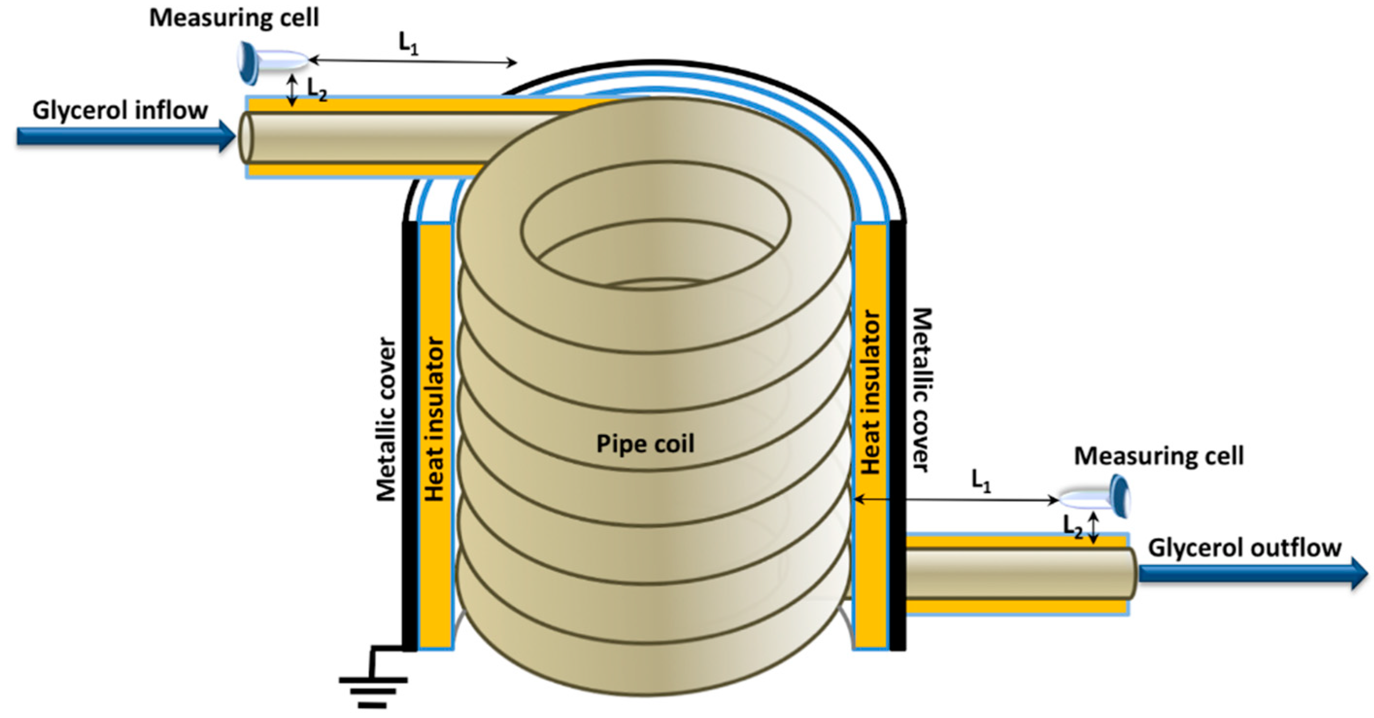
2.1. Experimental Setup

Figure 1 schematically illustrates the experimental setup used in our experiments.
The coiled section of the flow-based thermal stabilization system was imitated with a coiled polymeric pipe. An Eppendorf-type test tube (containing 1 mL of HRP protein solution) modeled a measuring cell. The cell was incubated near either the inflow or the outflow linear sections at the distance of L2 = 1 cm of the flow-based system in positions shown in Figure 1. In both cases, the distance between the cell and the coil was L1 = 20 cm. To avoid interference from the electromagnetic field induced in the coil, the latter was shielded with a grounded metallic cover. It should be emphasized that only the coiled section was ground shielded, while the linear section was only covered with the heat insulator layer of <1 cm in thickness.
In our experiments, peroxidase from horseradish (HRP-C; Cat.# P6782, Sigma, St. Louis, MO, USA) was used. A total of 1 mL of 10−7 M buffered protein solution (10 µM HRP in 2 mM Dulbecco’s modified phosphate buffered saline (PBSD) (pH 7.4, Pierce, Waltham, MA, USA) was placed into a 1.7-mL Eppendorf-type test tube, which modeled a measuring cell. Glycerol was pumped through the pipe at a volumetric flow rate of 9 L/s and a temperature of 65 °C. The increased temperature of glycerol provided its lower viscosity required for pumping the liquid at the desired flow rate. The spiral pipe was heat-insulated with a polymeric shield in order to conduct the measurements in the solution cuvette at the room temperature (RT). The cell with the HRP solution was incubated in the setup for 40 min. Then, this solution was analyzed by AFM and spectrophotometry. In the control experiments, the protein solution was incubated at a distance of 10 m from the setup for 40 min.

2.2. AFM Measurements

The AFM experiments were performed as described in our previous paper [5] by the direct surface adsorption method [23] by using muscovite mica sheets (SPI, West Chester, PA, USA) as AFM substrates. For the AFM sample preparation, a 7 × 15 mm piece of freshly cleaved mica was immersed into 800 µL of 0.1 μM buffered solution of HRP in PBSD, incubated either far away from the experimental setup (in control experiments), or near the inflow or the outflow sections of the pipe (working experiments). The AFM substrate was incubated in the HRP solution for 10 min at room temperature in a shaker at 300 rpm. After the incubation, each substrate was washed in 1 mL of ultrapure water (obtained with a Millipore Simplicity UV system, Millipore, Molsheim, France) and then dried.
The surface of the mica substrates with adsorbed HRP molecules was visualized by AFM in tapping mode. In this mode, the action on the protein objects under study is gentle. A Titanium multimode atomic force microscope (NT-MDT, Russia; this device pertains to the equipment of “Human Proteome” Core Facility of the Institute of Biomedical Chemistry, supported by Ministry of Education and Science of Russian Federation, agreement 14.621.21.0017, unique project ID RFMEFI62117X0017), equipped with NSG03 cantilevers (“TipsNano”, Zelenograd, Russia; 47 to 150 kHz resonant frequency, 0.35 to 6.1 N/m force constant). In each sample, the total number of imaged objects was no less than 500. The number of frames obtained for each sample was no less than 10. The density of distribution of the imaged objects with height (the density function ρ(h)) was calculated exactly as described elsewhere [5,7,24]. Preliminary experiments were performed with the use of a protein-free buffer instead of a protein solution; in the preliminary experiments, no objects with height exceeding 0.5 nm were visualized on the substrate surface.
Processing of AFM data and calculation of the number of objects, visualized in the AFM images, was performed as described in our previous papers [5,7,8,25].

2.3. Spectrophotometry Estimation of HRP Functional Activity

HRP activity monitoring was estimated exactly as described in our previous papers, following the technique by Sanders et al. [26], employing 2,2’-azino-bis(3-ethylbenzothiazoline-6-sulfonate) (ABTS; Sigma, St. Louis, MO, USA) as a reducing substrate.

3. Results

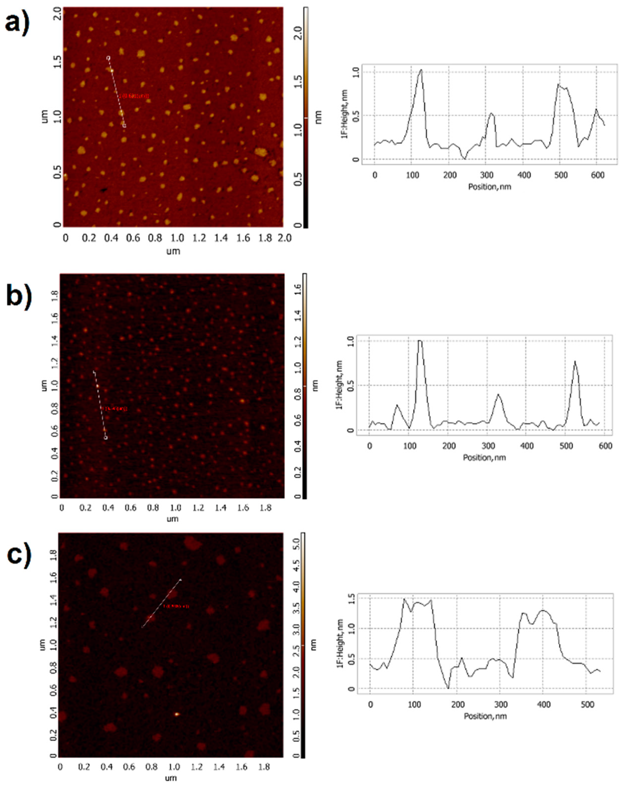
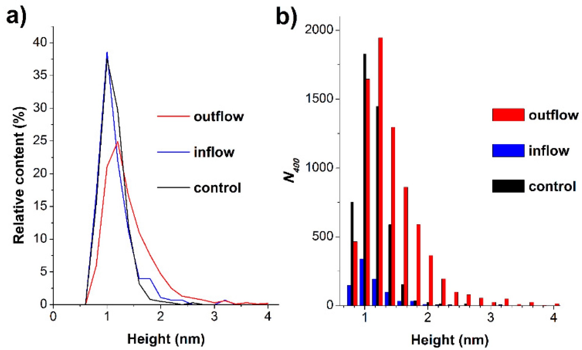
3.1. AFM Measurements

Figure 2 displays typical AFM images of mica substrate surface with HRP macromolecules, adsorbed from the analyzed solutions. In working experiments, the HRP solution was incubated in the experimental setup near either the inflow or the outflow section of the pipe. In control experiments, the protein solution was incubated far away (at a 10 m distance) from the setup.
In the typical AFM image, obtained in the control experiment (Figure 2a), one can clearly distinguish compact 1- to 1.2-nm-high objects. These objects can be attributed to HRP macromolecules, since no such objects was detected on the surface of a mica substrate incubated in a protein-free buffer.
In case of working experiments (Figure 2b,c), after the incubation of HRP solution near either the inflow or the outflow section of the pipe, compact-shaped objects are also observed, similar to the case with control experiments.
Figure 3 displays the distributions of AFM-visualized objects with height (density function plots) ρ(h) (Figure 3a) and the histogram representing the absolute number of visualized AFM-visualized objects, normalized per 400 µm2 area (Figure 3b).
The curves shown in Figure 3a indicate that in control experiments, the maximum of the density function corresponds to 1.0 ± 0.2 nm. The molecular weight Mr of HRP is 40 to 44 kDa [12,13]. According to the previously reported data, AFM images of proteins of similar molecular weight also have a height hmax of about 1 nm (putidaredoxin reductase, hmax = 1.8 nm [27], Mr = 45.6 kDa [28]; adrenodoxin reductase, hmax = 1.8 nm [29], Mr = 54 kDa [30]). For this consideration, the 1-nm-high objects, visualized on mica surface, can be attributed to monomeric HRP.
As regards the working experiments, when the protein solution was incubated near the inflow linear section of the pipe, the characteristics of the density function were similar to those obtained in control experiments. On the contrary, in the case of incubation near the outflow linear section of the pipe, the maximum of the density function plot shifted to the right (hmax ≈ 1.3 nm, Figure 3a, red line), and the contribution of the right distribution wing increased. That is, an appearance of objects with increased heights, which can be attributed to protein aggregates, was observed in the latter case.
As shown in Figure 3b, the absolute number of visualized objects (N400), obtained in our experiments, varies considerably. Thus, in the control experiment, N400 amounted to 4867 objects per 400 µm2. When the HRP solution was incubated near either the inflow or the outflow section of the flow-based system, N400 amounted to 851 and 7780 objects per 400 µm2, respectively. That is, the number of mica-adsorbed HRP macromolecules, observed in working experiments, significantly differs from that obtained in control experiments.

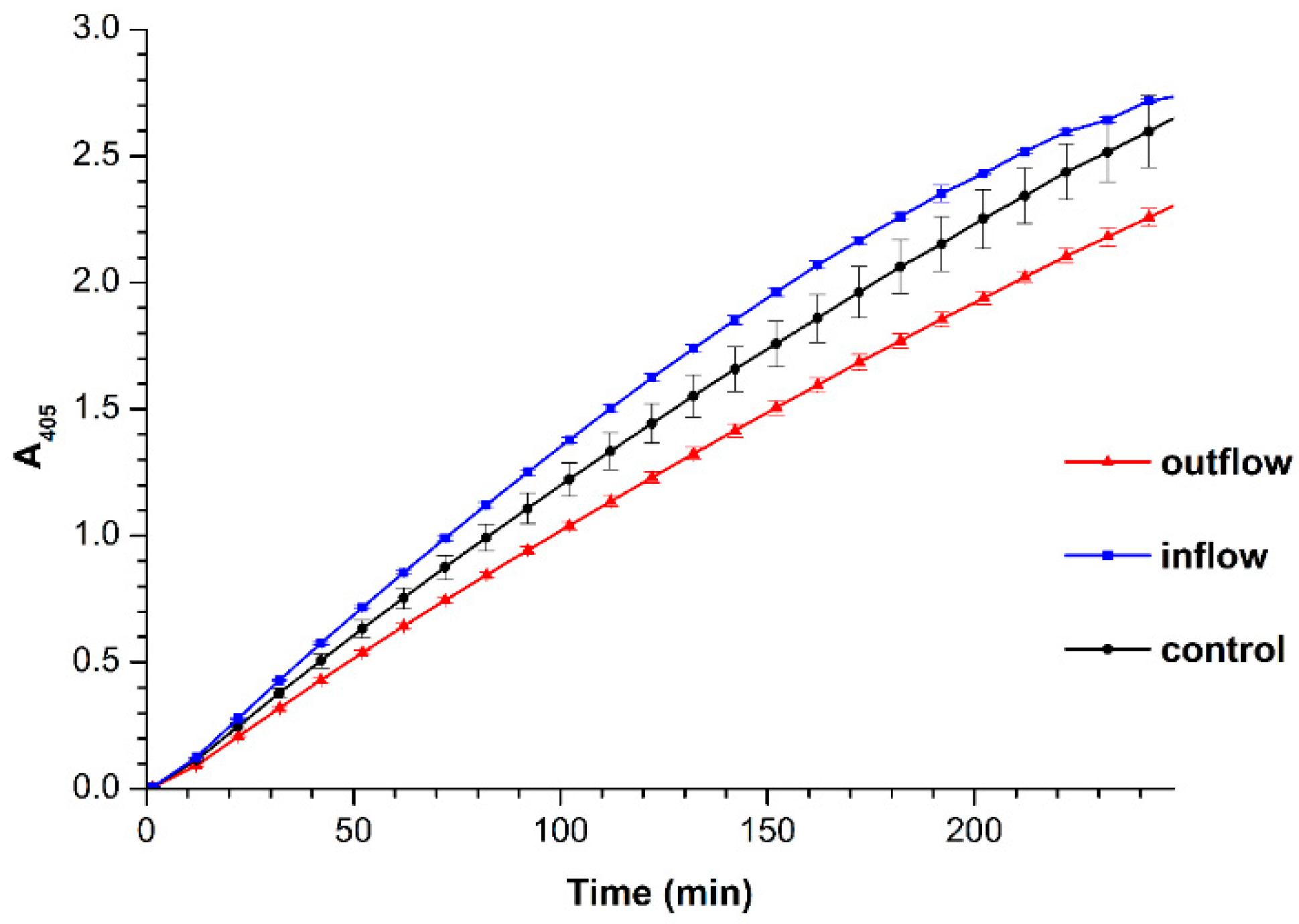
3.2. Spectrophotometry Measurements

Figure 4 displays the results of spectrophotometry-based estimation of HRP enzymatic activity by the reaction using 2,2′-azino-bis(3-ethylbenzothiazoline-6-sulfonate) (ABTS) substrate. As seen from Figure 4, in comparison with the control HRP solution, no significant change in the ABTS oxidation rate was observed after the incubation of HRP solution near the inflow section of the pipe. On the contrary, after the incubation of its solution near the outflow section of the flow-based system, the enzymatic activity of HRP significantly (by about 18%) decreased.

4. Discussion

This study looks into how glycerol flow through a polymeric pipe affects the properties of a model enzyme protein, the solution of which was placed near either the inflow or the outflow sections of the pipe. We have shown that after placing the enzyme solution near the linear sections of the pipe with flowing glycerol, changes in its properties are observed. Namely, after the HRP solution was incubated near the pipe inflow, a decreased adsorption of the protein onto mica was observed, being approximately 5.5 times lower than that observed for the control solution incubated far away from the pipe. At that, the enzymatic activity of the protein did not change. On the contrary, in the case when the HRP solution was incubated near the pipe outflow, an increased (approximately 1.6 times higher) protein adsorption was observed, while the enzymatic activity decreased considerably (by approximately 18%).
The data obtained indicate that the glycerol flow affects the physicochemical properties of HRP macromolecules. At that, the effect of the flow in different sections of the pipe varies. This is supposed to be caused by a difference in the intensity of the influence, which leads to variations in the character of protein structure deformations. Thus, a decreased protein adsorption in the case when the solution was incubated near the inflow pipe section is apparently caused by a decrease in electrostatic interaction between negatively charged surface groups of the mica substrate (whose zeta potential makes up −75 mV [31]) and positively charged groups on the surface of HRP (which represents a basic protein with an isoelectric point of about 9 [32]). Electrostatic interactions often play a governing role in protein adsorption [33]. Accordingly, the weaker the electrostatic interaction between the protein molecules and the substrate surface is, the lower the amount of the surface-adsorbed protein. Another cause of the observed decrease in the HRP adsorption can consist in an increased contribution of repulsion forces between negatively charged surface groups of the substrate and negatively charged groups of the protein globule. That is, an effect of a change in the local structure of the protein globule’s surface areas, which determine its interaction with the substrate surface, has been revealed. At that, the spectrophotometry data indicate no change in the protein’s functional activity. This indicates that the changes in the enzyme protein structure do not affect its active site.
In another case, when the HRP solution was incubated near the outflow section of the pipe, an increase in the number of objects on the substrate surface was observed, whereas their height increased from 1 nm up to 2 to 2.5 nm. That is, both protein adsorption properties and its aggregation state have changed (the content of aggregates has increased). Moreover, the spectrophotometry measurements also revealed a change in the HRP functional activity, which was found to decrease in comparison with the control experiment. This is why we can assume a different influence of glycerol flow on the protein in this case. This influence leads to a different kind of protein structure change, which affects not only the surface of the enzyme globule, but also its active site.
The data on the effect of the glycerol flow through the pipe on the properties of a protein are confirmed by the results obtained in our previous studies. Namely, in our previous paper [5], we studied the protein properties after the incubation of its solution near the similar coiled system with flowing glycerol, but over the coil (i.e., in a different position). In the latter case, a change in protein adsorption properties was also observed [5]. We discussed that a charge is being generated in the course of the flow of non-aqueous liquids such as glycerol [6]. This charge, accordingly, induces an electric field, which affects the protein properties. Such an effect can be observed for both water (and aqueous solutions [34,35,36,37]) and non-aqueous liquids [6,38]. We also demonstrated that the properties of a protein in a solution are affected by the flow of water flow through a coiled pipe [8]. The results obtained in the present study indicate that the liquid flow affects the properties of a protein in a solution even if the main (coiled) section of the pipe is ground shielded (as shown in Figure 1).
The results obtained herein are to be taken into consideration when conducting a highly sensitive analysis with using biosensor systems. Moreover, possible influence of the environment in terms of considering the particular characteristics of biosensor construction is important to interpret the data obtained in the analysis. Our results can also be used in modeling pathological processes associated with enzyme-mediated formation of functional multi-protein complexes, as well as for modeling hemodynamics in small vessels.

5. Conclusions

By AFM and spectrophotometry, glycerol flow through the flow-based system was found to affect the physicochemical properties of HRP enzyme protein. Namely, both the protein adsorption properties and its enzymatic activity change under the influence of the glycerol flow. The intensity of this influence was shown to depend on the location of the cell with the protein solution relative to the flow-based system’s pipe sections. The results obtained herein should be considered in biochemical and biomedical studies performed with the use of bioreactors and/or biosensors which contain flow-based systems.

Author Contributions

Conceptualization, Y.D.I.; data curation, I.A.I. and M.O.E.; formal analysis, A.F.K. and V.S.Z.; investigation, T.O.P., I.D.S., A.F.K., I.A.I., M.O.E., V.Y.T. and V.S.Z.; methodology, Y.D.I., T.O.P. and V.Y.T.; project administration, Y.D.I.; resources, V.Y.T. and V.S.Z.; software, V.S.Z.; supervision, Y.D.I.; validation, I.D.S.; visualization, T.O.P. and I.D.S.; writing—original draft, T.O.P. and I.D.S.; writing—review & editing, Y.D.I. All authors have read and agreed to the published version of the manuscript.

Funding

The work was performed in the framework of the Russian Federation fundamental research program for the long-term period for 2021–2030.

Data Availability Statement

The datasets generated during and/or analyzed during the current study are available from the corresponding author on reasonable request.

Acknowledgments

AFM measurements were performed employing a Titanium multimode atomic force microscope, which pertains to “Avogadro” large-scale research facilities.

Conflicts of Interest

The authors declare no conflict of interest.

References

  1. Doran, P.M. Heat transfer. In Bioprocess Engineering Principles, 2nd ed.; Doran, P.M., Ed.; Academic Press, Elsevier Ltd.: Oxford, UK, 2013; pp. 333–377. [Google Scholar]
  2. Daly, G.C. Heat Transfer Fluid. U.S. Patent 2008/0315152 A1, 25 December 2008. [Google Scholar]
  3. Ross, H.K. Cryoscopic Studies—Concentrated Solutions of Hydroxy Compounds. Ind. Eng. Chem. 1954, 46, 601–610. [Google Scholar] [CrossRef]
  4. Lane, L.B. Freezing Points of Glycerol and Its Aqueous Solutions. Ind. Eng. Chem. 1925, 17, 924. [Google Scholar] [CrossRef]
  5. Ziborov, V.S.; Pleshakova, T.O.; Shumov, I.D.; Kozlov, A.F.; Ivanova, I.A.; Valueva, A.A.; Tatur, V.Y.; Negodailov, A.N.; Lukyanitsa, A.A.; Ivanov, Y.D. Investigation of the Influence of Liquid Motion in a Flow-Based System on an Enzyme Aggregation State with an Atomic Force Microscopy Sensor: The Effect of Glycerol Flow. Appl. Sci. 2020, 10, 4825. [Google Scholar] [CrossRef]
  6. Tanasescu, F.; Cramariuc, R. Electroststica în Technica; Editura Technica: Bucuresti, Romania, 1977. [Google Scholar]
  7. Ivanov, Y.D.; Pleshakova, T.O.; Shumov, I.D.; Kozlov, A.F.; Ivanova, I.A.; Valueva, A.A.; Tatur, V.Y.; Smelov, M.V.; Ivanova, N.D.; Ziborov, V.S. AFM Imaging of Protein Aggregation in Studying the Impact of Knotted Electromagnetic Field on A Peroxidase. Sci. Rep. 2020, 10, 1–9. [Google Scholar] [CrossRef]
  8. Ivanov, Y.D.; Pleshakova, T.O.; Shumov, I.D.; Kozlov, A.F.; Romanova, T.S.; Valueva, A.A.; Tatur, V.Y.; Stepanov, I.N.; Ziborov, V.S. Investigation of the Influence of Liquid Motion in a Flow-based System on an Enzyme Aggregation State with an Atomic Force Mi-croscopy Sensor: The Effect of Water Flow. Appl. Sci. 2020, 10, 4560. [Google Scholar] [CrossRef]
  9. Metzler, D.E. Biochemistry: The Chemical Reactions of Living Cells; Academic Press: Oxford, UK, 1970. [Google Scholar]
  10. Gavrilenko, T.I.; Ryzhkova, N.A.; Parkhomenko, A.N. Myeloperoxidase and its role in the development of coronary heart disease. Ukr. J. Cardiol. 2014, 4, 119–126. [Google Scholar]
  11. Rogozhin, V.V.; Kutuzova, G.D.; Ugarova, N.N. Inhibition of horseradish peroxidase by N-ethylamide of o-sulfobenzoylacetic acid. Биоорганическая химия 2000, 26, 156–160. [Google Scholar]
  12. Davies, P.F.; Rennke, H.G.; Cotran, R.S. Influence of molecular charge upon the endocytosis and intracellular fate of peroxidase activity in cultured arterial endothelium. J. Cell Sci. 1981, 49, 69–86. [Google Scholar]
  13. Welinder, K.G. Amino Acid Sequence Studies of Horseradish Peroxidase. Amino and Carboxyl Termini, Cyanogen Bromide and Tryptic Fragments, the Complete Sequence, and Some Structural Characteristics of Horseradish Peroxidase C. JBIC J. Biol. Inorg. Chem. 1979, 96, 483–502. [Google Scholar] [CrossRef]
  14. Veitch, N.C. Horseradish peroxidase: A modern view of a classic enzyme. Phytochemistry 2004, 65, 249–259. [Google Scholar] [CrossRef]
  15. Shannon, L.M.; Kay, E.; Lew, J.Y. Peroxidase Isozymes from Horseradish Roots. J. Biol. Chem. 1966, 241, 2166–2172. [Google Scholar] [CrossRef]
  16. Tams, J.; Welinder, K.G. Mild Chemical Deglycosylation of Horseradish Peroxidase Yields a Fully Active, Homogeneous Enzyme. Anal. Biochem. 1995, 228, 48–55. [Google Scholar] [CrossRef]
  17. Dufrêne, Y.F.; Ando, T.; Garcia, R.; Alsteens, Y.F.D.D.; Martinez-Martin, D.; Engel, A.; Gerber, C.; Müller, D.M.-M.D.J. Imaging modes of atomic force microscopy for application in molecular and cell biology. Nat. Nanotechnol. 2017, 12, 295–307. [Google Scholar] [CrossRef]
  18. Braga, P.C.; Ricci, D. Atomic Force Microscopy Biomedical Methods and Applications; Humana Press: Totowa, NJ, USA, 2004. [Google Scholar]
  19. Pleshakova, T.O.; Bukharina, N.S.; Archakov, A.I.; Ivanov, Y.D. Atomic Force Microscopy for Protein Detection and Their Physicoсhemical Characterization. Int. J. Mol. Sci. 2018, 19, 1142. [Google Scholar] [CrossRef] [PubMed] [Green Version]
  20. Ignatenko, O.V.; Sjölander, A.; Hushpulian, D.M.; Kazakov, S.V.; Ouporov, I.V.; Chubar, T.A.; Poloznikov, A.A.; Ruzgas, T.; Tishkov, V.I.; Gorton, L.; et al. Electrochemistry of chemically trapped dimeric and monomeric recombinant horseradish peroxidase. Adv. Biosens. Bioelectron. 2013, 2, 25–34. [Google Scholar]
  21. Gouveia, M.; Xia, K.; Colón, W.; Vieira, S.I.; Ribeiro, F. Protein aggregation, cardiovascular diseases, and exercise training: Where do we stand? Ageing Res. Rev. 2017, 40, 1–10. [Google Scholar] [CrossRef]
  22. Xu, J.; Reumers, J.; Couceiro, J.R.; De Smet, F.; Gallardo, R.; Rudyak, S.; Cornelis, A.; Rozenski, J.; Zwolinska, A.; Marine, J.-C.; et al. Gain of function of mutant p53 by coaggregation with multiple tumor suppressors. Nat. Chem. Biol. 2011, 7, 285–295. [Google Scholar] [CrossRef] [PubMed] [Green Version]
  23. Kiselyova, O.I.; Yaminsky, I.V.; Ivanov, Y.D.; Kanaeva, I.P.; Kuznetsov, V.Y.; Archakov, A.I. AFM Study of Membrane Proteins, Cytochrome P450 2B4, and NADPH–Cytochrome P450 Reductase and Their Complex Formation. Arch. Biochem. Biophys. 1999, 371, 1–7. [Google Scholar] [CrossRef] [Green Version]
  24. Pleshakova, T.O.; Kaysheva, A.L.; Shumov, I.D.; Ziborov, V.S.; Bayzyanova, J.M.; Konev, V.A.; Uchaikin, V.F.; Archakov, A.I.; Ivanov, Y.D. Detection of Hepatitis C Virus Core Protein in Serum Using Aptamer-Functionalized AFM Chips. Micromachines 2019, 10, 129. [Google Scholar] [CrossRef] [Green Version]
  25. Ivanov, Y.D.; Pleshakova, T.O.; Shumov, I.D.; Kozlov, A.F.; Valueva, A.A.; Ivanova, I.A.; Ershova, M.O.; Larionov, D.I.; Repnikov, V.V.; Ivanova, N.D.; et al. AFM and FTIR Investigation of the Effect of Water Flow on Horseradish Peroxidase. Molecules 2021, 26, 306. [Google Scholar] [CrossRef]
  26. Sanders, S.A.; Bray, R.C.; Smith, A.T. pH-dependent Properties of a Mutant Horseradish Peroxidase Isoenzyme C in which Arg38 has been Replaced with Lysine. JBIC J. Biol. Inorg. Chem. 1994, 224, 1029–1037. [Google Scholar] [CrossRef] [PubMed]
  27. Ivanov, Y.D.; Bukharina, N.S.; Frantsuzov, P.A.; Pleshakova, T.O.; Kanashenko, S.L.; Medvedeva, N.V.; Argentova, V.V.; Zgoda, V.G.; Munro, A.W.; Archakov, A.I. AFM study of cytochrome CYP102A1 oligomeric state. Soft Matter 2012, 8, 4602–4608. [Google Scholar] [CrossRef]
  28. Unger, B.P.; Gunsalus, I.C.; Sligar, S.G. Nucleotide sequence of the Pseudomonas putida cytochrome P-450cam gene and its expression in Escherichia coli. J. Biol. Chem. 1986, 261, 1158–1163. [Google Scholar] [CrossRef]
  29. Ivanov, Y.D.; Frantsuzov, P.A.; Zollner, A.; Medvedeva, N.V.; Archakov, A.I.; Reinle, W.; Bernhardt, R. Atomic Force Microscopy Study of Protein–Protein Interactions in the Cytochrome CYP11A1 (P450scc)-Containing Steroid Hydroxylase System. Nanoscale Res. Lett. 2010, 6, 54. [Google Scholar] [CrossRef] [PubMed] [Green Version]
  30. Chu, J.-W.; Kimura, T. Studies on adrenal steroid hydroxylases. Molecular and catalytic properties of adrenodoxin reductase (a fla-voprotein). J. Biol. Chem. 1973, 248, 2089–2094. [Google Scholar] [CrossRef]
  31. Yamada, K.; Yoshii, S.; Kumagai, S.; Fujiwara, I.; Nishio, K.; Okuda, M.; Matsukawa, N.; Yamashita, I. High-Density and Highly Surface Selective Adsorption of Protein–Nanoparticle Complexes by Controlling Electrostatic Interaction. Jpn. J. Appl. Phys. 2006, 45, 4259–4264. [Google Scholar] [CrossRef]
  32. Aibara, S.; Yamashua, H.; Mori, E.; Kato, M.; Morita, Y. Isolation and Characterization of Five Neutral Isoenzymes of Horseradish Peroxidase. J. Biochem. 1982, 92, 531–539. [Google Scholar] [CrossRef]
  33. Hartvig, R.A.; Van De Weert, M.; Østergaard, J.; Jorgensen, L.; Jensen, H. Protein Adsorption at Charged Surfaces: The Role of Electrostatic Interactions and Interfacial Charge Regulation. Langmuir 2011, 27, 2634–2643. [Google Scholar] [CrossRef]
  34. Choi, D.; Lee, H.; Im, D.J.; Kang, I.S.; Lim, G.; Kim, D.S.; Kang, K.H. Spontaneous electrical charging of droplets by conventional pipetting. Sci. Rep. 2013, 3, srep02037. [Google Scholar] [CrossRef]
  35. Xu, W.; Zheng, H.; Liu, Y.; Zhou, X.; Zhang, C.; Song, Y.; Deng, X.; Leung, M.; Yang, Z.; Xu, R.X.; et al. A droplet-based electricity generator with high instantaneous power density. Nat. Cell Biol. 2020, 578, 392–396. [Google Scholar] [CrossRef]
  36. Zhao, L.; Liu, L.; Yang, X.; Hong, H.; Yang, Q.; Wang, J.; Tang, Q. Cumulative charging behavior of water droplet driven freestanding triboelectric nanogenerators toward hydrodynamic energy harvesting. J. Mater. Chem. A 2020, 8, 7880–7888. [Google Scholar] [CrossRef]
  37. Haque, R.I.; Arafat, A.; Briand, D. Triboelectric effect to harness fluid flow energy. J. Phys. Conf. Ser. 2019, 1407, 012084. [Google Scholar] [CrossRef]
  38. Balmer, R.T. Electrostatic Generation in Dielectric Fluids: The Viscoelectric Effect. In Proceedings of the World Tribology Congress III, Volume 1; ASME International: Washington, DC, USA, 2005; pp. 521–527. [Google Scholar]
Figure 1. Schematic representation of the experimental setup employed for studying the effect of liquid flow on the properties of a protein. The measuring cell with the enzyme solution was placed near either the inflow or the outflow linear section of the silicone pipe, whose central section forms a coil. The coil is shielded with a grounded metallic cover. Glycerol is pumped through the pipe.
Applsci 11 01723 g001
Figure 2. Typical AFM (atomic force microscopy) images of the mica substrate surface with adsorbed HRP (horseradish peroxidase) macromolecules and cross-sections, corresponding to the lines in the AFM images, obtained in control experiments (a), and when the HRP solution was incubated near either inflow (b) or outflow (c) sections of the pipe.
Applsci 11 01723 g002
Figure 3. Results of processing of AFM data obtained for HRP solutions. (a) Typical plots of distribution of the imaged objects with height ρ(h). (b) Absolute number of AFM-visualized particles, normalized per 400 µm2. The cell with HRP solution was placed far away at a 10-m distance from the experimental setup (control experiment, black line and bars), near the inflow (blue line and bars) or near the outflow (red line and bars) section of the pipe.
Applsci 11 01723 g003
Figure 4. Spectrophotometry-based estimation of HRP enzymatic activity. Characteristic time dependencies of change in solution absorbance at 405 nm obtained for control HRP sample and for HRP samples incubated in the experimental setup. The measuring cell (containing HRP solution) was placed either far away at a 10-m distance from the experimental setup (control experiment, black line), near the inflow (blue line) or near the outflow (red line) section of the pipe. Experimental conditions: HRP:ABTS:H2O2 = 10−9 M:3 mM:2.5 mM. T = 23 °C.
Applsci 11 01723 g004
Publisher’s Note: MDPI stays neutral with regard to jurisdictional claims in published maps and institutional affiliations.

