Article

L-Band Electron Paramagnetic Resonance Tooth Dosimetry Applied to Affected Cattle Teeth in Fukushima

1
Department of Environmental Health, National Institute of Public Health, Wako 351-0197, Japan
2
School of Dental Medicine, Tsurumi University of Dental Medicine, Yokohama 230-8501, Japan
3
School of Veterinary Medicine, Kitasato University, Towada 034-8628, Japan
4
Department of Radiation Biophysics, Research Institute for Radiation Biology and Medicine, Hiroshima University, Hiroshima 734-8553, Japan
5
Department of Oral and Maxillofacial Surgery, Kagawa University, Miki 761-0793, Japan
6
Department of Radiology, The Geisel School of Medicine at Dartmouth, Hanover, NH 03755-1404, USA
*
Author to whom correspondence should be addressed.
Appl. Sci. 2021, 11(3), 1187; https://doi.org/10.3390/app11031187
Submission received: 31 December 2020 / Revised: 19 January 2021 / Accepted: 21 January 2021 / Published: 28 January 2021

Abstract

:

Featured Application

The novel, non-destructive technique of tooth dosimetry using L-band electron paramagnetic resonance (EPR) presented in this paper could be applied to obtain assessments of the environmental impact after possible radiation exposures from nuclear or radiological accidents.

Abstract

We applied a non-destructive tooth dosimetry technique using L-band electron paramagnetic resonance (EPR) to assess radiation doses in cattle due to the Fukushima Daiichi Nuclear Power Station (FDNPS) accident, which occurred 10 years ago. The radiation exposure of cattle in the area affected by the FDNPS accident was estimated retrospectively with X-band and L-band EPR devices. Characteristic radiation-induced EPR signals were obtained from the teeth of the cattle in Fukushima, confirming their exposure. The estimated doses to the teeth were found to be consistent with the dose trends estimated for individual cows, while considerable uncertainties were seen in the doses of some tooth samples. This variation might be due to errors in the accuracy of the method but also might reflect the actual exposure because the cattle may have been exposed to higher areas of radioactivity in their quest for food and/or due to irradiation from absorption of the isotopes with localization in or near the teeth. However, at a minimum, these results confirm that L-band EPR can be used for non-destructive qualitative assessment of radiation exposure to animals using their teeth, which could be very valuable. Possible causes of the uncertainties should be investigated to enhance the value of the use of this technique.

1. Introduction

Electron paramagnetic resonance (EPR) tooth dosimetry is an established method for evaluating the absorbed dose ionizing radiation by measuring the unpaired electrons to in-teeth samples [1]. In some studies, EPR dating measurements are also carried out using the radiation-induced EPR signals from background radiation in the environment where the samples were located [2]. Most EPR measurements are performed in the X-band and some aim to detect signals in small amount of samples with the higher-frequency Q-band measurements [3]. Although EPR has a long history, its application to dosimetry is a novel application technique [4]. Attempts have also been made to measure the internal exposure of environmental organisms using radiation-induced signals in the sample [5]. In addition, EPR measurements using human bone samples have also been used to estimate exposure doses for atomic bomb survivors [6] and from individuals exposed in radiation accidents [7]. X-band EPR tooth dosimetry, which uses extracted teeth, has been used for atomic-bomb survivors of Hiroshima and Nagasaki [8] and residents of affected settlements due to radiation released into the environment [9]. This method was also applied to some animals affected by the Chernobyl accident in 1986 [10] and the Fukushima Daiichi Nuclear Power Station (FDNPS) accident which occurred in March 2011 [11,12].
Most of the above studies required the removal of the material with processing in vitro. In order to carry out radiation dosimetry in live subjects such as for potential radiation emergencies, a lower-frequency L-band EPR apparatus that can be applied to teeth in situ has been developed for measurement under triage conditions [13]. This method is also applicable for retrospective dose assessment for a large-scale radiation exposure incident [14,15].
It would be potentially valuable to use the EPR signals induced in large animals to assess the amount of radiation exposure that occurred. In the present study, we used a non-destructive L-band EPR tooth dosimetry technique to estimate the radiation dose in cattle affected by the FDNPS accident, and we validated the results by comparing them with values estimated from radiation monitoring using survey meters and personal dosimeters for each cow.

2. Materials and Methods

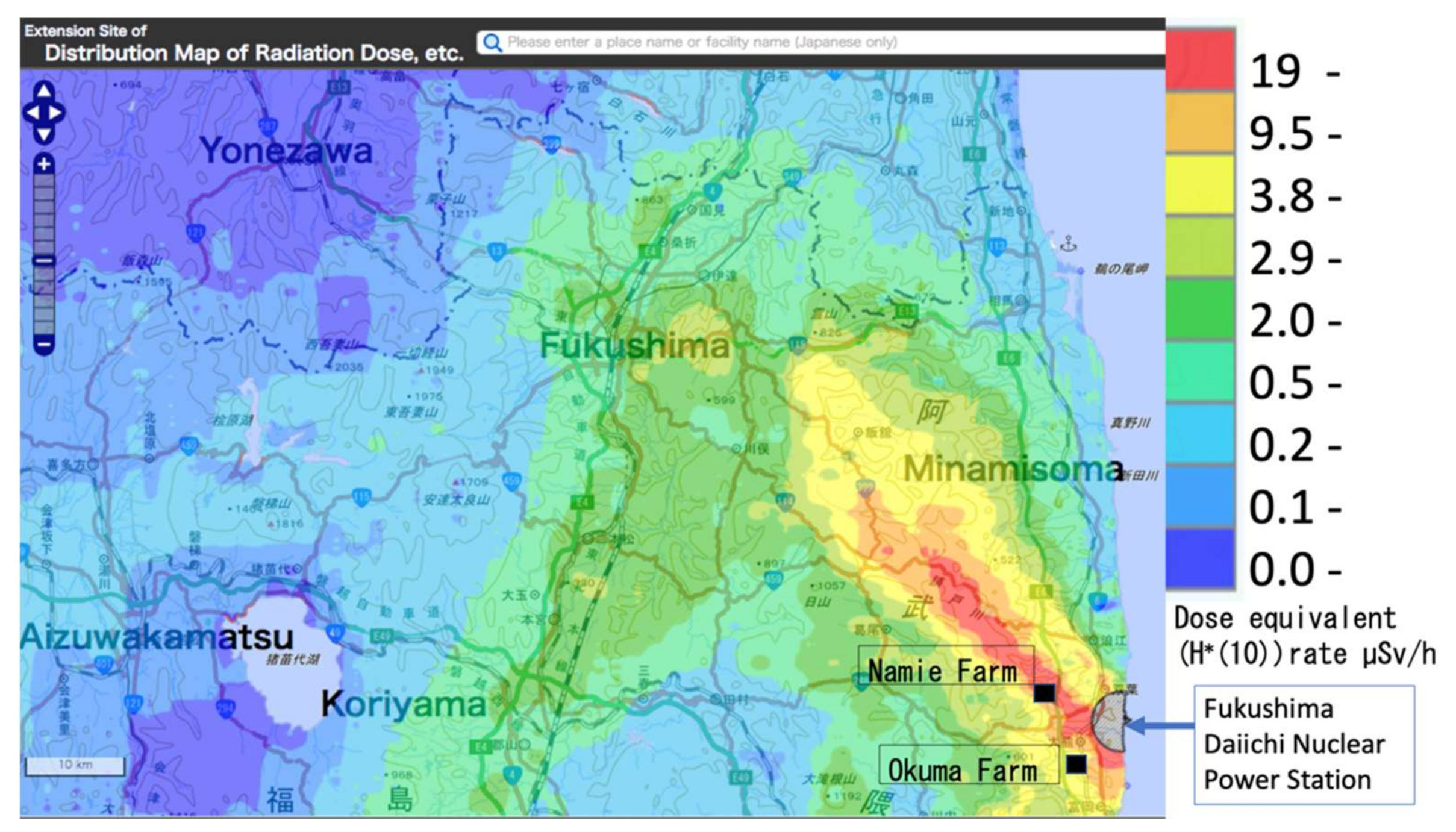
2.1. Sample Collection

Three to four incisor teeth were collected from three cows that remained in the town of Namie, which is located in the former evacuation zone of the FDNPS accident (10 teeth in total) (Figure 1).
For comparison, 10 incisors were collected from cattle under 3 years of age at locations where radiation was not released into the environment, Hokkaido, Iwate, Gunma, and Kagoshima (40 teeth in total).

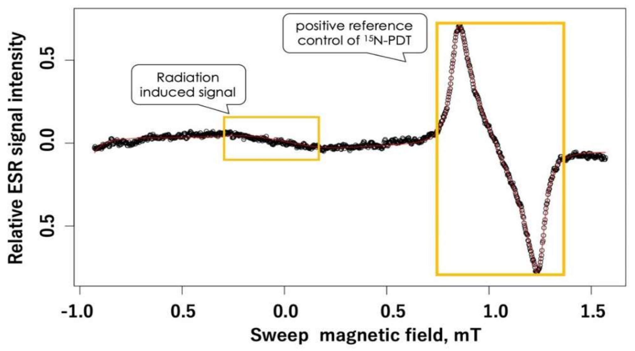
2.2. Non-Destructive Measurement Using L-Band EPR Spectroscopy

An EPR tooth dosimeter obtained from Clin-EPR, LLC was used for EPR tooth dosimetry. This instrument was based on instruments designed for in vivo dosimetry by the EPR Center for the Study of Viable Systems, Geisel School of Medicine at Dartmouth College in Hanover, NH, USA [15]. The spectrometer operates in continuous-wave (CW) mode and uses homodyne detection at an excitation frequency near 1.15 GHz (L-band) using a 41 mT dipole magnet weight 30 kg with 17 cm pole separation. The integrated field sweep and modulation coil provides a 4 mT sweep range and 0.4 mT modulation at 20 kHz. It was measured using a specially developed surface loop resonator for maxillary incisors which has a detection loop with an inner diameter of 6.0 mm. The detection loop was brought into contact with the enamel on the labial side of each incisor, and the radicals on that surface were measured.
For calibration of the L-band EPR response, we used 10 teeth that were irradiated with a 137Cs γ-rays at Hiroshima University by using a Gammacell 40 Exactor Low Dose Rate Research Irradiator (Best Theratronics Ltd., Ottaw, ON, Canada).
To adjust the resulting measured radiation-induced signal (RIS) amplitudes, the ratio of differentiated voltage as a radiation-induced signal (RIS) to the differentiated voltage of a stable standard that was simultaneously measured in the detection loop, 4-oxo-2, 2, 6, 6, -tetramethylpiperidine-d16-1-15N-1-oxyl (15N-PDT) for each measurement was normalized to the same ratio in the referral tooth.
The EPR spectra were acquired by using our standard measurement parameters of 20 scans, with a scan range of 2.5 mT, scan time of 3 s, and modulation amplitude of 0.4 mT [16]. This process was repeated three times. The spectra from each of the collected datasets were analyzed to estimate the peak-to-peak signal amplitudes of the RIS and 15N-PDT. The RIS and signals of 15N-PDT were then averaged to determine the mean amplitude for each tooth. For two cows (08411-03687, 08411-03274), the scan times were increased to 200, to increase signal/noise.

2.3. Measurement of Radioactivity Concentration

The radioactive concentrations of 137Cs and 90Sr in the molar teeth and lower jaw bones were measured. For 137Cs, each sample was powdered and analyzed by using a Ge semiconductor detector (CFG-SV-76, ORTEC, Oak Ridge, TN, USA). 90Sr was extracted by the use of an ion exchange method and was measured at Japan Chemical Analysis Center achieving radioactive equilibrium with 90Y.

3. Results

3.1. Non-Destructive Measurement Using L-Band EPR Spectroscopy

Positive RISs were detected in all samples from Namie. A typical signal is shown in Figure 1. In this spectrum, RISs were detected as the magnetic field was varied between 0.7 mT and 1.1 mT. The estimated doses are shown in Table 1.
No signals were detected in the control cattle from the other regions of Japan.
Differences in the dose response among the teeth are shown in Figure 2. The tooth with the largest dose response exhibited a signal magnitude 2.1 times larger than the tooth with the smallest response.
Note: The measurement conditions include 200 scans. Scan conditions are shown in the text. The Scheme 0.2 mT showed a positive reference of 15N-PDT. The estimated dose for this sample was 2.3 Gy.

3.2. Measurement of Radioactivity Concentration

The radionuclides and their concentrations in the tooth and lower jaw are shown in Table 2.

4. Discussion

4.1. Comparison of Estimated Radiation Doses

The estimated dose using L-band EPR tooth dosimetry was 0.9–3.9 Gy for the cattle in Namie, which was significantly different from the values in the tooth from Okuma [17]. This range was larger than the monitored cumulative radiation doses over a six-year period beginning in March 2011 in Namie and Okuma, which were estimated to be about 1 Gy and 0.16 Gy, respectively. By considering weathering, the accumulated dose before the start of monitoring for the FDNPS accident was calculated from the deposition density of 131I, 132Te, 132I, 134Cs, and 137Cs on the ground after the accident [18]. The measured dose was greater than the predicted dose. Possible reasons for this were experimental variation of the technique, the actual duration of the cattle in the area after the accident, and the radiation status of the cattle’s teeth such as exposures during eating contaminated grass. Furthermore, the age of the cattle from which the teeth of cattle that could be purchased were limited to those less than 30 months old due to countermeasures against bovine spongiform encephalopathy.

4.2. Effect of External Exposure to β-Rays from Deposited Radionuclides

Exposure from β-rays from radioactive materials in the environment is an additional possible source of radiation. Assuming that the deposited density of 137Cs is 1 MBq/m2, the skin dose rate from the ground was estimated to be about 0.14 mGy/h, based on the coefficient given in International Commission on Radiation Units and Measurements (ICRU) report 56 [12]. Assuming 8 h/day of direct contact with the ground, the accumulated skin dose from β-rays for six years after the FDNPS accident was 1.9 Gy. The 13352–73671 cow was dead when farmers found her and was skeletonized at the time of sampling, which could have exposed it to additional environmental radiation without shielding by the thickness of the body. This may have made them more exposed to radiation from radioactive materials in the environment.

4.3. Effect of Internal Exposure

Radioactive substances residing within the teeth and organs are another potential source of radiation exposure [19]. In the study of teeth from Techa residents, some ultra-high doses were measured; moreover, in some cases adjacent teeth differed by a factor of ten. These ultra-high doses were determined to be an anomaly attributed to enamel that was being formed during the period of release of radioactive materials into the environment. These teeth incorporated strontium-90 directly into the enamel. This observation unveils an added feature of EPR biodosimetry. Since enamel tissue formed during radionuclide releases exhibit anomalously high doses, one can target specific teeth from specific residents to use this feature to identify the time of release [20]. Assuming that our assays of the amount of radioactive material in the teeth reflect the full amount of such deposits, we find that, the self-absorption dose—that is, the radiation dose to which a tooth is subjected via radionuclides, including radioactive Cs and 89Sr, 90Sr—was limited, perhaps to as low as 20 mGy, This difference of internal radiation dose from radioactive Sr is due to the difference in the amount of radioactive strontium released into the atmosphere in the case of the Techa River and the nuclear accident at Fukushima. In cases where high concentrations of radionuclides are identified, the X-band may be able to detect the radiation dose due to radionuclides accumulated in the teeth.

4.4. Strength of this Method

L-band EPR tooth dosimetry can be applied to living organisms without the need to remove the samples. It also is valuable as a non-destructive method which is applicable for assessment of dose for samples that are too valuable to be damaged in during the measurements.

4.5. Limitations

Even in Futaba County where the nuclear power plant that caused the accident is located, the signals were not detected in cattle in Okuma, but only in cattle in Namie. This result was consistent with the different expected dose exposures in these areas. However, due to the small number of samples, the results are not fully generalizable for inter-regional comparisons. In addition, these cattle may have moved to different pastures as a consequence of the disruption due to the extreme circumstances caused by the nuclear accident and it is thought that the time of birth is related to exposure to radiation and tooth growth [21]; hence, the present authors do not know the full history of exposure. This effect needs to be taken into account to improve the quality of comparisons with expected doses since the calculations are based on some assumed history such as staying at the same location since the accident.

5. Conclusions

The novel technique of L-band EPR tooth dosimetry was successfully applied to cattle in the former evacuation zone affected by the FDNPS accident, which occurred 10 years ago. These results indicate that assessment for radiation exposure caused by radiation accidents can be performed using L-Band EPR dosimetry in the teeth of cattle and presumably in teeth of other animals as well. This was the first attempt to detect the EPR signals in the teeth of animals affected by a nuclear accident using a non-destructive method. The radiation doses recorded in the teeth of these animals were a good reflection of their exposure history over the last ten years. In addition, we found that the radiation doses to the sampled teeth tended to be higher than the doses determined from the data of environmental radiation monitoring, which suggests that there might have been sources of exposure beyond those estimated from the usual methods employed for monitoring of the environment. Each cow may also have a different level of radiation exposure, so we plan to study this further as planned slaughters are carried out.

Author Contributions

Conceptualization, I.Y., M.N., and H.M.S.; funding acquisition, I.Y., K.I., and M.M.; investigation, C.A.B.G. and Y.N.; project administration, M.N.; resources, K.I.; supervision, M.M.; writing—original draft, I.Y.; writing—review and editing, C.A.B.G., H.Y., and H.M.S. All authors have read and agreed to the published version of the manuscript.

Funding

This research was supported by the Program of the Network-type Joint Usage/Research Center for Radiation Disaster Medical Science and MEXT/JSPS KAKENHI Grant Number 18K09896 and 18K09724 This research was also partially funded by Industrial Disease Clinical Research Grants, Ministry of Health, Labour and Welfare, Japan.

Institutional Review Board Statement

Ethical review and approval were not applicable because it does not involve human or live animal subjects.

Informed Consent Statement

This study did not involve humans.

Data Availability Statement

The data presented in this study are openly available in the website at “https://ndrecovery.niph.go.jp/trustrad/invivo_EPR/data/”.

Acknowledgments

The authors are sincerely grateful to the farmers who donated their precious cattle with help from Keiji Okada and Jun Sasaki, AEN (Society for Animal Refugee and Environment post Nuclear Disaster) and to Tokyo Shibaura Organs Co. who provided the teeth of cows separated by region of origin for research purposes. The authors also thank Shin Toyoda for his kind encouragement and valuable suggestions. I.Y. thanks Seiko Hirota and Satoko Mikawa for their technical support.

Conflicts of Interest

Swartz is an owner of Clin-EPR, LLC, which manufactured the instrument used in this study. The other authors declare no conflict of interest. The funders had no role in the design of the study; in the collection, analyses, or interpretation of data; in the writing of the manuscript, or in the decision to publish the results.

References

  1. Brady, J.M.; Aarestad, N.O.; Swartz, H.M. In vivo dosimetry by electron spin resonance spectroscopy. Health Phys. 1968, 15, 43–47. [Google Scholar] [CrossRef] [PubMed]
  2. Lu, C.-C.; Lin, H.-H.; Hsu, C.-H.; Wang, F.-N.; Lin, J.-P.; Lai, L.-H. Potential Use of Environmental Biological Samples for Retrospective Electron Paramagnetic Resonance Dosimetry of Radiation Accidents. Appl. Sci. 2020, 10, 6867. [Google Scholar] [CrossRef]
  3. Romanyukha, A.; Trompier, F.; Reyes, R.A. Q-band electron paramagnetic resonance dosimetry in tooth enamel: Biopsy procedure and determination of dose detection limit. Radiat. Environ. Biophys. 2014, 53, 305–310. [Google Scholar] [CrossRef] [PubMed]
  4. Gonzales, C.A.; Taño, J.; Yasuda, H. An Attempt to Reduce the Background Free Radicals in Fingernails for Monitoring Accidental Hand Exposure of Medical Workers. Appl. Sci. 2020, 10, 8949. [Google Scholar] [CrossRef]
  5. Ivanov, D.V.; Shishkina, E.A.; Osipov, D.I.; Razumeev, R.A.; Pryakhin, E.A. Internal in vitro dosimetry for fish using hydroxyapatite-based EPR detectors. Radiat. Environ. Biophys. 2015, 54, 257–263. [Google Scholar] [CrossRef]
  6. Kinoshita, A.; Baffa, O.; Mascarenhas, S. Electron spin resonance (ESR) dose measurement in bone of Hiroshima A-bomb victim. PLoS ONE 2018, 13, e0192444. [Google Scholar] [CrossRef] [Green Version]
  7. Schauer, D.A.; Desrosiers, M.F.; Kuppusamy, P.; Zweier, J.L. Radiation dosimetry of an accidental overexposure using EPR spectrometry and imaging of human bone. Appl. Radiat. Isot. 1996, 47, 1345–1350. [Google Scholar] [CrossRef]
  8. Nakamura, N.; Hirai, Y.; Kodama, Y. Gamma-ray and neutron dosimetry by EPR and AMS, using tooth enamel from atomic-bomb survivors: A mini review. Radiat. Prot. Dosim. 2012, 149, 79–83. [Google Scholar] [CrossRef]
  9. Zhumadilov, K.S.; Ivannikov, A.I.; Stepanenko, V.F.; Toyoda, S.; Skvortsov, V.G.; Hoshi, M. Epr Dosimetry Study for Population Residing in the Vicinity of Fallout Trace for Nuclear Test on 7 August 1962. Radiat. Prot. Dosim. 2016, 172, 260–264. [Google Scholar] [CrossRef]
  10. Zdravkova, M.; Gallez, B.; Debuyst, R. A comparative in vivo and in vitro L-band EPR study of irradiated rat incisors. Radiat. Meas. 2005, 39, 143–148. [Google Scholar] [CrossRef]
  11. Jiao, L.; Liu, Z.-C.; Ding, Y.-Q.; Ruan, S.-Z.; Wu, Q.; Fan, S.-J.; Zhang, W.-Y. Comparison study of tooth enamel ESR spectra of cows, goats and humans. J. Radiat. Res. 2014, 55, 1101–1106. [Google Scholar] [CrossRef] [PubMed] [Green Version]
  12. Harshman, A.; Toyoda, S.; Johnson, T. Suitability of Japanese wild boar tooth enamel for use as an Electron Spin Resonance dosimeter. Radiat. Meas. 2018, 116, 46–50. [Google Scholar] [CrossRef]
  13. Miyake, M.; Liu, K.J.; Walczak, T.M.; Swartz, H.M. In vivo EPR dosimetry of accidental exposures to radiation: Experimental results indicating the feasibility of practical use in human subjects. Appl. Radiat. Isot. 2000, 52, 1031–1038. [Google Scholar] [CrossRef]
  14. Swartz, H.M.; Flood, A.B.; Williams, B.B.; Meineke, V.; Dörr, H. Comparison of the needs for biodosimetry for large-scale radiation events for military versus civilian populations. Health Phys. 2014, 106, 755–763. [Google Scholar] [CrossRef] [PubMed]
  15. Miyake, M.; Nakai, Y.; Yamaguchi, I.; Hirata, H.; Kunugita, N.; Williams, B.B.; Swartz, H.M. In-vivo radiation dosimetry using portable L band EPR: On-site measurement of volunteers in Fukushima prefecture, Japan. Radiat. Prot. Dosimetry 2016, 172, 248–253. [Google Scholar] [CrossRef] [PubMed] [Green Version]
  16. Yamaguchi, I.; Sato, H.; Kawamura, H.; Hamano, T.; Yoshii, H.; Suda, M.; Miyake, M.; Kunugita, N. L Band EPR Tooth Dosimetry for Heavy Ion Irradiation. Radiat. Prot. Dosim. 2016, 172, 81–86. [Google Scholar] [CrossRef] [PubMed] [Green Version]
  17. Inoue, K.; Yamaguchi, I.; Natsuhori, M. Preliminary Study on Electron Spin Resonance Dosimetry Using Affected Cattle Teeth Due to the Fukushima Daiichi Nuclear Power Plant Accident BT. In Low-Dose Radiation Effects on Animals and Ecosystems: Long-Term Study on the Fukushima Nuclear Accident; Fukumoto, M., Ed.; Springer: Singapore, 2020; pp. 165–177. ISBN 978-981-13-8218-5. [Google Scholar]
  18. Natsuhori, M.; Kojima, T.; Sato, I.; Okada, K.; Sasaki, J.; Sato, H.; Hojito, M.; Kobayashi, E.; Kakizaki, T.; Wada, S.; et al. Radioactive contamination profiles of soil, external and internal exposure to Japanese Black Cattle after Fukushima Daiichi Nuclear Plant Accident. Jpn. J. Large Anim. Clin. 2017, 8, 143–147. [Google Scholar] [CrossRef] [Green Version]
  19. Ignatiev, E.A.; Lyubashevskii, N.M.; Shishkina, E.A.; Romanyukha, A.A. EPR dose reconstruction for bone-seeking 90Sr. Appl. Radiat. Isot. 1999, 51, 151–159. [Google Scholar] [CrossRef]
  20. Romanyukha, A.A.; Seltzer, S.M.; Desrosiers, M.; Ignatiev, E.A.; Ivanov, D.V.; Bayankin, S.; Degteva, M.O.; Eichmiller, F.C.; Wieser, A.; Jacob, P. Correction factors in the EPR dose reconstruction for residents of the Middle and Lower Techa riverside. Health Phys. 2001, 81, 554–566. [Google Scholar] [CrossRef] [PubMed]
  21. Toyoda, S.; Murahashi, M.; Natsuhori, M.; Ito, S.; Ivannikov, A.; Todaka, A. Retrospective ESR reconstruction of cattle tooth enamel doses from the radioactive nuclei released by the accident of Fukushima dai-ichi atomic power plants. Radiat. Prot. Dosim. 2019, 186, 48–53. [Google Scholar] [CrossRef] [PubMed]
Figure 1. Radiation dose-rate map obtained by airborne monitoring results and the location of Okuma and Namie Farm as of 31 May 2012. This map was quoted from the website provided by Nuclear Regulation Authority, Japan. Dose rate shows μSv/h as a dose equivalent rate including background radiation. (http://ramap.jaea.go.jp/map/). Note: H*(10) means ambient dose equivalent.
Applsci 11 01187 g001
Figure 2. Representative electron paramagnetic resonance (EPR) spectrum of an incisor sample extracted from a cow (#08411-03274), determined by using L-band spectrometry.
Applsci 11 01187 g002
Table 1. Basic characteristics of the cattle and estimated radiation dose.
Location.
(Town)
Ear Tag IDSexBirth DateDose (Gy)
Mean ± SD
Autopsy Date
Ohkuma12416–04378female26 December 2006ND14 May 2017
Ohkuma12425–47537female2 September 2007ND14 May 2017
Ohkuma08597–08639female1 June 2012ND14 May 2017
Namie13352–73671female29 June 20103.85 ± 0.31
3.47 ± 0.89
2.29 ± 1.30
12 May 2018
Namie08411–03687male25 November 20102.09 ± 0.17
1.15 ± 0.81
0.93 ± 0.96
5 September 2020
Namie08411–03274female25 February 20113.84 ± 0.22
3.13 ± 0.36
2.71 ± 0.68
1.55 ± 0.75
6 September 2020
Note: ND means not detected.
Table 2. Radioactive concentrations in the lower jaw and molar of cows in Fukushima.
Cattle SamplesRadioactive Concentration 1 (Bq/kg)
PortionLocationCs–137Sr–90
MolarNamie855 ± 24270 ± 5
Okuma60.6 ± 1.746 ± 2.3
Lower jawNamie1174 ± 70190 ± 4
Okuma26.6 ± 2.428 ± 1.6
1 Each radioactive concentration was adjusted as of March 2011 and is presented along with the S.D. (statistical error). The ear tag of cow is 08597–08639 for Okuma and 13352–73671 for Namie.
Publisher’s Note: MDPI stays neutral with regard to jurisdictional claims in published maps and institutional affiliations.

