Article

Factors Affecting the Outcome of Periapical Surgery; a Prospective Longitudinal Clinical Study

1
Department of Operative Dentistry and Institute of Biomedical Sciences, Dow University of Health Sciences, Karachi 74200, Pakistan
2
Department of Restorative Dentistry and Endodontics, College of Dentistry, King Faisal University, Al-Ahsa 31982, Saudi Arabia
3
Department of Surgery, Aga Khan University, Karachi 74800, Pakistan
4
Department of Biological and Biomedical Sciences, Aga Khan University, Karachi 74800, Pakistan
5
Department of Pathology, Dr. Ishrat-ul-Ebad Khan Institute of Oral Health Sciences, Dow University of Health Sciences, Karachi 74200, Pakistan
6
Department of Conservative Dental Sciences, College of Dentistry, Prince Sattam Bin Abdulaziz University, Al-Kharj 11942, Saudi Arabia
7
Department of Prosthetic Dental Science, College of Dentistry, King Saud University, Riyadh 11545, Saudi Arabia
8
Engineer Abdullah Bugshan Research Chair for Dental and Oral Rehabilitation (Department), College of Dentistry, King Saud University, Riyadh 11545, Saudi Arabia
9
Department of Restorative Dentistry—Operative Division, College of Dentistry, King Saud University, Riyadh 11545, Saudi Arabia
*
Author to whom correspondence should be addressed.
Appl. Sci. 2021, 11(24), 11768; https://doi.org/10.3390/app112411768
Submission received: 23 August 2021 / Revised: 19 November 2021 / Accepted: 22 November 2021 / Published: 10 December 2021

Abstract

:
Wound healing following periapical surgery is influenced by age, gender, smoking, periapical lesion size, type of root-end filling, method of root-end cavity preparation and the use of microsurgical or conventional technique. This study aimed to evaluate the influence of various preoperative factors such as age, gender, smoking, preoperative pain, and preoperative periapical lesion size on the outcome of surgical endodontic treatment. A thorough history, examination, and investigation were performed to establish patient age, gender, smoking status, periapical lesion size, and the presence of preoperative pain. Forty patients aged between 15–57 years presented with persistent chronic apical periodontitis of single-rooted anterior teeth after conventional re-root canal treatment were enrolled for periapical surgery. Following periapical surgery, all patients were recalled for evaluation of periapical healing after 12 months based on clinical and periapical X-ray examination due to inaccessibility of an advanced imaging system (CBCT). Chi-squared and Fisher’s exact test were applied, which revealed a statistically significant association of periapical healing with age (p = 0.025), smoking (p = 0.029), and lesion size (p < 0.001). Although, the success of periapical healing was higher in males 78.6% (22/28) compared to females 58.3% (7/12) however, no statistically significant relationship was found between gender and healing (p = 0.254). Patient age, smoking status, and size of the preoperative lesion had a strong influence on periapical healing after surgical endodontic treatment.

1. Introduction

The treatment of choice for symptomatic, persistent or enlarging periapical lesion after conventional re-root canal treatment is surgical endodontic treatment, which is aimed at resecting the root end and then creating a barrier at the apical end of the root with an inert material [1,2,3]. The goal of this treatment protocol is to salvage the tooth and prevent it from further damage by limiting the entry of microbes and their byproducts into the canal system, thus promoting the health of the tooth.
The success of surgical endodontic treatment mainly relies on the absence of clinical signs and symptoms and radiographic resolution of periapical lesions. Studies have shown that multiple factors may influence the outcome of periapical surgery such as age, gender, smoking, type of root-end filling material, size of the preoperative periapical lesion, and the presence of pretreatment signs and symptoms [3,4].
Smoking is considered one of the risk factors that can impede periapical healing. Smoking disturbs healing by limiting the supply of oxygenated blood and nutrients to the periapical area [5]. This in turn makes periapical tissue more susceptible to bacterial infection [6]. Additionally, it alters the synthesis of collagen, expedites bone loss, and interferes in the tissue repair process around the periapical area [7,8]. Furthermore, smokers have restricted defense mechanisms due to the deficiency of several immunological factors such as Tumor necrosis factor alpha (TNF-α) and Human beta defensin-2 (hBD-2) [9].
There have been numerous studies investigating the size of preoperative periapical lesions with periapical wound healing. It was observed that fibroblasts proliferate into a large bony defect after periapical surgery and form scar tissues instead of allowing osteoblasts for osseous regeneration [10], which in turn disrupts healing. However, the involvement of several molecular and cellular factors makes it more intricate [11]. Few studies have reported a better healing outcome with smaller lesions [3,12,13] while others find no statistically significant difference in outcome in relation to the size of periapical lesions [14,15].
The presence of preoperative signs and symptoms indicates the acute stage of periapical lesion. Acute lesions usually present with pain and swelling while chronic lesions are usually asymptomatic with or without the presence of sinus tract. A consensus has been observed that the presence of preoperative clinical signs and symptoms do not significantly influence postoperative healing [16,17] however, vice versa has also been reported in some studies [3,18].
In light of conflicting evidence and lack of prospective studies regarding the factors that determine the successful outcome, the current study was designed to evaluate the various preoperative factors affecting the outcome of surgical endodontic treatment.

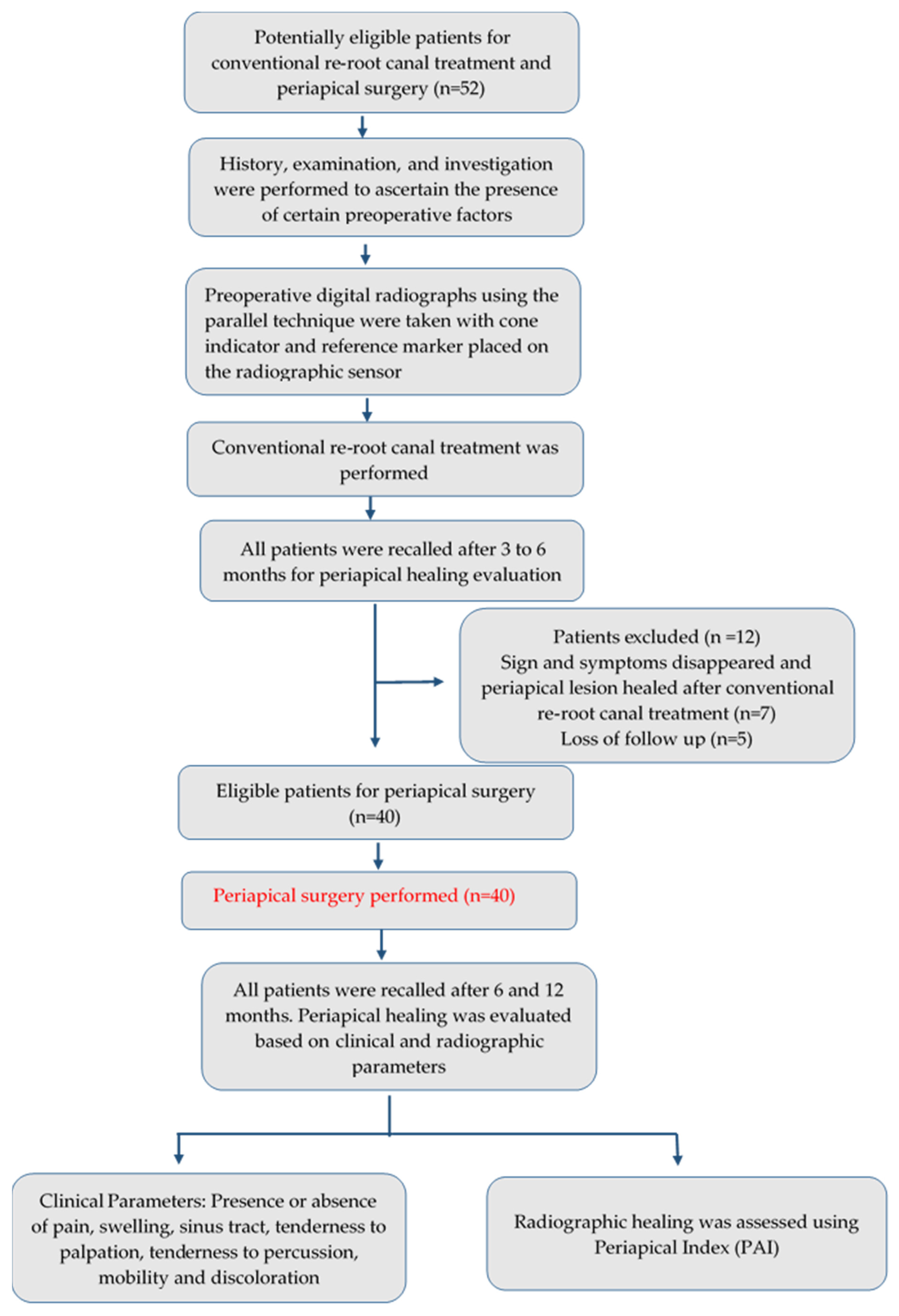
2. Materials and Methods

2.1. Study Setting and Ethical Approval

This study was conducted at the Department of Operative Dentistry in collaboration with the Institute of Biomedical Sciences, Dow University of Health Sciences, Karachi from July 2017 to December 2018 after obtaining ethical approval (IRB-862/DUHS/Approval/2017/50) from Institutional Review Board, Dow University of Health Sciences. All the recommendations of the Helsinki Declaration and its later amendments were strictly followed.

2.2. Inclusion and Exclusion Criteria

Initially, 52 patients aged between 15–57 years who presented with chronic apical periodontitis of single-rooted anterior teeth, characterized radiographically as radiolucency greater than 4 mm around the root apex with loss of lamina dura and periodontal ligament space due to incompletely attempted or previously failed root canal treatment or periapical lesion around an open root apex in the tooth, where root formation was halted prematurely due to trauma, were enrolled in this study. Medically compromised patients with any uncontrolled systemic disease (ASA III), patients with pre-existing periodontal disease or patients in whom the lesion was smaller than 4 mm, multirooted teeth, teeth with cracks and root fractures, pregnant or lactating women and patients who did not turn up for follow-up visits were excluded from this study.

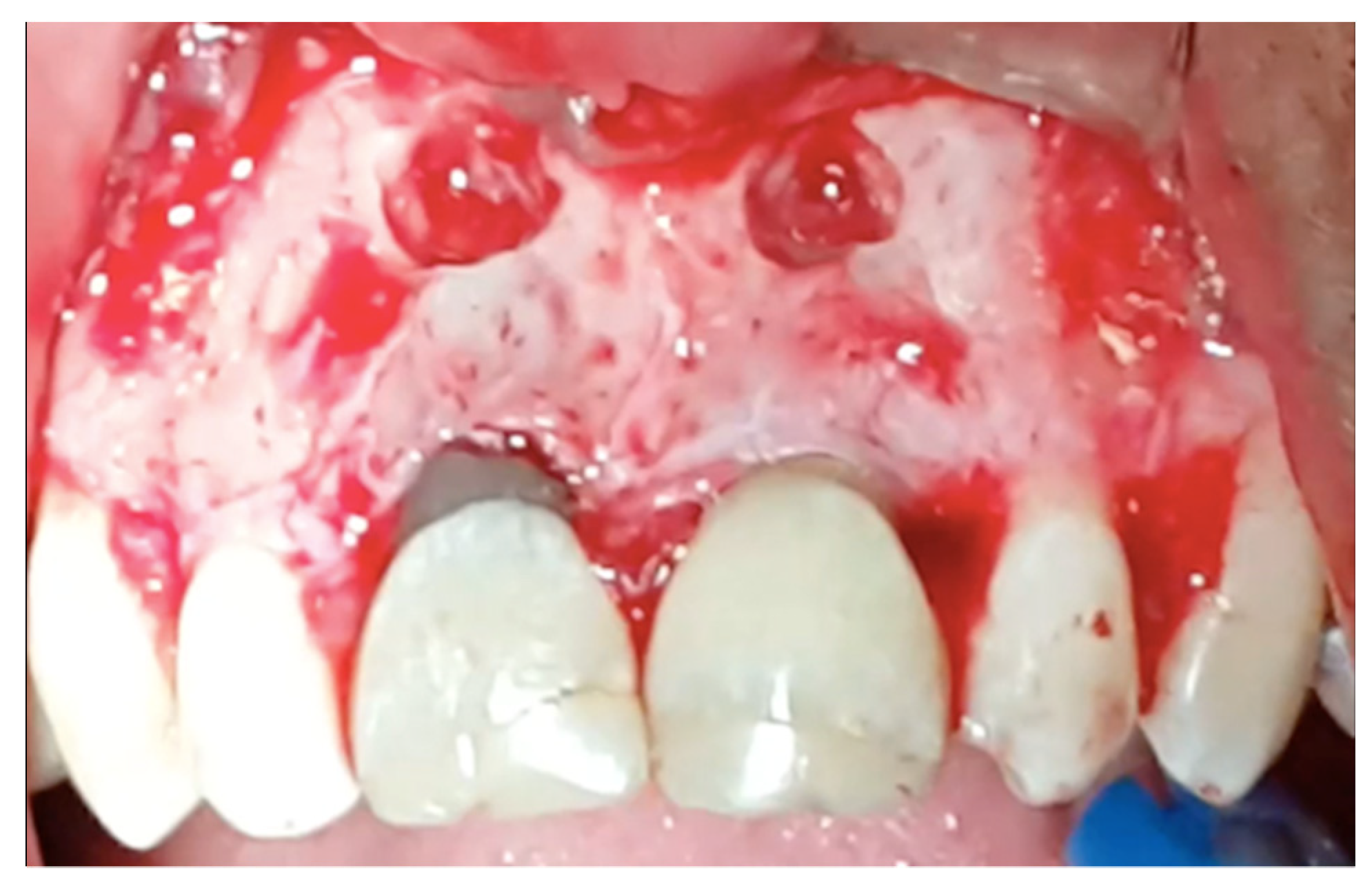
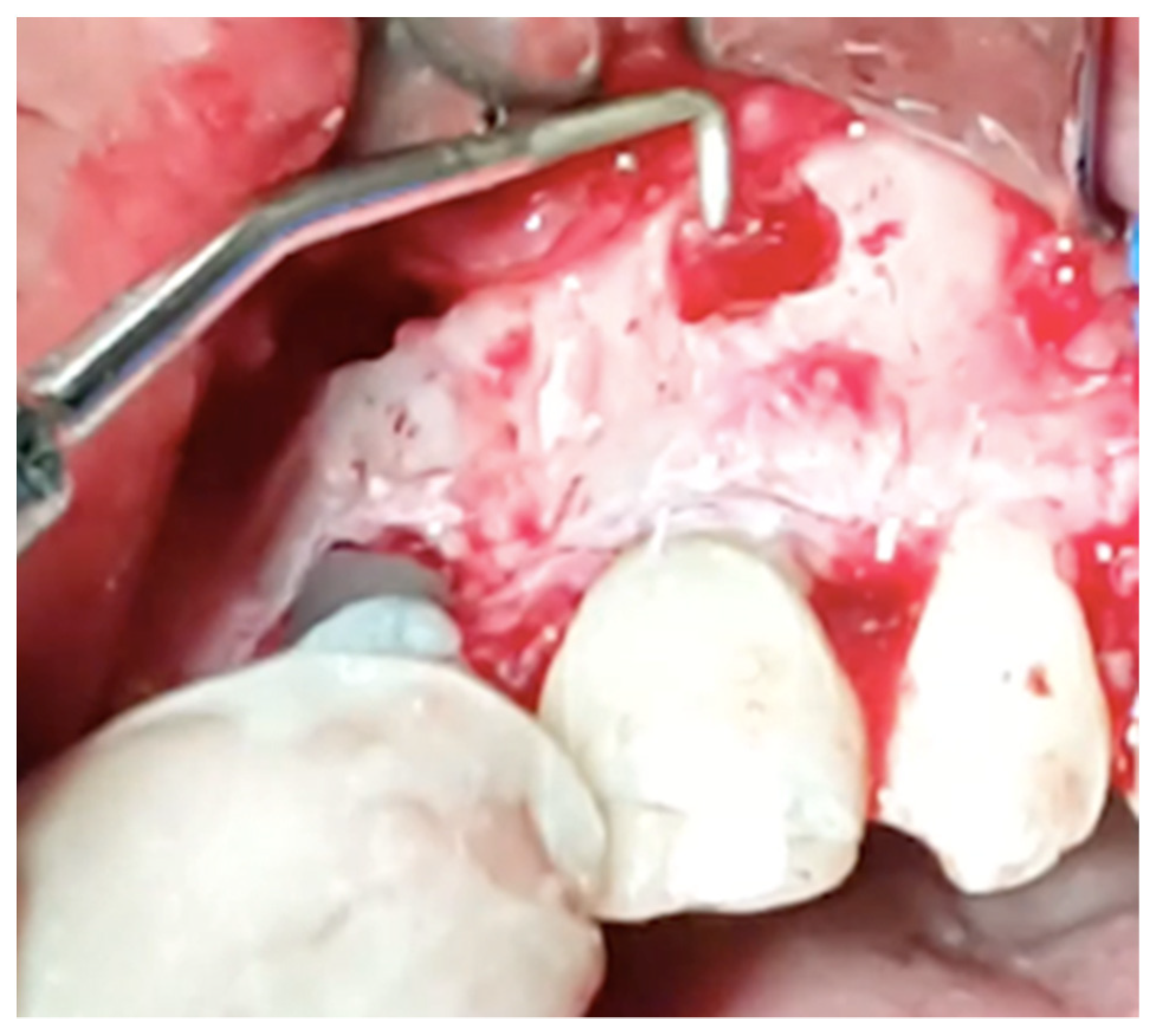
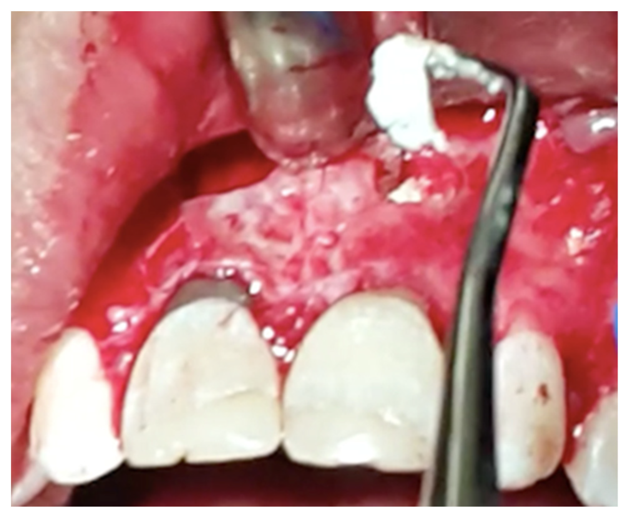
2.3. Clinical Procedure

History, examination, and investigation were performed to ascertain the presence of certain preoperative factors. Preoperative digital radiographs using the parallel technique were taken with a cone indicator and reference marker placed on the radiographic sensor Written informed consent was obtained from the patients.
Conventional re-root canal treatment: Local anesthesia 1:80,000 lidocaine with epinephrine was administered and a rubber dam was used for isolation. Access to the root was gained by removing the coronal restoration, followed by application of GP solvent (Carvene, PREVESTDenPro Prevest Denpro Limited, Jammu, India) and 25 or 30 # H file (Mani, Utsunomiya, Tochigi, Japan) for the removal of radicular filling. Working length was established by 35 or 40 # K file (Mani, Tochigi, Japan). Canal cleaning and shaping was performed by modified crown-down technique along with copious irrigation by 3.0% sodium hypochlorite (CanalPro NaOCl, Coltène/Whaledent, Altstätten, St. Gallen, Altstätten, Switzerland). Calcium hydroxide (Metapaste, Meta Biomed, Cheongju-si, Chungcheongbuk-do, Korea) was used as an intracanal medicament in between appointments. Finally, obturation was performed by cold lateral condensation technique followed by composite coronal restoration.
All patients were recalled after 3 to 6 months for clinical and radiographic evaluation of the periapical healing after conventional re-root canal treatment. Periapical healing was observed in seven patients and five patients failed to appear on follow-up visit, therefore 12 patients were excluded from the study. Finally, 40 patients underwent periapical surgery due to nonhealing periapical lesions (Figure 1).
Periapical Surgery: To begin with, local anesthesia 1:80,000 lidocaine with epinephrine was administered and a full thickness mucoperiosteal flap was elevated. After identifying the lesion site, access to the lesion was achieved by performing window preparation in cortical bone with small round bur No. 2 (Mani, Tochigi, Japan) in a slow-speed handpiece (Figure 2). The periapical lesion was identified and surgically removed by a surgical curette (Hibro Int, Tokyo, Japan). Zirconia-coated ultrasonic tip (Pro ultra, Maillefer DENTSPLY, Ballaigues, Switzerland) was used for apicoectomy and retrograde cavity preparation (Figure 3). The 3 mm apical fragment of the root was resected perpendicular to the long axis of the root with minimum or no bevel. Finally, MTA (Pro-root MTA, DENTSPLY Tulsa Dental Specialties, Johnson city, TX, USA) was used for retrograde filling (Figure 4). The flap was then repositioned and sutured with a 3/0 silk suture (ETHICON, Johnson & Johnsons, New Brunswick, NJ, USA). A periapical radiograph was taken after surgery. Analgesics and antibiotics were prescribed. These patients were recalled after 12 months for a follow-up visit. Both clinical and radiographic examination was performed at each follow-up visit (Figure 5). The parameters studied for clinical examination were pain, swelling, sinus tract, tenderness to palpation, tenderness to percussion, mobility, and discoloration. Radiographic healing was assessed using Periapical Index (PAI). The radiographic interpretation was carried out by two blinded endodontists. The interexaminer reliability in evaluating the postoperative radiographs was calculated and presented.

2.4. Statistical Analysis

The data were analyzed using a statistical package for social sciences (SPSS Version 25.0. Armonk, NY, USA). The results are presented as frequencies and percentages for age, gender, smoking, preoperative pain, size of the periapical lesion, and healing. Statistical associations were performed between periapical healing with age, gender, smoking, preoperative pain, size of the periapical lesion using chi-squared and Fisher’s exact test as per data assumptions. A p-value of 0.05 or less was considered statistically significant.

3. Results

Out of 40 patients, 32 (80%) patients were below 30 years of age while only 8 (20%) patients were above the age of 30 years. The gender distribution showed that 88 (58.7%) of the patients were male and 62 (41.3%) were female. Most of the patients, 26 (65.0%), were nonsmokers. The size of the preoperative periapical lesion was arbitrarily categorized into 6 mm or less and more than 6 mm for this study. The results showed that half of the patients had a periapical lesion size of 6 mm or less, while another half had more than 6 mm. Preoperative pain was present in 22 patients (55.01%) (Table 1).
There was a statistically significant association between periapical healing and age (p-value = 0.025), smoking (p-value = 0.029) and the size of periapical lesion (p-value < 0.001). After periapical surgery, healing was observed in 29 patients, in which 26 (81.3%) patients were below the age of 30 years while only 3 (37.5%) patients were above 30 years of age on one year follow-up. Similarly, 22 nonsmoking patients (84.6%) revealed periapical healing compared to 7 (50%) smoking patients. Although the success of healing was higher in males, at 78.6% (22/28), compared to females, at 58.3% (7/12), no statistically significant relationship was found between gender and healing (p-value = 0.254). Likewise, no significant association between the presence of preoperative pain and postoperative healing (p-value = 0.723) was observed (Table 2).

4. Discussion

The current study was conducted to evaluate the effect of various preoperative factors such as age, gender, smoking, preoperative pain, and preoperative periapical lesion size on the outcome of surgical endodontic treatment. The results of the study showed a statistically significant relationship between periapical wound healing and age, smoking, and preoperative lesion size.
Surgical endodontic retreatment is employed in managing recurrent secondary periapical lesions as a consequence of primary root canal treatment failure [19]. Surgical endodontic retreatment involves re-root canal treatment with subsequent sectioning of the apical end of the root followed by retrograde obturation and establishment of the apical seal of the root canal system. This apical seal facilitates healing of the periapical tissues by preventing the ingress of microbial irritants into the periapical area and concomitant tissue reaction [3].
It has now become clinically evident that even if the surgical endodontic treatment has been conducted using the same procedure, patients respond differently with respect to periapical wound healing [3,4]. This difference in periapical wound healing may be due to the presence of various preoperative factors such as pain, swelling, sinus tract, smoking, and size of the periapical lesion. However, evidence to support the influence of these preoperative factors on the outcome of periapical surgery are conflicting. In addition, most of the clinical studies that exist in the literature are retrospective [8,20,21]. The novelty of this study lies in following patients prospectively and opening an avenue for further research in this direction.
Age is considered a prognostic indicator of outcome for many surgical procedures [4]. In this study, 81.3% healing was achieved in patients below 30 years of age. This finding is in agreement with Kriesler et al. [22] They found a 95% success in patients between 21 to 40 years of age. Contrarily, Barone et al. [23] reported 84% success in periapical healing in patients above 45 years of age, compared to 68% in patients below 45 years of age. This study observed no significant relationship between healing and gender. Similarly, a large number of studies did not find gender as a prognostic factor for periapical surgery [18,24,25]. However, Peñarrocha-Diago et al. reported a higher success in males (60%) compared to females (40%) at 6 months [26].
The habit of smoking can influence the outcome of periapical surgery by several plausible biological mechanisms. First, smoking interferes with the function of lymphocytes, leukocytes, macrophages and increases the level of various proinflammatory mediators, such as TNF- α, IL-6, and C-reactive protein [27,28]. Second, smoking decreases the fibroblast migration to the periapical wound area and stimulates the function of osteoclastic cells, which encourage bone resorption [29]. Third, smoking causes morphological alteration in the microvasculature, which in turn disturbs the oxygen and nutrient supply to the periapical wound area [30]. In the current study, a positive trend of healing was observed in nonsmokers (84.6%). This finding conforms to Lopez et al. [8] and Kirkevang et al. [31], who found a statistically significant association between smoking and periapical healing. On the other hand, Rodriguez et al. [32] and Balto et al. [33] reported no significant difference in periapical periodontitis between smokers and nonsmokers.
The presence of preoperative pain and swelling or sinus tract may also govern the outcome of periapical surgery as indicated by many studies. Von Arc et al. [20] revealed that the presence of preoperative signs and symptoms discourages healing. They postulated that the healing potential of surgical wounds could be significantly affected by the stage of infection at the time of surgery, which is influenced by preoperative pain and signs. Similarly, Kreisler et al. [22] also claimed a lower success rate in patients with pretreatment pain. Contrastingly, the present study did not find any difference in the healing success rate in relation to the presence of preoperative pain, similarly to Song et al. [34] and Peñarrocha et al. [35]. The reason for there being no relation between the presence of preoperative pain and periapical healing after surgical endodontic treatment may be due to the fact that same treatment protocol was used for every patient and all treatment procedures were performed by the principal investigator.
Another important prognostic factor in periapical healing is the size of the lesion. In the present study, 100% successful healing was observed in patients with preoperative lesions of less than 6 mm, while 45% healing success was evident in patients with more than 6 mm of preoperative lesion size. A large number of studies have described the favorable prognosis after periapical surgery in patients with preoperative lesions of less than 5 mm in diameter [20,26,34]. Alternatively, Barone et al. [23] found an 80% success rate in periapical lesions of less than 10 mm in size at the time of surgery, compared to 53% success in periapical lesions of more than 10 mm in size.
Multiple systemic diseases are known to interfere with periapical healing, such as diabetes, hypertension, osteoporosis, or any uncontrolled systemic disease. Hyperglycemia increases the level of inflammatory markers and influences the various functions of the patient immune system [36]. Similarly, hypertension is associated with alterations in response and differentiation of bone cells at various levels [21]. Moreover, the drug bisphosphonate is commonly used for the treatment and prevention of osteoporosis and it is well-established that it reduces bone remodeling [37], therefore patients with these systemic diseases were excluded from this study, in addition to the patients with a pre-existing medical condition such as pregnancy and lactating mothers. Further prospective studies are required to evaluate postoperative healing in relation to the presence of different chronic systemic diseases using microsurgical techniques.
The findings of the study must be seen in light of some limitations. First, periapical surgery was performed by the conventional approach rather than microsurgical. The microsurgical approach offers numerous benefits such as small osteotomy, easy identification of root apices, visualization of minor anatomical variations, and preservation of cortical bone and root length by nearly 90-degree root resection. Several studies have shown the superiority of microsurgical endodontic treatment in comparison to traditional root-end surgery [38,39]. Second, the healing was assessed on a digital periapical radiograph, using a cone indicator and reference marker to ensure constant distance and angle between X-ray cone and sensor on every shoot. Moreover, exposure time, tube current, and voltage were the same on recall images. However, the X-ray image obtained was still two-dimensional, with a high probability of missing details in the third dimension. Nowadays, cone beam computed tomography (CBCT) is considered a standard of care [40] and has great value in establishing the correct diagnosis and in evaluating periapical healing after surgical endodontic treatment. Third, periapical surgery was performed on single-rooted anterior teeth only, multirooted posterior teeth where surgical access to treatment is difficult and high chances of variation in root morphology were excluded. Lastly, the sample size of the study was small due to the constrained study time, therefore; the results should be interpreted cautiously and may not represent the large population.
There is also a dire need to design a questionnaire which can evaluate the health-related quality of patients’ life after endodontic surgery, similar to The University of Washington Quality of Life Questionnaire (UW-QOL) for head-and-neck cancer patients [41]. Future studies are also desirable with the inclusion of a large sample size, multirooted teeth and various other treatment-related factors such as microsurgical technique, type of root-end filling, method of root-end resection and retrograde cavity preparation. Addition of tools such as fractal analysis used for the quantitative evaluation of bone trabeculation following periapical surgery will also help in the early detection of complex structural patterns in the trabecular bone [42].

5. Conclusions

Patient age, smoking status, and size of the preoperative lesion had a strong influence on periapical healing after surgical endodontic treatment. However, other perioperative (treatment-related) factors should also be taken into account to establish conclusive evidence.

Author Contributions

Conceptualization, M.A.A., N.M. and S.H.A.; methodology, M.A.A.; software, M.A.A.; validation, F.V., M.M., A.A. and M.F.B.; formal analysis, M.A.A.; investigation, M.A.A. and A.A.; resources, N.M. and S.H.A.; data curation, M.F.B.; writing—original draft preparation, M.A.A. and A.A.; writing—review and editing, M.A.A., F.V., M.M., A.A. and S.H.A.; visualization, N.M.; supervision, N.M., S.H.A. and M.F.B.; project administration, M.A.A. and A.A.; funding acquisition, M.A.A. All authors have read and agreed to the published version of the manuscript.

Funding

The authors are grateful to the Deanship of Scientific Research, King Saud University for supporting through Vice Deanship of Scientific Research Chairs, Engr. Abdullah Bugshan research chair for Dental and Oral Rehabilitation.

Institutional Review Board Statement

The study was conducted according to the guidelines of the Declaration of Helsinki, and approved by the Institutional Review Board, Dow University of Health Sciences (IRB-862/DUHS/Approval/2017/50) on 10 July 2017.

Informed Consent Statement

Informed consent was obtained from all subjects involved in the study.

Data Availability Statement

The data presented in this study are available on request from the corresponding author. The data are not publicly available due to ethical concerns.

Conflicts of Interest

The authors declare no conflict of interest.

References

  1. Schilder, H.; Hargreaves, K.M. Filling root canals in three dimensions. J. Endod. 2006, 32, 281–290. [Google Scholar] [CrossRef]
  2. Alghamdi, F.; Alhaddad, A.J.; Abuzinadah, S. Healing of Periapical Lesions After Surgical Endodontic Retreatment: A Systematic Review. Cureus 2020, 12, e6916. [Google Scholar] [CrossRef] [Green Version]
  3. Ahmed, M.A.; Anwar, M.F.; Ahmed, K.; Aftab, M.; Nazim, F.; Bari, M.F.; Mustafa, M.; Vohra, F.; Alrahlah, A.; Mughal, N.; et al. Baseline MMP expression in periapical granuloma and its relationship with periapical wound healing after surgical endodontic treatment. BMC Oral Health 2021, 21, 562. [Google Scholar] [CrossRef] [PubMed]
  4. Serrano-Giménez, M.; Sánchez-Torres, A.; Gay-Escoda, C. Prognostic factors on periapical surgery: A systematic review. Med. Oral Patol. Oral Cir. Bucal 2015, 20, e715–e722. [Google Scholar] [CrossRef]
  5. Segura-Egea, J.J.; Martín-González, J.; Castellanos-Cosano, L. Endodontic medicine: Connections between apical periodontitis and systemic diseases. Int. Endod. J. 2015, 48, 933–951. [Google Scholar] [CrossRef] [PubMed]
  6. Krall, E.A.; Sosa, C.A.; Garcia, C.; Nunn, M.E.; Caplan, D.J.; Garcia, R.I. Cigarette smoking increases the risk of root canal treatment. J. Dent. Res. 2006, 85, 313–317. [Google Scholar] [CrossRef]
  7. Peršić Bukmir, R.; Jurčević Grgić, M.; Brumini, G.; Spalj, S.; Pezelj-Ribaric, S.; Brekalo Pršo, I. Influence of tobacco smoking on dental periapical condition in a sample of Croatian adults. Wien. Klin. Wochenschr. 2016, 128, 260–265. [Google Scholar] [CrossRef]
  8. López-López, J.; Jané-Salas, E.; Martín-González, J.; Castellanos-Cosano, L.; Llamas-Carreras, J.M.; Velasco-Ortega, E.; Segura-Egea, J.J. Tobacco smoking and radiographic periapical status: A retrospective case-control study. J. Endod. 2012, 38, 584–588. [Google Scholar] [CrossRef]
  9. Ghattas Ayoub, C.; Aminoshariae, A.; Bakkar, M.; Ghosh, S.; Bonfield, T.; Demko, C.; Montagnese, T.A.; Mickel, A.K. Comparison of IL-1β, TNF-α, hBD-2, and hBD-3 Expression in the Dental Pulp of Smokers Versus Nonsmokers. J. Endod. 2017, 43, 2009–2013. [Google Scholar] [CrossRef]
  10. Melcher, A.H. On the Repair Potential of Periodontal Tissues. J. Periodontol. 1976, 47, 256–260. [Google Scholar] [CrossRef]
  11. Lin, L.; Chen, M.Y.H.; Ricucci, D.; Rosenberg, P.A. Guided Tissue Regeneration in Periapical Surgery. J. Endod. 2010, 36, 618–625. [Google Scholar] [CrossRef] [PubMed]
  12. Von Arx, T.; Jensen, S.S.; Hänni, S.; Friedman, S. Five-year longitudinal assessment of the prognosis of apical microsurgery. J. Endod. 2012, 38, 570–579. [Google Scholar] [CrossRef] [PubMed]
  13. Çalişkan, M.K.; Tekin, U.; Kaval, M.E.; Solmaz, M.C. The outcome of apical microsurgery using MTA as the root-end filling material: 2- to 6-year follow-up study. Int. Endod. J. 2016, 49, 245–254. [Google Scholar] [CrossRef] [PubMed]
  14. Shinbori, N.; Grama, A.M.; Patel, Y.; Woodmansey, K.; He, J. Clinical Outcome of Endodontic Microsurgery That Uses EndoSequence BC Root Repair Material as the Root-end Filling Material. J. Endod. 2015, 41, 607–612. [Google Scholar] [CrossRef]
  15. Lui, J.N.; Khin, M.M.; Krishnaswamy, G.; Chen, N.N. Prognostic factors relating to the outcome of endodontic microsurgery. J. Endod. 2014, 40, 1071–1076. [Google Scholar] [CrossRef]
  16. Lustmann, J.; Friedman, S.; Shaharabany, V. Relation of pre- and intraoperative factors to prognosis of posterior apical surgery. J. Endod. 1991, 17, 239–241. [Google Scholar] [CrossRef]
  17. Allen, R.K.; Newton, C.W.; Brown, C.E. A statistical analysis of surgical and nonsurgical endodontic retreatment cases. J. Endod. 1989, 15, 261–266. [Google Scholar] [CrossRef]
  18. Rahbaran, S.; Gilthorpe, M.S.; Harrison, S.D.; Gulabivala, K. Comparison of clinical outcome of periapical surgery in endodontic and oral surgery units of a teaching dental hospital: A retrospective study. Oral Surg. Oral Med. Oral Pathol. Oral Radiol. Endodontology 2001, 91, 700–709. [Google Scholar] [CrossRef]
  19. Kruse, C.; Spin-Neto, R.; Christiansen, R.; Wenzel, A.; Kirkevang, L.L. Periapical Bone Healing after Apicectomy with and without Retrograde Root Filling with Mineral Trioxide Aggregate: A 6-year Follow-up of a Randomized Controlled Trial. J. Endod. 2016, 42, 533–537. [Google Scholar] [CrossRef]
  20. Von Arx, T.; Peñarrocha, M.; Jensen, S. Prognostic factors in apical surgery with root-end filling: A meta-analysis. J. Endod. 2010, 36, 957–973. [Google Scholar] [CrossRef]
  21. Martins, C.M.; Sasaki, H.; Hirai, K.; Andrada, A.C.; Gomes-Filho, J.E. Relationship between hypertension and periapical lesion: An in vitro and in vivo study. Braz. Oral Res. 2016, 30, e78. [Google Scholar] [CrossRef] [PubMed] [Green Version]
  22. Kreisler, M.; Gockel, R.; Aubell-Falkenberg, S.; Kreisler, T.; Weihe, C.; Filippi, A.; Kühl, S.; Schütz, S.; D’Hoedt, B. Clinical outcome in periradicular surgery: Effect of patient- and tooth-related factors—A multicenter study. Quintessence Int. 2013, 44, 53–60. [Google Scholar]
  23. Barone, C.; Dao, T.T.; Basrani, B.B.; Wang, N.; Friedman, S. Treatment Outcome in Endodontics: The Toronto Study-Phases 3, 4, and 5: Apical Surgery. J. Endod. 2010, 36, 28–35. [Google Scholar] [CrossRef]
  24. Zuolo, M.L.; Ferreira, M.O.F.; Gutmann, J.L. Prognosis in periradicular surgery: A clinical prospective study. Int. Endod. J. 2000, 33, 91–98. [Google Scholar] [CrossRef]
  25. Wesson, C.M.; Gale, T.M. Molar apicectomy with amalgam root-end filling: Results of a prospective study in two district general hospitals. Br. Dent. J. 2003, 195, 707–714. [Google Scholar] [CrossRef] [PubMed] [Green Version]
  26. Peñarrocha-Diago, M.; Ortega-Sánchez, B.; García-Mira, B.; Maestre-Ferrín, L.; Peñarrocha-Oltra, D.; Gay-Escoda, C. A prospective clinical study of polycarboxylate cement in periapical surgery. Med. Oral Patol. Oral Cir. Bucal 2012, 17, 276–280. [Google Scholar] [CrossRef] [PubMed] [Green Version]
  27. Johnson, G.K.; Guthmiller, J.M. The impact of cigarette smoking on periodontal disease and treatment. Periodontol. 2000 2007, 44, 178–194. [Google Scholar] [CrossRef]
  28. Cabanillas-balsera, D.; Segura-Egea, J.J.; Bermudo-fuenmayor, M.; Martín-gonzález, J.; Jiménez-sánchez, M.C.; Areal-quecuty, V.; Sánchez-domínguez, B.; Montero-miralles, P.; Velasco-Ortega, E. Smoking and Radiolucent Periapical Lesions in Root Filled Teeth: Systematic Review and Meta-Analysis. J. Clin. Med. 2020, 9, 3506. [Google Scholar] [CrossRef]
  29. Bergström, J.; Babcan, J.; Eliasson, S. Tobacco smoking and dental periapical condition. Eur. J. Oral Sci. 2004, 112, 115–120. [Google Scholar]
  30. Krall, E.A.; Sosa, C.A.; Garcia, C.; Nunn, M.E.; Caplan, D.J.; Garcia, R.I. Cigarette smoking is associated with an acute impairment of microvascular function in humans. Clin. Sci. 2003, 104, 247–252. [Google Scholar]
  31. Kirkevang, L.L.; Wenzel, A. Risk indicators for apical periodontitis. Community Dent. Oral Epidemiol. 2003, 31, 59–67. [Google Scholar] [CrossRef]
  32. Rodriguez, F.R.; Taner, B.; Weiger, R.; Walter, C. Is smoking a predictor of apical periodontitis? Clin. Oral Investig. 2013, 17, 1947–1955. [Google Scholar] [CrossRef] [Green Version]
  33. Balto, H.A.; Alabdulaaly, L.; Bahammam, S.; Al-Ekrish, A.A. Comparative analysis of prevalence of apical periodontitis in smokers and non-smokers using cone-beam computed tomography. Saudi Dent. J. 2019, 31, 52–57. [Google Scholar] [CrossRef] [PubMed]
  34. Song, M.; Jung, I.Y.; Lee, S.J.; Lee, C.Y.; Kim, E. Prognostic factors for clinical outcomes in endodontic microsurgery: A retrospective study. J. Endod. 2011, 37, 927–933. [Google Scholar] [CrossRef] [PubMed]
  35. Peñarrocha Diago, M.; Ortega Sánchez, B.; García Mira, B.; Martí Bowen, E.; Von Arx, T.; Gay Escoda, C. Evaluation of healing criteria for success after periapical surgery. Med. Oral Patol. Oral Cir. Bucal 2008, 13, 143–147. [Google Scholar]
  36. Segura-Egea, J.J.; Martín-González, J.; Cabanillas-Balsera, D.; Fouad, A.F.; Velasco-Ortega, E.; López-López, J. Association between diabetes and the prevalence of radiolucent periapical lesions in root-filled teeth: Systematic review and meta-analysis. Clin. Oral Investig. 2016, 20, 1133–1141. [Google Scholar] [CrossRef]
  37. Hsiao, A.; Glickman, G.; He, J. A retrospective clinical and radiographic study on healing of periradicular lesions in patients taking oral bisphosphonates. J. Endod. 2009, 35, 1525–1528. [Google Scholar] [CrossRef] [PubMed]
  38. Tortorici, S.; Difalco, P.; Caradonna, L.; Tetè, S. Traditional endodontic surgery versus modern technique: A 5-year controlled clinical trial. J. Craniofac. Surg. 2014, 25, 804–807. [Google Scholar] [CrossRef]
  39. Setzer, F.C.; Shah, S.B.; Kohli, M.R.; Karabucak, B.; Kim, S. Outcome of endodontic surgery: A meta-analysis of the literature—part 1: Comparison of traditional root-end surgery and endodontic microsurgery. J. Endod. 2010, 36, 1757–1765. [Google Scholar] [CrossRef]
  40. Abdinian, M.; Katiraei, M.; Zahedi, H.; Rengo, C.; Soltani, P.; Spagnuolo, G. Age Estimation Based on Pulp–Tooth Volume Ratio of Anterior Teeth in Cone-Beam Computed Tomographic Images in a Selected Population: A Cross-Sectional Study. Appl. Sci. 2021, 11, 9984. [Google Scholar] [CrossRef]
  41. Lowe, D.; Rogers, S. University of Washington Quality of Life Questionnaire (UW-QOL v4 and v4.1). Available online: http://www.headandneckcancer.co.uk/sites/default/files/assets/pages/UW-QOL-update-2018.pdf (accessed on 10 June 2021).
  42. Soltani, P.; Sami, S.; Yaghini, J.; Golkar, E.; Riccitiello, F.; Spagnuolo, G. Application of Fractal Analysis in Detecting Trabecular Bone Changes in Periapical Radiograph of Patients with Periodontitis. Int. J. Dent. 2021, 7, 3221448. [Google Scholar] [CrossRef] [PubMed]
Figure 1. Flowchart of Methodology.
Applsci 11 11768 g001
Figure 2. A full-thickness mucoperiosteal flap was reflected and the window was created around the periapical area of teeth 11 and 21.
Applsci 11 11768 g002
Figure 3. Retrograde cavity preparation was performed by zirconia-coated ultrasonic tip used for periapical surgery.
Applsci 11 11768 g003
Figure 4. The retrograde filling was performed by MTA.
Applsci 11 11768 g004
Figure 5. (A). Immediate postoperative periapical radiograph showing periapical radiolucency around the apex of Tooth 12. (B). Periapical radiolucency decreased in six months. (C). Complete resolution of periapical radiolucency in one year.
Applsci 11 11768 g005
Table 1. Descriptive Statistics for Demographics.
CharacteristicsN = 40 (%)
Age
<30 years32 (80.0)
>30 years08 (20.0)
Gender
Male28 (70.0)
Female12 (30.0)
Smoking
Nonsmoker26 (65.0)
Smoker14 (35.0)
Preoperative pain22 (55.0)
Periapical lesion size
≤6 mm20 (50.0)
>6 mm20 (50.0)
Postoperative healing29 (72.5)
Table 2. Relationship of Periapical Healing with Age, Gender, Smoking, Preoperative pain, and Periapical lesion size on one year follow-up.
CharacteristicsPeriapical Healing
Yes
(n = 29)
No
(n = 11)
Totalp-Value
Age in years 0.025 ^
<3026 (81.3%)6 (18.8%)32
>303 (37.5%)5 (62.5%)8
Gender
Male22 (78.6%)6 (21.4%)280.254 ^
Female7 (58.3%)5 (41.7%)12
Smoking
Nonsmoker22 (84.6%)4 (15.4%)260.029 ^
Smoker7 (50.0%)7 (50.0%)14
Preoperative pain
Yes15 (68.2%)7 (31.8%)220.723 ^
No14 (77.8%)4 (22.2%)18
Periapical lesion size
≤6 mm20 (100%)0 (0.0%)20<0.001 ¥
>6 mm09 (45.0%)11 (55.0%)20
¥ Chi-squared test, ^ Fisher’s Exact Test.
Publisher’s Note: MDPI stays neutral with regard to jurisdictional claims in published maps and institutional affiliations.

