
Recent Developments in Plasmonic Sensors of Phenol and Its Derivatives


Abstract
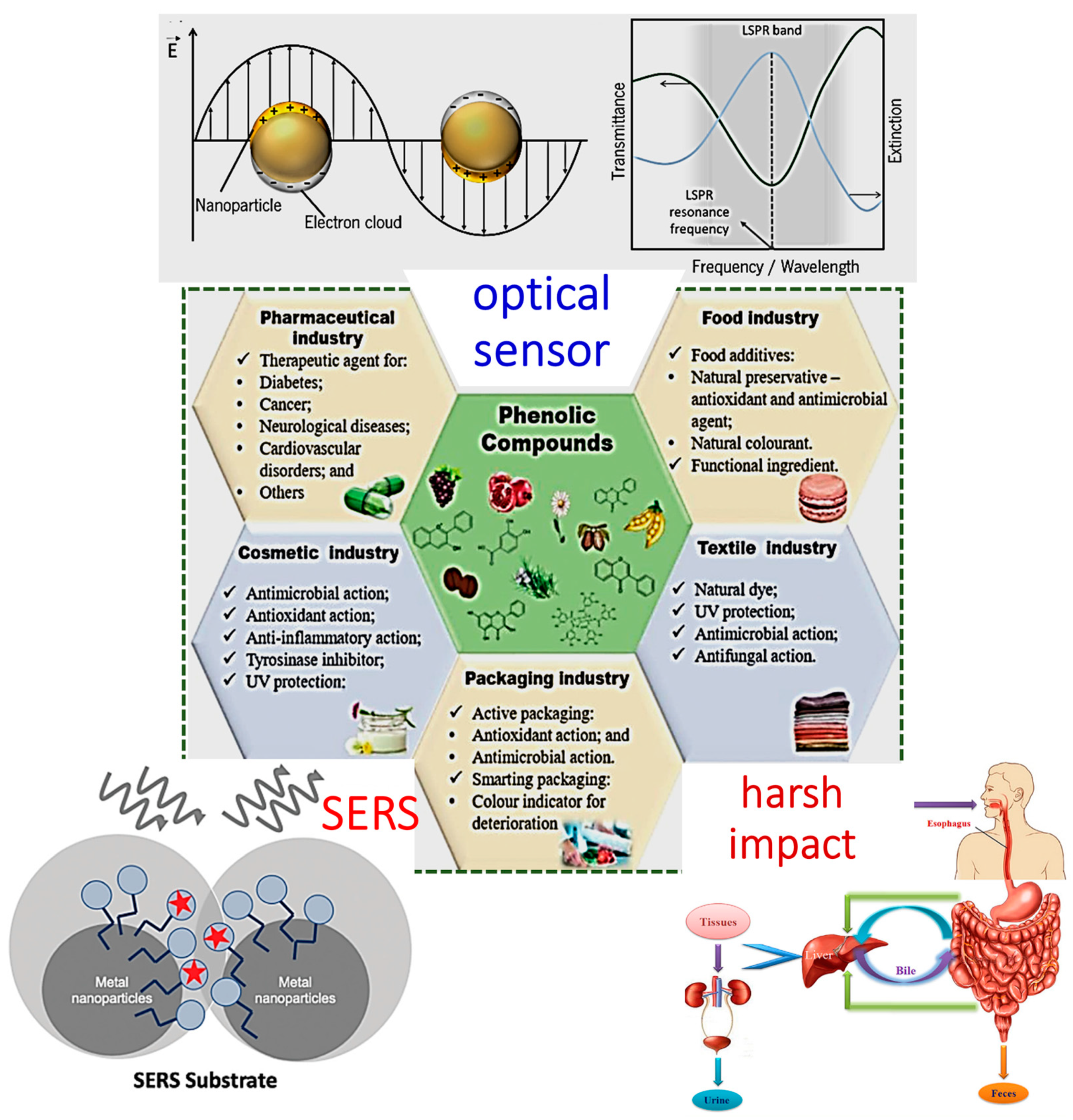
:1. Introduction
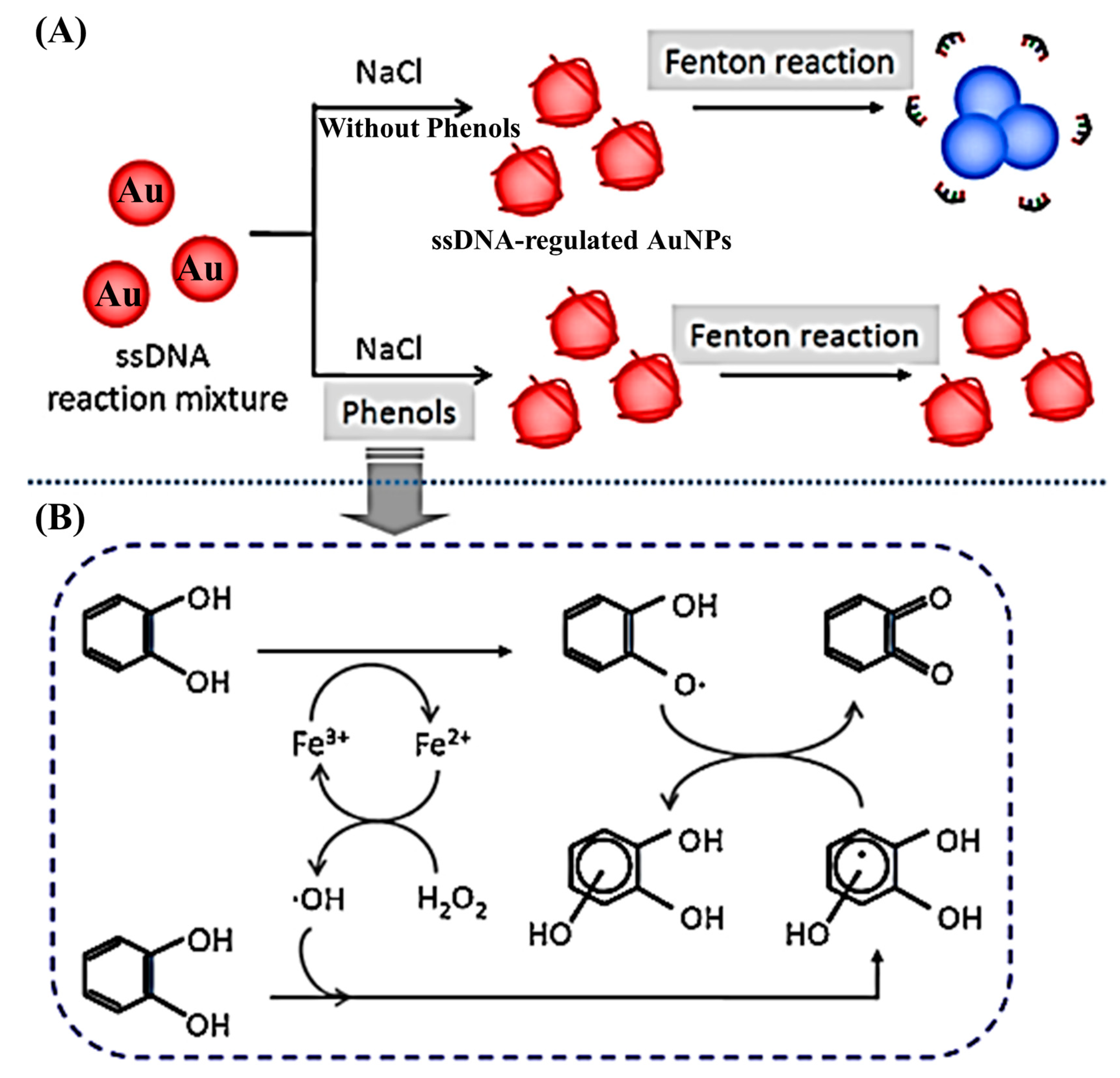
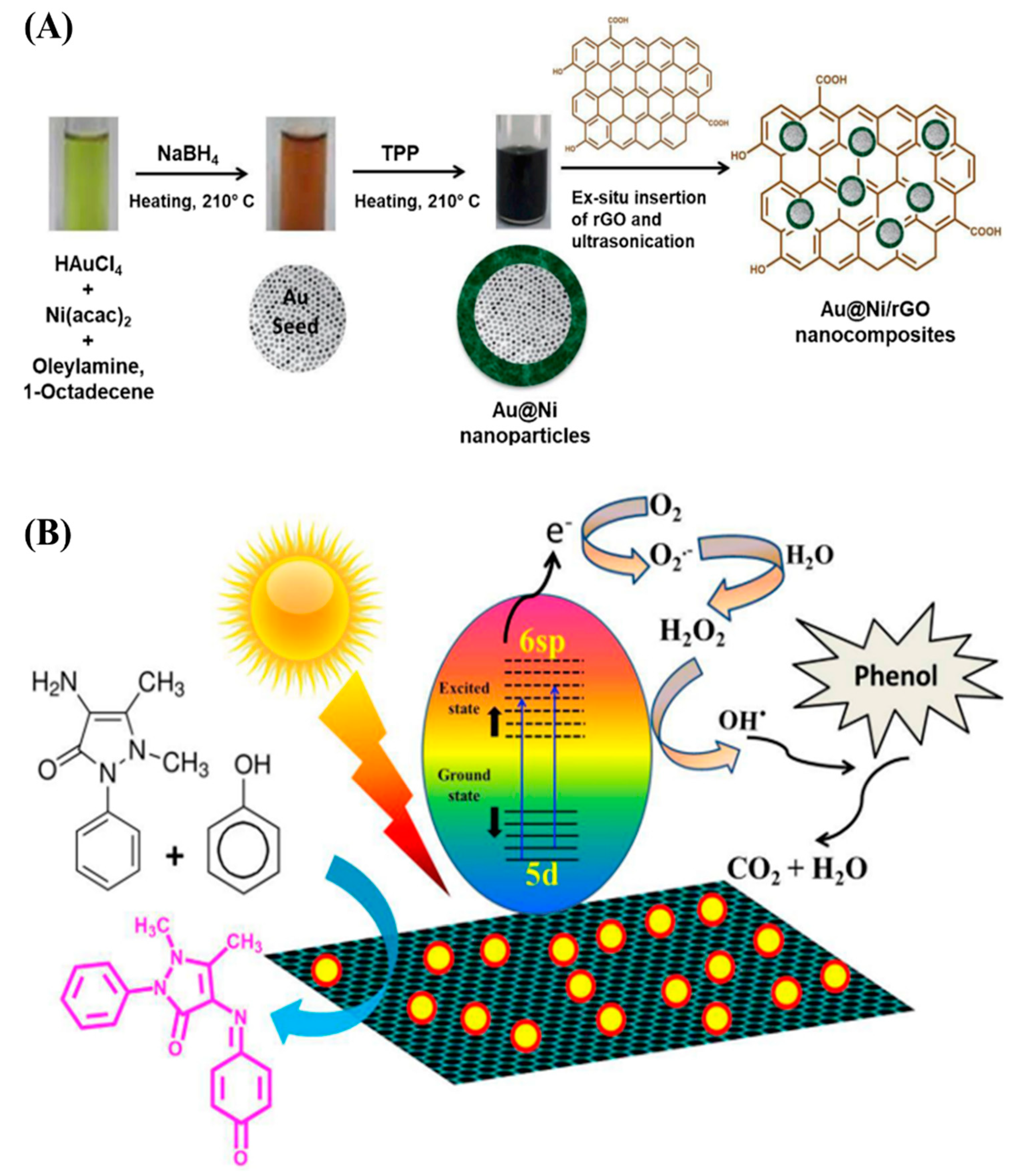
2. Plasmonic Resonance-Based Colorimetric Sensor for Phenolic Compounds
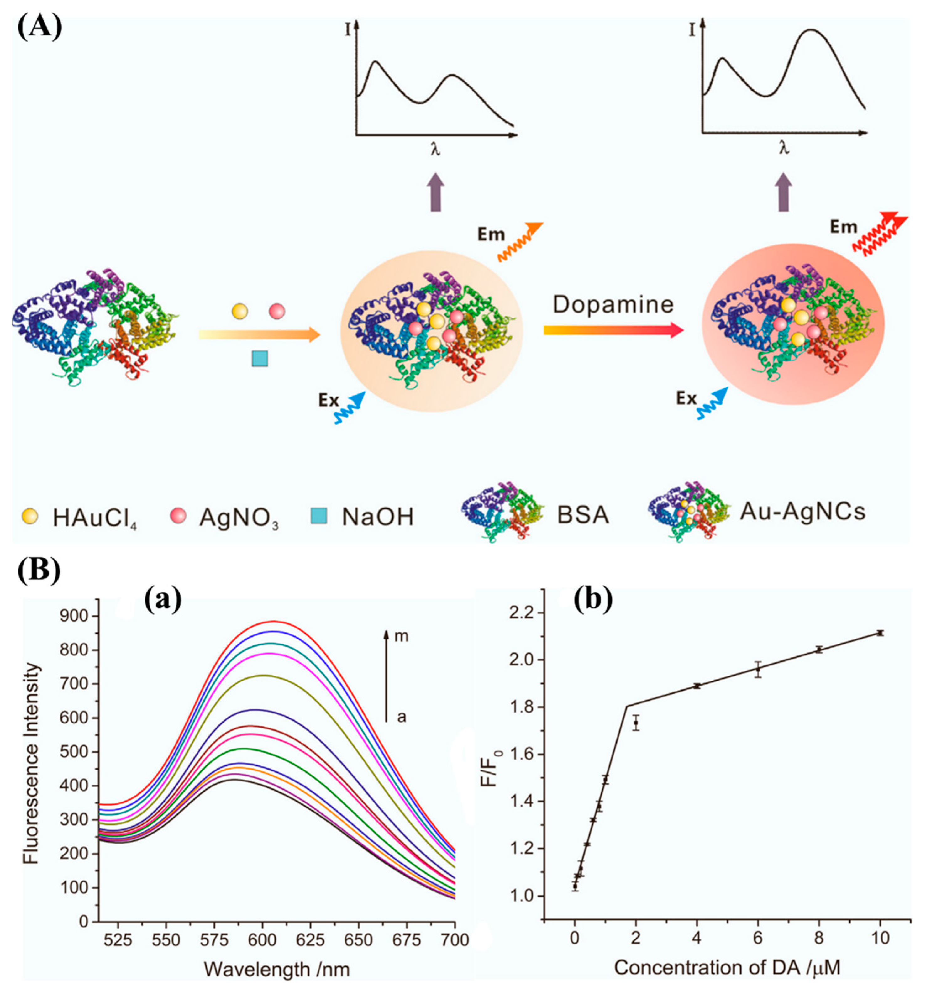
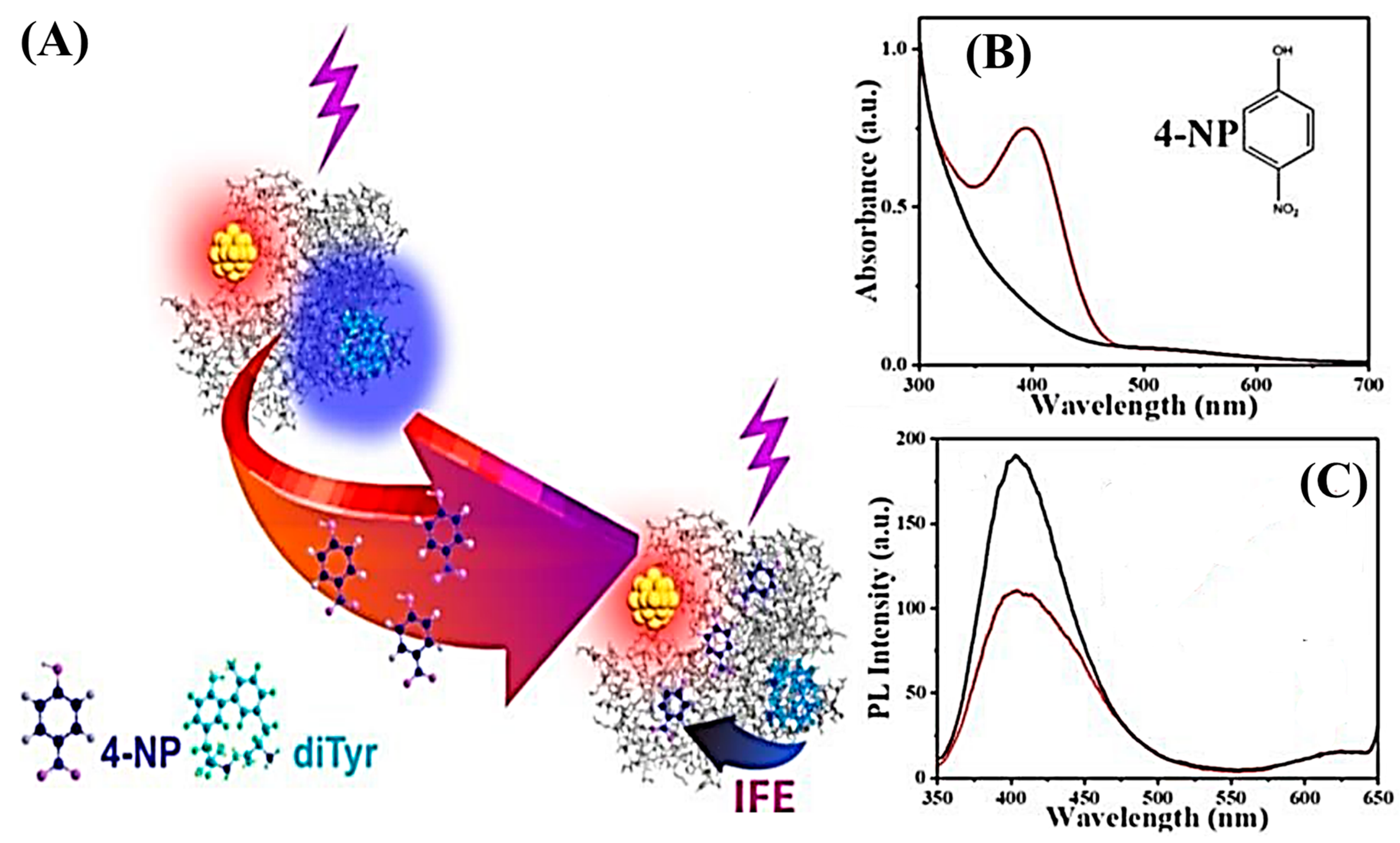
3. Plasmonic Resonance-Based Fluorescence Sensor for Phenolic Compounds
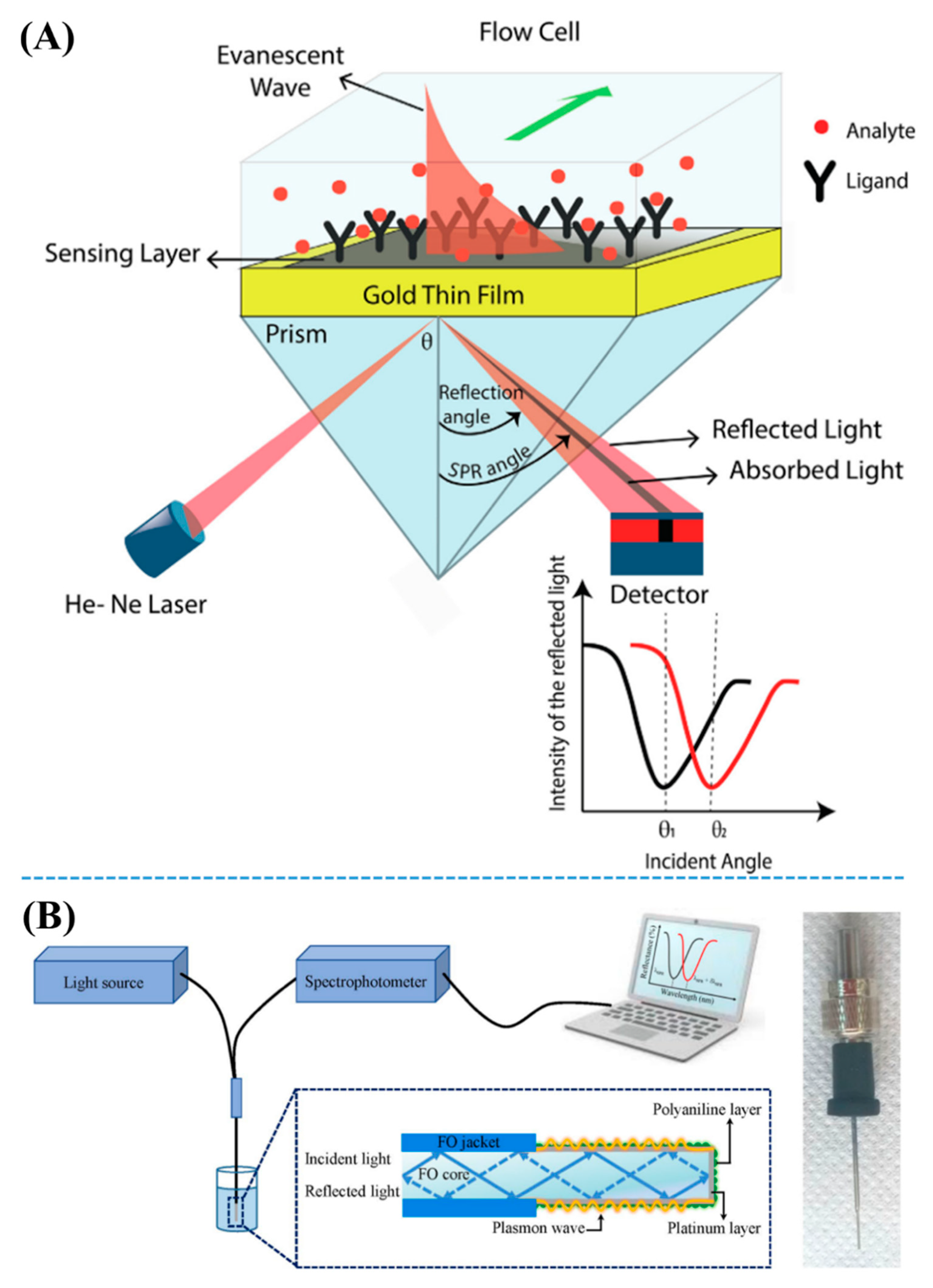
4. Localized Surface Plasmon Resonance Phenomenon-Based Optical Sensor for Phenolic Compounds
5. Plasmonic Resonance-Based Raman Sensors for Detection of Phenolic Compounds
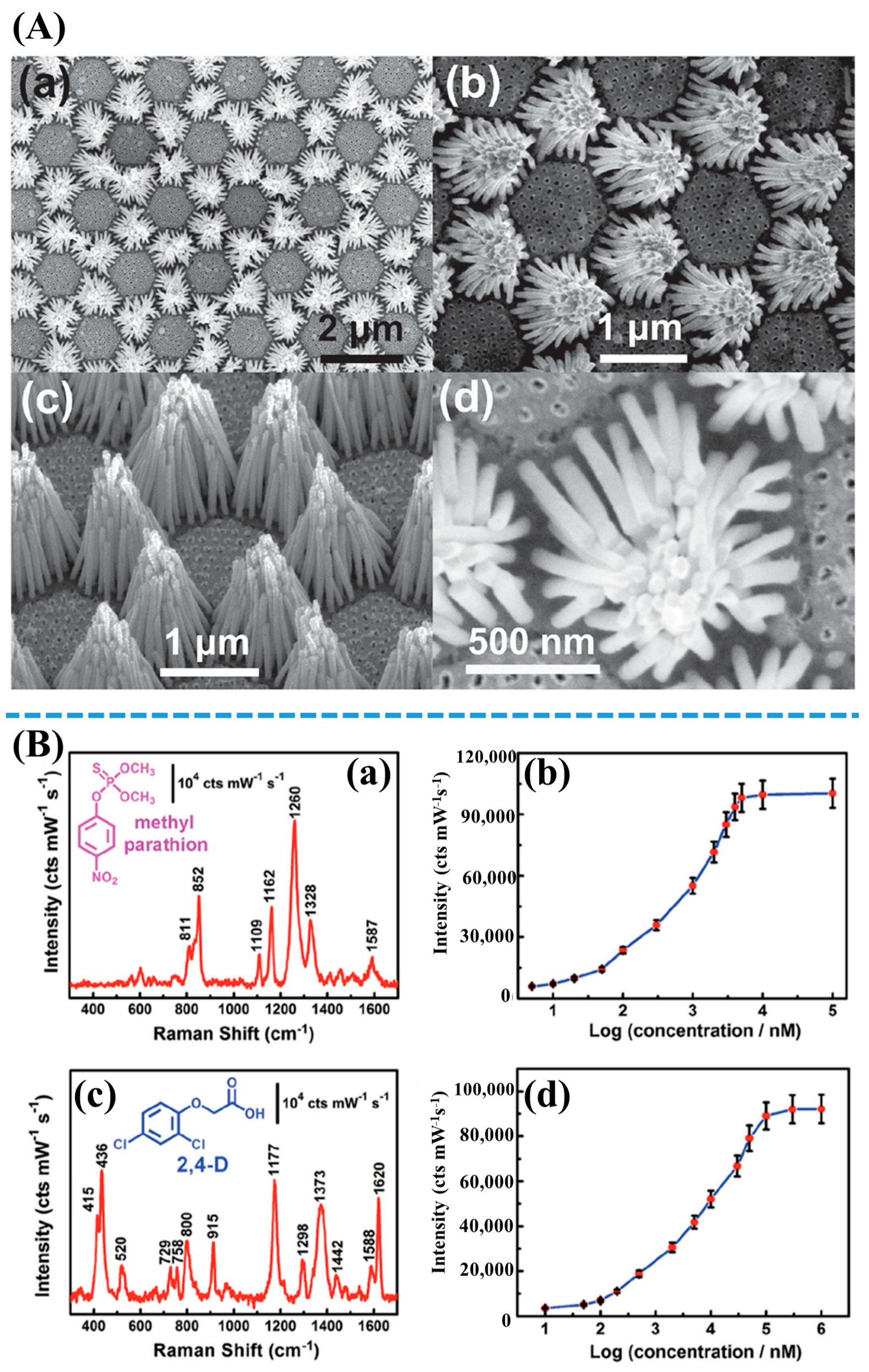
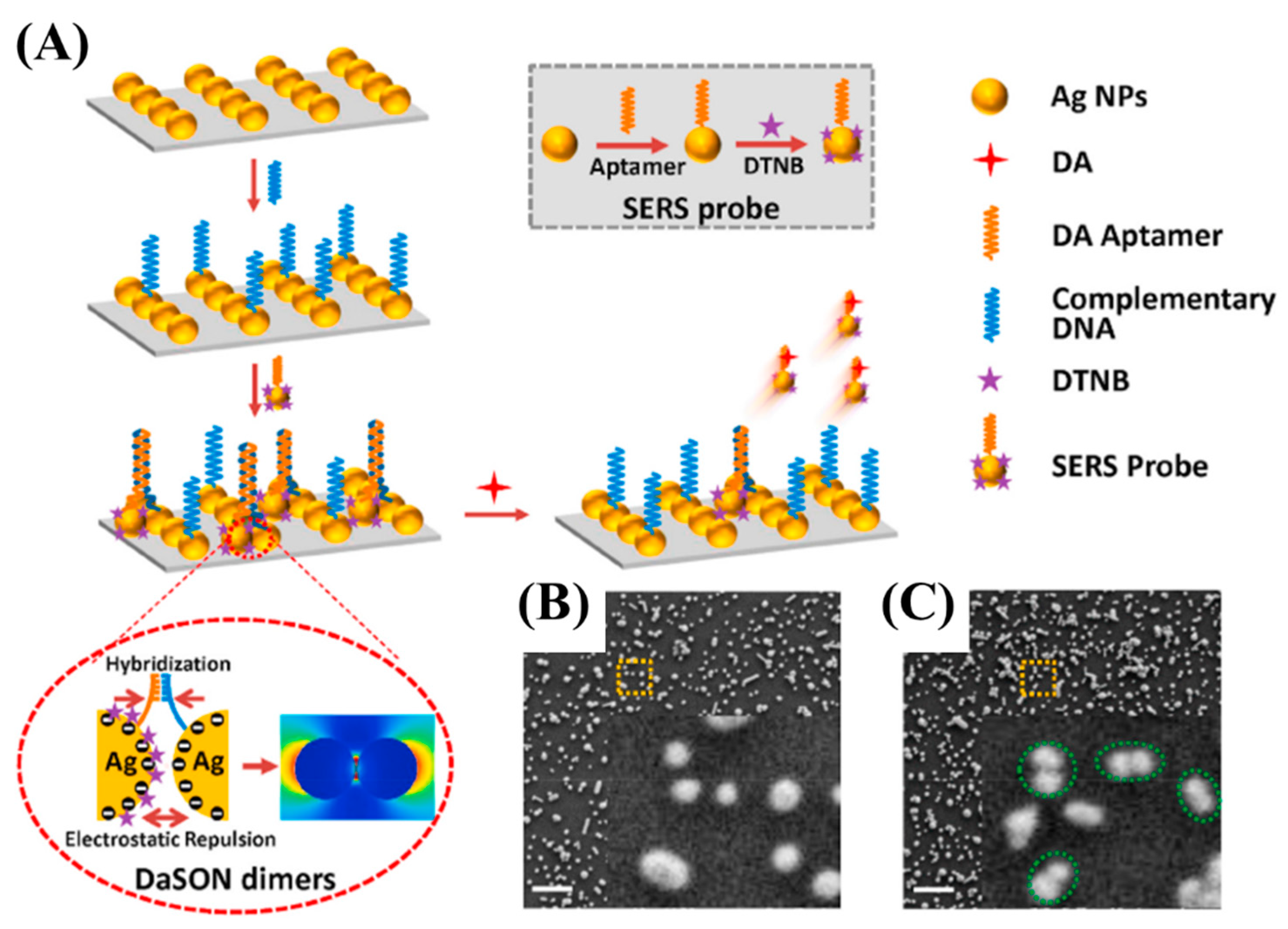
5.1. Pure Noble Metal Nanomaterial-Based Raman Sensors
5.2. Hybrid Nanomaterial-Based Raman Sensors
5.3. Metal–Organic Frameworks Structure-Introduced Raman Sensors
6. Conclusions
Author Contributions
Funding
Institutional Review Board Statement
Informed Consent Statement
Data Availability Statement
Conflicts of Interest
References
- Pérez-Ocón, F.; Pozo, A.M.; Cortina, J.; Rabaza, O. Surface plasmon resonance sensor of CO2 for indoors and outdoors. Appl. Sci. 2021, 11, 6869. [Google Scholar] [CrossRef]
- Rodrigues, M.S.; Borges, J.; Lopes, C.; Pereira, R.M.S.; Vasilevskiy, M.I.; Vaz, F. Gas sensors based on localized surface plasmon resonances: Synthesis of oxide films with embedded metal nanoparticles, theory and simulation, and sensitivity enhancement strategies. Appl. Sci. 2021, 11, 5388. [Google Scholar] [CrossRef]
- Zhang, L.; Chen, X.-R.; Wen, S.-H.; Liang, R.-P.; Qiu, J.-D. Optical sensors for inorganic arsenic detection. TrAC Trends Anal. Chem. 2019, 118, 869–879. [Google Scholar] [CrossRef]
- Zhu, C.; Zhao, Q.; Wang, X.; Li, Z.; Hu, X. Ag-nanocubes/graphene-oxide/Au-nanoparticles composite film with highly dense plasmonic hotspots for surface-enhanced Raman scattering detection of pesticide. Microchem. J. 2021, 165, 106090. [Google Scholar] [CrossRef]
- Omar, N.A.S.; Fen, Y.W.; Ramli, I.; Azmi, U.Z.M.; Hashim, H.S.; Abdullah, J.; Mahdi, M.A. Cellulose and vanadium plasmonic sensor to measure Ni2+ ions. Appl. Sci. 2021, 11, 2963. [Google Scholar] [CrossRef]
- Nagy-Simon, T.; Hada, A.-M.; Suarasan, S.; Potara, M. Recent advances on the development of plasmon-assisted biosensors for detection of C-reactive protein. J. Mol. Struct. 2021, 1246, 131178. [Google Scholar] [CrossRef]
- Varasteanu, P.; Kusko, M. A multi-objective optimization of 2D materials modified surface plasmon resonance (SPR) based sensors: An NSGA II approach. Appl. Sci. 2021, 11, 4353. [Google Scholar] [CrossRef]
- Zhong, X.; Ma, L.; Yin, G.; Gan, M.; Wei, Y. Hg2+ optical fiber sensor based on LSPR with PDDA-templated AuNPs and CS/PAA bilayers. Appl. Sci. 2020, 10, 4845. [Google Scholar] [CrossRef]
- Hashim, H.S.; Fen, Y.W.; Omar, N.A.S.; Abdullah, J.; Daniyal, W.M.E.M.M.; Saleviter, S. Detection of phenol by incorporation of gold modified-enzyme based graphene oxide thin film with surface plasmon resonance technique. Opt. Express 2020, 28, 9738–9752. [Google Scholar] [CrossRef]
- Du, J.; Jing, C. One-step fabrication of dopamine-inspired Au for SERS sensing of Cd2+ and polycyclic aromatic hydrocarbons. Anal. Chim. Acta 2019, 1062, 131–139. [Google Scholar] [CrossRef]
- Tang, L.; Li, S.; Han, F.; Liu, L.; Xu, L.; Ma, W.; Kuang, H.; Li, A.; Wang, L.; Xu, C. SERS-active Au@Ag nanorod dimers for ultrasensitive dopamine detection. Biosens. Bioelectron. 2015, 71, 7–12. [Google Scholar] [CrossRef] [PubMed]
- Matteini, P.; Cottat, M.; Tavanti, F.; Panfilova, E.; Scuderi, M.; Nicotra, G.; Menziani, M.C.; Khlebtsov, N.; De Angelis, M.; Pini, R. Site-selective surface-enhanced Raman detection of proteins. ACS Nano 2017, 11, 918–926. [Google Scholar] [CrossRef] [PubMed]
- Bhavya, M.B.; Prabhu, B.R.; Shenoy, B.M.; Bhol, P.; Swain, S.; Saxena, M.; John, N.S.; Hegde, G.; Samal, A.K. Femtomolar detection of thiram via SERS using silver nanocubes as an efficient substrate. Environ. Sci. Nano 2020, 7, 3999–4009. [Google Scholar] [CrossRef]
- Lê, Q.T.; Ly, N.H.; Kim, M.-K.; Lim, S.H.; Son, S.J.; Zoh, K.-D.; Joo, S.-W. Nanostructured Raman substrates for the sensitive detection of submicrometer-sized plastic pollutants in water. J. Hazard. Mater. 2021, 402, 123499. [Google Scholar] [CrossRef] [PubMed]
- Kim, N.; Ahn, Y.; Jo, J.; Pyo, H.; Lee, J.; Choi, J. Soil assessment after chemical accidents using metabolic profiling and microbial community evaluation. Chemosphere 2020, 268, 129362. [Google Scholar] [CrossRef] [PubMed]
- Duan, W.; Meng, F.; Lin, Y.; Wang, G. Toxicological effects of phenol on four marine microalgae. Environ. Toxicol. Pharmacol. 2017, 52, 170–176. [Google Scholar] [CrossRef]
- Duan, W.; Meng, F.; Cui, H.; Lin, Y.; Wang, G.; Wu, J. Ecotoxicity of phenol and cresols to aquatic organisms: A review. Ecotoxicol. Environ. Saf. 2018, 157, 441–456. [Google Scholar] [CrossRef] [PubMed]
- Chae, Y.; Kim, L.; Kim, D.; Cui, R.; Lee, J.; An, Y.-J. Deriving hazardous concentrations of phenol in soil ecosystems using a species sensitivity distribution approach. J. Hazard. Mater. 2020, 399, 123036. [Google Scholar] [CrossRef]
- Chandane, P.; Ladke, J.; Jori, C.; Deshmukh, S.; Zinjarde, S.; Chakankar, M.; Hocheng, H.; Jadhav, U. Synthesis of magnetic Fe3O4 nanoparticles from scrap iron and use of their peroxidase like activity for phenol detection. J. Environ. Chem. Eng. 2019, 7, 103083. [Google Scholar] [CrossRef]
- Tran, T.D.; Nguyen, P.T.; Le, T.N.; Kim, M.I. DNA-copper hybrid nanoflowers as efficient laccase mimics for colorimetric detection of phenolic compounds in paper microfluidic devices. Biosens. Bioelectron. 2021, 182, 113187. [Google Scholar] [CrossRef]
- Carvalho, D.G.; Ranzan, L.; Jacques, R.A.; Trierweiler, L.F.; Trierweiler, J.O. Analysis of total phenolic compounds and caffeine in teas using variable selection approach with two-dimensional fluorescence and infrared spectroscopy. Microchem. J. 2021, 169, 106570. [Google Scholar] [CrossRef]
- Mainali, K.; Garcia-Perez, M. Identification and quantification of trace oxygenated compounds in alternative jet fuels: Fluorescence methods for fast detection of phenolic compounds in operational field conditions. Fuel 2020, 271, 117652. [Google Scholar] [CrossRef]
- Antohe, I.; Iordache, I.; Antohe, V.-A.; Socol, G. A polyaniline/platinum coated fiber optic surface plasmon resonance sensor for picomolar detection of 4-nitrophenol. Sci. Rep. 2021, 11, 1–10. [Google Scholar] [CrossRef]
- Mendonça, C.D.; Khan, S.U.; Rahemi, V.; Verbruggen, S.W.; Machado, S.A.S.; De Wael, K. Surface plasmon resonance-induced visible light photocatalytic TiO2 modified with GNPs for the quantification of hydroquinone. Electrochim. Acta 2021, 389, 138734. [Google Scholar] [CrossRef]
- Carreira-Casais, A.; Montes-García, V.; Pastoriza-Santos, I.; Prieto, M.; Simal-Gandara, J.; Pérez-Juste, J. Multiple SERS detection of phenol derivatives in tap water. Proceedings 2020, 70, 88. [Google Scholar] [CrossRef]
- Dendisová, M.; Němečková, Z.; Člupek, M.; Prokopec, V. EC-SERS study of phenolic acids sorption behavior on Au, Ag and Cu substrates—Effect of applied potential and metal used. Appl. Surf. Sci. 2019, 470, 716–723. [Google Scholar] [CrossRef]
- Deneva, V.; Bakardzhiyski, I.; Bambalov, K.; Antonova, D.; Tsobanova, D.; Bambalov, V.; Cozzolino, D.; Antonov, L. Using Raman spectroscopy as a fast tool to classify and analyze Bulgarian wines-A feasibility study. Molecules 2019, 25, 170. [Google Scholar] [CrossRef] [PubMed] [Green Version]
- Wang, J.; Qiu, C.; Mu, X.; Pang, H.; Chen, X.; Liu, D. Ultrasensitive SERS detection of rhodamine 6G and p-nitrophenol based on electrochemically roughened nano-Au film. Talanta 2020, 210, 120631. [Google Scholar] [CrossRef]
- Wan, M.; Zhao, H.; Wang, Z.; Zou, X.; Zhao, Y.; Sun, L. Fabrication of Ag modified SiO2 electrospun nanofibrous membranes as ultrasensitive and high stable SERS substrates for multiple analytes detection. Colloids Interface Sci. Commun. 2021, 42, 100428. [Google Scholar] [CrossRef]
- Sun, J.; Xue, D.; Shan, W.; Liu, R.; Liu, R.; Zhao, H.; Li, T.; Wang, Z.; Zhang, J.; Shao, B. In situ growth large area silver nanostructure on metal phenolic network coated NAAO film and its SERS sensing application for monofluoroacetic acid. ACS Sens. 2021, 6, 2129–2135. [Google Scholar] [CrossRef]
- Sricharoen, N.; Sukmanee, T.; Pienpinijtham, P.; Ekgasit, S.; Kitahama, Y.; Ozaki, Y.; Wongravee, K. MCR-ALS with sample insertion constraint to enhance the sensitivity of surface-enhanced Raman scattering detection. Analyst 2021, 146, 3251–3262. [Google Scholar] [CrossRef] [PubMed]
- Shen, Y.; Yu, D.; Han, F.-Y.; Shen, A.-G.; Hu, J.-M. On-site and quantitative SERS detection of trace 1,2,3-benzotriazole in transformer oil with colloidal lignin particles-based green pretreatment reagents. Spectrochim. Acta A Mol. Biomol. Spectrosc. 2021, 252, 119469. [Google Scholar] [CrossRef] [PubMed]
- Rostami, S.; Zór, K.; Zhai, D.S.; Viehrig, M.; Morelli, L.; Mehdinia, A.; Smedsgaard, J.; Rindzevicius, T.; Boisen, A. High-throughput label-free detection of Ochratoxin A in wine using supported liquid membrane extraction and Ag-capped silicon nanopillar SERS substrates. Food Control 2020, 113, 107183. [Google Scholar] [CrossRef]
- Rao, Y.; Zhao, X.; Li, Z.; Huang, J. Phenolic acids induced growth of 3D ordered gold nanoshell composite array as sensitive SERS nanosensor for antioxidant capacity assay. Talanta 2018, 190, 174–181. [Google Scholar] [CrossRef] [PubMed]
- Aarthi, A.; Bindhu, M.; Umadevi, M.; Parimaladevi, R.; Sathe, G.; Al-Mohaimeed, A.M.; Elshikh, M.S.; Balasubramanian, B. Evaluating the detection efficacy of advanced bimetallic plasmonic nanoparticles for heavy metals, hazardous materials and pesticides of leachate in contaminated groundwater. Environ. Res. 2021, 201, 111590. [Google Scholar] [CrossRef]
- Chen, X.; Qin, L.; Kang, S.-Z.; Li, X. A special zinc metal-organic frameworks-controlled composite nanosensor for highly sensitive and stable SERS detection. Appl. Surf. Sci. 2021, 550, 149302. [Google Scholar] [CrossRef]
- Xu, J.; Shang, S.; Gao, W.; Zeng, P.; Jiang, S. Ag@ZIF-67 decorated cotton fabric as flexible, stable and sensitive SERS substrate for label-free detection of phenol-soluble modulin. Cellulose 2021, 28, 1–16. [Google Scholar] [CrossRef]
- Heredia-Cancino, J.A.; Carrillo-Torres, R.C.; Félix-Domínguez, F.; Álvarez-Ramos, M.E. Experimental characterization of chemical properties of engine oil using localized surface plasmon resonance sensing. Appl. Sci. 2021, 11, 8518. [Google Scholar] [CrossRef]
- Hashim, H.S.; Fen, Y.W.; Omar, N.A.S.; Fauzi, N.I.M.; Daniyal, W.M.E.M.M. Recent advances of priority phenolic compounds detection using phenol oxidases-based electrochemical and optical sensors. Measurement 2021, 184, 109855. [Google Scholar] [CrossRef]
- Tarafdar, A.; Sirohi, R.; Balakumaran, P.A.; Reshmy, R.; Madhavan, A.; Sindhu, R.; Binod, P.; Kumar, Y.; Kumar, D.; Sim, S.J. The hazardous threat of Bisphenol A: Toxicity, detection and remediation. J. Hazard. Mater. 2022, 423, 127097. [Google Scholar] [CrossRef]
- Ullah, R.; Wang, X. Raman spectroscopy of bisphenol ‘S’ and its analogy with bisphenol ‘A’ uncovered with a dimensionality reduction technique. J. Mol. Struct. 2019, 1175, 927–934. [Google Scholar] [CrossRef]
- Zhang, J.; Wang, D.; Li, Y.; Wang, J.; Bai, D.; Zhou, C.; He, J.; Shi, S.; Li, H. Construction of octahedral SERS blotting imprinted sensor for selective detection of 2,6-dichlorophenol. Opt. Mater. 2021, 112, 110764. [Google Scholar] [CrossRef]
- Albuquerque, B.R.; Heleno, S.A.; Oliveira, M.B.P.P.; Barros, L.; Ferreira, I.C.F.R. Phenolic compounds: Current industrial applications, limitations and future challenges. Food Funct. 2021, 12, 14–29. [Google Scholar] [CrossRef] [PubMed]
- Kumar, N.; Goel, N. Phenolic acids: Natural versatile molecules with promising therapeutic applications. Biotechnol. Rep. 2019, 24, e00370. [Google Scholar] [CrossRef]
- Arfin, T.; Sonawane, K.; Tarannum, A. Review on detection of phenol in water. Adv. Mater. Lett. 2019, 10, 753–785. [Google Scholar] [CrossRef]
- Javaid, S.; Lee, J.; Sofianos, M.V.; Douglas-Moore, Z.; Arrigan, D.W.M.; Silvester, D.S. Zinc oxide nanoparticles as antifouling materials for the electrochemical detection of methylparaben. ChemElectroChem 2021, 8, 187–194. [Google Scholar] [CrossRef]
- Rahman, M.M.; Alam, M.M.; Asiri, A.M. Development of an efficient phenolic sensor based on facile Ag2O/Sb2O3 nanoparticles for environmental safety. Nanoscale Adv. 2019, 1, 696–705. [Google Scholar] [CrossRef] [Green Version]
- Romih, T.; Menart, E.; Jovanovski, V.; Jerič, A.; Andrenšek, S.; Hočevar, S.B. Sodium-polyacrylate-based electrochemical sensors for highly sensitive detection of gaseous phenol at room temperature. ACS Sens. 2020, 5, 2570–2577. [Google Scholar] [CrossRef]
- Fu, S.; Zhu, Y.; Zhang, Y.; Zhang, M.; Zhang, Y.; Qiao, L.; Yin, N.; Song, K.; Liu, M.; Wang, D. Recent advances in carbon nanomaterials-based electrochemical sensors for phenolic compounds detection. Microchem. J. 2021, 171, 106776. [Google Scholar] [CrossRef]
- Ayhan, N.K.; Rosenberg, E. Development of comprehensive liquid chromatography with diode array and mass spectrometric detection for the characterization of (poly-)phenolic and flavonoid compounds and application to asparagus. Food Chem. 2021, 354, 129518. [Google Scholar] [CrossRef]
- Liu, W.; Xie, M.; Hao, X.; Xu, Q.; Jiang, X.; Liu, T.; Wang, M. Rapid synergistic cloud point extraction for simultaneous determination of five polar phenols in environmental water samples via high performance liquid chromatography with fluorescence detection. Microchem. J. 2021, 164, 105963. [Google Scholar] [CrossRef]
- Luo, X.; Zheng, H.; Zhang, Z.; Wang, M.; Yang, B.; Huang, L.; Wang, M. Cloud point extraction for simultaneous determination of 12 phenolic compounds by high performance liquid chromatography with fluorescence detection. Microchem. J. 2018, 137, 148–154. [Google Scholar] [CrossRef]
- Kolahchi, N.; Braiek, M.; Ebrahimipour, G.; Ranaei-Siadat, S.O.; Lagarde, F.; Jaffrezic-Renault, N. Direct detection of phenol using a new bacterial strain-based conductometric biosensor. J. Environ. Chem. Eng. 2018, 6, 478–484. [Google Scholar] [CrossRef]
- Li, K.; Yang, Y.; Bacha, A.-U.-R.; Feng, Y.; Ajmal, S.; Nabi, I.; Zhang, L. Efficiently complete degradation of 2,4-DCP using sustainable photoelectrochemical reduction and sequential oxidation method. Chem. Eng. J. 2019, 378, 122191. [Google Scholar] [CrossRef]
- Shahbakhsh, M.; Saravani, H.; Narouie, S.; Hashemzaei, Z. Poly (hydroquinone-oxovanadium (IV)) porous hollow microspheres for voltammetric detection of phenol. Microchem. J. 2021, 164, 105948. [Google Scholar] [CrossRef]
- Yi, Z.; Kun-Lin, Y. Quantitative detection of phenol in wastewater using square wave voltammetry with pre-concentration. Anal. Chim. Acta 2021, 1178, 338788. [Google Scholar] [CrossRef]
- Li, D.; Cheng, Y.; Zuov, H.; Zhang, W.; Pan, G.; Fu, Y.; Wei, Q. Dual-functional biocatalytic membrane containing laccase-embedded metal-organic frameworks for detection and degradation of phenolic pollutant. J. Colloid Interface Sci. 2021, 603, 771–782. [Google Scholar] [CrossRef]
- Della Pelle, F.; Compagnone, D. Nanomaterial-based sensing and biosensing of phenolic compounds and related antioxidant capacity in food. Sensors 2018, 18, 462. [Google Scholar] [CrossRef] [Green Version]
- Li, N.; Chen, H.; Zhang, M.; Zha, Y.; Mu, Z.; Ma, Y.; Chen, P. A universal ultrasensitive platform for enzyme-linked immunoassay based on responsive surface-enhanced Raman scattering. Sens. Actuators B Chem. 2020, 315, 128135. [Google Scholar] [CrossRef]
- Hariharan, A.; Chelli, S.M.; Belliraj, S.K.; Ferrari, M.; Narasimha, N.K.; Vishnubhatla, K.C. Paper-microfluidics based SERS substrate for PPB level detection of catechol. Opt. Mater. 2019, 94, 305–310. [Google Scholar] [CrossRef]
- Li, D.; Li, D.-W.; Fossey, J.S.; Long, Y.-T. Portable surface-enhanced Raman scattering sensor for rapid detection of aniline and phenol derivatives by on-site electrostatic preconcentration. Anal. Chem. 2010, 82, 9299–9305. [Google Scholar] [CrossRef]
- Yu, J.; Tsow, F.; Mora, S.J.; Tipparaju, V.V.; Xian, X. Hydrogel-incorporated colorimetric sensors with high humidity tolerance for environmental gases sensing. Sens. Actuators B Chem. 2021, 345, 130404. [Google Scholar] [CrossRef] [PubMed]
- Tang, Y.; Huang, X.; Wang, X.; Wang, C.; Tao, H.; Wu, Y. G-quadruplex DNAzyme as peroxidase mimetic in a colorimetric biosensor for ultrasensitive and selective detection of trace tetracyclines in foods. Food Chem. 2022, 366, 130560. [Google Scholar] [CrossRef] [PubMed]
- Alkasir, R.S.J.; Ornatska, M.; Andreescu, S. Colorimetric paper bioassay for the detection of phenolic compounds. Anal. Chem. 2012, 84, 9729–9737. [Google Scholar] [CrossRef] [PubMed]
- Zhang, L.-P.; Xing, Y.-P.; Liu, L.-H.; Zhou, X.-H.; Shi, H.-C. Fenton reaction-triggered colorimetric detection of phenols in water samples using unmodified gold nanoparticles. Sens. Actuators B Chem. 2016, 225, 593–599. [Google Scholar] [CrossRef]
- Zhang, D.; Yang, J.; Ye, J.; Xu, L.; Xu, H.; Zhan, S.; Xia, B.; Wang, L. Colorimetric detection of bisphenol A based on unmodified aptamer and cationic polymer aggregated gold nanoparticles. Anal. Biochem. 2016, 499, 51–56. [Google Scholar] [CrossRef]
- Jia, M.; Sha, J.; Li, Z.; Wang, W.; Zhang, H. High affinity truncated aptamers for ultra-sensitive colorimetric detection of bisphenol A with label-free aptasensor. Food Chem. 2020, 317, 126459. [Google Scholar] [CrossRef] [PubMed]
- Feng, Y.; Wang, Y.; Ying, Y. Structural design of metal–organic frameworks with tunable colorimetric responses for visual sensing applications. Coord. Chem. Rev. 2021, 446, 214102. [Google Scholar] [CrossRef]
- Hou, C.; Fu, L.; Wang, Y.; Chen, W.; Chen, F.; Zhang, S.; Wang, J. Co-MOF-74 based Co3O4/cellulose derivative membrane as dual-functional catalyst for colorimetric detection and degradation of phenol. Carbohydr. Polym. 2021, 273, 118548. [Google Scholar] [CrossRef]
- Wang, J.; Zhou, Y.; Zeng, M.; Zhao, Y.; Zuo, X.; Meng, F.; Lv, F.; Lu, Y. Zr(IV)-based metal-organic framework nanocomposites with enhanced peroxidase-like activity as a colorimetric sensing platform for sensitive detection of hydrogen peroxide and phenol. Environ. Res. 2022, 203, 111818. [Google Scholar] [CrossRef]
- Huang, H.; Lei, L.; Bai, J.; Zhang, L.; Song, D.; Zhao, J.; Li, J.; Li, Y. Efficient elimination and detection of phenolic compounds in juice using laccase mimicking nanozymes. Chin. J. Chem. Eng. 2021, 29, 167–175. [Google Scholar] [CrossRef]
- Darabdhara, G.; Das, M.R. Dual responsive magnetic Au@Ni nanostructures loaded reduced graphene oxide sheets for colorimetric detection and photocatalytic degradation of toxic phenolic compounds. J. Hazard. Mater. 2019, 368, 365–377. [Google Scholar] [CrossRef] [PubMed]
- Wang, Y.; Zhao, M.; Hou, C.; Yang, X.; Li, Z.; Meng, Q.; Liang, C. Graphene-based magnetic metal organic framework nanocomposite for sensitive colorimetric detection and facile degradation of phenol. J. Taiwan Inst. Chem. Eng. 2019, 102, 312–320. [Google Scholar] [CrossRef]
- Li, M.; Lei, P.; Song, S.; Shuang, S.; Wang, R.; Dong, C. A butterfly-shaped ESIPT molecule with solid-state fluorescence for the detection of latent fingerprints and exogenous and endogenous ONOO− by caging of the phenol donor. Talanta 2021, 233, 122593. [Google Scholar] [CrossRef] [PubMed]
- Balijapalli, U.; Manickam, S.; Thirumoorthy, K.; Sundaramoorthy, K.N.; Sathiyanarayanan, K.I. (Tetrahydrodibenzo[a,i]phenanthridin-5-yl)phenol as a fluorescent probe for the detection of aniline. J. Org. Chem. 2019, 84, 11513–11523. [Google Scholar] [CrossRef]
- Grzelakowska, A.; Zielonka, M.; Dębowska, K.; Modrzejewska, J.; Szala, M.; Sikora, A.; Zielonka, J.; Podsiadły, R. Two-photon fluorescent probe for cellular peroxynitrite: Fluorescence detection, imaging, and identification of peroxynitrite-specific products. Free Radic. Biol. Med. 2021, 169, 24–35. [Google Scholar] [CrossRef] [PubMed]
- Zeng, L.; Zhang, X.; Wang, X.; Cheng, D.; Li, R.; Han, B.; Wu, M.; Zhuang, Z.; Ren, A.; Zhou, Y.; et al. Simultaneous fluorescence determination of bisphenol A and its halogenated analogs based on a molecularly imprinted paper-based analytical device and a segment detection strategy. Biosens. Bioelectron. 2021, 180, 113106. [Google Scholar] [CrossRef]
- Santos, I.; Bosman, G.; Aleixandre-Tudo, J.L.; du Toit, W. Direct quantification of red wine phenolics using fluorescence spectroscopy with chemometrics. Talanta 2022, 236, 122857. [Google Scholar] [CrossRef]
- Liu, L.; Zhao, Q. A simple fluorescence anisotropy assay for detection of bisphenol A using fluorescently labeled aptamer. J. Environ. Sci. 2020, 97, 19–24. [Google Scholar] [CrossRef]
- Sivakumar, P.; Priyatharshni, S.; Kumar, K. Fluorescent silver nanoparticles for sensitive and selective detection of dopamine. Mater. Chem. Phys. 2020, 240, 122167. [Google Scholar] [CrossRef]
- Pan, Y.; Wei, X.; Guo, X.; Wang, H.; Song, H.; Pan, C.; Xu, N. Immunoassay based on Au-Ag bimetallic nanoclusters for colorimetric/fluorescent double biosensing of dicofol. Biosens. Bioelectron. 2021, 194, 113611. [Google Scholar] [CrossRef] [PubMed]
- Li, M.; Zhu, N.; Zhu, W.; Zhang, S.; Li, F.; Wu, P.; Li, X. Enhanced emission and higher stability ovalbumin-stabilized gold nanoclusters (OVA-AuNCs) modified by polyethyleneimine for the fluorescence detection of tetracyclines. Microchem. J. 2021, 169, 106560. [Google Scholar] [CrossRef]
- Kamel, R.M.; Mohamed, S.K. Highly sensitive solid-state fluorescent sensor immobilized on silica nanoparticles for direct detection dimethyl phenol in seawater samples. J. Mol. Struct. 2021, 1246, 131128. [Google Scholar] [CrossRef]
- Zhou, T.; Su, Z.; Tu, Y.; Yan, J. Determination of dopamine based on its enhancement of gold-silver nanocluster fluorescence. Spectrochim. Acta A Mol. Biomol. Spectrosc. 2021, 252, 119519. [Google Scholar] [CrossRef] [PubMed]
- Shu, T.; Wang, J.; Lin, X.; Zhou, Z.; Liang, F.; Su, L.; Zhang, X. Dual-emissive gold nanoclusters for label-free and separation-free ratiometric fluorescence sensing of 4-nitrophenol based on the inner filter effect. J. Mater. Chem. C 2018, 6, 5033–5038. [Google Scholar] [CrossRef]
- Pang, Y.; Cao, Y.; Han, J.; Xia, Y.; He, Z.; Sun, L.; Liang, J. A novel fluorescence sensor based on Zn porphyrin MOFs for the detection of bisphenol A with highly selectivity and sensitivity. Food Control 2022, 132, 108551. [Google Scholar] [CrossRef]
- Willets, K.A.; Van Duyne, R.P. Localized surface plasmon resonance spectroscopy and sensing. Annu. Rev. Phys. Chem. 2007, 58, 267–297. [Google Scholar] [CrossRef] [Green Version]
- Yazdian-Robati, R.; Hedayati, N.; Dehghani, S.; Ramezani, M.; Alibolandi, M.; Saeedi, M.; Abnous, K.; Taghdisi, S.M. Application of the catalytic activity of gold nanoparticles for development of optical aptasensors. Anal. Biochem. 2021, 629, 114307. [Google Scholar] [CrossRef]
- Szunerits, S.; Boukherroub, R. Sensing using localised surface plasmon resonance sensors. Chem. Commun. 2012, 48, 8999–9010. [Google Scholar] [CrossRef]
- Patil, P.O.; Pandey, G.R.; Patil, A.G.; Borse, V.B.; Deshmukh, P.K.; Patil, D.R.; Tade, R.S.; Nangare, S.N.; Khan, Z.G.; Patil, A.M.; et al. Graphene-based nanocomposites for sensitivity enhancement of surface plasmon resonance sensor for biological and chemical sensing: A review. Biosens. Bioelectron. 2019, 139, 111324. [Google Scholar] [CrossRef]
- Ramdzan, N.S.M.; Fen, Y.W.; Omar, N.A.S.; Anas, N.A.A.; Liew, J.Y.C.; Daniyal, W.M.E.M.M.; Hashim, H.S. Detection of mercury ion using surface plasmon resonance spectroscopy based on nanocrystalline cellulose/poly(3,4-ethylenedioxythiophene) thin film. Measurement 2021, 182, 109728. [Google Scholar] [CrossRef]
- Derazshamshir, A. Preparation of molecularly imprinted optical sensors for the real time detection of phenol. Hacettepe J. Biol. Chem. 2021, 49, 333–344. [Google Scholar] [CrossRef]
- Eddin, F.B.K.; Fen, Y.W.; Omar, N.A.S.; Liew, J.Y.C.; Daniyal, W.M.E.M.M. Femtomolar detection of dopamine using surface plasmon resonance sensor based on chitosan/graphene quantum dots thin film. Spectrochim. Acta A Mol. Biomol. Spectrosc. 2021, 263, 120202. [Google Scholar] [CrossRef] [PubMed]
- Zhang, L.; Zhang, H.; Chen, Q.; Zhai, C. Plasmon enhanced photo-assisted electrochemical detection of bisphenol A based on Au decorated La2Ti2O7/RGO nanosheets. Surf. Interfaces 2021, 26, 101331. [Google Scholar] [CrossRef]
- Deiminiat, B.; Rounaghi, G.H. A novel visible light photoelectrochemical aptasensor for determination of bisphenol A based on surface plasmon resonance of gold nanoparticles activated g-C3N4 nanosheets. J. Electroanal. Chem. 2021, 886, 115122. [Google Scholar] [CrossRef]
- Amiri, M.; Dadfarnia, S.; Shabani, A.M.H.; Sadjadi, S. Non-enzymatic sensing of dopamine by localized surface plasmon resonance using carbon dots-functionalized gold nanoparticles. J. Pharm. Biomed. Anal. 2019, 172, 223–229. [Google Scholar] [CrossRef] [PubMed]
- Petersen, M.; Yu, Z.; Lu, X. Application of Raman Spectroscopic Methods in Food Safety: A Review. Biosensors 2021, 11, 187. [Google Scholar] [CrossRef]
- Quan, Y.; Su, R.; Yang, S.; Chen, L.; Wei, M.; Liu, H.; Yang, J.; Gao, M.; Li, B. In-situ surface-enhanced Raman scattering based on MTi20 nanoflowers: Monitoring and degradation of contaminants. J. Hazard. Mater. 2021, 412, 125209. [Google Scholar] [CrossRef]
- Wang, L.; Wang, X.; Cheng, L.; Ding, S.; Wang, G.; Choo, J.; Chen, L. SERS-based test strips: Principles, designs and applications. Biosens. Bioelectron. 2021, 189, 113360. [Google Scholar] [CrossRef]
- Jahn, M.; Patze, S.; Hidi, I.J.; Knipper, R.; Radu, A.I.; Mühlig, A.; Yüksel, S.; Peksa, V.; Weber, K.; Mayerhöfer, T.; et al. Plasmonic nanostructures for surface enhanced spectroscopic methods. Analyst 2016, 141, 756–793. [Google Scholar] [CrossRef]
- Wang, Z.; Yan, R.; Liao, S.; Miao, Y.; Zhang, B.; Wang, F.; Yang, H. In situ reduced silver nanoparticles embedded molecularly imprinted reusable sensor for selective and sensitive SERS detection of bisphenol A. Appl. Surf. Sci. 2018, 457, 323–331. [Google Scholar] [CrossRef]
- Xie, J.; Li, L.; Khan, I.M.; Wang, Z.; Ma, X. Flexible paper-based SERS substrate strategy for rapid detection of methyl parathion on the surface of fruit. Spectrochim. Acta A Mol. Biomol. Spectrosc. 2020, 231, 118104. [Google Scholar] [CrossRef]
- Zhu, C.; Meng, G.; Zheng, P.; Huang, Q.; Li, Z.; Hu, X.; Wang, X.; Huang, Z.; Li, F.; Wu, N. A hierarchically ordered array of silver-nanorod bundles for surface-enhanced Raman scattering detection of phenolic pollutants. Adv. Mater. 2016, 28, 4871–4876. [Google Scholar] [CrossRef] [PubMed]
- Li, L.; Lu, Y.; Qian, Z.; Yang, Z.; Yang, K.; Zong, S.; Wang, Z.; Cui, Y. Ultra-sensitive surface enhanced Raman spectroscopy sensor for in-situ monitoring of dopamine release using zipper-like ortho-nanodimers. Biosens. Bioelectron. 2021, 180, 113100. [Google Scholar] [CrossRef] [PubMed]
- Juang, R.-S.; Wang, K.-S.; Cheng, Y.-W.; Fu, C.-C.; Chen, W.-T.; Liu, C.-M.; Chien, C.-C.; Jeng, R.-J.; Chen, C.-C.; Liu, T.-Y. Floating SERS substrates of silver nanoparticles-graphene based nanosheets for rapid detection of biomolecules and clinical uremic toxins. Colloids Surf. A Physicochem. Eng. Asp. 2019, 576, 36–42. [Google Scholar] [CrossRef]
- Li, H.; Wang, Y.; Li, Y.; Qiao, Y.; Liu, L.; Wang, Q.; Che, G. High-sensitive molecularly imprinted sensor with multilayer nanocomposite for 2,6-dichlorophenol detection based on surface-enhanced Raman scattering. Spectrochim. Acta A Mol. Biomol. Spectrosc. 2020, 228, 117784. [Google Scholar] [CrossRef]
- Wang, Y.; Zhu, G.; Li, M.; Singh, R.; Marques, C.; Min, R.; Kaushik, B.K.; Zhang, B.; Jha, R.; Kumar, S. Water pollutants p-cresol detection based on Au-ZnO nanoparticles modified tapered optical fiber. IEEE Trans. Nanobiosci. 2021, 20, 377–384. [Google Scholar] [CrossRef]
- Usha, S.P.; Gupta, B.D. Urinary p-cresol diagnosis using nanocomposite of ZnO/MoS2 and molecular imprinted polymer on optical fiber based lossy mode resonance sensor. Biosens. Bioelectron. 2018, 101, 135–145. [Google Scholar] [CrossRef]
- Pathak, A.; Gupta, B.D. Fiber-optic plasmonic sensor utilizing CTAB-functionalized ZnO nanoparticle-decorated carbon nanotubes on silver films for the detection of catechol in wastewater. ACS Appl. Nano Mater. 2020, 3, 2582–2593. [Google Scholar] [CrossRef]


| Plasmonic Structures | Detection Methods | Target Compounds | Limit of Detection | Reference |
|---|---|---|---|---|
| Au-modified tyrosinase-based GO thin film | SPR | phenol | 1 µM | [9] |
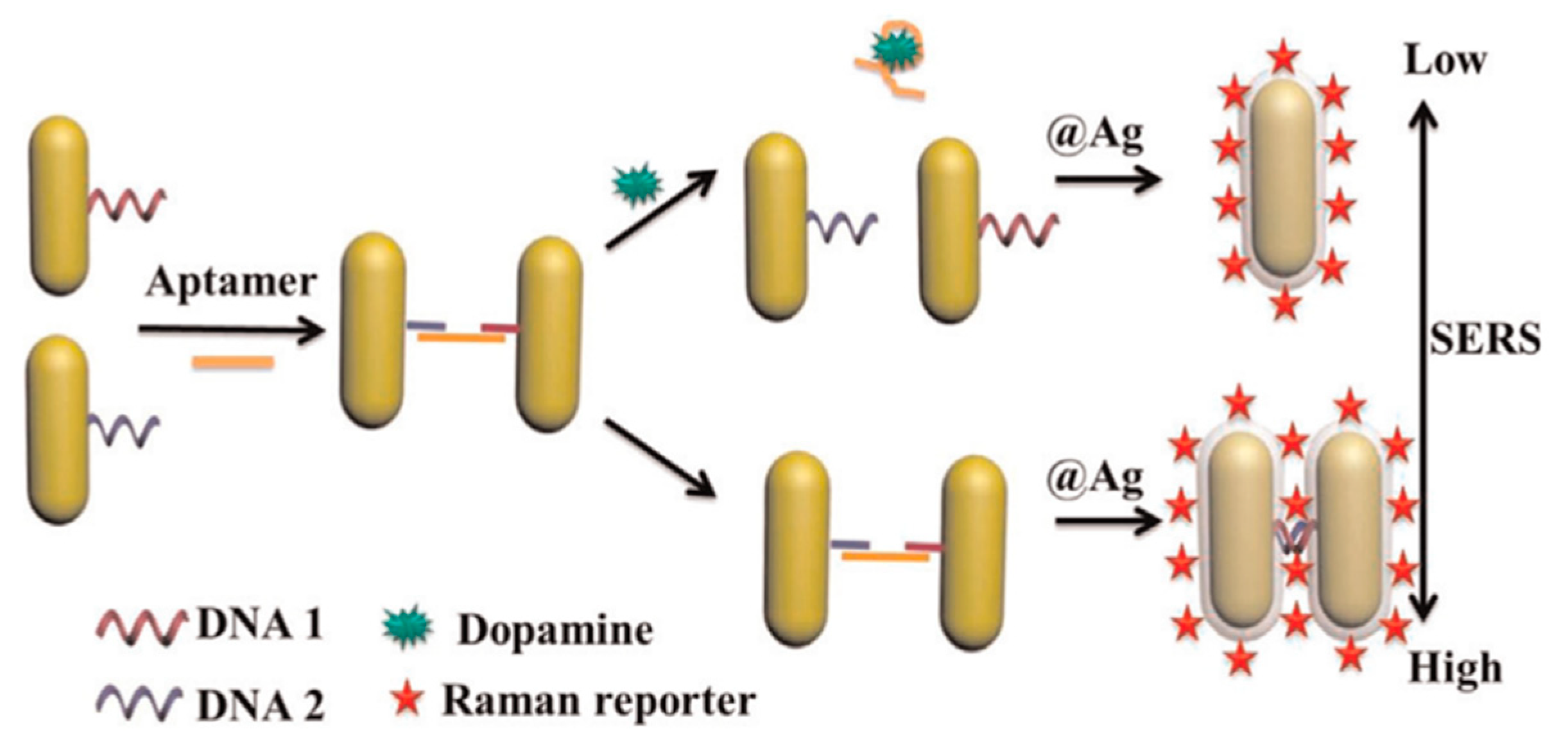
| Au@Ag nanorod dimers | Raman | dopamine | 0.006 pM | [11] |
| DNA–copper hybrid nanoflowers | colorimetric | dopamine | 4.5 μg/mL | [20] |
| catechol | 3.0 μg/mL | |||
| hydroquinone | 4.5 μg/mL | |||
| polyaniline/Pt-coated fiber optic | SPR | 4-nitrophenol | 0.34 pM | [23] |
| GNPs@TiO2 | SPR | hydroquinone | 33.8 nM | [24] |
| SiO2/GNPs | Raman | vanillic acid | 10–250 µM | [34] |
| syringic acid | 10–110 µM | |||
| gallic acid | 5–55 µM | |||
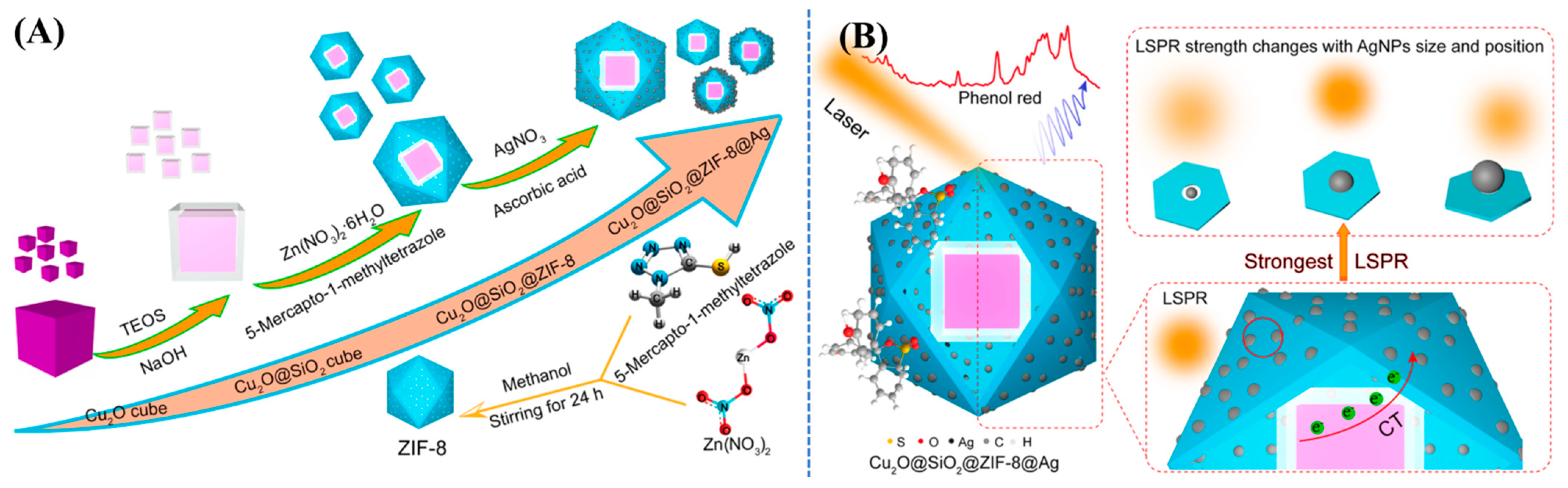
| Cu2O@SiO2@ZIF-8@Ag | Raman | phenol red | 5.76 × 10⁻¹² M | [36] |
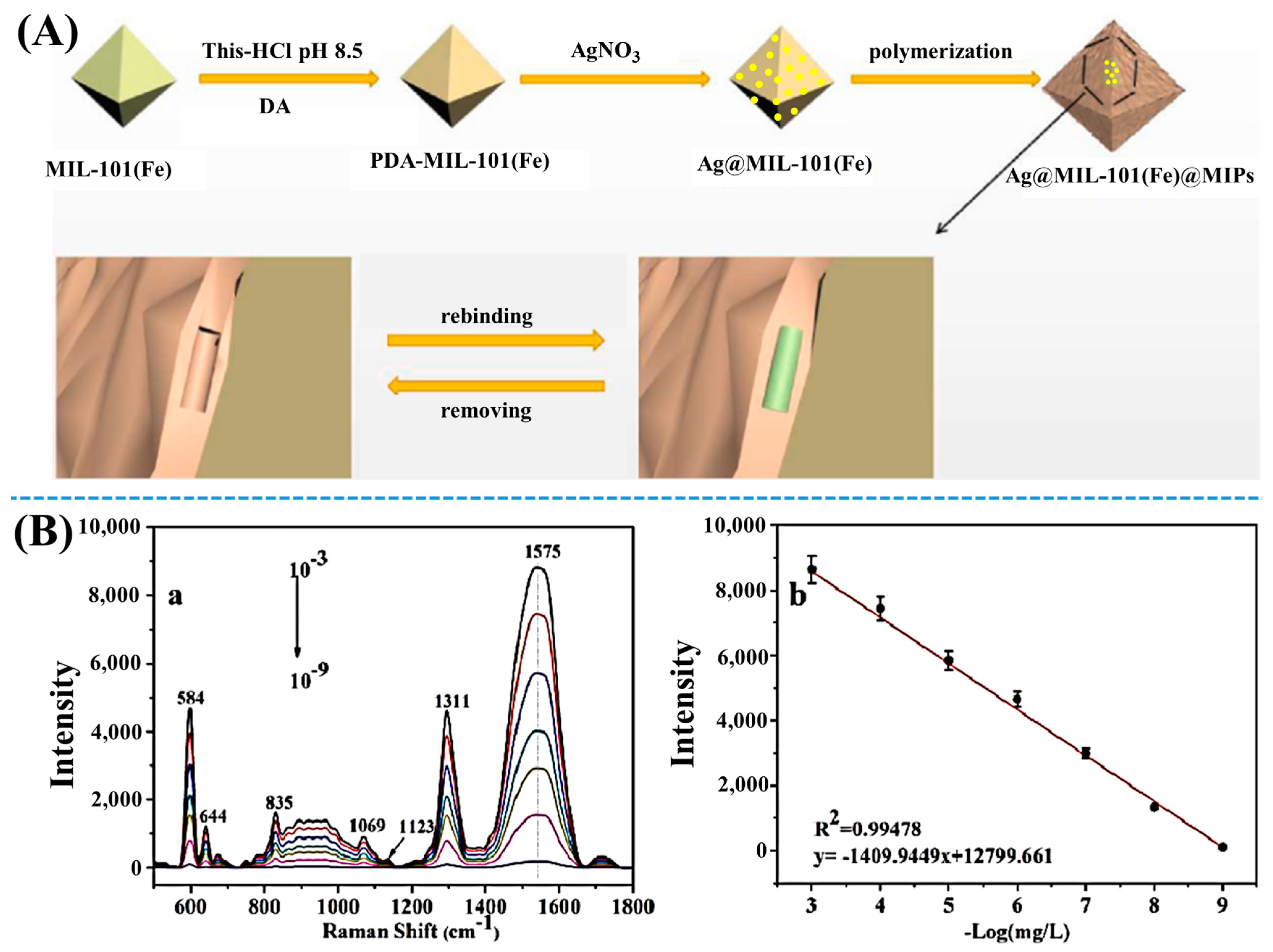
| Ag@MIL-101(Fe)@MIPs | Raman | 2,6-dichlorophenol | 4.5 nmol/L | [42] |
| GNPs | colorimetric | catechol | 0.11 µM | [65] |
| hydroquinone | 1.6 µM | |||
| Co-MOF-74-based Co3O4/cellulose | colorimetric | phenol | 1.02 μM | [69] |
| Zr(IV)-based MOFs | colorimetric | phenol | 1.28 μM | [70] |
| Adenosine monophosphate-Cu nanozymes | UV/Vis | phenolic compounds | 0.033 μmol/L | [71] |
| Au@Ni/rGO nanocomposite | colorimetric | phenol | 1.68 μM | [72] |
| Fe3O4/rGO/MOF | colorimetric | phenol | 3.33 × 10⁻⁶ M | [73] |
| AgNPs | fluorescence | dopamine | 5.3934 × 10⁻⁶ M | [80] |
| Au-Ag nanocluster | fluorescence | dopamine | 6.9 nM | [84] |
| Au nanoclusters | fluorescence | 4-nitrophenol | 13.8 nM | [85] |
| chitosan/graphene quantum dots/Au thin film | SPR | dopamine | 1.0 fM | [93] |
| carbon dot-functionalized GNPs | SPR | dopamine | 0.23 µM | [96] |
| GNPs | Raman | methyl parathion | 0.011 μg/cm2 | [102] |
| Ag-nanorod bundles | Raman | methyl parathion | 21.5 × 10⁻⁹ M | [103] |
| 2,4-D | 61.9 × 10⁻⁹ M | |||
| zipper-like ortho-Ag nanodimers | Raman | dopamine | 10 fM | [104] |
| AgNPs-graphene based nanosheets | Raman | p-cresol | 10−5 M | [105] |
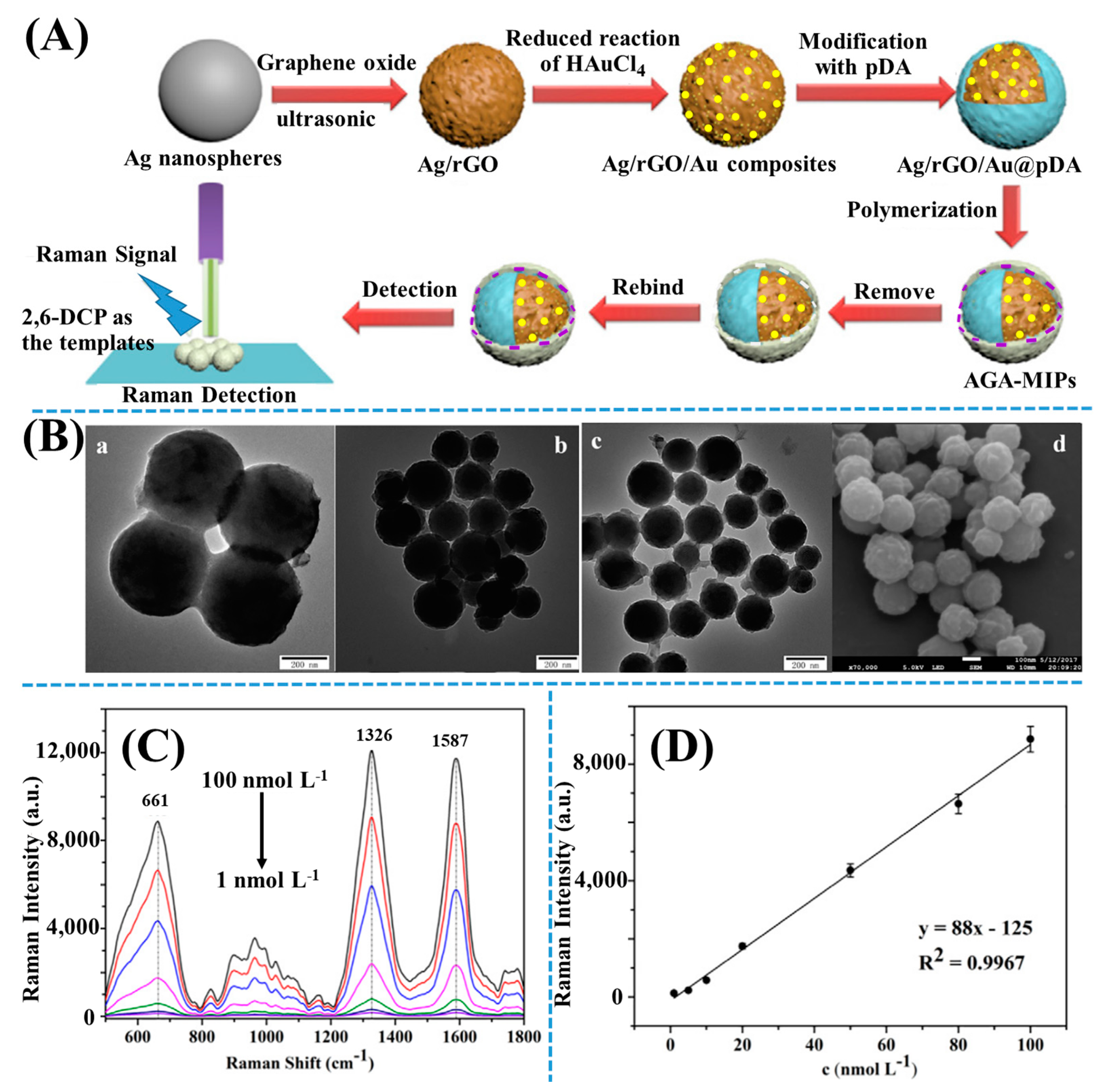
| SiO2/rGO/Au | Raman | 2,6-dichlorophenol | 100–1.0 nM | [106] |
| Au-ZnO nanoparticle-modified tapered optical fiber | SPR | p-cresol | 57.43 μM | [107] |
| ZnO/MoS2 | SPR | p-cresol | 28 nM | [108] |
| cetyltrimethylammonium-bromide-functionalized ZnO/carbon nanotube nanocomposite coated over Ag film | plasmonic sensor | catechol | 0.1 μM | [109] |
Publisher’s Note: MDPI stays neutral with regard to jurisdictional claims in published maps and institutional affiliations. |
© 2021 by the authors. Licensee MDPI, Basel, Switzerland. This article is an open access article distributed under the terms and conditions of the Creative Commons Attribution (CC BY) license (https://creativecommons.org/licenses/by/4.0/).
Share and Cite
Ly, N.H.; Son, S.J.; Kim, H.H.; Joo, S.-W. Recent Developments in Plasmonic Sensors of Phenol and Its Derivatives. Appl. Sci. 2021, 11, 10519. https://doi.org/10.3390/app112210519
Ly NH, Son SJ, Kim HH, Joo S-W. Recent Developments in Plasmonic Sensors of Phenol and Its Derivatives. Applied Sciences. 2021; 11(22):10519. https://doi.org/10.3390/app112210519
Chicago/Turabian StyleLy, Nguyễn Hoàng, Sang Jun Son, Ho Hyun Kim, and Sang-Woo Joo. 2021. "Recent Developments in Plasmonic Sensors of Phenol and Its Derivatives" Applied Sciences 11, no. 22: 10519. https://doi.org/10.3390/app112210519