Article

Monte Carlo Simulation-Based Calculations of Complex DNA Damage for Incidents of Environmental Ionizing Radiation Exposure

by
Spyridon A. Kalospyros
1,†,
Violeta Gika
1,†,
Zacharenia Nikitaki
1,
Antigoni Kalamara
1,
Ioanna Kyriakou
2,
Dimitris Emfietzoglou
2,
Michael Kokkoris
1 and
Alexandros G. Georgakilas
1,*
1
Physics Department, School of Applied Mathematical and Physical Sciences, National Technical University of Athens (NTUA), 15780 Zografou, Greece
2
Medical Physics Laboratory, Department of Medicine, University of Ioannina, 45110 Ioannina, Greece
*
Author to whom correspondence should be addressed.
Equal contribution for these authors.
Appl. Sci. 2021, 11(19), 8985; https://doi.org/10.3390/app11198985
Submission received: 12 July 2021 / Revised: 13 September 2021 / Accepted: 23 September 2021 / Published: 27 September 2021
(This article belongs to the Special Issue Advances in Environmental Applied Physics)

Abstract

:
In this paper, we present a useful Monte Carlo (MC)-based methodology that can be utilized to calculate the absorbed dose and the initial levels of complex DNA damage (such as double strand breaks-DSBs) in the case of an environmental ionizing radiation (IR) exposure incident (REI) i.e., a nuclear accident. Our objective is to assess the doses and complex DNA damage by isolating only one component of the total radiation released in the environment after a REI that will affect the health of the exposed individual. More specifically, the radiation emitted by radionuclide 137Cs in the ground (under the individual’s feet). We use a merging of the Monte Carlo N-Particle Transport code (MCNP) with the Monte Carlo Damage Simulation (MCDS) code. The DNA lesions have been estimated through simulations for different surface activities of a 137Cs ground-based γ radiation source. The energy spectrum of the emitted secondary electrons and the absorbed dose in typical mammalian cells have been calculated using the MCNP code, and then these data are used as an input in the MCDS code for the estimation of critical DNA damage levels and types. As a realistic application, the calculated dose is also used to assess the Excess Lifetime Cancer Risk (ELCR) for eight hypothetical individuals, living in different zones around the Chernobyl Nuclear Power Plant, exposed to different time periods at the days of the accident in 1986. We conclude that any exposition of an individual in the near zone of Chernobyl increases the risk of cancer at a moderate to high grade, connected also with the induction of complex DNA damage by radiation. Generally, our methodology has proven to be useful for assessing γ rays-induced complex DNA damage levels of the exposed population, in the case of a REI and for better understanding the long-term health effects of exposure of the population to IR.

Graphical Abstract

1. Introduction

Large-scale exposure to IR, in terms of both the amount of radiation and the number of people exposed occurs rarely due to international safeguards that are currently in force. All the nuclear accidents that have taken place have provided us with valuable information and experience about the health consequences of the leaked radioactivity to the public and the appropriate health management of such incidents. The nuclear accident of Chernobyl in April 1986, as the first large-scale REI, has been a great challenge to the scientific community that deals with the radiation protection, to revise much of its knowledge until that time and to improve the relevant nuclear safeguard rules. The value of the experience gained from that nuclear accident found application [1] to other accidents that followed of a smaller scale, such as in Brazil (the Goiânia 137Cs accident in 1987) and the two accidents that took place in Tokaimura of Japan on 11 March 1997 and 30 September 1999 [2,3], as well as of a larger scale, as with the accident that too place in Fukushima Daiichi on 11 March 2011 [4]. It is of note that environmental pollution due to man-made radionuclides began in the period 1946–1980, during the process of nuclear materials testing, when approximately 400 nuclear tests were performed in the northern hemisphere [5].
Radionuclides released in the air from a radioactive source may be in a gaseous, particulate, or multi-phase (i.e., simultaneously gaseous and particle) form [6]. These particles are generally transported through the air by adhesion to aerosols, construction particles or soil particles, as well as weather conditions such as the wind, rain and snow, which play a crucial role in transporting particles from the place of their release to the surrounding areas. Concerning the radionuclide spreading mechanism following an REI (i.e., a nuclear power plant accident, a nuclear weapon detonation, a radiological dispersal device, a transportation incident, sabotage or an improvised nuclear device), it is suggested that part of these radioparticles are deposited on the surface soil, while others are transported to the underground layers with rain, snow and watering, or they are transferred to surrounding geographic areas by re-floating [7]. The migration of radionuclides across the ground surface depends on the particle and surface characteristics. The transport of contaminants may vary depending on their physiochemical characteristics; for example, 137Cs is characterized as highly mobile because of its high-water solubility [8].
Two main pathways leading to radiation exposure of the general public due to a nuclear ‘fallout’ are the external exposure from radionuclides deposited on the ground, and the internal exposure through contaminated air inhalation, ingestion of contaminated food or water from the affected areas [9]. Radionuclide analysis of environmental samples is very practical for the evaluation of the current environmental radioactivity level. However, in the case of a nuclear accident, it is highly important to evaluate the environmental contamination, as well as the external and internal exposure risks for radiation protection and public health purposes. In the latter case, the risks of an internal exposure are extremely low because of the corresponding international regulations which demand restrictions in food and water intake (http://www.mhlw.go.jp/english/topics/2011eq/index.html (accessed on 29 July 2021)). On the other hand, the risks of external exposure for the public are enforced for safety reasons.
The Chernobyl Nuclear Power Plant (CNPP) accident, classified as Level 7 (‘major accident’) on the International Nuclear and Radiological Event Scale of the International Atomic Energy Agency (IAEA) [10], resulted in a massive release of radionuclides into the atmosphere and caused an extensive contamination of the environment. Ten-day major releases from the Unit 4 of the CNPP injected about 14 EBq of radioactive substances into the atmosphere, including radioactive gases, condensed aerosols, and a large amount of fuel particles. The total release of radioactive substances included the radionuclides: 132Te, 134Cs, 137Cs, 99Mo, 103Ru, 106Ru, 140Ba, 95Zr, 141Ce, 144Ce, 89Sr, and 90Sr. The noble gases contributed about 50% of the total release [11]. A terrestrial surface of more than 200,000 square kilometres in Europe received 137Cs with levels above 37 kBq m−2 [12]. Over 70% of the contaminated area comprised Belarus, Russia and Ukraine of the former USSR [13]. The radiation exposure of the population to these biologically dangerous releases was classified into two main phases: the earlier phase included exposure to rapidly decaying radionuclides, whose doses were delivered over a short period (no more than three months after the accident), while the later one comprised of an exposure to radionuclides of a long life that were deposited on the ground [14]. Most of the strontium and plutonium isotopes were deposited within 100 km of the destroyed reactor due to their larger particle sizes, while other important radionuclides decayed away. In the early months after the accident, the radionuclide levels of agricultural plants and plant-consuming animals were dominated by surface deposits [15]. The deposition of     53 131 I caused the most immediate concern, but the problem was confined to the first two-three months after the accident because of its decay. The radioiodine was rapidly absorbed into milk, leading to significant thyroid doses in people consuming milk products, especially children in Belarus, Russia and Ukraine. Increased levels of 131I in milk products were also detected in some southern areas of Europe, while dairy animals were being fed outdoors [16]. Radioisotopes of Caesium (137Cs and 134Cs) were the nuclides which led to the largest problems. The Cs hazard may be attributed to its incorporation into the human body from food consumption (especially dairy products) and via γ-irradiation from the ground. It is characterized by a relatively slow accumulation of doses, compared to radioiodine, but also by a whole-body irradiation [17]. 131I also concentrates inside the human body through dairy products and has a gamma-component, resulting in a significant whole-body dose. Once inside the body, this radionuclide is absorbed mainly by the thyroid gland, potentially increasing the risk for thyroid cancer, since this gland uses iodine to produce its hormones and cannot distinguish between the radioactive particles and its stable natural form. As 131I builds up in the thyroid gland, its emitted radiation can induce DNA damage, removing normal limits to cell growth and division and, thus, causing unchecked growth of thyroid tissue [18]. Additionally, the radionuclide 90Sr (half-life: ~28.79 years), due to its chemical similarity to calcium, accumulates in bones and irradiates the bone marrow, and therefore can also cause problems for human health. The latter was concentrated in a more restricted area around the reactor, as its larger particle size and its deposition levels at large distances were not radiologically significant. It is of note that gamma emitter nuclides (such as caesium and iodine isotopes) have been studied more extensively than the beta emitter strontium, since it is much simpler to measure gamma than beta emitters [19]. Therefore, 137Cs was chosen in our study for the mapping of the deposition because of its radiological importance and its easiness for measurements.
Increased contamination levels remained within the atmosphere for years after the accident, while radionuclide deposition values, depending on meteorological and environmental conditions, as well as the particle size, showed a substantial reduction in their transfer to vegetation and animals in agricultural systems during the first few years. The 137Cs activity concentrations still remain high in the surrounding areas of Chernobyl (in Ukraine, Belarus and Russia), especially in natural ecosystems (forests, rivers and lakes), while the surface contamination and the air dose rate have been reduced significantly due to radioactive decay, rain, wind and human activities [20]. Caesium-137 levels are still present, even in the southern areas of Europe, as monitored through plants such as lichens and mushrooms [21], which have a high retention capacity of such radionuclides; through the lichen Stereocaulon vasuvianum, Savino et al. [22] have accurately determined the effective half-life of 137Cs.
In general, many significant radionuclides released by the CNPP accident have already decayed away. Radioactive iodine isotopes caused great concern within the first few months after the accident since they are short-lived. In the future, 137Cs will continue to be of greater importance, with less attention paid to 90Sr. Over the longer term (hundreds of years), the plutonium isotopes and 241Am will remain radioactive, although at levels that are relatively low [9].
One recent “major” (according to IAEA 2008 criteria) nuclear accident occurred in Fukushima Daiichi Nuclear Power Plant (FDNPP) in Japan, on 11 March 2011, as a result of a tsunami (caused by a strong earthquake) that struck the east coast of the island. The radioactive releases from this nuclear accident were not as large as those from Chernobyl but were still considered substantial [23]. A radioactive plume derived from the Units 1, 2, 3 and 4 of the FDNPP was dispersed in the atmosphere, causing significant radioactive pollution (mainly due to 134Cs, 137Cs and 131I release) of the environment within a radius of 40 km from the damaged reactors. Immediately after the accident, the prevalent dose-forming radionuclides collected from soil samples from the nearby areas around the reactors were 131I, 134Cs, 137Cs, 129mTe, 95Nb and 136Cs, while, after several months, the corresponding ones were 134Cs, 137Cs and 129mTe. It was also estimated (some months after the accident) that the amount of radioactive materials released into the environment at that time was approximately 10% (1.6 × 10¹⁷ Bq for 131I and 1.5 × 10¹⁶ Bq for 137Cs) that of the Chernobyl accident [24].
Up until now, a large area of a few thousand square kilometres around CNPP (the ‘Chernobyl Exclusion Zone’) remains evacuated, with restricted human access. 131I (half-life: 8 days) has long decayed, and 134Cs (half-life: 2 years) has already been considered depleted, therefore, the main existing problem is the radioactive hazard from contamination by 137Cs (half-life: 30.17 years), although half of the initial release has now decayed. The whole restricted zone gives the opportunity for the study of radiation transmission to the animals of this area, since wildlife is flourishing in the absence of human activities [25]. In Fukushima, there are confined areas within a radius of 40 km around the FDNPP, where the annual effective dose exceeds 20 mSv (the effective dose limit established by the Japanese Government for the lifting of the human exclusion order) and other areas where the corresponding value is up to 50 mSv. Although remediation measures through the years have been taken for the maximum possible decrease in the radioactive nuclides of the terrestrial area, the radio-contamination levels do not permit the return of evacuees to their land. Gamma-radiation from the deposited 137Cs still remains a big problem for authorities [26]. As referred to above, the 137Cs content in these areas is also investigated through lichens and other plants [27]. As also occurred in Chernobyl, 90Sr dispersion over the Fukushima territory was limited and the biggest part of this released radionuclide was deposited in the proximity of the FDNPP [19].
One of the main pathways leading to human exposure after such a REI is the external exposure from radionuclides deposited on the ground [28]. Gamma-radiation from these radionuclides has been a major contributor to the external exposure of the public due to a nuclear accident [29]. In our study, we focus on the 137Cs as the most prominent radionuclide for ground contamination due to its substantial contribution to the lifetime effective dose to humans, its long radioactive half-life, and its ease of measurement.
Exposure to IR induces a range of DNA lesions to living cells. IR photons especially can target the nuclear DNA molecule in two ways: either by directly striking it, with the induction of secondary electrons, causing breaks in the phosphodiester bond connecting adjacent nucleotides on the same side of the DNA helix, or by water radiolysis, which can result in the formation of reactive oxidative species (ROS) and additional oxidative DNA damage. All the above can result in the formation of simple or complex lesions [30,31]. In such a DNA lesion, a single nucleotide is characterized by an abnormal chemical alteration, i.e., a missing or damaged base or a strand break. These lesions may comprise base damage (BD), single-strand breaks (SSBs, i.e., a cluster of lesions that contains at least one strand break and which has no other additional break within 10 bp on the opposed strand), double-strand breaks (DSBs, i.e., this is a lesion that consists of two SSBs, which are located on opposite strands within 10 bp of each other) and complex DNA damage (groups of several lesions within 1 or 2 helical turns of the DNA molecule) [32]. Among them, DSBs and non-DSB clustered damage (i.e., two or more lesions within one or two helical turns of DNA induced by the passage of a single radiation track) [33] are considered to be the primary cause of radiation-induced cell killing, mutagenesis and neoplastic transformation. Through the process of breaking and re-joining, DSBs are converted into small- or large- scale chromosomal exchanges with the ability to induce phenotypic tumor lesions and, finally, cell death. Other types of non-DSB lesions, such as clustered oxidized bases, are considered to be resistant to the repair process and more susceptible to DSB formation through their repair processing [34].
Throughout the last decade, Monte Carlo (MC) codes have proved to be a useful tool for assessing DNA lesions in a cell exposed to IR, since they have been constructed to simulate damage induction at the DNA scale [35,36]. Radiation risks for an individual exposed to IR depend on different factors. One factor of great importance is the overall dose of radiation absorbed by the human body, while another equally important one is the dose distribution within it.
In our study, we have used a serial combination of two MC codes with different roles in order to estimate the number of induced lesions (SSBs and DSBs) per cell of a person exposed to IR from a radioactive source (137Cs) placed in the ground, as a remnant from a hypothetical nuclear accident. These lesions were assessed for different values of surface activities and exposure times of the radioactive material to make conclusions about the health risk in such exposures. We have developed an efficient computational method with the combination of a small-scale MC biophysical model (MCDS), with a larger-scale and general-purpose MC transport code (MCNP) to reproduce the results induced in the cell environment after such an exposure. Specifically, we have used the general-purpose MC N-particle (MCNP) radiation transport code to estimate the absorbed dose and secondary electron spectra of 137Cs irradiation in cell DNA. Subsequently, we combined the estimates of the secondary electron spectrum and the absorbed dose acquired from MCNP with MCDS simulations in order to calculate the initial DNA Damage.
In synopsis, we have developed a computational method to study the potential biological effects on cell DNA after IR exposures. It is crucial to know the absorbed dose after the radiation exposure because, based on the recording of this magnitude and the analysis of the effects of radiation in living organisms (DNA damage), we can estimate the severity of each exposure. Ultimately, this information may help medical personnel to respond appropriately to such cases with individuals exposed to IR after a radiological incident.

2. Materials and Methods

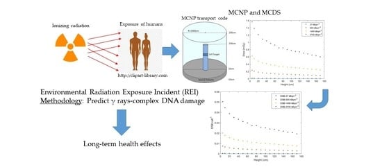
2.1. Monte Carlo (MC) Codes: MCNP and MCDS

Our calculations have been based on the Model Carlo simulation technique. MC codes have been widely used to simulate damage induction to a cell exposed to any type of the IR spectrum [37]. Theoretical studies, together with MC Track Structure codes (MCTS), have contributed remarkably to the understanding of the DNA damage dynamics and to the simulation of particle tracks in biological matter, leading scientific research to the estimation of the radiation effect parameters and, therefore, providing important information applied in radiation protection and radiotherapy [35,36]. In this way, MCTS simulations have proved to be the most sophisticated tool for studying and understanding the interactions of IR with biological matter and for determining the damage induced by it to the major target of the cell: its DNA macromolecule [31].
In our study, we have used two different MC codes for obtaining our calculations. The first one, MCNP, being one of the most accurate in its category, is a multipurpose code used widely in nuclear and medical applications, utilizing a large spectrum of particles. The simulation of particle interactions with living matter are based on databases that are embedded in the software and include internationally recognized libraries of cross sections [38]. The MCNP6.1 version of the code used by us, especially, has the ability to accurately describe the electron transport down to 10 eV [39]. Via this code, we have estimated the absorbed dose by our target and the spectrum of secondary electrons produced in it. As aforementioned, one part of the damage induced to a cell is due to secondary electrons ejected as a result of the ionizations in the medium.
The second MC code used in our study, in combination with MCNP, is the Monte Carlo damage simulation (MCDS) algorithm, which has been selected among the other MC codes of the bibliography because of its simplicity and its results production swiftness. It does not have the accuracy of other codes in its category, but it can yield major trends in the spectrum of DNA damage predicted by other detailed MCTS simulations. This code has been developed in order to predict the initial yield and types of DNA damage formed by IR and is much faster compared to conventional track structure simulations (it can give results within seconds to minutes), in addition to its easy-to-use algorithm [34]. MCDS algorithm is characterized as a quasi-phenomenological model, which can predict the full spectrum of DNA damage induced by electrons, protons, α-particles and ions up to 56Fe. Although this code does not possess the ability to directly simulate the damage in irradiated cells for photons or other neutral particles, secondary electron spectra for 137Cs in a monolayer cell geometry were used to produce DNA damage yields [40]. The estimates of the code parameters are based on the interpolated damage yields, derived mainly from the track structure simulations of Nikjoo et al. [41,42,43,44] and Friedland et al. [45,46]. MCDS [47] uses only four adjustable parameters, three of which are the same for electrons, protons and α-particles. In brief, simulations are performed in two steps: (1) random distribution of the expected number of lesions produced in a cell per Gy of radiation in a DNA segment and (2) subdivision of the lesions in the former segment into clusters [34].

2.2. MCNP Setting for the Simulations of Our Study

Estimation of the absorbed dose to tissues of the human body, from radiations emitted by an arbitrary distribution of a radionuclide in an environmental medium is an extremely difficult computational task. By using the MCNP code, we create the geometry of the experimental setup. In this case, we base our simulations on a “human phantom” (filled with water as content), which appears as a cylinder with a radius of 25 cm and 180 cm in height, simulating an average man standing on the soil at the air–ground interface. This phantom is separated into layers of 10 μm thickness, which represents the typical average size of a eukaryotic mammalian cell (Figure 1).
Additionally, an isotropically distributed cylindrical source of 137Cs has been placed in the ground, at a depth of 20 cm; the latter value represents a mean typical depth of radiocasium deposition in soil after its release as a consequence of a nuclear accident [48]. We have chosen to use this specific value for the deposition depth of 137Cs in all our measurements, since many fundamental studies about the vertical distribution of this key artificial radionuclide in the environment show its biggest concentrations in the topsoil layers, especially above the depth of 20 cm [5,49,50]. The content of this radionuclide in soil is influenced by vertical migration due to the physical processes of diffusion, convection transfer with the soil moisture, and migration through the roots of plants. Generally, the concentration of radiocaesium is decreased exponentially with depth, depending on the landscape and mainly the soil type; the latter includes factors such as the mineral and physical composition of the soil, its organic composition, cation exchange capacity, acidity, and the presence of certain kinds of vegetation (e.g., coniferous trees and mosses which play the role of a filter, engulfing radionuclides for long periods after the initial fallout) [51,52,53,54,55]. In our study, we have considered an isotropic cylindrical source (the base cylinder in Figure 1) and, therefore, in our simulations we have planned its surface area (base of the cylinder) to be 1256 m2 (radius 20 m). Of course, a larger source area would result in a higher effective dose to our phantom, but based on the results of a similar study by Han et al., 2010 [56] where the effective dose proves to increase <2% when the source area increases by 78%, we have assumed in all our simulations that a source area with the aforementioned value is a reasonable one, representable of a contaminated area beneath the ground. In our study, the material which is mixed with the source of gamma rays (137Cs) includes all those elements contained in soil in the respective proportions (mass percentage) (i.e., Si: 27.1183%, Fe: 5.6283%, Mg: 1.3303%, K: 1.4327%, Ca: 5.1167%, O: 51.3713%, Na: 0.614%, Al: 6.8563%, Ti: 0.4605% and Mn: 0.0716%), considering a typical value of dry density ~1.52 g/cm3 [57]. The entire assembly is surrounded by air (i.e., the external cylinder in which the human phantom stands). Varying the perpendicular distance of the DNA cell that we study from the source (keeping its position stable in the other two axes), we scanned the surface of the human body, i.e., the height between 5 cm and 180 cm from the ground level and estimated the absorbed dose (in MeV/g per particle) and the spectrum of secondary electrons produced in the DNA target for each different case. We applied a series of simulations by MCNP6.1 for the various distances mentioned above, having chosen various values of source activities of 137Cs for different specific values of surface exposure (i.e., 37, 555, 1480 and 3700 kBq/m2—these specific values are defined as the limit ones discerning areas of a different grade of 137Cs surface ground deposition, according to UNSCEAR 2000 Report [58]), in order to simulate the conditions of a nuclear accident. It is worth mentioning that the value of 37 kBq/m2 was taken as the lower limit defining the boundaries of contaminated zones, while the value of 555 kBq/m2 designate an area of strict control [58,59]. Likewise, for the exposure time we have chosen the typical time periods of one day, one week and one month.
In order to get the results from the MCNP code (through the output file), we completed a corresponding input file providing certain information about the geometry of our experiment, the definitions of the surfaces included, the materials contained (in the appropriate proportions), the choice of the particles emitted by the radioactive source and its dimensions. For each value of surface exposure and exposure time we have run the code for 1010 histories (‘NPS’) in order to get better results by reducing the error in the doses received to less than 5%. For the absorbed dose calculation, the energy pulse-height tally (energy balance tally *F8: P, E) was exported. Notably, this code extended electron transport down to 10 eV, adopting this value as the electron tracking and production threshold [60].
As one may notice, in our calculations via this code, we refer only to the secondary electron’s spectrum produced by γ radiation. This does not mean that we omitted the β radiation emitted by 137Cs [61], which is known to have a short range, a fact that could play a crucial role in our final results. For this reason, before performing all our calculations through MCNP6.1, we ran the same input files for the same geometry model in the MCNP-CP code [62], which has the ability to calculate the corresponding beta decay spectrum. Through the latter calculation, we inferred that the range of the electrons emitted by 137Cs during β decay in the soil towards the human phantom is only some μm; this happens because we use an extended source (source diameter: 40 m) in comparison to the dimensions of the phantom, and in a long distance from the standing “human”, in comparison to the range of beta-radiation. In this way, we may neglect the β radiation emitted from our source.

2.3. MCDS-Based Estimates of DNA Damage

The MCDS provides a simple algorithm that calculates the DNA damage yields for a given absorbed dose of a chosen particle, and for a set of parameters such as cell size, nucleus size, type of radiation, and other parameters related to cell environment (e.g., oxygen and water cell concentration). As a whole, MCDS calculates complex DNA damage (DSBs, SSBs, non-DSB oxidative lesions) that are induced through both the direct ionization and excitation of the DNA and the indirect action of hydroxyl, or other radicals formed in close proximity to DNA. DNA damage by the bystander effect is neglected in this code. The MCDS code “reads” data from particles and other necessary information for the simulation from an input file (in which we set the calculated value of the absorbed dose by MCNP, as well as the energy spectrum of the secondary electrons down to 10 eV) and yields its results through an output file [63]. In our calculations, we have considered cells irradiated under normoxic conditions (O2 concentration: 10% for tissues). Our simulations have been based on the default parameter values: σSb = 217 Gy−1 Gbp−1, f = 3 and nmin = 9 bp, where σSb is the number of individual strand breaks per unit dose per amount of DNA in the cell, f is the ratio of base damage to strand breaks and nmin the minimum length (in bp) of undamaged DNA between neighboring elementary damages, such that these elementary damages are considered to belong to two different lesions [63]. We also assumed that the DNA of each diploid cell has a length equal to 6.4 Gbp, which is the average value for a diploid human cell with 46 chromosomes, as well as the diameter of the cell nucleus is 5 μm, and the cell diameter is 10 μm. All the simulations in our study have been performed through the MCDS Version 3.10A [64].

3. Results

The results of this study comprise, firstly, the calculation of the absorbed dose through the MCNP6 code for the radionuclide 137Cs. In particular, the absorbed dose has been estimated as a function of height (distance from the ground) of a typical man (5–180 cm) for the surface activity values of 37, 555, 1480 and 3700 kBq/m2, respectively and for the exposure time of a week. These results are depicted in Figure 2.
As can be seen, when the height (distance from the source) is increased, an exponential decrease in absorbed dose occurs. In addition, an initial sharp decrease in the slope of the curve is observed at small heights on the water phantom; this happens because an increase in the distance from the source is equivalent to the reduction in the number of photons which penetrate a surface along the direction of the radiation. On the other hand, the dose becomes quite constant after the first 80 cm from the ground, with surface activity of 3700 kBq/m2, and after the first 30 cm for the corresponding values of 555 and 1480 kBq/m2, while it is constantly near zero for the value of 37 kBq/m2; this was expected, since the aforementioned values of distance are within the range of gamma rays in the air [65]. It is of note that the estimated doses for the deposition density of 37 kBq/m2, as the limit of the high radiation control safe zone according to the UNSCEAR 2000 Report [58] for the Chernobyl nuclear accident, are in the range of 1.1·× 10−4–1.99 × 10⁻²—mGy and are considered as “low absorbed doses”.
Next, we present our results of the MCDS for the calculation of the DSB and SSB damage (number of lesions per cell) (Figure 3a,b) for the same values of deposition densities of 137Cs and for the exposure time of one week.
The first thing to note is that the analogy between SSBs and DSBs seems to be in the range reported in irradiated typical mammalian cells [66]. It can also be noted that, for the higher value of surface activity, an exponential decrease is observed in both the numbers of the expected DNA lesions (DSBs and SSBs per cell) of the cell target as a function of distance (height) from the source of 137Cs. For the value of 1480 kBq·m−2 the corresponding numbers show a small decrease at the first 30 cm, while for the other two (and lesser) values of deposition density, the number of the lesions is quite stable as the distance from the source is increased. This means that for the highest value of surface activity, i.e., for a man standing on the ground, exposed to 137Cs radiation in the near zone (<100 km from the place of the nuclear accident), the number of induced SSBs and DSBs in the cells of his genitals and the rest of his body organs are quite the half of those induced to the cells of his feet. For places in the far zone (100 km to approximately 2000 km), the number of the same lesions is low and quite stable throughout his whole body. It is of note that as the distance from the source increases (and consequently the absorbed dose is reduced), the number of the expected SSBs per cell for the deposition density of 3700 kBq·m−2 decreases by a factor of 2.5, reducing, in this way the biological effects of radiation; the corresponding decrease in DSBs with distance is lesser, since this kind of lesion is more complex than SSBs and occurs more scarcely. All our MCDS simulations have been performed with a standard error of the mean better than 0.2%.
DSBs are considered to be the most biologically deleterious lesions, since one single unrepaired DSB can lead a cell death or can cause chromosomal aberrations with subsequent genomic instability, and possible malignant transformation [67]. Therefore, this kind of damage was chosen to be analysed mainly in association with the aforementioned results. As reported previously, the MCDS code does not involve the process of repair in its results and thus, we take into account the existing literature in order to translate the numbers of lesions shown in the above diagrams into cancer risk information.
In the dose range related to a REI, as in our study, the associated cancer risk cannot be deduced from epidemiological data, due to a lack of sensitivity. Such estimates for persons exposed to an environmental IR incident have been based on a linear extrapolation of high-dose data obtained from the study of atomic bomb survivors of Hiroshima and Nagasaki through the years; the linear-no-threshold (LNT) model assumes that the DNA damage is proportional to the dose and that the response of the irradiated cell functions equal efficiently from high to low doses [68]. However, the validity of such an extrapolation is questioned by phenomena such as the low-dose hypersensitivity, the adaptive and hormetic response, the bystander effects, and the threshold hypothesis [69,70,71,72]. In attempting to associate a low dose irradiation with cancer risk, it is important to make the distinction between acute exposures over a very short period of time and protracted ones [73]. Clearly, the atomic bomb survivor risks represent the average of all those exposed to an REI population. Considering the existing data, the fact that individuals are at lower or greater risk than the average, depends on genetic status, age, age of exposure and other factors [74].
Deep sequencing studies have confirmed that biomarkers (such as the micronucleus assay and scoring of chromosome aberrations) are important tools for the detection of the early stages of radiation-induced carcinogenesis. These validated techniques, however, do not have the sensitivity to study the effects of IR on cells at doses below 100 mGy [75,76]. For this reason, phosphorylated histone H2AX (γ-H2AX) foci immunodetection has become the internationally accepted quantitative biomarker of human low-level IR exposure [77]. DSBs activate histone H2AX right after their induction by phosphorylating a highly conserved serine (Ser-139). The phosphorylated γ-H2AX forms foci in the cell nucleus. In this way, a γ-H2AX focus represents a DSB [77,78].
Focused on the range of low doses (i.e., doses <0.05 Gy [79]), as this very low (≤1 mGy, see Figure 2) in our study, Rothkamm and Löbrich have shown that in non-dividing primary human fibroblasts cultured in vitro, irradiated by doses ~ 1 mGy of IR, the induced DSBs remain unrepaired for many days and the cells with unrepaired DSBs are eventually eliminated [80]. Similar results were observed by Osipov et al. in a study conducted on human MSCs, isolated from oral mucosa, where for the lower doses, after the initial rise in γH2AX foci number, there was no decrease observed [81]. It appears that, in cellular responses at low compared to high doses, different pathways may be activated, and non-linear responses prevail that are not compatible with the LNT model.
Unrepaired or mis-repaired DSBs may give rise to chromosomal aberrations and alterations, micronuclei formation, gene amplification, sister chromatid exchange and other genetic instability hallmarks. In vitro observations show the induction of unstable chromosomal aberrations in cells irradiated in G1 phase and chromatid-type aberrations in the ones exposed to IR at the G2 phase of the cell cycle [82]. Aberrations, such as a dicentric chromosome, or a ring with an acentric fragment, or a reciprocal translocation, permit the continuation of cell proliferation with the over expression of truncated oncogene which leads to oncogenic transformation. For example, rearrangements of the RET (rearranged during transfection) gene are observed in papillary thyroid carcinoma. Studies conducted after the CNPP accident show a sharp increase in the incidence of pediatric thyroid papillary cancer [83].
Examining the human breast tissue after its irradiation with very low doses (a few mGy) of IR, one may detect, except for induced DSBs, changes in the transcription level of genes [84]. Female breast tissue is proved to be very sensitive to radiation due to the presence of reproductive hormones, including estrogens; the latter may function as carcinogens since they energize the estrogen receptor-mediated cell proliferation [85]. Depuydt et al. investigated the irradiation of glandular epithelial cells of breast tissue in the dose range representative for mammography screening, with results indicating the existence of a hypersensitive response for DSB induction [86]. In a previous study, they showed that the number of mammography-induced DSBs resulted in chromosomal aberrations, which are a hallmark for cancer [87]. Other studies demonstrated that for very low doses of IR, the γH2AX foci induction is much higher than at higher doses [88,89,90].
Another effect which plays a key role in the IR-induced carcinogenesis is the bystander effect; this is more relevant to low-dose radiation [91]. At low doses of IR, the response to radiation becomes important in regard to how dominant and extended the bystander effect is in the vicinity of the irradiated cell, and the consequences this will have. Thus, the IR-induced genomic instability and the consequences of the bystander effects indicate a non-linear behavior in the low-dose area. The evidence arising from the published data indicate that the cellular response to low-dose IR is a complex interaction of various modulating factors [92].
Shimure and Kojima recently attempted to identify the lowest IR dose causing molecular changes in the human body [93]. They concluded that, although the extent of DSBs formation differed depending on the irradiated cell species that were investigated, the lowest limit at which these DNA lesions are formed is approximately 1 mGy. Halm et al. indicated that blood doses ranging between 0.22 and 1.22 mGy may induce somatic DNA lesions, one hour after C.T. examination [94]. Additionally, Vandervoorde et al. [95] showed that a very low blood dose 0.15 mGy caused DSBs five minutes after the C.T. examination. An increase in leukemia risk is suggested in children under five years old who were exposed to radioactive fallout from nuclear weapons testing (estimated fallout marrow dose: 1.5 mGy) [96].
According to all the aforementioned data, exposure of an individual to the dose of the underground caesium-137 in areas of such values of surface activity for a period not more than a week, seems to be of very low risk for any future health problems. It would be interesting to investigate the same cases but in a more extended exposure time to IR.
In order to study the absorbed dose as a function of the exposure time, we have calculated its values for the deposition density of 3700 kBq·m−2 (in the near zone of the nuclear accident) at the exposure times of one day, one week and one month through MCNP code (Figure 4).
It is of note that there is an exponential decrease (for the exposure time of one month) in the absorbed dose with the increase in height from the ground (source), while in shorter time periods the curve is converted to a nearly stable line and, simultaneously, a linear increase in this dose with the increase in exposure time. As can be seen in Figure 5, the same trends have the estimated DSBs and SSBs per cell as a function of height and exposure time, correspondingly. This linear trend also holds for the relationship between the absorbed dose and the number of inducted DSBs and SSBs. This trend could support, in part, the LNT model. On the other hand, the estimated absorbed dose for the exposure time of one month (see Figure 4) takes an average value ~3 mGy for the vital organs of a 1.80 m tall man; this fact may pose a risk for the induction of unrepairable DSBs and unstable chromosomal formations, which in turn, may enter an organ of the exposed organism to early potential stages of carcinogenesis. This would also mean that a man standing on the radioactive ground of such an area for the time period of one month (in practice, this means that the individual is exposed to a daily eight-hour-irradiation from such a source in the ground for three consecutive months) has a real risk of being a cancer patient in the future.
Since the exact details of the microscopic association between radiation and carcinogenesis is not yet known, it would be interesting to explain how the aforementioned calculated numbers are connected with the risk of carcinogenesis in the organs of an exposed individual in a relatively macroscopic scale. For this reason, we have used the National Cancer Institute NCI (USA) Radiation Risk Assessment Tool—Lifetime Cancer Risk from Ionizing Radiation: https://radiationcalculators.cancer.gov/radrat/model/inputs/ (accessed on 29 July 2021).
With this tool, we evaluated the risk for various organs of different characteristic adults exposed to such doses. In our following estimates, thyroid cancer has been excluded since it is by far attributed to the intake of 131I; this radionuclide is the main contributor to the thyroid doses received mainly through internal body irradiation (via milk consumption) [97]. On the other hand, 137Cs is considered the main contributor to doses of the body organs and tissues, other than the thyroid gland, from external and internal irradiation. For this reason, we have considered presumably eight different individuals (two males and six females) with different ages and different exposures to the nuclear accident of Chernobyl in 1986. Through this online risk calculator, by filling the age (at the year of the accident) of each individual (all considered about 1.80 m high) and the absorbed dose (in mGy) as calculated before (see Figs 1 and 4), in which they were exposed for a certain single exposure time (in days) and rate (acute or chronic), we estimate their Excess Lifetime Cancer Risk (ELCR) for different body organs in a certain height from the ground. In fact, the real exposure time of an individual is a longer one, since this person does not keep standing on the ground for the total time of exposure, as referred above. Based on these data, the possibility of a certain organ cancer development (chances expressed as cases per 100,000) is evaluated. By the ELCR, we refer to the average probability of cancer development to a certain organ of an individual exposed to IR higher than their unexposed peers. Our selection of the specific organs for the evaluation of their ELCR was based on the sensitivity of the human tissues to radiation [98], the availability of these organs in the quoted list of the NCI tool, and the volume that they occupy in the human body; in the latter criterion we selected such organs so that they are not extended (such as the bowel) and correspond to a certain height from the ground (for the use of a certain value of dose, according to Figure 2 and Figure 4).
More analytically (see Table 1), the first calculation is about an adolescent, 16 years old at the year of the Chernobyl nuclear accident, exposed to a 30-day irradiation because his family—for various reasons—delayed to evacuate their homestead in the near zone of 3700 kBq·m−2: assessing a total dose of 3.44 mGy for all those days of exposure, we evaluate an ELCR for his prostate gland equal to 3.91 × 10⁻⁵, which is considered high according to the World Health Organization [99], the Canadian Council of Ministers of the Environment [100] and the New Zealand Ministry for the Environment [101]. In the same way, the next examined case is that of a 60-year-old woman, living in the aforementioned zone, who refused to evacuate her home; considering a chronic exposure of 3.02 mGy for a period of 30 days, we assess an ELCR for her breast equal to 2.15 × 10⁻⁵, which is also considered high (according to the aforementioned criteria). The third case is that of a 16-year-old lady, who received only an acute dose of 0.1 mGy for only 1 day of exposure in the near zone before evacuating the contaminated area with her family; this dose yields an ELCR equal to 0.58 × 10⁻⁵ for her breast, which is considered as moderate. If, for the same young lady, we assess the ELCR for all her organs (in this case we considered an average height of 130 cm from the ground for her vital organs), we calculate a value of 2.4 × 10⁻⁵ which is considered high, while the corresponding value for leukemia is 0.08 × 10⁻⁵, considered low. Our next case is that of a 50-year-old cattleman who lived in the same area and promptly evacuated his ranch under the first governmental recommendations for the general public, but after some weeks returned to live again in his area at his own risk. This man, exposed to a 30-day-irradiation after the first days of his reinstallation of 3.20 mGy from the radiation under his feet, has an ELCR value for his pancreas equal to 0.66 × 10⁻⁵, which is considered as a moderate one. We then consider a 16-year–old lady, living in an area of the outer contaminated zone of 37 kBq m−2, exposed to a 7-day-external irradiation of 8 mGy (due to a delay in her family’s evacuation of their home) for whom we assess an ELCR for her ovaries equal to 0.005 × 10⁻⁵; this value is a low one. Our last case is that of a 30-year-old woman living those days in the contaminated zone of 555 kBq·m−2, who, for a 7-day-delay of evacuation, has been exposed to an external dose of 0.068 mGy, equivalent to a low ELCR value for her brain equal to 0.004 × 10⁻⁵.
As a conclusion to all the previous calculations, it may be noticed that any exposition of an individual in the near zone of 3700 kBq·m−2 increases the risk of cancer at a moderate to high grade (only in the case of leukemia there is a low-risk grade estimation for exposed individuals in this zone, and this happens due to the selected short time of exposure, i.e., 1 day).
The objective of this study is the calculation of the absorbed dose and the estimation of a critical marker for biological damage i.e. the one induced at the DNA of an individual exposed to the radiation emitted by the 137Cs in the ground. In this way, we have isolated only one component of the total radiation that can affect the health of a human exposed to it after a nuclear accident and could lead to carcinogenesis. The latter, as we know, is a multistage and multifactorial process and in order to assess any risk estimate with the most accurate approach possible for cancer, one has to investigate at the same time all the factors associated with this disease. For example, in the case of radiation-induced leukemia, one should know not only the absorbed dose and the time of exposure, but also diverse factors connected to genetic susceptibility to the disease. We should also know other environmental factors and dietary habits referred to the specific agricultural cohort of this area around the Chernobyl Nuclear Power Plant. Noshchenko et al. [102] estimated the radiation-induced risk of acute leukemia that occurred from 1987–1997 among residents 0–5 years of age at the time of the Chernobyl accident, who lived in the most radioactively contaminated territories of the Ukraine; this risk was significantly increased among those who were exposed to doses higher than 10 mGy. If we compare our results (Figure 4: lower doses in shorter exposure periods) to those above then we will have to extend the exposure time to much longer periods (e.g., more than 2 months) in order to reach similar doses. Some reports about an increase in infant leukemia due to the prenatal irradiation after the Chernobyl accident in more remote countries, such as Greece [103] and Germany [104], quote an average of ~2 mSv and 0.49 mSv correspondingly, (for radiation protection purposes, absorbed dose and effective dose are used, including a radiation-dependent weighting factor: for X-rays and γ-rays, 1mSv = 1 mGy) for the added radiation exposure during the first year after the accident, while others in neigboring countries such as Belarus [105] quote multiple values of doses.

4. Discussion

Simulating the biological DNA damage effects in the human body in the case of REI, utilizing MC methodologies, has proved to be a useful methodology to better understand the effects of environmental radiation exposure. MC methods are computational algorithms of high predictive accuracy and useful for modelling phenomena with significant uncertainty inputs. Thus, this is a powerful approach for confirming that, given the characteristics of the living matter and the medium, such as density, distance from the radiation source, radiation energy and geometry, as well as the kind of particles, effectively affect the photon transport path. The associated expected DNA damage levels (DSBs, SSBs) are usually accompanied by 5–10 times more non-DSB lesions, increasing the mutational potential even at low levels of radiation dose [31,106].
As expected, our results show that, for a given exposure time and 137Cs surface activity, the absorbed dose decreases exponentially as a function of the height from the ground surface of the gamma-ray source. In the same way, the number of initial complex damage levels per cell decreased exponentially when the height increased and depended on the duration of the exposure and the source activity. It is obvious that a higher surface activity of 137Cs can induce a greater number of DSBs and SSBs. It is worth noting that according to in vitro experiments on normal mammalian cells, 10–35 DSBs/cell and 200–1000 SSBs/cell are expected to occur with doses of 0.2–1 Gy, respectively [80,107]. This means that if an individual has received such a dose, they would have a 1–3% increased risk of cancer as well [108]. Additionally, if the number of DSBs per cell is greater than 35 in any irradiated cell, this cell will have a dramatic risk increase of mutations, probably leading to apoptosis or cell death [109].
Although we know that many of the DSB lesions induced to a cell after IR exposure can be repaired by endogenous mechanisms within 24 h, some of these lesions are difficult to repair, leading possibly to mutation or cell death [110]. What we really need in order to evaluate the damage induced to an individual after an IR exposure caused by a REI is a correlation between the absorbed dose and the DNA damage.
This study has several limitations. In particular, in order to obtain a reliable cancer risk estimate for a specific cohort of people affected by a REI, there are more factors that have to be taken into consideration i.e., chemical factors (smoking, alcohol drinking, heterocyclic amine intake by overcooked meat, occupational contact with pesticides, herbicides and fertilizers), biological factors (medical history of hepatitis B, C and D) and genetic ones (hereditary abnormalities in DNA repair and cell cycle genes), together with the IR relevant magnitudes measured in the affected area [111]. Another issue is the fact that it is difficult to isolate only one physical component of the total radiation relevant to a REI that affects a certain geographic area, since there no specific non variable irradiation limits in time and space, and the radionuclides released by such incidents affect public health both individually and as a whole. The latter issue has been overcome in part, via the use of MC techniques, which are useful tools utilized for modelling phenomena with uncertainty inputs.

5. Conclusions

Using Monte Carlo simulations, we have calculated the absorbed dose and estimated the induced complex DNA damage types in the cells of a hypothetical individual exposed to IR after a nuclear accident, in order to show the association between this type of radiation and the resulting damage yields. Specifically, in order to assess the potential biological consequences of 137Cs source activity more efficiently, we have used the MCNP radiation transport code for an accurate estimation of the secondary electron spectrum, produced by emitted photons in cells by using an irradiation geometry, similar to the conditions under which a possible radiological incident may occur. We have combined the aforementioned MCNP estimation with DNA damage yields assessed via the MCDS code. Based on the great need for biodosimetry combined with physical dosimetry, our methodology can be considered as an intermediate step between these, providing a useful estimate of the DNA damage that has been induced in cells after radiation exposure; these damage levels and types could be used by the scientific community for a better assessment of the long-term health effects of IR. All these, in conjunction with simple experimental measurements of the DSB lesions in the blood of exposed individuals in specific time periods after exposure, by using, for example, the γ-H2AX assay [112,113], can provide a reliable basis for calculating the level of initial DNA damage with very good approximation, and the expected radiation exposure levels.

Author Contributions

Conceptualization, A.G.G. and M.K.; methodology, S.A.K., V.G., A.K. and M.K.; formal analysis, S.A.K. and V.G.; data curation, D.E., I.K., Z.N. and M.K.; writing—original draft preparation, S.A.K. and V.G.; writing—review and editing, all authors; supervision, A.G.G. All authors have read and agreed to the published version of the manuscript.

Funding

This research received no external funding.

Data Availability Statement

Any logical additional data requests may be sent to A.G.G.

Acknowledgments

We sincerely thank Rob D. Stewart (University of Washington) for his helpful comments regarding DNA damage simulations using the Monte Carlo code MCDS.

Conflicts of Interest

The authors declare no conflict of interest.

References

  1. Balonov, M. The Chernobyl accident as a source of new radiological knowledge: Implications for Fukushima rehabilitation and research programmes. J. Radiol. Prot. 2013, 33, 27–40. [Google Scholar] [CrossRef]
  2. Steinhauser, G.; Brandl, A.; Johnson, T.E. Comparison of the Chernobyl and Fukushima nuclear accidents: A review of the environmental impacts. Sci. Total Environ. 2014, 470, 800–817. [Google Scholar] [CrossRef] [PubMed]
  3. United Nations Scientific Committee on the Effects of Atomic Radiation. Sources and Effects of Ionizing Radiation, United Nations Scientific Committee on the Effects of Atomic Radiation (UNSCEAR) 2008 Report; United Nations Scientific Committee on the Effects of Atomic Radiation: New York, NY, USA, 2011; Volume II. [Google Scholar] [CrossRef]
  4. Aliyu, A.S.; Evangeliou, N.; Mousseau, T.A.; Wu, J.; Ramli, A.T. An overview of current knowledge concerning the health and environmental consequences of the Fukushima Daiichi Nuclear Power Plant (FDNPP) accident. Environ. Int. 2015, 85, 213–228. [Google Scholar] [CrossRef] [PubMed]
  5. Ladygienė, R.; Orentienė, A.; Žukauskienė, L. Investigation into 137Cs found in the soil profile within Vilnius region and estimation of inhabitants exposed to 137Cs transfered through the food chain. J. Environ. Eng. Landsc. Manag. 2012, 20, 213–220. [Google Scholar] [CrossRef]
  6. Salbu, B.; Lind, O.C.; Skipperud, L. Radionuclide speciation and its relevance in environmental impact assessments. J. Environ. Radioact. 2004, 74, 233–242. [Google Scholar] [CrossRef] [PubMed]
  7. Karam, P.A. Radiological Terrorism. Hum. Ecol. Risk Assess. Int. J. 2005, 11, 501–523. [Google Scholar] [CrossRef]
  8. Andersson, K.G.; Roed, J.; Fogh, C.L. Weathering of radiocaesium contamination on urban streets, walls and roofs. J. Environ. Radioact. 2002, 62, 49–60. [Google Scholar] [CrossRef]
  9. Christodouleas, J.P.; Forrest, R.D.; Ainsley, C.G.; Tochner, Z.; Hahn, S.M.; Glatstein, E. Short-term and long-term health risks of nuclear-power-plant accidents. N. Engl. J. Med. 2011, 364, 2334–2341. [Google Scholar] [CrossRef]
  10. IAEA. INES: The International Nuclear and Radiological Event Scale User’s Manual; International Atomic Energy Agency (IAEA): Vienna, Austria, 2008. [Google Scholar]
  11. Kashparov, V.A.; Lundin, S.M.; Zvarych, S.I.; Yoshchenko, V.I.; Levchuk, S.E.; Khomutinin, Y.V.; Maloshtan, I.M.; Protsak, V.P. Territory contamination with the radionuclides representing the fuel component of Chernobyl fallout. Sci. Total Environ. 2003, 317, 105–119. [Google Scholar] [CrossRef]
  12. Publications Office of the EU. Atlas of Caesium Deposition on Europe after the Chernobyl Accident. Available online: https://op.europa.eu/el/publication-detail/-/publication/110b15f7-4df8-49a0-856f-be8f681ae9fd (accessed on 29 July 2021).
  13. Balonov, M.I. On protecting the inexperienced reader from Chernobyl myths. J. Radiol. Prot. 2012, 32, 181–189. [Google Scholar] [CrossRef]
  14. Ilyin, L.A.; Balonov, M.I.; Buldakov, L.A. Radiocontamination patterns and possible health consequences of the accident at the Chernobyl nuclear power station. J. Radiol. Prot. 1990, 10, 3–29. [Google Scholar] [CrossRef]
  15. Aarkrog, A. Past and recent trends in radioecology. Environ. Int. 1994, 20, 633–643. [Google Scholar] [CrossRef]
  16. Ostroumova, E.; Rozhko, A.; Hatch, M.; Furukawa, K.; Polyanskaya, O.; McConnell, R.J.; Nadyrov, E.; Petrenko, S.; Romanov, G.; Yauseyenka, V.; et al. Measures of thyroid function among Belarusian children and adolescents exposed to iodine-131 from the accident at the Chernobyl nuclear plant. Environ. Health Perspect. 2013, 121, 865–871. [Google Scholar] [CrossRef] [PubMed]
  17. Il’in, L.A. The Chernobyl Experience in the Context of Contemporary Radiation Protection Problems; International Atomic Energy Agency (IAEA): Vienna, Austria, 1988; pp. 47–63. [Google Scholar]
  18. Braverman, E.R.; Blum, K.; Loeffke, B.; Baker, R.; Kreuk, F.; Yang, S.P.; Hurley, J.R. Managing terrorism or accidental nuclear errors, preparing for iodine-131 emergencies: A comprehensive review. Int. J. Environ. Res. Public Health 2014, 11, 4158–4200. [Google Scholar] [CrossRef] [PubMed] [Green Version]
  19. Sahoo, S.K.; Kavasi, N.; Sorimachi, A.; Arae, H.; Tokonami, S.; Mietelski, J.W.; Łokas, E.; Yoshida, S. Strontium-90 activity concentration in soil samples from the exclusion zone of the Fukushima daiichi nuclear power plant. Sci. Rep. 2016, 6, 23925. [Google Scholar] [CrossRef] [PubMed]
  20. International Atomic Energy Agency, V. Chernobyl’s Legacy: Health, Environmental and Socio-Economic Impacts and Recommendations to the Governments of Belarus, the Russian Federation and Ukraine The Chernobyl Forum; International Atomic Energy Agency (IAEA): Vienna, Austria, 2005; p. 52. [Google Scholar]
  21. Chiaravalle, A.E.; Mangiacotti, M.; Marchesani, G.; Bortone, N.; Tomaiuolo, M.; Trotta, G. A ten-year survey of radiocontamination of edible Balkan mushrooms: Cs-137 activity levels and assessed dose to the population. Food Control 2018, 94, 263–267. [Google Scholar] [CrossRef]
  22. Savino, F.; Pugliese, M.; Quarto, M.; Adamo, P.; Loffredo, F.; De Cicco, F.; Roca, V. Thirty years after Chernobyl: Long-term determination of (137)Cs effective half-life in the lichen Stereocaulon vesuvianum. J. Environ. Radioact 2017, 172, 201–206. [Google Scholar] [CrossRef]
  23. UNSCEAR. Levels and Effects of Radiation Exposure Due to the Nuclear Accident after the 2011 Great East-Japan Earthquake and Tsunami; United Nations Scientific Committee on the Effects of Atomic Radiation: New York, NY, USA, 2013. [Google Scholar]
  24. Taira, Y.; Hayashida, N.; Yamashita, S.; Kudo, T.; Matsuda, N.; Takahashi, J.; Gutevitc, A.; Kazlovsky, A.; Takamura, N. Environmental contamination and external radiation dose rates from radionuclides released from the Fukushima Nuclear Power Plant. Radiat. Prot. Dosim. 2012, 151, 537–545. [Google Scholar] [CrossRef] [Green Version]
  25. Bondarkov, M.D.; Zheltonozhsky, V.A.; Zheltonozhskaya, M.V.; Kulich, N.V.; Maksimenko, A.M.; Farfan, E.B.; Jannik, G.T.; Marra, J.C. Vertical migration of radionuclides in the vicinity of the chernobyl confinement shelter. Health Phys. 2011, 101, 362–367. [Google Scholar] [CrossRef] [Green Version]
  26. IAEA. The Fukushima Daiichi Accident; International Atomic Energy Agency (IAEA): Vienna, Austria, 2015. [Google Scholar]
  27. Dohi, T.; Ohmura, Y.; Yoshimura, K.; Sasaki, T.; Fujiwara, K.; Kanaizuka, S.; Nakama, S.; Iijima, K. Radiocaesium accumulation capacity of epiphytic lichens and adjacent barks collected at the perimeter boundary site of the Fukushima Dai-ichi Nuclear Power Station. PLoS ONE 2021, 16, e0251828. [Google Scholar] [CrossRef]
  28. IAEA. Environmental Consequences of the Chernobyl Accident and their Remediation: Twenty Years of Experience; International Atomic Energy Agency (IAEA): Vienna, Austria, 2006. [Google Scholar]
  29. UNSCEAR. Sources and Effects of Ionizing Radiation; United Nations Scientific Committee on the Effects of Atomic Radiation: New York, NY, USA, 2008. [Google Scholar]
  30. Santivasi, W.L.; Xia, F. Ionizing radiation-induced DNA damage, response, and repair. Antioxid. Redox Signal. 2014, 21, 251–259. [Google Scholar] [CrossRef]
  31. Mavragani, I.V.; Nikitaki, Z.; Kalospyros, S.A.; Georgakilas, A.G. Ionizing Radiation and Complex DNA Damage: From Prediction to Detection Challenges and Biological Significance. Cancers 2019, 11, 1789. [Google Scholar] [CrossRef] [Green Version]
  32. Liang, Y.; Yang, G.; Liu, F.; Wang, Y. Monte Carlo simulation of ionizing radiation induced DNA strand breaks utilizing coarse grained high-order chromatin structures. Phys. Med. Biol. 2016, 61, 445–460. [Google Scholar] [CrossRef] [PubMed]
  33. Eccles, L.J.; O’Neill, P.; Lomax, M.E. Delayed repair of radiation induced clustered DNA damage: Friend or foe? Mutat. Res. 2011, 711, 134–141. [Google Scholar] [CrossRef] [PubMed]
  34. Semenenko, V.A.; Stewart, R.D. A fast Monte Carlo algorithm to simulate the spectrum of DNA damages formed by ionizing radiation. Radiat. Res. 2004, 161, 451–457. [Google Scholar] [CrossRef] [PubMed]
  35. Chatzipapas, K.P.; Papadimitroulas, P.; Emfietzoglou, D.; Kalospyros, S.A.; Hada, M.; Georgakilas, A.G.; Kagadis, G.C. Ionizing Radiation and Complex DNA Damage: Quantifying the Radiobiological Damage Using Monte Carlo Simulations. Cancers 2020, 12, 799. [Google Scholar] [CrossRef] [Green Version]
  36. Nikjoo, H.; Emfietzoglou, D.; Liamsuwan, T.; Taleei, R.; Liljequist, D.; Uehara, S. Radiation track, DNA damage and response—A review. Rep. Prog. Phys. 2016, 79, 116601. [Google Scholar] [CrossRef]
  37. Nikjoo, H.; Uehara, S.; Emfietzoglou, D.; Cucinotta, F.A. Track-structure codes in radiation research. Radiat. Meas. 2006, 41, 1052–1074. [Google Scholar] [CrossRef]
  38. Hendricks, J.S.; Adam, K.J.; Booth, T.E.; Briesmeister, J.F.; Carter, L.L.; Cox, L.J.; Favorite, J.A.; Forster, R.A.; McKinney, G.W.; Prael, R.E. Present and future capabilities of MCNP. Appl. Radiat. Isot. 2000, 53, 857–861. [Google Scholar] [CrossRef]
  39. Goorley, J.T. Initial MCNP6 Release Overview—MCNP6 Version 1.0; Los Alamos National Lab.(LANL): Los Alamos, NM, USA, 2013.
  40. Hsiao, Y.; Stewart, R.D. Monte Carlo simulation of DNA damage induction by X-rays and selected radioisotopes. Phys. Med. Biol. 2008, 53, 233–244. [Google Scholar] [CrossRef]
  41. Nikjoo, H.; O’Neill, P.; Terrissol, M.; Goodhead, D.T. Modelling of radiation-induced DNA damage: The early physical and chemical event. Int. J. Radiat. Biol. 1994, 66, 453–457. [Google Scholar] [CrossRef] [PubMed]
  42. Nikjoo, H.; O’Neill, P.; Goodhead, D.T.; Terrissol, M. Computational modelling of low-energy electron-induced DNA damage by early physical and chemical events. Int. J. Radiat. Biol. 1997, 71, 467–483. [Google Scholar] [CrossRef] [PubMed]
  43. Nikjoo, H.; O’Neill, P.; Wilson, W.E.; Goodhead, D.T. Computational approach for determining the spectrum of DNA damage induced by ionizing radiation. Radiat. Res. 2001, 156, 577–583. [Google Scholar] [CrossRef]
  44. Nikjoo, H.; Bolton, C.E.; Watanabe, R.; Terrissol, M.; O’Neill, P.; Goodhead, D.T. Modelling of DNA damage induced by energetic electrons (100 eV to 100 keV). Radiat. Prot. Dosim. 2002, 99, 77–80. [Google Scholar] [CrossRef]
  45. Friedland, W.; Jacob, P.; Bernhardt, P.; Paretzke, H.G.; Dingfelder, M. Simulation of DNA damage after proton irradiation. Radiat. Res. 2003, 159, 401–410. [Google Scholar] [CrossRef]
  46. Friedland, W.; Dingfelder, M.; Jacob, P.; Paretzke, H.G. Calculated DNA double-strand break and fragmentation yields after irradiation with He ions. Radiat. Phys. Chem. (1993) 2005, 72, 279–286. [Google Scholar] [CrossRef]
  47. Semenenko, V.A.; Stewart, R.D. Fast Monte Carlo simulation of DNA damage formed by electrons and light ions. Phys. Med. Biol. 2006, 51, 1693–1706. [Google Scholar] [CrossRef] [Green Version]
  48. Butkus, D.; Konstantinova, M. Modelling vertical migration of 137Cs in Lithuanian soils. J. Environ. Eng. Landsc. Manag. 2008, 16, 23–29. [Google Scholar] [CrossRef] [Green Version]
  49. Vukašinović, I.; Todorović, D.; Životić, L.; Kaluđerović, L.; Đorđević, A. An analysis of naturally occurring radionuclides and 137Cs in the soils of urban areas using gamma-ray spectrometry. Int. J. Environ. Sci. Technol. 2018, 15, 1049–1060. [Google Scholar] [CrossRef]
  50. Liu, W.; Li, Y.; Yu, H.; Saggar, S.; Gong, D.; Zhang, Q. Distribution of 137Cs and 60Co in plough layer of farmland: Evidenced from a lysimeter experiment using undisturbed soil columns. Pedosphere 2021, 31, 180–190. [Google Scholar] [CrossRef]
  51. Hrabovskyy, V.; Dzendzelyuk, O.; Katerynchuk, I.; Furgala, Y. Monitoring of radionuclides contamination of soils in Shatsk National Natural Park (Volyn region, Ukraine) during 1994–2001. J. Environ. Radioact 2004, 72, 25–33. [Google Scholar] [CrossRef]
  52. Keum, D.-K.; Kim, B.-H.; Lim, K.-M.; Choi, Y.-H. Radiation exposure to Marine biota around the Fukushima Daiichi NPP. Environ. Monit. Assess. 2014, 186, 2949–2956. [Google Scholar] [CrossRef]
  53. Kiss, E.; Volford, P. Depth and Areal Distribution of Cs-137 in the Soil of a Small Water Catchment in the Sopron Mountains. Acta Silv. Lignaria Hung. 2014, 9, 147–159. [Google Scholar] [CrossRef] [Green Version]
  54. Evangeliou, N.; Hamburger, T.; Talerko, N.; Zibtsev, S.; Bondar, Y.; Stohl, A.; Balkanski, Y.; Mousseau, T.A.; Møller, A.P. Reconstructing the Chernobyl Nuclear Power Plant (CNPP) accident 30 years after. A unique database of air concentration and deposition measurements over Europe. Environ. Pollut. 2016, 216, 408–418. [Google Scholar] [CrossRef] [PubMed]
  55. IAEA. Present and Future Environmental Impact of the Chernobyl Accident; International Atomic Energy Agency (IAEA): Vienna, Austria, 2001. [Google Scholar]
  56. Han, B.; Zhang, J.; Na, Y.H.; Caracappa, P.F.; Xu, X.G. Modelling and Monte Carlo organ dose calculations for workers walking on ground contaminated with Cs-137 and Co-60 gamma sources. Radiat. Prot. Dosim. 2010, 141, 299–304. [Google Scholar] [CrossRef] [PubMed] [Green Version]
  57. Bunzl, K.; Schimmack, W.; Zelles, L.; Albers, B.P. Spatial variability of the vertical migration of fallout 137Cs in the soil of a pasture, and consequences for long-term predictions. Radiat. Environ. Biophys. 2000, 39, 197–205. [Google Scholar] [CrossRef] [PubMed]
  58. UNSCEAR. Sources and Effects of Ionizing Radiation; United Nations Scientific Committee on the Effects of Atomic Radiation: New York, NY, USA, 2000. [Google Scholar]
  59. Gerasimova, N.V.; Marchenko, T.A.; Shoigu, S.K. Twenty Years of the Chernobyl Accident: Results and Problems in Eliminating Its Consequences in Russia 1986–2006 Russian National Report; International Atomic Energy Agency (IAEA): Vienna, Austria, 2006; p. 86. [Google Scholar]
  60. Hughes, I.; Grady, H. Quick-Start Guide to Low-Energy Photon/Electron Transport in MCNP6; Los Alamos National Lab.(LANL): Los Alamos, NM, USA, 2013.
  61. Delacroix, D.; Guerre, J.P.; Leblanc, P.; Hickman, C. Radionuclide and radiation protection data handbook 2nd edition (2002). Radiat. Prot. Dosim. 2002, 98, 9–168. [Google Scholar] [CrossRef] [PubMed]
  62. Berlizov, A. MCNP-CP: A correlated particle radiation source extension of a general purpose monte carlo N-particle transport code. ACS Symp. Ser. 2006, 945, 183–194. [Google Scholar]
  63. Stewart, R.D.; Streitmatter, S.W.; Argento, D.C.; Kirkby, C.; Goorley, J.T.; Moffitt, G.; Jevremovic, T.; Sandison, G.A. Rapid MCNP simulation of DNA double strand break (DSB) relative biological effectiveness (RBE) for photons, neutrons, and light ions. Phys. Med. Biol. 2015, 60, 8249–8274. [Google Scholar] [CrossRef] [PubMed] [Green Version]
  64. Stewart, R.D.; Yu, V.K.; Georgakilas, A.G.; Koumenis, C.; Park, J.H.; Carlson, D.J. Effects of radiation quality and oxygen on clustered DNA lesions and cell death. Radiat. Res. 2011, 176, 587–602. [Google Scholar] [CrossRef]
  65. Endo, S.; Fujii, K.; Kajimoto, T.; Tanaka, K.; Stepanenko, V.; Kolyzhenkov, T.; Petukhov, A.; Akhmedova, U.; Bogacheva, V. Comparison of calculated beta- and gamma-ray doses after the Fukushima accident with data from single-grain luminescence retrospective dosimetry of quartz inclusions in a brick sample. J. Radiat. Res. 2018, 59, 286–290. [Google Scholar] [CrossRef] [Green Version]
  66. Ward, J.F. DNA damage produced by ionizing radiation in mammalian cells: Identities, mechanisms of formation, and reparability. Prog. Nucleic Acid Res. Mol. Biol. 1988, 35, 95–125. [Google Scholar] [CrossRef] [PubMed]
  67. Polo, S.E.; Jackson, S.P. Dynamics of DNA damage response proteins at DNA breaks: A focus on protein modifications. Genes Dev. 2011, 25, 409–433. [Google Scholar] [CrossRef] [PubMed] [Green Version]
  68. Breckow, J. Linear-no-threshold is a radiation-protection standard rather than a mechanistic effect model. Radiat. Environ. Biophys. 2006, 44, 257–260. [Google Scholar] [CrossRef]
  69. Löbrich, M.; Rief, N.; Kühne, M.; Heckmann, M.; Fleckenstein, J.; Rübe, C.; Uder, M. In vivo formation and repair of DNA double-strand breaks after computed tomography examinations. Proc. Natl. Acad. Sci. USA 2005, 102, 8984–8989. [Google Scholar] [CrossRef] [PubMed] [Green Version]
  70. Brenner, D.J.; Doll, R.; Goodhead, D.T.; Hall, E.J.; Land, C.E.; Little, J.B.; Lubin, J.H.; Preston, D.L.; Preston, R.J.; Puskin, J.S.; et al. Cancer risks attributable to low doses of ionizing radiation: Assessing what we really know. Proc. Natl. Acad. Sci. USA 2003, 100, 13761–13766. [Google Scholar] [CrossRef] [PubMed] [Green Version]
  71. Averbeck, D. Does scientific evidence support a change from the LNT model for low-dose radiation risk extrapolation? Health Phys. 2009, 97, 493–504. [Google Scholar] [CrossRef]
  72. Averbeck, D. Non-targeted effects as a paradigm breaking evidence. Mutat. Res. 2010, 687, 7–12. [Google Scholar] [CrossRef]
  73. Howard, A. Influence of Dose and Its Distribution in Time on Dose-Response Relationships for Low-LET Radiations; National Council on Radiation Protection and Measurements: Bethesda, MD, USA, 1981; Volume 39, p. 233. [Google Scholar] [CrossRef]
  74. Herold, D.M.; Hanlon, A.L.; Hanks, G.E. Diabetes mellitus: A predictor for late radiation morbidity. Int. J. Radiat. Oncol. Biol. Phys. 1999, 43, 475–479. [Google Scholar] [CrossRef]
  75. Bauchinger, M. Quantification of low-level radiation exposure by conventional chromosome aberration analysis. Mutat. Res. 1995, 339, 177–189. [Google Scholar] [CrossRef]
  76. Chaudhry, M.A. Biomarkers for human radiation exposure. J. Biomed. Sci. 2008, 15, 557–563. [Google Scholar] [CrossRef]
  77. Fernandez-Capetillo, O.; Lee, A.; Nussenzweig, M.; Nussenzweig, A. H2AX: The histone guardian of the genome. DNA Repair 2004, 3, 959–967. [Google Scholar] [CrossRef] [PubMed] [Green Version]
  78. Sedelnikova, O.A.; Rogakou, E.P.; Panyutin, I.G.; Bonner, W.M. Quantitative detection of (125)IdU-induced DNA double-strand breaks with gamma-H2AX antibody. Radiat. Res. 2002, 158, 486–492. [Google Scholar] [CrossRef]
  79. Kadhim, M.; Salomaa, S.; Wright, E.; Hildebrandt, G.; Belyakov, O.V.; Prise, K.M.; Little, M.P. Non-targeted effects of ionising radiation--implications for low dose risk. Mutat. Res. 2013, 752, 84–98. [Google Scholar] [CrossRef] [Green Version]
  80. Rothkamm, K.; Löbrich, M. Evidence for a lack of DNA double-strand break repair in human cells exposed to very low X-ray doses. Proc. Natl. Acad. Sci. USA 2003, 100, 5057–5062. [Google Scholar] [CrossRef] [PubMed] [Green Version]
  81. Osipov, A.N.; Pustovalova, M.; Grekhova, A.; Eremin, P.; Vorobyova, N.; Pulin, A.; Zhavoronkov, A.; Roumiantsev, S.; Klokov, D.Y.; Eremin, I. Low doses of X-rays induce prolonged and ATM-independent persistence of γH2AX foci in human gingival mesenchymal stem cells. Oncotarget 2015, 6, 27275–27287. [Google Scholar] [CrossRef] [PubMed] [Green Version]
  82. Natarajan, A.T.; Obe, G.; van Zeeland, A.A.; Palitti, F.; Meijers, M.; Verdegaal-Immerzeel, E.A. Molecular mechanisms involved in the production of chromosomal aberrations. II. Utilization of Neurospora endonuclease for the study of aberration production by X-rays in G1 and G2 stages of the cell cycle. Mutat. Res. 1980, 69, 293–305. [Google Scholar] [CrossRef]
  83. Nikiforov, Y.E.; Rowland, J.M.; Bove, K.E.; Monforte-Munoz, H.; Fagin, J.A. Distinct pattern of ret oncogene rearrangements in morphological variants of radiation-induced and sporadic thyroid papillary carcinomas in children. Cancer Res. 1997, 57, 1690–1694. [Google Scholar]
  84. Franco, N.; Lamartine, J.; Frouin, V.; Le Minter, P.; Petat, C.; Leplat, J.-J.; Libert, F.; Gidrol, X.; Martin, M.T. Low-Dose Exposure to γ Rays Induces Specific Gene Regulations in Normal Human Keratinocytes. Radiat. Res. 2005, 163, 623–635. [Google Scholar] [CrossRef] [PubMed]
  85. Shao, C.; Folkard, M.; Held, K.D.; Prise, K.M. Estrogen enhanced cell-cell signalling in breast cancer cells exposed to targeted irradiation. BMC Cancer 2008, 8, 184. [Google Scholar] [CrossRef]
  86. Depuydt, J.; Viaene, T.; Blondeel, P.; Roche, N.; Van den Broecke, R.; Thierens, H.; Vral, A. DNA double strand breaks induced by low dose mammography X-rays in breast tissue: A pilot study. Oncol. Lett. 2018, 16, 3394–3400. [Google Scholar] [CrossRef]
  87. Depuydt, J.; Baert, A.; Vandersickel, V.; Thierens, H.; Vral, A. Relative biological effectiveness of mammography X-rays at the level of DNA and chromosomes in lymphocytes. Int. J. Radiat. Biol. 2013, 89, 532–538. [Google Scholar] [CrossRef] [PubMed]
  88. Beels, L.; Bacher, K.; De Wolf, D.; Werbrouck, J.; Thierens, H. gamma-H2AX foci as a biomarker for patient X-ray exposure in pediatric cardiac catheterization: Are we underestimating radiation risks? Circulation 2009, 120, 1903–1909. [Google Scholar] [CrossRef] [PubMed] [Green Version]
  89. Beels, L.; Werbrouck, J.; Thierens, H. Dose response and repair kinetics of gamma-H2AX foci induced by in vitro irradiation of whole blood and T-lymphocytes with X- and gamma-radiation. Int. J. Radiat. Biol. 2010, 86, 760–768. [Google Scholar] [CrossRef] [PubMed]
  90. Groesser, T.; Cooper, B.; Rydberg, B. Lack of bystander effects from high-LET radiation for early cytogenetic end points. Radiat. Res. 2008, 170, 794–802. [Google Scholar] [CrossRef] [PubMed] [Green Version]
  91. Prise, K.M.; Folkard, M.; Michael, B.D. A review of the bystander effect and its implications for low-dose exposure. Radiat. Prot. Dosim. 2003, 104, 347–355. [Google Scholar] [CrossRef] [PubMed]
  92. Hei, T.K.; Zhou, H.; Chai, Y.; Ponnaiya, B.; Ivanov, V.N. Radiation induced non-targeted response: Mechanism and potential clinical implications. Curr. Mol. Pharm. 2011, 4, 96–105. [Google Scholar] [CrossRef]
  93. Shimura, N.; Kojima, S.J.D.-R. The Lowest Radiation Dose Having Molecular Changes in the Living Body. Dose Response 2018, 16. [Google Scholar] [CrossRef] [Green Version]
  94. Halm, B.M.; Franke, A.A.; Lai, J.F.; Turner, H.C.; Brenner, D.J.; Zohrabian, V.M.; DiMauro, R. γ-H2AX foci are increased in lymphocytes in vivo in young children 1 h after very low-dose X-irradiation: A pilot study. Pediatr. Radiol. 2014, 44, 1310–1317. [Google Scholar] [CrossRef] [Green Version]
  95. Vandevoorde, C.; Franck, C.; Bacher, K.; Breysem, L.; Smet, M.H.; Ernst, C.; De Backer, A.; Van De Moortele, K.; Smeets, P.; Thierens, H. γ-H2AX foci as in vivo effect biomarker in children emphasize the importance to minimize X-ray doses in paediatric CT imaging. Eur. Radiol. 2015, 25, 800–811. [Google Scholar] [CrossRef] [Green Version]
  96. Darby, S.C.; Olsen, J.H.; Doll, R.; Thakrar, B.; Brown, P.D.; Storm, H.H.; Barlow, L.; Langmark, F.; Teppo, L.; Tulinius, H. Trends in childhood leukaemia in the Nordic countries in relation to fallout from atmospheric nuclear weapons testing. BMJ 1992, 304, 1005–1009. [Google Scholar] [CrossRef] [Green Version]
  97. Likhtarev, I.A.; Sobolev, B.G.; Kairo, I.A.; Tronko, N.D.; Bogdanova, T.I.; Oleinic, V.A.; Epshtein, E.V.; Beral, V. Thyroid cancer in the Ukraine. Nature 1995, 375, 365. [Google Scholar] [CrossRef]
  98. Grundmann, O.; Mitchell, G.C.; Limesand, K.H. Sensitivity of salivary glands to radiation: From animal models to therapies. J. Dent. Res. 2009, 88, 894–903. [Google Scholar] [CrossRef]
  99. Joint FAO/WHO Expert Committee on Food Additives; World Health Organization. Evaluation of certain food contaminants: Sixty-fourth report of the Joint FAO/WHO Expert Committee on Food Additives; WHO Technical Report Series 930; World Health Organization: Geneva, Switzerland, 2006; Volume 930, pp. 1–99. [Google Scholar]
  100. Canadian Council of Ministers of the Environment. A Protocol for the Derivation of Environmental and Human Health Soil Quality Guidelines; Canadian Council of Ministers of the Environment: Winnipeg, MB, Canada, 2006.
  101. Ministry for the Environment. Toxicological Intake Values for Priority Contaminants in Soil; Ministry for the Environment: Wellington, New Zealand, 2011.
  102. Noshchenko, A.G.; Bondar, O.Y.; Drozdova, V.D. Radiation-induced leukemia among children aged 0–5 years at the time of the Chernobyl accident. Int. J. Cancer 2010, 127, 412–426. [Google Scholar] [CrossRef]
  103. Petridou, E.; Trichopoulos, D.; Dessypris, N.; Flytzani, V.; Haidas, S.; Kalmanti, M.; Koliouskas, D.; Kosmidis, H.; Piperopoulou, F.; Tzortzatou, F. Infant leukaemia after in utero exposure to radiation from Chernobyl. Nature 1996, 382, 352–353. [Google Scholar] [CrossRef]
  104. Michaelis, J.; Kaletsch, U.; Burkart, W.; Grosche, B. Infant leukaemia after the Chernobyl accident. Nature 1997, 387, 246. [Google Scholar] [CrossRef]
  105. Ivanov, E.P.; Tolochko, G.V.; Shuvaeva, L.P.; Ivanov, V.E.; Iaroshevich, R.F.; Becker, S.; Nekolla, E.; Kellerer, A.M. Infant leukemia in Belarus after the Chernobyl accident. Radiat. Environ. Biophys. 1998, 37, 53–55. [Google Scholar] [CrossRef] [PubMed]
  106. Lumniczky, K.; Impens, N.; Armengol, G.; Candéias, S.; Georgakilas, A.G.; Hornhardt, S.; Martin, O.A.; Rödel, F.; Schaue, D. Low dose ionizing radiation effects on the immune system. Environ. Int. 2021, 149, 106212. [Google Scholar] [CrossRef] [PubMed]
  107. Thompson, L.H. Origin, Recognition, Signaling and Repair of DNA Double-Strand Breaks in Mammalian Cells; Landes Bioscience: Austin, TX, USA, 2013. [Google Scholar]
  108. National Research Council. Health Risks from Exposure to Low Levels of Ionizing Radiation: BEIR VII, Phase I; The National Academies Press: Washington, DC, USA, 2006. [CrossRef]
  109. Asaithamby, A.; Chen, D.J. Cellular responses to DNA double-strand breaks after low-dose gamma-irradiation. Nucleic Acids Res. 2009, 37, 3912–3923. [Google Scholar] [CrossRef] [PubMed] [Green Version]
  110. Löbrich, M.; Rydberg, B.; Cooper, P.K. Repair of X-ray-induced DNA double-strand breaks in specific Not I restriction fragments in human fibroblasts: Joining of correct and incorrect ends. Proc. Natl. Acad. Sci. USA 1995, 92, 12050–12054. [Google Scholar] [CrossRef] [PubMed] [Green Version]
  111. Saeki, H.; Sugimachi, K. Carcinogenic Risk Factors. J. Jpn. Med. Assoc. 2001, 125, 297–300. [Google Scholar]
  112. Ivashkevich, A.N.; Martin, O.A.; Smith, A.J.; Redon, C.E.; Bonner, W.M.; Martin, R.F.; Lobachevsky, P.N. γH2AX foci as a measure of DNA damage: A computational approach to automatic analysis. Mutat. Res. 2011, 711, 49–60. [Google Scholar] [CrossRef] [Green Version]
  113. Bonner, W.M.; Redon, C.E.; Dickey, J.S.; Nakamura, A.J.; Sedelnikova, O.A.; Solier, S.; Pommier, Y. GammaH2AX and cancer. Nat. Rev. Cancer 2008, 8, 957–967. [Google Scholar] [CrossRef] [PubMed]
Figure 1. MC geometry model of the MCNP6.1 simulation including the source, the phantom and all the patterns used in it. The whole setting is as follows: the human phantom (depicted as a blue thin cylinder of 1.80 m height and 25 cm radius) stands in the middle of the outer azure cylinder (radius 20 m) which is filled with air (surrounding environment). The grey base cylinder (radius 20 m) represents the source volume on which the “human” stands (i.e., the surrounding ground, composed of soil and contaminated with the radionuclide 137Cs in different surface activity values). There is no proportionality in the depicted dimensions of the patterns for the sake of simplicity.
Applsci 11 08985 g001
Figure 2. Absorbed dose to the cell (target) as a function of height (on the water phantom used in our simulations) induced by different 137Cs surface activities for the exposure time of one week, using MCNP6.1 code.
Applsci 11 08985 g002
Figure 3. Estimation of the number of (a) DSBs and (b) SSBs per cell, respectively, induced by different levels of 137Cs contamination in the ground, for the exposure time of one week, according to our MCDS simulations.
Applsci 11 08985 g003
Figure 4. Cell–target absorbed dose of the water phantom at various values of height from the ground surface, induced by the 137Cs ionizing radiation of 3700 kBq/m2 for the exposure time of one day, one week and a month, using MCNP6.1 simulations.
Applsci 11 08985 g004
Figure 5. Estimation of the number of (a) DSBs and (b) SSBs per cell, formed by dose induced by a 137Cs ionizing radiation of surface activity 3700 kBq·m−2 for an exposure time of a day, one week and a month, using MCDS simulations.
Applsci 11 08985 g005
Table 1. Data of hypothetical individuals exposed to the radiation from Chernobyl nuclear accident [sex (male, female), birth year, exposure year, exposed organ, surface activity (kBq/m2), total exposure time (days), exposure rate, dose (mGy)] for the assessment of ELCR and risk grade (High, Medium and Low).
SexBirthYearExp. YearOrganS. Activ. (kBq/m2)Tot. Exp. Time (Days)Exp. RateDose (mGy)ElcrRisk Grade
M19701986Prostate370030chronic3.443.91 × 10−5H
F19261986Breast370030chronic3.022.15 × 10−5H
F19701986Breast37001acute0.10.58 × 10−5M
F19701986all organs37001acute0.12.4 × 10−5H
F19701986leukemia37001acute0.10.08 × 10−5L
M19361986pancreas370030chronic3.200.66 × 10−5M
F19701986ovaries377acute0.0080.005 × 10−5L
F19561986brain5557acute0.0680.004 × 10−5L
Publisher’s Note: MDPI stays neutral with regard to jurisdictional claims in published maps and institutional affiliations.

