Article

Effect of Fiber-Reinforced Composite and Elastic Post on the Fracture Resistance of Premolars with Root Canal Treatment—An In Vitro Pilot Study

by
Jesús Mena-Álvarez
1,*,
Rubén Agustín-Panadero
2 and
Alvaro Zubizarreta-Macho
1
1
Department of Endodontics, Faculty of Health Sciences, Alfonso X El Sabio University, Villanueva de la Cañada, 28691 Madrid, Spain
2
Faculty of Medicine and Dentistry, University of Valencia, 46010 Valencia, Spain
*
Author to whom correspondence should be addressed.
Appl. Sci. 2020, 10(21), 7616; https://doi.org/10.3390/app10217616
Submission received: 12 September 2020 / Revised: 18 October 2020 / Accepted: 26 October 2020 / Published: 28 October 2020

Abstract

:
(1) Background: To analyze the fracture resistance of endodontically upper premolar teeth restored with glass fiber reinforced posts, glass fiber elastic posts, conventional composite resin (CR) and glass fiber reinforced composite (FRC) resins as restorations. (2) Methods: Seventy premolars were submitted to root canal treatment and restored with the following restorative materials (n = 10): A. FRC posts restored with resin; B. Elastic FRC posts restored with resin; C. FRC posts restored with FRC resin; D. Elastic FRC posts restored with FRC resin; E. Direct restoration with resin; F. Direct restoration with FRC resin; G. Untreated teeth. The teeth were embedded in an epoxy resin model, thermal cycling fatigued in distilled water and mechanical cycling fatigued inducing 80 N load. Loading was applied axially on the center of the occlusal surface with a vertical displacement. The fracture was produced by a universal machine at a crosshead speed of 0.5 mm/s with a 5000 N load cell. The results were analyzed by ANOVA and Tukey’s test and Weibull characteristic strength and modulus were calculated. (3) Results: The group that obtained the greatest fracture resistance was D (3620 ± 470 N) and the least resistant was group A (2420 ± 1010 N). Statistically significant differences were observed between the groups restored with Elastic FRC posts-CR versus FRC post-CR and only CR (p = 0.043 and p = 0.008). (4) Conclusions: The glass fiber reinforced restorative materials increase the fracture resistance of endodontically treated teeth.

1. Introduction

Vital tooth behaves like an empty and laminated structure and the cusp morphology allows receiving functional loads, distributing them evenly without causing any damage [1].
However, endodontically treated teeth present a different biomechanical behavior at different levels [2]. When pulp is removed the protective feedback is lost [3], increasing the risk of fracture [4], due to the significant loss of the dental structure [5] during endodontic access to the pulp chamber and caries removal [6,7]. The absence of pulp tissue causes irreversible alterations in dentin, [6] reducing its wettability and the collagen content [8], affecting the Young’s elastic modulus of dentin and its proportional limit, in other words, the proportional limit determines the greatest stress that is directly proportional to strain The proportional limit is the point on a stress-strain curve where the linear, elastic deformation region transitions into a non-linear, plastic deformation region.
The use of chemical solutions during root canal treatment can reduce the mineral content of dentin, rendering it weaker [2].
All changes that occur in endodontically treated posterior teeth can induce it to fracture during chewing [9] causing a greater cusp deflection [10], increasing according to the size of the restoration; where the teeth will be subjected to greater stress.
The mechanical behavior of upper premolars is fully different from molars by anatomical reasons, bicuspids not multicuspids, internal fracture resistance and architecture. Upper premolars require extra coronal support to be restored [11] since they are more likely than molars to be subjected to lateral forces during chewing due to their smaller diameter [12]. However, Zarow et al. reported by means of a finite element analysis that the resistance of upper premolars restored through fiber post with mesio-occlusal-distal (MOD) cavity allow a positive distribution of occlusal forces; preventing dangerous stress concentration [13]. In addition, if the tooth has been restored, the type of fracture will not only depend on whether the tooth has been previously endodontically treated or not but also on the mechanical properties of the restorative material [14].
To improve fracture resistance of endodontic teeth, new materials with greater physical properties have been tried [15] and also new techniques. Many kind of investigations were carried out not only depending on destructive test but including virtual and real simulations by means of CAD-FEM analyses [16,17] With the latest advances in adhesive restorations, a concept of minimal intervention in dentistry has been introduced to preserve the dental structure as much as possible [18], enhancing its mechanical properties and its ability to adhere to the tooth [19]. Although composite resins (CR) core, glass fiber reinforced CR, fiber reinforced composite (FRC) posts and elastics FRC posts have been used in clinic, we want to analyze their mechanical behavior because we have not found publications where they compare. The aim of this work was to analyze and compare the fracture resistance of endodontically single-rooted first upper premolar teeth restored with glass FRC posts, FRC reinforced elastic posts, conventional composite resin and glass fiber reinforced composite resins as root canal treatment restorations, with a null hypothesis (H0), which states that the restoring materials tested has no statistically significant effect on the fracture resistance of endodontically treated tooth.

2. Materials and Methods

2.1. Study Design

Seventy single-rooted first upper premolar teeth extracted for orthodontic or periodontal reasons were selected at random in this study. The inclusion criteria were patients 15–65 years of age, absence of caries, cervical abfraction or root fracture, curvature of less than 5°, according to Schneider’s technique [20] and root length of 14 ± 1 mm and rather similar mesiodistal and buccolingual dimensions (±10%). Furthermore, the teeth were submitted to a radiograph exam to analyze the number of root canals, absence of previous endodontic treatment, restorations and root resorptions.
A randomized controlled experimental trial was conducted in accordance with the principles defined in the German Ethics Committee´s statement for the use of organic tissues in medical research (Zentrale Ethikkommission, 2003) and was approved by the University Ethics Committee (Process No. 03/2019). All patients gave their informed consent to transfer the teeth for the study. The sample size was determined based on the study by Fráter et al. [21] with statistical significance

2.2. Experimental Procedure

The freshly extracted teeth were stored in 1% tymol (Braun®, Melsungen, Germany) before being used (1 month maximum) at room temperature and randomly (Epidat 4.1, Galicia, Spain) distributed into the following study groups (in order to standardize the results only compare a selection of specific commercial products): A. Conventional glass FRC posts (Fiber Post® 0.8 mm, GC Europe, Leuven, Belgium), restored with a dual-cure CR core material (Gradia Core®, GC Europe, Leuven, Belgium) (n = 10); B. Elastic FRC posts (EverStick Post® 0.9 mm, GC Europe, Leuven, Belgium), restored with a dual-cure CR core material (Gradia Core®, GC Europe, Leuven, Belgium) (n = 10); C. Conventional FRC posts (Fiber Post® 0.8 mm, GC Europe, Leuven, Belgium), restored with a FRC resin (Everx X Posterior®, GC Europe, Leuven, Belgium) (n = 10); D. Elastic FRC posts (EverStick Post® 0.9 mm, GC Europe, Leuven, Belgium), restored with a FRC resin (Everx X Posterior®, GC Europe, Leuven, Belgium) (n = 10); E. mesio-occlusal-distal (MOD) cavity directly restored with a dual-cure CR core material (Gradia Core®, GC Europe, Leuven, Belgium) (n = 10); F. MOD cavity directly restored with a FRC resin (Everx X Posterior®, GC Europe, Leuven, Belgium) (n = 10); G. intact teeth without MOD cavity, nor endodontic treatment nor restorations (n = 10) (Figure 1).
The preparation of the samples, except for the control group comprising, began with the preparation of a MOD cavity. This cavity was performed with a diamond bur (Ref. 882 314 012, Komet Medical, Lemgo, Germany) in a high-speed handpiece and under constant irrigation on the occlusal surface of each tooth with the following characteristics: a depth of 4 mm at the occlusal cavity and 1.5 mm at the proximal cavities. A vestibule-palatal width of 2/3 (mean 3.7 mm) of the intercuspal distance (mean 5.5 mm), a mesio-distal width of the proximal cavities of 3 mm [22]. Subsequently, the endodontic access cavity was performed to allow a straight access to the root canal system and a chamber opening of 5 mm of depth (Figure 2).
The working length of the root canal was established using a direct method, by subtracting 1 mm from the actual root length determined by introducing a 10/.02 K-file (Dentsply Maillefer, Ballaigues, Switzerland) until it was visible through the apical foramen. Canal instrumentation was performed using an R25 rotary file (Reciproc®, VDW, Munich, Germany) and irrigated with 5 mL of 5.25% sodium hypochlorite (NaOCl) (Clorox, Oakland, CA, USA), 5 mL of 17% EDTA (SmearClear®, SybronEndo, CA, USA), 5 mL of 5.25% NaOCl (Clorox, Oakland) and sterile saline solution (Braun®, Melsungen, Germany) using an endodontic needle (Miraject Endo Luer®, Hager & Werken, Duisburg, Germany) with a diameter of 0.3 mm inserted 1 mm into the working length. Afterwards, the root canal system was dried with sterile paper points (Dentsply Maillefer, Ballaigues, Switzerland) and finally, each root canal system was sealed using a warm gutta-percha system (Calamus®, Dentsply Maillefer, Ballaigues, Switzerland) and an epoxy-amine resin-based sealer (AH Plus®, Dentsply DeTrey, Konstanz, Germany) until the cement-enamel junction and the endodontic access cavity was temporarily sealed with Cavit® (3M ESPE, Saint Paul, MN, USA). The teeth were embedded into an epoxy resin models (Ref.: 20-8130-128. EpoxiCure®, Buehler, IL, USA) that simulate periodontal ligament to absorb some of the mechanical loading (Flexural strength (DIN 53452) 50 N/mm2, e-modulus (DIN 53452) 3900 N/mm2) and then they were stored in an incubator (mco-18aic, Sanyo, Moriguchi, Osaka, Japan) for 1 week (37 °C, 100% relative humidity). Elastic Modulus (GPa) of different teeth’s components are described in Table 1.

2.3. Restorations Procedure

The post space for the experimental groups A–D was prepared to a depth of 17 mm from the buccal cusp with a size 3 Gates Glidden bur (Dentsply Maillefer, Ballaigues, Switzerland), leaving an apical seal of 4 mm of gutta-percha in the canal. Experimental groups A and C were restored with a conventional FRC post of 0.8 mm diameter. Experimental groups B and D were restored with an elastic FRC post of 0.9 mm diameter handled according to the manufacturer’s instructions. A dual-cure one-step self-etch adhesive system (Gradia Core Self-Etching Bond®, GC Europe, Leuven, Belgium) was used for bonding and the posts were cemented with a dual-cure CR core material (groups A and B) or by means of a lightcure FRC resin core material (groups C and D). After the insertion of the posts, the composite core material was polymerized from the top of the post with a quartz-tungsten-halogen light-curing unit (Elipar DeepCure®, 3M ESPE, Saint Paul, MN, USA) for 60 s from each side (a total of 240 s/tooth). Endodontic access and MOD cavities of experimental groups E and F were directly restored with a dual-cure resin composite core material or a lightcure FRC resin core material, respectively, without placing post. All restorations manufactured with lightcure FRC resin core material were made using the bulk one technique (effective depth of cure: 5.5 mm). All restorative materials are indicated in Table 2.
Afterwards, the samples were stored in a stove (P-Selecta, JP Selecta, Abrera, Barcelona, Spain) with a phosphate-capped saline solution (PBS, Dulbecco’s Phosphate Buffered Saline, Sigma Adrich, St Louis, MO, USA) at 37 °C for 24 h for 30 days before the cyclic loading test.

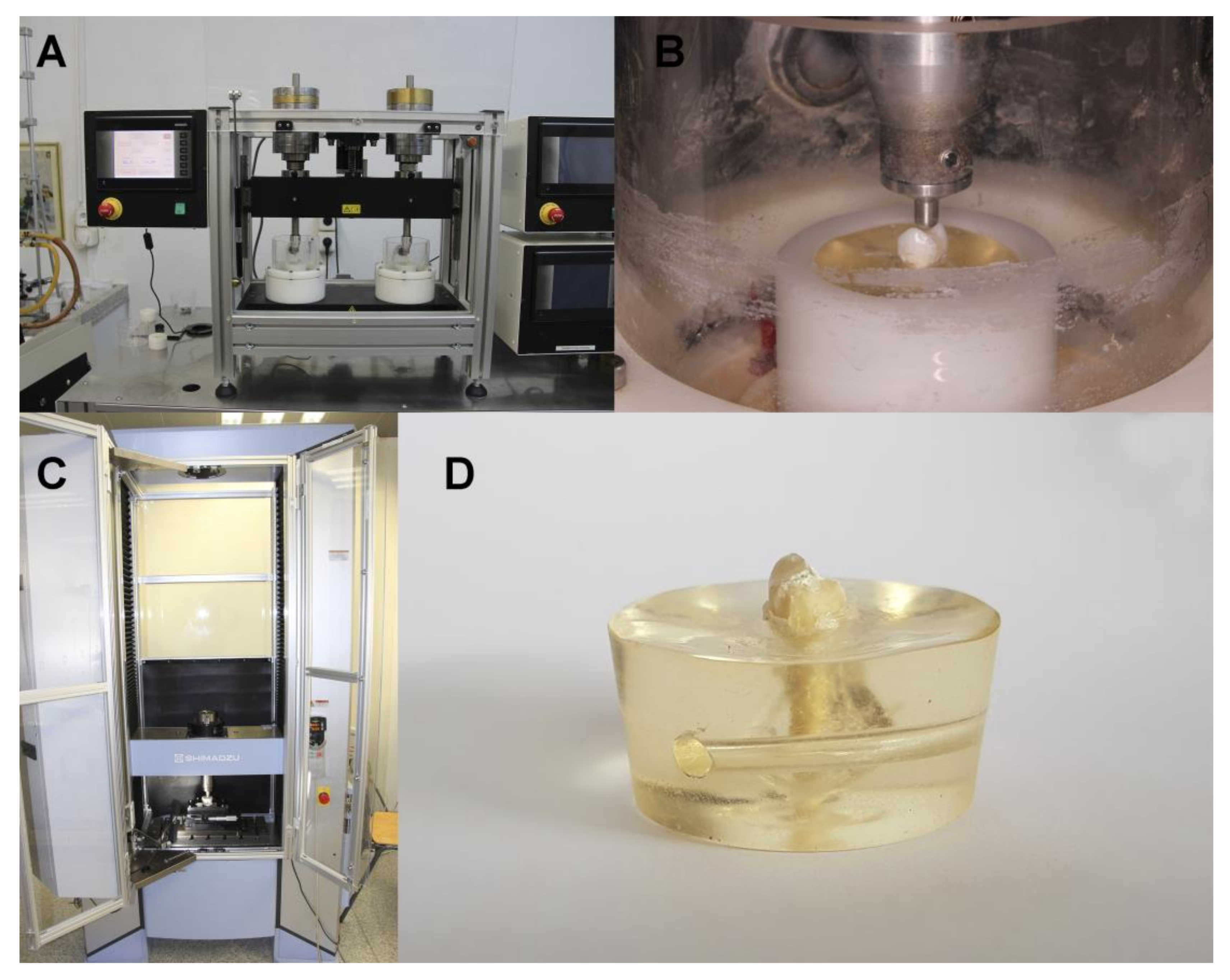
2.4. Thermal and Mechanical Cycling Fatigue

The experimental groups were subjected to thermal and mechanical cycling. The specimens were thermal fatigued (Thermocycling TC-3, SD Mechatronik, Feldkirchen-Westerham, Germany) in distilled water for 43 cycles/h during 24 h (6000 thermal cycles) between 5 and 55 °C with a 30 s dwell time. A masticatory simulator (SD Mechatronik, Chewing Simulator CS-4. Mechatronik GmbH, Feldkirchen, Germany) (Supplementary Material Figure S1 and Video S1) was used for mechanical cycling fatigued inducing 80 N load for 240,000 masticatory cycles. Loading was applied axially on the center of the occlusal surface with a vertical displacement of 2 mm at 2 Hz frequency and 40 mm/s speed by a point [12] (Figure 3A,B).

2.5. Fracture Load Test

After fatigue simulation, all specimens were subjected to a bending test until fracture using a universal testing machine (UTM) (Shimadzu® AG-100 KN, Shimadzu corporation, Kyoto, Japan) at a crosshead speed of 0.5 mm/s (ME 405/10, SERVOSIS, Madrid, Spain) with a load cell of 5000 N and at a room temperature of 23 ± 1 °C, moving vertically downward perpendicular to the occlusal plane. This test reproduces the action of forces similar to the masticatory forces, exerted in vitro on endodontic treated teeth. Axial compressive loads were exerted by sliding a cone shaped stainless-steel bar finished in a rounded tip (diameter: 1 mm) adapted to the UTM. This customized load piston was perpendicularly applied at the center of the occlusal surface, touching only restoration material, until the fracture of the testing restoration materials (Figure 3C,D), defined as a sharp decrease in the stress plot. The load force applicator’s aluminum ball was applied on the internal slopes of vestibular and palatal cusps of teeth. The results were recorded using inbuilt software for the testing machine (PCD2K, SERVOSIS) and force (N)-displacement (mm) curves were automatically created.

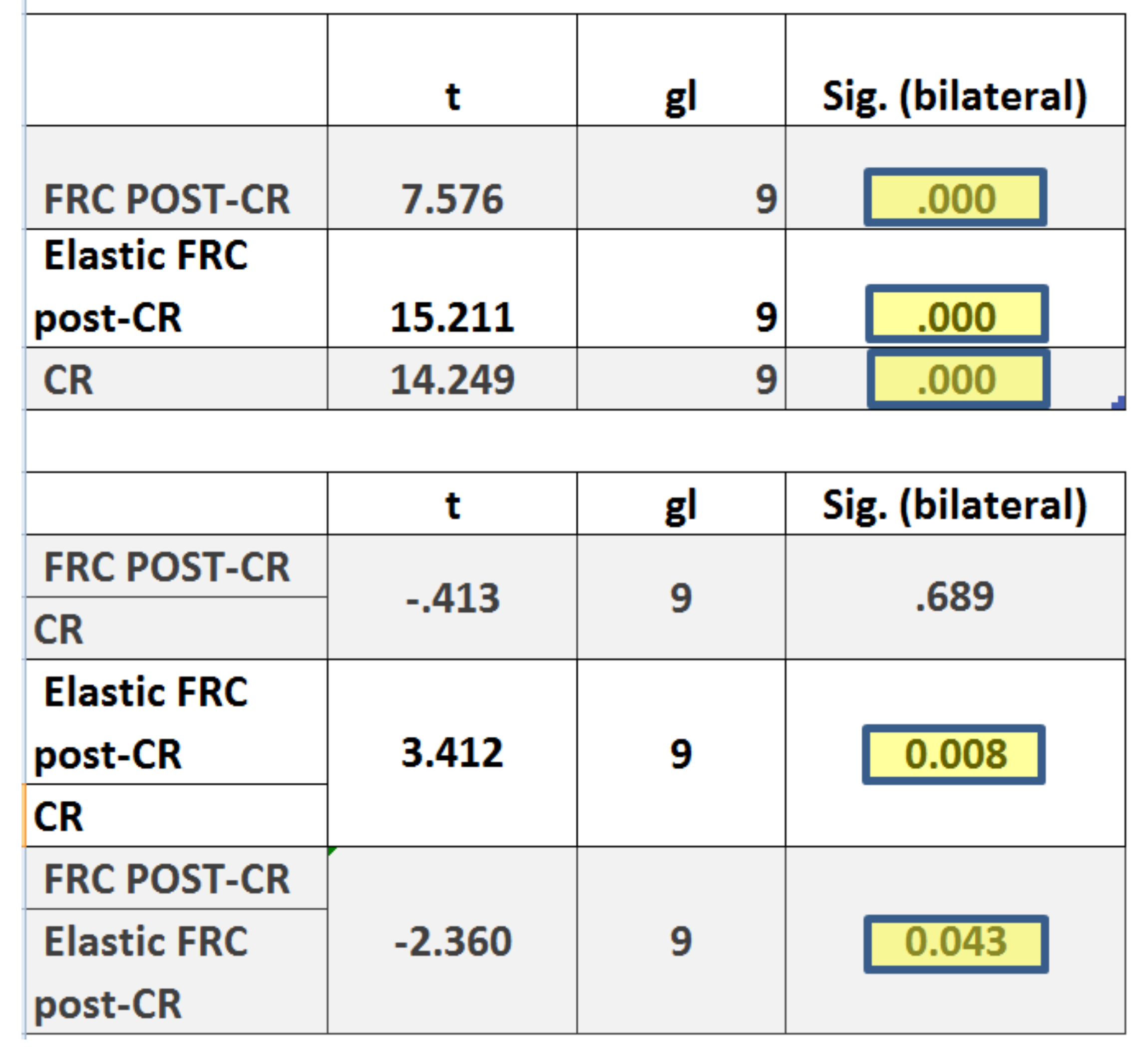
2.6. Statistical Analysis

Statistical analysis of all variables was carried out using SPSS 22.00 (IBM, Armonk, NY, USA) and Graph Pad Prism 7.0 (Graph Pad Software, San Diego, CA, USA). Descriptive statistics were expressed as means and standard deviations (SD) for quantitative variables. Comparative analysis was performed (data were normally distributed analyzed by Shapiro-Wilk) by comparing the fracture resistance load (N), the one-way ANOVA was used for comparing the means of normally-distributed data between multiple groups and Tukey tests was used to compare means between groups. In addition, Weibull characteristic strength (σ0) and Weibull modulus (m) were calculated. The statistical significance was set at p ˂ 0.05.

3. Results

The means and SD values for the fracture resistance (N) in the study groups are displayed in Table 3 and Figure 4.
The highest mean fracture resistance value was observed at the FRC post-FRC resin core study group (3620 ± 470) and the lowest mean fracture resistance value was observed at the FRC post-CR study group (2420 ± 1010) (Table 3 and Figure 4). Three study groups evidenced more fracture resistance than Control group (3290 ± 830): Elastics Post-CR study group (3510 ± 730), FRC post-FRC resin core study group (3620 ± 470) and Elastic Post-FRC resin core study group (3520 ± 730), as if they could be increasing the fracture resistance of the specimens (Table 3 and Figure 4). The paired Tukey test revealed statistically significant differences between the mean fracture resistance values between FRC post-CR and Elastic FRC Post-CR (p = 0.043) and between Elastic FRC post-CR and CR (p = 0.008) (Figure 5). Also, there was statistically significant difference between FRC post-CR and FRC post-FRC CR (Figure 6). However, there were no statistically significant differences between the mean fracture resistance values of the study groups.
The scale distribution parameter (η) of Weibull statistics showed statistically significant differences between CR core study group and control group (p = 0.0065), however, there were not statistically significant differences between the mean fracture resistance values of the study groups. There were also statistical significant differences at the shape distribution parameter (β) between FRC post-FRC resin core study group and control group (p = 0.0161), however, there were not statistical significant differences between the mean fracture resistance values of the study groups (Table 4 and Figure 7).

4. Discussion

The results obtained in the present study lead to the acceptation of the null hypothesis (H0), which states that the restoring materials tested has no statistically significant effect on the fracture resistance of endodontically treated teeth, however there was statistically significant differences between elastic FRC post restored with CR (3510 N) and FRC post with CR (2420 N) and only CR (2560 N). Besides, if we compare the FRC post/CR (2420 N) and FRC post/FRC resin core (3620 N) pairs, we also find statistically significant differences.
In endodontically treated teeth with substantial loss of structure, intraradicular posts are recommended to provide sufficient retention of restorative materials [23]. Mortazavi et al. [15] evidenced that elastic fiber reinforced posts offered a better behavior to physiological occlusal forces. Likewise, we reported that posts, especially elastic FRC post, showed greater fracture resistance to compression forces and this may be due to a modulus of elasticity more similar to dentin.
Bolay et al. [24] stated that elastic fiber reinforced posts have a better resistance to physiological occlusal forces. In our study, fiber post, especially elastic ones, showed greater resistance to compression forces and this may be because these posts have an elastic module which is more similar to dentin. Fráter et al. [21] concluded that teeth restored with an elastic fiber posts showed a significantly greater fracture resistance than those restored with conventional fiber posts. In our study, the elastic posts showed better results in terms of fracture resistance; although, in some tests the conventional posts achieved some positive results, this could be due to the fact that they were used with a reinforced fiberglass composite. The elastic FRC posts significantly increased the mean fracture resistance values (3510 N) with respect to non-elastic FRC posts (2420 N) when they were directly restored with CR. Nevertheless, non-elastic FRC posts slightly increased the mean fracture resistance values (3620 N) with respect to elastic FRC posts (3520 N) when they were restored with FRC resin core. The mechanical properties of elastic FRC posts should influence over the fracture resistance of the endodontically treated premolar teeth but it seems that FRC resin core influences to a greater extent. Rocca et al. [25] highlighted that the presence and orientation of the glass fibers inside the FRC resin core might influence over the fracture resistance of the restoration of the endodontically treated teeth.
Upper premolars have been shown to be more prone to root fractures, what justifies the selection of these teeth for this study [5,26,27,28]. Hannig et al. [28] stated that maxillary bicuspids with MOD restorations showed the lowest overall survival rate. Soares et al. [14] also confirmed that MOD cavities preparations and endodontic treatment increased the stress concentration within the dental structure, mainly due to the greater tissue removal. To extrapolate the data obtained in this in vitro study with those clinically observed in the oral environment, the samples were exposed to cyclical masticatory loads with thermocycling to analyze their fracture resistance.
Göktürk et al. [29] observed that direct restorations can distribute the functional stress through the interface of the restorative material and the tooth in a better way and also have the potential to support the weaker. However, these types of restorations tend to suffer shrinkage during polymeration causing cusp deflections [4,9,10,12,29]. To overcome these inconveniences, glass fibers were added to the resins changing their behavior by altering the elastic modulus of the material and therefore modifying the distribution of tensions to the walls of the tooth cavity. FRC has been considered as a new alternative for the restoration of endodontically treated tooth. In addition, placing composites with fibers in the cavity, allows a better distribution and dissipation of tension in the structure, which decreases and homogenizes the transmission of tension to the support teeth [29,30].
Moezizadeh et al. [30] indicated that fiber reinforced composites do not significantly increase the fracture resistance of endodontically treated premolar teeth. This statement is not in line with our findings since; in the present study the use of FRC resin core showed significant results in terms of resistance of the endodontically treated tooth with MOD cavities, when used as a final restoration or combined with a fiber post build-up restoration. It obtained better mean fracture resistance values (3040 N) of the endodontically treated premolar teeth directly restored with CR (2560 N).
Moezizadeh et al. [30] also stated in their study that the orientation of the glass fibers placed within the reinforced composites in MOD cavities showed greater fracture toughness. This statement is proven in our study where FRC resin core has a better resistance due to the intrinsic placement of its fibers and its orientation. In endodontic teeth with substantial loss of tissue, intraradicular posts are advisable to provide enough retention of restorative materials [19]. Nicola et al. [31] in their study found that resistance to fracture was significantly reduced in upper jaw premolars treated endodontically with direct restorations without fiber posts.
This may be because they did not previously fatigue the samples and used a higher crosshead speed (1 mm/min). The thermal and mechanical cycling fatigue device and the universal static load testing machine used in this in vitro study tried to simulate the oral environment [32] and have been described as the most effective procedure of evaluating the fracture resistance of dental restorations [33]. It also fulfils with the requirements and recommended established in ISO 6872:2015 [34] and have been management with the cycling fatigue parameters established in other similar trials [35]. The highest mean fracture resistance value and best biomechanical behavior was observed at the FRC post-FRC resin core study group (3620 ± 470). Weibull distribution analyzes the material’s variable failure rate. Weibull statistical analysis expresses the probability of failure of restore materials and allow a greater understanding of a material’s biomechanical behavior [36].
Some potential limitations of the present research was that is a destructive method and many studies have shown that the use of finite element stress analysis to evaluate stress in endodontically treated teeth is the ideal method for assessing post-core application, compared to several other methods of stress analysis [16,17,37,38,39]. Nonetheless, further in vitro non-destructive studies should be conducted in conjunction with clinical studies.
To date, FRC materials have proven to be an efficient restorative option and FRC posts and FRC resin core materials offer promising results as restorative therapy of endodontically treated teeth. However, more research is needed to determine the potential of FRC materials. Prefabricated fiberglass posts restored with conventional composite resins and reinforced fiberglass composites offered resistance values similar to the control group (premolars without treatment).

5. Conclusions

Within the limitations of this study, our results showed:
-
The use of elastic FRC post increase the fracture resistance of endodontically treated single-rooted upper first premolar teeth versus FRC post and only CR core. Besides, the restoration using FRC core resin also presents greater resistance to fracture than when they are restored with composite resin (CR).
-
The use of fiber-reinforced composites both in the core restoration and inside the root canal can help to reduce the potential risk of fracture associated to endodontically teeth.

Supplementary Materials

The following are available online at https://www.mdpi.com/2076-3417/10/21/7616/s1, Figure S1: Load applied on occlusal surface in masticatory simulation, Video S1: Masticatory simulation used (SD Mechatronik, Chewing Simulator CS-4. Mechatronik GmbH, Feldkirchen, Germany).

Author Contributions

Conceptualization, J.M.-Á. and A.Z.-M., data curation, writing and editing, J.M.-Á., A.Z.-M. and R.A.-P.; visualization and supervision, J.M.-Á. and A.Z.-M. All authors have read and agreed to the published version of the manuscript.

Funding

This research received no external funding.

Acknowledgments

The authors would like to thank Cristina Cueva Romero for his invaluable assistance in this study.

Conflicts of Interest

The authors declare no conflict of interest.

References

  1. Ausiello, P.; Apicella, A.; Davidson, C.; Rengo, S. 3D-finite element analyses of cusp movements in a human upper premolar, restored with adhesive resin-based composites. J. Biomech. 2001, 34, 1269–1277. [Google Scholar] [CrossRef]
  2. Dietschi, D.; Duc, O.; Krejci, I.; Sadan, A. Biomechanical considerations for the restoration of endodontically treated teeth: A systematic review of the literature—Part 1. Composition and micro- and macrostructure alterations. Quintessence Int. 2007, 38, 733–743. [Google Scholar] [PubMed]
  3. Goodacre, C.J.; Spolnik, K.J. The Prosthodontic Management of endodontically treated teeth: A literature review. Part I. Success and failure data, treatment concepts. J. Prosthodont. 1994, 3, 243–250. [Google Scholar] [CrossRef] [PubMed]
  4. Dietschi, D.; Duc, O.; Krejci, I.; Sadan, A. Biomechanical considerations for the restoration of endodontically treated teeth: A systematic review of the literature, Part II (Evaluation of fatigue behavior, interfaces, and in vivo studies). Quintessence Int. 2008, 39, 117–129. [Google Scholar] [PubMed]
  5. Tamse, A.; Zilburg, I.; Halpern, J. Vertical root fractures in adjacent maxillary premolars: An endodontic-prosthetic perplexity. Int. Endod. J. 1998, 31, 127–132. [Google Scholar] [CrossRef] [PubMed]
  6. Sorrentino, R.; Monticelli, F.; Goracci, C.; Zarone, F.; Tay, F.R.; García-Godoy, F.; Ferrari, M. Effect of post-retained composite restorations and amount of coronal residual structure on the fracture resistance of endodontically-treated teeth. Am. J. Dent. 2007, 20, 269–274. [Google Scholar] [PubMed]
  7. Peroz, I.; Blankenstein, F.; Lange, K.-P.; Naumann, M. Restoring endodontically treated teeth with posts and cores—A review. Quintessence Int. 2005, 36, 737–746. [Google Scholar]
  8. Saritha, M.K.; Paul, U.; Keswani, K.; Jhamb, A.; Mhatre, S.H.; Sahoo, P.K. Comparative evaluation of fracture resistance of different post systems. J. Int. Soc. Prev. Community Dent. 2017, 7, 356–359. [Google Scholar] [CrossRef]
  9. Da Rocha, D.M.; Tribst, J.P.; Ausiello, P.; Piva, A.M.D.O.D.; Da Rocha, M.C.; Di Nicoló, R.; Borges, A.L.S. Effect of the restorative technique on load-bearing capacity, cusp deflection, and stress distribution of endodontically-treated premolars with MOD restoration. Restor. Dent. Endod. 2019, 44, e33. [Google Scholar] [CrossRef]
  10. Taha, N.A.; Palamara, J.E.; Messer, H.H. Cuspal deflection, strain and microleakage of endodontically treated premolar teeth restored with direct resin composites. J. Dent. 2009, 37, 724–730. [Google Scholar] [CrossRef]
  11. Signore, A.; Benedicenti, S.; Covani, U.; Ravera, G. A 4-to 6-year retrospective clinical study of cracked teeth restored with bonded indirect resin composite onlays. Int. J. Prosthodont. 2007, 20, 609–616. [Google Scholar] [CrossRef]
  12. Pereira, R.; Bicalho, A.; Franco, S.; Tantbirojn, D.; Versluis, A.; Soares, C.J.; Tantbirojin, D. Effect of restorative protocol on cuspal strain and residual stress in endodontically treated molars. Oper. Dent. 2016, 41, 23–33. [Google Scholar] [CrossRef] [PubMed] [Green Version]
  13. Zarow, M.; Vadini, M.; Chojnacka-Brozek, A.; Szczeklik, K.; Milewski, G.; Biferi, V.; D’Arcangelo, C.; De Angelis, F. Effect of fiber posts on stress distribution of endodontically treated upper premolars: Finite element analysis. Nanomaterials 2020, 10, 1708. [Google Scholar] [CrossRef] [PubMed]
  14. Ausiello, P.; Ciaramella, S.; Garcia-Godoy, F.; Martorelli, M.; Sorrentino, R.; Gloria, A. Stress distribution of bulk-fill resin composite in class II restorations. Am. J. Dent. 2017, 30, 227–232. [Google Scholar]
  15. Mortazavi, V.; Fathi, M.; Katiraei, N.; Shahnaseri, S.; Badrian, H.; Khalighinejad, N. Fracture resistance of structurally compromised and normal endodontically treated teeth restored with different post systems: An in vitro study. Dent. Res. J. 2012, 9, 185–191. [Google Scholar] [CrossRef]
  16. Gloria, A.; Maietta, S.; Martorelli, M.; Lanzotti, A.; Watts, D.; Ausiello, P. FE analysis of conceptual hybrid composite endodontic post designs in anterior teeth. Dent. Mater. 2018, 34, 1063–1071. [Google Scholar] [CrossRef] [PubMed] [Green Version]
  17. Ausiello, P.; Gloria, A.; Maietta, S.; Watts, D.C.; Martorelli, M. Stress distributions for hybrid composite endodontic post designs with and without a ferrule: FEA study. Polymers 2020, 12, 1836. [Google Scholar] [CrossRef]
  18. Vadavadagi, S.V.; Dhananjaya, K.M.; Yadahalli, R.P.; Lahari, M.; Shetty, S.R.; Bhavana, B. Comparison of different post systems for fracture resistance: An in vitro study. J. Contemp. Dent. Pract. 2017, 18, 205–208. [Google Scholar] [CrossRef] [Green Version]
  19. Saker, S.; Özcan, M. Retentive strength of fiber-reinforced composite posts with composite resin cores: Effect of remaining coronal structure and root canal dentin conditioning protocols. J. Prosthet. Dent. 2015, 114, 856–861. [Google Scholar] [CrossRef] [Green Version]
  20. Schneider, S.W. A comparison of canal preparations in straight and curved root canals. Oral Surg. Oral Med. Oral Pathol. 1971, 32, 271–275. [Google Scholar] [CrossRef]
  21. Fráter, M.; Forster, A.; Jantyik, Á.; Braunitzer, G.; Nagy, K.; Grandini, S. In vitro fracture resistance of premolar teeth restored with fibre-reinforced composite posts using a single or a multi-post technique. Aust. Endod. J. 2017, 43, 16–22. [Google Scholar]
  22. Pottmaier, L.; Linhares, L.A.; Baratieri, L.; Vieira, L.C. Evaluation of the fracture resistance of premolars with extensive and medium cavity preparations restored with direct restoring systems. Indian J. Dent. Res. 2018, 29, 465–469. [Google Scholar] [CrossRef] [PubMed]
  23. Nothdurft, F.P.; Seidel, E.; Gebhart, F.; Naumann, M.; Motter, P.; Pospiech, P. The fracture behavior of premolar teeth with class II cavities restored by both direct composite restorations and endodontic post systems. J. Dent. 2008, 36, 444–449. [Google Scholar] [CrossRef] [PubMed]
  24. Bolay, Ş.; Ozturk, E.; Tuncel, B.; Ertan, A. Fracture resistance of endodontically treated teeth restored with or without post systems. J. Dent. Sci. 2012, 7, 148–153. [Google Scholar] [CrossRef] [Green Version]
  25. Rocca, G.T.; Saratti, C.M.; Poncet, A.; Feilzer, A.J.; Krejci, I. The influence of FRCs reinforcement on marginal adaptation of CAD/CAM composite resin endocrowns after simulated fatigue loading. Odontology 2016, 104, 220–232. [Google Scholar] [CrossRef]
  26. Mohammadi, N.; Kahnamoii, M.A.; Yeganeh, P.K.; Navimipour, E.J. Effect of fiber post and cusp coverage on fracture resistance of endodontically treated maxillary premolars directly restored with composite resin. J. Endod. 2009, 35, 1428–1432. [Google Scholar] [CrossRef] [PubMed]
  27. Xie, K.X.; Wang, X.Y.; Gao, X.J.; Yuan, C.Y.; Li, J.X.; Chu, C.H. Fracture resistance of root filled premolar teeth restored with direct composite resin with or without cuspcoverage. Int. Endod. J. 2012, 45, 524–529. [Google Scholar] [CrossRef]
  28. Hannig, C.; Westphal, C.; Becker, K.; Attin, T. Fracture resistance of endodontically treated maxillary premolars restored with CAD/CAM ceramic inlays. J. Prosthet. Dent. 2005, 94, 342–349. [Google Scholar] [CrossRef]
  29. Göktürk, H.; Karaarslan, E.Ş.; Tekin, E.; Hologlu, B.; Sarıkaya, I. The effect of the different restorations on fracture resistance of root-filled premolars. BMC Oral Health 2018, 18, 196. [Google Scholar]
  30. Moezizadeh, M.; Shokripour, M. Effect of fiber orientation and type of restorative material on fracture strength of the tooth. J. Conserv. Dent. 2011, 14, 341–345. [Google Scholar] [CrossRef] [Green Version]
  31. Scotti, N.; Forniglia, A.; Tempesta, R.M.; Comba, A.; Saratti, C.M.; Pasqualini, D.; Alovisi, M.; Berutti, E.; Nicola, S.; Alberto, F.; et al. Effects of fiber-glass-reinforced composite restorations on fracture resistance and failure mode of endodontically treated molars. J. Dent. 2016, 53, 82–87. [Google Scholar] [CrossRef] [PubMed] [Green Version]
  32. Scotti, N.; Scansetti, M.; Rota, R.; Pera, F.; Pasqualini, D.; Berutti, E. The effect of the post length and cusp coverage on the cycling and static load of endodontically treated maxillary premolars. Clin. Oral Investig. 2011, 15, 923–929. [Google Scholar] [CrossRef] [PubMed]
  33. López-Suárez, C.; Castillo-Oyagüe, R.; Rodríguez-Alonso, V.; Lynch, C.D.; Suárez-García, M.-J. Fracture load of metal-ceramic, monolithic, and bi-layered zirconia-based posterior fixed dental prostheses after thermo-mechanical cycling. J. Dent. 2018, 73, 97–104. [Google Scholar] [CrossRef] [PubMed]
  34. Augstin-Panadero, R.; Fons-Font, A.; Rodríguez, J.L.R.; Granell-Ruiz, M.; Del Rio-Highsmith, J.; Ruiz, M.F.S. Zirconia versus metal: A preliminary comparative analysis of ceramic veneer behavior. Int. J. Prosthodont. 2012, 25, 294–300. [Google Scholar] [PubMed]
  35. Lopez-Suarez, C.; Tobar, C.; Sola-Ruiz, M.F.; Pelaez, J.; Suarez, M.J. Effect of thermomechanical and static loading on the load to fracture of metal-ceramic, monolithic and veneered zirconia posterior fixed partial dentures. J. Prosthodont. 2019, 28, 171–178. [Google Scholar] [CrossRef]
  36. Quinn, J.B.; Quinn, G.D. A practical and systematic review of Weibull statistics for reporting strengths of dental materials. Dent. Mater. 2010, 26, 135–147. [Google Scholar] [CrossRef] [Green Version]
  37. Ausiello, P.; Ciaramella, S.; Martorelli, M.; Lanzotti, A.; Gloria, A.; Watts, D.C. CAD-FE modeling and analysis of class II restorations incorporating resin-composite, glass ionomer and glass ceramic materials. Dent. Mater. 2017, 33, 1456–1465. [Google Scholar] [CrossRef] [Green Version]
  38. Coelho, C.S.D.M.; Biffi, J.C.G.; Da Silva, G.R.; Abrahão, A.; Campos, R.E.; Soares, C.J. Finite element analysis of weakened roots restored with composite resin and posts. Dent. Mater. J. 2009, 28, 671–678. [Google Scholar] [CrossRef] [Green Version]
  39. Falakaloğlu, S.; Adıgüzel, Ö.; Özdemir, G. Root canal reconstruction using biological dentin posts: A 3D finite element analysis. J. Dent. Res. Dent. Clin. Dent. Prospect. 2019, 13, 274–280. [Google Scholar] [CrossRef] [Green Version]
Figure 1. Schematic representation of experimental groups.
Applsci 10 07616 g001
Figure 2. (A,B) Representation of the cavity preparation used in this work. In blue, endodontic access cavity.
Applsci 10 07616 g002
Figure 3. (A,B) Samples placed in the thermal and mechanical cycling fatigue device and (C,D) sample fractured by static load after bending test.
Applsci 10 07616 g003
Figure 4. Box plots of fracture resistance values (N) of the study groups after bending test. The horizontal line in each box represents median value.
Applsci 10 07616 g004
Figure 5. Paired Tukey test differences inter group. p value < 0.05.
Applsci 10 07616 g005
Figure 6. Paired Tukey test differences inter group. p value < 0.05.
Applsci 10 07616 g006
Figure 7. Weibull probability plot of the fracture resistance values (N) of the study groups after bending test.
Applsci 10 07616 g007
Table 1. Elastic Modulus of teeth’s components.
ComponentElastic Modulus
(GPa)
Dentin 14–18.6
Enamel 80
Periodontal Ligament0.05
Compact Bone 13.8
Medullar Bone 0.345
Table 2. Restorative materials description.
Material/ManufactureClassificationElastic Modulus (GPa)
Gradia Core Self-Etching Bond®, GC Europe, Leuven, BelgiumDual-cure one-step self-etch adhesive system4.5
Fiber Post® 0.8 mm, GC Europe, Leuven, BelgiumConventional glass Fiber Reinforced Composite posts24
EverStick Post® 0.9 mm, GC Europe, Leuven, BelgiumElastic Fiber Reinforced Composite posts 13–16
Gradia Core®, GC Europe, Leuven, BelgiumDual-cure Composite Resin core material10.8
Everx X Posterior®, GC Europe, Leuven, BelgiumFiber Reinforced Composite resin lightcured14.6
Table 3. Descriptive statistics of the fracture resistance values (N) of the study groups after bending test. * p value < 0.05. (One-way ANOVA test for comparing the means between multiple groups).
GroupnMeanSDMinimumMaximum
Conventional FRC post-CR Core 102420 *101011904010
Elastic FRC post-CR Core10351073020104230
Conventional FRC post-FRC CR10362047028004020
Elastic FRC post-FRC CR10352073020704020
CR Core 102560 *57016903400
FRC CR 103040108013104070
Control: intact teeth without MOD cavity, nor endodontic treatment nor restorations10329083018304930
Table 4. Weibull statistics of the fracture resistance of the study groups after bending test.
m = Weibull Shape (β)σ0 = Weibull Scale (η)
EstimateSt ErrorLowerUpperEstimateSt ErrorLowerUpper
Conventional FRC Post-CR Core27.4100.668616.99244.21327.2700.333321.46134.651
Elastic FRC post-CR Core70.05519.50840.589120.91437.7890.177434.46841.431
Conventional FRC Post-FRC CR106.74429.24762.390182.62838.1290.118435.87840.521
Elastic FRC Post-FRC CR72.26220.69041.228126.65537.8140.171834.59241.336
CR Core (Gradia Core)56.24014.40734.04092.91727.7730.164524.72931.191
FRC CR (Ever-X)35.2110.951420.73559.79533.9930.320228.26340.884
Control44.83010.50828.31770.97435.9970.268731.09941.667
Publisher’s Note: MDPI stays neutral with regard to jurisdictional claims in published maps and institutional affiliations.

