Electrochemical Biosensors Based on Conducting Polymers: A Review


UFR Sciences et Techniques, University Bourgogne Franche-Comté, Institut Utinam UMR CNRS 6213, 16 Route de Gray, 25030 Besançon, France
Appl. Sci. 2020, 10(18), 6614; https://doi.org/10.3390/app10186614
Submission received: 31 August 2020
/
Revised: 18 September 2020
/
Accepted: 20 September 2020
/
Published: 22 September 2020
(This article belongs to the Special Issue Advanced Electrochemical Biosensors)
Abstract
:Conducting polymers are an important class of functional materials that has been widely applied to fabricate electrochemical biosensors, because of their interesting and tunable chemical, electrical, and structural properties. Conducting polymers can also be designed through chemical grafting of functional groups, nanostructured, or associated with other functional materials such as nanoparticles to provide tremendous improvements in sensitivity, selectivity, stability and reproducibility of the biosensor’s response to a variety of bioanalytes. Such biosensors are expected to play a growing and significant role in delivering the diagnostic information and therapy monitoring since they have advantages including their low cost and low detection limit. Therefore, this article starts with the description of electroanalytical methods (potentiometry, amperometry, conductometry, voltammetry, impedometry) used in electrochemical biosensors, and continues with a review of the recent advances in the application of conducting polymers in the recognition of bioanalytes leading to the development of enzyme based biosensors, immunosensors, DNA biosensors, and whole-cell biosensors.
1. Introduction
Conducting polymers have attracted much interest since Shirakawa et al. demonstrated in 1977 that halogen doping of polyacetylene strongly increased its conductivity [1]. Thanks to this revolutionary research, Shirakawa, MacDiarmid, and Heeger were awarded the Nobel Prize in Chemistry in 2000, and opened the way to the development of other conducting polymers combining properties of organic polymers and electronic properties of semiconductors. Another major breakthrough in this field was achieved by Diaz et al., who reported the electrodeposition of highly conductive, stable and processable polypyrrole films [2,3,4]. Following these pioneering studies, numerous conducting polymers have been prepared and used in various applications, such as polyacetylene, polypyrrole (PPy), polyaniline (PANI), polycarbazole, polythiophene (PTh), poly(3,4-ethylenedioxythiophene) (PEDOT), polyphenylene, poly(phenylene vinylene), and polyfluorene (Table 1). All these organic polymers are characterized by alternating single (σ) and double (π) bonds and by the presence of π electrons delocalized across their entire conjugated structure, thus resulting in polymers which can be easily oxidized or reduced [5]. This doping, that can be performed upon oxidation (p-doping) or reduction (n-doping), increases significantly the conductivity of the polymers since this conductivity can vary from less than 10−6 S/cm in the neutral state [5] to more than 105 S/cm in the doped state [6,7]. The conductivity of the polymers is also dependent on a number of factors including the nature and concentration of the dopant [8,9,10], temperature [11,12,13], swelling/deswelling [14], polymer morphology [8], pH and applied potential [15], and polymer chain length [16]. For most heterocyclic polymers, such as PPy [17] or PTh [18], the mechanism of conduction corresponds to a p-doping and starts with the removal of one electron from the initial monomer leading to the formation of an unstable radical cation (named polaron). Then, a second electron is removed from another monomer or from an oligomer, leading to the formation of a dication (named bipolaron) [19]. Under an applied electric field, these polarons and bipolarons serve as charge carriers which are delocalized over the polymer chains and their movement along polymer chains produces electronic conductivity [20].
Conducting polymers have become an important class of materials since they combine some useful properties of organic polymers (such as strength, plasticity, flexibility, toughness or elasticity) with unusual electronic [5], optical [21,22] and thermoelectric [23,24] properties due to the charge mobility along the π electron polymer chains. These unique properties explain the use of conducting polymers in a wide variety of applications including energy storage with rechargeable batteries [25,26] and supercapacitors [27,28], photovoltaics with solar cells [29,30,31,32], light-emitting diodes [33,34], electrocatalysis [35], anti-corrosion [36,37] or electrochromic applications such as electrochromic displays [38,39] or rearview mirrors and smart windows [40,41].
2. Preparation of Sensitive Materials
2.1. Preparation of Conducting Polymers
Although it is possible to prepare conducting polymers using gas phase techniques such as CVD [42] or plasma polymerization [43,44], conducting polymers are mostly prepared via chemical or electrochemical oxidative polymerization even if it is sometimes possible to use non-oxidative chemical polymerization methods such as Grignard metathesis [45] or dehydrobrominative polycondensation [46]. In traditional chemical oxidative polymerization [47], the synthesis of polymers can be done under harsh oxidative conditions with the use of oxidants such as K2Cr2O7, KMnO4, K2S2O8, KIO3 and FeCl3 [48], or under mild conditions by using, for example, the catalytic action of redox enzymes to produce hydrogen peroxide that initiates the polymerization [49], or less frequently at the liquid/air interface [50]. However, the electrochemical oxidative polymerization is the most frequently used method, mainly because it allows a better control of the polymer deposition [51]. Electrochemical polymerization is carried out with a classical three-electrode set-up in an electrochemical cell containing a monomer, a solvent and a supporting salt. The electropolymerization can be achieved either with a potentiodynamic technique such as cyclic voltammetry where the current response to a linearly cycled potential sweep between two or more set values is measured, with a potentiostatic technique where a constant potential is applied to initiate the polymerization, or with a galvanostatic technique where a constant current is applied to initiate polymerization. The potentiostatic technique allows easy control of the film thickness through Faraday’s law, whereas potentiodynamic techniques lead to more homogeneous and adherent films on the electrode. Additionally, the galvanostatic technique is generally considered as the best approach since it allows to follow the growth of the conducting polymer film by monitoring the potential changes with time which reflects the conductivity.
Conducting polymers have been widely used in the area of bioanalytical and biomedical science [52,53], drug delivery [54,55,56], tissue engineering [57,58,59], and cell culture [60,61,62] due to their intrinsic properties and biocompatibility [63,64,65,66]. In addition, conducting polymers represent an attractive sensitive material for biosensors due to their electrical properties that allow to convert biochemical information into electrical signals. Additionally, conducting polymers can be easily modified by grafting of functional groups which offers the possibility to enhance their abilities to detect and quantify bioanalytes or to maximize the interactions between the biomolecules and the functionalized polymer. Therefore, after a short description of the electrochemical techniques used in conducting polymer-based biosensors, a series of examples of such biosensors will be described to highlight the recent advances in the field of conducting polymer-based electrochemical biosensors.
2.2. Strategies for Immobilizing Biological Sensing Elements into Conducting Polymers
Biological sensing element immobilization plays a fundamental role in the performance characteristics of biosensors since biomolecules must be directly attached to the surface of the biosensor to obtain a good sensitivity and a long operational life. The most commonly used methods to immobilize biomolecules to polymers are physical adsorption, covalent attachment and entrapment (Figure 1). The choice of immobilization strategy mainly depends on the type of biological element. Indeed, antibodies and ssDNA are preferentially immobilized by adsorption or covalent binding onto the surface of the conducting polymer films to facilitate the access of the analyte to these biorecognition molecules when entrapment is generally used to immobilize oxidoreductases within the polymer film to facilitate the electron transfer from the enzyme’s redox center to the analyte solution surrounding the conducting polymer and the rapid redox reaction of electroactive species such as hydrogen peroxide generated by enzymatic catalysis.
The method of covalent immobilization uses the functional groups of biomolecules (such as –COOH, -NH2, or -SH) for binding with a conducting polymer. Thus, a biomolecule containing amino groups has the capacity to form amide bonds with a conducting polymer bearing carboxylic groups. For example, Kim et al. have developed a glucose biosensor with a conducting electrosynthesized poly(terthiophene benzoic acid) bearing benzoic acid groups which allow the immobilization of glucose oxidase (GOx) through amide bond formation [67]. Similarly, Tuncagil et al. electrosynthesized the conducting polymer 4-(2,5-di(thiophen-2-yl)-1H-pyrrol-1-yl) benzenamine to immobilize GOx through amide bonds [68]. Moreover, covalent attachment of biomolecules is frequently achieved by initial synthesis of functionalized monomers with an amino side group, followed by electrochemical polymerization of these functionalized monomers leading to conducting polymer films with interfacial attachable side groups that can be covalently bound to biomolecules containing the corresponding groups. To facilitate the formation of covalent bonds between biomolecules and polymers, crosslinking agents such as glutaraldehyde [69,70] or 1-ethyl-3-(3-dimethylaminopropyl) carbodiimide (EDC) [71,72] are commonly used. The covalent immobilization method has the benefit of providing low diffusional resistance, giving strong binding force between biomolecule and polymer, thus reducing loss of biomolecule. Therefore, these electrodes are more stable in time even if it may be difficult in some cases to retain the biomolecule activity.
The adsorption method is very simple and only consists in the physical adsorption of the biomolecule on the polymer surface. Sometimes, the presence of opposite charges into the conducting polymer and the biomolecule facilitates the immobilization of the biomolecule. Thus, negatively charged glucose oxidase was successfully adsorbed onto positively charged polyaniline-polyisoprene films at pH 4.5 to provide a material sensitive to glucose concentration changes [73]. This method has the benefit of providing small perturbation of the biomolecule native structure and function and so generally leads to very sensitive responses. However, a strong drawback is that direct physical adsorption of biomolecule on a surface generally leads to poor long-term stability of the sensor because of biomolecule leakage from the surface when changes in the environment arise (pH, ionic strength) even if the modification of the surface by a polymer film can slow this leakage [74,75].
Entrapment is another method widely used for the immobilization of enzymes [76,77], antibodies [78] or DNA [79]. It involves the preparation of an electrolyte solution containing both monomer and biomolecule, followed by the electropolymerization of the whole solution. Thus, a polymer film containing biomolecules is formed at the electrode surface. Entrapment is an interesting technique since it leads to a strong adhesion between biomolecule and polymer film in a single step. Additionally, this strategy includes the possibility of controlling the amount of entrapped biomolecules simply by controlling the thickness of the electrodeposited polymer film. Entrapment generally leads to biosensors with a good sensitivity and a long lifetime. On the contrary, entrapment can generate problems associated with inaccessibility of the embedded biomolecule. Additionally, some conducting polymers require very acidic conditions or high oxidation potential during the electropolymerization process to be prepared but these conditions are not favorable to biomolecules [80]. It is also important to note that supporting electrolytes are usually used during the electropolymerization process to increase the conductivity of the monomer solution. Besides, the electrolytes tend to compete with the biomolecules for the polymer doping sites, and so reduce the amount of biomolecule entrapped which is a problem especially for costly biomolecules. A solution to this problem is the use of biomolecules as counter-ions during the growth of the conducting polymer film to allow a more efficient entrapment as previously done with polypyrrole and GOx enzyme [81]. To enhance the incorporation of enzymes into polymers during their electropolymerization, it is also possible to use sinusoidal voltages as evidenced by Lupu et al. who developed dopamine biosensors based on tyrosinase entrapped into PEDOT film [82].
3. Electroanalytical Methods
When conducting polymers are used as sensitive material in electrochemical sensors, the capture of a target analyte to a bioreceptor immobilized in a conducting polymer generates an analytical measurable signal which is converted into an electrical signal (Figure 2). The presence of the conducting polymer is beneficial for improving sensitivity and selectivity of the biosensor while reducing the effect of interfering species. The selectivity of the biosensor strongly depends on the presence of specific interactions between the analyte and the bioreceptor when the quality of the immobilization of the bioreceptor to the conducting polymer and of the conducting polymer to the surface of the biosensor is mainly responsible for the long-term efficiency of the biosensor. The sensitivity of the biosensor depends on many factors, the main one being the intensity of the electrochemical signal generated by the reaction between the analyte, the bioreceptor and the conducting polymer. This electrochemical signal can be a change in the value of the voltage, current, conductivity/resistance, impedance, or number of electrons exchanged through an oxidation or reduction reaction leading to the fabrication of potentiometric, amperometric, conductimetric, impedimetric and voltametric biosensors, respectively.
3.1. Potentiometry
In potentiometric conducting polymer-based biosensors, the potential between a reference electrode and an electrode coated either with a conducting polymer and a biorecognition element is measured using a high impedance voltmeter. The conducting polymer must be sensitive to the products of a reaction involving the analyte bound to the biorecognition element. This modified electrode senses the variation in protons (or other ions) amount since potential and pH are linked by the Nernst equation, leading to a recorded analytical signal which is generally logarithmically correlated with the analyte concentration.
The most widely studied potentiometric biosensors are enzymatic biosensors that use an enzyme incorporated in the conducting polymer to catalyze a reaction producing protons. For example, in potentiometric urea enzymatic biosensors, urease is immobilized in a polymer and is used to catalyze the conversion of urea to carbon dioxide, ammonia and protons which produce a pH increase detected by the potentiometric biosensor (see Section 4.1).
3.2. Amperometry
In amperometric conducting polymer-based biosensors, the current produced during the oxidation or reduction of an electroactive biological element at a constant potential that is applied between a reference electrode and a polymer-modified electrode is measured, thus providing specific quantitative analytical information. Such biosensors are inspired by the first and simplest amperometric biosensor developed by Clark in 1956 who fabricated an amperometric oxygen sensor that produced a current proportional to the oxygen concentration when a potential of −0.6 V vs. Ag/AgCl electrode was applied to a platinum electrode [83].
The most widely studied amperometric biosensor is the glucose biosensor. In this system, glucose oxidase catalyzes the reaction of glucose with oxygen to produce gluconolactone and hydrogen peroxide. By monitoring the amount of hydrogen peroxide produced by this reaction in the presence of GOx through amperometric measurements, it is possible to determine the glucose concentration. In such biosensors, the GOx is immobilized in the conducting polymer either by electropolymerization of a solution containing a monomer and GOx or by addition of GOx in an electrodeposited conducting polymer film (see Section 4.1).
3.3. Conductometry
In conductometric conducting polymer-based biosensors, a change in electrical conductivity or resistivity is measured against the analyte concentration when a constant or sweeping potential is applied between a reference electrode and a polymer-modified electrode. To increase the sensitivity of the sensor, the conducting polymer must be highly conductive when it is charged (doped) and lowly conductive when it is neutral (dedoped), thus leading to a strong conductivity change when the conducting polymer reacts with the analyte. Furthermore, the morphology of the conducting polymer is important since the charges that are created within the backbone of the polymer must be able to interact with the surrounding environment and thus to change the polymer’s conductivity. However, these biosensors often suffer from their lack of selectivity since any change in conductivity in the solution modifies their signal.
An example of conductometric biosensor was fabricated by Forzani et al. [84] who coated a pair of nanoelectrodes with PANI/GOx. Their exposure to glucose resulted in the reduction of GOx which was spontaneously reoxidized in the presence of oxygen to form hydrogen peroxide which oxidized the PANI, leading to a change in conductivity that can be monitored and used to determine the glucose concentration.
3.4. Voltammetry
In voltametric conducting polymer-based biosensors, a current is produced by sweeping the potential applied between a reference electrode and a polymer-modified electrode over a range that is associated with the redox reaction of the analyte. This redox reaction generates a change in the peak current which can be correlated with the analyte concentration, thus providing specific quantitative analytical information. All the voltametric methods that can be used, such as linear voltammetry, cyclic voltammetry, differential pulse voltammetry, or square wave voltammetry, have the advantage of providing both qualitative information deduced from the potential location of the current peak and quantitative information deduced from the peak current intensity.
For example, the detection of acetylcholine was successfully achieved using a conductive PEDOT film loaded with Fe3O4 nanoparticles and reduced graphene oxide since the intensity of the oxidation peak present in the cyclic voltammograms was linear to the acetylcholine concentration [85]. Similarly, the detection of serotonin in banana was done by square wave voltammetry using conducting polypyrrole/Fe3O4 nanocomposites [86] and the detection of danazol was performed by differential pulse voltammetry using conducting electrodeposited polyaniline [87].
3.5. Impedancemetry
Electrochemical impedance spectroscopy (EIS) is a sensitive technique for the analysis of biomolecular recognition events of specific binding proteins, nucleic acids, whole cells, antibodies or antibody-related substances, occurring at the modified surface [88,89,90]. In particular, many studies on impedometric biosensors are focused on immunosensors since the bonding between antibodies and antigens leads to the formation of an immunocomplex resulting in electron transfers and impedance changes (see Section 4.2). Moreover, impedometric biosensors allow direct detection of biomolecular recognition events without using enzyme labels and have the advantages of low cost, ease of use, portability and ability to perform both screening and online monitoring without being destructive. In impedometric immunosensors, conducting polymers are generally used to immobilize the antigens, commonly through covalent attachment, thus allowing the detection of the antibodies due to the high antigen-antibody affinity [91,92,93].
4. Conducting Polymer-Based Electrochemical Biosensors
4.1. Conducting Polymer-Based Enzyme Biosensors
Enzymatic electrochemical biosensors utilize the biospecificity of an enzymatic reaction, along with an electrode reaction that generates a current or potential for quantitative analysis. Many biomolecules such as glucose, cholesterol, or urea are important analytes due to their adverse effects on health. Enzymatic biosensors utilize the biochemical reactions between analyte and enzyme resulting in a product (hydrogen peroxide, protons, ammonium ions) that can be quantified by a transducer. In general, many oxidoreductases, for example glucose oxidase, catalyze the oxidation of substrates by electron transfer to oxygen to form hydrogen peroxide. These oxidoreductase enzymes can be immobilized on conducting polymer films and the H2O2 formed as a result of enzyme-catalyzed reactions induces an amperometric signal measured by the electrochemical biosensor. As a consequence, many biosensors have already been prepared that use conducting polymers as a matrix to immobilize enzymes at the surface of the biosensors.
My objective here is not to describe all the very numerous works in the field of enzymatic biosensors, but to focus on some examples of glucose sensors, illustrating the major current trends and the progress made in recent years in this research area. Indeed, in the field of biosensors, glucose biosensors have given rise to the highest number of studies due to the clinical significance of measuring blood glucose levels in patients with diabetes. Thus, some potentiometric biosensors used polyaniline films to detect pH changes due to the production of protons by oxidation of hydrogen peroxide [94,95]. However, the vast majority of classical glucose biosensors have been prepared by electropolymerization of a solution containing glucose oxidase and a monomer and used an amperometric detection. Thus, many glucose biosensors associated GOx with polyaniline or GOx with polypyrrole as extensively described in the reviews from Lai et al. [96] and Singh et al. [97], respectively. However, such conventional conducting-based glucose biosensors still present some problems such as unsatisfactory sensitivity or detection limit and relatively high applied potential. That is why more recent biosensors have substituted polymer matrix with polymer nanocomposites having the properties of increasing permselectivity, sensitivity and stability, and to decrease applied potential. Thus, conducting polymers were combined with artificial mediators such as benzoquinone derivatives [98], ferrocene derivatives [99], and Os-complex mediators [100], which were able to reoxidize the reduced GOx to its oxidized state. Thus, the released electrons from the reduced GOx are transferred to the polymer modified electrode through the redox process of the mediators. Therefore, the incorporation of a mediator leads to a better charge transport which is responsible for an enhancement of the biosensor’s sensitivity. Another problem encountered in classical biosensors is that a high anodic potential (exceeding +0.6 V) is applied, leading to interference from other oxidizable substances, such as ascorbic acid, acetaminophen or uric acid. To solve this problem of interferences, it is possible to add to the sensitive layer of the biosensor a polymeric membrane (for example in Nafion or polyphenol) permeable to glucose and hydrogen peroxide but impermeable to the interfering species [101,102]. Similarly, electrocatalysts for reduction of hydrogen peroxide, such as Prussian Blue, can be used to lower reduction potential of H2O2 and solve the selectivity problem. Thus, Chen et al. have prepared a glucose biosensor where GOx was entrapped in a polyaniline and Prussian Blue film and which operated at the low potential of 0.0 V/SCE. This biosensor does not exhibit any interference with ascorbic acid and uric acid. It also shows a good stability, high sensitivity, rapid response, good reproducibility, and long-term stability [103].
Recently, rapid progress in the field of nanotechnology has contributed to new achievements in glucose biosensing. Indeed, association of conducting polymers with metal nanoparticles such as Au [104,105], Pt [106] or carbon materials, such as carbon nanotubes (CNT) or graphene, allowed higher GOx loading to be accessed and facilitated the electron transfer between GOX and the electrode due to their remarkable electrochemical and electrocatalytic properties. For example, Chowdhury et al. immobilized GOx onto Au nanoparticles decorated polyaniline nanowires by covalent attachment for sensing of glucose leading to lower detection limit, higher sensitivity, and greater stability than those obtained without nanoparticles [104]. Concerning carbon materials, Yuan et al. constructed a glucose biosensor based on GOx adsorbed onto a film of polyaniline containing multiwall nanotubes and Pt nanoparticles via covalent interaction with glutaraldehyde [107]. The resulting biosensor exhibited very high sensitivity because of the synergistic catalytic activity between polyaniline, multiwall nanotubes and Pt nanoparticles. Another electrochemical glucose biosensor based on glucose oxidase immobilized on a surface containing Pt nanoparticles electrodeposited on conducting poly(Azure A) previously electropolymerized on activated screen-printed carbon electrodes has been developed [108]. The resulting biosensor was validated towards glucose oxidation in real samples and further electrochemical measurement associated with the generated H2O2 (Figure 3). The electrochemical biosensor operated at a low potential (0.2 V vs. Ag/AgCl) and was successfully applied to glucose quantification in several real samples (commercial juices and a plant cell culture medium), exhibiting a high accuracy when compared with a classical spectrophotometric method.
Enzymatic electrochemical biosensors can also be based on a conducting polymer and metal oxide nanoparticles. For example, a biosensor based on lipase was developed for amlodipine besylate (AMD) drug using a mixture of polyaniline, iron oxide and gelatin (Figure 4). After preparation of the sensitive material (step 1), the enzyme was entrapped in the biocomposite matrix film with the aid of a glutaraldehyde cross-linking reagent (step 2) to establish the immobilization of the lipase (step 3) [109]. Cyclic voltammetry (A) and impedometry (B) were then used for detection experiments which proved that such material was a good candidate for the construction of a sensitive biosensor for AMD analysis (Figure 4b). Similarly, another glucose biosensor was prepared from polyaniline, nickel oxide nanoparticles and graphene oxide in order to exploit the synergy between those kinds of materials The biosensor showed a good sensitivity, a low detection limit of 0.5 mM, and a good selectively since glucose could be detected in the presence of common interfering species such as ascorbic acid, uric acid and dopamine. [110].
At nanoscale, conducting polymer are processable and it is possible to prepare polymer nanostructures, using chemical template-based syntheses or template free methods, that allow higher GOx loading and more sensitive response to glucose. For example, a glucose biosensor has been reported which is based on GOx electrochemically entrapped into the inner wall of highly ordered polyaniline nanotubes synthesized using anodic aluminium oxide as template [111]. This biosensor-enhanced electrocatalysis as indicated by its high sensitivity (97 μA·mM−1·cm−2), fast response time (3 s), efficient preservation of enzyme activity, and effective discrimination to common interfering species. The strategy consisting in the use of polymer nanowires was also chosen by Xu et al. who developed a glucose biosensor, based on the modification of well-aligned polypyrrole nanowires array with Pt nanoparticles and subsequent surface adsorption of GOx [112]. This biosensor showed evidence of direct electron transfer due mainly to modification incorporating Pt nanoparticles and allowed either potentiometric or amperometric detection. Using another strategy, Komathi et al. prepared polyaniline nanoflowers with protruded whiskers at the edge of the flowers using cetyltrimethylammonium bromide as a soft template and fine tuning the graft co-polymerization conditions. These nanostructures exhibited wider linear concentration range, low detection limit, and high sensitivity compared to most of the previously reported classical enzyme glucose sensors [113].
An ultimate goal of the biosensors is to eliminate the usage of the mediator to lower fabrication cost and complexity while increasing the durability of the biosensor. Therefore, the third-generation biosensors based on the direct electron transfer from immobilized enzyme to the working electrode are a more progressive type of sensor. Such direct electron transfer have been evidenced from redox enzymes to electrode in conducting polymer-based biosensors by Ramanivicius et al. who reported for the first time that direct electron-transfer processes between a polypyrrole entrapped quinohemoprotein alcohol dehydrogenase from Gluconobacter sp. 33 and a platinum electrode take place via the conducting-polymer network [114]. The cooperative action of the enzyme-integrated prosthetic groups is assumed to allow this electron-transfer pathway from the enzyme’s active site to the conducting-polymer backbone. This electron-transfer pathway leads to a significantly increased linear detection range of an ethanol sensor. Since this work, dehydrogenase based bioelectrocatalysis has been increasingly exploited in order to develop electrochemical biosensors with improved performances since dehydrogeases are able to directly exchange electrons with an appropriately designed electrode surface, without the need for an added redox mediator, allowing bioelectrocatalysis based on a direct electron transfer process [115]. A direct electron transfer can also occur from immobilized glucose oxidase via grafted and electropolymerized 1,10-phenanthroline [116]. Such polymer-modified biosensor showed superior electron transfer to/from flavine adenine dinucleotide cofactor of GOx as well as an excellent selectivity towards glucose and a good operational-stability. Similarly, a biosensor based on electrodeposited polycarbazole was fabricated and exhibited good electrocatalytic activity toward enzymatic glucose sensors with a high sensitivity, a wide linear range of detection up to 5 mM due to direct electron transfer from the enzyme to electrode and direct glucose oxidation on the electrode [117]. Table 2 summarizes the performances of these conducting polymer-based glucose amperometric biosensors.
4.2. Conducting Polymer-Based Immunosensors
An immunosensor is a type of affinity solid-state based biosensor in which a specific target analyte, antigen (Ag), is detected by formation of a stable immunocomplex between antigen and antibody as a capture agent (Ab) due to the generation of a measurable signal. Thanks to the strong affinity between antigen and antibody, electrochemical immunosensors are among the most promising bioanalytical sensors. Another advantage of the antibody-based recognition method is that the target analyte, the antigen, does not need to be purified prior to detection contrary to enzymes for example. Thus, a variety of conducting polymer-based electrochemical biosensors have been developed in recent years that showed promising sensing performances.
For example, Grennan et al. fabricated an amperometric immunosensor allowing a very low level detection of atrazine (0.1 ppb) using recombinant single-chain antibody fragments electrostatically attached to classical polyaniline associated with poly(vinylsulphonic acid) which enables direct mediatorless coupling to take place between the redox centers of antigen-labelled horseradish peroxidase and the electrode surface [118]. Similarly, Grant et al. reported the fabrication of an impedimetric immunosensor based on the direct incorporation of antibodies (anti-BSA) into polypyrrole films that allows the detection of BSA proteins with a linear response from 0 to 75 ppm [91]. Darain et al. synthesized a more original terthiophene monomer having a carboxylic acid group, 5,2′:5′2″-terthiophene-3′-carboxylic acid, and used it to immobilize the antibody monoclonal anti-vitellogenin (Vtg) through covalent amine bonds. The resulting layer allowed the detection of vitellogenin, a biomarker for xenobiotic estrogens responsible for causing endocrine disruption through antibody-antigen interactions with high selectivity and sensitive response to Vtg [119]. Similarly, Aydin et al. synthesized poly(2-thiophen-3-yl-malonic acid), an original polythiophene derivative bearing two acid side groups per monomer allowing the immobilization of anti-Interleukin-1β antibody through amide bonds after EDC-NHS treatment [92]. This sensitive layer was then used to detect Interleukin 1β in human serum and saliva by impedometric detection leading to low detection limit (3 fg/mL), good specificity, reproducibility, and stability. The immunosensor was applicable for detection of IL-1β samples.
Recently, Wang et al. proposed an immunosensor for the detection of the tumor marker neuron specific enolase (NSE) based on a complex and original sensitive layer [120]. Indeed, they prepared a hydrogel containing polypyrrole and polythionine along with GOx as a doping agent, and gold nanoparticles used to enhance the conductivity and provide a binding surface for the antibody, anti-neuron specific enolase (anti-NSE). Moreover, glucose was added to the analyte solution to react with GOx, and so generate H2O2 which amplified the biosensor’s response. Square wave voltammetry was then used to detect NSE levels, leading to a low detection limit (0.65 pg/mL) and a wide linear range. Another group developed an immunosensor for the detection of carcinoma antigen-125 (CA 125) which was based on a hydrogel composed of polypyrrole, polythionine, gold nanoparticles, and phytic acid used as a polymer crosslinker to increase hydrophilicity and provide an antifouling capability. The as-prepared immunosensor exhibited a wide linear range from 0.1 mU/mL to 1 kU/mL, a low limit of detection (1.25 mU/mL), a high sensitivity and an excellent specificity [121]. Another immunosensor dedicated to the detection of CA 125 and based on electrodeposited poly(anthranilic acid) and gold nanoparticles was prepared by Taleat et al. [94]. The monoclonal anti-CA 125 antibodies were covalently immobilized on poly(anthranilic acid) using EDC-NHS and labeled with gold nanoparticles before being used to capture and detect CA 125 using electrochemical impedimetric measurements with a good sensitivity and reproducibility which matches the request of clinical needs (cancer antigens CA 125 are cancer biomarkers). Similarly, Shaikh et al. developed an impedimetric immunosensor for the sensitive, specific, and label-free detection of human serum albumin (HSA, a valuable clinical biomarker for the early detection of chronic kidney disease) in urine. To enable efficient antibody immobilization and improved sensitivity, the carbon working electrode was sequentially modified with electropolymerized polyaniline and electrodeposited gold nanocrystals (Figure 5). Indeed, polyaniline (b) and Au nanocrystals (c) were successively electrodeposited on the screen-printed working electrode (a). Then, oxidized HSA antibody (d) and BSA (e) were immobilized onto the polyaniline-Au layer. Finally, the evolution of impedance (g) was used to quantify the amount of HSA (f). The normalized impedance variation during immunosensing increased linearly with HSA concentration and the biosensor displayed highly repeatable and highly specific response to HSA concentration [122].
Recently, the first immunosensors based on conducting polymer nanostructures using template methods have been developed. For example, an immunosensor for the determination of alpha-fetoprotein (AFP) was fabricated based on the three-dimensional macroporous polyaniline doped with poly (sodium 4-styrene sulfonate) by using a hard-template method [123]. The 3D macroporous PANI possessed large surface area, high conductivity and many functional groups, which allowed the immobilization of anti-AFP. Based on differential pulse voltammetry measurements, the prepared AFP immunosensor showed a wide linear range for AFP from 0.01 to 1000 pg/mL, with a detection limit of 3.7 fg/mL. Table 3 summarizes the performances of these conducting polymer-based immunosensors.
4.3. Conducting Polymer-Based DNA Biosensors
There is a great interest in the development of easy-to-use DNA biosensors, since detection of specific DNA sequences is of great importance in medical research and clinical diagnosis, in particular for DNA diagnostics and gene analysis. DNA biosensors generally rely on the immobilization of a single stranded DNA (ssDNA) probe onto a surface to recognize its complementary DNA target sequence by hybridization. Conducting polymers are frequently used for fabrication of DNA biosensors since the immobilization of oligonucleotide (ODN) probes onto conducting polymers generally provides an electrochemical response which allows a direct way to detect hybridization events.
Adsorption method for probe DNA immobilization into conducting polymers offers the simplest methodology but suffers from poor stability and response if not properly optimized. One of the most convincing studies using the adsorption method has been carried out by Dutta et al. who used few layered MoS2 nanosheets blended with conducting polyaniline to perform DNA sensing via differential pulse voltammetry technique [124]. This biosensor worked well even at concentrations as low as 10−15 M of target DNA and showed highly satisfactory results in case of serum samples. It is also possible to use ODN entrapment in conducting polymer films even if this method is not the most widely used in the area of DNA sensors since it can be difficult for the DNA target to access the entrapped ODN. However, some works exist such as the one of Eguiluz et al. who entrapped an ODN probe in a polypyrrole film during its electropolymerization, leading to a low detection limit of Alicyclobacillus acidoterrestris DNA [125]. Another strategy was used by Tlili et al. who synthesized a polypyrrole derivative to facilitate the covalent attachment of an ODN. Indeed, the copolymer poly [3-acetic acid pyrrole, 3-N-hydroxyphthalimide pyrrole)] was electropolymerized, then a direct chemical substitution of the leaving N-hydroxyphthalimide group by the oligonucleotide was realized leading to the formation of amide bonds between the ODN probe bearing a terminal amino group on its 5′ phosphorylated position and the copolymer film [126]. The hybridization reactions with the DNA complementary target and non-complementary target were then investigated by both amperometric and impedimetric analyses which demonstrated a good sensitivity and low detection limit (1 pmol).
The elaboration of nanocomposites by combination of conducting polymers and metallic or carbon materials can also be used to covalently attach ODN and enhance the response of DNA biosensors. Thus, Wilson et al. fabricated a DNA biosensor in which a DNA labelled at 5′ end using 6-mercapto-1-hexhane was covalently immobilized by the Au-thiol chemistry onto a PPy-PANI-Au film obtained by successive chemical oxidation of PPy and PANI, followed by electrodeposition of gold [127]. This association of conducting polymers with Au nanoparticles increased the conductivity and provided an enhancement of the hybridization efficiency. Similarly, the electrochemical DNA hybridization sensing of bipolymer polypyrrole and PEDOT functionalized with Ag nanoparticles has been investigated [128]. DNA labeled at 5′ end using 6-mercapto-1-hexhane was immobilized on the PPy-PEDOT-Ag surface, and the resulting impedometric biosensor effectively allowed the detection of target DNA sequences with a wide dynamic detection range and a low detection limit of 5.4 × 10⁻¹⁵ M. It is also possible to combine polymers and carbon nanotubes as done by Xu et al. who prepared an impedometric DNA biosensor by using a composite material of polypyrrole electropolymerized in the presence of carboxylic groups ended multiwalled carbon nanotubes [129]. Amino group ended single-stranded DNA probe was linked onto the PPy/MWNTs-COOH using carbodiimide for crosslinking amine and carboxylic acid group. The PPy/MWNTs-COOH film exhibited a good electronic transfer property and a large specific surface area and led to a high sensitivity and selectivity of this biosensor. In this work, PPy did not consist in a film deposited on a substrate but it consisted of nanotubes. This is a recent trend to use nanostructured polymers for biosensing applications and PANI nanotubes have also be used in DNA biosensors [130] as well as PANI nanowires [131] or PANI nanofibers [132,133]. Chang et al. prepared conducting polyaniline nanotubes to induce a signal enhancement compared to classical polyaniline [130]. A PANI nanotube array with a highly organized structure was fabricated under a well-controlled nanoscale dimension on a graphite electrode using a nanoporous layer as a template, and 21-mer oligonucleotide probes were immobilized on these nanotubes. The electrochemical results showed that the DNA biosensor detected the target oligonucleotide at a concentration as low as 1.0 fM. In addition, this biosensor demonstrated good capability of differentiating the perfect matched target ODN from one-nucleotide mismatched ODN even at a low concentration. Another electrochemical DNA biosensor based on electrochemically fabricated polyaniline nanowires and methylene blue was used for DNA hybridization [131]. Nanowires of conducting polymers, with diameters in the range from 80 to 100 nm, were directly synthesized through an electrochemical deposition procedure. Oligonucleotides with phosphate groups at the 5′ end were covalently linked onto the amino groups of polyaniline nanowires on the electrode. The hybridization events were monitored with differential pulse voltammetry measurement using methylene blue as an indicator. The approach described here can effectively discriminate complementary from non-complementary DNA sequence, with a detection limit of 1.0 × 10⁻¹² mol/L of complementary target, suggesting that the polyaniline nanowires hold great promises for sensitive electrochemical biosensor applications. Du et al. have electrodeposited reduced graphene oxide on polyaniline nanofibers, and the formed nanocomposites were applied to bind ssDNA probe via the non-covalent assembly [132]. After the hybridization of ssDNA probe with complementary DNA, the response of the biosensor changed obviously, and allowed selective detection of the sequence-specific DNA of cauliflower mosaic virus gene with a detection limit of 3.2 × 10⁻¹⁴ mol/L. Finally, polyaniline and graphene composite nanofibers (ranging from 90 to 360 nm in diameter) were prepared by oxidative polymerization in the presence of a solution containing poly(methyl vinyl ether-alt-maleic acid) (Figure 6). The composite nanofibers with an immobilized DNA probe were used for the detection of Mycobacterium tuberculosis by using differential pulse voltammetry method leading to a detection range of 10−6–10−9 M with the detection limit of 7.8 × 10⁻⁷ M under optimum conditions [133]. These results show that the composite nanofibers have a great potential in a range of applications for DNA sensors. Table 4 summarizes the performances of these conducting polymer-based DNA biosensors.
4.4. Conducting Polymer-Based Whole Cell Biosensors
Whole cells are more complex biological recognition elements than isolated components such as enzymes, but they offer many advantages including low cost (no cost for isolation process), less time consuming due to reduced processing, better resistance to pH and temperature. Therefore, whole cells hold the promise of allowing significant progress in the field of cell-based electrochemical biosensors having a wide range of applications in pharmacology, medicine, cell biology, toxicology, and neuroscience [134].
Until now, conducting polymers have been mainly used as conductive scaffolds to enhance the adhesion and proliferation of cells on substrates [135,136,137,138]. Thus, El-Said et al. electrodeposited a film of conductive polyaniline on the ITO electrode of a cell-based chip which was used to measure the cellular electrochemical properties of HeLa carcinoma cells and monitor the effects of different anticancer drugs on the cell viability [137]. To go further and to develop interface electrical devices with neural cells allowing long-term implantation, some research groups develop nanoelectrode arrays incorporating nanostructured conducting polymers. For example, Nguyen-Vu et al. achieved the culture of neural cells on electrodeposited vertically aligned polypyrrole nanoarrays that can serve as a 3D interface between neural tissues and electronic biosensors [139]. In another study, polypyrrole nanowires electrodeposited in highly ordered nanoporous alumina substrates were used to immobilize cancer cells. These polypyrrole nanowires were found to exhibit better cell adhesion and proliferation than traditional culture substrates showing the potential of biocompatible electroactive polymer for both healthy and cancer cell cultures applications [140].
There are also a few examples of biosensors using both whole cells (mainly microbial cells) and conductive polymers. Thus, a rapid and sensitive determination of glucose in biological samples was performed using conducting polypyrrole and whole Aspergillus niger microbial cells, rather than pure enzymes, as bioreceptors. The use of whole microbial cells enabled a reduction in the cost of the biosensor and an improvement of the adaptability of the biosensor to adverse conditions [141]. Another amperometric biosensor based on Gluconobacter oxydans whole cells and electrodeposited poly(10-(4H-dithiyeno [3,2-b:2′,3′-d]pyroll-4-il)decan-1-amine) was fabricated for the detection of glucose and exhibited good analytical performances in terms of sensitivity and dynamic range [142]. In another study, the same Gluconobacter oxydans whole cells were immobilized on an electrodeposited poly(4,7-di(2,3)-dihydrothienol[3,4-b][1,4]dioxin-5-yl-benzo[1,2,5]thiadiazole) film used to sense glucose since the respiratory activity of the cells was found to be directly proportional to the glucose concentration [143]. Similar results were obtained with another glucose biosensor designed by the same researchers and associating Gluconobacter oxydans whole cells with electrodeposited poly(4-amino-N-(2,5-di(thiophen-2-yl)-1H-pyrrol-1-yl)benzamide) [144,145]. Furthermore, an efficient conductometric urea biosensor was fabricated which used the change in resistivity generated by an increase of the pH due to the catalytic action of urease contained in the whole Brevibacterium ammoniagenes cells previously immobilized in a polystyrene sulphonate–polyaniline (PSS–PANI) conducting film [146].
4.5. Biosensors Based on Molecularly Imprinted Polymers
A new trend in the area of biosensors concerns the use of molecularly imprinted polymers (MIPs). MIPs are biomimetic receptors that are synthetically prepared by polymerizing monomers in the presence of the target analyte (used as a template). Upon template removal, this process generated a three-dimensional polymer matrix that provides cavities (biomimetic receptors) with the correct size, shape, and electrostatic environment to specifically interact with the molecular target.
Thus, the group of Ramanavicius electrodeposited a polypyrrole layer molecularly imprinted by caffeine and studied its properties [147]. Using quartz crystal microbalance, they demonstrated that the equilibrium of the interaction between the MIP and dissolved caffeine was shifted towards the formation of MIP/caffeine complex while the equilibrium for the interaction of MIP and theophylline was shifted towards dissociation of MIP/theophylline complex. Therefore, the obtained MIP evidenced much higher selectivity towards caffeine in comparison with the selectivity towards its homologue-theophylline. Additionally, an imprinted amperometric biosensor based on polypyrrole-sulfonated graphene/hyaluronic acid-multiwalled carbon nanotubes was fabricated for sensitive detection of tryptamine [148]. The biosensor was based on MIPs previously synthesized by electropolymerization using tryptamine as the template, and para-aminobenzoic acid as the monomer. The presence of the MIP induced an enhancement of the current response of the biosensor. The good selectivity of the sensor allowed discrimination of tryptamine from interferents (tyramine, dopamine and tryptophan). Another electrochemical sensor was developed for the recognition and detection of epinephrine by combining a MIP, silica nanoparticles and multiwalled carbon nanotubes [149]. A molecular imprinted polypyrrole film was electropolymerized on the surface of a glassy carbon electrode modified with silica nanoparticles and carbon nanotubes in the presence of epinephrine. With the etching of silica nanoparticles, the obtained amperometric biosensor exhibited a multiporous network structure which increased the efficiency of imprinted sites of the biosensor. The resulting MIP-based biosensor showed high sensitivity, good selectivity and reproducibility for epinephrine determination. Similarly, clopidol-imprinted polypyrrole films were electrochemically prepared on screen printed carbon electrodes in aqueous solutions of pyrrole and clopidol [150]. The clopidol template molecules were successfully trapped in the polypyrrole film where they created artificial recognition sites. After extraction of the template, the polypyrrole film acted as a MIP for the specific and selective recognition of clopidol. Using differential pulse voltammetry, the calibration curve of the biosensor was found to be linear for a wide concentration range, sensitive, stable, and reproducible without any influence of interferents existing in real samples. MIPs were also used to fabricate immunosensors. For example, an AFP immunosensor based on polythionine and gold nanoparticles coated by a polydopamine-AFP MIP was fabricated [151]. Indeed, a polydopamine–AFP complex was electropolymerized on a polythionine/Au nanoparticles film, applying AFP as template and dopamine as imprinted monomers. After elution, the specific cavities served to adsorb the target molecules. Using differential pulse voltammetry detection, the peak current decreased with the increase in concentration of AFP, and the linear response range of the biosensor was from 0.001 ng/mL to 800 ng/mL with a low detection limit of 0.8 pg/mL. Another immunosensor based on a MIP was developed to detect simultaneously prostate-specific antigen (PSA) and myoglobin (Myo) in human serum and urine samples. Thus, target proteins were attached covalently to 3,3′-dithiodipropionic acid di(N-hydroxysuccinimide ester) previously deposited on a gold substrate. The MIP was then fabricated on this surface using acrylamide as monomer, N,N′-methylenebisacrylamide as a crosslinker, and PSA and Myo as the templates, respectively [152]. After that, a nanocomposite was synthesized based on the decorated magnetite nanoparticles with multi-walled carbon nanotubes, graphene oxide and specific antibody for PSA. The ability of proposed biosensor to detect PSA and Myo simultaneously with high sensitivity and specificity offers an opportunity for a new generation of immunosensors.
5. Conclusions
This review presents an overview of the diverse strategies used for developing electrochemical biosensors based on conducting polymers and outlines the significant advances in this field. Indeed, conducting polymers have many advantages including their charge transport properties and their chemical versatility that can be used to fabricate efficient biosensors through potentiometric, amperometric, conductometric, voltametric and impedometric detection. Additionally, conductive polymers with functional groups can be synthesized and used to facilitate the immobilization of biorecognition molecules through covalent attachment which is the most commonly used method to immobilize biomolecules, but adsorption or entrapment are also often used. As a result, conducting polymers are now considered as good sensitive materials for the development of selective, specific, and stable sensing devices. However, electrodeposited polymers still have many unexplored possibilities, and so a lot of future research will probably be dedicated to the development of new polymer-based biosensors. Another promising way for the future is the nanostructuration of electrodeposited polymers since the electrosynthesis of polymer nanowires or nanotubes recently led to strong improvements in the sensing properties of conducting polymers. In addition, the landscape for hybrid conducting polymer systems combining polymers and conducting inorganic materials, especially metallic nanoparticles and carbon nanomaterials, is rich in potential with numerous promising materials, each with their own chemical, electrical and physical properties, yet to be explored for biosensing.
Funding
This research received no external funding.
Conflicts of Interest
The authors declare no conflict of interest.
References
- Shirakawa, H.; Louis, E.J.; MacDiarmid, A.G.; Chiang, C.K.; Heeger, A.J. Synthesis of electrically conducting organic polymers: Halogen derivatives of polyacetylene, (CH)x. J. Chem. Soc. Chem. Commun. 1977, 578–580. [Google Scholar] [CrossRef]
- Diaz, A. Electrochemical preparation and characterisation of conducting polymers. Chem. Scr. 1981, 17, 145–148. [Google Scholar]
- Diaz, A.F.; Kanazawa, K.K. Electrochemical polymerisation of pyrrole. J. Chem. Soc. Chem. Commun. 1979, 635. [Google Scholar] [CrossRef]
- Kanazawa, K.K.; Diaz, A.F.; Geiss, R.H.; Gill, W.D.; Kwak, J.F.; Logan, J.A. ‘Organic metals’: Polypyrrole a stable synthetic ‘metallic’ polymer. J. Chem. Soc. Chem. Commun. 1979, 854. [Google Scholar] [CrossRef]
- Le, T.H.; Kim, Y.; Yoon, H. Electrical and Electrochemical Properties of Conducting Polymers. Polymers 2017, 9, 150. [Google Scholar] [CrossRef]
- Tsukamoto, J. Recent advances in highly conductive polyacetylene. Adv. Phys. 1992, 41, 509–546. [Google Scholar] [CrossRef]
- Tsukamoto, J.; Takahashi, A.; Kawasaki, K. Structure and electrical properties of polyacetylene yielding a conductivity of 105 S/cm. Jpn. J. Appl. Phys. 1990, 29, 125. [Google Scholar] [CrossRef]
- Patois, T.; Lakard, B.; Martin, N.; Fievet, P. Effect of various parameters on the conductivity of free standing electrosynthesized polypyrrole films. Synth. Met. 2010, 160, 2180–2185. [Google Scholar] [CrossRef]
- Zhang, Y.; de Boer, B.; Blom, P.W.M. Controllable molecular doping and charge transport in solution-processed polymer semiconducting layers. Adv. Funct. Mater. 2009, 19, 1901–1905. [Google Scholar] [CrossRef] [Green Version]
- Guimard, N.K.; Gomez, N.; Schmidt, C.E. Conducting polymers in biomedical engineering. Prog. Polym. Sci. 2007, 32, 876–921. [Google Scholar] [CrossRef]
- Ahlskog, M.; Reghu, M.; Heeger, A.J. The temperature dependence of the conductivity in the critical regime of the metal-insulator transition in conducting polymers. J. Phys. Condens. Matter 1997, 9, 4145–4156. [Google Scholar] [CrossRef]
- Aleshin, A.; Kiebooms, R.; Menon, R.; Wudl, F.; Heeger, A.J. Metallic conductivity at low temperatures in poly (3,4-ethylenedioxythiophene) doped with PF6. Phys. Rev. B 1997, 56, 3659–3663. [Google Scholar] [CrossRef]
- Lee, K.; Cho, S.; Heum Park, S.; Heeger, A.J.; Lee, C.W.; Lee, S.H. Metallic transport in polyaniline. Nature 2006, 441, 65–68. [Google Scholar] [CrossRef] [PubMed]
- Wataru, T.; Shyam, S.P.; Masaki, F.; Keiichi, K. Cyclic step-voltammetric analysis of cation-driven and anion-driven actuation in polypyrrole films. Jpn. J. Appl. Phys. 2002, 41, 7532–7536. [Google Scholar]
- Paul, E.W.; Ricco, A.J.; Wrighton, M.S. Resistance of polyaniline films as a function of electrochemical potential and the fabrication of polyaniline-based microelectronic devices. J. Phys. Chem. 1985, 89, 1441–1447. [Google Scholar] [CrossRef]
- Saxena, V.; Malhotra, B.D.; Menon, R. Charge transport and electrical properties of doped conjugated polymers. In Handbook of Polymers in Electronics; Malhotra, B.D., Ed.; Rapra Technology Limited: Shrewsbury, Shropshire, UK, 2002; pp. 3–65. [Google Scholar]
- Wan, M. Conducting Polymers with Micro or Nanometer Structure; Springer: New York, NY, USA, 2008; pp. 1–13. [Google Scholar]
- Li, Y. Conducting polymer. In Organic Optoelectronic Materials; Li, Y., Ed.; Springer International Publishing: New York, NY, USA, 2015; pp. 23–50. [Google Scholar]
- Bredas, J.L.; Chance, R.R.; Silbey, R. Theoretical Studies of Charged Defect States in Doped Polyacetylene and Polyparaphenylene. Mol. Cryst. Liq. Cryst. 1981, 77, 319–332. [Google Scholar] [CrossRef]
- Scrosati, B. Electrochemical Properties of Conducting Polymers. Prog. Solid State Chem. 1988, 18, 1–77. [Google Scholar] [CrossRef]
- Beaujuge, P.M.; Reynolds, J.R. Color Control in π—Conjugated Organic Polymers for Use in Electrochromic Devices. Chem. Rev. 2010, 110, 268–320. [Google Scholar] [CrossRef]
- Moliton, A.; Hiorns, R.C. Review of Electronic and Optical Properties of Semiconducting π-Conjugated Polymers: Applications in Optoelectronics. Polym. Int. 2004, 53, 1397–1412. [Google Scholar] [CrossRef]
- Bharti, M.; Singh, A.; Samanta, S.; Aswal, D.K. Conductive Polymers for Thermoelectric Power Generation. Prog. Mater. Sci. 2018, 93, 270–310. [Google Scholar] [CrossRef]
- Fan, Z.; Ouyang, J. Thermoelectric Properties of PEDOT: PSS. Adv. Electron. Mater. 2019, 5, 1800769. [Google Scholar] [CrossRef]
- Guerfi, A.; Trottier, J.; Boyano, I.; De Meatza, I.; Blazquez, J.; Brewer, S.; Ryder, K.; Vijh, A.; Zaghib, K. High cycling stability of zinc-anode/conducting polymer rechargeable battery with non-aqueous electrolyte. J. Power Sources 2014, 248, 1099–1104. [Google Scholar] [CrossRef]
- Katz, H.E.; Searson, P.C.; Poehler, T.O. Batteries and Charge Storage Devices Based on Electronically Conducting Polymers. J. Mater. Res. 2010, 25, 1561–1574. [Google Scholar] [CrossRef]
- Ehsani, A.; Shiri, H.M.; Kowsari, E.; Safari, R.; Torabian, J.; Hajghani, S. High performance electrochemical pseudocapacitors from ionic liquid assisted electrochemically synthesized p-type conductive polymer. J. Colloid Interface Sci. 2017, 490, 91–96. [Google Scholar] [CrossRef]
- Kao, P.; Best, A.S. Conducting-polymer-based supercapacitor devices and electrodes. J. Power Sources 2011, 196, 1–12. [Google Scholar]
- Lee, J.; Kang, H.; Kee, S.; Lee, S.H.; Jeong, S.Y.; Kim, G.; Kim, J.; Hong, S.; Back, H.; Lee, K. Long-Term Stable Recombination Layer for Tandem Polymer Solar Cells Using Self-Doped Conducting Polymers. ACS Appl. Mater. Interfaces 2016, 8, 6144–6151. [Google Scholar] [CrossRef] [PubMed]
- Mengistie, D.A.; Ibrahem, M.A.; Wang, P.C.; Chu, C.W. Highly conductive PEDOT: PSS treated with formic acid for ITO-free polymer solar cells. ACS Appl. Mater. Interfaces 2014, 6, 2292–2299. [Google Scholar] [CrossRef]
- Zhang, J.; Hao, Y.; Yang, L.; Mohammadi, H.; Vlachopoulos, N.; Sun, L.; Hagfeldt, A.; Sheibani, E. Electrochemically polymerized poly (3, 4-phenylenedioxythiophene) as efficient and transparent counter electrode for dye sensitized solar cells. Electrochim. Acta 2019, 300, 482–488. [Google Scholar] [CrossRef]
- Boudreault, P.L.T.; Najari, A.; Leclerc, M. Processable Low-Bandgap Polymers for Photovoltaic Applications. Chem. Mater. 2011, 23, 456–469. [Google Scholar] [CrossRef]
- Kim, Y.H.; Lee, J.; Hofmann, S.; Gather, M.C.; Müller-Meskamp, L.; Leo, K. Achieving high efficiency and improved stability in ITO free transparent organic light-emitting diodes with conductive polymer electrodes. Adv. Funct. Mater. 2013, 23, 3763–3769. [Google Scholar] [CrossRef]
- Bhuvana, K.P.; Joseph Bensingh, R.; Abdul Kader, M.; Nayak, S.K. Polymer Light Emitting Diodes: Materials, Technology and Device. Polym. Plast. Technol. Eng. 2018, 57, 1784–1800. [Google Scholar] [CrossRef]
- Dutta, K.; Das, S.; Rana, D.; Kundu, P.P. Enhancements of Catalyst Distribution and Functioning Upon Utilization of Conducting Polymers as Supporting Matrices in DMFCs: A Review. Polym. Rev. 2015, 55, 1–56. [Google Scholar] [CrossRef]
- Baldissera, A.F.; Freitas, D.B.; Ferreira, C.A. Electrochemical impedance spectroscopy investigation of chlorinated rubber-based coatings containing polyaniline as anticorrosion agent. Mater. Corros. 2010, 61, 790–801. [Google Scholar] [CrossRef]
- Deshpande, P.P.; Jadhav, N.G.; Gelling, V.J.; Sazou, D. Conducting polymers for corrosion protection: A review. J. Coat. Techn. Res. 2014, 11, 473–494. [Google Scholar] [CrossRef]
- Soganci, T.; Gumusay, O.; Soyleyici, H.C.; Ak, M. Synthesis of highly branched conducting polymer architecture for electrochromic applications. Polymer 2018, 134, 187–195. [Google Scholar] [CrossRef]
- Pagès, H.; Topart, P.; Lemordant, D. Wide band electrochromic displays based on thin conducting polymer films. Electrochim. Acta 2001, 46, 2137–2143. [Google Scholar] [CrossRef]
- Barnes, A.; Despotakis, A.; Wong, T.C.P.; Anderson, A.P.; Chambers, B.; Wright, P.V. Towards a ‘smart window’ for microwave applications. Smart Mater. Struct. 1998, 7, 752. [Google Scholar] [CrossRef]
- Barus, D.A.; Sebayang, K.; Ginting, J.; Ginting, R.T. Effect of Chemical Treatment on Conducting Polymer for Flexible Smart Window Application. J. Phys. Conf. Ser. 2018, 1116, 032006. [Google Scholar] [CrossRef]
- Wang, M.; Wang, X.; Moni, P.; Liu, A.; Kim, D.H.; Jo, W.J.; Sojoudi, H.; Gleason, K.K. CVD Polymers for Devices and Device Fabrication. Adv. Mater. 2017, 29, 1604606. [Google Scholar] [CrossRef]
- Park, C.S.; Kim, D.H.; Shin, B.J.; Tae, H.S. Synthesis and Characterization of Nanofibrous Polyaniline Thin Film Prepared by Novel Atmospheric Pressure Plasma Polymerization Technique. Materials 2016, 9, 39. [Google Scholar] [CrossRef] [Green Version]
- Liu, C.; Goeckner, M.; Walker, A.V. Plasma polymerization of poly (3,4-ethylenedioxyethene) films: The influence of plasma gas phase chemistry. J. Vac. Sci. Technol. A 2017, 35, 021302. [Google Scholar] [CrossRef]
- Loewe, R.S.; Ewbank, P.C.; Liu, J.; Zhai, L.; Mccullough, R.D. Regioregular, Head-to-Tail Coupled Poly(3-Alkylthiophenes) Made Easy by the GRIM Method: Investigation of the Reaction and the Origin of Regioselectivity. Macromolecules 2001, 34, 4324–4333. [Google Scholar] [CrossRef]
- Tamba, S.; Fuji, K.; Meguro, H.; Okamoto, S.; Tendo, T.; Komobuchi, R.; Sugie, A.; Nishino, T.; Mori, A. Synthesis of High-Molecular-Weight Head-to-Tail-Type Poly(3-Substituted-Thiophene)s by Cross-Coupling Polycondensation with [CpNiCl(NHC)] as a Catalyst. Chem. Lett. 2013, 42, 281–283. [Google Scholar] [CrossRef] [Green Version]
- Malinauskas, A. Chemical deposition of conducting polymers. Polymer 2001, 42, 3957–3972. [Google Scholar] [CrossRef]
- Erdem, E.; Karakisla, M.; Sacak, M. The chemical synthesis of conductive polyaniline doped with dicarboxylic acids. Eur. Polym. J. 2004, 40, 785–791. [Google Scholar] [CrossRef]
- Ramanavicius, A.; Kausaite, A.; Ramanaviciene, A. Polypyrrole-coated glucose oxidase nanoparticles for biosensor design. Sens. Actuators B 2005, 111, 532–539. [Google Scholar] [CrossRef]
- Jha, P.; Koiry, S.P.; Saxena, V.; Veerender, P.; Chauhan, A.K.; Aswal, D.K.; Gupta, S.K. Growth of Free-Standing Polypyrrole Nanosheets at Air/Liquid Interface Using J-Aggregate of Porphyrin Derivative as in-Situ Template. Macromolecules 2011, 44, 4583–4585. [Google Scholar] [CrossRef]
- Heinze, J.; Frontana-Uribe, B.A.; Ludwigs, S. Electrochemistry of Conducting Polymers—Persistent Models and New Concepts. Chem. Rev. 2010, 110, 4724–4771. [Google Scholar] [CrossRef]
- Park, Y.; Jung, J.; Chang, M. Research Progress on Conducting Polymer-Based Biomedical Applications. Appl. Sci. 2019, 9, 1070. [Google Scholar] [CrossRef] [Green Version]
- Nair, S.S.; Mishra, S.K.; Kumar, D. Recent progress in conductive polymeric materials for biomedical applications. Polym. Adv. Technol. 2019, 30, 2932–2953. [Google Scholar] [CrossRef]
- Geetha, S.; Rao, C.R.K.; Vijayan, M.; Trivedi, D.C. Biosensing and drug delivery by polypyrrole. Anal. Chim. Acta 2006, 568, 119–125. [Google Scholar] [CrossRef] [PubMed]
- Boehler, C.; Oberueber, F.; Asplund, M. Tuning drug delivery from conducting polymer films for accurately controlled release of charged molecules. J. Control. Release 2019, 304, 173–180. [Google Scholar] [CrossRef]
- Krukiewicz, K.; Bednarczyk, B.; Turczyn, R.; Zak, J.K. EQCM verification of the concept of drug immobilization and release from conducting polymer matrix. Electrochim. Acta 2016, 212, 694–700. [Google Scholar] [CrossRef]
- Guo, B.; Ma, P.X. Conducting polymers for tissue engineering. Biomacromolecules 2018, 19, 1764–1782. [Google Scholar] [CrossRef] [PubMed]
- Dong, R.; Ma, P.X.; Guo, B. Conductive biomaterials for muscle tissue engineering. Biomaterials 2020, 229, 119584. [Google Scholar] [CrossRef]
- Zarrintai, P.; Bakhshandeh, B.; Saeb, M.R.; Sefat, F.; Rezaeian, I.; Ganjali, M.R.; Ramakrishna, S.; Mozafari, M. Oligoaniline-based conductive biomaterials for tissue engineering. Acta Biomater. 2018, 72, 16–34. [Google Scholar] [CrossRef]
- Inal, S.; Hama, A.; Ferro, M.; Pitsalidis, C.; Oziat, J.; Iandolo, D.; Pappa, A.M.; Hadida, M.; Huerta, M.; Marchat, D.; et al. Conducting polymer scaffolds for hosting and monitoring 3D cell culture. Adv. Biosyst. 2017, 1, 1700052. [Google Scholar] [CrossRef] [Green Version]
- Lakard, S.; Morrand-Villeneuve, N.; Lesniewska, E.; Lakard, B.; Michel, G.; Herlem, G.; Gharbi, T.; Fahys, B. Synthesis of polymer materials for use as cell culture substrates. Electrochim. Acta 2007, 53, 1114–1126. [Google Scholar] [CrossRef]
- Ateh, D.D.; Navsaria, H.A.; Vadgama, P. Polypyrrole-based conducting polymers and interactions with biological tissues. J. R. Soc. Interface 2006, 3, 741–752. [Google Scholar] [CrossRef]
- He, H.; Zhang, L.; Guan, X.; Cheng, H.; Liu, X.; Yu, S.; Wei, J.; Ouyang, J. Biocompatible conductive polymers with high conductivity and high stretchability. ACS Appl. Mater. Interfaces 2019, 11, 26185–26193. [Google Scholar] [CrossRef]
- Humpolicek, P.; Kasparkova, V.; Pachernik, J.; Stejskal, J.; Bober, P.; Capakova, Z.; Radaszkiewicz, K.A.; Junkar, I.; Lehocky, M. The biocompatibility of polyaniline and polypyrrole: A comparative study of their cytotoxicity, embryotoxicity and impurity profile. Mat. Sci. Eng. C 2018, 91, 303–310. [Google Scholar] [CrossRef]
- Humpolicek, P.; Kasparkova, V.; Saha, P.; Stejskal, J. Biocompatibility of polyaniline. Synth. Met. 2012, 162, 722–727. [Google Scholar] [CrossRef]
- George, P.M.; Lyckman, A.W.; LaVan, D.A.; Hegde, A.; Leung, Y.; Avasare, R.; Testa, C.; Alexander, P.M.; Langer, R.; Sur, M. Fabrication and biocompatibility of polypyrrole implants suitable for neural prosthetics. Biomaterials 2005, 26, 3511–3519. [Google Scholar] [CrossRef] [PubMed]
- Kim, D.M.; Cho, S.J.; Cho, C.H.; Kim, K.B.; Kim, M.Y.; Shim, Y.B. Disposable all-solid-state pH and glucose sensors based on conductivepolymer covered hierarchical AuZn oxide. Biosens. Bioelectron. 2016, 79, 165–172. [Google Scholar] [CrossRef] [PubMed]
- Tuncagil, S.; Ozdemir, C.; Demirkol, D.O.; Timur, S.; Toppare, L. Gold nanoparticle modified conducting polymer of 4-(2,5-di(thiophen-2-yl)-1H-pyrrole-1-l) benzenamine for potential use as a biosensing material. Food Chem. 2011, 127, 1317–1322. [Google Scholar] [CrossRef]
- Kausaite-Minkstimiene, A.; Glumbokaite, L.; Ramanaviciene, A.; Dauskaite, E.; Ramanavicius, A. An Amperometric Glucose Biosensor Based on Poly (Pyrrole-2-Carboxylic Acid)/Glucose Oxidase Biocomposite. Electroanalysis 2018, 30, 1642–1652. [Google Scholar] [CrossRef]
- Gaikwad, P.; Shirale, D.; Gade, V.; Savale, P.; Kharat, H.; Kakde, K.; Shirsat, M. Immobilization of GOD on electrochemically synthesized PANI film by cross-linking via glutaraldehyde for determination of glucose. Int. J. Electrochem. Sci. 2006, 1, 425–434. [Google Scholar]
- Lakard, B.; Herlem, G.; Lakard, S.; Antoniou, A.; Fahys, B. Urea potentiometric biosensor based on modified electrodes with urease immobilized on polyethylenimine films. Biosens. Bioelectron. 2004, 19, 1641–1647. [Google Scholar] [CrossRef]
- Magnin, D.; Callegari, V.; Matefi-Tempfli, S.; Matefi-Tempfli, M.; Glinel, K.; Jonas, A.M.; Demoustier-Champagne, S. Functionalization of Magnetic Nanowires by Charged Biopolymers. Biomacromolecules 2008, 9, 2517–2522. [Google Scholar] [CrossRef]
- Xue, H.G.; Shen, Z.Q.; Li, Y.F. Polyaniline-polyisoprene composite film based glucose biosensor with high permselectivity. Synth. Met. 2001, 124, 345–349. [Google Scholar] [CrossRef]
- Molino, P.J.; Higgins, M.J.; Innis, P.C.; Kapsa, R.M.I.; Wallace, G.G. Fibronectin and bovine serum albumin adsorption and conformational dynamics on inherently conducting polymers: A QCM-D study. Langmuir 2012, 28, 8433–8445. [Google Scholar] [CrossRef] [PubMed]
- Lakard, B.; Magnin, D.; Deschaume, O.; Vanlancker, G.; Glinel, K.; Demoustier-Champagne, S.; Nysten, B.; Jonas, A.M.; Bertrand, P.; Yunus, S. Urea potentiometric enzymatic biosensor based on charged biopolymers and electrodeposited polyaniline. Biosens. Bioelectron. 2011, 26, 4139–4145. [Google Scholar] [CrossRef] [PubMed]
- Yang, G.; Kampstra, K.L.; Abidian, M.R. High performance conducting polymer nanofiber biosensors for detection of biomolecules. Adv. Mater. 2014, 26, 4954–4960. [Google Scholar] [CrossRef] [PubMed] [Green Version]
- Soares, J.C.; Brisolari, A.; da Cruz Rodrigues, V.; Sanches, E.A.; Gonçalves, D. Amperometric urea biosensors based on the entrapment of urease in polypyrrole films. React. Funct. Polym. 2012, 72, 148–152. [Google Scholar] [CrossRef]
- Minett, A.I.; Barisci, J.N.; Wallace, G.G. Immobilisation of anti-Listeria in a polypyrrole film. React. Funct. Polym. 2002, 53, 217–227. [Google Scholar] [CrossRef]
- Mandli, J.; Amine, A. Impedimetric genosensor for miRNA-34a detection in cell lysates using polypyrrole. J. Solid State Electrochem. 2018, 22, 1007–1014. [Google Scholar] [CrossRef]
- Ramanavicius, A.; Ramanaviciene, A.; Malinauskas, A. Electrochemical sensors based on conducting polymer- polypyrrole. Electrochim. Acta 2006, 51, 6025–6037. [Google Scholar] [CrossRef]
- Adeloju, S.B.; Moline, A.N. Fabrication of ultra-thin polypyrrole–glucose oxidase film from supporting electrolyte-free monomer solution for potentiometric biosensing of glucose. Biosens. Bioelectron. 2001, 16, 133–139. [Google Scholar] [CrossRef]
- Leite, C.; Lakard, B.; Hihn, J.Y.; del Campo, F.J.; Lupu, S. Use of sinusoidal voltages with fixed frequency in the preparation of tyrosinase based electrochemical biosensors for dopamineelectroanalysis. Sens. Actuators B 2017, 240, 801–809. [Google Scholar] [CrossRef]
- Clark, L.C. Monitor and control of blood and tissue oxygen tensions. Trans. Am. Soc. Artif. Intern. Organs 1956, 2, 41–48. [Google Scholar]
- Forzani, E.S.; Zhang, H.; Nagahara, L.A.; Amlani, I.; Tsui, R.; Tao, N. A Conducting Polymer Nanojunction Sensor for Glucose Detection. Nano Lett. 2004, 4, 1785–1788. [Google Scholar] [CrossRef]
- Chauhan, N.; Chawla, S.; Pundir, C.S.; Jain, U. An electrochemical sensor for detection of neurotransmitter-acetylcholine using metal nanoparticles, 2D material and conducting polymer modified electrode. Biosens. Bioelectron. 2017, 89, 377–383. [Google Scholar] [CrossRef]
- Uwaya, G.E.; Fayemi, O.E. Electrochemical detection of serotonin in banana at green mediated PPy/Fe3O4 NPs nanocomposites modified electrodes. Sens. Bio Sens. Res. 2020, 28, 100338. [Google Scholar] [CrossRef]
- Hamid, H.H.; Harb, M.E.; Elshaer, A.M.; Erahim, S.; Soliman, M.M. Electrochemical Preparation and Electrical Characterization of Polyaniline as a Sensitive Biosensor. Microsyst. Technol. 2018, 24, 1775–1781. [Google Scholar] [CrossRef]
- Bahadir, E.B.; Sezgintürk, M.K. A review on impedimetric biosensors. Artif. Cells Nanomed Biotechn. 2016, 44, 248–262. [Google Scholar] [CrossRef]
- He, S.; Yuan, Y.; Nag, A.; Feng, S.; Afsarimanesh, N.; Han, T.; Mukhopadhyay, S.C.; Organ, D.R. A Review on the Use of Impedimetric Sensors for the Inspection of Food Quality. Int. J. Environ. Res. Public Health 2020, 17, 5220. [Google Scholar] [CrossRef] [PubMed]
- Leva-Bueno, J.; Peyman, S.A.; Millner, P.A. A review on impedimetric immunosensors for pathogen and biomarker detection. Med. Microbiol. Immunol. 2020, 209, 343–362. [Google Scholar] [CrossRef] [Green Version]
- Grant, S.; Davis, F.; Law, K.A.; Barton, A.C.; Collyer, S.D.; Higson, S.P.J.; Gibson, T.D. Label-free and reversible immunosensor based upon an ac impedance interrogation protocol. Anal. Chim. Acta 2005, 537, 163–168. [Google Scholar] [CrossRef]
- Aydin, E.B.; Aydin, M.; Sezgintürk, M.K. Highly sensitive electrochemical immunosensor based on polythiophene polymer with densely populated carboxyl groups as immobilization matrix for detection of interleukin 1β in human serum and saliva. Sens. Actuators B 2018, 270, 18–27. [Google Scholar] [CrossRef]
- Taleat, Z.; Ravalli, A.; Mazloum-Ardakami, M.; Marrazza, G. CA 125 Immunosensor Based on Poly-Anthranilic Acid Modified Screen-Printed Electrodes. Electroanalysis 2013, 25, 269–277. [Google Scholar] [CrossRef]
- Jugovic, B.; Grgur, B.; Antov, M.; Knezevic-Jugovic, Z.; Stevanovic, J.; Gvozdenovic, M. Polypyrrole-based Enzyme Electrode with Immobilized Glucose Oxidase for Electrochemical Determination of Glucose. Int. J. Electrochem. Sci. 2016, 11, 1152–1161. [Google Scholar]
- Gvozdenovic, M.M.; Jugovic, B.Z.; Bezbradica, D.I.; Antov, M.G.; Knezevic-Jugovic, Z.D.; Grgur, B.N. Electrochemical determination of glucose using polyaniline electrode modified by glucose oxidase. Food Chem. 2011, 124, 396–400. [Google Scholar] [CrossRef]
- Lai, J.; Yi, Y.; Zhu, P.; Shen, J.; Wu, K.; Zhang, L.; Liu, J. Polyaniline-based glucose biosensor: A review. J. Electroanal. Chem. 2016, 782, 138–153. [Google Scholar] [CrossRef]
- Singh, M.; Kathuroju, P.K.; Jampana, N. Polypyrrole based amperometric glucose biosensors. Sens. Actuators B 2009, 143, 430–443. [Google Scholar] [CrossRef]
- Lau, K.T.; de Fortescu, S.A.L.; Murphy, L.J.; Slater, J.M. Disposable glucose sensors for flow injection analysis using substituted 1,4-benzoquinonemediators. Electroanalysis 2003, 15, 975–981. [Google Scholar] [CrossRef]
- Qiu, J.D.; Zhou, W.M.; Guo, J.; Wang, R.; Liang, R.P. Amperometric sensor based on ferrocene-modified multiwalled carbon nanotube nanocomposites as electron mediator for the determination of glucose. Anal. Biochem. 2009, 385, 264–269. [Google Scholar] [CrossRef]
- Jian, L.; Shanhui, S.; Changchun, L.; Shoushui, W. An amperometric glucose biosensor based on a screen-printed electrode and Os-complex mediator for flow injection analysis. Measurement 2011, 44, 1878–1883. [Google Scholar]
- Shrestha, B.K.; Ahmad, R.; Mousa, H.M.; Kim, I.G.; Kim, J.I.; Neupane, M.P.; Park, C.H.; Kim, C.S. High-performance glucose biosensor based on chitosane-glucose oxidase immobilized polypyrrole/Nafion/functionalized multi-walled carbon nanotubes bio-nanohybrid film. J. Colloid Interf. Sci. 2016, 482, 39–47. [Google Scholar] [CrossRef]
- Shrestha, B.K.; Ahmad, R.; Shrestha, S.; Park, C.H.; Kim, C.S. Globular Shaped Polypyrrole Doped Well-Dispersed Functionalized Multiwall Carbon Nanotubes/Nafion Composite for Enzymatic Glucose Biosensor Application. Sci. Rep. 2017, 7, 16191. [Google Scholar] [CrossRef]
- Chen, X.; Chen, Z.; Tian, R.; Yan, W.; Yao, C. Glucose biosensor based on three dimensional ordered macroporous self-doped polyaniline/Prussian blue bicomponent film. Anal. Chim. Acta 2012, 723, 94–100. [Google Scholar] [CrossRef]
- Chowdhury, A.D.; Gangopadhyay, R.; De, A. Highly sensitive electrochemical biosensor for glucose, DNA and protein using gold-polyaniline nanocomposites as a common matrix. Sens. Actuators B 2014, 190, 348–356. [Google Scholar] [CrossRef]
- Mazeiko, V.; Kausaite-Minkstimiene, A.; Ramanaviciene, A.; Balevicius, Z.; Ramanavicius, A. Gold Nanoparticle and Conducting Polymer—Polyaniline—Based Nanocomposites for Glucose Biosensor Design. Sens. Actuators B 2013, 189, 187–193. [Google Scholar] [CrossRef]
- Zhai, D.; Liu, B.; Shi, Y.; Pan, L.; Wang, Y.; Li, Y.; Zhang, R.; Yu, G. Highly sensitive glucose sensor based on Pt nanoparticle/polyaniline hydrogel heterostructures. ACS Nano 2013, 7, 3540–3546. [Google Scholar] [CrossRef] [PubMed]
- Zhong, H.; Yuan, R.; Chai, Y.; Li, W.; Zhong, X.; Zhang, Y. In situ chemo-synthesized multi-wall carbon nanotube-conductive polyaniline nanocomposites: Characterization and application for a glucose amperometric biosensor. Talanta 2011, 85, 104–111. [Google Scholar] [CrossRef]
- Jimenez-Fierrez, F.; Gonzalez-Sanchez, M.I.; Jimenez-Perez, R.; Iniesta, J.; Valero, E. Glucose Biosensor Based on Disposable Activated Carbon Electrodes Modified with Platinum Nanoparticles Electrodeposited on Poly(Azure A). Sensors 2020, 20, 4489. [Google Scholar] [CrossRef]
- Djaalab, E.; El Hadi Samar, M.; Zougar, S.; Kherrat, R. Electrochemical Biosensor for the Determination of Amlodipine Besylate Based on Gelatin-Polyaniline Iron Oxide Biocomposite Film. Catalysts 2018, 8, 233. [Google Scholar] [CrossRef] [Green Version]
- Zhuang, X.; Tian, C.; Luan, F.; Wu, X.; Chen, L. One-step electrochemical fabrication of a nickel oxide nanoparticle/polyaniline nanowire/graphene oxide hybrid on a glassy carbon electrode for use as a non-enzymatic glucose biosensor. RSC Adv. 2016, 6, 92541–92546. [Google Scholar] [CrossRef]
- Wang, Z.; Liu, S.; Wu, P.; Cai, C. Detection of Glucose Based on Direct Electron Transfer Reaction of Glucose Oxidase Immobilized on Highly Ordered Polyaniline Nanotubes. Anal. Chem. 2009, 81, 1638–1645. [Google Scholar] [CrossRef]
- Xu, G.; Adeloju, S.B.; Wu, Y.; Zhang, X. Modification of polypyrrole nanowires array with platinum nanoparticles and glucose oxidase for fabrication of a novel glucose biosensor. Anal. Chim. Acta 2012, 755, 100–107. [Google Scholar] [CrossRef]
- Komathi, S.; Gopalan, A.I.; Muthuchamy, N.; Lee, K.P. Polyaniline nanoflowers grafted onto nanodiamonds via a soft template-guided secondary nucleation process for high-performance glucose sensing. RSC Adv. 2017, 7, 15342–15351. [Google Scholar] [CrossRef] [Green Version]
- Ramanavicius, A.; Habermüller, K.; Csöregi, E.; Laurinavicius, V.; Schuhmann, W. Polypyrrole-Entrapped Quinohemoprotein Alcohol Dehydrogenase. Evidence for Direct Electron Transfer via Conducting-Polymer Chains. Anal. Chem. 1999, 71, 3581–3586. [Google Scholar] [CrossRef]
- Bollela, P.; Gorton, L.; Antiochia, R. Direct Electron Transfer of Dehydrogenases for Development of 3rd Generation Biosensors and Enzymatic Fuel Cells. Sensors 2018, 18, 1319. [Google Scholar] [CrossRef] [PubMed] [Green Version]
- Oztekin, Y.; Ramanaviciene, A.; Yazicigil, Z.; Solak, A.O.; Ramanavicius, A. Direct electron transfer from glucose oxidase immobilized on polyphenanthroline-modified glassy carbon electrode. Biosens. Bioelectron. 2011, 26, 2541–2546. [Google Scholar] [CrossRef] [PubMed]
- Bagdziunas, G.; Palinauskas, D. Poly (9H-carbazole) as a Organic Semiconductor for Enzymatic and Non-Enzymatic Glucose Sensors. Biosensors 2020, 10, 104. [Google Scholar] [CrossRef] [PubMed]
- Grennan, K.; Strachan, G.; Porter, A.J.; Killard, A.J.; Smyth, M.R. Atrazine analysis using an amperometric immunosensor based on single-chain antibody fragments and regeneration-free multi-calibrant measurement. Anal. Chim. Acta 2003, 500, 287–298. [Google Scholar] [CrossRef] [Green Version]
- Darain, F.; Park, D.S.; Park, J.S.; Shim, Y.B. Development of an immunosensor for the detection of vitellogenin using impedance spectroscopy. Biosens. Bioelectron. 2004, 19, 1245–1252. [Google Scholar] [CrossRef]
- Wang, H.; Ma, F. A cascade reaction signal-amplified amperometric immunosensor platform for ultrasensitive detection of tumour marker. Sens. Actuators B 2018, 254, 642–647. [Google Scholar] [CrossRef]
- Zhao, L.; Ma, Z. Facile Synthesis of Polyaniline-Polythionine Redox Hydrogel: Conductive, Antifouling and Enzyme-Linked Material for Ultrasensitive Label-Free Amperometric Immunosensor toward Carcinoma Antigen-125. Anal. Chim. Acta 2018, 997, 60–66. [Google Scholar] [CrossRef]
- Shaikh, M.O.; Srikanth, B.; Zhu, P.Y.; Chuang, C.H. Impedimetric Immunosensor Utilizing Polyaniline/Gold Nanocomposite-Modified Screen-Printed Electrodes for Early Detection of Chronic Kidney Disease. Sensors 2019, 19, 3990. [Google Scholar] [CrossRef] [Green Version]
- Liu, S.; Ma, Y.; Cui, M.; Luo, X. Enhanced Electrochemical Biosensing of Alpha-Fetoprotein Based on Three-Dimensional Macroporous Conducting Polymer Polyaniline. Sens. Actuators B 2018, 255, 2568–2574. [Google Scholar] [CrossRef]
- Dutta, S.; Chowdhury, A.D.; Biswas, S.; Park, E.Y.; Agnihotri, N.; De, A.; De, S. Development of an effective electrochemical platform for highly sensitive DNA detection using MoS2—polyaniline nanocomposites. Biochem. Eng. J. 2018, 140, 130–139. [Google Scholar] [CrossRef]
- Eguiluz, K.I.V.; Salazar-Banda, G.R.; Elizabeth, M.; Huacca, F.; Alberice, J.V.; Carrilho, E.; Machado, S.A.S.; Avaca, L.A. Sequence-specific electrochemical detection of Alicyclobacillus acidoterrestris DNA using electroconductive polymer-modified fluorine tin oxide electrodes. Analyst 2009, 134, 314–319. [Google Scholar] [CrossRef] [PubMed]
- Tlili, C.; Jaffrezic-Renault, N.J.; Martelet, C.; Korri-Youssoufi, H. Direct electrochemical probing of DNA hybridization on oligonucleotide-functionalized polypyrrole. Mater. Sci. Eng. C 2008, 28, 848–854. [Google Scholar] [CrossRef]
- Wilson, J.; Radhakrishnan, S.; Sumathi, C.; Dharuman, V. Polypyrrole-polyaniline-Au (PPy-PANi-Au) nano composite films for label-free electrochemical DNA sensing. Sens. Actuators B 2012, 171, 216–222. [Google Scholar] [CrossRef]
- Radhakrishnan, S.; Sumathi, C.; Umar, A.; Kim, S.J.; Wilson, J.; Dharuman, V. Polypyrrole-poly (3,4-ethylenedioxythiophene)-Ag (PPy-PEDOT-Ag) nanocomposite films for label-free electrochemical DNA sensing. Biosens. Bioelectron. 2013, 47, 133–140. [Google Scholar] [CrossRef] [PubMed]
- Xu, Y.; Ye, X.; Yang, L.; He, P.; Fang, Y. Impedance DNA biosensor using electropolymerized polypyrrole/multiwalled carbon nanotubes modified electrode. Electroanalysis 2006, 18, 1471–1478. [Google Scholar] [CrossRef]
- Chang, H.; Yuan, Y.; Shi, N.; Guan, Y. Electrochemical DNA biosensor based on conducting polyaniline nanotube Array. Anal. Chem. 2007, 79, 5111–5115. [Google Scholar] [CrossRef]
- Zhu, N.; Chang, Z.; He, P.; Fang, Y. Electrochemically fabricated polyaniline nanowire-modified electrode for voltammetric detection of DNA hybridization. Electrochim. Acta 2006, 51, 3758–3762. [Google Scholar] [CrossRef]
- Du, M.; Yang, T.; Li, X.; Jiao, K. Fabrication of DNA/graphene/polyaniline nanocomplex for label-free voltammetric detection of DNA hybridization. Talanta 2012, 88, 439–444. [Google Scholar] [CrossRef]
- Mohamad, F.S.; Zaid, M.H.M.; Abdullah, J.; Zawawi, R.M.; Lim, H.N.; Sulaiman, Y.; Rahman, N.A. Synthesis and Characterization of Polyaniline/Graphene Composite Nanofiber and Its Application as an Electrochemical DNA Biosensor for the Detection of Mycobacterium tuberculosis. Sensors 2017, 17, 2789. [Google Scholar] [CrossRef] [Green Version]
- Ding, L.; Du, D.; Zhang, X.; Ju, H. Trends in Cell-Based Electrochemical Biosensors. Curr. Med. Chem. 2008, 15, 3160–3170. [Google Scholar] [CrossRef] [PubMed]
- Wei, Y.; Mo, X.; Zhang, P.; Li, Y.; Liao, J.; Li, Y.; Zhang, J.; Ning, C.; Wang, S.; Deng, X.; et al. Directing Stem Cell Differentiation via Electrochemical Reversible Switching between Nanotubes and Nanotips of Polypyrrole Array. ACS Nano 2017, 11, 5915–5924. [Google Scholar] [CrossRef]
- Lakard, B.; Ploux, L.; Anselme, K.; Lallemand, F.; Lakard, S.; Nardin, M.; Hihn, J.Y. Effect of ultrasounds on the electrochemical synthesis of polypyrrole. Application to the adhesion and growth of biological cells. Bioelectrochemistry 2009, 75, 148–157. [Google Scholar] [CrossRef] [PubMed]
- El-Said, W.A.; Yea, C.H.; Choi, J.W.; Kwon, I.K. Ultrathin polyaniline film coated on an indium-tin oxide cell-based chip for study of anticancer effect. Thin Solid Film 2009, 518, 661–667. [Google Scholar] [CrossRef]
- Lee, J.Y.; Lee, J.W.; Schmidt, C.E. Neuroactive conducting scaffolds: Nerve growth factor conjugation on active ester-functionalized polypyrrole. J. R. Soc. Interface 2009, 6, 801–810. [Google Scholar] [CrossRef] [Green Version]
- Nguyen-Vu, T.D.B.; Chen, H.; Cassell, A.M.; Andrews, R.; Meyyappan, M.; Li, J. Vertically Aligned Carbon Nanofiber Arrays: An Advance toward Electrical-Neural Interfaces. Small 2006, 2, 89–94. [Google Scholar] [CrossRef]
- El-Said, W.A.; Yea, C.H.; Jung, M.; Kim, H.C.; Choi, J.W. Analysis of effect of nanoporous alumina substrate coated with polypyrrole nanowire on cell morphology based on AFM topography. Ultramicroscopy 2010, 110, 676–681. [Google Scholar] [CrossRef]
- Apetrei, R.M.; Carac, G.; Bahrim, G.; Camurlu, P. Glucose biosensor based on whole cells of Aspergillus niger MIUG 34 coated with polypyrrole. J. Biotechnol. 2017, 256, S55–S56. [Google Scholar] [CrossRef]
- Cevik, E.; Cerit, A.; Tombuloglu, H.; Sabit, H.; Yildiz, H.B. Electrochemical Glucose Biosensors: Whole Cell Microbial and Enzymatic Determination based on 10-(4H-dithiyeno [3,2-b:2′,3′-d]pyroll-4-il) decan-1-amine Interfaces Glassy Carbon Electrodes. Anal. Lett. 2019, 52, 1138–1152. [Google Scholar] [CrossRef]
- Baskurt, E.; Ekiz, F.; Demirkol, D.O.; Timur, S.; Toppare, L. A conducting polymer with benzothiadiazole unit: Cell based biosensing applications and adhesion properties. Colloids Surf. B 2012, 97, 13–18. [Google Scholar] [CrossRef]
- Guler, E.; Soylevici, H.C.; Demirkol, D.O.; Ak, M.; Timur, S. A novel functional conducting polymer as an immobilization platform. Mat. Sci. Eng. C 2014, 40, 148–156. [Google Scholar] [CrossRef] [PubMed]
- Tuncagil, S.; Odaci, D.; Yildiz, E.; Timur, S.; Toppare, L. Design of a microbial sensor using conducting polymer of 4-(2,5-di(thiophen-2-yl)-1H-pyrrole-1-l) benzenamine. Sens. Actuators B 2009, 137, 42–47. [Google Scholar] [CrossRef]
- Jha, S.K.; Kanungo, M.; Nath, A.; D’Souza, S.F.D. Entrapment of live microbial cells in electropolymerized polyaniline and their use as urea biosensor. Biosens. Bioelectron. 2009, 24, 2637–2642. [Google Scholar] [CrossRef] [PubMed]
- Ratautaite, V.; Plausinaitis, D.; Baleviciute, I.; Mikoliunaite, L.; Ramanaviciene, A.; Ramanavicius, A. Characterization of caffeine-imprinted polypyrrole by a quartz crystalmicrobalance and electrochemical impedance spectroscopy. Sens. Actuators B 2015, 212, 63–71. [Google Scholar] [CrossRef]
- Xing, X.; Liu, S.; Yu, J.; Lian, W.; Huan, J. Electrochemical sensor based on molecularly imprinted film at polypyrrole-sulfonated graphene/hyaluronic acid-multiwalled carbon nanotubes modified electrode for determination of tryptamine. Biosens. Bioelectron. 2012, 31, 277–283. [Google Scholar] [CrossRef]
- Zhou, H.; Xu, G.; Zhu, A.; Zhao, Z.; Ren, C.; Nie, L.; Kan, X. A multiporous electrochemical sensor for epinephrine recognition and detection based on molecularly imprinted polypyrrole. RSC Advances 2012, 2, 7803–7808. [Google Scholar] [CrossRef]
- Radi, A.E.; El-Naggar, A.E.; Nassef, H.M. Determination of coccidiostat clopidol on an electropolymerized-molecularly imprinted polypyrrole polymer modified screen printed carbon electrode. Anal. Methods 2014, 6, 7967–7972. [Google Scholar] [CrossRef]
- Lai, Y.X.; Zhang, C.X.; Deng, Y.; Yang, G.J.; Li, S.; Tang, C.L.; He, N.Y. A novel alpha-fetoprotein-MIP immunosensor based on AuNPs/PTh modified glass carbon electrode. Chin. Chem. Lett. 2019, 30, 160–162. [Google Scholar] [CrossRef]
- Karami, P.; Bagheri, H.; Johari-Ahar, M.; Khoshsafar, H.; Arduini, F.; Afkhami, A. Dual-modality impedimetric immunosensor for early detection of prostate-specific antigen and myoglobin markers based on antibody-molecularly imprinted polymer. Talanta 2019, 202, 111–122. [Google Scholar] [CrossRef]
Figure 1.
Strategies of immobilization of biomolecules in/on conducting polymers: advantages and drawbacks.
Figure 1.
Strategies of immobilization of biomolecules in/on conducting polymers: advantages and drawbacks.

Figure 2.
Detection principles of conducting polymer-based electrochemical biosensors.

Figure 3.
Anodic linear scan voltammetry responss of the different electrode modification steps in the absence (dashed lines) and the presence of 5M of H2O2 (solid lines) in 0.1 M phosphate buffer solution. SPCE: Screen-printed carbon electrodes, aSPCE: activated SPCE, PAA: Poly(Azur A), PtNPs: Platinum Nanoparticles, GOx: glucose oxidase. Reproduced with permission of [108], Copyright 2020, MDPI.

Figure 4.
(a) Schematic representation of the biosensor elaboration using PANI@Fe2O3; (b) Voltammograms at 50 mV/s (A) and Diagram of Nyquist at −200 mV potential of Pt bare electrode and Pt electrode modified with an enzymatic membrane in 0.1 M phosphate buffered saline solution in the presence of Fe(CN6)3−/4−. (B) impedometry Reproduced with permission of [109], Copyright 2018, MDPI.

Figure 5.
Schematic representation of the protocol for surface modification and immunosensing. SPCE: Screen-printed carbon electrodes, PANI: polyailine, AuNCs: gold nanocrystals, HSA: human serum albumin, Ab-HSA: anti-HAS, EIS: electrochemical impedance spectroscopy. (a) screen-printed working electrode ; (b) polyaniline ; (c) Au nanocrystals (d) oxidized HSA antibody (e) BSA (g) the evolution of impedance (g) HSA Reproduced with permission of [122], Copyright 2019, MDPI.

Figure 6.
Schematic illustration of the stepwise electrochemical fabrication process for DNA biosensor. Reproduced with permission of [133], Copyright 2017, MDPI.
Figure 6.
Schematic illustration of the stepwise electrochemical fabrication process for DNA biosensor. Reproduced with permission of [133], Copyright 2017, MDPI.


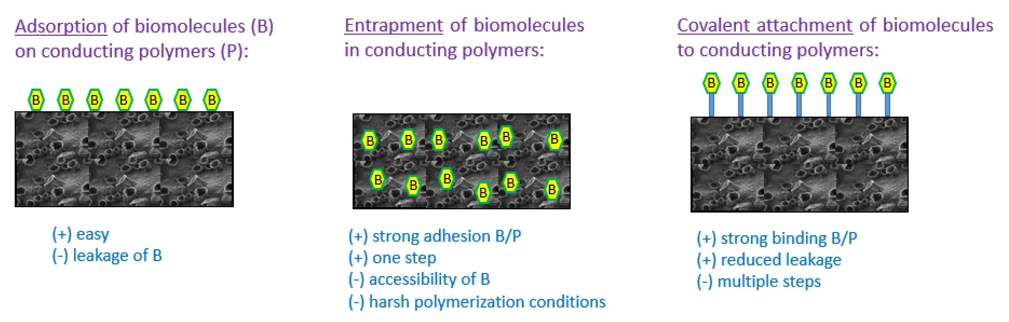
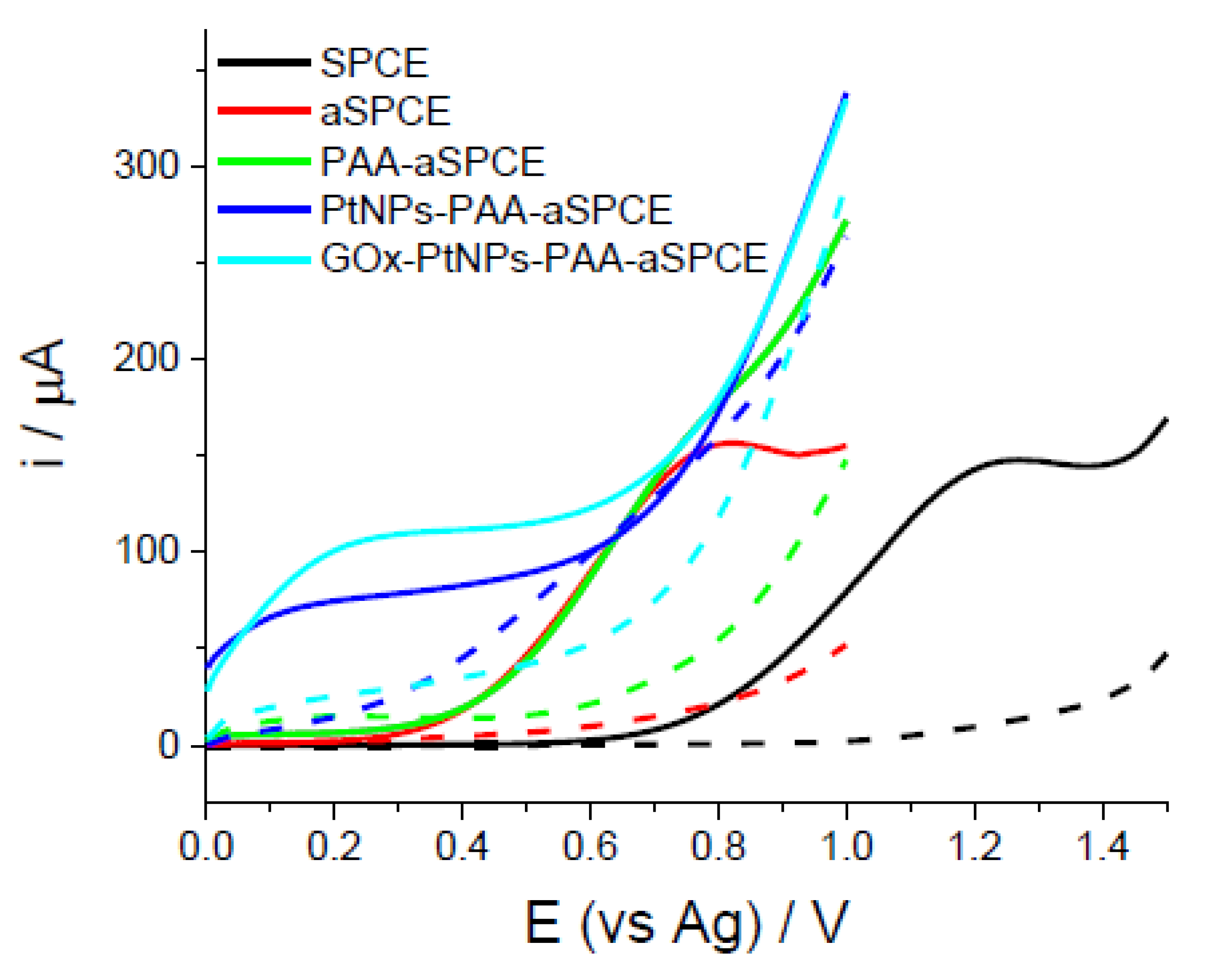
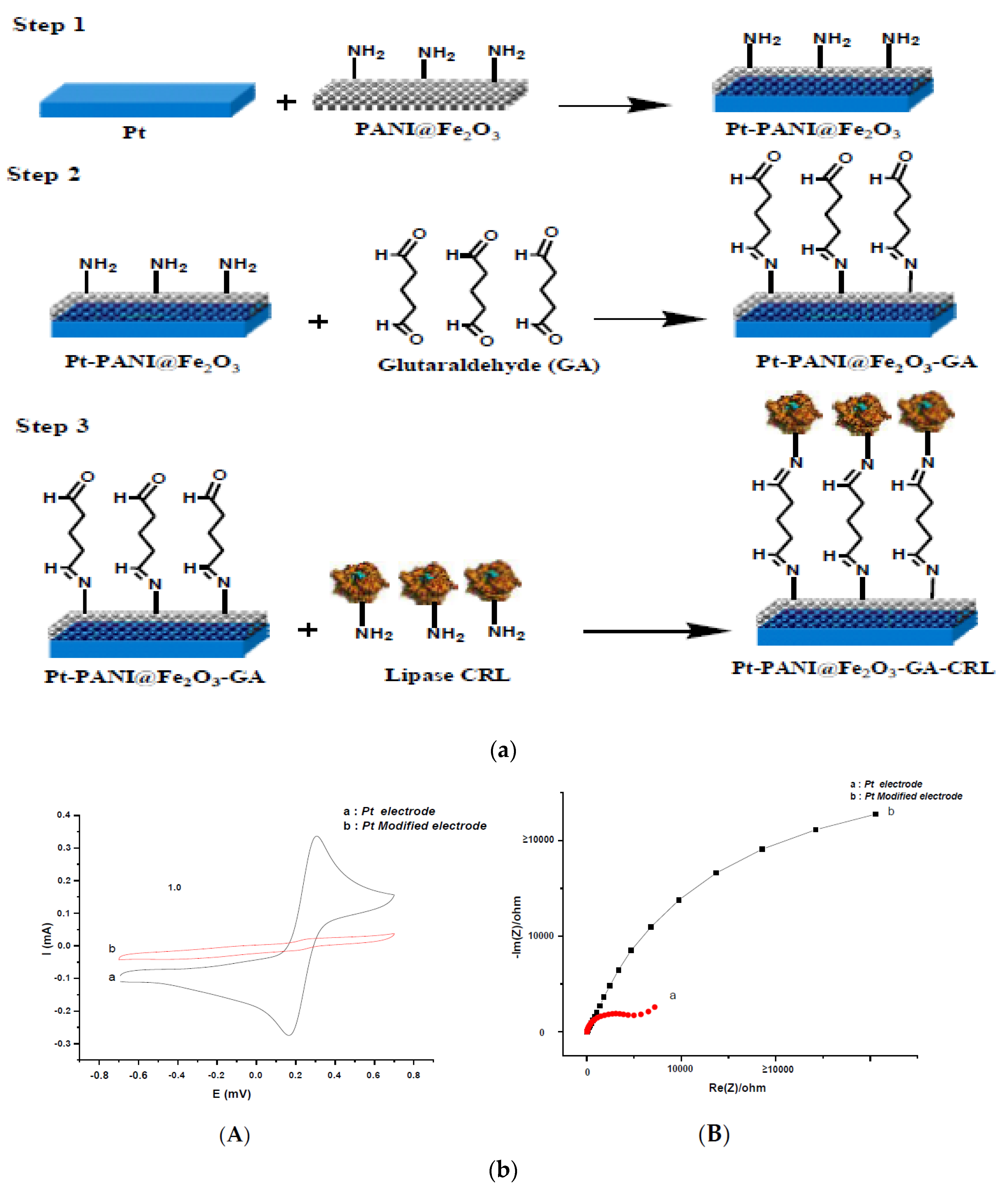
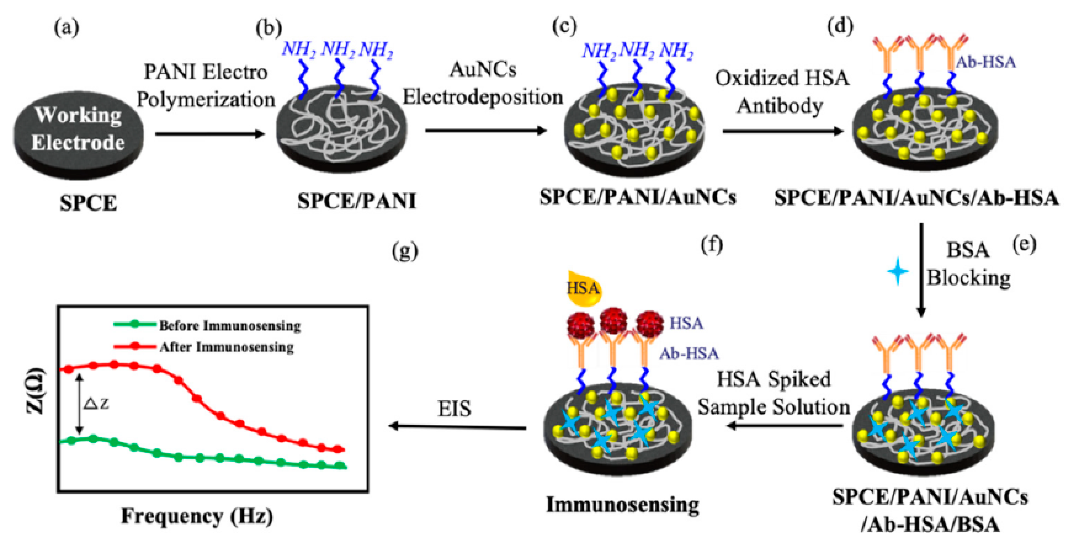
Table 1.
Chemical structures of some common conducting polymers: (a) polyacetylene, (b) polypyrrole, (c) polyaniline, (d) polycarbazole, (e) polythiophenes, (f) poly(3,4-ethylenedioxythiophene), (g) polyphenylenes, (h) poly(phenylene vinylene), (i) polyfluorene.
Table 1.
Chemical structures of some common conducting polymers: (a) polyacetylene, (b) polypyrrole, (c) polyaniline, (d) polycarbazole, (e) polythiophenes, (f) poly(3,4-ethylenedioxythiophene), (g) polyphenylenes, (h) poly(phenylene vinylene), (i) polyfluorene.
 |  |  |
| (a) | (b) | (c) |
 |  |  |
| (d) | (e) | (f) |
 |  |  |
| (g) | (h) | (i) |
Table 2.
Comparison of conducting-polymer-based glucose amperometric biosensor performances.
| Active Layer | Linear Range | Sensitivity | Detection Limit | Stability | Real Samples? | Ref. |
|---|---|---|---|---|---|---|
| Polypyrrole-CNT-chitosan | 1–4.7 mM | 2860 μA mM−1 cm−2 | 5.0 μM | 45 days | serum | [101] |
| Polypyrrole-CNT | 1–4.1 mM | 54.2 μA mM−1 cm−2 | 5.0 μM | 45 days | serum | [102] |
| Polyaniline-Prussian Blue | 2–1.6 MM | 99.4 μA mM−1 cm−2 | 0.4 μM. | 15 days | serum | [103] |
| Polyaniline-Au NP | 1–20 mM | 14.6 μA mM−1 cm−2 | 1.0 μM | --- | --- | [104] |
| Polyaniline-Pt NP | 0.01–8 mM | 96.1 μA mM−1 cm−2 | 0.7 μM | --- | --- | [106] |
| Polyaniline-CNT | 3–8.2 mM | 16.1 μA mM−1 cm−2 | 1.0 μM | 48 days | serum | [107] |
| Poly(Azure A)-Pt NP | 0.02–2.3 mM | 42.7μA mM−1 cm−2 | 7.6 μM | 3 month | fruit juice | [108] |
| Polyaniline-Graphene-NiO2 | 0.02–5.56 mM | 376.2 μA mM−1 cm−2 | 0.5 μM | --- | serum | [110] |
| Polyaniline | 0.01–5.5 mM | 97.2 μA mM−1 cm−2 | 0.3 μM | 15 days | urine | [111] |
| Polypyrrole-Pt NP | 0.1–9 mM | 34.7 μA mM−1 cm−2 | 27.7 μM | --- | --- | [112] |
| Polyaniline-nanodiamonds | 1–30 mM | 2.03 mA mM−1 cm−2 | 18.0 μM | 30 days | serum | [113] |
| Polycarbazole | 0.01–5 mM | 14.0 μA mM−1 cm−2 | 0.2 μM | --- | --- | [117] |
Table 3.
Comparison of conducting polymer-based immunosensor performances.
| Active Layer | Target | Detection Mode | Linear Range | Detection Limit | Ref. |
|---|---|---|---|---|---|
| Polyaniline-poly(vinylsulfonic acid) | atrazine | amperometry | 0.12–5 µM | 0.1 µg/L | [118] |
| Polythiophene derivative (with—COOH groups) | Carp vittelogenin | impedometry | 1–8 µg/L | 0.42 µg/L | [119] |
| Polypyrrole-polythionine | neuron-specific enolase | voltammetry | 0.001–100 pg/mL | 0.65 pg/mL | [120] |
| Polypyrrole-polythionine | carcinoma antigen-125 | 1–20 mM | 0.0001–1000 U/mL | 0.00125 U/mL | [121] |
| Polyaniline/Au nanocrystals | human serumalbumin | voltammetry + impedometry | 3–300 µg/mL | 3 µg/mL | [122] |
| Polyaniline-poly(sodium styrene sulfonate) | voltammetry | 0.01–1000 pg/L | 3.7 fg/mL | [123] |
Table 4.
Comparison of conducting polymer-based DNA biosensor performances.
| Active Layer | Detection Mode | Linear Range | Detection Limit | Ref. |
|---|---|---|---|---|
| Polyaniline–MoS2 | voltammetry | 10−15–10−6 M | 10−15 M | [124] |
| Polypyrrole–Au and Ag NPs | voltammetry | 7–150 nM | 7 nM | [125] |
| poly [3–acetic acid pyrrole,3–N–hydroxyphthalimide pyrrole)] | impedometry | 0.05–5.5 nM | 1 pM | [126] |
| Polypyrrole–Polyaniline– | impedometry | 10−13–10−6 M | 10−13 M | [127] |
| Polypyrrole–PEDOT–Ag NP | impedometry | 10−15–10−11 M | 5 × 10⁻¹⁵ M | [128] |
| Polypyrrole–CNT–COOH | impedometry | 10−12–10−7 M | 5 × 10⁻¹² M | [129] |
| Polyaniline | voltammetry | 10−15–10−12 M | 10−15 M | [130] |
| Polyaniline–methylene blue | voltammetry | 10−12–10−10 M | 10−12 M | [131] |
| Polyaniline–graphene | voltammetry | 10−13–10−7 M | 3 × 10⁻¹⁴ M | [132] |
| Polyaniline–graphene | voltammetry | 10−9–10−6 M | 8 × 10⁻⁷ M | [133] |
© 2020 by the author. Licensee MDPI, Basel, Switzerland. This article is an open access article distributed under the terms and conditions of the Creative Commons Attribution (CC BY) license (http://creativecommons.org/licenses/by/4.0/).
Share and Cite
MDPI and ACS Style
Lakard, B. Electrochemical Biosensors Based on Conducting Polymers: A Review. Appl. Sci. 2020, 10, 6614. https://doi.org/10.3390/app10186614
AMA Style
Lakard B. Electrochemical Biosensors Based on Conducting Polymers: A Review. Applied Sciences. 2020; 10(18):6614. https://doi.org/10.3390/app10186614
Chicago/Turabian StyleLakard, Boris. 2020. "Electrochemical Biosensors Based on Conducting Polymers: A Review" Applied Sciences 10, no. 18: 6614. https://doi.org/10.3390/app10186614
Note that from the first issue of 2016, this journal uses article numbers instead of page numbers. See further details here.