Article

RRM Prediction of Erythrocyte Band3 Protein as Alternative Receptor for SARS-CoV-2 Virus

1
AMALNA Consulting, Black Rock, Melbourne, VIC 3193, Australia
2
College of Science, Engineering and Health, RMIT University, Melbourne, VIC 3000, Australia
3
QuantBioRes A/S - QBR A/S, 2860 Copenhagen, Denmark
*
Author to whom correspondence should be addressed.
Appl. Sci. 2020, 10(11), 4053; https://doi.org/10.3390/app10114053
Submission received: 10 May 2020 / Revised: 5 June 2020 / Accepted: 10 June 2020 / Published: 11 June 2020

Abstract

:

Featured Application

The results of this research can be used for understanding how SARS-CoV-2 infects human cells, particularly in severe cases of COVID-19 and, consequently, this research can aid the development of new treatments.

Abstract

Severe acute respiratory syndrome coronavirus 2 (SARS-CoV-2) is a new coronavirus causing a worldwide pandemic. It is infecting respiratory organs and, in more severe cases, the lungs, where it is infecting the human cells through the angiotensin-converting enzyme 2 (ACE2) receptor. In severe cases, it is characterized not only by difficulties in breathing through infected lungs, but also with disproportionally and, thus far, unexplained low levels of oxygen in the blood. Here, we propose that, besides the infection of respiratory organs through ACE2 receptors, there is an additional infection in the red blood cells (erythrocytes). There could be a possible for SARS-CoV-2 to pass through the alveoli membrane in the lungs and infect the red blood cells through another receptor. Using our own biophysical model, the Resonant Recognition Model, we propose that the red blood cell (RBC) Band3 protein on the surface of red blood cells is a possible entry point for the SARS-CoV-2 virus into red blood cells.

1. Introduction

The current coronavirus disease 2019 (COVID-19) pandemic has caused havoc across the world, triggering a number of medical problems from mild symptoms to severe and critical symptoms, and even death. Furthermore, it has created huge economic problems due to worldwide lockdowns. The main problem is that this pandemic was caused by the new and, until now, unknown virus severe acute respiratory syndrome coronavirus 2 (SARS-CoV-2), which triggered fear regarding how this highly infectious virus will affect the human population. The SARS-CoV-2 virus belongs to the group of coronaviruses, all of which are RNA viruses. Coronaviruses are widely spread throughout nature, mostly infecting animals, but some infect humans as well, usually with mild or non-existent symptoms. However, there have been, thus far, three instances where coronavirus has infected humans, jumping from animals and causing severe symptoms, namely severe acute respiratory syndrome (SARS, 2003), Middle East respiratory syndrome (MERS, 2012) and COVID-19 (2019 novel coronavirus (2019-nCoV) or SARS-CoV-2, 2019). SARS and MERS had infected relatively limited areas of the population in China and the Middle East, respectively, and had been less infectious, but with a higher mortality rate, while the new coronavirus, SARS-CoV-2, is more infectious, infecting people across the world, but with a much lower mortality rate [1].
As SARS-CoV-2 is a completely new virus, causing a worldwide pandemic, there is an urgent need to investigate all aspects of SARS-CoV-2 virus infection and its activity. Bearing in mind that 80–90% of infected individuals have only mild or no symptoms, while the rest of infected individuals may present severe symptoms, which can even cause death, there is particular need to find out the difference between virus activity in mild versus severe cases. In severe cases, SARS-CoV-2 infects the lungs, but, apart from affecting lung function, it also extremely and dangerously lowers the oxygen level in the blood, disproportional to lung damage and the ability to breathe. In such cases, patients’ oxygen levels are falling dangerously low, while they are not experiencing the shortness of breath that usually signals the life-threating condition. This “silent hypoxia” is making some SARS-CoV-2 virus infected patients critically ill and, in some cases, is causing death [2].
Our research is specifically focused on investigating an anomaly of severe cases of COVID-19 infection, where the function of infected lungs is not severely impaired, but oxygen levels in the blood become dangerously low. Here, we have utilized our own biophysical Resonant Recognition Model (RRM) [3,4,5,6] to investigate the possibility that SARS-CoV-2 can not only infect lung cells, but can also pass through the alveoli membrane to infect red blood cells and consequently cause an unexpectedly disproportional drop in the oxygen levels in blood.

2. Methods and Materials

2.1. Methods-Resonant Recognition Model (RRM)

The Resonant Recognition Model (RRM) is a biophysical model that can analyze proteins and their DNA or RNA targets [3,4,5,6]. It is based on findings that certain periodicities (frequencies) in the distribution of free electron energy along proteins characterize their biological functions and interactions with their targets. Thus, by just using these RRM frequencies, it is possible to perform a detailed analysis of the interaction between proteins and their targets [3,4,5,6,7,8,9], without any analysis of their homology, 3D structures or complicated physical calculations. A full description of the Resonant Recognition Model (RRM) has been presented in number of our earlier publications and here we present an extract of the method description that is relevant to this paper [6].
“All proteins can be considered as a linear sequence of their constitutive elements, i.e., amino acids and biological function of proteins is determined primarily by this linear sequence. The RRM [3,4,5] interprets this linear information by transforming a protein sequence into a numerical series and then into the frequency domain using digital signal processing methods: The Fast Fourier Transform (FFT).
Protein primary structure can be presented as numerical series by assigning the relevant physical parameter value to each amino acid. Our investigations have shown that the best correlation can be achieved with parameters which are related to the energy of delocalized electrons of each amino acid, calculated as Electron Ion Interaction Potential (EIIP), as electrons delocalized from the particular amino acid, have the strongest impact on the electronic distribution of the whole protein [3,4,5]. The resulting numerical series represents the distribution of the free electrons energies along the protein molecule.
Such numerical series are then analyzed by digital signal analysis methods, using FFT, in order to extract information pertinent to the biological function. As the distance between amino acid residues in a polypeptide chain is 3.8A, it can be assumed that the points in the numerical sequence are equidistant. For further numerical analysis, the distance between points in these numerical sequences is set at an arbitrary value d = 1. Therefore, the maximum frequency in the spectrum is F = 1/2d = 0.5. The total number of points in the sequence influences the resolution of the spectrum only. Therefore, for N-point sequence the resolution in the spectrum is equal to 1/N. The n-th point in the spectral function corresponds to the frequency f = n/N.
In order to extract common spectral characteristics of sequences having the same or similar biological function, the cross-spectral function is used. Peak frequencies in the amplitude cross-spectral function define common frequency components of the two sequences analyzed. Peak frequencies in such a multiple cross-spectral function present common frequency component for all sequences analyzed. Such common frequency components are found to be related to the common biological function of the analyzed proteins leading to the conclusion that each specific biological function within the protein or DNA or RNA is characterized by one frequency [3,4,5,6,7,8,9,10].
Each biological function and/or process is driven by proteins that selectively interact with other proteins, DNA regulatory segments or small molecules. Using the RRM, it has been shown that proteins and their targets share the same, matching, characteristic frequency [3,4,5,6,7,8,9,10]. The matching of periodicities within the distribution of energies of free electrons along the interacting proteins can be regarded as the resonant recognition and is highly selective. Thus, the RRM frequencies characterize not only protein function, but also recognition and interaction between a protein and its targets: receptors, binding proteins, and inhibitors. In addition, it has been also shown that interacting proteins have opposite phases at their characteristic recognition frequency [3,4,5,6,7,10]. Every frequency can be presented by one sinusoid characterized with its frequency, amplitude and phase. The phase is presented in radians and can be between −π and +π (−3.14 and +3.14). The phase difference of or about 3.14 is considered opposite phase. The phase value can be presented in the phase circle where it is easier to observe graphically opposite phases.”
Once the characteristic biological function of the protein’s functional group has been identified, it is possible to design new proteins with the desired frequency components and, consequently, with the desired biological functions [3,4,5,6,11,12]. This approach has already been successfully applied and experimentally tested in design of the FGF analogue [3,4,5,13], HIV envelope protein analogue [3,4,5,14,15,16] and a peptide to mimic the oncolytic function of the myxoma virus [17,18].
The RRM concept has already been extensively published and is presented in detail within the Supplementary Materials.

2.2. Materials-Protein Sequences Analysed by RRM
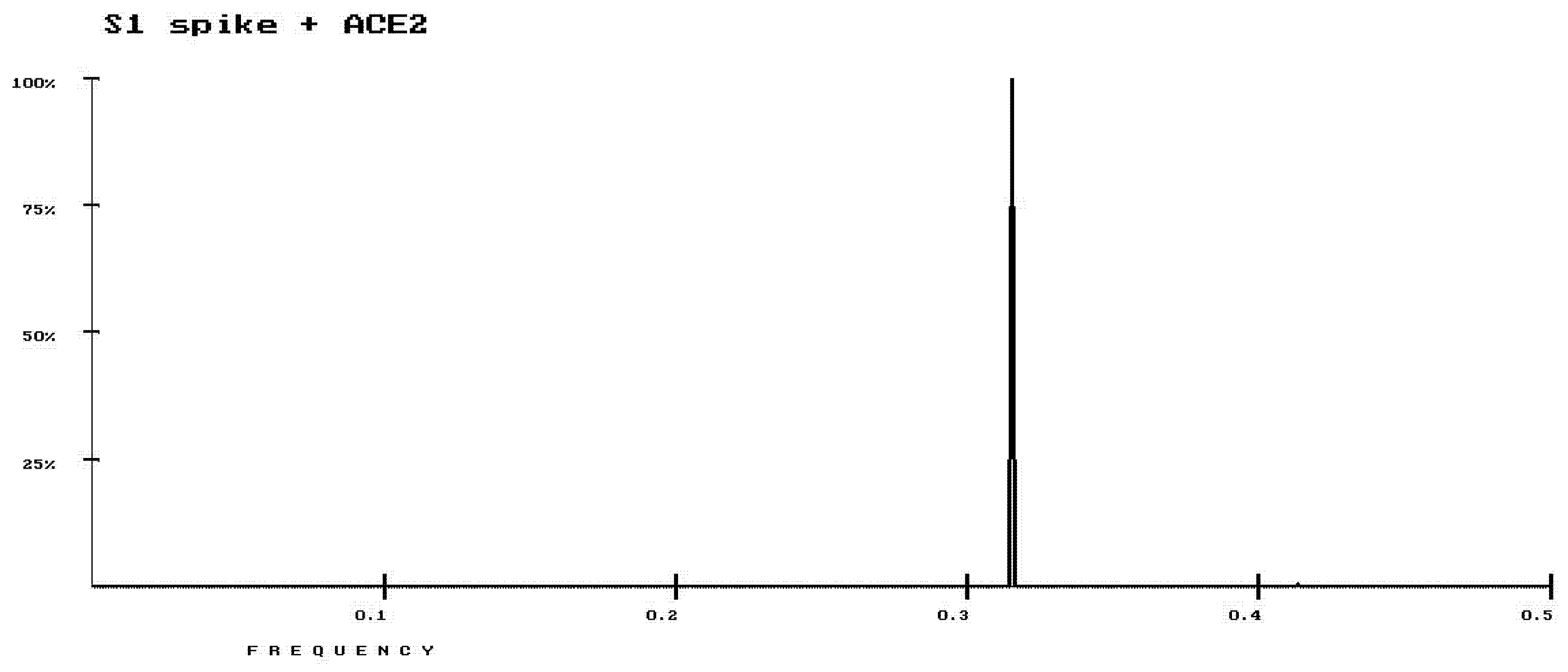
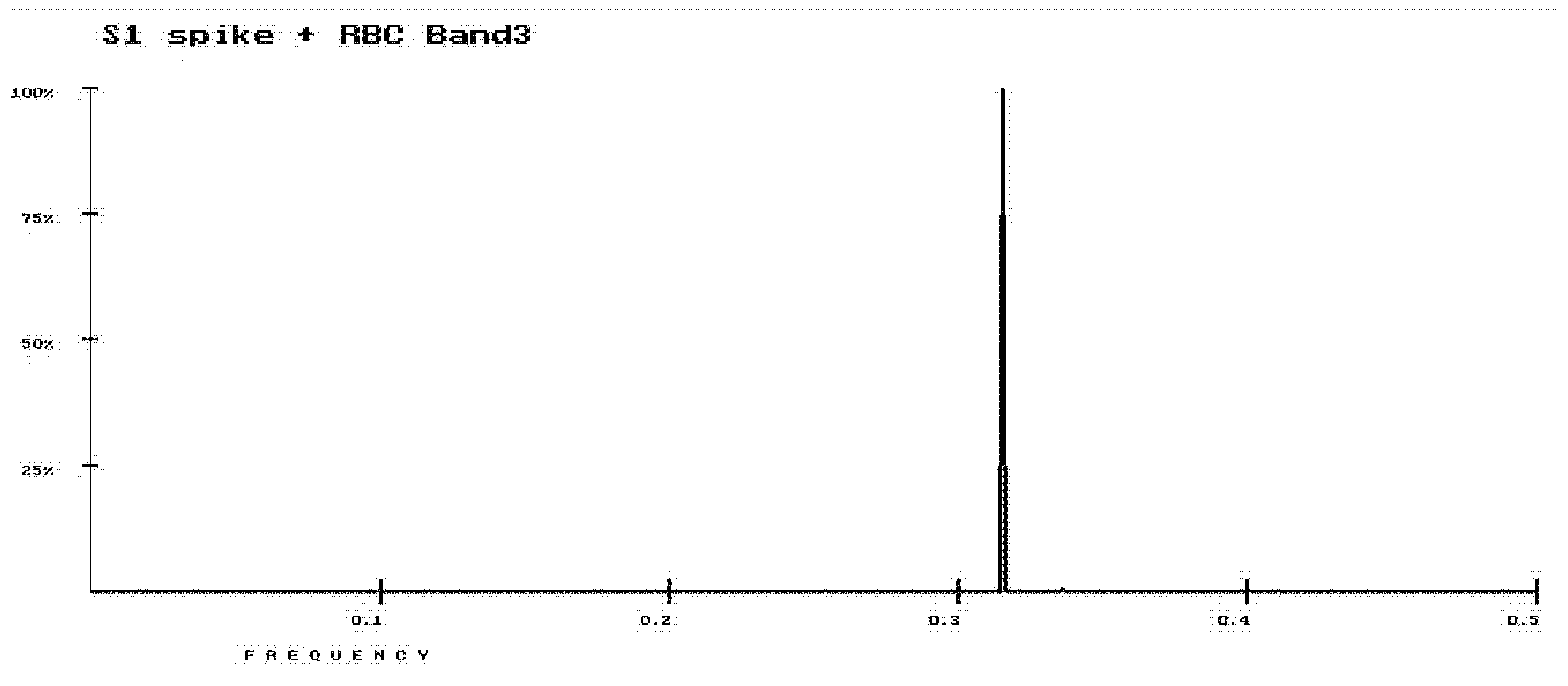
The following protein sequences from UniProt Database have been analyzed using the RRM:
Nine S1 spike proteins:
>active fragment s1 sp|P0DTC2|SPIKE_SARS2 Spike glycoprotein OS=Severe acute respiratory syndrome coronavirus 2 OX=2697049 GN=S PE=1 SV=1
>active fragment s1 sp|P15423|SPIKE_CVH22 Spike glycoprotein OS=Human coronavirus 229E OX=11137 GN=S PE=1 SV=1
>active fragment s1 sp|P36334|SPIKE_CVHOC Spike glycoprotein OS=Human coronavirus OC43 OX=31631 GN=S PE=3 SV=1
>active fragment s1 sp|Q6Q1S2|SPIKE_CVHNL Spike glycoprotein OS=Human coronavirus NL63 OX=277944 GN=S PE=1 SV=1
>active fragment s1 sp|P59594|SPIKE_CVHSA Spike glycoprotein OS=Human SARS coronavirus OX=694009 GN=S PE=1 SV=1
>active fragment s1 sp|K9N5Q8|SPIKE_CVEMC Spike glycoprotein OS=Middle East respiratory syndrome-related coronavirus (isolate United Kingdom/H123990006/2012) OX=1263720 GN=S PE=1 SV=1
>active fragment s1 sp|Q0ZME7|SPIKE_CVHN5 Spike glycoprotein OS=Human coronavirus HKU1 (isolate N5) OX=443241 GN=S PE=1 SV=1
>active fragment s1 sp|Q5MQD0|SPIKE_CVHN1 Spike glycoprotein OS=Human coronavirus HKU1 (isolate N1) OX=443239 GN=S PE=1 SV=1
>active fragment s1 sp|Q14EB0|SPIKE_CVHN2 Spike glycoprotein OS=Human coronavirus HKU1 (isolate N2) OX=443240 GN=S PE=3 SV=1
Ten ACE2 receptors:
>sp|Q5EGZ1|ACE2_RAT Angiotensin-converting enzyme 2 OS=Rattus norvegicus OX=10116 GN=Ace2 PE=1 SV=1
>sp|Q56NL1|ACE2_PAGLA Angiotensin-converting enzyme 2 OS=Paguma larvata OX=9675 GN=ACE2 PE=1 SV=1
>sp|Q9BYF1|ACE2_HUMAN Angiotensin-converting enzyme 2 OS=Homo sapiens OX=9606 GN=ACE2 PE=1 SV=2
>sp|Q8R0I0|ACE2_MOUSE Angiotensin-converting enzyme 2 OS=Mus musculus OX=10090 GN=Ace2 PE=1 SV=1
>sp|Q59RR0|ACE2_CANAL Cell wall transcription factor ACE2 OS=Candida albicans (strain SC5314 / ATCC MYA-2876) OX=237561 GN=ACE2 PE=4 SV=2
>sp|P21192|ACE2_YEAST Metallothionein expression activator OS=Saccharomyces cerevisiae (strain ATCC 204508 / S288c) OX=559292 GN=ACE2 PE=1 SV=1
>sp|Q56H28|ACE2_FELCA Angiotensin-converting enzyme 2 OS=Felis catus OX=9685 GN=ACE2 PE=2 SV=1
>sp|Q58DD0|ACE2_BOVIN Angiotensin-converting enzyme 2 OS=Bos taurus OX=9913 GN=ACE2 PE=2 SV=1
>sp|Q5RFN1|ACE2_PONAB Angiotensin-converting enzyme 2 OS=Pongo abelii OX=9601 GN=ACE2 PE=2 SV=1
>sp|Q6FJQ9|ACE2_CANGA Cell wall transcription factor ACE2 OS=Candida glabrata (strain ATCC 2001 / CBS 138 / JCM 3761 / NBRC 0622 / NRRL Y-65) OX=284593 GN=ACE2 PE=3 SV=1
Five RBC Band3 proteins:
>sp|P02730|B3AT_HUMAN Band3 anion transport protein OS=Homo sapiens GN=SLC4A1 PE=1 SV=3
>sp|P04919|B3AT_MOUSE Band3 anion transport protein OS=Mus musculus GN=Slc4a1 PE=1 SV=1
>sp|P23562|B3AT_RAT Band3 anion transport protein OS=Rattus norvegicus GN=Slc4a1 PE=2 SV=3
>sp|P32847|B3AT_ONCMY Band3 anion exchange protein OS=Oncorhynchus mykiss GN=slc4a1 PE=2 SV=2
>sp|P15575|B3AT_CHICK Band3 anion transport protein OS=Gallus gallus GN=SLC4A1 PE=2 SV=1

3. Results

Our focus here is to find out how SARS-CoV-2 infects different cell types. The main entry point into cells for some coronaviruses, including HCoV-NL63, SARS-CoV (the virus that causes SARS) [19] and SARS-CoV-2 (the virus that causes COVID-19) [20] is angiotensin-converting enzyme 2 (ACE2), which is attached to the outer surface of the cell membranes of cells in the lungs, arteries, heart, kidney and intestines. More specifically, the binding of the spike S1 protein of SARS-CoV and SARS-CoV-2 to the enzymatic domain of ACE2 on the surface of cells results in endocytosis and the translocation of both the virus and the enzyme into endosomes located within cells [21].
We have utilized the RRM model to analyze the interaction between spike S1 protein and the ACE2 receptor to find the RRM frequency characterizing this interaction. To achieve this, we have compared, using the RRM cross-spectra function, ACE2 receptors and S1 spike proteins from coronaviruses that are interacting with the ACE2 receptor, as listed in the Section 2.2. The prominent common characteristic frequency appears to be at an RRM frequency of 0.3145, as presented in Figure 1.
According to RRM principles, this frequency is characterizing the interaction between S1 spikes and ACE2 receptors. To confirm that the S1 spike from SARS-CoV-2 can interact with the human ACE2 receptor, we calculated phases for those two proteins at an RRM characteristic frequency of 0.3145 to find out if they are opposite, as required by RRM for successful interaction. The phase for S1 spike was found to be at −1.74rad, while the phase for human ACE2 receptor was found to be at +2.26rad, indicating that their phase difference is 2.28rad, which is empirically shown to be opposite enough to enable successful interaction [3,4,5,6].
To explain why severe cases of SARS-CoV-2 virus infection are causing extremely and dangerously low levels of oxygen in the blood in the absence of a lung infection, and where the patient’s ability to breathe is not proportionally affected, we propose that there may be another target receptor for SARS-CoV-2. Thus, we propose that SARS-CoV-2 can not only infect cells through the ACE2 receptor, but also through some other pathway. As oxygen is captured and transported by the red blood cells, it is logical to investigate the possibility that red blood cells are also infected by SARS-CoV-2, causing the capture of oxygen to be disturbed and explaining the disproportionally low level of oxygen in the blood (hypoxia) in severely ill patients. Red blood cells do not have ACE2 receptors on their surface and thus we propose that there might be another pathway on the surface of red blood cells that can interact with S1 spike proteins. One such possible pathway is through the interaction with the red blood cell (RBC) Band3 surface protein, which is a ubiquitous membrane transport protein found in the plasma membrane. It is the major integral transmembrane protein of the erythrocyte plasma membrane, comprising 25% of the total membrane proteins. Interestingly, it has been hypothesized that the main route of RBC invasion by merozoites from malaria parasites occurs through the RBC Band3-dependent pathway [10,22].
When we compared coronavirus S1 spike proteins to RBC Band3 surface proteins, using the RRM cross-spectra function, strikingly, we found the prominent common RRM characteristic frequency of 0.3145 was exactly the same as the frequency for the interaction between S1 spikes and ACE2 receptors, as presented in Figure 2.
This indicates not only the possibility that S1 spikes interact with RBC Band3 proteins, but also that this interaction uses the same mechanism as the interaction between S1 spikes and ACE2 receptors. If the interaction between S1 spikes and RBC Band3 surface proteins is possible, then it means that red blood cells can be infected by SARS-CoV-2 and thus the capture of oxygen could be affected, causing severely low levels of oxygen in the blood (hypoxia).
To confirm that the S1 spike from SARS-CoV-2 can interact with human RBC Band3 surface proteins, we calculated phases for those two proteins at an RRM characteristic frequency of 0.3145 to find out if they are opposite, as required by RRM for successful interaction. The phase for S1 spike was found to be at -1.74rad, while the phase for human RBC Band3 surface protein was found to be at +1.91rad, indicating that their phase difference is 2.43rad, which is empirically shown to be opposite enough to enable successful interaction.
The results above show that the RBC Band3 surface protein has the same RRM characteristics as the ACE2 protein regarding interaction with the S1 spike protein from coronaviruses. Thus, it can be considered as another pathway for SARS-CoV-2 virus to affect human cells. In addition, it has been shown that the human RBC Band3 surface protein has not only the same RRM frequency of 0.3145 as the human ACE2 receptor, but also that it has a very similar phase at this characteristic frequency, approximately opposite to the phase for the S1 spike from SARS-CoV-2, as presented in Figure 3 (within the phase circle).
These findings indicate, according to RRM principles, that the RBC Band3 surface protein is most probably another pathway for SARS-CoV-2 to enter into human cells.

4. Discussion

SARS-CoV-2, as a completely new virus that has caused a worldwide pandemic, is very infectious, but 80–90% of infected individuals have only mild or no symptoms. However, a small percentage of infected individuals experience severe symptoms, mostly through infected lungs, difficulties with their breathing and, more intriguingly, seriously low levels of oxygen in their blood. Here, we focused on an anomaly of severe cases of COVID-19 infection, where the function of infected lungs is not severely damaged, but the oxygen level in the blood becomes dangerously low. We propose that this anomaly is due to the possibility that SARS-CoV-2 virus not only infects lung cells, but can also pass through the alveoli membrane to infect red blood cells and consequently cause an unexpected disproportional drop in the oxygen levels in the blood. To find out if such red blood cell infection is possible, we utilized our own biophysical Resonant Recognition Model (RRM), which is able to analyze protein interactions with their targets, particularly receptors, by identifying matching periodicities (frequencies) in the distribution of free electron energies along the interacting proteins, characterizing their mutual recognition. Firstly, we analyzed the interaction between the S1 spike protein and the ACE2 receptor, as known receptors of SARS-CoV-2, and we identified the common RRM characteristic frequency of 0.3145. Secondly, we analyzed the interaction between S1 spike proteins and possible proteins on the surface of red blood cells. Strikingly, we found that the RBC Band3 surface proteins have the same RRM characteristics as ACE2 receptors when compared with S1 spike proteins from coronaviruses. In addition, it has been shown that the human RBC Band3 surface protein and the human ACE2 receptor have similar phases at this characteristic RRM frequency of 0.3145, both approximately opposite to the phase at the same RRM frequency for the S1 spike from SARS-CoV-2 (Figure 3). Thus, we propose that the RBC Band3 surface protein can also interact with the S1 spike protein from coronaviruses and therefore could be an entry point of SARS-CoV-2 into the red blood cells (erythrocytes). The hypothesis that the RBC Band3 surface protein can interact with SARS-CoV-2 could be of huge importance, as RBC Band3 protein integrity is mandatory for RBC physiology and, if disturbed, it can severely hamper RBC function, i.e., oxygen transport [23]. If the RBC oxygen transport function is disturbed by an interaction between the S1 fragment of SARS-CoV-2 and the RBC Band3 surface protein, it can cause severe hypoxia, as detected in severe cases of COVID-19. Thus, we propose that hypoxia in severe cases of COVID-19 is caused by SARS-CoV-2 interacting with the RBC Band3 surface protein and hampering RBC oxygen transport function. Our future research will be directed towards the use of the RRM protein design capability to design and test peptides that are able to interfere with and counteract interactions between the S1 fragment of SARS-CoV-2 and both ACE2 receptors and RBC Band3 surface proteins, using the identified RRM frequencies and phases.
It is interesting to note that the RBC Band3 protein has been already found to be an entry point for the malarial parasite agents merozoites from plasmodium falciparum into red blood cells [24]. We have already previously analyzed, using the RRM model, the interaction between merozoites from plasmodium falciparum and RBC Band3 proteins and identified the RRM characteristic frequency for this interaction [10]. However, the characteristic RRM frequency for malaria merozoites and the RBC Band3 protein is different from the RRM frequency found for SARS-CoV-2 and the RBC Band3 protein, indicating different mechanisms for these two interactions.
Bearing all of the above in mind, we propose here that the severity of symptoms in some COVID-19 infection cases, where the oxygen levels in the blood are drastically low, are due to the interaction of SARS-CoV-2 with the RBC Band3 protein on the red blood cell surface. This finding could be extremely important in adjusting the treatment of severe COVID-19 cases, where, instead of focusing on improving lung function only, the treatment should also be focused on blood cells—particularly red blood cells.

Supplementary Materials

The following are available online at https://www.mdpi.com/2076-3417/10/11/4053/s1, Resonant Recognition Model.

Author Contributions

Conceptualization, I.C.; methodology, I.C. and D.C.; software, D.C.; resources I.L.; writing—original draft preparation—review and editing, I.C., D.C. and I.L. All authors have read and agreed to the published version of the manuscript.

Funding

This research received no external funding.

Conflicts of Interest

The authors declare no conflict of interest.

References

  1. Makin, S. How Coronaviruses Cause Infection—from Colds to Deadly Pneumonia. Available online: https://www.scientificamerican.com/article/how-coronaviruses-cause-infection-from-colds-to-deadly-pneumonia1 (accessed on 5 February 2020).
  2. Couzin-Frankel, J. Why Don’t Some Coronavirus Patients Sense Their Alarmingly Low Oxygen Levels? Available online: https://www.sciencemag.org/news/2020/04/why-don-t-some-coronavirus-patients-sense-their-alarmingly-low-oxygen-levels (accessed on 28 April 2020).
  3. Cosic, I. Macromolecular Bioactivity: Is it Resonant Interaction between Macromolecules? -Theory and Applications. IEEE Trans Biomed. Eng. 1994, 41, 1101–1114. [Google Scholar] [CrossRef] [PubMed]
  4. Cosic, I. Virtual spectroscopy for fun and profit. Biotechnology 1995, 13, 236–238. [Google Scholar] [CrossRef] [PubMed]
  5. Cosic, I. The Resonant Recognition Model of Macromolecular Bioactivity: Theory and Applications; Birkhäuser Verlag: Basel, Switzerland, 2012. [Google Scholar]
  6. Cosic, I.; Cosic, D.; Lazar, K. Analysis of Tumor Necrosis Factor Function Using the Resonant Recognition Model. Cell Biochem. Biophys. 2016, 74, 175–180. [Google Scholar] [CrossRef] [PubMed]
  7. Cosic, I.; Paspaliaris, V.; Cosic, D. Analysis of Protein-Receptor on an Example of Leptin-Leptin Receptor Interaction Using the Resonant Recognition Model. Appl. Sci. 2019, 9, 5169. [Google Scholar] [CrossRef] [Green Version]
  8. Cosic, I.; Hearn, M.T.W. Studies on Protein-DNA Interactions Using the Resonant Recognition Model: Application to Repressors and Transforming Proteins. Eur. J. Biochem. 1992, 205, 613–619. [Google Scholar] [CrossRef] [PubMed]
  9. Cosic, I.; Hodder, A.N.; Aguilar, M.I.; Hearn, M.T.W. Resonant Recognition Model and protein topography: Model studies with myoglobin, haemoglobin and lysozyme. Eur. J. Biochem. 1991, 198, 113–119. [Google Scholar] [CrossRef] [PubMed]
  10. Caceres, J.L.H.; Cosic, I.; Cosic, D. Application of the Resonant Recognition Model to the Study of Plasmodium Proteins Involved in Malaria Infection. MD Med. Data 2015, 7, 7–14. [Google Scholar]
  11. Pirogova, E.; Istivan, T.; Gan, E.; Cosic, I. Advances in methods for therapeutic peptide discovery, design and development. Curr. Pharm. Biotechnol. 2011, 12, 1117–1127. [Google Scholar] [CrossRef] [PubMed]
  12. Cosic, I.; Pirogova, E. Bioactive Peptide Design using the Resonant Recognition Model. Nonlinear Biomed. Phys. 2007, 1, 7. [Google Scholar] [CrossRef] [PubMed]
  13. Cosic, I.; Drummond, A.E.; Underwood, J.R.; Hearn, M.T.W. In vitro inhibition of the actions of basic FGF by novel 16 amino acid peptides. Mol. Cell. Biochem. 1994, 130, 1–9. [Google Scholar] [CrossRef] [PubMed]
  14. Krsmanovic, V.; Biquard, J.M.; Sikorska-Walker, M.; Cosic, I.; Desgranges, C.; Trabaud, M.A.; Whitfield, J.F.; Durkin, J.P.; Achour, A.; Hearn, M.T. Investigation Into the Cross-reactivity of Rabbit Antibodies Raised against Nonhomologous Pairs of Synthetic Peptides Derived from HIV-1 gp120 proteins. J. Peptide. Res. 1998, 52, 410–412. [Google Scholar] [CrossRef] [PubMed]
  15. Krsmanovic, V.; Cosic, I.; Biquard, J.M.; Hearn, M.T.W. Peptides immunologically related to known viral protein. U.S. Patent No. 6,294,174, 25 September 2001. [Google Scholar]
  16. Achour, A.; Biquard, J.M.; Krsmanovic, V.; M’Bika, J.P.; Ficheux, D.; Sikorska, M.; Cozzone, A.J. Induction of Human Immunodeficiency Virus (HIV-1) Envelope Specific Cell-Mediated Immunity by a Non-Homologus Synthetic Peptide. PLoS ONE 2007, 2, e1214. [Google Scholar] [CrossRef] [PubMed]
  17. Almansour, N.; Pirogova, E.; Coloe, P.; Cosic, I.; Istivan, T. Investigation of cytotoxicity of negative control peptides versus bioactive peptides on skin cancer and normal cells: A comparative study. Future Med. Chem. 2012, 4, 1553–1565. [Google Scholar] [CrossRef] [PubMed]
  18. Istivan, T.; Pirogova, E.; Gan, E.; Almansour, N.; Coloe, P.; Cosic, I. Biological effects of a De Novo designed myxoma virus peptide analogue: Evaluation of cytotoxicity on tumor cells. PLoS ONE 2011, 6, e24809. [Google Scholar] [CrossRef] [PubMed] [Green Version]
  19. Li, F. Receptor recognition and cross-species infections of SARS coronavirus. Antivir. Res. 2013, 100, 246–254. [Google Scholar] [CrossRef] [PubMed]
  20. Xu, X.; Chen, P.; Wang, J.; Feng, J.; Zhou, H.; Li, X. Evolution of the novel coronavirus from the ongoing Wuhan outbreak and modeling of its spike protein for risk of human transmission. Sci. China Life Sci. 2020, 63, 457–460. [Google Scholar] [CrossRef] [PubMed] [Green Version]
  21. Millet, J.K.; Whittaker, G.R. Physiological and molecular triggers for SARS-CoV membrane fusion and entry into host cells. Virology 2018, 517, 3–8. [Google Scholar] [CrossRef] [PubMed]
  22. Li, X.; Chen, H.; Oh, S.S.; Chishti, A.H. A Presenilin-like protease associated with Plasmodium falciparum micronemes is involved in erythrocyte invasion. Mol. Biochem. Parasitol. 2008, 158, 22–31. [Google Scholar] [CrossRef] [PubMed] [Green Version]
  23. Gallagher, P.G. Hemolytic anemias: Red blood cell membrane and metabolic defects. Available online: https://www.semanticscholar.org/paper/HEMOLYTIC-ANEMIAS-%3A-RED-BLOOD-CELL-MEMBRANE-AND-Gallagher/0f01efe030314c2fadd42d007f0683721f854156 (accessed on 5 February 2020).
  24. Perkins, M.E. Binding of glycophorins to Plasmodium falciparum merozoites. Mol. Biochem. Parasitol. 1984, 10, 67–78. [Google Scholar] [CrossRef]
Figure 1. Resonant Recognition Model (RRM) cross-spectrum for interaction between S1 spike proteins and angiotensin-converting enzyme 2 (ACE2) receptors with common RRM characteristic frequency of 0.3145.
Applsci 10 04053 g001
Figure 2. RRM cross-spectrum for interaction between S1 spike proteins and red blood cell (RBC) Band3 surface proteins with common RRM characteristic frequency of 0.3145.
Applsci 10 04053 g002
Figure 3. Phase circles at frequency of 0.3145 for S1 spike from severe acute respiratory syndrome coronavirus 2 (SARS-CoV-2) with phase of −1.7rad in green, for human ACE2 receptor with phase of +2.26rad in blue and for human RBC Band3 surface protein with phase of +1.91rad in red. It can be easily observed that both receptor phases are almost opposite to the S1 spike phase, supporting the RRM approach that proteins and protein receptors should have opposite phases at frequencies characterizing their recognition and interaction.
Applsci 10 04053 g003

