Article

Uncovering the Hidden Dangers of Microplastic Pollution in Lake Ecosystems: Effects of Ingestion on Talitrid Amphipods

1
ENEA, Italian National Agency for New Technologies, Energy and Sustainable Economic Development, C.R. Casaccia, Via Anguillarese 301, 00123 Rome, Italy
2
Department of Biology and Biotechnologies ‘Charles Darwin’, Sapienza University of Rome, Piazzale Aldo Moro 5, 00185 Rome, Italy
*
Author to whom correspondence should be addressed.
Environments 2023, 10(7), 115; https://doi.org/10.3390/environments10070115
Submission received: 31 May 2023 / Revised: 23 June 2023 / Accepted: 29 June 2023 / Published: 4 July 2023

Abstract

:
Microplastic (MP) contamination is a globally recognised issue in aquatic environments, and recently, there has been an increase in investigations focusing on lake contamination, revealing significant amounts of dispersed MPs. However, our understanding of the ingestion and effects of MPs on organisms living in lake ecosystems remains limited. This study aims to develop an effective protocol for assessing the ingestion of MPs by the talitrid amphipod Cryptorchestia garbinii, with the goal of verifying and evaluating the biological effects following ingestion. Individuals sampled from the shores of Lake Albano were exposed to four different polymers, namely low-density polyethylene (PE), polyethylene terephthalate (PET), polyester (PES), and polypropylene (PP), under laboratory conditions. To deliver MPs through the diet, we decided to employ DECOTABs (DEcomposition and COnsumption TABlets) which have been successfully used as a food source in aquatic toxicity tests. At the end of the experiments, we employed the solvatochromic and fluorescent dye Nile red to detect and quantify the MPs present in the digestive tube contents of the animals. The results clearly demonstrate the ingestion of the supplied polymers through the tabs, validating this method of exposure as effective. Furthermore, the measurement of glucose, glycogen, and lipid levels reveals that within 24 h of ingestion, MPs had an impact on the macromolecules involved in the energy metabolism of C. garbinii. This research underscores the suitability of this species as a model organism for studying MP uptake and its effects.

Graphical Abstract

1. Introduction

Plastic debris has become a pervasive global pollutant, contaminating all habitats worldwide. Over the past decade, it has become a prevalent international concern [1]. With a continuous increase in the global production and consumption of plastic, reaching 390.7 million tonnes in 2022, plastic waste generation is constantly on the rise [2]. While recycling is the preferred solution for plastic waste, only 8.3% of all post-consumer plastic is recycled. The remaining plastic waste is partially used for energy recovery, but a significant portion ends up in landfills, ultimately finding its way into the environment [1]. Plastic litter encompasses items that result from the breakdown of larger plastic items, spanning a broad range of sizes. This includes large and easily removable objects, as well as small or invisible items, referred to as ‘microplastics (MPs)’ when smaller than 5 mm [3]. Both primary MPs (dispersed in the environment in the form of small particles, including microbeads present in personal care products, textile microfibres, erosion of tyre rubber, road markings, or marine coatings) and secondary MPs (originating from the fragmentation of larger plastic items) have contaminated all water and terrestrial environments, from deep-sea to polar areas, posing a threat to organisms that can mistake them for food and ingest them [4,5,6,7]. Such ingestion can have detrimental effects on individual organisms and can enter the food chain, impacting entire associated food webs [8].
Amphipods are excellent indicators for biomonitoring and ecotoxicological studies of environmental contaminants due to their widespread distribution, ease of collection, and significant role in nutrient cycling [9]. The talitrid species, in particular, plays a crucial role in the energy transfer of sandy shore ecosystems, as already stated by Griffiths et al. [10]. These organisms are among the dominant macrofaunal groups on sandy beaches and contribute significantly to the ecosystem by feeding on both terrestrial and aquatic matter [11]. Consequently, they assimilate various sources of pollution and serve as a significant food source for numerous invertebrates, fish, and birds, as indicated by several researchers [12,13]. These semi-terrestrial crustaceans thrive in humid or water-saturated substrates and represent one of the most abundant taxa in temperate regions [9,14]. Talitrid amphipods have been extensively studied in terms of their ecology and behaviour, making them a potential choice as bioindicators of environmental disturbance, due to their gene flow, phylogeography, and population features, including life history and behavioural traits [15].
The amphipod species Cryptorchestia garbinii, described by Ruffo et al., 2014 (Figure 1a,b), resides in the supralittoral zone of fresh and brackish waters found in the Mediterranean Basin, Central Europe, and England [16]. It has been utilised in various studies as a model species for cellular differentiation and gene expression, as pointed out by Davolos et al. [17]. As a detritivorous species, similar to other talitrids, C. garbinii feeds on organic debris and has the capacity to ingest small fragments of plastic present in the environment. Previous research conducted by Iannilli et al. [18] and Battistin et al. [19] has demonstrated this ability of plastic ingestion by the species.
MPs are pervasive in lake environments, with lakes being more susceptible to MP contamination compared to oceanic and coastal regions [20]. These systems are strongly influenced by both terrestrial and aquatic sources [21,22]. The characteristics of lentic water bodies and long water-residence times contribute to the accumulation of MP contaminants in these environments, making them a sink for MP pollutants [23]. Despite the increasing focus on lake MPs, studies have primarily concentrated on assessing their occurrence characteristics, while fewer studies have investigated the biological effects of MP contamination on organisms [24]. Moreover, research efforts have primarily concentrated on studying MPs with smooth edges and spherical shapes, which account for less than 1% of the total MPs found in nature. Conversely, studies regarding fragment-type MPs are notably scarce and are still in early stages of development [25].
Exposure to plastic polymers in water can pose operational challenges due to the characteristics of the material. Some polymers have low density, causing them to float on the water’s surface, while others may stick to the walls of containers due to electrostatic reasons. To overcome this issue, MP particles were therefore incorporated into special tabs and used as food sources for the animals. DECOTABs (DEcomposition and COnsumption TABlets) [26] have been employed in previous studies for incorporating MP particles into the diet of organisms, such as gammarids, as suggested by Götz et al. [27]. These tabs have been successfully used to deliver food supplements or chemicals in toxicity test procedures with shredder and grazer species [27]. Due to their stability in water and particle distribution, DECOTABs have been proven to be highly suitable for dietary exposure to MPs.
To advance our understanding of C. garbinii, a highly promising candidate species for MP studies, we investigated the uptake of MP particles under laboratory conditions and evaluated the resulting biological effects, specifically focusing on energy reserve changes. To achieve this, we conducted laboratory exposures using four different types of MP particles derived from commonly used objects.

2. Methods

Samples of C. garbinii were collected from Lake Albano shores using an aspirator or by hand, and immediately transferred to temperature-controlled conditions in the laboratory. They were kept in a glass tank in a thermostatic cabinet at 20 °C with a 12 h/12 h photoperiod, taking care to maintain humidity by adding water and only feeding them with fish food and pieces of blotting paper, due to their preference for cellulose-rich material [28]. Soil taken from the survey site was used as the substrate. The specimens were acclimated in the lab for three weeks before exposing them to microplastics.

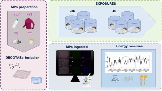
2.1. Preparation of MPs

C. garbinii specimens were fed with DECOTABs prepared in the laboratory and which contained four different types of polymers to verify the possible ingestion of MPs in laboratory conditions: low-density polyethylene (PE) particles smaller than 100 µm, polypropylene (PP) and polyethylene terephthalate (PET) particles below 200 µm in size, and polyester (PES) fibres with a maximum length of 1.5 mm (Figure 2, Table S1). The selection of these microplastic types is based on several factors: PE, PP, PET, and PES are widely used in various consumer products and packaging materials. They represent some of the most common types of plastics found in the environment due to their widespread use and disposal. Moreover, they are frequently found in aquatic environments, including oceans, lakes, and rivers. By studying these commonly encountered microplastics, we can better understand their potential ecological impacts.
PE microparticles had already been obtained in granular form (irregularly shaped). To produce particles of the other polymers tested, we used commonly used objects with clearly stated polymeric compositions. A lanyard was used to obtain PP microparticles. Following the approach described by Cole et al. [29] in their MP studies, the lanyard was incorporated into the Cryo-M-Bed compound, typically used for tissue inclusion and freezing in histological analysis. The coated wire was frozen, ensuring that the fibres remained aligned within a compact block. Subsequently, the block was cut at 20 µm intervals using a cryostat microtome (CM1520, Leica Biosystems, Nussloch, Germany). The resulting sections were rinsed multiple times with distilled water to remove the freezing-solution coating and left in water until completely evaporated to recover only the PP particles.
A plastic bottle was used for PET particles and the surface was shredded using a rotating steel brush. PES fibres were obtained from a 100% PES fleece sweater. Steel razors were used to cut the fibres, followed by further sectioning with a scalpel.
Before exposure, all the obtained MPs were carefully examined under bright-field and fluorescence microscopy (Leica DM750, Leica Microsystems, Wetzlar, Germany), photographed with a digital HD camera (Leica ICC50HD, Leica Microsystems, Wetzlar, Germany), and documented to ensure easy recognition during subsequent analyses (Figure S1).

2.2. Food Preparation

The tabs for microplastic inclusion were made following the procedure described by Straub et al. [30] for DECOTABS (DEcomposition and COnsumption TABlets, [1]), modified according to the needs of this study. Agar 4% and L-ascorbic acid 100 mg/L were hot melted in a beaker with 50 mL of distilled water and, after cooling below 50 °C, microplastic was added to reach a concentration of 280 mg/L. Tabs were made for each polymer used for testing. Afterwards, the solution was transferred and solidified on a 96-cell multi-well plate. For each well, 200 µL of the MP suspension was transferred and allowed to cool under a sterile hood until solidification, thus obtaining the tablets. We performed a numerical estimate of the MP density in the tabs for each polymer used. For this purpose, a section of a tab was weighted, pressed on a microscope slide, and observed. The number of MP per milligram of tab was therefore obtained.

2.3. Exposure Conditions

Ingestion. Two exposure experiments were conducted, one lasting 24 h and the other 48 h. In each experiment, five individuals were individually exposed in separate glass containers with one tab containing MP particles (Figure 3). Additionally, an equal number of control samples with tabs without MPs were included for comparison. The entire experiment was replicated twice, resulting in a total of 50 specimens analysed. Each specimen was placed in a transparent glass beaker containing 100 g of 1 mm diameter glass beads to simulate sediment. To maintain a wet environment, 15 mL of water was added to each beaker along with the respective tab containing MP particles. In order to ensure that the specimens were not stressed or forced to feed, they were not fasted prior to the exposures. The glass containers were then placed in a thermostatic cabinet set at 20 °C and a 12/12 photoperiod for the duration of the experiments. At the end of each exposure period, the individuals were preserved in 80% ethanol. The digestive tube of each specimen was carefully dissected under a stereomicroscope (Leica M80, Leica Microsystems, Wetzlar, Germany) and transferred to a tube containing 1 mL of 30% hydrogen peroxide to degrade the organic matter. The samples were left at room temperature for the next 7 days for further processing and analysis.
Subsequently, the digested material obtained from 5 animals was filtered using one black polycarbonate membrane (Cyclopore Track Etched, 0.4 µ, 25 mm, Whatman™), following the method described by Iannilli et al. 2019, 2020. The filters were then washed with 100 µL of n-hexane under a fume hood to remove any lipid residues, placed on a slide, allowed to dry, and subsequently stained with 100 µL of Nile red at a concentration of 5 mg/L in n-hexane. After staining, the filters were examined using a fluorescence microscope (Leica DM750 equipped with a Digital HD camera Leica ICC50HD, Leica Microsystems, Wetzlar, Germany) in green (excitation wavelength at 450–490 nm and emission wavelength 515−565 nm) and in blue (excitation at 365 nm and 445 nm emission). The microplastic particles highlighted were photographed and measured using the Leica Application Suite, version 49 software. Three negative procedural control samples were processed with the same protocol to check for any MP contamination during the analysis, and no microplastics were observed.
Glucose, glycogen, and lipid assessment. In order to evaluate the impact of microplastic (MP) ingestion on metabolic processes, an analysis of energy reserves, including glucose, glycogen, and lipids, was conducted after the exposure period. The experimental setup for the ingestion experiments was as previously described, with 5 individuals exposed individually to each type of microplastic for both 24 h and 48 h durations. The control specimens were exposed to MP-free tabs. At the conclusion of the exposure period, each specimen was weighed and the energy reserves were assessed following the protocol of van Hendel [31,32], as revisited by Foray et al. [33]. This analysis was carried out twice, resulting in a total of 50 specimens being analysed for their energy reserve levels.
The specimens were individually homogenized using a Potter–Elvehjem tissue homogenizer (Merck KGaA, Darmstadt, Germany) with 200 μL of ultrapure water. The homogenate was then centrifuged at 300× g for 2 min. From the resulting supernatant, 100 μL was transferred to a new tube and mixed with 50 μL of 20% sodium sulphate solution and 1.2 mL of chloroform:methanol solution in a 1:1 ratio. After centrifugation at 3000 rpm for 2 min, the glycogen precipitated into a pellet. The supernatant was transferred to another test tube and 300 μL of distilled water was added. Following centrifugation at 3000 rpm for 2 min, two distinct phases formed: the lower denser phase containing the lipid fraction, and the upper phase containing the sugars. For the glucose analysis, the fraction containing glucose was reduced to a volume of 100 μL using a thermoblock at 90 °C. Then, 1.5 mL of an anthrone solution (0.5% in 70% H2SO4) was added, and the samples were kept at 90 °C for 17 min. A glucose solution with known concentrations (0, 1, 3, 5, 10, and 20 μL of a 0.1% solution in water) was prepared as the standard and also used for glycogen analysis. The absorbance of the samples was measured at 625 nm using a photometer (Hach Lange DR1900, Hach Company, Loveland, Colorado) after cooling to room temperature. For the glycogen analysis, the pellet containing glycogen was dissolved in 1.5 mL of the anthrone solution and kept at 90 °C for 17 min. The absorbance was then determined at a wavelength of 625 nm.
For the lipid analysis, the fraction was evaporated to a volume of 100 μL at 90 °C under a fume hood. Then, 100 μL of H2SO4 was added and the mixture was maintained at 90 °C for 13 min. The same amount of sulfuric acid was added to solutions containing 0, 1, 3, 5, 10, and 20 μL of a 0.1% oil solution in chloroform to obtain the standards for the calibration curve. After cooling the samples to room temperature, 1000 μL of a vanillin solution (0.1% vanillin in 64% H3PO4) was added. The tubes were kept at room temperature for 5 min and then the absorbance was measured at 525 nm.

2.4. Statistical Analysis

Analyses were performed to determine the significance of differences between the values of the energy reserves after the exposures and the untreated samples, and between the two exposure times; p-values below a confidence level of 0.05 were considered statistically significant.
After checking for the data normal distribution, t-tests were carried out on pairs of groups of values resulting from the dosages of the energy reserves. When the data did not follow a normal distribution, the Mann–Whitney test was used.
The data analysis was carried out using Past software, Paleontological Statistics, Version 3.26.

3. Results and Discussions

This study aimed to investigate the ingestion of MPs by C. garbinii under laboratory conditions and assess their metabolic effects using an effective dietary-exposure protocol. Low-density PE, PET, PES, and PP were incorporated into DECOTABs. As C. garbinii is a semiterrestrial species abundant along lake shores in close proximity to water, it serves as an important food source for vertebrates, particularly birds, that may be found in this environment. Therefore, it constitutes an entry point for plastics into the trophic chain and a potential pathway for the transfer of MPs from aquatic to terrestrial organisms. The density of the MPs in the DECOTABs prepared to feed the amphipods yielded the following results: 1.32 PES fibres/mgTab, 25.82 PP MPs/mgTab, 34.03 PE MPs/mgTab, and 68.13 PET MPs/mgTab. Using the Nile red staining method, we confirmed the presence of MPs in all of the tested specimens. Through fluorescence observations, MP fragments were identified and counted in the stomach contents (Table 1). To ensure that only the MPs provided to the animals during the exposure were considered, a visual comparison was made between the MPs in the tabs and the particles found in the digestive tract based on shape and size, using both bright-field and green and blue fluorescence. Characteristic patterns emerged for each polymer: PE exhibited green and light-blue fluorescence, PET showed green and pink fluorescence, and PP displayed green and purple fluorescence (Figure S1). The green fluorescence of PE and PP was also observed by Stanton et al. [34]. However, PES did not exhibit fluorescent staining with Nile red, as reported by Stanton et al. [34]. Nevertheless, the PES fibres could still be identified based on their shape and colour. The fibres appeared green under green wavelengths, as observed by Stanton et al. [34], and light-blue under blue wavelengths, consistent with the findings of Shim et al. [35].
We observed that the C. garbinii ingested all the supplied polymers through the tabs, both in the 24 h and 48 h exposures. The number of MPs we found in the digestive tract was comparable to those observed in specimens collected in natural conditions from Lake Garda (7.13 ± 2.11), Lake Bracciano (5 ± 4.6), and Lake Albano (2.2 ± 1.8). After 24 h of exposure, we recorded the ingestion of 4.5 ± 0.7 MP particles of PE, 3.3 ± 0.7 of PET, 1.8 ± 0.4 of PP, and 8 ± 2.4 fibres of PES. This number appeared to increase for all the polymers with longer exposure times (Table 1). However, the opposite trend was observed for PES, although it was not statistically significant. PES, supplied in the form of coloured fibres (rather than as particulates and transparent, as in the case of the other polymers), was more present in the digestive tracts of animals exposed for 24 h compared to those exposed for 48 h. This could be attributed to the initial attractiveness of the colour, which entices the animals to feed, but the taste might later discourage them from further ingestion.
Previous studies have shown that the characteristics of MPs, such as polymer composition, shape, and size, can significantly influence their uptake [36]. For example, research on the wild fish species Psalidodon eigenmanniorum showed that yellow and blue fragments were the most consumed, indicating the visual attraction of colours [37]. In addition, taste and food preferences can also play a role in ingestion. The essential role of taste has been recognised in zooplankton [38,39,40], particularly when comparing feeding studies involving synthetic microbeads and labelled bacteria or algae [40,41,42]. DeMott [38] observed significant differences in feeding rates on flavoured and unflavoured polymeric particles among rotifers, Bosmina (cladocerans), and copepods (calanoids and cyclopoids). Bosmina and the rotifer Filinia terminalis showed a preference for aromatised algal spheres over untreated ones, unlike Daphnia magna and Brachionus calyciflorus, which did not exhibit the same preference. The degree of selectivity was even greater in experiments with copepods, particularly calanoid (e.g., Diaptomus siciloides) and cyclopoid species (e.g., Cyclops bicuspidatus thomasi), which strongly avoided unflavoured polymeric spheres [38].
A post-exposure dosage was given to evaluate the ingestion of MPs and their effects on the energy reserves of C. garbinii. Notably, a previous study by Straub et al. [30] used similar tabs to incorporate biodegradable polyhydroxybutyrate (PHB) and conventional petroleum-based polymethylmethacrylate (PMMA) as food for Gammarus fossarum, resulting in the ingestion of both polymers. Other studies have demonstrated the ingestion of MPs by other amphipods in laboratory conditions. For instance, Blarer and Burkhardt-Holm [43] demonstrated the ingestion of polyamide (PA) fibres (20 μm) and polystyrene (PS) microbeads (1.6 μm in diameter) in lab conditions by the same species. Scherer et al. [40] observed that G. pulex also ingested PS microbeads of varying sizes (1, 10, and 90 μm). Furthermore, Hyalella azteca individuals were found to ingest MPs during 10-day exposures to PE particles (10–27 μm in diameter) and PP fibres (20 μm–75 μm in length and 20 μm in diameter). Recent research by Iannilli et al. [8] demonstrated the ingestion of MPs by Echinogammarus veneris in laboratory conditions when the organisms were exposed to Spirodela polyrhiza plants contaminated with PE microparticles measuring approximately 50 μm.
The literature provides evidence that the ingestion of MPs can lead to adverse effects on organisms, including digestive tube obstruction, reduced food intake, translocation, physical irritation, and gill blockage [40]. However, it is worth noting that the effects of MPs vary among species and depend on factors such as feeding patterns, anatomy, and physiology. Therefore, it is crucial to evaluate the physiological effects, such as those studied in this work, to understand the biological basis and mechanisms of interaction between plastics and ecosystem parts.
The effects of MP ingestion detected in our work reveal interesting findings. Despite the relatively low number of particles ingested (Table 1), there was a significant difference in glucose and glycogen reserves between the 24 h and 48 h exposure periods for all the polymers, except for PP, which elicited contrasting effects. The content normalised based on animal weight is illustrated in Figure 4. Notably, a reduction in glucose and glycogen concentrations was evident after the 24 h exposure compared to the negative control. Statistical tests made it possible to compare the obtained values to those of the negative control. The glucose content results were significantly reduced after 24 h for all the polymers, except for PP. While a reduction was also observed after 48 h, it was only statistically significant for PE. A similar trend was observed for glycogen, with a substantial decrease in concentration after 24 h across all the samples analysed, which consistently showed a statistical significance when compared to the untreated control.
These results are in accordance with the study by Kratina et al. [44], conducted on G. pulex under laboratory conditions. They reported a decrease in metabolic rates following 24 h of exposure to PMMA spheres with a diameter of 40.2 μm. After 24 h, the respiration rate significantly decreased as the concentration of MPs increased.
The results of our study demonstrate a consistent decrease in the concentrations of energy reserves after 24 h exposures. However, the observed effects became less pronounced after 48 h, suggesting a possible adjustment of metabolism between 24 and 48 h following the initial stress recorded. Interestingly, despite the overall increase in the number of ingested MPs over time (Table 1), the physiological responses appear to have reached a plateau. This mechanism was also observed by Shen et al. [45] for the marine amphipod Allorchestes compressa, where exposure to copper, pyrene, and their mixtures for 24 and 48 h resulted in changes primarily observed after 24 h, returning to control levels after 48 h.
All the polymers tested caused a decrease in lipid content after both 24 and 48 h, except for PET. Statistical t-tests show that this reduction is significant only for some polymers. Concerning the effects caused by PET, for which there was an increase in lipids at both 24 and 48 h, it has been observed that it may not have a detrimental impact on metabolism in terms of lipid reserves. The same result was obtained by Weber et al. [46]. The authors, who exposed individuals of G. pulex for 48 days to PET, did not observe a negative impact on feeding activity or energy reserves (glycogen and lipids).
Other statistical tests were carried out to compare the values of the concentrations of each energy reserve after 24 h to those at 48 h for each polymer. Significant differences were found in the glucose and glycogen reserves between the 24 h and 48 h exposures, except for PP, which showed contrasting effects. However, when examining the energy reserves after each exposure, we can observe that in all cases, the reduction in concentration is more evident after 24 h of exposure than after 48 h, except for lipids in PET exposures. This observation supports the hypothesis of metabolic regulation occurring after the initial 24 h period.
According to the literature, the ingestion of MPs can affect the health and energy reserves of amphipods in multiple ways [47]. Firstly, MPs can occupy space in the digestive apparatus of amphipods [48], preventing the digestion and absorption of essential nutrients, such as proteins and lipids. In this way, the intake of MPs can reduce the amount of energy available to amphipods [49]. MPs may contain toxic compounds that can disrupt the amphipods’ endocrine system [50], affecting hormone production and altering energy metabolism, leading to a reduction in energy reserves. Additionally, the presence of MPs in the gastrointestinal tract can cause physical and physiological stress, resulting in irritation, inflammation [8], and the need for increased energy expenditure to repair damaged tissues. Environmental stress caused by the presence of MPs can have a significant impact on an organism’s energy balance, as it requires additional energy to recover and maintain homeostasis. This can put a strain on the systems involved in energy acquisition, conversion, and storage [51].
Although variable effects have been reported [29], MPs can also adversely affect metabolic rates through impaired oxygen uptake or altered enzyme activity [52]. Ultimately, changes in energy demand (metabolism) and energy supply (nutrition) have the potential to modify community structure and ecosystem function [44].
It will be necessary to conduct additional experiments with longer exposure times to evaluate the long-term effects of MPs. Previous studies have demonstrated significant impacts on amphipods due to chronic exposure [30,43]. The survival and functioning of an organism, as well as its adaptation and stress tolerance, are heavily influenced by energy metabolism. Every organism has inherent limitations on the amount of energy that can be acquired, metabolised, and stored. The allocation of these energy reserves is important, as they play a crucial role in providing high energy demands during stress exposure. Determining the bioenergetic dimensions of the stress response can be valuable in predicting tolerance limits in realistic environmental scenarios when multiple stressors, which are often variable, act simultaneously on an organism [51].

4. Conclusions

The results of this study provide clear evidence of the ingestion of the polymers supplied through the tabs, thereby validating the effectiveness of this method of exposure. The presence of the ingested polymers in the digestive tracts of the tested organisms confirms that the tabs successfully delivered the MPs to the study subjects. This finding supports the use of this exposure protocol for future studies investigating MP ingestion under similar laboratory conditions.
Our findings provide evidence that exposure to MP particles can significantly affect the energy reserves of C. garbinii (Talitridae). Ingestion of MPs can interfere with the absorption of nutrients and energy from food, leading to a reduction in energy reserves.
Our results suggest that the lipid and glycogen stores of amphipods are particularly vulnerable to chemical stressors, with potentially negative consequences for their survival and reproductive success.
Overall, this study suggests that MPs and chemical pollutants can have similar toxic effects on species’ energy reserves, ultimately affecting their population dynamics and ecological roles. More research is needed to better understand the specific mechanisms by which these pollutants disrupt energy metabolism and to develop effective strategies for mitigating their impacts on freshwater ecosystems.
Our findings have important implications for the management and conservation of lake ecosystems, as C. garbinii plays a critical role in the food chain and nutrient cycling. Moreover, they highlight the urgent need for further research to identify the specific mechanisms by which toxic substances disrupt energy metabolism in this and other organisms, as well as to develop effective strategies to mitigate and prevent their harmful effects.

Supplementary Materials

The following supporting information can be downloaded at: https://www.mdpi.com/article/10.3390/environments10070115/s1. Table S1: Size of the microplastic particles used in DECOTABs. Figure S1: Images in bright-field (left) and fluorescence microscope in green (middle-excitation wavelength at 450–490 nm and emission wavelength 515–565 nm) and in blue (right excitation at 365 nm and 445 nm emission) after Nile red staining of the microplastics from digestive tracts of Cryptorchestia garbinii.

Author Contributions

Conceptualization, C.C. and V.I.; data curation, C.C., V.I. and A.S.; formal analysis, C.C., V.I. and A.S.; investigation, C.C., V.I. and F.L.; methodology, V.I., C.C. and F.L.; project administration, V.I.; resources, C.C. and F.L.; supervision, V.I.; validation, V.I. and A.S.; writing—original draft, C.C., V.I. and A.S.; writing—review and editing, C.C., V.I. and A.S. All authors have read and agreed to the published version of the manuscript.

Funding

This research received no external funding.

Conflicts of Interest

The authors declare no conflict of interest.

References

  1. OECD. Global Plastics Outlook: Economic Drivers, Environmental Impacts and Policy Options; OECD Publishing: Paris, France, 2022. [Google Scholar] [CrossRef]
  2. Plastics Europe. Plastics—The Facts 2022: An Analysis of European Plastics Production, Demand and End-of-Life Management; Plastic Europe: Paris, France, 2022; pp. 1–81. Available online: https://plasticseurope.org/knowledge-hub/plastics-the-facts-2022/ (accessed on 7 February 2023).
  3. Wu, P.; Huang, J.; Zheng, Y.; Yang, Y.; Zhang, Y.; He, F.; Chen, H.; Quan, G.; Yan, J.; Li, T.; et al. Environmental occurrences, fate, and impacts of microplastics. Ecotoxicol. Environ. Saf. 2019, 184, 109612. [Google Scholar] [CrossRef] [PubMed]
  4. Choy, C.A.; Robison, B.H.; Gagne, T.O.; Erwin, B.; Firl, E.; Halden, R.U.; Hamilton, J.A.; Katija, K.; Lisin, S.E.; Rolsky, C.; et al. The vertical distribution and biological transport of marine microplastics across the epipelagic and mesopelagic water column. Sci. Rep. 2019, 9, 7843. [Google Scholar] [CrossRef] [Green Version]
  5. Courtene-Jones, W.; Quinn, B.; Gary, S.F.; Mogg, A.O.M.; Narayanaswamy, B.E. Microplastic pollution identified in deep-sea water and ingested by benthic invertebrates in the Rockall Trough, North Atlantic Ocean. Environ. Pollut. 2017, 231, 271–280. [Google Scholar] [CrossRef] [Green Version]
  6. Cincinelli, A.; Scopetani, C.; Chelazzi, D.; Lombardini, E.; Martellini, T.; Katsoyiannis, A.; Fossi, M.C.; Corsolini, S. Microplastic in the surface waters of the Ross Sea (Antarctica): Occurrence, distribution and characterization by FTIR. Chemosphere 2017, 175, 391–400. [Google Scholar] [CrossRef]
  7. Iannilli, V.; Pasquali, V.; Setini, A.; Corami, F. First evidence of microplastics ingestion in benthic amphipods from Svalbard. Environ. Res. 2019, 179, 108811. [Google Scholar] [CrossRef] [PubMed]
  8. Iannilli, V.; Passatore, L.; Carloni, S.; Lecce, F.; Sciacca, G.; Zacchini, M.; Pietrini, F. Microplastic Toxicity and Trophic Transfer in Freshwater Organisms: Ecotoxicological and Genotoxic Assessment in Spirodela polyrhiza (L.) Schleid. and Echinogammarus veneris (Heller, 1865) Treated with Polyethylene Microparticles. Water 2023, 15, 921. [Google Scholar] [CrossRef]
  9. McLachlan, A.; Defeo, O. The Ecology of Sandy Shores; Academic Press: Cambridge, MA, USA, 2017. [Google Scholar]
  10. Griffiths, C.L.; Stenton-Dozey, J.M.E.; Koop, K. Kelp wrack and the flow of energy through a sandy beach ecosystem. In Sandy Beaches as Ecosystems; McLachlan, A., Erasmus, T., Eds.; W. Junk: The Hague, The Netherlands, 1983; pp. 547–556. [Google Scholar]
  11. Lastra, M.; de La Huz, R.; Sánchez-Mata, A.G.; Rodil, I.F.; Aerts, K.; Beloso, S.; López, J. Ecology of exposed sandy beaches in northern Spain: Environmental factors controlling macrofauna communities. J. Sea Res. 2006, 55, 128–140. [Google Scholar] [CrossRef]
  12. Ugolini, A.; Borghini, F.; Calosi, P.; Bazzicalupo, M.; Chelazzi, G.; Focardi, S. Mediterranean Talitrus saltator (Crustacea, Amphipoda) as a biomonitor of heavy metals contamination. Mar. Pollut. Bull. 2004, 48, 526–532. [Google Scholar] [CrossRef]
  13. Olabarria, C.; Incera, M.; Garrido, J.; Rodil, I.F.; Rossi, F. Intraspecific diet shift in Talitrus saltator inhabiting exposed sandy beaches. Estuar. Coast. Shelf Sci. 2009, 84, 282–288. [Google Scholar] [CrossRef]
  14. Fanini, L.; Coleman, C.O.; Lowry, J.K. Insights into the ecology of Cryptorchestia garbinii on the shores of the urban lake Tegel (Berlin, Germany). Vie Milieu Life Environ. 2019, 69, 187–191. [Google Scholar]
  15. De Matthaeis, E.; Davolos, D.; Cobolli, M.; Ketmaier, V. Isolation by Distance in Equilibrium and Nonequilibrium Populations of Four Talitrid Species in the Mediterranean Sea. Evolution 2000, 54, 1606–1613. Available online: http://www.jstor.org/stable/2640659 (accessed on 1 April 2023).
  16. Ruffo, S.; Tarocco, M.; Latella, L. Cryptorchestia garbinii n. sp. (Amphipoda: Talitridae) from Lake Garda (Northern Italy), previously referred to as Orchestia cavimana Heller, 1865, and notes on the distribution of the two species. Ital. J. Zool. 2014, 81, 91–98. [Google Scholar] [CrossRef] [Green Version]
  17. Davolos, D.; Vonk, R.; Latella, L.; De Matthaeis, E. The name of a model species: The case of Orchestia cavimana (Crustacea: Amhipoda: Talitridae). Eur. Zool. J. 2018, 85, 229–231. [Google Scholar] [CrossRef] [Green Version]
  18. Iannilli, V.; Corami, F.; Grasso, P.; Lecce, F.; Buttinelli, M.; Setini, A. Plastic abundance and seasonal variation on the shorelines of three volcanic lakes in Central Italy: Can amphipods help detect contamination? Environ. Sci. Pollut. Res. 2020, 27, 14711–14722. [Google Scholar] [CrossRef]
  19. Battistin, G.; Latella, L.; Iannilli, V. Microplastic pollution in the food web: Observation of ingestion by the talitrid amphipod Cryptorchestia garbinii on the shores of Lake Garda Microplastic pollution in the food web: Observation of ingestion by the talitrid amphipod Cryptorchestia garbinii. Eur. Zool. J. 2023, 90, 73–82. [Google Scholar] [CrossRef]
  20. Malygina, N.; Mitrofanova, E.; Kuryatnikova, N.; Biryukov, R.; Zolotov, D.; Pershin, D.; Chernykh, D. Microplastic pollution in the surface waters from plain and mountainous Lakes in Siberia, Russia. Water 2021, 13, 2287. [Google Scholar] [CrossRef]
  21. Hammer, J.; Kraak, M.H.S.; Parsons, J.R. Reviews of Environmental Contamination and Toxicology; Whitacre, D.M., Ed.; Springer: New York, NY, USA, 2012; pp. 1–44. [Google Scholar]
  22. Li, J.; Liu, H.; Paul Chen, J. Microplastics in freshwater systems: A review on occurrence, environmental effects, and methods for microplastics detection. Water Res. 2018, 137, 362–374. [Google Scholar] [CrossRef]
  23. Free, C.M.; Jensen, O.P.; Mason, S.A.; Eriksen, M.; Williamson, N.J.; Boldgiv, B. High-levels of microplastic pollution in a large, remote, mountain lake. Mar. Pollut. Bull. 2014, 85, 156–163. [Google Scholar] [CrossRef]
  24. Li, T.; Zhang, W.; Yu, H. Research status and prospects of microplastic pollution in lakes. Environ. Monit. Assess. 2023, 195, 485. [Google Scholar] [CrossRef]
  25. Park, J.H.; Hong Seungwoo Kim, O.H.; Kim, C.H.; Kim, J.; Kim, J.W.; Hong Sungguan Lee, H.J. Polypropylene microplastics promote metastatic features in human breast cancer. Sci. Rep. 2023, 13, 6252. [Google Scholar] [CrossRef]
  26. Kampfraath, A.A.; Hunting, E.R.; Mulder, C.; Breure, A.M.; Gessner, M.O.; Kraak, M.H.S.; Admiraal, W. DECOTAB: A multipurpose standard substrate to assess effects of litter quality on microbial decomposition and invertebrate consumption. Freshw. Sci. 2012, 31, 1156–1162. [Google Scholar] [CrossRef] [Green Version]
  27. Götz, A.; Imhof, H.K.; Geist, J.; Beggel, S. Moving Toward Standardized Toxicity Testing Procedures with Particulates by Dietary Exposure of Gammarids. Environ. Toxicol. Chem. 2021, 40, 1463–1476. [Google Scholar] [CrossRef] [PubMed]
  28. Abdelrhman, K.F.A.; Bacci, G.; Nistri, A.; Mengoni, A.; Ugolini, A. Diet and gut microbiota of two supralittoral amphipods Orchestia montagui and Talitrus saltator living in different microhabitats. Estuar. Coast. Shelf Sci. 2017, 197, 119–125. [Google Scholar] [CrossRef]
  29. Cole, M.; Lindeque, P.; Fileman, E.; Halsband, C.; Galloway, T.S. The impact of polystyrene microplastics on feeding, function and fecundity in the marine copepod Calanus helgolandicus. Environ. Sci. Technol. 2015, 49, 1130–1137. [Google Scholar] [CrossRef] [PubMed]
  30. Straub, S.; Hirsch, P.E.; Burkhardt-Holm, P. Biodegradable and petroleum-based microplastics do not differ in their ingestion and excretion but in their biological effects in a freshwater invertebrate Gammarus fossarum. Int. J. Environ. Res. Public Health 2017, 14, 774. [Google Scholar] [CrossRef] [Green Version]
  31. van Handel, E. Rapid determination of glycogen and sugars in mosquitoes. J. Ofthe Am. Mosq. Control. Assoc. 1985, 1, 299–301. [Google Scholar]
  32. van Handel, E. Rapid determination of total lipids in mosquitoes. J. Am. Mosq. Control. Assoc. 1985, 1, 302–304. [Google Scholar]
  33. Foray, V.; Pelisson, P.F.; Bel-Venner, M.C.; Desouhant, E.; Venner, S.; Menu, F.; Giron, D.; Rey, B. A handbook for uncovering the complete energetic budget in insects: The van Handel’s method (1985) revisited. Physiol. Entomol. 2012, 37, 295–302. [Google Scholar] [CrossRef]
  34. Stanton, T.; Johnson, M.; Nathanail, P.; Gomes, R.L.; Needham, T.; Burson, A. Exploring the Efficacy of Nile Red in Microplastic Quantification: A Costaining Approach. Environ. Sci. Technol. Lett. 2019, 6, 606–611. [Google Scholar] [CrossRef]
  35. Shim, W.J.; Song, Y.K.; Hong, S.H.; Jang, M. Identification and quantification of microplastics using Nile Red staining. Mar. Pollut. Bull. 2016, 113, 469–476. [Google Scholar] [CrossRef]
  36. O'Connor, J.D.; Mahon, A.M.; Ramsperger, A.F.; Trotter, B.; Redondo-Hasselerharm, P.E.; Koelmans, A.A.; Lally, H.T.; Murphy, S. Microplastics in Freshwater Biota: A Critical Review of Isolation, Characterization, and Assessment Methods. Glob. Chall. 2019, 4, 1800118. [Google Scholar] [CrossRef] [Green Version]
  37. Ríos, J.M.; Tesitore, G.; de Mello, F.T. Does color play a predominant role in the intake of microplastics fragments by freshwater fish: An experimental approach with Psalidodon eigenmanniorum. Environ. Sci. Pollut. Res. 2022, 29, 49457–49464. [Google Scholar] [CrossRef]
  38. DeMott, W.R. The role of taste in food selection by freshwater zooplankton. Oecologia 1986, 69, 334–340. [Google Scholar] [CrossRef] [PubMed]
  39. DeMott, W.R. Discrimination between algae and artificial particles by freshwater and marine copepods. Limnol. Oceanogr. 1988, 33, 397–408. [Google Scholar] [CrossRef]
  40. Scherer, C.; Weber, A.; Lambert, S.; Wagner, M. Interactions of microplastics with freshwater biota. In Freshwater Microplastics, 58th ed.; Wagner, M., Lambert, S., Eds.; Springer International Publishing: Cham, Switzerland, 2018; pp. 153–180. [Google Scholar] [CrossRef] [Green Version]
  41. Sanders, R.W.; Porter, K.G.; Bennett, S.J.; De Biase, A.E. Seasonal patterns of bacterivory by flagellates, ciliates, rotifers, and cladocerans in a freshwater planktonic community. Limnol. Oceanogr. 1989, 34, 673–687. [Google Scholar] [CrossRef] [Green Version]
  42. Thouvenot, A.; Richardot, M.; Debroas, D.; Devaux, J. Bacterivory of metazooplankton, ciliates and flagellates in a newly flooded reservoir. J. Plankton Res. 1999, 21, 1659–1679. [Google Scholar] [CrossRef] [Green Version]
  43. Blarer, P.; Burkhardt-Holm, P. Microplastics affect assimilation efficiency in the freshwater amphipod Gammarus fossarum. Environ. Sci. Pollut. Res. 2016, 23, 23522–23532. [Google Scholar] [CrossRef]
  44. Kratina, P.; Watts, T.J.; Green, D.S.; Kordas, R.L.; O’Gorman, E.J. Interactive effects of warming and microplastics on metabolism but not feeding rates of a key freshwater detritivore. Environ. Pollut. 2019, 255, 113259. [Google Scholar] [CrossRef]
  45. Shen, H.; Nzabanita, D.; Sinclair, G.M.; Vu, H.; Grist, S.; Nugegoda, D.; Long, S.M. Changes in metabolic profiles of amphipods Allorchestes compressa after acute exposures to copper, pyrene, and their mixtures. Environ. Toxicol. Pharmacol. 2023, 99, 104120. [Google Scholar] [CrossRef]
  46. Weber, A.; Scherer, C.; Brennholt, N.; Reifferscheid, G.; Wagner, M. PET microplastics do not negatively affect the survival, development, metabolism and feeding activity of the freshwater invertebrate Gammarus pulex. Environ. Pollut. 2018, 234, 181–189. [Google Scholar] [CrossRef]
  47. Rani-Borges, B.; Meitern, R.; Teesalu, P.; Raudna-Kristoffersen, M.; Kreitsberg, R.; Heinlaan, M.; Tuvikene, A.; Ivask, A. Effects of environmentally relevant concentrations of microplastics on amphipods. Chemosphere 2022, 309, 136599. [Google Scholar] [CrossRef]
  48. Moyo, S. An enigma: A meta-analysis reveals the effect of ubiquitous microplastics on different taxa in aquatic systems. Front. Environ. Sci. 2022, 10, 1–17. [Google Scholar] [CrossRef]
  49. Jiang, W.; Fang, J.; Du, M.; Gao, Y.; Fang, J.; Jiang, Z. Microplastics influence physiological processes, growth and reproduction in the Manila clam, Ruditapes philippinarum. Environ. Pollut. 2022, 293, 118502. [Google Scholar] [CrossRef] [PubMed]
  50. Cosentino, S.; Aureli, F.; Iannilli, V. Bisphenols A and Its Analogues Induce Genotoxic Damage in Marine and Freshwater Amphipods. Environ. Adv. 2022, 7, 100183. [Google Scholar] [CrossRef]
  51. Sokolova, I.M.; Frederich, M.; Bagwe, R.; Lannig, G.; Sukhotin, A.A. Energy homeostasis as an integrative tool for assessing limits of environmental stress tolerance in aquatic invertebrates. Mar. Environ. Res. 2012, 79, 1–15. [Google Scholar] [CrossRef]
  52. Wen, B.; Jin, S.R.; Chen, Z.Z.; Gao, J.Z.; Liu, Y.N.; Liu, J.H.; Feng, X.S. Single and combined effects of microplastics and cadmium on the cadmium accumulation, antioxidant defence and innate immunity of the discus fish (Symphysodon aequifasciatus). Environ. Pollut. 2018, 243, 462–471. [Google Scholar] [CrossRef]
Figure 1. Cryptorchestia garbinii females (a) and male (b) specimens.
Environments 10 00115 g001
Figure 2. Microplastic particles prepared in the laboratory: low-density polyethylene (PE), polyethylene terephthalate (PET), polyester (PES), and polypropylene (PP), included in DECOTABS for C. garbinii feeding.
Environments 10 00115 g002
Figure 3. Cryptorchestia garbinii feeding on a DECOTAB.
Environments 10 00115 g003
Figure 4. Evaluation of energetic molecule quantities in C. garbinii specimens exposed to microplastics. For each sample, a box plot is used to represent the 25th and 75th percentiles, with the median indicated by a horizontal line inside the box. The minimum and maximum values are depicted by short horizontal lines. * Significantly different from control group (NT) (p of t-test < 0.05).
Environments 10 00115 g004
Table 1. Number of microplastics found in the digestive tract of animals per individual. * Significantly different from 48 h (p of t-test < 0.05).
PEPET *PESPP
24 h4.5 ± 0.73.3 ± 0.78 ± 2.41.8 ± 0.4
48 h7.1 ± 2.28.6 ± 1.67 ± 0.82.4 ± 0.4
Disclaimer/Publisher’s Note: The statements, opinions and data contained in all publications are solely those of the individual author(s) and contributor(s) and not of MDPI and/or the editor(s). MDPI and/or the editor(s) disclaim responsibility for any injury to people or property resulting from any ideas, methods, instructions or products referred to in the content.

