
The Importance of Mitral Valve Prolapse Doming Volume in the Assessment of Left Ventricular Stroke Volume with Cardiac MRI

 , , , add
Show full author list
, , , add
Show full author list
Abstract
:1. Introduction
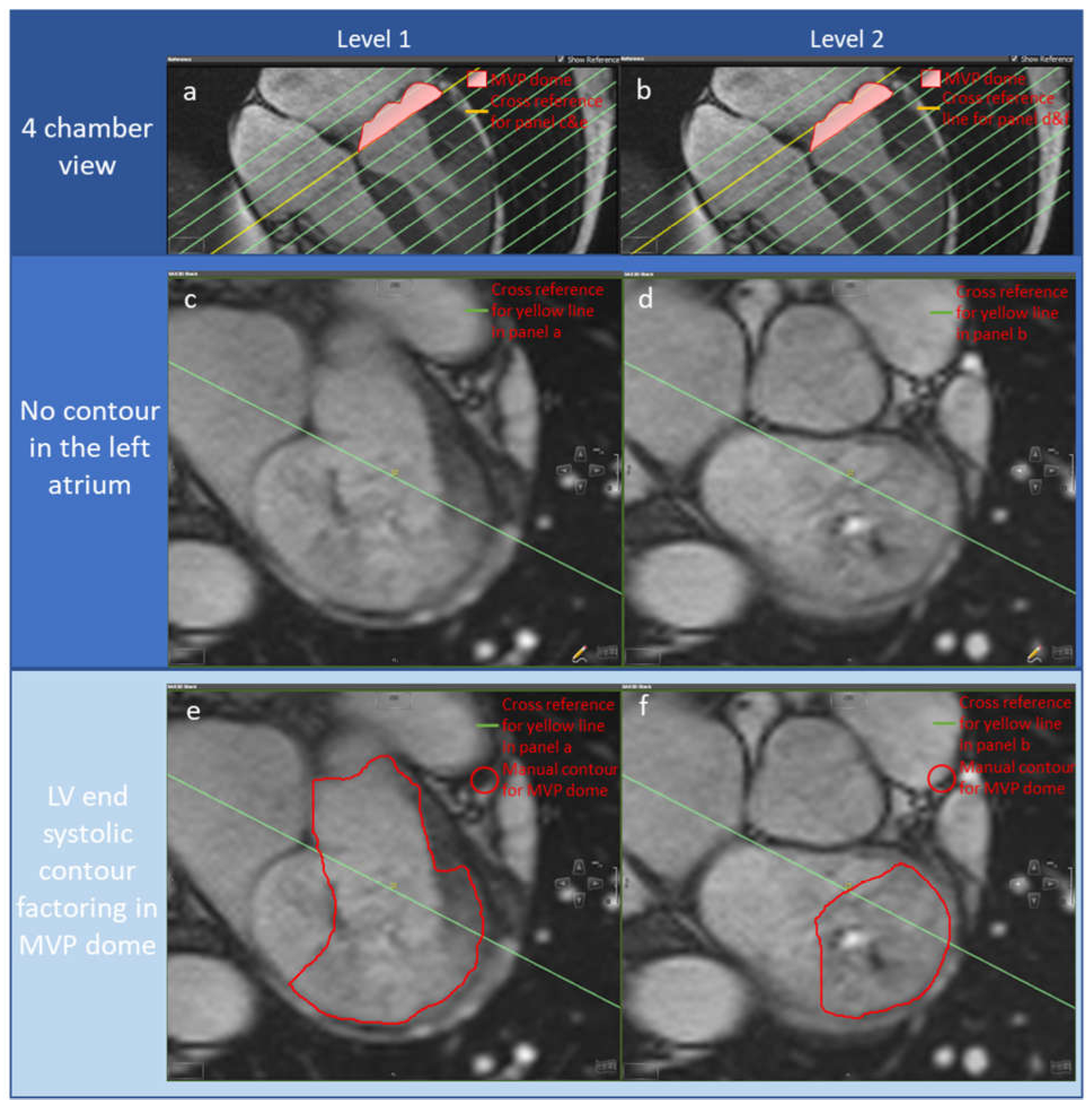
2. Materials and Methods
2.1. Study Cohort
2.2. CMR Protocol LV Volume Assessment
2.3. 4DF CMR Acquisition
2.4. 4D Flow CMR Analysis
2.5. MR Severity Assessment
2.6. Statistical Analysis
3. Results
4. Discussion
5. Limitations
6. Conclusions
Author Contributions
Funding
Institutional Review Board Statement
Informed Consent Statement
Data Availability Statement
Conflicts of Interest
References
- Kodali, S.K.; Velagapudi, P.; Hahn, R.T.; Abbott, D.; Leon, M.B. Valvular Heart Disease in Patients ≥80 Years of Age. J. Am. Coll. Cardiol. 2018, 71, 2058–2072. [Google Scholar] [CrossRef] [PubMed]
- Iung, B.; Baron, G.; Butchart, E.G.; Delahaye, F.; Gohlke-Bärwolf, C.; Levang, O.W.; Tornos, P.; Vanoverschelde, J.-L.; Vermeer, F.; Boersma, E.; et al. A prospective survey of patients with valvular heart disease in Europe: The Euro Heart Survey on Valvular Heart Disease. Eur. Heart J. 2003, 24, 1231–1243. [Google Scholar] [CrossRef] [PubMed] [Green Version]
- Prakash, R.; Horsfall, M.; Markwick, A.; Pumar, M.; Lee, L.; Sinhal, A.; Tornos, P.; Vanoverschelde, J.-L.; Vermeer, F.; Boersma, E.; et al. Prognostic impact of moderate or severe mitral regurgitation (MR) irrespective of concomitant comorbidities: A retrospective matched cohort study. BMJ Open 2014, 4, e004984. [Google Scholar] [CrossRef] [PubMed] [Green Version]
- Dziadzko, V.; Clavel, M.-A.; Dziadzko, M.; Medina-Inojosa, J.R.; Michelena, H.; Maalouf, J.; Nkomo, V.; Thapa, P.; Enriquez-Sarano, M. Outcome and undertreatment of mitral regurgitation: A community cohort study. Lancet 2018, 391, 960–969. [Google Scholar] [CrossRef]
- Freed, L.A.; Levy, D.; Levine, R.A.; Larson, M.G.; Evans, J.C.; Fuller, D.L.; Lehman, B.; Benjamin, E.J. Prevalence and Clinical Outcome of Mitral-Valve Prolapse. N. Engl. J. Med. 1999, 341, 1–7. [Google Scholar] [CrossRef]
- Spartalis, M.; Tzatzaki, E.; Spartalis, E.; Athanasiou, A.; Moris, D.; Damaskos, C.; Garmpis, N.; Voudris, V. Mitral valve prolapse: An underestimated cause of sudden cardiac death—A current review of the literature. J. Thorac. Dis. 2017, 9, 5390–5398. [Google Scholar] [CrossRef] [Green Version]
- Essayagh, B.; Sabbag, A.; Antoine, C.; Benfari, G.; Yang, L.-T.; Maalouf, J.; Asirvatham, S.; Michelena, H.; Enriquez-Sarano, M. Presentation and Outcome of Arrhythmic Mitral Valve Prolapse. J. Am. Coll. Cardiol. 2020, 76, 637–649. [Google Scholar] [CrossRef]
- VVahanian, A.; Beyersdorf, F.; Praz, F.; Milojevic, M.; Baldus, S.; Bauersachs, J.; Capodanno, D.; Conradi, L.; De Bonis, M.; De Paulis, R.; et al. 2021 ESC/EACTS Guidelines for the management of valvular heart disease: Developed by the Task Force for the management of valvular heart disease of the European Society of Cardiology (ESC) and the European Association for Cardio-Thoracic Surgery (EACTS). Eur. Heart J. 2022, 43, 561–632. [Google Scholar] [CrossRef]
- Otto, C.M.; Nishimura, R.A.; Bonow, R.O.; Carabello, B.A.; Erwin, J.P.; Gentile, F.; Jneid, H.; Krieger, E.V.; Mack, M.; McLeod, C.; et al. 2020 ACC/AHA Guideline for the Management of Patients With Valvular Heart Disease: A Report of the American College of Cardiology/American Heart Association Joint Committee on Clinical Practice Guidelines. Circulation 2021, 143, e72–e227. [Google Scholar]
- Biner, S.; Rafique, A.; Rafii, F.; Tolstrup, K.; Noorani, O.; Shiota, T.; Gurudevan, S.; Siegel, R.J. Reproducibility of Proximal Isovelocity Surface Area, Vena Contracta, and Regurgitant Jet Area for Assessment of Mitral Regurgitation Severity. JACC Cardiovasc. Imaging 2010, 3, 235–243. [Google Scholar] [CrossRef]
- Uretsky, S.; Gillam, L.; Lang, R.; Chaudhry, F.A.; Argulian, E.; Supariwala, A.; Gurram, S.; Jain, K.; Subero, M.; Jang, J.J.; et al. Discordance Between Echocardiography and MRI in the Assessment of Mitral Regurgitation Severity: A Prospective Multicenter Trial. J. Am. Coll. Cardiol. 2015, 65, 1078–1088. [Google Scholar] [CrossRef] [Green Version]
- Bellenger, N.; Burgess, M.; Ray, S.; Lahiri, A.; Coats, A.S.; Cleland, J.; Pennell, D. Comparison of left ventricular ejection fraction and volumes in heart failure by echocardiography, radionuclide ventriculography and cardiovascular magnetic resonance. Are they interchangeable? Eur. Heart J. 2000, 21, 1387–1396. [Google Scholar] [CrossRef] [Green Version]
- Grothues, F.; Smith, G.C.; Moon, J.C.; Bellenger, N.G.; Collins, P.; Klein, H.U.; Pennell, D.J. Comparison of interstudy reproducibility of cardiovascular magnetic resonance with two-dimensional echocardiography in normal subjects and in patients with heart failure or left ventricular hypertrophy. Am. J. Cardiol. 2002, 90, 29–34. [Google Scholar] [CrossRef]
- Mewton, N.; Liu, C.Y.; Croisille, P.; Bluemke, D.; Lima, J.A. Assessment of Myocardial Fibrosis with Cardiovascular Magnetic Resonance. J. Am. Coll. Cardiol. 2011, 57, 891–903. [Google Scholar] [CrossRef] [Green Version]
- Perazzolo Marra, M.; Basso, C.; De Lazzari, M.; Rizzo, S.; Cipriani, A.; Giorgi, B.; Lacognata, C.; Rigato, I.; Migliore, F.; Pilichou, K.; et al. Morphofunctional Abnormalities of Mitral Annulus and Arrhythmic Mitral Valve Prolapse. Circ. Cardiovasc. Imaging. 2016, 9, e005030. [Google Scholar] [CrossRef] [Green Version]
- Fidock, B.; Archer, G.; Barker, N.; Elhawaz, A.; Al-Mohammad, A.; Rothman, A.; Hose, R.; Hall, I.R.; Grech, E.; Briffa, N.; et al. Standard and emerging CMR methods for mitral regurgitation quantification. Int. J. Cardiol. 2021, 331, 316–321. [Google Scholar] [CrossRef]
- Garg, P.; Swift, A.J.; Zhong, L.; Carlhäll, C.-J.; Ebbers, T.; Westenberg, J.; Hope, M.D.; Bucciarelli-Ducci, C.; Bax, J.J.; Myerson, S.G. Assessment of mitral valve regurgitation by cardiovascular magnetic resonance imaging. Nat. Rev. Cardiol. 2020, 17, 298–312. [Google Scholar] [CrossRef]
- Dyverfeldt, P.; Bissell, M.; Barker, A.J.; Bolger, A.F.; Carlhäll, C.-J.; Ebbers, T.; Francios, C.J.; Frydrychowicz, A.; Geiger, J.; Giese, D.; et al. 4D flow cardiovascular magnetic resonance consensus statement. J. Cardiovasc. Magn. Reson. 2015, 17, 72. [Google Scholar] [CrossRef] [Green Version]
- Garg, P.; Broadbent, D.A.; Swoboda, P.P.; Foley, J.R.; Fent, G.; Musa, T.A.; Ripley, D.; Erhayiem, B.; Dobson, L.E.; McDiarmid, A.K.; et al. Acute Infarct Extracellular Volume Mapping to Quantify Myocardial Area at Risk and Chronic Infarct Size on Cardiovascular Magnetic Resonance Imaging. Circ. Cardiovasc. Imaging 2017, 10, e006182. [Google Scholar] [CrossRef] [Green Version]
- Garg, P.; Crandon, S.; Swoboda, P.P.; Fent, G.J.; Foley, J.R.J.; Chew, P.G.; Brown, L.A.E.; Vijayan, S.; Hassell, M.E.C.J.; Nijveldt, R.; et al. Left ventricular blood flow kinetic energy after myocardial infarction—Insights from 4D flow cardiovascular magnetic resonance. J. Cardiovasc. Magn. Reson. 2018, 20, 61. [Google Scholar] [CrossRef] [Green Version]
- Garg, P.; Van Der Geest, R.J.; Swoboda, P.; Crandon, S.; Fent, G.J.; Foley, J.R.J.; Dobson, L.E.; Al Musa, T.; Onciul, S.; Vijayan, S.; et al. Left ventricular thrombus formation in myocardial infarction is associated with altered left ventricular blood flow energetics. Eur. Heart J. Cardiovasc. Imaging 2019, 20, 108–117. [Google Scholar] [CrossRef] [PubMed] [Green Version]
- Assadi, H.; Uthayachandran, B.; Li, R.; Wardley, J.; Nyi, T.H.; Grafton-Clarke, C.; Swift, A.J.; Solana, A.B.; Aben, J.-P.; Thampi, K.; et al. Kat-ARC accelerated 4D flow CMR: Clinical validation for transvalvular flow and peak velocity assessment. Eur. Radiol. Exp. 2022, 6, 46. [Google Scholar] [CrossRef] [PubMed]
- Vincenti, G.; Masci, P.G.; Rutz, T.; De Blois, J.; Prša, M.; Jeanrenaud, X.; Schwitter, J.; Monney, P. Impact of bileaflet mitral valve prolapse on quantification of mitral regurgitation with cardiac magnetic resonance: A single-center study. J. Cardiovasc. Magn. Reson. 2017, 19, 56. [Google Scholar] [CrossRef] [PubMed] [Green Version]
- Hatipoglu, S.; Mohiaddin, R.H.; Gatehouse, P.; Alpendurada, F.; Baksi, A.J.; Izgi, C.; Prasad, S.K.; Pennell, D.J.; Krupickova, S. Performance of artificial intelligence for biventricular cardiovascular magnetic resonance volumetric analysis in the clinical setting. Int. J. Cardiovasc. Imaging 2022, 38, 2413–2424. [Google Scholar] [CrossRef]
- Jin, C.N.; Salgo, I.S.; Schneider, R.J.; Kam, K.K.; Chi, W.K.; So, C.Y.; Tang, Z.; Wan, S.; Wong, R.; Underwood, M.; et al. Using Anatomic Intelligence to Localise Mitral Valve Prolapse on Three-Dimensional Echocardiography. J. Am. Soc. Echocardiogr. 2016, 29, 938–945. [Google Scholar] [CrossRef]
- Alenazy, A.; Eltayeb, A.; Alotaibi, M.K.; Anwar, M.K.; Mulafikh, N.; Aladmawi, M.; Vriz, O. Diagnosis of Mitral Valve Prolapse: Much More than Simple Prolapse. Multimodality Approach to Risk Stratification and Therapeutic Management. J. Clin. Med. 2022, 11, 455. [Google Scholar] [CrossRef]
- Han, Y.; Peters, D.C.; Salton, C.J.; Bzymek, D.; Nezafat, R.; Goddu, B.; Kissinger, K.V.; Zimetbaum, P.J.; Manning, W.J.; Yeon, S.B. Cardiovascular magnetic resonance characterisation of mitral valve prolapse. JACC Cardiovasc. Imaging. 2008, 1, 294–303. [Google Scholar] [CrossRef] [Green Version]
- Delling, F.N.; Kang, L.L.; Yeon, S.B.; Kissinger, K.V.; Goddu, B.; Manning, W.J.; Han, Y. CMR Predictors of Mitral Regurgitation in Mitral Valve Prolapse. JACC Cardiovasc. Imaging 2010, 3, 1037–1045. [Google Scholar] [CrossRef] [Green Version]
- Morningstar, J.E.; Nieman, A.; Wang, C.; Beck, T.; Harvey, A.; Norris, R.A. Mitral Valve Prolapse and Its Motley Crew-Syndromic Prevalence, Pathophysiology, and Progression of a Common Heart Condition. J. Am. Heart Assoc. 2021, 10, e020919. [Google Scholar] [CrossRef]
- Sattur, S.; Bates, S.; Movahed, M.R. Prevalence of mitral valve prolapse and associated valvular regurgitations in healthy teenagers undergoing screening echocardiography. Exp. Clin. Cardiol. 2010, 15, e13–e15. [Google Scholar]
- Delling, F.N.; Vasan, R.S. Epidemiology and Pathophysiology of Mitral Valve Prolapse: New Insights into Disease Progression, Genetics, and Molecular Basis. Circulation 2014, 129, 2158–2170. [Google Scholar] [CrossRef] [Green Version]
- Kelley, B.P.; Chaudry, A.M.; Syed, F.F. Developing a Mechanistic Approach to Sudden Death Prevention in Mitral Valve Prolapse. J. Clin. Med. 2022, 11, 1285. [Google Scholar] [CrossRef]
- Morningstar, J.E.; Gensemer, C.; Moore, R.; Fulmer, D.; Beck, T.C.; Wang, C.; Moore, K.; Guo, L.; Sieg, F.; Nagata, Y.; et al. Mitral Valve Prolapse Induces Regionalised Myocardial Fibrosis. J. Am. Heart Assoc. 2021, 10, e022332. [Google Scholar] [CrossRef]
- David, T.E.; David, C.M.; Tsang, W.; Lafreniere-Roula, M.; Manlhiot, C. Long-Term Results of Mitral Valve Repair for Regurgitation Due to Leaflet Prolapse. J. Am. Coll. Cardiol. 2019, 74, 1044–1053. [Google Scholar] [CrossRef]
- Spampinato, R.A.; Jahnke, C.; Crelier, G.; Lindemann, F.; Fahr, F.; Czaja-Ziolkowska, M.; Sieg, F.; Strotdrees, E.; Hindricks, G.; Borger, M.A.; et al. Quantification of regurgitation in mitral valve prolapse with four-dimensional flow cardiovascular magnetic resonance. J. Cardiovasc. Magn. Reson. 2021, 23, 87. [Google Scholar] [CrossRef]
- El-Tallawi, K.C.; Kitkungvan, D.; Xu, J.; Cristini, V.; Yang, E.Y.; Quinones, M.A.; Lawrie, G.M.; Zoghbi, W.A.; Shah, D.J. Resolving the Disproportionate Left Ventricular Enlargement in Mitral Valve Prolapse Due to Barlow Disease: Insights From Cardiovascular Magnetic Resonance. JACC Cardiovasc. Imaging. 2021, 14, 573–584. [Google Scholar] [CrossRef]
- Levy, F.; Iacuzio, L.; Marechaux, S.; Civaia, F.; Dommerc, C.; Wautot, F.; Tribouilloy, C.; Eker, A. Influence of Prolapse Volume in Mitral Valve Prolapse. Am. J. Cardiol. 2021, 157, 64–70. [Google Scholar] [CrossRef]
- Nkomo, V.T.; Gardin, J.M.; Skelton, T.N.; Gottdiener, J.S.; Scott, C.G.; Enriquez-Sarano, M. Burden of valvular heart diseases: A population-based study. Lancet 2006, 368, 1005–1011. [Google Scholar] [CrossRef]
- McDonagh, T.A.; Metra, M.; Adamo, M.; Gardner, R.S.; Baumbach, A.; Böhm, M.; Burri, H.; Butler, J.; Čelutkienė, J.; Chioncel, O.; et al. 2021 ESC Guidelines for the diagnosis and treatment of acute and chronic heart failure: Developed by the Task Force for the diagnosis and treatment of acute and chronic heart failure of the European Society of Cardiology (ESC) With the special contribution of the Heart Failure Association (HFA) of the ESC. Eur. Heart J. 2021, 42, 3599–3726. [Google Scholar]
- Bennett, S.; Thamman, R.; Griffiths, T.; Oxley, C.; Khan, J.N.; Phan, T.; Patwala, A.; Heatlie, G.; Kwok, C.S. Mitral annular disjunction: A systematic review of the literature. Echocardiography 2019, 36, 1549–1558. [Google Scholar] [CrossRef]
- Bennett, S.; Tafuro, J.; Duckett, S.; Appaji, A.; Khan, J.N.; Heatlie, G.; Cubukcu, A.; Kwok, C.S. Definition, prevalence, and clinical significance of mitral annular disjunction in different patient cohorts: A systematic review. Echocardiography 2022, 39, 514–523. [Google Scholar] [CrossRef] [PubMed]
- Essayagh, B.; Sabbag, A.; Antoine, C.; Benfari, G.; Batista, R.; Yang, L.T.; Maalouf, J.; Thapa, P.; Asirvatham, S.; Michelena, H.I.; et al. The Mitral Annular Disjunction of Mitral Valve Prolapse: Presentation and Outcome. JACC Cardiovasc. Imaging 2021, 14, 2073–2087. [Google Scholar] [CrossRef] [PubMed]
- Konda, T.; Tani, T.; Suganuma, N.; Nakamura, H.; Sumida, T.; Fujii, Y.; Kawai, J.; Kitai, T.; Kim, K.; Kaji, S.; et al. The analysis of mitral annular disjunction detected by echocardiography and comparison with previously reported pathological data. J. Echocardiogr. 2017, 15, 176–185. [Google Scholar] [CrossRef] [PubMed]
- Delling, F.N.; Aung, S.; Vittinghoff, E.; Dave, S.; Lim, L.J.; Olgin, J.E.; Connolly, A.; Moffatt, E.; Tseng, Z.H. Antemortem and Post-Mortem Characteristics of Lethal Mitral Valve Prolapse Among All Countywide Sudden Deaths. JACC Clin. Electrophysiol. 2021, 7, 1025–1034. [Google Scholar] [CrossRef]


| Baseline Characteristic | n (%) or Mean ± SD |
|---|---|
| Male | 4 (26.7%) |
| Female | 11 (73.3%) |
| Age (years) | 49.8 ± 19.6 |
| BSA (m2) | 1.91 ± 0.2 |
| Sinus rhythm | 13 (86.7) |
| DM | 0 (0%) |
| HTN | 2 (13.3%) |
| Previous MI | 0 (0%) |
| Smoker | 5 (33.3%) |
| HYHA type I | 9 (60%) |
| HYHA type II | 4 (26.7%) |
| HYHA type III | 2 (13.3%) |
| HYHA type IV | 1 (6.7) |
| Beta-blocker | 9 (60%) |
| Loop diuretic | 3 (20%) |
| Other diuretic | 1 (6.7%) |
| Ca channel blocker | 1 (6.7%) |
| ARB blocker | 0 (0%) |
| ACEi | 6 (40%) |
| Groups | Mean ± SD |
|---|---|
| 4D flow derived | |
| Aortic valve backward flow (mL) | 1.4 ± 2.6 |
| Mitral valve backward flow (mL) | 13.3 ± 13.5 |
| Mitral valve forward flow (mL) | 100 ± 28 |
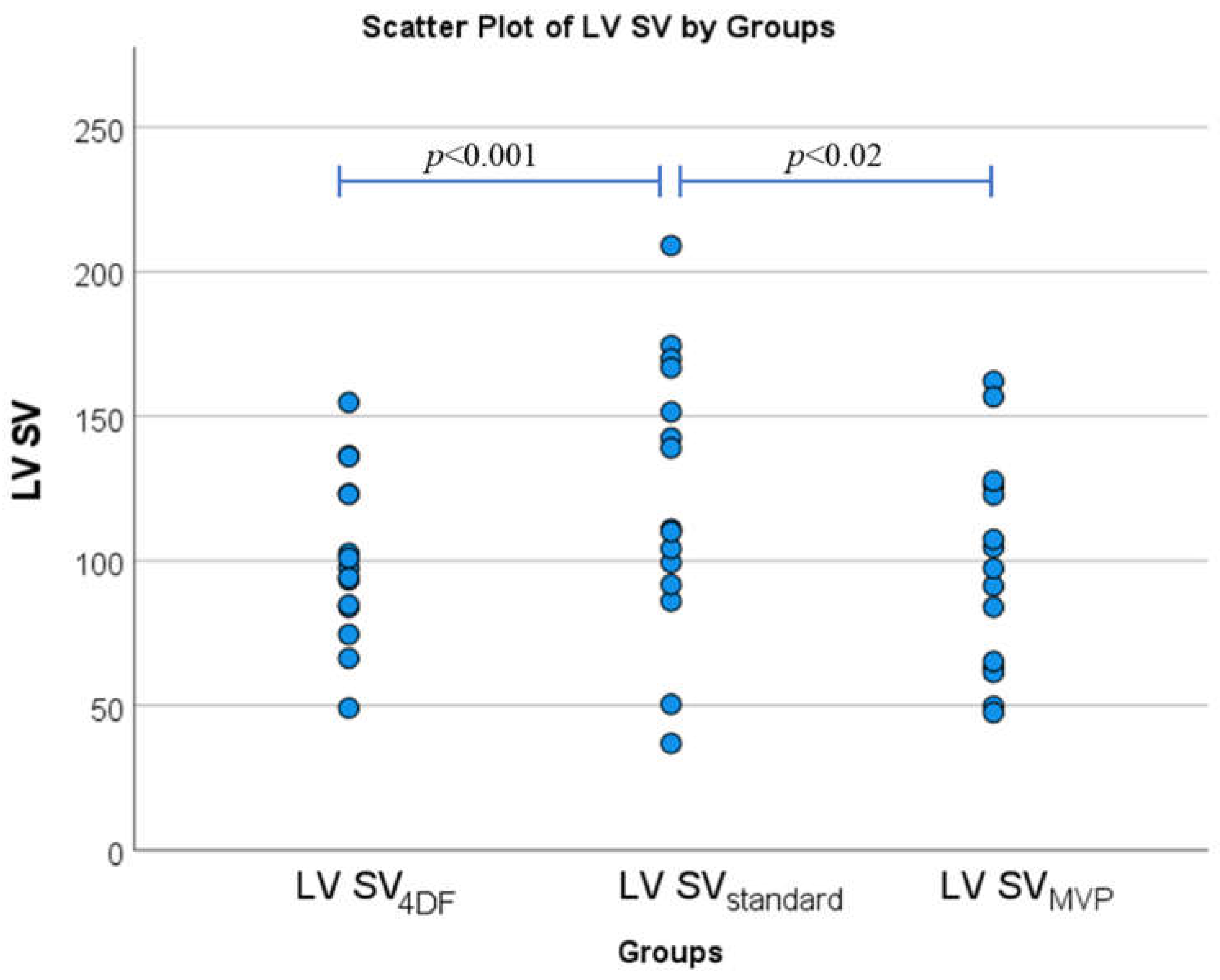
| LV stroke volume (mL) | 101 ± 29 |
| AI-derived without including MVP doming volume in LVESV | |
| LV end-diastolic volume (mL) | 200 ± 66 |
| LV end-systolic volume (mL) | 77 ± 33 |
| LV stroke volume (mL) | 123 ± 48 |
| LV ejection fraction (%) | 61 ± 14 |
| MV ejection fraction (%) | 11 ±11 |
| Manually refined including MVP doming volume in LVESV | |
| LV end-diastolic volume (mL) | 202 ± 63 |
| LV end-systolic volume (mL) | 98 ± 37 |
| LV stroke volume (mL) | 105 ± 38 |
| LV ejection fraction (%) | 52 ± 11 |
| MV ejection fraction (%) | 13 ± 12 |
Disclaimer/Publisher’s Note: The statements, opinions and data contained in all publications are solely those of the individual author(s) and contributor(s) and not of MDPI and/or the editor(s). MDPI and/or the editor(s) disclaim responsibility for any injury to people or property resulting from any ideas, methods, instructions or products referred to in the content. |
© 2023 by the authors. Licensee MDPI, Basel, Switzerland. This article is an open access article distributed under the terms and conditions of the Creative Commons Attribution (CC BY) license (https://creativecommons.org/licenses/by/4.0/).
Share and Cite
Li, R.; Assadi, H.; Matthews, G.; Mehmood, Z.; Grafton-Clarke, C.; Kasmai, B.; Hewson, D.; Greenwood, R.; Spohr, H.; Zhong, L.; et al. The Importance of Mitral Valve Prolapse Doming Volume in the Assessment of Left Ventricular Stroke Volume with Cardiac MRI. Med. Sci. 2023, 11, 13. https://doi.org/10.3390/medsci11010013
Li R, Assadi H, Matthews G, Mehmood Z, Grafton-Clarke C, Kasmai B, Hewson D, Greenwood R, Spohr H, Zhong L, et al. The Importance of Mitral Valve Prolapse Doming Volume in the Assessment of Left Ventricular Stroke Volume with Cardiac MRI. Medical Sciences. 2023; 11(1):13. https://doi.org/10.3390/medsci11010013
Chicago/Turabian StyleLi, Rui, Hosamadin Assadi, Gareth Matthews, Zia Mehmood, Ciaran Grafton-Clarke, Bahman Kasmai, David Hewson, Richard Greenwood, Hilmar Spohr, Liang Zhong, and et al. 2023. "The Importance of Mitral Valve Prolapse Doming Volume in the Assessment of Left Ventricular Stroke Volume with Cardiac MRI" Medical Sciences 11, no. 1: 13. https://doi.org/10.3390/medsci11010013