Article

Effects of Dietary Tuber Ethanolic Extract of Nut Grass (Cyperus rotundus Linn.) on Growth, Immune Response, and Disease Resistance in Nile Tilapia (Oreochromis niloticus)

by
Supranee Wigraiboon
1,2,
Ruamruedee Panchan
1,2,
Vijitra Luang-In
3,
Wilailak Ounjit
4,
Paiboon Panase
5,6,
Sontaya Sookying
6,7 and
Nantaporn Sutthi
1,2,6,*
1
Department of Agricultural Technology, Faculty of Technology, Mahasarakham University, Maha Sarakham 44150, Thailand
2
Applied Animal and Aquatic Sciences Research Unit, Division of Fisheries, Faculty of Technology, Mahasarakham University, Maha Sarakham 44150, Thailand
3
Natural Antioxidant Innovation Research Unit, Department of Biotechnology, Faculty of Technology, Mahasarakham University, Maha Sarakham 44150, Thailand
4
Department of Sociology and Anthropology, Faculty of Humanities and Social Sciences, Mahasarakham University, Maha Sarakham 44150, Thailand
5
Fisheries Division, School of Agriculture and Natural Resources, University of Phayao, Phayao 56000, Thailand
6
Unit of Excellence Physiology and Sustainable Production of Terrestrial and Aquatic Animals (FF66-UoE014), School of Agriculture and Natural Resources, University of Phayao, Phayao 56000, Thailand
7
Division of Pharmaceutical Sciences, School of Pharmaceutical Sciences, University of Phayao, Phayao 56000, Thailand
*
Author to whom correspondence should be addressed.
Animals 2024, 14(3), 503; https://doi.org/10.3390/ani14030503
Submission received: 18 December 2023 / Revised: 28 January 2024 / Accepted: 31 January 2024 / Published: 2 February 2024
(This article belongs to the Special Issue Functional Feeds to Improve Shrimp and Fish Aquaculture)

Abstract

:

Simple Summary

This study investigates the use of tuber extract from nut grass (Cyperus rotundus Linn.) as growth and immune boosters in aquaculture. Although C. rotundus is considered a weed of low value, it has been found to enhance growth and act as an immunostimulant in Nile tilapia. C. rotundus exhibited antimicrobial activity against S. agalactiae, resulting in increased fish survival. The study suggests that the immunostimulant properties of C. rotundus, typically regarded as a weed of low value, could serve as a value-added source in a bio-green aquaculture model, promoting fish-friendly practices for consumer health.

Abstract

Nut grass (Cyperus rotundus Linn.) is a weed that grows in all tropical, subtropical and temperate regions of the world, including areas where it grows on saline soil. This research was conducted to evaluate the effect of C. rotundus tuber extract in the diet on the growth performance and disease resistance of Nile tilapia. Various components of phytochemical importance of nut grass, including sugars/carbohydrates, terpenoids, tannins, and flavonoids were found in C. rotundus. Tilapia (n = 25 fish/group in triplicate) were fed with different levels of nut grass extract including 0 (control; T1), 0.4 (T2), 0.8 (T3), and 1.6 (T4) g/kg for 60 days in a completely randomized design (CRD) experiment. After the feeding trial, the highest weight gain and average daily gain (ADG) were observed in the T4 group, but it was not significantly different from T3 (Nile tilapia fed with a 0.8 g/kg) (p > 0.05). The lowest feed conversion ratio (FCR) was observed in the T3 group. Moreover, the fillet, crud lipid content, and blood chemical profiles (aspartate aminotransferase (AST), cholesterol, and malondialdehyde (MDA)) in fish fed with 1.6 g/kg were highest when compared in all groups. In addition, the T3 group presented with the immune response parameter found in red blood cells (RBC), lysozyme activity, and antioxidant (superoxide dismutase activity (SOD)) being higher than those of the control group (p < 0.05). The highest survival (93.33%) was observed in fish fed with 0.8 g/kg (T3) after a 14 day challenge with Streptococcus agalactiae. Thus, it was concluded that nut grass extract at 0.8 g/kg can be used to improve the growth performance and the tendency for resistance to S. agalactiae in Nile tilapia.

1. Introduction

Aquaculture production across the world has shown a continuous increase associated with high consumer demands including the dwindling of global capture fisheries. Concerning fisheries output, Thailand was ranked among the top 25 countries in 2023. Oreochromis niloticus, commonly referred to as Nile tilapia, holds significance in aquaculture due to its rapid growth, good survival and reproductive capabilities in captivity, high-quality meat, and favorable market value [1]. It is adaptable to a wide range of environmental conditions such as water temperature (8–42 °C) [2,3,4,5] and fresh to brackish water (0–20 ppt. salinity level) [6,7,8]. As of 2021, global production of this fish exceeded 4,827,581 tons across more than 120 countries [1]. Nile tilapia is economically importance as a food fish for many countries and is ranked as the world’s third most produced fish species after grass carp (Ctenopharyngodon idellus) and silver carp (Hypophthalmichthys molitrix) [1,9]. As the development of Nile tilapia has increased over the last few decades, the fish are mostly farmed using floating net cages, tanks, or earth ponds with a high fish stocking density. They are fed with high protein levels and water quality is monitored to obtain the highest production in a short period of time [10,11]. However, during intensive culture, the use of inappropriate rearing conditions and rapid changes in water qualities expose the fish to stress with many subsequent problems such as disease outbreak [12,13]. Fish farmers often encounter disease infection caused by bacteria, viruses, and parasites during farming, leading to a significant economic loss and subsequent loss globally, with extensive waste for the fish culture industry [14,15,16,17,18,19,20]. Among such disease infections, Streptococcus agalactiae (or group B streptococcus; GBS) is the one of the main causes of considerable morbidity and mortality in cultured tilapia worldwide [21,22,23].
To treat streptococcus outbreaks, Nile tilapia vaccines have been recently developed [24,25]. Antibiotics are also applied (lincomycin, norfloxacin, oxytetracycline, ampicillin, erythromycin and chloramphenicol, oxolinic acid, gentamicin, sulfamethoxazole, and trimethoprim [26]). However, antibiotic resistance and residues in meat are recent concerns, particularly for export producers. Many herbs are promoted as alternative plant medicinals to treat disease outbreaks and reduce antibiotic usage. If these herbaceous plant organs are effective, they may be fish-friendly and beneficial to consumer health [27,28].
Nut grass (Cyperus rotundus), belonging to the family Cyperaceae (sedges), is a weed with an erect triangular stem branching into three stems of purple, antenna-like seedpods. It is widely distributed in tropical, subtropical, and temperate regions throughout the world [29]. This plant is among the world’s most invasive species, absorbing nutrients from the soil and thereby reducing agronomic and horticultural crop yields [30,31,32,33]. However, the plant is traditionally used as a folk medicinal herb for the treatment of many diseases in humans, such as diarrhea, diabetes, pyresis, inflammation, malaria, and stomach and bowel disorders [34,35,36,37,38] due to the plant containing highly active ingredients in the form of essential oils, phenolic acids, tannins, glycosides, ascorbic acids, furochromones, monoterpenes, sesquiterpenes, sitosterol, alkaloids saponins, terpenoids, and flavonoids in the tuber and rhizomes [29,39,40,41,42]. In a few reports, nut grass (C. rotundus) has been employed in aquaculture. For instance, Citarasu et al. (2015) [43] recorded that extracts from C. rotundus effectively suppressed the white spot syndrome virus (WSSV) and enhanced the immune system in Indian white shrimp (Fenneropenaeus indicus), providing protection against WSSV infection. Additionally, Guo et al. (2019) [44] demonstrated the antibacterial activity of an aqueous extract of C. rotundus against Streptococcus agalactiae. The supplementation of C. rotundus tuber extract was observed to enhance feed palatability and stimulate the growth of Cirrhinus mrigala [45]. Nevertheless, there has been little research on the impact of extracted C. rotundus, as a supplementary feed, on fish growth, particularly in terms of immune responses against S. agalactiae in Nile tilapia. The aim of this study is to assess the impact of diet supplementation with the extract of C. rotundus on the growth performance, hematological parameters, and immune response of Nile tilapia against S. agalactiae. The hypothesis of this study is that the utilization of nut grass (C. rotundus) as an effective immunomodulator could enable the reduction of antibiotic usage in aquaculture, promoting fish-friendly practices for consumer health.

2. Materials and Methods

2.1. Preparation Ethanolic Extract from Tuber C. rotundus

Pure nut grass tuber (C. rotundus) dry powder was obtained from Thaprachan Herb Co., Ltd. (Bangkok, Thailand). Fresh C. rotundus tubers (size ranged 0.5–1.0 cm; aged 9–10 months) were collected from the wild at a location in Surin Province, Thailand. It was cleaned and sliced into small segments and subjected to an air-drying procedure. Subsequently, the dried pieces were mashed into a powder, which was then stored at a temperature of 4 °C until use. The tuber extraction procedure followed our previous report [46] with a slight modification. Briefly, the C. rotundus powder was combined with a 95% ethanol solution in a weight-to-volume ratio of 1:2, within 1000 mL conical flasks that were securely covered with aluminum foil. Flask contents were then macerated for 2 days at ambient temperature without agitation. After macerating, each mixture was filtrated through a sterile muslin cloth and Whatman qualitative filter paper. The resulting liquid residue was collected and dried using a rotary evaporator maintained at a temperature of 65 °C, and the resulting extract was kept at −20 °C until further analysis.

2.2. Phytochemical Screening

The standard procedures for C. rotundus phytochemical screening, as detailed in the protocol in [47,48], were used in the current study. Alkaloid presence was assessed through Dragendorff and Hager’s tests. The identification of cardiac glycosides, steroids, and terpenoids utilized the Liebermann-Burchard’s test. Molisch, Keller-Killiani, and Kedde’s tests were used to ascertain normal sugar, deoxy sugar, and the presence of an unsaturated lactone ring, respectively. For detecting cyanogenic glycosides, the sodium picrate method was applied. The frothing test was executed to determine the existence of saponins. In the case of screening for tannins, the lead acetate method was used to identify tannins and pseudotannins. Shinoda’s test was implemented for assessing the presence of flavonoids and Borntrager’s method was utilized to detect anthraquinones. The comprehensive protocol in these screenings follows our previously reported methodology [46].

2.3. Evaluation of Antioxidant Activity and Bioactive Compounds

The evaluation of the antioxidant activity of C. rotundus was carried out according to the methodology outlined [49]. The assessment encompassed the 2,2-diphenyl-1-picrylhydrazyl (DPPH) radical scavenging method, as well as the ferric reducing antioxidant power (FRAP) assay. The resultant values were expressed in terms mg Trolox equivalent (TE)/g DW for DPPH scavenging activity and mg Fe2+/g DW for FRAP activity. Furthermore, the bioactive constituents present in C. rotundus were analyzed for total phenolic content (TPC) and total flavonoid content (TFC), employing the method detailed in a previous study [50]. The quantified values were reported in mg gallic acid equivalent (GAE)/g DW for TPC, and mg rutin equivalent (RE)/g DW for TFC.

2.4. Fish and Experimental Diet

Sex reversed Nile tilapia (Oreochromis niloticus) fingerlings were bred at and purchased from Maha Sarakham Inland Fisheries Research and Development Center (Maha Sarakham, Thailand). The fish were placed in concrete ponds (size: 10 × 5 × 1.2 m) and allowed to acclimate for two weeks with a commercial diet containing 32% protein (Betagro®, Bangkok, Thailand). Afterward, 300 apparently healthy fish (weight 11.45 ± 1.41 g and length 6.85 ± 0.31 cm) were randomly distributed into 12 net cages (1 × 1 × 1 m) in concrete ponds, where the photoperiod was 12 h of light and 12 h of darkness throughout the experimental period. A completely randomized design (CRD) was used in this experiment. The fish was randomly divided into four groups (n = 25 fish/net cage) in triplicate experiments (T1: fish given only a basal diet, T2: given a C. rotundus extract 0.4 g/kg diet, T3: given a C. rotundus extract 0.8 g/kg diet, T4: given a C. rotundus extract 1.6 g/kg diet).
For the experimental diets, the basal diet (commercial fish feed, Betagro®, Thailand) contained protein 32%, lipid 4%, moisture 12%, and fiber 6%. The ingredients of the commercial fish feed included fish meal, soybean meal, corn, broken rice, rice bran, as well as vitamins and minerals. The C. rotundus ethanol extract was mixed with the basal diet in four different proportions: 0, 0.4, 0.8 and 1.6 g/kg diets. In brief, crude C. rotundus extract was dissolved in 100 mL of 95% ethanol solution per kg diet, then sprayed into the experimental diet at different concentrations of 0.4, 0.8, and 1.6 g/kg per diet, respectively, and dried at room temperature. The feed pellets from all 4 groups were mixed with 20 g of guar gum (pellet binder), then coated with 4% agar solution at 20 mL/kg of the diet and air dried at 40 °C for 24 h. For the basal diet, added guar gum was also added with a coating of 4% agar solution but without the C. rotundus extract supplement. Diets were packed and stored at −20 °C until used, and this method was performed every 14 days. Proximate analysis data of the diets was obtained using standard methods [51], the results are shown in Table 1. Fish were manually fed with the experimental diets at a level of 5% of their respective body weight in two equal parts [28] for a period of 60 days. During the feeding trial, water was changed every two weeks and average water quality was monitored periodically, water temperature was recorded as 27.72 ± 0.47 °C, dissolved oxygen was 10.52 ± 0.73 mg/L, and pH was 8.69 ± 0.25.

2.5. Growth Measurements and Sample Collection

The growth rate of all fish was determined using the mathematical growth model of Bagenal (1978) [52] to determine the weight gain (WG; g), average daily gain (ADG; g/day, specific growth rate (SGR; %/day), feed conversion ratio (FCR) and survival rate (%).
Weight gain (WG; g) = final weight (g) − initial weight (g);
Average daily gain (ADG; g/day) = [final weight (g) − initial weight (g)]/days;
Specific growth rate (SGR; %/day) = 100 × [{Ln final weight (g) − Ln initial weight (g)}/days];
Feed conversion ratio (FCR) = total feed (g)/weight gain (g);
Survival Rate (SR, %) = [number of survived fish/initial number of fish] × 100.
After 60 days of administering the experimental diets, blood was collected following our previous protocol [53]. Briefly, fish were anesthetized with 100 mg/L of clove oil and caudal vein blood samples were collected immediately. Blood (0.5 mL) was collected in and transferred to anticoagulant containers for hematological analysis. Another 1 mL of blood was deposited in sterile Eppendorf containers without anticoagulant in order to collect serum. Serum samples were kept at −20 °C until further analysis.

2.6. Body Chemical Composition and Organosomatic Indices

Fillets of six fish/treatments were used for proximate body chemical composition using standard methods [51]. The organosomatic indices (%fillet, %carcass, hepatosomatic index (%HSI) and viscerosomatic index (%VSI)) were computed following the methods of Van Doan et al. (2023) [53].

2.7. Blood Chemical and Hematological Analysis

The collected blood and serum samples were transported to the Veterinary Central Laboratory (Khon Kaen, Thailand) for comprehensive analysis of blood chemistry and blood indices parameters. These included measurements of serum aspartate aminotransferase (AST), alanine aminotransferase (ALT), blood urea nitrogen (BUN), total cholesterol (TC), total protein (TP), albumin (ALB), and globulin (GLB). Evaluation of hematological parameters involved the assessment of red blood cell (RBC), white blood cell (WBC), neutrophils (Neut), lymphocytes (Lymphs), hematocrit (Hct), and hemoglobin (Hb), following the detailed methodology outlined [53]. Furthermore, blood indices, such as mean cell volume (MCV), mean cell hemoglobin (MCH), and mean corpuscular hemoglobin concentration (MCHC), were determined according to the methodology established in [54].

2.8. Lipid Peroxidation and Activity of Antioxidant Enzymes

The concentration of serum malondialdehyde (MDA) was determined by assessing thiobarbituric acid reactive substances (TBARS) using the method suggested in [55], with minor modifications as detailed in our previous report [56]. Catalase activity (CAT) in the liver was determined following the protocol outlined in [54]. The change in absorbance was measured after 20 s (A1) and 80 s (A2) of incubation at 240 nm and room temperature. The CAT value was calculated using the formula (A1 − A2)/0.0008. Superoxide dismutase (SOD) activity in the liver was measured and calculated according to the formulas provided in [54]. The percent of inhibition (%) = 100 − ((ΔA control − ΔA sample/ΔA control) × 100).
SOD activity (U/g liver) = % inhibition × 3.75.

2.9. Immunological Analysis

Lysozyme activity in serum samples was determined using the turbidimetric method, based on the lysis of the lysozyme-sensitive Gram-positive bacterium Micrococcus lysodeikticus (Sigma, Missouri, USA), as described [57] with some modifications as previously reported in [53]. The activity of myeloperoxidase (MPO) in serum was measured following the method suggested in [58], with slight modifications as detailed in [54]. And the serum bactericidal activity against Streptococcus agalactiae (103 CFU/mL) was estimated using viable counts, in accordance with our previously established method [53].

2.10. Challenge Test

After the feeding trial for 60 days, different sets of healthy fish (n = 10 fish/group in triplicate) were transferred and intraperitoneally challenged with a 0.3 mL suspension containing Streptococcus agalactiae at a dose of 108 CFU/mL in 1× phosphate-buffered saline (PBS), following the method outlined in our previous study [53]. The negative control group was inoculated with 0.3 mL of 1× PBS. Following a post-challenge period of 14 days, the percentage of survival (%) of tilapia was observed, and the relative percentage of survival (RPS) was determined using the equation provided [59].
RPS = 1 − (% test mortality/% control mortality) × 100.

2.11. Statistical Analysis

The acquired data were analyzed using a one-way analysis of variance (ANOVA), followed by Duncan’s post hoc test for multiple comparisons among the treatment groups. The significance level was set at p < 0.05. Results are presented as mean ± standard deviation (SD).

3. Results

3.1. Phytochemical Screening, Antioxidant Activity and Bioactive Compounds

The results for screened phytochemical constituents are shown in Table 2. It was found that sugars/carbohydrates, terpenoids, tannins, and flavonoids were present in the ethanolic extract of C. rotundus but alkaloids, anthraquinones, coumarins, saponins, steroids, cardiac glycosides, and cyanogenic glycosices were not. Antioxidant activity and bioactive contents were determined in C. rotundus as shown in Table 3. The results showed that C. rotundus had antioxidant activity such as DPPH scavenging activity and FRAP activity at 4.33 ± 0.04 mg TE/g of extract and 4.94 ± 0.05 mg Fe2+/g of extract, respectively. Moreover, the bioactive contents, total phenolic content (TPC) was 7.98 ± 0.17 mg GAE/g of extract, and the total flavonoid content (TFC) was concentration at 9.15 ± 0.81 mg RE/g of extract.

3.2. Growth Performances

The effects of supplementing C. rotundus ethanolic extract in feed for 60 days on the growth performances of Nile tilapia are presented in Table 4. Fish fed with C. rotundus at a concentration of 1.6 g/kg of the diet (T4) demonstrated significantly improved growth performance (p < 0.05), particularly in terms of WG and ADG, when compared to the T2 and control group. The most favorable FCR was observed in fish fed the T3 diet. However, no significant differences were observed in SGR and SR among fish groups fed with C. rotundus and those on the control diet for all treatments (p > 0.05).

3.3. Blood Chemical and Hematological Profiles

The blood chemical profiles in this study are shown in Table 5. The activity of AST and TC were the highest in the T4 group, and significant differences were observed when compared to all other groups (p < 0.05). However, no significant differences (p > 0.05) were noticed in TP, GLB, ALT and BUN among all treatment groups. Hematological indices were assessed at the conclusion of the experiment and are presented in Table 6. The RBC count was notably lower in the T4 group in comparison to the T3 group (p < 0.05), although no significant differences were observed in comparison to the T2 and control groups (p > 0.05). Additionally, no significant differences were identified among all treatments (p > 0.05) with respect to WBC, Hb, Hct, Neut, Lymphs, MCV, MCH, and MCHC.

3.4. Body Composition and Organosomatic Indices

The body composition and organosomatic indices were investigated at the end of the experiment and are presented in Table 7. There was a significant difference (p < 0.05) in the values for the fillet values, where fish fed with a C. rotundus supplement in their diet (T4) exhibited higher values compared to fish fed the T2, T3, and control diets. However, no significant differences were observed in the crude protein, crude lipid, moisture, ash, and carcass composition among the various experimental treatments (p > 0.05). Likewise, in terms of the organosomatic indices (HSI, VSI and SSI), the results indicated no significant differences (p > 0.05).

3.5. Immunological and Antioxidant Observations

The serum immunological and antioxidant activity observations of Nile tilapia receiving C. rotundus supplement in their diet were investigated at the end of the experiment and are presented in Table 8. A significant difference (p < 0.05) was observed in the content of serum LZM of fish fed the T3 diet, which was significantly higher than with the control diet (T1). However, for serum MPO, the results indicated no significant differences (p > 0.05) among the various treatments. In terms of antioxidant activity, liver SOD exhibited the highest values in fish fed the T3 diet, which was significantly higher than with the control diet (T1). Similarly, both the T3 and T2 groups demonstrated significantly greater CAT compared to the T4 group (p < 0.05). The MDA showed significant differences (p > 0.05) observed in the T4 group when compared to fish fed the T2, T3, and control diets (Table 8).

3.6. Bactericidal Activity and Challenge Test

The results of the bacterial count of Streptococcus agalactiae after incubation with fish serum are presented in Figure 1. The results indicated that the lowest bacterial counts were obtained in fish fed the T4 diet, in comparison to the T2 and T1 diets (p < 0.05). However, no significant difference in bacterial counts between the T4 and T3 diets was observed. In the challenge test, the study revealed a significant improvement in the survival rate of Nile tilapia following the challenge with S. agalactiae, as depicted in Figure 2. This improvement was particularly pronounced in the group that received C. rotundus at a dietary concentration of 0.8 g/kg (T3), when compared to the T2, T4, and T1 control groups. Mortality within the control group was evident as early as day 3 post bacterial injection, while the C. rotundus-treated groups experienced fish mortality on days 4 to 5 following the S. agalactiae challenge. Notably, the relative percent of survival (RPS%) reached its highest value within the T3 diet-fed group (81.81%), followed by the T4 diet-fed group (45.45%) and the T2 diet-fed group (45.45%).

4. Discussion

The persistent issue of environmental contamination and the impact of chemical pollutants on aquatic organisms is of growing concern. Consequently, there is an increasing interest in exploring herbal remedies as alternatives for managing disease outbreaks, with a preference for these over antibiotics. To the best of our knowledge, nut grass (C. rotundus) serves as the focus of this study. Here, we embark on transforming C. rotundus, initially considered a low-value weed, into a high-value product. The ethanolic extract of C. rotundus showed significant improvements in parameters such as WG and ADG, along with an increase in the fillet percentage in Nile tilapia over a 60-day feeding period. The results of our study suggest that a C. rotundus extract dosage of 1.6 g/kg in the diet is suitable and can serve as an effective natural aquafeed additive to enhance growth in practical aquaculture. To date, there have been no reported investigations into the potential effects of C. rotundus on the growth of Tilapia species. This study represents the first exploration of C. rotundus as a dietary herbal supplement for fish, with a specific focus on Nile tilapia. Our findings align with the broader research in this domain. Notably, Cyperus powder, derived from this plant, is frequently employed as a cost-effective substitute for conventional feed components such as wheat bran or maize meal in fish diets. For instance, a study conducted by Rambabu et al. (2004) [45] demonstrated the successful replacement of wheat bran with C. rotundus powder, resulting in a significant increase in the WG of C. mrigala after a 45-day feeding period. Similarly, research by Oladele et al. (2010) [60] showed that substituting maize meal with Tigernut (Cyperus esculentus) at a 100% level led to substantial improvements in parameters such as WG, SGR, and overall catfish (Clarias gariepinus) production after an 8-week feeding period. The application of medicinal plants as growth enhancers, aimed at modulating physiological processes, is subject to a complex interplay of endogenous and exogenous factors. Within this context, the central nervous system (CNS) assumes a pivotal role in sensing and integrating these signals, and the resultant responses can significantly influence various aspects of development, growth, metabolism, and appetite [61]. Additionally, plant-derived compounds hold the potential to stimulate fish growth by modulating the somatotropic axis (growth hormones (GH), insulin-like growth factors (IGF-I and IGF-II)) and neuroendocrine pathways (ghrelin and leptin) [61]. However, further investigation is needed to elucidate the regulatory mechanisms involving C. rotundus in this context.
In line with these above findings, our study demonstrates that the ethanolic extract of C. rotundus tubers significantly promotes tilapia growth, possibly due to the presence of phytochemical compounds and bioactive elements within the plant. The screening of the ethanolic extract revealed the presence of four phytochemical elements: sugars/carbohydrates, terpenoids, tannins, and flavonoids. It is well-documented in earlier research that the extract of C. rotundus is rich in bioactive constituents, and the results of this study align with previous investigations [29,39,40,41,42]. In accordance with our findings, the phytochemical composition revealed in our data aligns with the conclusions of Babiaka et al. (2021) [62], who conducted an extensive review. They identified terpenoids and flavonoids as the primary bioactive constituents within C. rotundus, especially in plant samples primarily harvested from regions in Asia and Africa.
Furthermore, in our findings, we observed that C. rotundus exhibited antioxidant and bioactive compounds, as evidenced by its DPPH scavenging activity, FRAP activity, TPC, and TFC, as presented in Table 2. These results unequivocally establish the antioxidative potential of C. rotundus, consistent with previous reports [63,64]. Phenolic compounds hold a significant place in the composition of plants due to their antioxidant effects via deactivating lipid free radicals or inhibiting the breakdown of hydroperoxides into free radicals [65]. In fact, flavonoids have been documented to possess various beneficial properties, including antioxidative, antiviral, antimicrobial, antiplatelet, and antitoxic activities [66]. The biological functions of these polyphenols in diverse biological systems are attributed to their redox characteristics, which are believed to play a pivotal role in the absorption and neutralization of free radicals, extinguishing singlet and triplet oxygens, or breaking down peroxides [67,68].
The antioxidant defense system in animals primarily relies on enzymes like CAT and SOD to protect cells from free radicals and facilitate their removal [28]. These enzymes serve as essential indicators of oxidative stress in aquatic animals [28,69]. In our study, we observed significantly higher levels of SOD and CAT activities in the liver, particularly in the group fed with C. rotundus (T3) at 0.8 g/kg. However, the activity decreased when a higher amount (1.6 g/kg) of C. rotundus was added. This decline may be attributed to the high sugar or carbohydrate content in the ethanolic C. rotundus extract. This, in turn, raises the possibility that this carbohydrate content contributes to the observed elevation in cholesterol levels. In line with our findings, a study on Nile tilapia [70] revealed the adverse effects of a high-cholesterol diet on liver function and mitochondrial abundance. This dietary regimen hindered endogenous cholesterol synthesis, upregulated genes associated with cholesterol esterification and efflux, and inhibited lipid catabolic processes, including mitochondrial β-oxidation and lysosome-mediated lipophagy, while also reducing insulin sensitivity [70].
Additional studies, such as one on juvenile turbot [71], have shown that high dietary cholesterol intake can affect key genes involved in cholesterol synthesis (hmgcr) and promote bile acid synthesis-related genes (cyp7a1). This was further corroborated by findings from studies on Japanese flounder (Paralichthys olivaceus) [72] and channel catfish (Ictalurus punctatus) [73], where increased body weight was associated with elevated plasma and hepatic cholesterol levels. Moreover, cholesterol accumulation in the liver, driven by high-carbohydrate diets, has been observed in Nile tilapia [74], potentially compromising hepatic antioxidant capacity [75]. Although high cholesterol intake seemed to stimulate growth in Nile tilapia, it also gave rise to metabolic disorders in the fish [70]. Our study supports these findings, as the T4 group exhibited high growth rates but showed signs of liver damage, indicated by elevated levels of serum MDA and AST. Serum AST levels are commonly employed as markers and indicators of illness in fish, providing insights into the extent of liver injury. These enzymes are released into the bloodstream, contributing to liver damage [76,77]. Furthermore, MDA serves as a marker for oxidative stress and damage to cellular membranes [69]. Typically, certain herbs, such as Artemisia apiacea, are known to exert antioxidant effects, resulting in significant increases in the activity of SOD and CAT in the livers of rats, accompanied by a marked reduction in MDA production [78]. Furthermore, the heightened activity of antioxidant enzymes has been observed to play a role in maintaining stable MDA levels in fish [79]. Based on these findings, it becomes apparent that a high dose of C. rotundus is associated with reduced antioxidant enzyme activity, leading to the elevation of AST and MDA levels. These observations suggest that C. rotundus not only contributes to an increase in cholesterol levels but may also be unsuitable for use in high concentrations as a component of tilapia feed. It is noteworthy that acute oral toxicity studies involving the administration of 95% ethanol-extracted C. rotundus rhizomes at a dosage of 5 g/kg and subacute toxicity studies entailing daily dosages of 1 g/kg over a 14 day period in rats did not yield toxic effects [80]. However, it is advisable to consider chronic toxicity evaluations to assess the long-term safety of the extract. Consequently, our research findings lead us to conclude that tilapia exposed to a high dose (1.6 g/kg of the diet) of C. rotundus over a 60 day period may experience chronic toxicity. The appropriate dose of C. rotundus for Nile tilapia was determined to be 0.8 g/kg of the diet, which proved suitable for promoting growth and maintaining blood parameters, as well as supporting optimal antioxidant enzyme activity.
In previous studies of the immunology of Nile tilapia fed C. rotundus, it was demonstrated that C. rotundus aqueous extracts can be used for their antibacterial activity against S. agalactiae, as reported in the in vitro screening of candidate herbs [44]. Their results agree with our study showing 
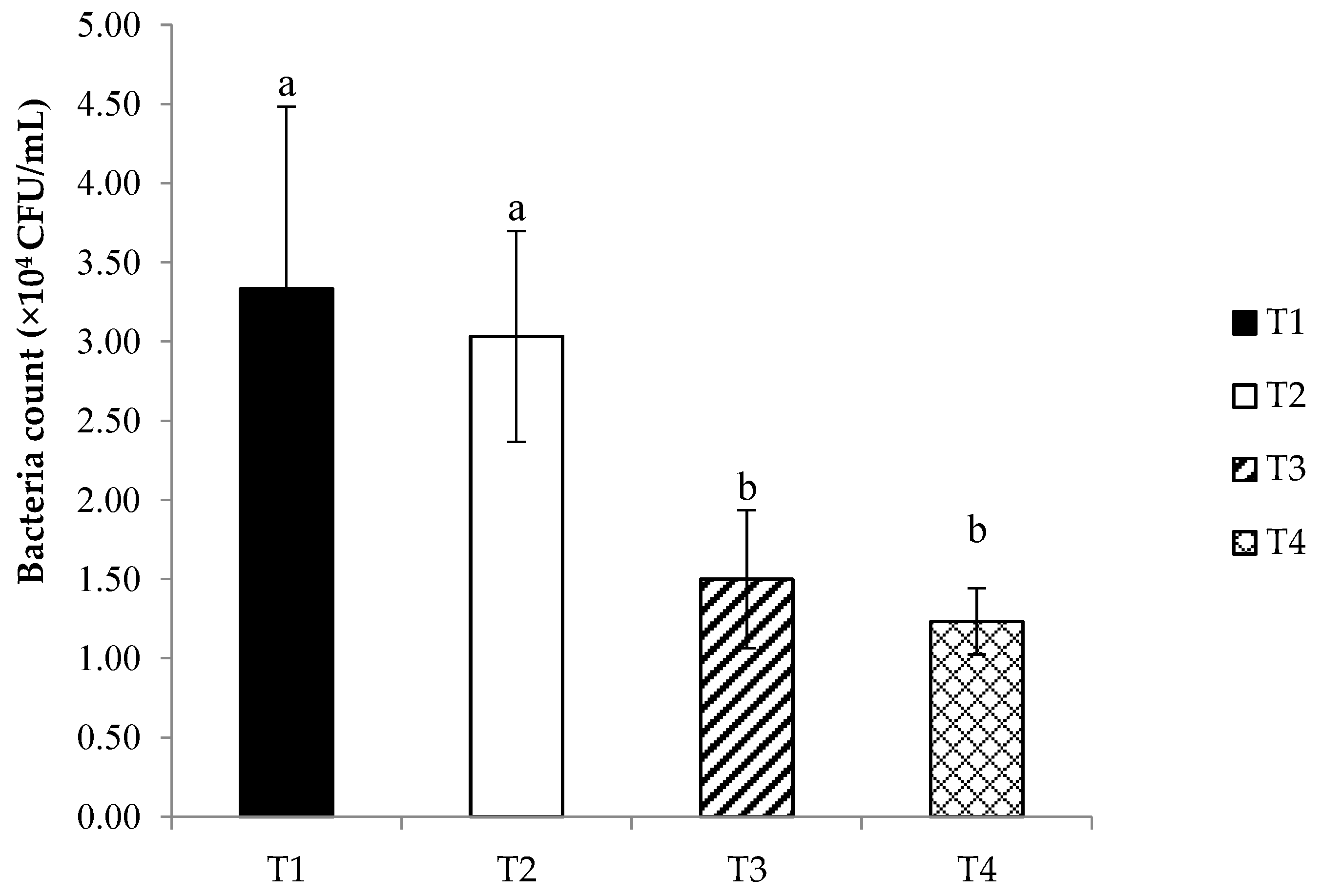
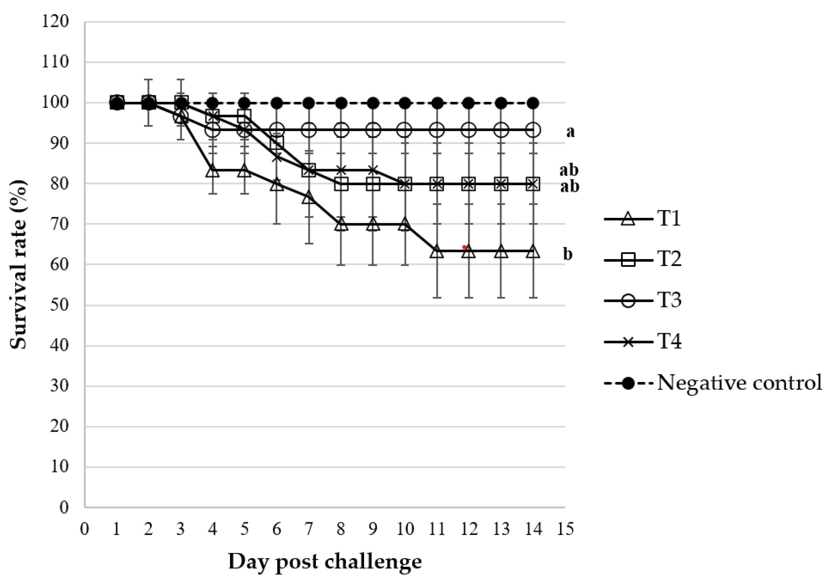
C. rotundus enhancement of the non-specific immune system which appears to be the most promising approach for disease prevention in this fish. Following the administration of supplementation to the T3 group fed with C. rotundus (0.8 g/kg of the diet), a protective effect against Streptococcus agalactiae was observed. Upon incubation with fish serum, the bacterial counts showed a significant decrease, along with a notable improvement in the survival rate of Nile tilapia after being challenged with S. agalactiae for 14 days. This result may have been correlated with the elevation of nonspecific immune parameters such as LZM and MPO activity. After feeding, fish fed with C. rotundus showed enhancements in lysozyme activity in the T3 group. LZM is a crucial element within the immune system; it is present in tissues of various animals, including fish [81], and is utilized as a biomarker for evaluating the innate immune system’s ability to defend against microbial infections in the host [82,83].
Moreover, the bioactive constituents extracted from the tuber of C. rotundus exhibited significant antibacterial properties. Phenolic compounds, recognized as natural substances synthesized through secondary metabolites [84,85], play a crucial role in this context. Phenolic compounds possess diverse biological activities, including anti-inflammatory, antioxidant, and antimicrobial properties [86]. Notably, plant flavonoids exhibit antimicrobial effects, particularly targeting Gram-positive bacteria. The primary site of flavonoid action on Gram-positive bacteria is the cell membrane, potentially involving phospholipid bilayer damage, respiratory chain inhibition, or interference in ATP synthesis [87]. Tannins, another group of bioactive compounds, exert their antibacterial action by inhibiting fatty acid biosynthesis, demonstrating iron chelation activity, impeding iron uptake, and hindering cell wall synthesis. Additionally, tannins cause damage to the outer membrane and cell membrane [88]. Terpenoids present in essential oils also contribute to antibacterial activity and antioxidative potential, as evidenced by studies [89,90].
A singular instance within the previous literature highlights the effectiveness of C. rotundus extracts, not specifically in Nile tilapia but notably against the white spot syndrome virus (WSSV) in Indian white shrimp [43]. The present study constitutes the first documentation of the application of C. rotundus tuber extracts to enhance disease resistance in Nile tilapia. Our findings are consonant with previous studies that have reported an increase in disease resistance in Nile tilapia when exposed to various medicinal plants or their extracts [28,91,92,93,94]. Many studies suggest that medicinal plants possess immunomodulatory capabilities. The way a plant functions, either by enhancing the immune system or displaying anti-inflammatory effects, is mainly influenced by the particular plant species and its constituent active compounds. In certain instances, the combination of various bioactive compounds exhibits distinct behavior [61]. Thus, our results indicate that C. rotundus, a medicinal plant/herb, has immunomodulatory potential for use in Nile tilapia.

5. Conclusions

This study investigated the feasibility of integrating the weed C. rotundus into fish feed. Our results indicate that adding C. rotundus at a level of 0.8 g/kg diet (T3) significantly improved immune responses and disease resistance in Nile tilapia. Furthermore, there was an observed upward trend in growth data; however, it was not statistically significant. These findings underscore the potential of C. rotundus as a natural product with promising applications in the aquafeed industry. The data presented herein offers valuable insights into the immunostimulant properties of C. rotundus, thereby emphasizing its potential significance in the context of tilapia culture practices.

Author Contributions

Conceptualization, N.S.; methodology, N.S., S.W., V.L.-I., P.P. and S.S.; formal analysis, N.S., S.W., P.P., S.S. and V.L.-I.; investigation, N.S., S.W., V.L.-I., P.P. and S.S.; resources, S.W., P.P., V.L.-I. and S.S.; data curation, N.S., S.W., R.P., P.P., V.L.-I. and W.O.; writing—original draft preparation, N.S., R.P., W.O. and V.L.-I.; writing—review and editing, N.S., R.P., W.O. and V.L.-I.; supervision, N.S. and S.W.; funding acquisition, N.S. All authors have read and agreed to the published version of the manuscript.

Funding

This research project was financially supported by Thailand Science Research and Innovation (TSRI) (Grant no. 6506030/2565).

Institutional Review Board Statement

The handling and experimental protocols in the fish study were approved and authorized by the Institutional Animal Care and Use Committee, Mahasarakham University (IACUC-MSU), Thailand (IACUC-MSU-09/2022).

Informed Consent Statement

Not applicable.

Data Availability Statement

The datasets generated during and/or analyzed during the current study are available from the corresponding author upon reasonable request.

Acknowledgments

The authors would like to thank the Division of Fisheries, Department of Agricultural Technology, Faculty of Technology, Mahasarakham University for research laboratory facilities, Nanthana Pothakam, Conservation and Research Section, Animal Management Division, Chiang Mai Night Safari for kindly supporting the laboratory collaboration. Special thanks went to Monthicha Romwichian and Ekkalak Yotsakun for their kind assistance during the preparation of this research.

Conflicts of Interest

The authors declare no conflicts of interest.

References

  1. FAO. Oreochromis niloticus Linnaeus, 1758, Fisheries and Aquaculture Division. Available online: https://www.fao.org/fishery/en/aqspecies/tln (accessed on 12 May 2023).
  2. Likongwe, J.S.; Stecko, T.D.; Stauffer, J.R.; Carline, R.F. Combined effects of water temperature and salinity on growth and feed utilization of juvenile Nile tilapia Oreochromis niloticus (Linneaus). Aquaculture 1996, 146, 37–46. [Google Scholar] [CrossRef]
  3. Atwood, H.L.; Tomasso, J.R.; Webb, K.; Gatlin, D.M. Low-temperature tolerance of Nile tilapia, Oreochromis niloticus: Effects of environmental and dietary factors. Aquac. Res. 2003, 34, 241–251. [Google Scholar] [CrossRef]
  4. El-Sherif, M.S.; El-Feky, A.M.I. Performance of nile tilapia (Oreochromis niloticus) fingerlings. II. Influence of different water temperatures. Int. J. Agric. Biol. 2009, 11, 301–305. [Google Scholar]
  5. Xie, S.; Zheng, K.; Chen, J.; Zhang, Z.; Zhu, X.; Yang, Y. Effect of water temperature on energy budget of Nile tilapia, Oreochromis niloticus. Aquac. Nutr. 2011, 17, e683–e690. [Google Scholar] [CrossRef]
  6. Suresh, A.V.; Lin, C.K. Tilapia culture in saline waters: A review. Aquaculture 1992, 106, 201–226. [Google Scholar] [CrossRef]
  7. Dawood, M.A.O.; Gewaily, M.S.; Sewilam, H. The growth performance, antioxidative capacity, and histological features of intestines, gills, and livers of Nile tilapia reared in different water salinities and fed menthol essential oil. Aquaculture 2022, 554, 738122. [Google Scholar] [CrossRef]
  8. Mashaii, N.; Rajabipour, F.; Bitaraf, A.; Hosseinzadeh, H.; Rohani, M.S.; Sarsangi, H.; Mohammadi, M. Reproductive Biology of Nile Tilapia, Oreochromis niloticus under the Brackish Water Culture Condition. Int. J. Food Sci. Agric. 2022, 6, 4–7. [Google Scholar] [CrossRef]
  9. Makori, A.J.; Abuom, P.O.; Kapiyo, R.; Anyona, D.N.; Dida, G.O. Effects of water physico-chemical parameters on tilapia (Oreochromis niloticus) growth in earthen ponds in Teso North Sub-County, Busia County. Fish. Aquat. Sci. 2017, 20, 30. [Google Scholar] [CrossRef]
  10. El-Sayed, A.F.M. Effects of stocking density and feeding levels on growth and feed efficiency of Nile tilapia (Oreochromis niloticus L.) fry. Aquac. Res. 2002, 33, 621–626. [Google Scholar] [CrossRef]
  11. Mohammad, R.T. Comparative study of growth performance of three strains of Nile tilapia, Oreochromis niloticus, L. at two stocking densities. Aquac. Res. 2006, 37, 172–179. [Google Scholar] [CrossRef]
  12. Garcia, F.; Romera, D.M.; Gozi, K.S.; Onaka, E.M.; Fonseca, F.S.; Schalch, S.H.C.; Candeira, P.G.; Guerra, L.O.; Carmo, F.J.; Carneiro, D.J.; et al. Stocking density of Nile tilapia in cages placed in a hydroelectric reservoir. Aquaculture 2013, 410, 51–56. [Google Scholar] [CrossRef]
  13. Li, M.; Wang, X.; Qi, C.; Li, E.; Du, Z.; Qin, J.G.; Chen, L. Metabolic response of Nile tilapia (Oreochromis niloticus) to acute and chronic hypoxia stress. Aquaculture 2018, 495, 187–195. [Google Scholar] [CrossRef]
  14. Bigarré, L.; Cabon, J.; Baud, M.; Heimann, M.; Body, A.; Lieffrig, F.; Castric, J. Outbreak of betanodavirus infection in tilapia, Oreochromis niloticus (L.), in fresh water. J. Fish. Dis. 2009, 32, 667–673. [Google Scholar] [CrossRef] [PubMed]
  15. Dong, H.T.; Nguyen, V.V.; Le, H.D.; Sangsuriya, P.; Jitrakorn, S.; Saksmerprome, V.; Senapin, S.; Rodkhum, C. Naturally concurrent infections of bacterial and viral pathogens in disease outbreaks in cultured Nile tilapia (Oreochromis niloticus) farms. Aquaculture 2015, 448, 427–435. [Google Scholar] [CrossRef]
  16. Dong, H.T.; Siriroob, S.; Meemetta, W.; Santimanawong, W.; Gangnonngiw, W.; Pirarat, N.; Khunrae, P.; Rattanarojpong, T.; Vanichviriyakit, R.; Senapin, S. Emergence of tilapia lake virus in Thailand and an alternative semi-nested RT-PCR for detection. Aquaculture 2017, 476, 111–118. [Google Scholar] [CrossRef]
  17. Fathi, M.; Dickson, C.; Dickson, M.; Leschen, W.; Baily, J.; Muir, F.; Ulrich, K.; Weidmann, M. Identification of Tilapia Lake Virus in Egypt in Nile tilapia affected by ‘summer mortality’ syndrome. Aquaculture 2017, 473, 430–432. [Google Scholar] [CrossRef]
  18. Nicholson, P.; Fathi, M.A.; Fischer, A.; Mohan, C.; Schieck, E.; Mishra, N.; Heinimann, A.; Frey, J.; Wieland, B.; Jores, J. Detection of Tilapia Lake Virus in Egyptian fish farms experiencing high mortalities in 2015. J. Fish Dis. 2017, 40, 1925–1928. [Google Scholar] [CrossRef]
  19. Surachetpong, W.; Janetanakit, T.; Nonthabenjawan, N.; Tattiyapong, P.; Sirikanchana, K.; Amonsin, A. Outbreaks of tilapia lake virus infection, Thailand, 2015–2016. Emerg. Infect. Dis. 2017, 23, 1031–1033. [Google Scholar] [CrossRef]
  20. Behera, B.K.; Pradhan, P.K.; Swaminathan, T.R.; Sood, N.; Paria, P.; Das, A.; Verma, D.K.; Kumar, R.; Yadav, M.K.; Dev, A.K.; et al. Emergence of Tilapia Lake Virus associated with mortalities of farmed Nile Tilapia Oreochromis niloticus (Linnaeus 1758) in India. Aquaculture 2018, 484, 168–174. [Google Scholar] [CrossRef]
  21. Evans, J.; Klesius, P.; Shoemaker, C.; Pasnik, D. Identification and Epidemiology of Streptococcus iniae and S. agalactiae in tilapias Oreochromis spp. In Proceedings of the 7th International Symposium on Tilapia in Aquaculture, Veracruz, Mexico, 6–8 September 2006; pp. 25–42. [Google Scholar]
  22. Geng, Y.; Wang, K.Y.; Huang, X.L.; Chen, D.F.; Li, C.W.; Ren, S.Y.; Liao, Y.T.; Zhou, Z.Y.; Liu, Q.F.; Du, Z.J.; et al. Streptococcus agalactiae, an emerging pathogen for cultured Ya-Fish, Schizothorax prenanti, in China. Transbound. Emerg. Dis. 2012, 59, 369–375. [Google Scholar] [CrossRef]
  23. Kayansamruaj, P.; Pirarat, N.; Katagiri, T.; Hirono, I.; Rodkhum, C. Molecular characterization and virulence gene profiling of pathogenic Streptococcus agalactiae populations from tilapia (Oreochromis sp.) farms in Thailand. J. Vet. Diagn. Investig. 2014, 26, 488–495. [Google Scholar] [CrossRef]
  24. Klesius, P.; Evans, J.; Shoemaker, C.; Pasnik, D. Vaccines to prevent S. iniae and S. agalactiae disease in tilapia, O. niloticus. In Proceedings of the 7th International Symposium on Tilapia in Aquaculture, Veracruz, Mexico, 6–8 September 2006; pp. 15–24. [Google Scholar]
  25. Liu, G.; Zhu, J.; Chen, K.; Gao, T.; Yao, H.; Liu, Y.; Zhang, W.; Lu, C. Development of Streptococcus agalactiae vaccines for tilapia. Dis. Aquat. Organ. 2016, 122, 163–170. [Google Scholar] [CrossRef] [PubMed]
  26. Dangwetngam, M.; Suanyuk, N.; Kong, F.; Phromkunthong, W. Serotype distribution and antimicrobial susceptibilities of Streptococcus agalactiae isolated from infected cultured tilapia (Oreochromis niloticus) in Thailand: Nine-year perspective. J. Med. Microbiol. 2016, 65, 247–254. [Google Scholar] [CrossRef] [PubMed]
  27. Stratev, D.; Zhelyazkov, G.; Noundou, X.S.; Krause, R.W. Beneficial effects of medicinal plants in fish diseases. Aquac. Int. 2018, 26, 289–308. [Google Scholar] [CrossRef]
  28. Wangkahart, E.; Wachiraamonloed, S.; Lee, P.T.; Subramani, P.A.; Qi, Z.; Wang, B. Impacts of Aegle marmelos fruit extract as a medicinal herb on growth performance, antioxidant and immune responses, digestive enzymes, and disease resistance against Streptococcus agalactiae in Nile tilapia (Oreochromis niloticus). Fish Shellfish Immunol. 2022, 120, 402–410. [Google Scholar] [CrossRef] [PubMed]
  29. Uddin, S.J.; Mondal, K.; Shilpi, J.A.; Rahman, M.T. Antidiarrhoeal activity of Cyperus rotundus. Fitoterapia 2006, 77, 134–136. [Google Scholar] [CrossRef]
  30. Andrews, F.W. A Study of Nut Grass (Cyperus rotundus L.) in the Cotton Soil of the Gezira: I. The Maintenance of Life in the Tuber Author (s): F.W. ANDREWS Source: Annals of Botany, New Series. Ann. Bot. 1940, 4, 177–193. [Google Scholar] [CrossRef]
  31. Quayyum, H.A.; Mallik, A.U.; Leach, D.M.; Gottardo, C. Growth inhibitory effects of nutgrass (Cyperus rotundus) on rice (Oryza sativa) seedlings. J. Chem. Ecol. 2000, 26, 2221–2231. [Google Scholar] [CrossRef]
  32. Peerzada, A.M. Biology, agricultural impact, and management of Cyperus rotundus L.: The world’s most tenacious weed. Acta Physiol. Plant. 2017, 39, 270. [Google Scholar] [CrossRef]
  33. Coleman, M.; Kristiansen, P.; Sindel, B.; Fyfe, C. Nutgrass (Cyperus rotundus): Weed Management Guide for Australian Vegetable Production; University of New England: Lebanon, UK, 2019; pp. 1–16. [Google Scholar]
  34. Seo, W.G.; Pae, H.O.; Oh, G.S.; Chai, K.Y.; Kwon, T.O.; Yun, Y.G.; Kim, N.Y.; Chung, H.T. Inhibitory effects of methanol extract of Cyperus rotundus rhizomes on nitric oxide and superoxide productions by murine macrophage cell line, RAW 264.7 cells. J. Ethnopharmacol. 2001, 76, 59–64. [Google Scholar] [CrossRef]
  35. Singh, N.; Pandey, B.R.; Verma, P.; Bhalla, M.; Gilca, M. Phyto-pharmacotherapeutics of cyperus rotundus linn. (motha): An overview. Indian J. Nat. Prod. Resour. 2012, 3, 467–476. [Google Scholar]
  36. Srivastava, R.K.; Singh, A.; Shukla, S.V. Chemical Investigation and Pharmaceutical Action of Cyperus rotundus—A Review. J. Biol. Act. Prod. Nat. 2013, 3, 166–172. [Google Scholar] [CrossRef]
  37. Bajpay, A.; Nainwal, R.C.; Singh, D.; Tewari, S.K. Medicinal value of Cyperus rotundus Linn: An updated review. Med. Plants 2018, 10, 165–170. [Google Scholar] [CrossRef]
  38. Muhammad Arshad, U.; Ali, H. Medical treatment of various diseases through Nagarmotha (Cyperus rotundus) plant. Eur. J. Biol. Med. Sci. Res. 2022, 10, 26–43. [Google Scholar] [CrossRef]
  39. Meena, A.K.; Meena, A.K.; Yadav, A.K.; Niranjan, U.S.; Singh, B.; Nagariya, A.K.; Verma, M. Review on Cyperus rotundus—A potential herb. Int. J. Pharm. Clin. Res. 2010, 2, 20–22. [Google Scholar]
  40. Shivpalan, S.R. Medicinal uses and Pharmacological activities of Cyperus rotundus Linn—A Review. Int. J. Sci. Res. Publ. 2013, 3, 2250–3153. [Google Scholar]
  41. Pirzada, A.M.; Ali, H.H.; Naeem, M.; Latif, M.; Bukhari, A.H.; Tanveer, A. Cyperus rotundus L.: Traditional uses, phytochemistry, and pharmacological activities. J. Ethnopharmacol. 2015, 174, 540–560. [Google Scholar] [CrossRef] [PubMed]
  42. Al-Snafi, A.E. A review on Cyperus rotundus A potential medicinal plant. IOSR J. Pharm. 2016, 6, 32–48. [Google Scholar] [CrossRef]
  43. Citarasu, T.; Michaelbabu, M.; Vakharia, V.N. Influence of Cyperus rotundus active principles inhibit viral Multiplication and stimulate immune system in Indian White Shrimp Fenneropenaeus indicus against white spot syndrome virus infection. World Acad. Sci. Eng. Technol. Int. J. Anim. Vet. Sci. 2015, 9, 360–364. [Google Scholar]
  44. Guo, W.L.; Deng, H.W.; Wang, F.; Wang, S.F.; Zhong, Z.H.; Sun, Y.; Chen, X.F.; Wang, J.-H.; Zhou, Y.-C. In vitro and in vivo screening of herbal extracts against Streptococcus agalactiae in Nile tilapia (Oreochromis niloticus). Aquaculture 2019, 503, 412–421. [Google Scholar] [CrossRef]
  45. Rambabu, M.; Patro, B.; Pal, A.K.; Venkateshwarlu, G. Feeding stimulatory effectes of Cyperus rotundus tuber on Cirrhinus mrigala. J. Indian Fish. Assoc. 2004, 31, 145–153. [Google Scholar]
  46. Sutthi, N.; Panase, A.; Chitmanat, C.; Sookyin, S.; Panase, P. Effects of dietary leaf ethanolic extract of Apium graveolens L. on growth performance, serum biochemical indices, bacterial resistance and lysozyme activity in Labeo chrysophekadion (Bleeker, 1849). Aquac. Rep. 2020, 18, 100551. [Google Scholar] [CrossRef]
  47. Edeoga, H.O.; Okwu, D.E.; Mbaebie, B.O. Phytochemical constituents of some Nigerian medicinal plants. Afr. J. Biotechnol. 2005, 4, 685–688. [Google Scholar] [CrossRef]
  48. Singh, K.H.L.; Bag, G.C. Phytochemical analysis and determination of total phenolics content in water extracts of three species of Hedychium. Int. J. PharmTech Res. 2013, 5, 1516–1521. [Google Scholar]
  49. Saengha, W.; Karirat, T.; Buranrat, B.; Katisart, T.; Ma, N.L.; Luang-In, V. Antioxidant properties and cytotoxic effects of Alternanthera sissoo and Alternanthera bettzickiana extracts against cancer cells. Not. Bot. Horti Agrobot. Cluj-Napoca 2022, 50, 12776. [Google Scholar] [CrossRef]
  50. Luang-In, V.; Saengha, W.; Karirat, T.; Buranrat, B.; Matra, K.; Deeseenthum, S.; Katisart, T. Effect of cold plasma and elicitors on bioactive contents, antioxidant activity and cytotoxicity of Thai rat-tailed radish microgreens. J. Sci. Food Agric. 2021, 101, 1685–1698. [Google Scholar] [CrossRef]
  51. AOAC. Official Methods of Analysis of the Association of Official Analytical Chemistry, 16th ed.; AOAC International: Washington, DC, USA, 1995; p. 1141. [Google Scholar]
  52. Bagenal, T. Method for Assessment of Fish Production in Fresh Waters, 3rd ed.; IBP Handbook No 3; Blackwell Scientific Publication: Oxford, UK, 1978; p. 365. [Google Scholar]
  53. Van Doan, H.; Wangkahart, E.; Thaimuangphol, W.; Panase, P.; Sutthi, N. Effects of Bacillus spp. mixture on growth, immune responses, expression of immune-related genes, and resistance of Nile tilapia against Streptococcus agalactiae infection. Probiotics Antimicrob. Proteins 2023, 15, 363–378. [Google Scholar] [CrossRef] [PubMed]
  54. Mansour, A.T.E.; Goda, A.A.; Omar, E.A.; Khalil, H.S.; Esteban, M.Á. Dietary supplementation of organic selenium improves growth, survival, antioxidant and immune status of meagre, Argyrosomus regius, juveniles. Fish Shellfish Immunol. 2017, 68, 516–524. [Google Scholar] [CrossRef] [PubMed]
  55. Aengwanich, W.; Kongbuntad, W.; Boonsorn, T. Effects of shade on physiological changes, oxidative stress, and total antioxidant power in Thai Brahman cattle. Int. J. Biometeorol. 2011, 55, 741–748. [Google Scholar] [CrossRef] [PubMed]
  56. Sutthi, N.; Thaimuangphol, W.; Rodmongkoldee, M.; Leelapatra, W.; Panase, P. Growth performances, survival rate, and biochemical parameters of Nile tilapia (Oreochromis niloticus) reared in water treated with probiotic. Comp. Clin. Path. 2018, 27, 597–603. [Google Scholar] [CrossRef]
  57. Parry, R.M.; Chandan, R.C.; Shahani, K.M. A rapid and sensitive assay of muramidase. Proc. Soc. Exp. Biol. Med. 1965, 119, 384–386. [Google Scholar] [CrossRef]
  58. Sahoo, P.; Kumari, J.; Mishra, B. Non-specific immune responses in juveniles of Indian major carps. J. Appl. Ichthyol. 2005, 21, 151–155. [Google Scholar] [CrossRef]
  59. Amend, D.F. Potency testing of fish vaccines. Dev. Biol. Stand. 1981, 49, 8. [Google Scholar]
  60. Oladele, A.K.; Alatise, P.S.; Ogundele, O. Evaluation of Tigernut (Cyperus esculentus) meal as a replacement for maize meal in the diet of catfish (Clarias gariepinus) fingerlings. World J. Agric. Sci. 2010, 6, 18–22. [Google Scholar]
  61. Faheem, M.; Abbas, R.Z.; Liaqat, I.; Hoseinifar, S.H.; Maneepitaksanti, W.; Van Doan, H. Bio-active components in medicinal plants: A mechanistic review of their effects on fish growth and physiological parameters—A Review. Ann. Anim. Sci. 2022, 22, 1127–1149. [Google Scholar] [CrossRef]
  62. Babiaka, S.B.; Moumbock, A.F.A.; Günther, S.; Ntie-Kang, F. Natural products in Cyperus rotundus L. (Cyperaceae): An update of the chemistry and pharmacological activities. RSC Adv. 2021, 11, 15060–15077. [Google Scholar] [CrossRef]
  63. Yazdanparast, R.; Ardestani, A. In vitro antioxidant and free radical scavenging activity of Cyperus rotundus. J. Med. Food 2007, 10, 667–674. [Google Scholar] [CrossRef] [PubMed]
  64. Soumaya, K.J.; Zied, G.; Nouha, N.; Mounira, K.; Kamel, G.; Genviève, F.D.; Leila, G.C. Evaluation of in vitro antioxidant and apoptotic activities of Cyperus rotundus. Asian Pac. J. Trop. Med. 2014, 7, 105–112. [Google Scholar] [CrossRef] [PubMed]
  65. Norra, I. Free radical scavenging activity and phenolic content of Ficus deltoidea accessions MFD4 and MFD6 leaves. J. Trop. Agric. Fd. Sc. 2011, 39, 85–92. [Google Scholar]
  66. Ben Ammar, R.; Kilani, S.; Bouhlel, I.; Ezzi, L.; Skandrani, I.; Boubaker, J.; Ben Sghaier, M.; Naffeti, A.; Mahmoud, A.; Chekir-Ghedira, L.; et al. Antiproliferative, antioxidant, and antimutagenic activities of flavonoid-enriched extracts from (Tunisian) Rhamnus alaternus L.: Combination with the phytochemical composition. Drug Chem. Toxicol. 2008, 31, 61–80. [Google Scholar] [CrossRef] [PubMed]
  67. Fabri, R.L.; Nogueira, M.S.; Braga, F.G.; Coimbra, E.S.; Scio, E. Mitracarpus frigidus aerial parts exhibited potent antimicrobial, antileishmanial, and antioxidant effects. Biores Technol. 2009, 100, 428–433. [Google Scholar] [CrossRef] [PubMed]
  68. Kumbhare, M.R.; Guleha, V.; Sivakumar, T. Estimation of total phenolic content, cytotoxicity and in-vitro antioxidant activity of stem bark of Moringa oleifera. Asian Pac. J. Trop. Dis. 2012, 2, 144–150. [Google Scholar] [CrossRef]
  69. Winston, G.W. Oxidants and antioxidants in aquatic animals. Comp. Biochem. Physiol. C Comp. Pharmacol. Toxicol. 1991, 100, 173–176. [Google Scholar] [CrossRef] [PubMed]
  70. Li, R.X.; Chen, L.Y.; Limbu, S.M.; Qian, Y.C.; Zhou, W.H.; Chen, L.Q.; Luo, Y.; Qiao, F.; Zhang, M.-L.; Du, Z.-Y. High cholesterol intake remodels cholesterol turnover and energy homeostasis in Nile tilapia (Oreochromis niloticus). Mar. Life Sci. Technol. 2023, 5, 56–74. [Google Scholar] [CrossRef]
  71. Zhu, T.; Mai, K.; Xu, W.; Ai, Q. Effect of dietary cholesterol and phospholipids on feed intake, growth performance and cholesterol metabolism in juvenile turbot (Scophthalmus maximus L.). Aquaculture 2018, 495, 443–445. [Google Scholar] [CrossRef]
  72. Deng, J.; Mai, K.; Ai, Q.; Zhang, W.; Wang, X.; Tan, B.; Xu, W.; Liufu, Z.; Ma, H. Interactive effects of dietary cholesterol and protein sources on growth performance and cholesterol metabolism of Japanese founder (Paralichthys olivaceus). Aquac. Nutr. 2010, 16, 419–429. [Google Scholar] [CrossRef]
  73. Twibell, R.G.; Wilson, R.P. Preliminary evidence that cholesterol improves growth and feed intake of soybean meal-based diets in aquaria studies with juvenile channel catfish, Ictalurus punctatus. Aquaculture 2004, 236, 539–546. [Google Scholar] [CrossRef]
  74. Xu, R.; Li, M.; Wang, T.; Zhao, Y.W.; Shan, C.J.; Qiao, F.; Chen, L.Q.; Zhang, W.-B.; Du, Z.-Y.; Zhang, M.-L. Bacillus amyloliquefaciens ameliorates high-carbohydrate diet-induced metabolic phenotypes by restoration of intestinal acetate-producing bacteria in Nile Tilapia. Br. J. Nutr. 2022, 127, 653–665. [Google Scholar] [CrossRef]
  75. Tao, Y.F.; Qiang, J.; Bao, J.W.; Chen, D.J.; Yin, G.J.; Xu, P.; Zhu, H.J. Changes in physiological parameters, lipid metabolism, and expression of microRNAs in genetically improved farmed tilapia (Oreochromis niloticus) with fatty liver induced by a high-fat diet. Front. Physiol. 2018, 9, 1521. [Google Scholar] [CrossRef] [PubMed]
  76. Pakhira, C.; Nagesh, T.; Abraham, T.; Dash, G.; Behera, S. Stress responses in rohu, Labeo rohita transported at different densities. Aquac. Rep. 2015, 2, 39–45. [Google Scholar] [CrossRef]
  77. Park, I.S.; Hur, J.; Choi, J. Hematological responses, survival, and respiratory exchange in the olive flounder, Paralichthys olivaceus, during starvation. Asian-Australas. J. Anim. Sci. 2012, 25, 1276. [Google Scholar] [CrossRef]
  78. Kim, K.S.; Lee, S.; Lee, Y.S.; Jung, S.H.; Park, Y.; Shin, K.H.; Kim, B.K. Anti-oxidant activities of the extracts from the herbs of Artemisia apiacea. J. Ethnopharmacol. 2003, 85, 69–72. [Google Scholar] [CrossRef]
  79. Khezrian, S.; Salati, A.P.; Agh, N.; Pasha-Zanoosi, H. Effect of replacement of fish oil with different plant oils in Oncorhynchus mykiss broodstocks diets on egg and larval antioxidant defense development. Vet. Res. Forum. 2020, 11, 83–88. [Google Scholar] [CrossRef] [PubMed]
  80. Thanabhorn, S.; Jaijoy, K.; Thamaree, S.; Ingkaninan, K.; Panthong, A. Acute and subacute toxicities of the ethanol extract from the rhizomes of Cyperus rotundus Linn. Mahidol Univ. J. Pharm. Sci. 2005, 32, 15–22. [Google Scholar]
  81. Costa, A.A.; Leef, M.J.; Bridle, A.R.; Carson, J.; Nowak, B.F. Effect of vaccination against yersiniosis on the relative percent survival, bactericidal and lysozyme response of Atlantic salmon, Salmo salar. Aquaculture 2011, 315, 201–206. [Google Scholar] [CrossRef]
  82. Nhu, T.Q.; Hang, B.T.B.; Hue, B.T.B.; Quetin-Leclercq, J.; Scippo, M.L.; Phuong, N.T.; Lestemont, P. Plant extract-based diets differently modulate immune responses and resistance to bacterial infection in striped catfish (Pangasianodon hypophthalmus). Fish Shellfish Immunol. 2019, 92, 913–924. [Google Scholar] [CrossRef]
  83. Bae, J.; Hamidoghli, A.; Won, S.; Choi, W.; Lim, S.I.; Lim, L.W.; Lee, B.J.; Hur, S.W.; Bai, S.W. Evaluation of seven different functional feed additives in a low fish meal diet for olive flounder, Paralichthys olivaceus. Aquaculture 2020, 525, 735333. [Google Scholar] [CrossRef]
  84. Randhir, R.; Lin, Y.T.; Shetty, K. Stimulation of phenolics, antioxidant and antimicrobial activities in dark germinated mung bean sprouts in response to peptide and phytochemical elicitors. Process Biochem. 2004, 39, 637–646. [Google Scholar] [CrossRef]
  85. Lin, D.; Xiao, M.; Zhao, J.; Li, Z.; Xing, B.; Li, X.; Kong, M.; Li, L.; Zhang, Q.; Liu, Y.; et al. An overview of plant phenolic compounds and their importance in human nutrition and management of type 2 diabetes. Molecules 2016, 21, 1374. [Google Scholar] [CrossRef] [PubMed]
  86. Kauffmann, A.C.; Castro, V.S. Phenolic Compounds in Bacterial Inactivation: A Perspective from Brazil. Antibiotics 2023, 12, 645. [Google Scholar] [CrossRef]
  87. Yuan, G.; Guan, Y.; Yi, H.; Lai, S.; Sun, Y.; Cao, S. Antibacterial activity and mechanism of plant flavonoids to gram-positive bacteria predicted from their lipophilicities. Sci. Rep. 2021, 11, 10471. [Google Scholar] [CrossRef]
  88. Farha, A.K.; Yang, Q.Q.; Kim, G.; Li, H.B.; Zhu, F.; Liu, H.Y.; Gan, R.Y.; Corke, H. Tannins as an alternative to antibiotics. Food Biosci. 2020, 38, 100751. [Google Scholar] [CrossRef]
  89. Guimarães, A.C.; Meireles, L.M.; Lemos, M.F.; Guimarães, M.C.C.; Endringer, D.C.; Fronza, M.; Scherer, R. Antibacterial activity of terpenes and terpenoids present in essential oils. Molecules 2019, 24, 2471. [Google Scholar] [CrossRef] [PubMed]
  90. Wang, C.Y.; Chen, Y.W.; Hou, C.Y. Antioxidant and antibacterial activity of seven predominant terpenoids. Int. J. Food Prop. 2019, 22, 230–238. [Google Scholar] [CrossRef]
  91. Abdel-Tawwab, M.; Samir, F.; Abd El-Naby, A.S.; Monier, M.N. Antioxidative and immunostimulatory effect of dietary cinnamon nanoparticles on the performance of Nile tilapia, Oreochromis niloticus (L.) and its susceptibility to hypoxia stress and Aeromonas hydrophila infection. Fish Shellfish Immunol. 2018, 74, 19–25. [Google Scholar] [CrossRef] [PubMed]
  92. Abd El-Hakim, Y.M.; El-Houseiny, W.; Abd Elhakeem, E.M.; Ebraheim, L.L.; Moustafa, A.A.; Mohamed, A.A.R. Melamine and curcumin enriched diets modulate the haemato-immune response, growth performance, oxidative stress, disease resistance, and cytokine production in Oreochromis niloticus. Aquat. Toxicol. 2020, 220, 105406. [Google Scholar] [CrossRef]
  93. Naiel, M.A.E.; Ismael, N.E.M.; Negm, S.S.; Ayyat, M.S.; Al-Sagheer, A.A. Rosemary leaf powder–supplemented diet enhances performance, antioxidant properties, immune status, and resistance against bacterial diseases in Nile Tilapia (Oreochromis niloticus). Aquaculture 2020, 526, 735370. [Google Scholar] [CrossRef]
  94. Mansour, A.T.; Mahboub, H.H.; Elshopakey, G.E.; Aziz, E.K.; Alhajji, A.H.; Rayan, G.; Ghazzawy, H.S.; El-Houseiny, W. Physiological performance, antioxidant and immune status, columnaris resistance, and growth of Nile tilapia that received Alchemilla vulgaris-supplemented diets. Antioxidants 2022, 11, 1494. [Google Scholar] [CrossRef]
Figure 1. Streptococcus agalactiae counts after incubation with serum of Nile tilapia fed different concentrations of C. rotundus: T1 (0—control), T2 (0.4 g/kg diet), T3 (0.8 g/kg diet), and T4 (1.6 g/kg diet) for 60 days. Data are given as mean ± SD and different superscripts are significant differences (p < 0.05).
Animals 14 00503 g001
Figure 2. Percentage of survival rate of Nile tilapia fed different concentrations of C. rotundus: T1 (0.0—control), T2 (0.4 g/kg diet), T3 (0.8 g/kg diet), T4 (1.6 g/kg diet), and negative control (injected with PBS) for 14 days post the challenge test with Streptococcus agalactiae. Data are given as mean ± SD and different superscripts are significant differences (p < 0.05).
Animals 14 00503 g002
Table 1. Proximate analysis of experimental diets (% dry matter).
Chemical CompositionT1T2T3T4
(Control)(0.4 g/kg Diet)(0.8 g/kg Diet)(1.6 g/kg Diet)
Crude lipid (%)4.384.404.494.88
Crude fiber (%)4.114.404.375.41
Crude protein (%)36.4835.2035.1734.18
Moisture (%) 12.9215.0714.6714.49
Ash (%)10.099.869.9011.19
NFE% 144.9446.1446.0644.34
GE (MJ/kg) 218.0717.9818.0017.62
1 Nitrogen-free extract (NFE%) = 100 − (crude protein + crude lipid + ash + crude fiber). 2 Gross energy (GE) was calculated based on 23.6, 39.5 and 17.2 kJ/g protein, lipid, and carbohydrates, respectively.
Table 2. Preliminary phytochemical constituents of tuber C. rotundus ethanolic extract.
PhytochemicalsResults
Flavonoids+
Alkaloids-
Anthraquinones-
Tannins+
Saponins-
Terpenoids++
Steroids-
Cardiac glycosides-
Cyanogenic glycosides-
Sugars/Carbohydrates+++
+ present (+ = low; ++ = moderate; +++ high), - absent.
Table 3. Antioxidant activity and bioactive compounds of tuber C. rotundus ethanolic extract.
Antioxidant Activity and Bioactive CompoundsExtracts of C. rotundus
DPPH scavenging activity (mg TE/g of extract)4.33 ± 0.04
FRAP activity (mg Fe2+/g of extract)4.94 ± 0.05
TPC (mg GAE/g of extract)7.98 ± 0.17
TFC (mg RE/g of extract)9.15 ± 0.81
Data are given as mean ± SD (n = 3). 2,2-diphenyl-1-picrylhydrazyl assay (DPPH), Ferric reducing antioxidant power assay (FRAP), The total phenolic content (TPC), the total flavonoid content (TFC), GAE = gallic acid equivalent, RE = rutin equivalent.
Table 4. Growth performances and survival rate of Nile tilapia fed diets supplemented with C. rotundus ethanolic extract for 60 days.
ParametersT1T2T3T4
(Control)(0.4 g/kg Diet)(0.8 g/kg Diet)(1.6 g/kg Diet)
Weight gain (WG; g)22.86 ± 1.93 b21.67 ± 4.67 b26.30 ± 0.80 ab28.60 ± 1.90 a
Average daily growth gain (ADG; g/day)0.38 ± 0.03 b0.36 ± 0.07 b0.44 ± 0.01 ab0.48 ± 0.03 a
Specific growth rate (SGR; %/day)1.94 ± 0.161.81 ± 0.361.96 ± 0.211.99 ± 0.17
Feed conversion ratio (FCR)2.27 ± 0.26 ab2.59 ± 0.37 b1.90 ± 0.15 a2.16 ± 0.18 ab
Survival rate (SR; %)85.33 ± 6.1182.66 ± 12.2282.66 ± 6.1184.00 ± 0.00
Data are given as mean ± SD. Different superscripts mean significant differences (p < 0.05).
Table 5. Blood chemical profiles of Nile tilapia fed diets supplemented with C. rotundus ethanolic extract for 60 days.
ParametersT1T2T3T4
(Control)(0.4 g/kg Diet)(0.8 g/kg Diet)(1.6 g/kg Diet)
Total protein (TP; g/dL)3.86 ± 0.253.46 ± 0.303.86 ± 0.323.83 ± 0.05
Albumin (ALT; g/dL)1.53 ± 0.111.16 ± 0.251.33 ± 0.301.36 ± 0.05
Globulin (GLB; g/dL)2.33 ± 0.152.30 ± 0.092.53 ± 0.052.46 ± 0.11
Aspartate aminotransferase (AST; U/L)66.00 ± 20.80 b64.00 ± 18.35 b60.33 ± 3.78 b98.33 ± 11.93 a
Alanine aminotransferase (ALT; U/L)25.33 ± 6.8023.33 ± 9.0724.33 ± 5.0328.00 ± 3.46
Blood Urea Nitrogen (BUN; mg/dL)2.14 ± 0.162.06 ± 0.122.13 ± 0.152.30 ± 0.20
Total cholesterol (TC; mg/dL)121.33 ± 3.21 b123.00 ± 8.18 b128.00 ± 4.58 b144.01 ± 6.24 a
Data are given as mean ± SD. Different superscripts mean significant differences in the rows (p < 0.05). No superscripts mean the values were not significantly different.
Table 6. Hematological values of Nile tilapia fed diets supplemented with C. rotundus ethanolic extract for 60 days.
ParametersT1T2T3T4
(Control)(0.4 g/kg Diet)(0.8 g/kg Diet)(1.6 g/kg Diet)
Red blood cell (RBC; ×106 cells/mm3)1.84 ± 0.07 ab1.98 ± 0.11 ab2.10 ± 0.13 a1.75 ± 0.06 b
Hemoglobin (Hb; g/dL)7.60 ± 0.608.06 ± 0.708.53 ± 0.707.40 ± 0.26
Hematocrit (Hct; %)32.00 ± 3.4634.66 ± 4.7232.33 ± 2.0831.66 ± 1.52
White blood cell (WBC; ×103 cells/mm3)2.20 ± 0.221.61 ± 0.441.50 ± 0.611.76 ± 0.29
Neutrophils (Neut; %)42.33 ± 8.3845.33 ± 8.6243.33 ± 6.8047.33 ± 10.59
Lymphocytes (Lymphs; %)53.66 ± 9.2952.67 ± 9.0155.33 ± 6.6550.67 ± 10.26
Mean cell volume (MCV; fL)173.7 ± 9.07175.70 ± 26.36154.93 ± 22.44180.43 ± 7.83
Mean cell hemoglobin (MCH; pg)41.26 ± 2.1240.66 ± 2.0040.56 ± 1.8042.10 ± 1.30
Mean corpuscular hemoglobin concentration (MCHC; g/dL)23.83 ± 2.4523.40 ± 2.6026.40 ± 2.5523.37 ± 1.09
Data are given as mean ± SD. Different superscripts mean significant differences (p < 0.05). No superscripts mean the values were not significantly different.
Table 7. The body composition (dry weight basis) and organosomatic indices of Nile tilapia fed diets supplemented with C. rotundus ethanolic extract for 60 days.
ParametersT1T2T3T4
(Control)(0.4 g/kg Diet)(0.8 g/kg Diet)(1.6 g/kg Diet)
Crude protein (%)83.92 ± 0.5483.55 ± 0.4984.85 ± 0.9485.11 ± 1.68
Crude lipid (%)2.53 ± 0.092.87 ± 0.372.76 ± 0.082.54 ± 0.11
Moisture (%) 1.51 ± 0.011.53 ± 0.431.50 ± 0.021.36 ± 0.13
Ash (%)6.19 ± 0.026.18 ± 0.506.20 ± 0.066.12 ± 0.11
Fillet (%)25.42 ± 2.02 b24.54 ± 0.83 b26.10 ± 3.56 b29.66 ± 2.06 a
Carcass (%)57.47 ± 3.3658.12 ± 1.7656.07 ± 2.7054.27 ± 1.86
Hepatosomatic index (HSI; %)2.69 ± 0.543.10 ± 1.193.73 ± 0.713.53 ± 0.72
Viscerosomatic index (VSI; %)10.26 ± 2.0611.24 ± 1.5110.93 ± 0.6110.65 ± 1.47
Spleenosomatic index (SSI; %)0.30 ± 0.080.28 ± 0.140.33 ± 0.100.26 ± 0.08
Data are given as mean ± SD. Different superscripts mean significant differences (p < 0.05). No superscripts mean the values were not significantly different.
Table 8. Immunological and antioxidant parameters of Nile tilapia fed diets supplemented with C. rotundus ethanolic extract for 60 days.
ParametersT1T2T3T4
(Control)(0.4 g/kg Diet)(0.8 g/kg Diet)(1.6 g/kg Diet)
Lysozyme activity9.22 ± 4.34 b15.33 ± 11.77 ab29.55 ± 11.88 a21.27 ± 9.86 ab
(LZM; U/mL)
Myeloperoxidase3.14 ± 0.45 a3.07 ± 0.36 a3.43 ± 0.32 a3.49 ± 0.12 a
(MPO; OD450nm)
Superoxide dismutase 13.12 ± 3.79 b15.93 ± 5.54 ab37.87 ± 4.09 a20.00 ± 9.53 ab
(SOD; U/g liver)
Malondialdehyde 94.82 ± 7.58 b87.25 ± 8.89 b99.16 ± 7.88 b117.39 ± 14.47 a
(MDA; µmol/L)
Catalase activity37.50 ± 8.84 ab43.75 ± 8.84 a46.88 ± 8.07 a28.13 ± 8.07 b
(CAT; U/g liver)
Data are given as mean ± SD. Different superscripts mean significant differences (p < 0.05).
Disclaimer/Publisher’s Note: The statements, opinions and data contained in all publications are solely those of the individual author(s) and contributor(s) and not of MDPI and/or the editor(s). MDPI and/or the editor(s) disclaim responsibility for any injury to people or property resulting from any ideas, methods, instructions or products referred to in the content.

