Review

The Importance of Animal Models in Biomedical Research: Current Insights and Applications

by
Adriana Domínguez-Oliva
1,
Ismael Hernández-Ávalos
2,
Julio Martínez-Burnes
3,
Adriana Olmos-Hernández
4,
Antonio Verduzco-Mendoza
4 and
Daniel Mota-Rojas
5,*
1
Master’s Program in Agricultural and Livestock Sciences [Maestría en Ciencias Agropecuarias], Xochimilco Campus, Universidad Autónoma Metropolitana (UAM), Mexico City 04960, Mexico
2
Clinical Pharmacology and Veterinary Anesthesia, Facultad de Estudios Superiores Cuautitlán, Universidad Nacional Autónoma de México (UNAM), Cuautitlán 54714, Mexico
3
Facultad de Medicina Veterinaria y Zootecnia, Universidad Autónoma de Tamaulipas, Victoria City 87000, Mexico
4
Division of Biotechnology—Bioterio and Experimental Surgery, Instituto Nacional de Rehabilitación-Luis, Guillermo Ibarra Ibarra (INR-LGII), Mexico City 14389, Mexico
5
Neurophysiology, Behavior and Animal Welfare Assessment, DPAA, Universidad Autónoma Metropolitana (UAM), Mexico City 04960, Mexico
*
Author to whom correspondence should be addressed.
Animals 2023, 13(7), 1223; https://doi.org/10.3390/ani13071223
Submission received: 21 February 2023 / Revised: 19 March 2023 / Accepted: 30 March 2023 / Published: 31 March 2023

Abstract

:

Simple Summary

The present review highlights and examines the importance of animal models in relevant topics concerning current human and animal health. Over the past five years, different animal species have been used to study pandemics, such as the 2019 Coronavirus, diabetes, and obesity. Through murine, primate, porcine, and even aquatic models (e.g., zebrafish), several neurological, behavioral, cardiovascular, and oncological disorders are being understood while developing new therapeutic approaches. Nematodes and arthropods are some of the new alternatives for biomedical science; however, regardless of the species, many animal research studies show the vital role of animal models in advancing biomedical research.

Abstract

Animal research is considered a key element in advance of biomedical science. Although its use is controversial and raises ethical challenges, the contribution of animal models in medicine is essential for understanding the physiopathology and novel treatment alternatives for several animal and human diseases. Current pandemics’ pathology, such as the 2019 Coronavirus disease, has been studied in primate, rodent, and porcine models to recognize infection routes and develop therapeutic protocols. Worldwide issues such as diabetes, obesity, neurological disorders, pain, rehabilitation medicine, and surgical techniques require studying the process in different animal species before testing them on humans. Due to their relevance, this article aims to discuss the importance of animal models in diverse lines of biomedical research by analyzing the contributions of the various species utilized in science over the past five years about key topics concerning human and animal health.

1. Introduction

The use of animals in scientific research is controversial [1]. However, the transformation of medicine from an art to a science can be mainly attributed to using a wide range of animal models [2], selected according to their functional and genetic characteristics for specific research lines [3]. Animal models contribute significantly to the advance of biomedical science through their meaningful contributions to our growing understanding of pathological and biological processes [4]. Moreover, they enable the development and testing of drugs, vaccines, and surgical techniques applicable to human and veterinary medicine [5].
The term “animal model” comes from the Latin animae (alma or spirit) and the word model, which means to imitate or be similar to [6]. Animal models are based on the principle of comparative medicine [7] as instruments that can replicate physiological and pathological processes [8]. The species is selected according to each project’s objective and hypothesis [3] but also considers biological, anatomical, functional, and genetic similarities to humans or other animals [6]. Today, most of the species utilized in biomedical research are rodents [9], as they are deemed ideal models for studying pathologies that affect human populations due to their physiological homology [10], which allows them to be employed to further our understanding of such processes as sepsis, obesity, cancer, organ transplants, and biological development, among many others [11,12].
The species used in experimentation are not limited to small mammals. Rhesus monkeys (Macaca mulata) are utilized to study high-priority diseases such as the pandemic caused by the severe, acute respiratory syndrome type 2 coronavirus (SARS-CoV-2) [13]. Domestic pigs (Sus scrofa) are crucial for organ transplant medicine and immune therapies [14]. New species, including some invertebrates such as fruit flies (Drosophila melanogaster), are used to study neurological disorders such as epilepsy [15], nematodes such as Caenorhabditis elegans to study obesity [16], and aquatic models, such as the zebrafish (Danio rerio), to treat metabolic disorders, including diabetes [17].
The broad range of species used in research has brought exponential advances in medicine, especially with the introduction of genetically modified (transgenic) animals [18] and the implementation of supporting technologies such as nanotechnology and artificial intelligence [19]. In light of this, this article aims to discuss the importance of animal models in diverse lines of biomedical research by analyzing the contributions of the various species utilized in science over the past five years concerning key topics of human and animal health.

2. Search Methodology

The literature search was performed in the Web of Science, Scopus, and PubMed. Keywords related to the use of animal models applied to current research priorities were searched to select the relevant articles, for example, “emerging infectious disease”, “diabetes and obesity”, “neurodegenerative diseases”, “pain therapies”, “surgical techniques”, “cancer models”, and “alternative animal models”. The search was limited to articles published in English in the last five years (2019–2023) and related to human and non-human medicine and therapeutics.

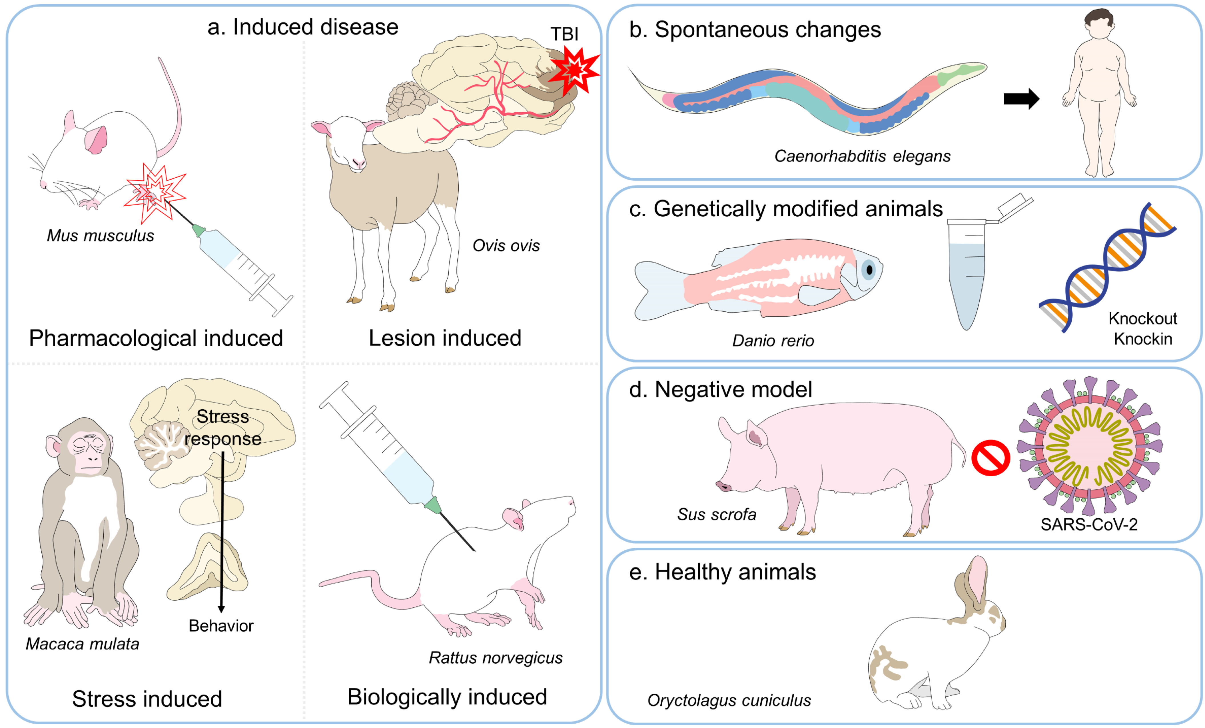
3. A Review of Animal Experimentation

Animal models are essential for several biomedical research fields such as cancer biology and therapeutics, neuroscience, pharmacology and toxicology, neurobiology of diseases, endocrinology, public health, palliative medicine, also, in studies in human and animal biology and for the discovery and testing of new drugs, vaccines, and other biologicals (e.g., antibodies, hormones) whose validation requires preclinical studies in animals [6,20]. Currently, these models address current research priorities, considered as those imposing major global threats to human and animal health. These include diseases that have afflicted humankind or increased exponentially in recent years such as SARS-CoV-2, different types of cancer and their therapy, cardiovascular diseases, metabolic and neurodegenerative disorders, and experimental refinement of surgical techniques to treat these issues [21]. The models may involve complete animals or only particular cells, tissues, organs, genes, or other agents that reproduce pathological processes (Figure 1) [8,22]. Species include rats, mice, guinea pigs, dogs, rabbits, birds, ruminants (cows, sheep), horses, fish, frogs, monkeys, cats, reptiles, squid, crabs, bees, chimpanzees, hamsters, sea slugs, pigs, nematodes (roundworm), fruit flies, and protozoans, among others [7].
The importance of animals in medical science is reflected, for example, in the percentage of Nobel Prizes studies in Physiology or Medicine using animal models (90%) [5]. From 1901 to 2020, two-thirds of those awards (186 of 222 projects [7]) employed animal models to understand pathogenic mechanisms, metabolic diseases, diagnostic and therapeutic procedures, develop vaccines, or test the efficacy of novel drugs [22]. At least 144 species used in those animal-based studies were mammals, and 42% were rodents [7]. Dogs were the first animal model used in metabolic research on gastric secretions [23] and for discovering insulin [24]. To date, rodents are the predominant species in research (Table 1) [9]. However, non-mammal species are trending, and the number of animals depends on the country and its legal regulation regarding the use and reporting of animals in research. Moreover, in some countries, there is no official annual report on animal research (e.g., South America), and not every country counts the same animals (e.g., the United States does not consider rats, mice, fish, birds, amphibians, reptiles, and cephalopods, they are not covered by the Animal Welfare Act). Although it might differ, Table 1 and Table 2 show an overview of the use of animals according to species in some countries and a summary of the reported statistics worldwide.
Several Nobel Prizes have been awarded for animal research, and the increasing number of animal models in different countries demonstrates these studies’ importance for scientific advancement [7]. However, just as necessary, their use also entails ethical challenges that require surveillance through laws, norms, guides, and strict bioethical committees to monitor the use and care of laboratory animals based on the principles of the 3Rs [33]. In this regard, for 50 years, the National Center for the Replacement, Reduction, and Refinement of Animals in Research (NC3Rs) has promoted Russel and Burch’s initiative of the 3Rs to reduce, replace, and refine procedures to improve the conditions of animals used in experimental protocols [34].
These norms differ from one nation to the next. However, one guide recognized internationally is ARRIVE (Animal Research: Reporting of in vivo Experiments), developed in 2010 to improve the in vivo experiments description to increase the reproducibility of results, refine the stages of study design, and clearly report the methods so they can be repeated and tested [35]. A second guide is PREPARE (Planning Research and Experimental Procedures on Animals), which seeks to determine and guarantee quality control in animal studies [36]. Today, for any experimental protocol requiring animals, proposals such as the Animal Study Registry (ASR) help researchers thoroughly plan their study design, methods, and statistical analyses to ensure transparency and reproducibility in their results [37]. Additionally, it is essential to mention that Ethic Committees must approve current experimental protocols within each institute to promote an appropriate use and care for animals in research.
Animal models certainly provide valuable information on the nature of diseases [38]. However, it is important to remember that inter-species limitations exist in anatomy, metabolism, physiology, and genetics [39], so a single preclinical model cannot represent all aspects of pathogenesis due to differences in resistance or susceptibility [38]. Currently, many animals used in biomedical studies undergo some genetic modification, such as transgenesis or the utilization of knockout or knockin genes, to visualize specific changes that would take years to develop under normal conditions [40]. Therefore, the selection of the animals depends on the specific research field; through their use, researchers develop scientific knowledge focused on human and veterinary medicine.

4. Animal Models and Their Application in Distinct Fields of Current Biomedical Science

4.1. Emerging Infectious Diseases

The SARS-CoV-2 virus is the etiologic agent of the coronavirus 2019 disease (COVID-19) [41]. This disease has claimed the lives of over 6.3 million people worldwide since 2019 [42,43]. The lack of knowledge of this virus and its rapid propagation at the onset of the pandemic made it essential to determine its physiopathology and identify therapeutic agents and vaccines that could mitigate its threatening consequences. These fundamental issues were solved using in vivo assays that replicated the virus in animals to untangle its pathogenesis, the immune response, and the adverse effects that might result from the vaccines and therapies proposed before testing in humans and their release to the public [41,44].
The choice of an animal model that would allow researchers to observe the histopathological, radiological, or immune changes that the virus caused required that the test animals be susceptible to lung tissue damage and capable of developing an inflammatory process [45]. Potential species included nonhuman primates, ferrets, rats, mice, Syrian hamsters, lagomorphs, minks, cats, camelids, and even zebrafish [46].
The transgenic mice can express the human angiotensin-converting enzyme II (hACE2), a functional receptor for the SARS-CoV-2 virus that mimics clinical signs observed in humans [47]. Sun et al.’s [48] research with 4.5–30-week-old transgenic mice successfully replicated the virus after intranasal and intragastric inoculation. It led to the discovery of viral loads in the lung, trachea, brain, and feces. Those authors also detected an immune and inflammatory response due to the presence of interleukins (IL). Adult mice showed more lesions in the alveolar epithelial cells, focal pulmonary hemorrhage, and more significant apoptosis of macrophages. Those findings concurred with human reports showing that COVID-19 affected older adults more severely, with the over-65 population representing 80% of all hospitalizations and a 23-fold greater risk of mortality. Reports emphasized clinical signs, such as respiratory distress and cytokine release syndromes [49]. Studies with Syrian hamsters found that while the virus is lung-tropic and infects the respiratory tract by binding to the ACE2 cell surface in the alveoli, causing pneumonia in 67% of the animals, the gastrointestinal signs reported in humans are due to viral replication and dissemination in enterocytes [50].
One animal model that shares multiple similarities with humans for the physiopathology of the SARS-CoV-2 virus is based on Rhesus macaques, African green monkeys (Chlorocebus aethiops), and crab-eating macaques (Cynomolgus macaques) [51]. The latter has been utilized to replicate the infection conditions in young (males and females of 3–9 years) and old-aged animals (23–29 years-old females). After intranasal and intratracheal viral inoculations, researchers found that nasal swabs (peak viral load of 106 copies/µL) had higher viral loads than pharynx and rectal ones (a maximum of 104 copies/µL). Additionally, viruses from nasal and pharynx samples were detected for longer periods in elderly monkeys [52]. This relation between age and disease mortality was also reported in Rhesus monkeys. Comparative studies of three nonhuman primates (three 3–5 years and two 15 years old macaques) infected intratracheally revealed that the viral replication detected by nasopharyngeal and anal swabs was persistently detected from 3 days post-infection (dpi) to 11 dpi in elderly animals. In older macaques, 104–107.5 copies/mL were also detected (while young individuals had approximately 104 copies/mL), often accompanied by the development of diffuse severe interstitial pneumonia [53].
The reinfection processes prevalent in human populations were replicated in studies with C. aethiops. Infection in six animals caused signs such as fever (50%), hypercapnia (66%), 2–7-fold increases in C-reactive protein concentrations (100%), and coagulopathy (100%) were recorded. That research proved that anal, oral, and nasal swabs could detect viral loads up to 15 dpi [44]. These findings are similar to those from other works with M. mulata, where viral RNA was found in swabs from the nose, pharynx, and anus, with amounts increasing up to 3 dpi (in an approximate range of 4–7 copies/mL) [53]. These nonhuman primate models undoubtedly contributed significantly to our understanding of the pathogenicity of COVID-19 and the physiological bases for implementing preventive and diagnostic measures and treatment.
Another important aspect of using animals is that they helped understand the transmission of the virus to other domestic species and showed that pets could acquire the SARS-CoV-2 virus through contact with an infected human. However, there is no evidence of active pet-to-human transmission [54]. Studies with dogs, pigs, chickens, and ducks showed they were not susceptible to COVID-19 infection due to low viral replication [55]. Identifying susceptible species made it possible to choose appropriate models for developing and testing vaccines [55]. Ferrets, Syrian hamsters, rabbits, transgenic mice [47], and cats were all found to be susceptible, the latter even vulnerable to airborne transmission with the development of clinical signs such as hair loss and pulmonary alterations similar to those seen in humans [56,57]. Apart from domestic cats, wild felines (tigers, lions, pumas, snow leopards) [58] have been reported to show infections by this virus. Kang et al. [59], who reported the first Delta variant (SARS-CoV-2 Delta) case in three domestic cats with COVID-19-positive owners in China, insist that transmission to pets is a topic of concern due to their possible role as silent intermediate hosts.

4.2. Endocrinology and Metabolic Pathologies

Obesity is a public health problem affecting over 600 million people worldwide [60]. Obesity and its associated metabolic syndromes have consequences such as knee osteoarthritis, a disease prevalent in approximately 60% of the overweight population [61], but this is also associated with cancer, cardiovascular disease, hypertension, coronary artery disease, stroke, sleep apnea, asthma, gallstones, steatohepatitis, and dyslipidemia. Over one-third of the world’s overweight or obese population is at risk of developing type 2 diabetes mellitus [23]. Using rodent models, researchers have determined that one element that promotes the development of type 2 diabetes mellitus is adipose tissue inflammation due to insulin resistance and excess fat mass [62]. The increase in the presentation of these comorbidities has led to the use of animal models to test new, improved strategies for reducing the incidence of this disease.
The role of the different types of adipose tissue in humans and animals is a crucial line of research that has developed with the use of rodents. For example, adipogenesis suppression and the browning of white adipose tissue (WAT) [63] have been suggested as strategies for preventing obesity [60]. The browning process creates a brown adipose-like tissue (BAT) that can participate in thermogenesis by transforming caloric intake into heat [64]. Since this is part of a central nervous system response to cold, certain medications and exercise can trigger browning as has been observed in obese and lean rats subjected to high-intensity training. In C57BL/6J mice, the transformation of beige adipocytes into WAT can be promoted with diets complemented with resveratrol for 16 weeks, as this induces a change in the intestinal microbiota in treated animals (p < 0.01) (increasing microorganisms of the genera Bacteroides, Lachnospiraceae, Blautia, Lachnoclostridium, and Parabacteroides, among others) that modulates lipid metabolism and has anti-inflammatory properties and anti-obesity effects [65].
The importance of physical activity in treating these conditions has been demonstrated in experiments with 48 Sprague-Dawley male rats, where aerobic exercise for 12 weeks combined with prebiotic fiber supplementation prevented knee joint damage, dyslipidemia, endotoxemia and normalized the effects of insulin resistance (p < 0.001) [61]. Studies with these supplements as part of a therapeutic protocol in Wistar rats, administered in presentations such as yogurt, have shown that supplementation with 5% of yogurt reduces levels of oxidative stress (significant decreases in NO levels, p < 0.05), and had fewer amounts of inflammatory cell infiltration and collagen deposits in the liver (p < 0.05) when compared to animals fed high-fat diets. According to these studies, this supplement could be a potential human therapeutic option [66].
Studies of the human genome have identified hundreds of genetic variants associated with obesity and opened the way to examining these genes in species such as C. elegans, a nematode capable of storing fat in the form of lipid droplets inside hypodermal and intestinal cells. C. elegans has 14 genes that promote diet-induced obesity and three that prevent it [67]. Those genes are now recognized as potential targets for anti-obesity treatment. Ke et al. [68] found that the knockdown of 23 fat-storing not only reduced excessive fat accumulation but also improved the health and lifespan of this species (p < 0.05). The inhibitory effect of flavonoids such as butein on lipogenesis in C. elegans succeeded in reducing triglyceride levels by up to 27% without altering food intake or energy expenditure, an effect due to the downregulation of proteins involved in lipid metabolism [69]. Likewise, the appetite suppressant effect of administering vegetable extracts from the Lentinus strigosus mushroom (300 and 1000 µg/mL) to C. elegans functioned as a natural means of preventing obesity [70]. Studies of this kind allow researchers to address obesity as a complex pathology affected by diverse factors: diet, physical activity, developmental stage, age, genes, and environmental interaction [67].
Another animal species considered a promising model for studying metabolic syndromes is the zebrafish (D. rerio). This species has genetic homology with humans, so through genetic mutation, chemical induction, and changes in diet, they can be used to study hyperglycemia, obesity, diabetes, and hypertriglyceridemia [71]. Pigs, meanwhile, share similarities with humans in terms of organ size, lifespan, anatomy, physiology, and metabolic profile [40]. A study of obesity in Iberian pigs showed the pathogenesis of chronic kidney disease caused by overweight and obesity. Although the administration of high-fat diets did not generate diabetes in those pigs by day 100, analyses revealed hypercholesterolemia (142 ± 27 mg/dl), hypertriglyceridemia (75 ± 43), insulin resistance, and glomerular hyperfiltration [72]. These effects also occur in humans [73] and have been studied in obese male mice and ovariectomized females [74].
The domestic dog has been postulated as a valuable model for studying chronic morbidities brought on by environmental conditions since they share morbidity and mortality factors with humans. In this field, Hoffman et al. [75] reported that comorbidities behind chronic conditions such as obesity, arthritis, hypothyroidism, and diabetes reported in humans were also present in 73,835 canines and that those dogs showed a positive association between age and the number of morbidities (p < 0.001). Other studies have revealed that obesity in dogs (137/198) is closely linked to the alimentary habits of their owners, finding that the 79.8% of dogs from overweight owners (114 persons) were obese (p < 0.001) [76]. Therefore, studies of these animals could provide information on disease interaction.

4.3. Cancer in Biomedicine

According to the World Health Organization [77] and the National Cancer Institute [77,78], the most common types of cancer in humans in 2020 were breast (2.26 million cases), lung (2.21 million), colorectal (1.93 million), prostate (1.41 million), skin (1.20 million), and stomach (1.09 million). These cancers cause 10 million deaths per year. Projections for 2022 estimate that around 1,918,030 new cancer cases will be diagnosed in the United States, with 350 cancer-induced deaths per day, making this disease a primary cause of mortality [79]. The pathogeny of these cancers and testing new treatment options is another field that extensively uses animal models. Over 95% of studies use rats and mice to inject cancer cell lines subcutaneously, study the primary cancer lesion, and follow its growth before excising tumors [80,81]. However, one disadvantage of this subcutaneous tumor model, is that injections in athymic nude mice may not accurately represent the interaction among tumor cells, local stroma, and the tumor’s microenvironment, depending on its precise location [82]. Contrarily, orthotopic murine models have been shown to replicate the tumor microenvironment –including metastasis– when inoculated in the original anatomical site of the tumor. In female BALB7c mice, inoculation of mammary cancer cell line 4T1 as a fat pad tumor model showed that 50% of the animals had metastasis to the ovaries, spleen, liver, and sternum. However, when compared to a heterotopic model, orthotopic tumors were smaller (1993.7 ± 197.15 mm3 vs. 1078.4 ± 300.26 mm3, p < 0.05) and had a significantly lower percentage of infiltrating cells (p < 0.05) [83]. Moreover, these orthotropic models, together with in vivo optical metabolic imaging, are proposed as an approach to studying how, for example, the fatty acid uptake by breast cancer cells increases accordingly to tumor aggressiveness and metastatic process (p < 0.05) [84] Attacking this complication in tumor development is the principal objective of anticancer therapies, since most deaths from prostate cancer, for example, are due to metastasis into bone structures [80].
Koosha et al. [85] used diosmetin, an anti-tumorigenic, in colon cancer xenografts in 24 male nude mice. Results showed that tumor volume in the group treated with 100 mg/kg of diosmetin was significantly smaller than in the untreated group (264 ± 238.3 vs. 1428 ± 459.6 mm3, p < 0.01). Promisingly, the drug did not produce toxicity even when administered at high doses. Studies of this kind show that laboratory animals allow researchers to test new drugs and better understand disease development but also aid in determining non-toxic doses that can be applied to humans or animals. Using these models as translational media for studying cancer has also revealed the importance of identifying the pain that animals may experience. Pain assessment is important in in human medicine and laboratory animal welfare. In this regard, recognizing degrees of cancer-induced bone pain has been studied by observing behavioral changes in rats and mice, where innate behaviors, such as burrowing, are reduced 9 days after inoculation when compared to control groups (p < 0.05) as a result of the nociception associated with the degree of severity of cancer due to reduced bone density [86].
The fact that the canine and human genomes share a high degree of similarity (75%) and that the risks of death due to neoplastic, congenital, and metabolic diseases are comparable means that the dog is an ideal translational model for studying human morbidity and mortality [75,87]. For example, the percentage of neoplasia is similar between dogs and humans (27.4 vs. 25.3%). However, because the types of cancer that affect each species correlate only marginally (Spearman rank p = 0.661) [75], dogs have been replaced in many preclinical studies by genetically-modified pigs [87].
Another novel anticancer strategy involves managing nerve-tumor interaction [88] since tumor-specific denervation can suppress neoplasia growth [89]. A study by Kamiya et al. [90] with female Balb/c-nu mice and the use of xenografts in Hras128 rats in a model of chemically-induced breast cancer showed that sympathetic stimulation of the nerves in tumors accelerated cancer growth but that parasympathetic stimulation reduced growth and downregulated the expression of programmed death. In contrast, in the case of late-stage colorectal cancer, parasympathetic denervation via vagotomy and atropine administration in 150 male Wistar rats reduced the incidence of tumors and their weight and volume after eight weeks, as well as cell proliferation, angiogenesis, and regulated expression of the nerve growth factor [89].
These neural anticancer therapies in humans and animals indicate that while sympathetic nerves show cancer-promoting effects in prostate and breast cancer, and melanoma cases, the parasympathetic/vagal nerves are believed to trigger both reactions. For example, vagal nerves can promote prostate, gastric, and colorectal cancers, but suppress breast and pancreatic cancers, due to β-adrenergic and muscarinic effects that modify the behavior of cancer cells, angiogenesis, tumor-associated macrophages, and antitumor immunity [88]. The axonogenesis process in species such as mice, linked to the development of metastasis in breast cancer, showed through immunofluorescence that nerve twigs tend to be sympathetic-like, with no expression of parasympathetic fibers [91].
In addition to the support of laboratory techniques such as immunofluorescence, non-invasive diagnostic methods are a priority in oncology. In immunocompetent genetically-engineered mouse models, Kirkpatrick et al. [92] utilized nanosensors with urine tests to detect protease activity in diverse types of cancer, including lung cancer, achieving 100% specificity and 81% sensitivity. In this way, monitoring with nanosensors and clinical assays in animals has demonstrated that this technique can be an option for conducting accurate, radiation-free diagnostic tests.
Nanoparticles and their application, together with in vivo imaging, can help to test novel luminescent particles and assess their tissue penetration to improve cancer therapy [93]. In vivo imaging enables us to understand tumor growth-related processes such as oxidative mitochondrial metabolism in mouse models with cell lung cancer [94]. Likewise, in a mouse model of brain tumor –glioblastoma– under general anesthesia, modified in vivo optical imaging (Surface enhanced spatially offset Raman scattering) covers the inability of conventional techniques that rely on subcutaneous inoculation of cancerous cells because they cannot read deep tissues [95]. These techniques are the basis for imaging-guided phototherapies that are a current research field to find agents capable of inducing tumor cell apoptosis, such as photodynamic y and photothermal therapy [96].

4.4. Pharmacology and Therapeutics

Parallel to the advances in our knowledge of the physiopathology of diverse conditions, developing and testing new therapeutic options is another field destined for animal models. Algology is a science in constant actualization to provide new and efficient drugs to prevent the consequences of pain by reducing the number and severity of secondary effects in both human and veterinary patients [97,98]. Adequate models are needed to evaluate analgesic efficacy accurately. In the case of treatments for open wounds, Parra et al. [99] applied carprofen (5 mg/kg) and buprenorphine (0.1 mg/kg) to the left hind paw of Sprague Dawley rats of both sexes using a punch biopsy to assess analgesia in an open wound model. Using four behavioral tests associated with aspects of nociception, mechanical and thermal stimulation, guarding behavior, and the weight-bearing test, they found that carprofen promoted recovery of the thermal response to basal levels after just 2 h. The same rat species were utilized to test the renal and gastrointestinal safety of non-steroidal anti-inflammatory drugs (NSAIDs) such as ibuprofen by administering single and multiple oral doses to pediatric patients. Furthermore, the necropsies performed on pigs of different ages (8-week-old and 6-to-7-months-old) in the study by Millecam et al. [100] revealed no severe lesions in the stomach after multiple doses of ibuprofen at 5 mg/kg. However, significant histological score differences (p < 0.025) were observed in the duodenum (1.38 vs. 4) and jejunum (3.63 vs. 1.25) between the experimental and control group. Additionally, an increased clearance time for the drug after multiple doses was found, an effect similar to reports in human pediatric patients.
Due to the adverse effects that NSAIDs can generate, especially for treating chronic afflictions such as arthritis and cancer, opioids are another therapeutic option [101]. However, since the long-term use of these drugs is also associated with complications, research has begun to new concepts and explore directions. The opioid-free anesthesia technique was introduced to prevent tolerance and hyperalgesia and reduce the use of these drugs in the postoperative period. This method uses agents such as alpha-2-agonists, ketamine, and local analgesics with distinct action mechanisms in multimodal analgesia [102,103,104]. Other new opioid-based pharmacological options are transdermal patches impregnated with morphine-like compounds. In 6–12-week-old C57BL/6JJmsSlc mice, patches synthesized with two new opioids (new-opioids 1 and 2, N1 at 3 mg/kg; N2 at 10 mg/kg) showed the same analgesic efficacy as morphine at 3 mg/kg. The effect remained constant, even under repeated administration (in contrast to fentanyl), and the cutaneous trans-permeability rate was greater, at 1.71 ± 0.35 and 3.94 ± 1.36 µg/cm/h [105]. The administration of opioid nanoparticles has also been suggested to prevent opioid tolerance and reduce the severity of adverse effects. Leucine-enkephalin hydrochloride-based nanoparticles with a size of 100–200 nm have been tested in male Sprague Dawley rats by applying them intranasally, reaching the brain directly. After dosing, high concentrations were found in the olfactory bulb and cerebrum between the first 60 min (approximately 80 ng/g and 160 ng/g, respectively), while plasma concentrations were not detected at any evaluation time (p < 0.0001). This prevents the side effects of drug transit through peripheral pathways [106].
Techniques based on local anesthesia temporarily relieve pain by inhibiting nerve impulse transmission. However, when used to complement multimodal analgesia protocols, they can be associated with neurotoxicity in both human and veterinary patients [107]. Administration via polymer-based encapsulation is a new strategy designed to prevent toxicity and permit the prolonged release of the active ingredient to give a long-term analgesic effect for up to seven days [107]. A ketamine-polymer-based drug was applied transdermally to Wistar rats to determine its analgesic effects [98]. Results of the tail-flick test and readings from an analgesiometer led them to determine a significant analgesic effect (p < 0.01) maintained for 24 h with a peak effect at 8 h and a response time on the test 5.72 s vs. a basal time of 2.44 s. The compound did not produce irritation when tested on rabbit skin. It prevented the secondary effects of intravenous, nasal, or oral administration, so it is a potential option for treating neuropathic pain [108].

4.5. Experimental Surgical Tecniques

In addition to developing novel drugs, advances in surgical technology and techniques have opened fields in microsurgery in human and animal medicine since the 1900s when Carrel and Guthrie performed the first transplants in dogs [109]. Later, in 1950–1960, Buncke and Schultz tested the first microsurgery techniques using models of digital amputations and reimplantation in Rhesus monkeys, performing vascular microsurgery to restore circulatory connections successfully [110]. Anastomosis of 1-mm blood vessels in the ears of adult rabbits by reimplantation was the first demonstration of microsurgery in reconstructive medicine [111].
Today, rodents are considered models for reimplanting extremities and restoring blood vessels because their vascularization is homologous to the human finger [112]. For example, developing heterotrophic osteomyocutaneus flap transplant protocols in Lewis rats furthered our understanding of the mechanisms and pathways involved in the immune response underlying tissue transplant rejection [113]. Likewise, in an experiment with five syngeneic mice and allografts—using a donor-supplied aorta and inferior vena cava—end-to-end anastomosis of those structures showed a 74% success rate as a technique for hind limb transplants [114]. In another study, Tee et al. [115] performed grafts of engineered cardiac muscle flaps in the epicardium of 8 rats. The flaps were transplanted by microsurgery to resolve one of the first limitations: failed vascular anastomosis. Those researchers performed successful end-to-end anastomosis of the carotid artery and jugular vein by placing the flap on the epicardium, achieving a survival rate of 75% during 4 weeks post-surgery, with viable cardiomyocytes and vascular connections between the flap and the epicardium by week 10 [115]. These techniques, tested first in animals, were later used with human patients with coronary artery disease caused by diseases such as squamous cell carcinoma, with a 96% survival rate of the flap in individuals subjected to neck and head surgery [116].
Another advance in biomedicine achieved thanks to experimental work with animal species such as pigs are based on animal-to-human organ transplants. On 7 January 2022, Bartley Griffith’s team performed the first heart transplant from a genetically-modified pig to a 57-year-old human patient with terminal heart disease [117]. Although the patient’s condition who received that xenotransplant deteriorated two months after surgery, and he died, the procedure set an important precedent. It showed the need to continue research on genetically-engineered animal organs and immunosuppressor drugs since the immune response and organ rejection are still the leading causes of transplant failure, especially when the organs come from other animal species [118].
Due to the physiological similarity between nonhuman primates and humans, procedures for organ transplants are often tested in those species. Over seven years, Lee et al. [119] performed 22 xenotransplants using hearts from transgenic pigs eliminating alpha-galactosidase transferase knockout or expression of the regulatory proteins CD46, CD39, or CD73 in Cynomolgus monkeys (Macaca fascicularis). Results showed that survival of the grafts was significantly higher in hearts with double or triple genetic manipulation (11.63 ± 11.29 days vs. 30.83 ± 20.34 days, p = 0.03). This is similar to the report by Cui et al. [120] on triple knockout cells from pigs (that do not express any of the three carbohydrate xenoantigens). The complement-dependent cytotoxicity response and the amount of anti-pig IgG/IgM immunoglobulins (Ig) were evaluated in serum from 72 specific pathogen-free (SPF) baboons and in human serum. Results for humans and old-world monkeys showed similar antibody binding, but the cytotoxicity measured in IgM and IgG was lower in the humans (p < 0.05 vs. p < 0.01).
Observations on the immunosuppressor response to compounds such as anti-thymocyte globulin (20 mg/kg) and rituximab (20 mg/kg) demonstrate that, in addition to the use of transgenic animals, a strict immunosuppressor regimen is a critical element in allotransplants [119]. In this regard, drugs injected in nanoparticles such as mycophenolate mofetil allow low-water soluble compounds to be combined with other compounds and administered as solid lipid nanoparticles to improve their absorption and release by as much as 68% in acid media [121].
In this field, sustained release options such as nanoparticle-anchoring hydrogel scaffolds of the immunosuppressant tacrolimus allowed the localized release of the drug with tissue regeneration in nude female mice or those of the BALB/c line that were given the drug in the hind limb. Those combinations allowed the sustained release of 77% of the drug, without toxicity, within 28 days at <100 ng/mL [122]. Thus, refining these drugs in the future will make it possible to reduce the cases of organ rejection due to the immune response. This finding is significant because their benefits are not accompanied by systemic toxicity, complications, or dose reduction without pharmacological efficacy [123].

4.6. Neurosciences

The field of neuroscience includes surgical and therapeutic procedures involving the central nervous system and conducts studies focused on specific diseases or pathologies of that system. With the discovery of neurological sequelae in COVID-19-infected patients, animal models have allowed researchers to observe the effects that the SARS-CoV-2 virus generates in sporadic cases, including epileptic seizures and encephalitis with a mortality rate of approximately 5.3% [124].
Estimates suggest that approximately 42 million people worldwide suffer brain injuries annually and that 80% of cases are classified as traumatic brain injury (TBI). Animal models based on rodent species are being used to improve our understanding of the physiopathology of TBI [125], though authors such as Vink [126] caution that neuroanatomical differences in the mouse’s lissencephalic brain can generate biomechanical responses distinct from those in humans. Moreover, the replication of trauma may be greater in rodents since traumatisms in these animals tend to generate focal instead of diffuse lesions [127]. Grovola et al. [128] used male Yucatán miniature pigs to analyze neurological dysfunction in animals with mild traumatism 1-year postevent. They found a persistent neuroimmune response in animals with morphological changes to the microglia, with increased branches and junctions per cell (p = 0.026 and p = 0.045, respectively). In other research, models of medullar lesions are widely utilized with species such as rats, which are particularly important because between 236 and 1009 per million humans annually suffer a spinal cord injury [129]. Although this species is the one most often employed to replicate medullar damage, Filipp et al. [129] affirm that between-species differences (quadrupeds, bipeds) must be considered when evaluating the neuroplasticity of the spinal neurons.
Epilepsy is one of the most common neurological conditions, affecting over 50 million people worldwide [130] and 0.6–0.75% of the domestic canine population [131]. Recent studies of the physiopathology of this disorder and the testing of anti-seizure drugs have used fruit flies (D. melanogaster) because they manifest seizure-like behavior and share 70% of their genes with humans [15]. The use of the endocannabinoid anandamide (at 2, 20, and 200 µg/mL) in Drosophilas prevented induced seizures (p < 0.0001). This led to the discovery that the action mechanism of their metabolites is not linked to the cannabinoid receptors but, instead, to transient potential receptors (TRP). This makes the fruit fly a suitable medium for studying this type of drug [132].
Despite its nature and supposed organic simplicity, Drosophila has been used to understand the neurobiological bases of processes still considered mysteries by biology, such as sleep, plasticity, and memory [133]. After studying 12,000 exemplars of D. melanogaster, Toda et al. [134] reported the existence of the “nemuri” gene, a peptide with antimicrobial properties that favors sleep and helps these flies survive the infection. This suggests that its function could be linked to the immune competence of the sleep process in animals and humans. The association of sleep with long-term memory, known as post-learning sleep, was studied by Lei et al. [135], who found a neural circuit that excites the mushroom body neurons and a connection to the fan-shaped ventral neurons that promotes post-learning sleep during courtship. This finding underlined the association between the longer learning experience and the reinforcement of long-term memory, mechanisms sometimes found in mammals.
Neuroscience techniques applied to species such as nonhuman primates and transgenic models of those species have recently been proposed as useful for studying human evolution and the cerebral functioning of people with autism disorders and neurodegenerative diseases such as Alzheimer’s [136]. In humans, Alzheimer’s disease is considered the most common neurodegenerative disease accounting for around 80% of cases of dementia worldwide [137]. It is widely recognized that mitochondrial dysfunction is an event that precedes the onset of Alzheimer’s, and this has been studied in two lines of mice (APPswe/PSEN1 ∆E9 and C57BL/6J). There, the alteration of mitochondrial homeostasis and increased mitochondrial calcium levels caused damage and neuronal death (p < 0.0001) due to deposits of amyloid plaques. Recognition of this physiopathology helped scientists establish the goal of preventing this process as a novel therapeutic approach [138].
Another neurogenerative disease, Parkinson’s, has been studied primarily with murine models [139]. Recently, however, researchers recognized that the zebrafish shares more neuroanatomical traits with humans and that mutations of the PARK7 gene in adult fish were associated with the development of Parkinson’s in humans [140,141]. Exposure of zebrafish larvae to neurotoxins that act directly on the dopaminergic neurons constitutes a method to mimic the phenotype of Parkinson’s disease. Specifically, the MPP+ neurotoxin affected the locomotor function (total distance and velocity) of fish, reducing its performance by 80% and 85%, respectively (p < 0.001). Furthermore, no systemic effects were observed, presenting a condition similar to Parkinson’s [142].
Palliative treatments to control movement disorders such as dystonia, Huntington’s, and Parkinson’s disease have also been tested in zebrafish [143]. Treatment of Parkinsonian embryos with substances such as rosmarinic acid (RA) prevents the loss of dopaminergic neurons due to neurotoxicity. This acid has been proposed as a neuroprotector and antioxidant that reduces locomotor deficits measured, for example, by increasing the swimming distance in zebrafish treated with RA at concentrations of 10 or 100 µm (approximately 130 to 150 cm, p < 0.01) [144]. Similarly, it has been suggested that herbal medicines based on Tongtian oral liquid have neuroprotective and antioxidant properties. The administration of Tongtian to zebrafish prevented neurotoxicity and the degeneration of dopaminergic neurons (p < 0.01 when compared to non-treated fish) while reducing larval behavioral impairment measured as improvements in the total distance (peak distance around 180 cm) and velocity (peak values around 3.5 cm/s) (p < 0.001) [145].
Aquatic models are also utilized to study other neurodevelopmental problems, such as autism spectrum disorder in zebrafish and Medaka fish (Oryzas celebensis) [146]. Chen et al. [147] found that prenatal exposure to valproic acid (at 5 and 50 µM) in AB lines of zebrafish produced embryos and larvae with signs similar to those seen in autistic humans, including hyperactivity, manifested in a greater frequency of tail-bending, greater distances traveled after touching of the dorsal tail (p < 0.001, p < 0.05), increased swimming speed under both light and dark conditions, and deficient social interaction, anxiety, and macrocephaly, all as consequences of neuronal cerebral cell proliferation. In a separate study, when applied to 28 neonate rat pups, this acid generated oxidative stress in the cerebellar hemispheres and reduced the count and nuclear size of the Purkinje cells [148]. These findings appeared, as well, in the brains of children with this condition. In the case of rats, administering grape seed extract served as a neuroprotector thanks to its antioxidant effect.
Referring to neurodegenerative disorders, a key strategy is to improve symptomatology through physiotherapy and rehabilitation protocols, another line of research that has increased in importance due to the prevalence of neurological conditions that can affect the quality of life of both humans and animals.

4.7. Physiotherapy and Rehabilitation

Because the number of neurodegenerative and traumatic diseases in humans and animals has been increasing in recent years, one of the main options for these cases is developing and implementing physiotherapy techniques. For example, stimulation of the lateral cerebellar nucleus with low-intensity ultrasound is a non-invasive therapy for reducing the consequences of cerebrovascular accidents in mice after induced ischemic stroke. In those test animals, functional asymmetry of the brain was restored, and pathological electrical cerebral delta activity was reduced, leading to improved performance on the beam-walking test [149].
In cases of osteoarthritis, for example, transcutaneous electrical nerve stimulation techniques (TENS) in physiotherapy protocols utilized in male Sprague Dawley rats with induced pain showed that when applied to the knee joint for 20 min a day for two weeks, TENS reduced the expression of c-fos (p < 0.05) (a biomarker of pain) on the day following the intervention (7302.80 ± 152.40% vs. 5074.50 ± 199.50%) in all the test animals that, in addition to TENS, did exercise on a treadmill (7333.40 ± 156.70% vs. 2790.00 ± 111.88%) [150]. In canine patients, functional neurorehabilitation after Hansen type I intervertebral disc surgery has been tested using a technique with bases similar to TENS called transcutaneous electrical spinal cord stimulation (TESCS). Combined with pharmacological treatment (4-AP) for 90 days, this approach restored ambulation in 88% of 16 animals thanks to the so-called multimodal neurorehabilitation protocol in a study by Martins et al. [151].
In human medicine, TENS has been used with patients with knee osteoarthritis. It improved performance on the stair-climbing test by 0.41 s [152] and reduced pain in individuals with head and neck cancer who had received radiation and developed oral mucositis with the pain. In those patients, 30 min of high-frequency TENS functioned as a non-pharmacological intervention that reduced pain levels at rest by approximately 3.0 from visit #1 to visit #3, as measured by the McGill Pain Questionnaire. However, this approach did not show results for controlling functional pain [153]. Pain reduction allowed the patients to exercise the limb and prevent the loss of mass, muscular strength, and joint instability with some cartilage recovery.
Electroacupuncture is a similar technique used to control chronic inflammatory pain. The action mechanisms of this technique have been studied in murine models after administering the complete Freund’s adjuvant to the hind paw. In those animals, electroacupuncture produced analgesia by attenuating neuronal signaling in the dorsal ganglia of the spinal cord, the anterior cingulate cortex, and neurons of the somatosensorial cortex. This suggests that the analgesia generated affects cortical pain pathways and means that the somatosensorial and anterior cingulate cortices may be potential therapeutic targets for developing new options for pain management [154], one of the principal objectives of rehabilitative medicine in humans and animals.

5. New Models and Strategies Applied in Animal Research

The use of poorly developed or unconventional species is expanding to other areas of biomedicine. For example, the zebrafish is used to study anomalies in limbs and craniofacial regions [155]. In those fish, Bergen et al. [156] found 604 genes associated with processes of the formation, mineralization, and regeneration of scales, which demonstrated that those structures are reminiscent of bone. Mutations of these genes in humans generate bone mineralization disease. This suggests that scales could be a model for studying the pathogenesis of skeletal diseases, calcification, and matrix formation [156]. In another fish species –Medaka, the Japanese rice fish (Oryzias latipes)– researchers found that the electrocardiogram pattern was more similar to that of humans than those of rats and mice. This led authors such as Yonekura et al. [157] to use it as a model for testing cardiovascular therapies and the response of action potentials to verapamil, which causes bradycardia, an effect also seen in humans [158].
In addition to the use of mammals such as domesticated dogs as models for research on urinary pathologies due to their anatomical and physiological similarity to humans [159], the diverse species that have been incorporated into biomedical science include protozoans, platyhelminths, planarians, cnidarians, bivalve mollusks, gastropods, cephalopods, annelids such as the tardigrades, and arthropods such as hexapods, crustaceans, arachnids, and various insects in studies in broad fields of investigation [160]. In dermo-cosmetology, extraction of hyaluronic acid from mollusks such as Mytilus galloprovincialis and Crassostrea gigas to treat wounds in Wistar rats accelerated the processes of wound repair and re-epithelization, allowing lesions to heal completely within 15 days of treatment, in contrast to the results attained with commercial healing creams [161]. Another application of a cephalopod (Octopus vulguris) is in reconstructive medicine due to its capacity to regenerate nerves and adjacent tissues such as muscle and blood vessels. Despite these technical advances s in medical research, additional studies are required to determine markers, antibodies, and imaging techniques designed to take advantage of those species [162].
Non-animal alternatives such as cell cultures, 3D tissue cultures or organs-on-chips, mathematical models, stem cells, bioprinting, in silico tests, and advanced computer simulations have been increasing in recent years [163]. In leading research countries and regions such as the United States, United Kingdom, China, Germany, Japan, Canada, and Australia, among others [164], there has been a particular interest in replacing animal models with another methods. This is promoted by ethical pressures, the 3Rs initiative, and official instances such as the National Institute of Health [165]. An example of this is the new US law sponsored by the Food and Drug Administration (FDA), which states that drugs no longer require animal testing before human clinical trials [166]. Another example could be Canada and the statistics regarding the number of rats and fish used as animal models from 2019 to 2020. In 2019, rats and fish went from 3.9% and 19.9% to 2.6% and 11.7%, respectively [26,167].
When mentioning tissue engineering, the so-called “organoids”—transplantable tissues created by engineering—have raised expectations for replacing animals, resolving specific bioethical issues by making the study of pathologies and drug testing more specialized [168]. Protocols for head and neck squamous carcinoma have been published, using patient-derived organoids to study therapeutic agents and their drug sensitivity [169]. However, as materials that depend on in vitro handling and do not come from organisms that provide blood flow or the biochemical conditions of a live individual, their development and clinical application require further advances, not only in medicine but also in applicable biotechnologies [168]. Current trials aim to establish the vascularization of organoids, such as in human brains [170] or kidney organoids., In vitro culturing under millifluidic chips and endothelial cells is an alternative to creating vascular networks that need future studies but can be an option to research nephropathies [171]. Complex vascular networks made with mesodermal progenitor cells by Wörsdörfer et al. [172] replicated the ultrastructure of a blood vessel in tumor organoids with endothelial cell junctions, luminal caveolae, microvesicles, and antiangiogenic responsiveness to stimuli. Moreover, 3D bioprinting of organoids derived from stem cells (e.g., ectoderm, mesoderm, and endoderm) is another alternative to replicate developmental diseases in the brain, skin, kidney, heart, intestine, lung, and liver [173]. Those biotechnological advances include approaches in which animal models are accompanied by artificial intelligence [174].
The support that robotics and artificial intelligence provide to the advance of science has improved the technologies involved in techniques of robot-assisted, minimally-invasive surgery [175]. Recently, machine learning techniques have been used with animal models to help diagnose or identify specific behavioral or physiological changes in species. In this regard, models of Parkinson’s disease in zebrafish have used video recording to teach the machine to differentiate between a movement disorder and a parkinsonian fish, a technique that may apply to cases of motor diseases in humans [140]. Deep learning algorithms, a type of machine learning, are another approach to the future of biomedical science, particularly in diagnosing a wide range of diseases. Based on CT images, it has been tested in hepatocellular carcinomas [176] and COVID-19 diagnosis, showing 85.2% accuracy a specificity and sensitivity of 88 and 87%, respectively [177]. A similar accuracy percentage (91%) was also obtained when testing deep learning to identify genetic syndromes according to facial features [178]. In veterinary medicine, automatizing facial recognition to assess pain is a current approach applied in cats, with an accuracy above 72% [179]. These applications suggest that new diagnostic tools might not require animal models. Nonetheless, implementing these technologies depends on their ability to simulate the physiology of a live organism, especially humans, to improve the replicability of results [180].
The replicability of animal models in preclinical protocols depends on their internal and external validity for transposing results to humans. However, the complexity of some human conditions and the physiological differences among species have led authors such as Pound and Ritskes-Hoitinga [181] to recommend focusing on techniques and technologies prioritizing human research. However, it is important to remember that experimentation with human subjects involves many serious ethical and legal controversies such as those surrounding experimentation with nonhuman primates [182]. One ethical way to deal with this topic consists in establishing and following norms and guidelines such as the 3R principles that promote the rational and humane use of laboratory species [33].
In summary, important advances in human and veterinary medicine have been mainly achieved thanks to animal species that allow us to improve our understanding of the etiology, pathology, physiology, and toxicology of diverse conditions that affect both humans and nonhuman animals [5]. However, using these species requires evaluating ethical considerations, existing limitations, the options available, earlier studies, and, above all, focusing on the welfare of laboratory species to fully recognize their enormous contributions to science.

6. Conclusions

Animal models—including a broad diversity of species of vertebrates and invertebrates—are a key element for experimental research aimed at replicating human and animal pathologies. Over the past five years, significant advances regarding worldwide priority diseases such as COVID-19, breast cancer, diabetes, obesity, and Parkinson, among others, were made in species such as nonhuman primates, rodents, lagomorphs, dogs, pigs, and even invertebrates such as zebrafish and nematodes. Moreover, before human clinical trials, novel therapeutic drugs, diagnostic techniques, and surgical procedures such as flaps or organ transplants have also been refined in animals.
These examples show the importance of using animals in biomedical research to study emerging or poorly understood human and animal diseases, and development of novel therapeutic options, including nanoparticles and in vivo techniques. Although animals will remain an essential element of science in the near future, due to their remarkable contributions, the ethical aspect of animal experimentation is significant.
The ethical pressure and the application of initiatives to reduce and replace the number of animals used in experimental protocols is leading to new strategies such as genetic engineering, artificial intelligence, organs-on-chips, mathematical models, bioprinting of organs, and advanced machine learning technologies. This multimodal approach is considered the best option for addressing the ethical dilemmas raised by using laboratory animals while emphasizing their valuable contributions to human and animal medicine.

Author Contributions

All the authors contributed to the conceptualization, writing, reading, and approval of the final manuscript. All authors have read and agreed to the published version of the manuscript.

Funding

This research received no external funding.

Institutional Review Board Statement

Not applicable.

Informed Consent Statement

Not applicable.

Data Availability Statement

Not applicable.

Conflicts of Interest

The authors declare no conflict of interest.

References

  1. Davies, G.; Gorman, R.; Greenhough, B.; Hobson-West, P.; Kirk, R.G.W.; Message, R.; Myelnikov, D.; Palmer, A.; Roe, E.; Ashall, V.; et al. Animal research nexus: A new approach to the connections between science, health and animal welfare. Med. Humanit. 2020, 46, 499–511. [Google Scholar] [CrossRef] [PubMed] [Green Version]
  2. Hobson-West, P.; Davies, A. Societal Sentience: Constructions of the public in animal research policy and practice. Sci. Technol. Hum. Values 2018, 43, 671–693. [Google Scholar] [CrossRef] [PubMed] [Green Version]
  3. Robinson, N.B.; Krieger, K.; Khan, F.M.; Huffman, W.; Chang, M.; Naik, A.; Yongle, R.; Hameed, I.; Krieger, K.; Girardi, L.N.; et al. The current state of animal models in research: A review. Int. J. Surg. 2019, 72, 9–13. [Google Scholar] [CrossRef]
  4. Lee, K.H.; Lee, D.W.; Kang, B.C. The ‘R’ principles in laboratory animal experiments. Lab. Anim. Res. 2020, 36, 45. [Google Scholar] [CrossRef]
  5. Andersen, M.L.; Winter, L.M.F. Animal models in biological and biomedical research—Experimental and ethical concerns. An. Acad. Bras. Cienc. 2019, 91, e20170238. [Google Scholar] [CrossRef] [Green Version]
  6. Juárez-Portilla, C.; Zepeda-Hernández, R.C.; Sánchez-Salcedo, J.A.; Flores-Muñoz, M.; López-Franco, Ó.; Cortés-Sol, A. El uso de los animales en la investigación y en la enseñanza: Lineamientos y directrices para su manejo. Rev. Eduscientia. Divulg. Cienc. Educ. 2019, 2, 4–19. [Google Scholar]
  7. Jota Baptista, C.V.; Faustino-Rocha, A.I.; Oliveira, P.A. Animal models in pharmacology: A brief history awarding the nobel prizes for physiology or medicine. Pharmacology 2021, 106, 356–368. [Google Scholar] [CrossRef] [PubMed]
  8. Swearengen, J.R. Choosing the right animal model for infectious disease research. Anim. Model. Exp. Med. 2018, 1, 100–108. [Google Scholar] [CrossRef]
  9. Makowska, I.J.; Weary, D.M. A good life for laboratory rodents? ILAR J. 2020, 60, 373–388. [Google Scholar] [CrossRef]
  10. Carbone, L. Estimating mouse and rat use in American laboratories by extrapolation from Animal Welfare Act-regulated species. Sci. Rep. 2021, 11, 493. [Google Scholar] [CrossRef]
  11. Smith, J.R.; Bolton, E.R.; Dwinell, M.R. The rat: A model used in biomedical research. In Rat Genomics; Hayman, T., Smith, J.R., Dwinell, M.R., Shimoyama, M., Eds.; Springer: Hertfordshire, UK, 2019; pp. 1–41. [Google Scholar]
  12. Joyce, C.; Scallan, C.D.; Mateo, R.; Belshe, R.B.; Tucker, S.N.; Moore, A.C. Orally administered adenoviral-based vaccine induces respiratory mucosal memory and protection against RSV infection in cotton rats. Vaccine 2018, 36, 4265–4277. [Google Scholar] [CrossRef] [PubMed]
  13. Munster, V.J.; Feldmann, F.; Williamson, B.N.; van Doremalen, N.; Pérez-Pérez, L.; Schulz, J.; Meade-White, K.; Okumura, A.; Callison, J.; Brumbaugh, B.; et al. Respiratory disease in rhesus macaques inoculated with SARS-CoV-2. Nature 2020, 585, 268–272. [Google Scholar] [CrossRef] [PubMed]
  14. Mariscal, A.; Caldarone, L.; Tikkanen, J.; Nakajima, D.; Chen, M.; Yeung, J.; Cypel, M.; Liu, M.; Keshavjee, S. Pig lung transplant survival model. Nat. Protoc. 2018, 13, 1814–1828. [Google Scholar] [CrossRef] [PubMed]
  15. Lasko, P.; Lüthy, K. Investigating rare and ultrarare epilepsy syndromes with Drosophila models. Fac. Rev. 2021, 10, 10. [Google Scholar] [CrossRef]
  16. Babac, B.D.; Milton Dulay, R.R.; Arwen Calpito, R.S.; Domingo, M.A.; Grace Macamos, M.M.; Mangabat, A.R.; Zhyra Zoberiaga, N.L. In-vitro activity of ethanolic extract of Lentinus strigosus mycelia in N2 wild strain Caenorhabditis elegans-An animal model for obesity and its chemical composition. J. Appl. Biol. Biotech. 2021, 9, 41–46. [Google Scholar] [CrossRef]
  17. Zang, L.; Maddison, L.A.; Chen, W. Zebrafish as a Model for Obesity and Diabetes. Front. Cell Dev. Biol. 2018, 6, 91. [Google Scholar] [CrossRef] [Green Version]
  18. Fernandes, M.R.; Pedroso, A.R. Animal experimentation: A look into ethics, welfare and alternative methods. Rev. Assoc. Med. Bras. 2017, 63, 923–928. [Google Scholar] [CrossRef] [Green Version]
  19. Adir, O.; Poley, M.; Chen, G.; Froim, S.; Krinsky, N.; Shklover, J.; Shainsky-Roitman, J.; Lammers, T.; Schroeder, A. Integrating artificial intelligence and nanotechnology for precision cancer medicine. Adv. Mater. 2020, 32, 1901989. [Google Scholar] [CrossRef]
  20. Bale, T.L.; Abel, T.; Akil, H.; Carlezon, W.A.; Moghaddam, B.; Nestler, E.J.; Ressler, K.J.; Thompson, S.M. The critical importance of basic animal research for neuropsychiatric disorders. Neuropsychopharmacology 2019, 44, 1349–1353. [Google Scholar] [CrossRef] [Green Version]
  21. Zeggini, E.; Baumann, M.; Götz, M.; Herzig, S.; Hrabe de Angelis, M.; Tschöp, M.H. Biomedical research goes viral: Dangers and opportunities. Cell 2020, 181, 1189–1193. [Google Scholar] [CrossRef]
  22. Okechukwu, I.B. Introductory Chapter: Animal models for human diseases, a major contributor to modern medicine. In Experimental Animal Models of Human Diseases—An Effective Therapeutic Strategy; Batholomew, I., Ed.; InTech: London, UK, 2018; pp. 3–10. [Google Scholar]
  23. Kleinert, M.; Clemmensen, C.; Hofmann, S.M.; Moore, M.C.; Renner, S.; Woods, S.C.; Huypens, P.; Beckers, J.; de Angelis, M.H.; Schürmann, A.; et al. Animal models of obesity and diabetes mellitus. Nat. Rev. Endocrinol. 2018, 14, 140–162. [Google Scholar] [CrossRef] [Green Version]
  24. Wright, J.R. Frederick Banting’s actual great idea: The role of fetal bovine islets in the discovery of insulin. Islets 2021, 13, 121–133. [Google Scholar] [CrossRef]
  25. Understanding Animal Research. EU-Wide Animal Research Statistics. Available online: https://www.understandinganimalresearch.org.uk/news/eu-wide-animal-research-statistics-2019 (accessed on 9 March 2023).
  26. Canadian Council on Animal Care. CCAC Animal Data Report 2020. Available online: https://speakingofresearch.files.wordpress.com/2021/11/canada-2020.pdf (accessed on 9 March 2020).
  27. Speaking of Research. UK Animal Research Statistics. Available online: https://speakingofresearch.com/facts/uk-statistics/ (accessed on 9 March 2023).
  28. Speaking of Research. US animal Research Statistics. Available online: https://speakingofresearch.com/facts/statistics/ (accessed on 9 March 2023).
  29. Speaking of Research. Rise in Animal Research in South Korea in 2017. Available online: https://speakingofresearch.com/2018/04/12/rise-in-animal-research-in-south-korea-in-2017/ (accessed on 9 March 2023).
  30. Statista. Annual Number of Animals Used in Research and Testing in Selected Countries Worldwide as of 2020. Available online: https://www.statista.com/statistics/639954/animals-used-in-research-experiments-worldwide/ (accessed on 9 March 2023).
  31. Speaking of Research. Worldwide Animal Research Statistics. Available online: https://speakingofresearch.com/facts/animal-research-statistics/ (accessed on 9 March 2023).
  32. Gil, G.; Rodríguez, X. Experimentos con Animales en México: 20 Años de Caos y Riesgo. Available online: https://aristeguinoticias.com/2405/mexico/__trashed-25/ (accessed on 9 March 2023).
  33. Hubrecht, R.C. The 3Rs and humane experimental technique: Implementing change. Animals 2019, 9, 754. [Google Scholar] [CrossRef] [PubMed] [Green Version]
  34. Marquardt, N.; Feja, M.; Hünigen, H.; Plendl, J.; Menken, L.; Fink, H.; Bert, B. Euthanasia of laboratory mice: Are isoflurane and sevoflurane real alternatives to carbon dioxide? PLoS ONE 2018, 13, e0203793. [Google Scholar] [CrossRef] [PubMed] [Green Version]
  35. Percie du Sert, N.; Hurst, V.; Ahluwalia, A.; Alam, S.; Avey, M.T.; Baker, M.; Browne, W.J.; Clark, A.; Cuthill, I.C.; Dirnagl, U.; et al. The ARRIVE guidelines 2.0: Updated guidelines for reporting animal research. J. Cereb. Blood Flow Metab. 2020, 40, 1769–1777. [Google Scholar] [CrossRef]
  36. Smith, A.J.; Clutton, R.E.; Lilley, E.; Hansen, K.E.A.; Brattelid, T. PREPARE: Guidelines for planning animal research and testing. Lab. Anim. 2018, 52, 135–141. [Google Scholar] [CrossRef] [PubMed] [Green Version]
  37. Bert, B.; Heinl, C.; Chmielewska, J.; Schwarz, F.; Grune, B.; Hensel, A.; Greiner, M.; Schönfelder, G. Refining animal research: The Animal Study Registry. PLoS Biol. 2019, 17, e3000463. [Google Scholar] [CrossRef] [Green Version]
  38. Singh, A.; Gupta, U. Animal models of tuberculosis: Lesson learnt. Indian J. Med. Res. 2018, 147, 456. [Google Scholar] [CrossRef]
  39. Kim, T.-W.; Che, J.-H.; Yun, J.-W. Use of stem cells as alternative methods to animal experimentation in predictive toxicology. Regul. Toxicol. Pharmacol. 2019, 105, 15–29. [Google Scholar] [CrossRef]
  40. Yang, H.; Wu, Z. Genome editing of pigs for agriculture and biomedicine. Front. Genet. 2018, 9, 360. [Google Scholar] [CrossRef] [Green Version]
  41. Muñoz-Fontela, C.; Dowling, W.E.; Funnell, S.G.P.; Gsell, P.-S.; Riveros-Balta, A.X.; Albrecht, R.A.; Andersen, H.; Baric, R.S.; Carroll, M.W.; Cavaleri, M.; et al. Animal models for COVID-19. Nature 2020, 586, 509–515. [Google Scholar] [CrossRef] [PubMed]
  42. Dissanayake, H.A.; de Silva, N.L.; Sumanatilleke, M.; de Silva, S.D.N.; Gamage, K.K.K.; Dematapitiya, C.; Kuruppu, D.C.; Ranasinghe, P.; Pathmanathan, S.; Katulanda, P. Prognostic and therapeutic role of vitamin D in COVID-19: Systematic review and meta-analysis. J. Clin. Endocrinol. Metab. 2022, 107, 1484–1502. [Google Scholar] [CrossRef] [PubMed]
  43. World Health Organization (WHO). WHO Coronavirus (COVID-19) Dashboard. Available online: https://covid19.who.int/ (accessed on 9 June 2022).
  44. Woolsey, C.; Borisevich, V.; Prasad, A.N.; Agans, K.N.; Deer, D.J.; Dobias, N.S.; Heymann, J.C.; Foster, S.L.; Levine, C.B.; Medina, L.; et al. Establishment of an African green monkey model for COVID-19 and protection against re-infection. Nat. Immunol. 2021, 22, 86–98. [Google Scholar] [CrossRef] [PubMed]
  45. Cleary, S.J.; Pitchford, S.C.; Amison, R.T.; Carrington, R.; Robaina Cabrera, C.L.; Magnen, M.; Looney, M.R.; Gray, E.; Page, C.P. Animal models of mechanisms of SARS-CoV-2 infection and COVID-19 pathology. Br. J. Pharmacol. 2020, 177, 4851–4865. [Google Scholar] [CrossRef]
  46. Da Costa, C.B.P.; Cruz, A.C.D.M.; Penha, J.C.Q.; Castro, H.C.; Da Cunha, L.E.R.; Ratcliffe, N.A.; Cisne, R.; Martins, F.J. Using in vivo animal models for studying SARS-CoV-2. Expert Opin. Drug Discov. 2022, 17, 121–137. [Google Scholar] [CrossRef]
  47. Younes, S.; Younes, N.; Shurrab, F.; Nasrallah, G.K. Severe acute respiratory syndrome coronavirus-2 natural animal reservoirs and experimental models: Systematic review. Rev. Med. Virol. 2021, 31, e2196. [Google Scholar] [CrossRef]
  48. Sun, S.-H.; Chen, Q.; Gu, H.-J.; Yang, G.; Wang, Y.-X.; Huang, X.-Y.; Liu, S.-S.; Zhang, N.-N.; Li, X.-F.; Xiong, R.; et al. A mouse model of SARS-CoV-2 infection and pathogenesis. Cell Host Microbe 2020, 28, 124–133.e4. [Google Scholar] [CrossRef]
  49. Mueller, A.L.; McNamara, M.S.; Sinclair, D.A. Why does COVID-19 disproportionately affect older people? Aging 2020, 12, 9959–9981. [Google Scholar] [CrossRef]
  50. Devaux, C.A.; Lagier, J.-C.; Raoult, D. New insights into the physiopathology of COVID-19: SARS-CoV-2-associated gastrointestinal illness. Front. Med. 2021, 8, 640073. [Google Scholar] [CrossRef]
  51. Bi, Z.; Hong, W.; Yang, J.; Lu, S.; Peng, X. Animal models for SARS-CoV-2 infection and pathology. MedComm 2021, 2, 548–568. [Google Scholar] [CrossRef]
  52. Urano, E.; Okamura, T.; Ono, C.; Ueno, S.; Nagata, S.; Kamada, H.; Higuchi, M.; Furukawa, M.; Kamitani, W.; Matsuura, Y.; et al. COVID-19 cynomolgus macaque model reflecting human COVID-19 pathological conditions. Proc. Natl. Acad. Sci. USA 2021, 118, e2104847118. [Google Scholar] [CrossRef] [PubMed]
  53. Yu, P.; Qi, F.; Xu, Y.; Li, F.; Liu, P.; Liu, J.; Bao, L.; Deng, W.; Gao, H.; Xiang, Z.; et al. Age-related rhesus macaque models of COVID-19. Anim. Model. Exp. Med. 2020, 3, 93–97. [Google Scholar] [CrossRef] [PubMed] [Green Version]
  54. Csiszar, A.; Jakab, F.; Valencak, T.G.; Lanszki, Z.; Tóth, G.E.; Kemenesi, G.; Tarantini, S.; Fazekas-Pongor, V.; Ungvari, Z. Companion animals likely do not spread COVID-19 but may get infected themselves. GeroScience 2020, 42, 1229–1236. [Google Scholar] [CrossRef] [PubMed]
  55. Kumar, R.; Harilal, S.; Al-Sehemi, A.G.; Pannipara, M.; Behl, T.; Mathew, G.E.; Mathew, B. COVID-19 and domestic animals: Exploring the species barrier crossing, zoonotic and reverse zoonotic transmission of SARS-CoV-2. Curr. Pharm. Des. 2021, 27, 1194–1201. [Google Scholar] [CrossRef]
  56. Shi, J.; Wen, Z.; Zhong, G.; Yang, H.; Wang, C.; Huang, B.; Liu, R.; He, X.; Shuai, L.; Sun, Z.; et al. Susceptibility of ferrets, cats, dogs, and other domesticated animals to SARS–coronavirus 2. Science 2020, 368, 1016–1020. [Google Scholar] [CrossRef] [Green Version]
  57. Hosie, M.J.; Hofmann-Lehmann, R.; Hartmann, K.; Egberink, H.; Truyen, U.; Addie, D.D.; Belák, S.; Boucraut-Baralon, C.; Frymus, T.; Lloret, A.; et al. Anthropogenic infection of cats during the 2020 COVID-19 pandemic. Viruses 2021, 13, 185. [Google Scholar] [CrossRef]
  58. Pramod, R.K.; Nair, A.V.; Tambare, P.K.; Chauhan, K.; Kumar, T.V.; Rajan, R.A.; Mani, B.M.; Asaf, M.; Pandey, A.K. Reverse zoonosis of coronavirus disease-19: Present status and the control by one health approach. Vet. World 2021, 14, 2817–2826. [Google Scholar] [CrossRef]
  59. Kang, K.; Chen, Q.; Gao, Y.; Yu, K. Detection of SARS-CoV-2 B.1.617.2 (Delta) variant in three cats owned by a confirmed COVID-19 patient in Harbin, China. Vet. Med. Sci. 2022, 8, 945–946. [Google Scholar] [CrossRef]
  60. da Costa Rodrigues, K.C.; Pereira, R.M.; de Campos, T.D.P.; de Moura, R.F.; da Silva, A.S.R.; Cintra, D.E.; Ropelle, E.R.; Pauli, J.R.; de Araújo, M.B.; de Moura, L.P. The role of physical exercise to improve the browning of white adipose tissue via POMC neurons. Front. Cell. Neurosci. 2018, 12, 88. [Google Scholar] [CrossRef]
  61. Rios, J.L.; Bomhof, M.R.; Reimer, R.A.; Hart, D.A.; Collins, K.H.; Herzog, W. Protective effect of prebiotic and exercise intervention on knee health in a rat model of diet-induced obesity. Sci. Rep. 2019, 9, 3893. [Google Scholar] [CrossRef] [Green Version]
  62. Burhans, M.S.; Hagman, D.K.; Kuzma, J.N.; Schmidt, K.A.; Kratz, M. Contribution of adipose tissue inflammation to the development of type 2 diabetes mellitus. Compr. Physiol. 2018, 9, 1–58. [Google Scholar] [CrossRef] [PubMed]
  63. Khalafi, M.; Mohebbi, H.; Symonds, M.E.; Karimi, P.; Akbari, A.; Tabari, E.; Faridnia, M.; Moghaddami, K. The impact of moderate-intensity continuous or high-intensity interval training on adipogenesis and browning of subcutaneous adipose tissue in obese male rats. Nutrients 2020, 12, 925. [Google Scholar] [CrossRef] [Green Version]
  64. Lee, H.E.; Yang, G.; Han, S.-H.; Lee, J.-H.; An, T.-J.; Jang, J.-K.; Lee, J.Y. Anti-obesity potential of Glycyrrhiza uralensis and licochalcone A through induction of adipocyte browning. Biochem. Biophys. Res. Commun. 2018, 503, 2117–2123. [Google Scholar] [CrossRef]
  65. Wang, P.; Li, D.; Ke, W.; Liang, D.; Hu, X.; Chen, F. Resveratrol-induced gut microbiota reduces obesity in high-fat diet-fed mice. Int. J. Obes. 2020, 44, 213–225. [Google Scholar] [CrossRef] [PubMed]
  66. Lasker, S.; Rahman, M.M.; Parvez, F.; Zamila, M.; Miah, P.; Nahar, K.; Kabir, F.; Sharmin, S.B.; Subhan, N.; Ahsan, G.U.; et al. High-fat diet-induced metabolic syndrome and oxidative stress in obese rats are ameliorated by yogurt supplementation. Sci. Rep. 2019, 9, 20026. [Google Scholar] [CrossRef] [PubMed] [Green Version]
  67. Bouyanfif, A.; Jayarathne, S.; Koboziev, I.; Moustaid-Moussa, N. The nematode Caenorhabditis elegans as a model organism to study metabolic effects of ω-3 polyunsaturated fatty acids in obesity. Adv. Nutr. 2019, 10, 165–178. [Google Scholar] [CrossRef] [PubMed] [Green Version]
  68. Ke, W.; Reed, J.N.; Yang, C.; Higgason, N.; Rayyan, L.; Wählby, C.; Carpenter, A.E.; Civelek, M.; O’Rourke, E.J. Genes in human obesity loci are causal obesity genes in C. elegans. PLoS Genet. 2021, 17, e1009736. [Google Scholar] [CrossRef]
  69. Farias-Pereira, R.; Zhang, Z.; Park, C.; Kim, D.; Kim, K.; Park, Y. Butein inhibits lipogenesis in Caenorhabditis elegans. BioFactors 2020, 46, 777–787. [Google Scholar] [CrossRef]
  70. Yusela Kris, A.; Rich Milton, D.; Sofronio, K. Effect of Lentinus strigosus extract on the food intake and locomotion of N2 wild strain Caenorhabditis elegans as model for obesity. J. Appl. Pharm. Sci. 2020, 10, 23–28. [Google Scholar] [CrossRef]
  71. Benchoula, K.; Khatib, A.; Jaffar, A.; Ahmed, Q.U.; Sulaiman, W.M.A.W.; Wahab, R.A.; El-Seedi, H.R. The promise of zebrafish as a model of metabolic syndrome. Exp. Anim. 2019, 68, 407–416. [Google Scholar] [CrossRef] [Green Version]
  72. Rodríguez, R.R.; González-Bulnes, A.; Garcia-Contreras, C.; Elena Rodriguez-Rodriguez, A.; Astiz, S.; Vazquez-Gomez, M.; Luis Pesantez, J.; Isabel, B.; Salido-Ruiz, E.; González, J.; et al. The Iberian pig fed with high-fat diet: A model of renal disease in obesity and metabolic syndrome. Int. J. Obes. 2020, 44, 457–465. [Google Scholar] [CrossRef] [PubMed]
  73. Chagnac, A.; Zingerman, B.; Rozen-Zvi, B.; Herman-Edelstein, M. Consequences of glomerular hyperfiltration: The role of physical forces in the pathogenesis of chronic kidney disease in diabetes and obesity. Nephron 2019, 143, 38–42. [Google Scholar] [CrossRef] [PubMed]
  74. Rodríguez-Rodríguez, A.E.; Donate-Correa, J.; Luis-Lima, S.; Díaz-Martín, L.; Rodríguez-González, C.; Pérez-Pérez, J.A.; Acosta-González, N.G.; Fumero, C.; Navarro-Díaz, M.; López-Álvarez, D.; et al. Obesity and metabolic syndrome induce hyperfiltration, glomerulomegaly, and albuminuria in obese ovariectomized female mice and obese male mice. Menopause 2021, 28, 1296–1306. [Google Scholar] [CrossRef]
  75. Hoffman, J.M.; Creevy, K.E.; Franks, A.; O’Neill, D.G.; Promislow, D.E.L. The companion dog as a model for human aging and mortality. Aging Cell 2018, 17, e12737. [Google Scholar] [CrossRef]
  76. Suarez, L.; Bautista-Castaño, I.; Peña Romera, C.; Montoya-Alonso, J.A.; Corbera, J.A. Is dog owner obesity a risk factor for canine obesity? a “one-health” study on human–animal interaction in a region with a high prevalence of obesity. Vet. Sci. 2022, 9, 243. [Google Scholar] [CrossRef]
  77. World Health Organization (WHO). Cancer. Available online: https://www.who.int/news-room/fact-sheets/detail/cancer (accessed on 9 March 2023).
  78. National Cancer Institute. Common Cancer Types. Available online: https://www.cancer.gov/types/common-cancers (accessed on 9 March 2023).
  79. Siegel, R.L.; Miller, K.D.; Fuchs, H.E.; Jemal, A. Cancer statistics, 2022. CA Cancer J. Clin. 2022, 72, 7–33. [Google Scholar] [CrossRef] [PubMed]
  80. Berish, R.B.; Ali, A.N.; Telmer, P.G.; Ronald, J.A.; Leong, H.S. Translational models of prostate cancer bone metastasis. Nat. Rev. Urol. 2018, 15, 403–421. [Google Scholar] [CrossRef]
  81. Onaciu, A.; Munteanu, R.; Munteanu, V.C.; Gulei, D.; Raduly, L.; Feder, R.-I.; Pirlog, R.; Atanasov, A.G.; Korban, S.S.; Irimie, A.; et al. Spontaneous and induced animal models for cancer research. Diagnostics 2020, 10, 660. [Google Scholar] [CrossRef]
  82. Valcourt, D.M.; Kapadia, C.H.; Scully, M.A.; Dang, M.N.; Day, E.S. Best practices for preclinical in vivo testing of cancer nanomedicines. Adv. Healthc. Mater. 2020, 9, 2000110. [Google Scholar] [CrossRef]
  83. Santana-Krímskaya, S.E.; Kawas, J.R.; Zarate-Triviño, D.G.; Ramos-Zayas, Y.; Rodríguez-Padilla, C.; Franco-Molina, M.A. Orthotopic and heterotopic triple negative breast cancer preclinical murine models: A tumor microenvironment comparative. Res. Vet. Sci. 2022, 152, 364–371. [Google Scholar] [CrossRef]
  84. Madonna, M.C.; Duer, J.E.; Lee, J.V.; Williams, J.; Avsaroglu, B.; Zhu, C.; Deutsch, R.; Wang, R.; Crouch, B.T.; Hirschey, M.D.; et al. In vivo optical metabolic imaging of long-chain fatty acid uptake in orthotopic models of triple-negative breast cancer. Cancers 2021, 13, 148. [Google Scholar] [CrossRef] [PubMed]
  85. Koosha, S.; Mohamed, Z.; Sinniah, A.; Alshawsh, M.A. Evaluation of anti-tumorigenic effects of diosmetin against human colon cancer xenografts in athymic nude mice. Molecules 2019, 24, 2522. [Google Scholar] [CrossRef] [Green Version]
  86. Sliepen, S.H.J.; Diaz-del Castillo, M.; Korioth, J.; Olsen, R.B.; Appel, C.K.; Christoph, T.; Heegaard, A.-M.; Rutten, K. Cancer-induced bone pain impairs burrowing behaviour in mouse and rat. In Vivo 2019, 33, 1125–1132. [Google Scholar] [CrossRef] [PubMed]
  87. Overgaard, N.H.; Fan, T.M.; Schachtschneider, K.M.; Principe, D.R.; Schook, L.B.; Jungersen, G. Of Mice, Dogs, Pigs, and Men: Choosing the Appropriate Model for Immuno-Oncology Research. ILAR J. 2018, 59, 247–262. [Google Scholar] [CrossRef] [PubMed]
  88. Kamiya, A.; Hiyama, T.; Fujimura, A.; Yoshikawa, S. Sympathetic and parasympathetic innervation in cancer: Therapeutic implications. Clin. Auton. Res. 2021, 31, 165–178. [Google Scholar] [CrossRef]
  89. Sadighparvar, S.; Darband, S.G.; Ghaderi-Pakdel, F.; Mihanfar, A.; Majidinia, M. Parasympathetic, but not sympathetic denervation, suppressed colorectal cancer progression. Eur. J. Pharmacol. 2021, 913, 174626. [Google Scholar] [CrossRef]
  90. Kamiya, A.; Hayama, Y.; Kato, S.; Shimomura, A.; Shimomura, T.; Irie, K.; Kaneko, R.; Yanagawa, Y.; Kobayashi, K.; Ochiya, T. Genetic manipulation of autonomic nerve fiber innervation and activity and its effect on breast cancer progression. Nat. Neurosci. 2019, 22, 1289–1305. [Google Scholar] [CrossRef]
  91. Grelet, S.; Fréreux, C.; Obellianne, C.; Noguchi, K.; Howley, B.V.; Dalton, A.C.; Howe, P.H. TGFβ-induced expression of long noncoding lincRNA Platr18 controls breast cancer axonogenesis. Life Sci. Alliance 2022, 5, e202101261. [Google Scholar] [CrossRef]
  92. Kirkpatrick, J.D.; Warren, A.D.; Soleimany, A.P.; Westcott, P.M.K.; Voog, J.C.; Martin-Alonso, C.; Fleming, H.E.; Tammela, T.; Jacks, T.; Bhatia, S.N. Urinary detection of lung cancer in mice via noninvasive pulmonary protease profiling. Sci. Transl. Med. 2020, 12, eaaw0262. [Google Scholar] [CrossRef]
  93. Xu, X.; An, H.; Zhang, D.; Tao, H.; Dou, Y.; Li, X.; Huang, J.; Zhang, J. A self-illuminating nanoparticle for inflammation imaging and cancer therapy. Sci. Adv. 2019, 5, eaat2953. [Google Scholar] [CrossRef] [Green Version]
  94. Momcilovic, M.; Jones, A.; Bailey, S.T.; Waldmann, C.M.; Li, R.; Lee, J.T.; Abdelhady, G.; Gomez, A.; Holloway, T.; Schmid, E.; et al. In vivo imaging of mitochondrial membrane potential in non-small-cell lung cancer. Nature 2019, 575, 380–384. [Google Scholar] [CrossRef]
  95. Nicolson, F.; Andreiuk, B.; Andreou, C.; Hsu, H.-T.; Rudder, S.; Kircher, M.F. Non-invasive in vivo imaging of cancer using surface-enhanced spatially offset Raman Spectroscopy (SESORS). Theranostics 2019, 9, 5899–5913. [Google Scholar] [CrossRef] [PubMed]
  96. Chen, L.; Zuo, W.; Xiao, Z.; Jin, Q.; Liu, J.; Wu, L.; Liu, N.; Zhu, X. A carrier-free metal-coordinated dual-photosensitizers nanotheranostic with glutathione-depletion for fluorescence/photoacoustic imaging-guided tumor phototherapy. J. Colloid Interface Sci. 2021, 600, 243–255. [Google Scholar] [CrossRef]
  97. Hernández-Avalos, I.; Mota-Rojas, D.; Mendoza-Flores, J.E.; Casas-Alvarado, A.; Flores-Padilla, K.; Miranda-Cortes, A.E.; Torres-Bernal, F.; Gómez-Prado, J.; Mora-Medina, P. Nociceptive pain and anxiety in equines: Physiological and behavioral alterations. Vet. World 2021, 14, 2984–2995. [Google Scholar] [CrossRef] [PubMed]
  98. Singh, S.; Kumar, A.; Mittal, G. Ketamine-polymer based drug delivery system for prolonged analgesia: Recent advances, challenges and future prospects. Expert Opin. Drug Deliv. 2021, 18, 1117–1130. [Google Scholar] [CrossRef] [PubMed]
  99. Parra, S.; Thanawala, V.J.; Rege, A.; Giles, H. A novel excisional wound pain model for evaluation of analgesics in rats. Korean J. Pain 2021, 34, 165–175. [Google Scholar] [CrossRef] [PubMed]
  100. Millecam, J.; van Bergen, T.; Schauvliege, S.; Antonissen, G.; Martens, A.; Chiers, K.; Gehring, R.; Gasthuys, E.; Vande Walle, J.; Croubels, S.; et al. Developmental pharmacokinetics and safety of ibuprofen and its enantiomers in the conventional pig as potential pediatric animal model. Front. Pharmacol. 2019, 10, 505. [Google Scholar] [CrossRef] [Green Version]
  101. Domínguez-Oliva, A.; Casas-Alvarado, A.; Miranda-Cortés, A.E.; Hernández-Avalos, I. Clinical pharmacology of tramadol and tapentadol, and their therapeutic efficacy in different models of acute and chronic pain in dogs and cats. J. Adv. Vet. Anim. Res. 2021, 8, 404. [Google Scholar] [CrossRef]
  102. Mulier, J.P. Is opioid-free general anesthesia for breast and gynecological surgery a viable option? Curr. Opin. Anaesthesiol. 2019, 32, 257–262. [Google Scholar] [CrossRef]
  103. Cicirelli, V.; Burgio, M.; Lacalandra, G.M.; Aiudi, G.G. Local and regional anaesthetic techniques in canine ovariectomy: A review of the literature and technique description. Animals 2022, 12, 1920. [Google Scholar] [CrossRef]
  104. Miranda-Cortés, A.E.; Ruiz-García, A.G.; Olivera-Ayub, A.E.; Garza-Malacara, G.; Ruiz-Cervantes, J.G.; Toscano-Zapien, J.A.; Hernández-Avalos, I. Cardiorespiratory effects of epidurally administered ketamine or lidocaine in dogs undergoing ovariohysterectomy surgery: A comparative study. Iran. J. Vet. Res. 2020, 21, 92–96. [Google Scholar] [PubMed]
  105. Komatsu, A.; Miyano, K.; Nakayama, D.; Mizobuchi, Y.; Uezono, E.; Ohshima, K.; Karasawa, Y.; Kuroda, Y.; Nonaka, M.; Yamaguchi, K.; et al. Novel opioid analgesics for the development of transdermal opioid patches that possess morphine-like pharmacological profiles rather than fentanyl: Possible opioid switching alternatives among patch formula. Anesth. Analg. 2022, 134, 1082–1093. [Google Scholar] [CrossRef]
  106. Godfrey, L.; Iannitelli, A.; Garrett, N.L.; Moger, J.; Imbert, I.; King, T.; Porreca, F.; Soundararajan, R.; Lalatsa, A.; Schätzlein, A.G.; et al. Nanoparticulate peptide delivery exclusively to the brain produces tolerance free analgesia. J. Control. Release 2018, 270, 135–144. [Google Scholar] [CrossRef]
  107. Wang, B.; Wang, S.; Zhang, Q.; Deng, Y.; Li, X.; Peng, L.; Zuo, X.; Piao, M.; Kuang, X.; Sheng, S.; et al. Recent advances in polymer-based drug delivery systems for local anesthetics. Acta Biomater. 2019, 96, 55–67. [Google Scholar] [CrossRef] [PubMed]
  108. Wang, R.; Gan, J.; Li, R.; Duan, J.; Zhou, J.; Lv, M.; Qi, R. Controlled delivery of ketamine from reduced graphene oxide hydrogel for neuropathic pain: In vitro and in vivo studies. J. Drug Deliv. Sci. Technol. 2020, 60, 101964. [Google Scholar] [CrossRef]
  109. Kania, K.; Chang, D.K.; Abu-Ghname, A.; Reece, E.M.; Chu, C.K.; Maricevich, M.; Buchanan, E.P.; Winocour, S. Microsurgery training in plastic surgery. Plast. Reconstr. Surg.—Glob. Open 2020, 8, e2898. [Google Scholar] [CrossRef]
  110. Ozols, D.; Zarins, J.; Petersons, A. Novel technique for toe-to-hand transplantation: The fourth-toe as an alternative option for toe-to-hand transplantation for pediatric patients. Tech. Hand Up. Extrem. Surg. 2019, 23, 74–80. [Google Scholar] [CrossRef]
  111. Dvořák, Z.; Stupka, I. Atypical replantation and reconstruction of frozen ear. Medicine 2020, 99, e20068. [Google Scholar] [CrossRef]
  112. Khachatryan, A.; Tevosyan, A.; Novoselskiy, D.; Arakelyan, G.; Yushkevich, A.; Nazaretovich Nazarian, D. Experimental reimplantation models. In Microsurgery Manual for Medical Students and Residents; Springer International Publishing: Cham, Switzerland, 2021; pp. 139–144. [Google Scholar]
  113. Goutard, M.; Randolph, M.A.; Taveau, C.B.; Lupon, E.; Lantieri, L.; Uygun, K.; Cetrulo, C.L.; Lellouch, A.G. Partial heterotopic hindlimb transplantation model in rats. J. Vis. Exp. 2021, 172, e62586. [Google Scholar] [CrossRef]
  114. Vernon, R.; Wang, J.; Song, M.; Wilson, N.; Moris, D.; Cendales, L. Vascularized Composite Allotransplantation: A Functional Hind Limb Model in Mice. J. Surg. Res. 2020, 250, 119–124. [Google Scholar] [CrossRef]
  115. Tee, R.; Morrison, W.A.; Dilley, R.J. A novel microsurgical rodent model for the transplantation of engineered cardiac muscle flap. Microsurgery 2018, 38, 544–552. [Google Scholar] [CrossRef]
  116. Scaglioni, M.; Giovanoli, P.; Scaglioni, M.F.; Yang, J.C. Microsurgical head and neck reconstruction in patients with coronary artery disease: A perioperative assessment algorithm. Microsurgery 2019, 39, 290–296. [Google Scholar] [CrossRef] [PubMed]
  117. Kotz, D. UM Medicine Performs Historic Xenotransplantation. Available online: https://www.umaryland.edu/news/archived-news/january-2022/um-medicine-performs-historic-xenotransplantation.php (accessed on 7 June 2022).
  118. Wang, W.; He, W.; Ruan, Y.; Geng, Q. First pig-to-human heart transplantation. Innovation 2022, 3, 100223. [Google Scholar] [CrossRef]
  119. Lee, S.J.; Kim, J.S.; Chee, H.K.; Yun, I.J.; Park, K.S.; Yang, H.S.; Park, J.H. Seven years of experiences of preclinical experiments of xeno-heart transplantation of pig to non-human primate (Cynomolgus Monkey). Transplant. Proc. 2018, 50, 1167–1171. [Google Scholar] [CrossRef]
  120. Cui, Y.; Yamamoto, T.; Raza, S.S.; Morsi, M.; Nguyen, H.Q.; Ayares, D.; Cooper, D.K.C.; Hara, H. Evidence for GTKO/β4GalNT2KO pigs as the preferred organ-source for old world nonhuman primates as a preclinical model of xenotransplantation. Transplant. Direct. 2020, 6, e590. [Google Scholar] [CrossRef] [PubMed]
  121. Iqbal, A.; Zaman, M.; Wahab Amjad, M.; Adnan, S.; Abdul Ghafoor Raja, M.; Haider Rizvi, S.F.; Mustafa, M.W.; Farooq, U.; Abbas, G.; Shah, S. Solid lipid nanoparticles of mycophenolate mofetil: An attempt to control the release of an immunosuppressant. Int. J. Nanomed. 2020, 15, 5603–5612. [Google Scholar] [CrossRef] [PubMed]
  122. Li, R.; Liang, J.; He, Y.; Qin, J.; He, H.; Lee, S.; Pang, Z.; Wang, J. Sustained release of immunosuppressant by nanoparticle-anchoring hydrogel scaffold improved the survival of transplanted stem cells and tissue regeneration. Theranostics 2018, 8, 878–893. [Google Scholar] [CrossRef]
  123. Xie, H.; Zhu, H.; Zhou, K.; Wan, J.; Zhang, L.; Yang, Z.; Zhou, L.; Chen, X.; Xu, X.; Zheng, S.; et al. Target-oriented delivery of self-assembled immunosuppressant cocktails prolongs allogeneic orthotopic liver transplant survival. J. Control. Release 2020, 328, 237–250. [Google Scholar] [CrossRef]
  124. Natoli, S.; Oliveira, V.; Calabresi, P.; Maia, L.F.; Pisani, A. Does SARS-Cov-2 invade the brain? Translational lessons from animal models. Eur. J. Neurol. 2020, 27, 1764–1773. [Google Scholar] [CrossRef]
  125. Badaut, J.; Adami, A.; Huang, L.; Obenaus, A. Noninvasive magnetic resonance imaging stratifies injury severity in a rodent model of male juvenile traumatic brain injury. J. Neurosci. Res. 2019, 98, 129–140. [Google Scholar] [CrossRef] [Green Version]
  126. Vink, R. Large animal models of traumatic brain injury. J. Neurosci. Res. 2018, 96, 527–535. [Google Scholar] [CrossRef] [Green Version]
  127. Ackermans, N.L.; Varghese, M.; Wicinski, B.; Torres, J.; De Gasperi, R.; Pryor, D.; Elder, G.A.; Gama Sosa, M.A.; Reidenberg, J.S.; Williams, T.M.; et al. Unconventional animal models for traumatic brain injury and chronic traumatic encephalopathy. J. Neurosci. Res. 2021, 99, 2463–2477. [Google Scholar] [CrossRef] [PubMed]
  128. Grovola, M.R.; Paleologos, N.; Brown, D.P.; Tran, N.; Wofford, K.L.; Harris, J.P.; Browne, K.D.; Shewokis, P.A.; Wolf, J.A.; Cullen, D.K.; et al. Diverse changes in microglia morphology and axonal pathology during the course of 1 year after mild traumatic brain injury in pigs. Brain Pathol. 2021, 31, e12953. [Google Scholar] [CrossRef]
  129. Filipp, M.; Travis, B.; Henry, S.; Idzikowski, E.; Magnuson, S.; Loh, M.; Hellenbrand, D.; Hanna, A. Differences in neuroplasticity after spinal cord injury in varying animal models and humans. Neural Regen. Res. 2019, 14, 7. [Google Scholar] [CrossRef]
  130. Perucca, P.; Bahlo, M.; Berkovic, S.F. The genetics of epilepsy. Annu. Rev. Genom. Hum. Genet. 2020, 21, 205–230. [Google Scholar] [CrossRef] [PubMed]
  131. Hobbs, S.L.; Law, T.H.; Volk, H.A.; Younis, C.; Casey, R.A.; Packer, R.M.A. Impact of canine epilepsy on judgement and attention biases. Sci. Rep. 2020, 10, 17719. [Google Scholar] [CrossRef] [PubMed]
  132. Jacobs, J.A.; Sehgal, A. Anandamide metabolites protect against seizures through the TRP channel water witch in Drosophila melanogaster. Cell Rep. 2020, 31, 107710. [Google Scholar] [CrossRef]
  133. Dissel, S. Drosophila as a model to study the relationship between sleep, plasticity, and memory. Front. Physiol. 2020, 11, 533. [Google Scholar] [CrossRef]
  134. Toda, H.; Williams, J.A.; Gulledge, M.; Sehgal, A. A sleep-inducing gene, nemuri, links sleep and immune function in Drosophila. Science 2019, 363, 509–515. [Google Scholar] [CrossRef]
  135. Lei, Z.; Henderson, K.; Keleman, K. A neural circuit linking learning and sleep in Drosophila long-term memory. Nat. Commun. 2022, 13, 609. [Google Scholar] [CrossRef]
  136. Shi, L.; Su, B. A transgenic monkey model for the study of human brain evolution. Zool. Res. 2019, 40, 236–238. [Google Scholar] [CrossRef] [PubMed] [Green Version]
  137. Hoffe, B.; Holahan, M.R. The use of pigs as a translational model for studying neurodegenerative diseases. Front. Physiol. 2019, 10, 838. [Google Scholar] [CrossRef]
  138. Calvo-Rodriguez, M.; Hou, S.S.; Snyder, A.C.; Kharitonova, E.K.; Russ, A.N.; Das, S.; Fan, Z.; Muzikansky, A.; Garcia-Alloza, M.; Serrano-Pozo, A.; et al. Increased mitochondrial calcium levels associated with neuronal death in a mouse model of Alzheimer’s disease. Nat. Commun. 2020, 11, 2146. [Google Scholar] [CrossRef]
  139. Kin, K.; Yasuhara, T.; Kameda, M.; Date, I. Animal models for parkinson’s disease research: Trends in the 2000s. Int. J. Mol. Sci. 2019, 20, 5402. [Google Scholar] [CrossRef] [PubMed] [Green Version]
  140. Hughes, G.L.; Lones, M.A.; Bedder, M.; Currie, P.D.; Smith, S.L.; Pownall, M.E. Machine learning discriminates a movement disorder in a zebrafish model of Parkinson’s disease. Dis. Model. Mech. 2020, 13, dmm045815. [Google Scholar] [CrossRef] [PubMed]
  141. Barnhill, L.M.; Murata, H.; Bronstein, J.M. Studying the pathophysiology of Parkinson’s disease using zebrafish. Biomedicines 2020, 8, 197. [Google Scholar] [CrossRef]
  142. Kalyn, M.; Hua, K.; Mohd Noor, S.; Wong, C.E.D.; Ekker, M. Comprehensive analysis of neurotoxin-induced ablation of dopaminergic neurons in zebrafish larvae. Biomedicines 2019, 8, 1. [Google Scholar] [CrossRef] [Green Version]
  143. Vaz, R.L.; Outeiro, T.F.; Ferreira, J.J. Zebrafish as an animal model for drug discovery in parkinson’s disease and other movement disorders: A systematic review. Front. Neurol. 2018, 9, 347. [Google Scholar] [CrossRef] [Green Version]
  144. Zhao, Y.; Han, Y.; Wang, Z.; Chen, T.; Qian, H.; He, J.; Li, J.; Han, B.; Wang, T. Rosmarinic acid protects against 1-methyl-4-phenyl-1,2,3,6-tetrahydropyridine-induced dopaminergic neurotoxicity in zebrafish embryos. Toxicol. Vitr. 2020, 65, 104823. [Google Scholar] [CrossRef]
  145. Dongjie, S.; Rajendran, R.S.; Xia, Q.; She, G.; Tu, P.; Zhang, Y.; Liu, K. Neuroprotective effects of Tongtian oral liquid, a Traditional Chinese Medicine in the Parkinson’s disease-induced zebrafish model. Biomed. Pharmacother. 2022, 148, 112706. [Google Scholar] [CrossRef]
  146. Kodera, K.; Matsui, H. Zebrafish, medaka and turquoise killifish for understanding human neurodegenerative/neurodevelopmental disorders. Int. J. Mol. Sci. 2022, 23, 1399. [Google Scholar] [CrossRef] [PubMed]
  147. Chen, J.; Lei, L.; Tian, L.; Hou, F.; Roper, C.; Ge, X.; Zhao, Y.; Chen, Y.; Dong, Q.; Tanguay, R.L.; et al. Developmental and behavioral alterations in zebrafish embryonically exposed to valproic acid (VPA): An aquatic model for autism. Neurotoxicol. Teratol. 2018, 66, 8–16. [Google Scholar] [CrossRef] [PubMed]
  148. Arafat, E.A.; Shabaan, D.A. The possible neuroprotective role of grape seed extract on the histopathological changes of the cerebellar cortex of rats prenatally exposed to Valproic Acid: Animal model of autism. Acta Histochem. 2019, 121, 841–851. [Google Scholar] [CrossRef]
  149. Baek, H.; Sariev, A.; Lee, S.; Dong, S.-Y.; Royer, S.; Kim, H. Deep cerebellar low-intensity focused ultrasound stimulation restores interhemispheric balance after ischemic stroke in mice. IEEE Trans. Neural Syst. Rehabil. Eng. 2020, 28, 2073–2079. [Google Scholar] [CrossRef]
  150. Chen, C.-D.; Kim, S.-K.; HwangBo, G. Effect of Acute Phase Pain Control Using TENS on Pain Relief in Knee Osteoarthritis in a Rat Model. J. Korean Soc. Phys. Med. 2021, 16, 15–20. [Google Scholar] [CrossRef]
  151. Martins, Â.; Gouveia, D.; Cardoso, A.; Carvalho, C.; Silva, C.; Coelho, T.; Gamboa, Ó.; Ferreira, A. Functional neurorehabilitation in dogs with an incomplete recovery 3 months following intervertebral disc surgery: A case series. Animals 2021, 11, 2442. [Google Scholar] [CrossRef] [PubMed]
  152. Iijima, H.; Eguchi, R.; Shimoura, K.; Yamada, K.; Aoyama, T.; Takahashi, M. Transcutaneous electrical nerve stimulation improves stair climbing capacity in people with knee osteoarthritis. Sci. Rep. 2020, 10, 7294. [Google Scholar] [CrossRef]
  153. Lee, J.E.; Anderson, C.M.; Perkhounkova, Y.; Sleeuwenhoek, B.M.; Louison, R.R. Transcutaneous electrical nerve stimulation reduces resting pain in head and neck cancer patients. Cancer Nurs. 2019, 42, 218–228. [Google Scholar] [CrossRef]
  154. Hsiao, I.-H.; Liao, H.-Y.; Lin, Y. Optogenetic modulation of electroacupuncture analgesia in a mouse inflammatory pain model. Sci. Rep. 2022, 12, 9067. [Google Scholar] [CrossRef]
  155. Truong, B.T.; Artinger, K.B. The power of zebrafish models for understanding the co-occurrence of craniofacial and limb disorders. Genesis 2021, 59, e23407. [Google Scholar] [CrossRef]
  156. Bergen, D.J.M.; Tong, Q.; Shukla, A.; Newham, E.; Zethof, J.; Lundberg, M.; Ryan, R.; Youlten, S.E.; Frysz, M.; Croucher, P.I.; et al. Regenerating zebrafish scales express a subset of evolutionary conserved genes involved in human skeletal disease. BMC Biol. 2022, 20, 21. [Google Scholar] [CrossRef]
  157. Yonekura, M.; Kondoh, N.; Han, C.; Toyama, Y.; Ohba, T.; Ono, K.; Itagaki, S.; Tomita, H.; Murakami, M. Medaka as a model for ECG analysis and the effect of verapamil. J. Pharmacol. Sci. 2018, 137, 55–60. [Google Scholar] [CrossRef]
  158. Marzęda, P.; Drozd, M.; Tchórz, M.; Kisiel, K. Importance of early intervention in verapamil overdose—Case Report and antidotes review. J. Pre-Clin. Clin. Res. 2021, 15, 142–147. [Google Scholar] [CrossRef]
  159. Ruetten, H.; Vezina, C.M. Relevance of dog as an animal model for urologic diseases. In Progress in Molecular Biology and Translational Science; Tao, Y.-X., Ed.; Academic Press: Oxford, UK, 2022; pp. 35–65. [Google Scholar]
  160. Gajski, G.; Žegura, B.; Ladeira, C.; Pourrut, B.; Del Bo’, C.; Novak, M.; Sramkova, M.; Milić, M.; Gutzkow, K.B.; Costa, S.; et al. The comet assay in animal models: From bugs to whales—(Part 1 Invertebrates). Mutat. Res. Mutat. Res. 2019, 779, 82–113. [Google Scholar] [CrossRef] [Green Version]
  161. Bouriga, N.; Mili, S.; Bahri, W.R.; Jammeli, B.; Ben-Attia, M.; Quignard, J.-P.; Trabelsi, M. Skin Wound Healing Potential and Antioxidant Effect of Hyaluronic Acid Extracted from Mytilus galloprovincialis and Crassostrea gigas. Pharm. Chem. J. 2022, 56, 381–386. [Google Scholar] [CrossRef]
  162. Imperadore, P.; Uckermann, O.; Galli, R.; Steiner, G.; Kirsch, M.; Fiorito, G. Nerve regeneration in the cephalopod mollusc Octopus vulgaris: Label-free multiphoton microscopy as a tool for investigation. J. R. Soc. Interface 2018, 15, 20170889. [Google Scholar] [CrossRef] [PubMed] [Green Version]
  163. NIH When Are Alternatives to Animals Used in Research? Available online: https://grants.nih.gov/grants/policy/air/alternatives (accessed on 10 March 2023).
  164. Conroy, G. These Are the 10 Best Countries for Life Sciences Research. Available online: https://www.nature.com/nature-index/news-blog/ten-best-countries-life-sciences-research-rankings (accessed on 10 March 2023).
  165. Van Norman, G.A. Limitations of animal studies for predicting toxicity in clinical trials. JACC Basic Transl. Sci. 2019, 4, 845–854. [Google Scholar] [CrossRef]
  166. Hernandez, J. The FDA No Longer Requires All Drugs to Be Tested on Animals before Human Trials. Available online: https://www.npr.org/2023/01/12/1148529799/fda-animal-testing-pharmaceuticals-drug-development (accessed on 10 March 2023).
  167. Canadian Council on Animal Care. CCAC Animal Data Report 2019. Available online: https://speakingofresearch.files.wordpress.com/2021/08/canada-2019.pdf (accessed on 10 March 2023).
  168. Xinaris, C. Organoids for replacement therapy: Expectations, limitations and reality. Curr. Opin. Organ Transplant. 2019, 24, 555–561. [Google Scholar] [CrossRef] [PubMed]
  169. Driehuis, E.; Kretzschmar, K.; Clevers, H. Establishment of patient-derived cancer organoids for drug-screening applications. Nat. Protoc. 2020, 15, 3380–3409. [Google Scholar] [CrossRef]
  170. Matsui, T.K.; Tsuru, Y.; Hasegawa, K.; Kuwako, K. Vascularization of human brain organoids. Stem Cells 2021, 39, 1017–1024. [Google Scholar] [CrossRef]
  171. Homan, K.A.; Gupta, N.; Kroll, K.T.; Kolesky, D.B.; Skylar-Scott, M.; Miyoshi, T.; Mau, D.; Valerius, M.T.; Ferrante, T.; Bonventre, J.V.; et al. Flow-enhanced vascularization and maturation of kidney organoids in vitro. Nat. Methods 2019, 16, 255–262. [Google Scholar] [CrossRef] [PubMed]
  172. Wörsdörfer, P.; Dalda, N.; Kern, A.; Krüger, S.; Wagner, N.; Kwok, C.K.; Henke, E.; Ergün, S. Generation of complex human organoid models including vascular networks by incorporation of mesodermal progenitor cells. Sci. Rep. 2019, 9, 15663. [Google Scholar] [CrossRef] [PubMed] [Green Version]
  173. Rawal, P.; Tripathi, D.M.; Ramakrishna, S.; Kaur, S. Prospects for 3D bioprinting of organoids. Bio-Des. Manuf. 2021, 4, 627–640. [Google Scholar] [CrossRef]
  174. Briceño, J. Artificial intelligence and organ transplantation: Challenges and expectations. Curr. Opin. Organ Transpl. 2020, 25, 393–398. [Google Scholar] [CrossRef]
  175. Fattahi Sani, M.; Ascione, R.; Dogramadzi, S. Mapping surgeons hand/finger movements to surgical tool motion during conventional microsurgery using machine learning. J. Med. Robot. Res. 2021, 6, 2150004. [Google Scholar] [CrossRef]
  176. Azer, S.A. Deep learning with convolutional neural networks for identification of liver masses and hepatocellular carcinoma: A systematic review. World J. Gastrointest Oncol. 2019, 11, 1218–1230. [Google Scholar] [CrossRef]
  177. Wang, S.; Kang, B.; Ma, J.; Zeng, X.; Xiao, M.; Guo, J.; Cai, M.; Yang, J.; Li, Y.; Meng, X.; et al. A deep learning algorithm using CT images to screen for Corona virus disease (COVID-19). Eur. Radiol. 2021, 31, 6096–6104. [Google Scholar] [CrossRef]
  178. Gurovich, Y.; Hanani, Y.; Bar, O.; Nadav, G.; Fleischer, N.; Gelbman, D.; Basel-Salmon, L.; Krawitz, P.M.; Kamphausen, S.B.; Zenker, M.; et al. Identifying facial phenotypes of genetic disorders using deep learning. Nat. Med. 2019, 25, 60–64. [Google Scholar] [CrossRef]
  179. Feighelstein, M.; Shimshoni, I.; Finka, L.R.; Luna, S.P.L.; Mills, D.S.; Zamansky, A. Automated recognition of pain in cats. Sci. Rep. 2022, 12, 9575. [Google Scholar] [CrossRef]
  180. Herrmann, K. Beyond the 3Rs: Expanding the use of human-relevant replacement methods in biomedical research. ALTEX 2019, 36, 343–352. [Google Scholar] [CrossRef] [Green Version]
  181. Pound, P.; Ritskes-Hoitinga, M. Is it possible to overcome issues of external validity in preclinical animal research? Why most animal models are bound to fail. J. Transl. Med. 2018, 16, 304. [Google Scholar] [CrossRef] [PubMed] [Green Version]
  182. Carvalho, C.; Gaspar, A.; Knight, A.; Vicente, L. Ethical and scientific pitfalls concerning laboratory research with non-human primates, and possible solutions. Animals 2018, 9, 12. [Google Scholar] [CrossRef] [PubMed] [Green Version]
Figure 1. Classification of various animal models. The animals used in science can be divided into five broad types. (a) The main ones are models in which animals are induced to present a pathology similar to one that affects humans or other animals by administering drugs or other biologicals, inflicting injuries, or subjecting them to stress or other environmental conditions. In contrast, models based on spontaneous changes (b) include animals where the normal course of their life predisposes them to develop a specific disease. (c) Genetically-modified test subjects are animals with knockin or knockout genes or proteins. In contrast to using healthy animals (e), negative models (d) employ individuals that are not susceptible to certain diseases but serve to evaluate susceptibility to a specific pathology. TBI: traumatic brain injury.
Animals 13 01223 g001
Table 1. Overview of the number of animals used in research, according to the species.
CountryYearSpeciePercentage (%)Reference
European Union2019Rodents61.9[25]
Fish24.6
Birds6.2
Amphibians0.5
Cephalopods0.2
Dogs0.1
Non-human primates0.07
Other mammals6.5
Canada2020Birds50.0[26]
Rodents24.5
Fish11.7
Cattle11.3
Amphibians1.1
Pigs0.4
Dogs0.2
Non-human primates0.1
Reptiles0.1
Other animals0.5
United Kingdom 12021Mice68.2[27]
Fish12.9
Rats6.5
Birds8
Dogs0.14
Non-human primates0.09
Cats0.01
Other animals3.3
United States 22019Guinea pigs23[28]
Rabbits18
Hamsters12
Non-human primates9
Dogs7
Pigs6
Cats2
Sheep2
Other species21
South Korea2017Rodents91.8[29]
Fish3.3
Birds2.3
Rabbits1
Non-human primates0.08
Amphibians0.07
Other species1.21
Total3,085,259
1 Excluding Northern Ireland; 2 Rats, mice, fish, birds, amphibians, reptiles, and cephalopods are not included.
Table 2. Approximate of the number of animals used in research worldwide between 2019–2020.
CountryNumber of AnimalsReferences
United States20,000,000–24,000,000[30,31,32]
China16,000,000
Japan11,000,000
European Union9,400,000
Australia6,700,000
Canada5,067,778
South Korea4,141,433
United Kingdom3,300,000
Norway2,282,710
Germany2,151,805
France1,865,403
Spain761,012
Mexico685,315
Switzerland556,107
Belgium437,275
New Zealand240,000
Disclaimer/Publisher’s Note: The statements, opinions and data contained in all publications are solely those of the individual author(s) and contributor(s) and not of MDPI and/or the editor(s). MDPI and/or the editor(s) disclaim responsibility for any injury to people or property resulting from any ideas, methods, instructions or products referred to in the content.

