Article

CRFB5a, a Subtype of Japanese Eel (Anguilla japonica) Type I IFN Receptor, Regulates Host Antiviral and Antimicrobial Functions through Activation of IRF3/IRF7 and LEAP2

1
Key Laboratory of Healthy Mariculture for the East China Sea, Ministry of Agriculture and Rural Affairs, Fisheries College, Jimei University, Xiamen 361021, China
2
The Open Program of Key Laboratory of Cultivation and High-Value Utilization of Marine Organisms in Fujian Province, Xiamen 361000, China
*
Author to whom correspondence should be addressed.
Animals 2023, 13(19), 3157; https://doi.org/10.3390/ani13193157
Submission received: 23 July 2023 / Revised: 22 September 2023 / Accepted: 6 October 2023 / Published: 9 October 2023
(This article belongs to the Special Issue Advances in Fish Pathology)

Abstract

:

Simple Summary

In this study, we investigated the response mechanism of type I interferon receptor in the Japanese eel (Anguilla japonica) after an immune challenge, and we also explored the key role of type I interferon receptor in its immune response mechanism, which will provide new evidence for the study of type I interferon receptor in vertebrates, especially in teleost.

Abstract

IFNAR1, one of the type I IFN receptors, is crucial to mammalian host defense against viral invasion. However, largely unknown is the immunological role of the fish teleost protein IFNAR1, also known as CRFB5. We have successfully cloned the whole cDNA of the Japanese eel’s (Anguilla japonica) CRFB5a homolog, AjCRFB5a. The two fibronectin-3 domains and the transmembrane region (238–260 aa) of AjCRFB5a are normally present, and it shares a three-dimensional structure with zebrafish, Asian arowana, and humans. According to expression analyses, AjCRFB5a is highly expressed in all tissues found, particularly the liver and intestine. In vivo, Aeromonas hydrophila, LPS, and the viral mimic poly I:C all dramatically increased AjCRFB5a expression in the liver. Japanese eel liver cells were reported to express AjCRFB5a more strongly in vitro after being exposed to Aeromonas hydrophila or being stimulated with poly I: C. The membranes of Japanese eel liver cells contained EGFP-AjCRFB5a proteins, some of which were condensed, according to the results of fluorescence microscopy. Luciferase reporter assays showed that AjCRFB5a overexpression strongly increased the expression of immune-related genes in Japanese eel liver cells, such as IFN1, IFN2, IFN3, IFN4, IRF3, IRF5, and IRF7 of the type I IFN signaling pathway, as well as one of the essential antimicrobial peptides LEAP2, in addition to significantly inducing human IFN-promoter activities in HEK293 cells. Additionally, RNA interference (RNAi) data demonstrated that knocking down AjCRFB5a caused all eight of those genes to drastically lower their expression in Japanese eel liver cells, as well as to variable degrees in the kidney, spleen, liver, and intestine. Our findings together showed that AjCRFB5a participates in the host immune response to bacterial infection by inducing antimicrobial peptides mediated by LEAP2 and favorably modulates host antiviral immune responses by activating IRF3 and IRF7-driven type I IFN signaling pathways.

1. Introduction

Interferon (IFN) is noted as a crucial cytokine of glycoproteins exhibiting multiple activities in anti-virus, anti-tumor, and immunomodulatory effects [1]. In mammals, based on cell origin, sequence homology, and different receptor system inducible factor types [2], IFNs have been classified into three groups: type I IFN (IFN-α, IFN-β, IFN-δ, IFN-ε, IFN-ζ, IFN-κ, IFN-τ, IFN-ω), type II IFN (IFN-γ), and type III (IFNλ) [3,4,5]. Type I IFN plays a central role in host antiviral immunity, in which IFN-α and IFN-β as two classical Type I IFNs were found to first bind to their receptors and then initiate the JAK/STAT signaling pathway, resulting in the expression of Mx proteins and other antiviral proteins [6]. To date, two subunits of type I interferon receptors, IFNAR1 and IFNAR2, have been identified in humans [7]. Compared with IFNAR1, IFNAR2 has a higher affinity for interferon. IFNAR2 can first combine with interferon and then associate with IFNAR1 to form a complex leading to the activation of the JAK/STAT signal pathway [8,9]. Further studies showed that the change in the expression level of IFNAR2 mainly affects the activation level of the JAK/STAT signaling pathway [10,11].
In teleost, the first type I IFN receptor was identified in the pufferfish (Tetraodontidae) cytokine receptor family B (CRFB) gene, which shares commonality with the cluster of IFN receptor genes found in mammals [12,13]. Different types of CRFB genes have been characterized in teleost fishes, such as CRFB1 and CRFB5 in zebrafish (Danio rerio), Atlantic salmon (Salmo salar), and grass carp (Ctenopharyngodon idella), CRFB2 and CRFB5 in tilapia (Oreochromis mossambicus) [14,15,16], and CRFB13 and CRFB17 in Arapaima (Arapaima gigas) [17]. Protein structure analysis showed that two of the fibronectin type-3 (FN3) domains were conserved in fish CRFB5, whereas four FN3 domains were present in human IFNAR1 [18]. It has been proven that CRFB5 is a homolog of IFNAR1, while CRFB1 and CRFB2 are homologs of IFNAR2, as proven by sequence analysis and correlation experiments [19]. Some studies have found that virus infection or poly I:C stimulation could induce CRFB5 expression in some teleost fishes, including orange-spotted grouper (Epinephelus coioides), Asian Arowana (Scleropages formosus), Nile tilapia (Oreochromis niloticus), and grass carp [14,15,17,18]. For example, poly I:C stimulation caused the up-regulation of CRFB5 in the liver of tilapia and Asian arowana [14,20], as well as in grass carp kidney cells (CIK cell) [15], which demonstrated that CRFB5 was involved in antiviral immune responses in fish [20]. However, whether fish CRFB5 could be induced by pathogen infection and involved in host antibacterial immunity remains unclear. Hence, in vivo and in vitro gene expression analysis of CRFB5 upon various PAMPs or pathogen infections is needed to reveal its functional regulation in teleost fish antiviral and antibacterial immune responses. So far, the majority of the immune function research on fish CRFB proteins has focused on how to recognize and interact with their corresponding IFNs. It has been documented that complexes of CRFB1 and CRFB5 binding with IFNd and IFNh, or CRFB2 and CRFB5 binding with IFNc, occur in tilapia [14]. In Asian arowana, the receptor complexes of CRFB1/CRFB5b, CRFB2/CRFB5a, and CRFB2/CRFB5b were found to interact with IFNa1 or IFNa2, IFNb, and IFNc, respectively [20]. Based on bioinformatics analysis, in grass carp, group I IFN binds to dimers of CRFB1 and CRFB5, while group II IFN binds to dimers of CRFB2 and CRFB5 [21]. Recent research has revealed that group I IFN IFNa from grass carp was found to bind to CRFB1, CRFB2, and CRFB5 [22]. However, whether fish CRFB5 is involved in the activation of known antiviral immune-related type I IFN pathway, or inflammatory signaling pathways, including NF-κB and MAPK signaling pathways induced by viral or bacterial infections, remains unclear. Therefore, more information on the immune function of CRFB5 in the control of innate immune responses is needed in teleost fishes.
In East Asian nations, the Japanese eel (Anguilla japonica) is a significant aquaculture species [23]. However, the frequent occurrence of diseases caused by bacterial pathogens, parasites, and especially viruses has resulted in huge economic losses for the Japanese eel farming industry [23,24,25,26]. Therefore, it is crucial to have a thorough understanding of the immunological system of this species. A homolog of CRFB5a, AjCRFB5a, was first cloned from Japanese eel, and its gene expression pattern was examined in vivo and in vitro following A. hydrophila infection and several PAMP stimulations. Then, using AjCRFB5a overexpression or RNAi in vivo and in vitro, we examined its possible functions in regulating IRF3/IRF7-mediated type I IFN and inflammation-related NF-κB and MAPK signaling pathways. These discoveries will further be understanding of CRFB5′s role in controlling inflammatory reactions to bacterial and viral infections in teleost and provide the functional investigation of CRFB5 in vertebrates fresh impetus.

2. Materials and Methods

2.1. Fish Tissue Collection and Immune Challenge

Fuqing eel farms provided healthy Japanese eels (weighing 40–45 g), which were temporarily raised in circulating water for two weeks. Different tissues were collected under natural conditions and tissue RNA was extracted to detect tissue distribution as previously studied [27]. The procedure for in vivo and in vitro immunostimulation experiments is described in a previous report from our laboratory [27]. Tryptone soy broth (TSB) was infected with Aeromonas hydrophila, and the mixture was shaken at 28 °C for 24 h. The bacteria were collected and diluted in 0.01 mmol/L PBS (pH 7.4) to a concentration of 4 × 10⁴ cfu/mL. Fish immune stimulation was carried out by intraperitoneal injection of 250 μL each of LPS (Sigma-Aldrich®, St. Louis, MO, USA, 4 mg/mL), poly I:C (Sigma, 2 mg/mL), and A. hydrophila (4 × 10⁴ cfu/mL) in phosphate-buffered saline (PBS). Control fish were injected with 250 μL of PBS. For each time point, four fish were slaughtered for each of the control and experimental groups. Each group’s liver, spleen, and kidney were removed and stored for a quantitative real-time polymerase chain reaction (RT-qPCR) at 0, 6-, 12-, 24-, 48-, and 72-h following injection. The day before the treatment, Japanese eel liver cells were sown in 6-well culture plates and were at a confluence of around 85%. Then, 30 mg/mL LPS (Sigma), 50 mg/mL poly I:C (Sigma), 30 mg/mL CpG-DNA (Sangon Biotech, Shanghai, China), and 30 mg/mL PGN (Sigma) were added to the cells, with the untreated cells acting as the control. Each group had four parallel samples taken at 0, 3, 6, 12, 24, and 48 h following treatment.

2.2. Cloning of the Full-Length cDNA of CRFB5a and Bioinformatics Analysis

The cDNA of each tissue was synthesized using a PrimeScript RT reagent Kit with gDNA Eraser (Takara, Beijing, China), as described previously [27]. The one-step cloning technique was used to create the plasmid (In-Fusion Snap Assembly Master Mix, Takara, Beijing, China). The endonucleases Xbal and BamHI (Thermo Fisher Scientific, Waltham, MA, USA) were used to doubly digest the pcDNA3.1 plasmid (Invitrogen, USA), which was then recombined with AjCRFB5a to create pcDNA3.1-AjCRFB5a. HindIII and BamHI (Thermo Fisher Scientific, Waltham, MA, USA) endonucleases were used to doubly digest the EGFP-N1 plasmid (Youbio, Hunan, China). The result was EGFP-AjCRFB5a, which was then recombined with AjCRFB5a. To extract the whole cDNA sequence of AjCRFB5a, special primers (Table 1) were created. Protein 3D structures were predicted using the phyre2 online website and protein structure models were analyzed using Pymol software (Version 2.5.4, https://pymol.org, accessed on 13 January 2023). Using the MEGA 6 program and the Neighbor-joining method with a bootstrapping of 1000 iterations, a phylogenetic tree was created, and the phylogenetic tree was landscaped using the iTOL online website (https://itol.embl.de/itol.cgi, accessed on 29 January 2023). The BLAST tool was used to analyze the similarity of the sequences (http://blast.ncbi.nlm.nih.gov/Blast.cgi, accessed on 10 January 2023). Utilizing the ExPASy Molecular Biology server (http://www.us.expasy.org/tools/, accessed on 13 January 2023), the deduced amino acid sequence was examined. The CLUSTALW tool was used to align several sequences (http://www.ebi.ac.uk/clustaw/, accessed on 13 January 2023).

2.3. Subcellular Localization Analysis

Subcellular localization of CRFB5a in Japanese eel liver cells using electrotransfection as our previous study described [27]. HEK293 cells were cultured in DMEM medium (GibcoTM, Thermo Fisher Scientific, Waltham, MA, USA) containing 10% fetal bovine serum (FBS, GibcoTM, Thermo Fisher Scientific, Waltham, MA, USA) and antibiotics (100 IU/mL penicillin and 100 mg/mL erythromycin, Gibco) at 37 °C with a CO2 concentration of 5%. Japanese eel liver cells and FHM cells were cultured in DMEM/F12 medium (Gibco) containing 15% fetal bovine serum (FBS, Gibco) and antibiotics (100 IU/mL penicillin and 100 mg/mL erythromycin, Gibco) at 27 °C without CO2. Cells in logarithmic growth were first selected, and when cell coverage reached 70–80%, the medium was changed to a complete medium without penicillin and erythromycin one day before electrotransfection. The next day, cells were collected and suspended in 25 μL Opti MEM I (1 × 10⁶/tube) to bring the resistance to 200 ohms. Next, 3 μg of EGFP-AjCRFB5a or EGFP-N1 empty vector was mixed thoroughly with cells and electroporated at 150 v/10 ms using a 2 mm electrode cup in an electroporator (BEX CUY21 EDIT II, Japan). Cells were examined under a confocal microscope and photographed (Leica TCS SP8, Leica Microsystems GmbH, Wetzlar, Germany).

2.4. Expression Analysis of CRFB5a by RT-qPCR

On a Roche Light Cycler 480 device (Roche, Sussex, UK), RT-qPCR was carried out using the following reaction protocol: an initial incubation at 95 °C for 1 min, followed by 40 cycles of 95 °C for 15 s and 60 °C for 1 min. For further information, please refer to our prior investigations [27]. The relative mRNA expression level of AjCRFB5a was calculated using the comparative CT method (2−ΔΔCT method).

2.5. Luciferase Activity Assay

HEK293 cells (Human Embryonic Kidney 293 cell line) and FHM cells (Fathead Minnow muscle cell line) were used for this experiment and cells were collected 24 h after transfection for assay, as our previous study described [27]. Utilizing Lipofectamine 3000 Reagent (Invitrogen, Carlsbad, CA, USA), HEK 293 cells were transfected with 20 ng of the pRL-TK reference plasmid, 80 ng of the human IFN-luc luciferase reporter plasmid, and 300 ng of the pcDNA-CRFB5a. These cells were seeded overnight in 48-well plates at a density of 1 × 10⁵ cells per well.

2.6. Western Blotting

Japanese eel liver cells were used, and the proteins were extracted 24 h after transfection. The overexpressed cells were washed twice with PBS, then lysed by adding Western and IP cell lysates (RIPA, Beyotime, Shanghai, China) for 15 min on ice, and centrifuged at 12,000× g for 5 min, and the supernatant containing the proteins was retained. Then, 2× SDS Protein Sampling Buffer was added to the supernatant, boiled for 15 min, and stored at −80 °C for Western blot analysis. His Tag Mouse Monoclonal Antibody (Product No. AF5060, Beyotime, Shanghai, China) was used as the primary antibody and HRP-labeled Goat Anti-Mouse IgG (Product No. A0216, Beyotime, Shanghai, China) was used as the secondary antibody.

2.7. Overexpression of CRFB5a

To reveal the effects of CRFB5a on the expression of immune-related genes, including IFN1, IFN2, IFN3, IFN4, IRF3, IRF5, IRF7, and LEAP2, Japanese eel liver cells were seeded into 6-well plates at a density of 1 × 10⁶ cells per well for 24 h. Subsequently, pcDNA3.1-CRFB5a and pcDNA3.1 empty vectors (control) were transfected with lipo3000. Twenty-four hours later, cells were harvested to extract total RNA for preparing cDNA templates, and then the expression of the above immune-related genes was detected using RT-qPCR by specific primers listed in Table 1.

2.8. RNA Interference of CRFB5a In Vivo and In Vitro

Using RNA interference (RNAi), CRFB5a was silenced. Based on the ORF sequence of CRFB5a, the gene-specific primers with a T7 promoter sequence (Table 1) were designed to amplify a 341 bp fragment using the online program (https://www.flyrnai.org/cgi-bin/RNAi_find_primers.pl, accessed on 5 August 2022). The enhanced green fluorescent protein (EGFP) gene was used as an exogenous control dsRNA (dsEGFP). The detail of dsRNA synthesis referenced our previous studies, as described [28]. For a short Twelve Japanese eels weighing 20 to 25 g were separated into two groups and given intraperitoneal injections of CRFB5a dsRNA (40 g/individual) and EGFP dsRNA (40 g/individual) for 24 h, respectively, in an RNA interference experiment in vivo. Then, the tissues of the liver, spleen, kidney, and intestine were collected for RNA extraction. For an in vitro short RNA interference experiment, Japanese eel liver cells were cultured as described in our previous study [27]. The expression levels of CRFB5a, IFN1, IFN3, IFN4, IRF3, IRF5, IRF7, and LEAP2 genes in Japanese eel were detected using RT-qPCR (Table 1).

2.9. Statistical Analysis

SPSS 15.0 system software was used to statistically evaluate experimental data. To identify the significant difference between the two groups, the Student’s t-test was performed. By using a one-way analysis of variance (ANOVA) in accordance with Duncan’s multiple ranges, the significant differences between the various groups were assessed. p values of less than 0.05 were considered statistically significant.

3. Results

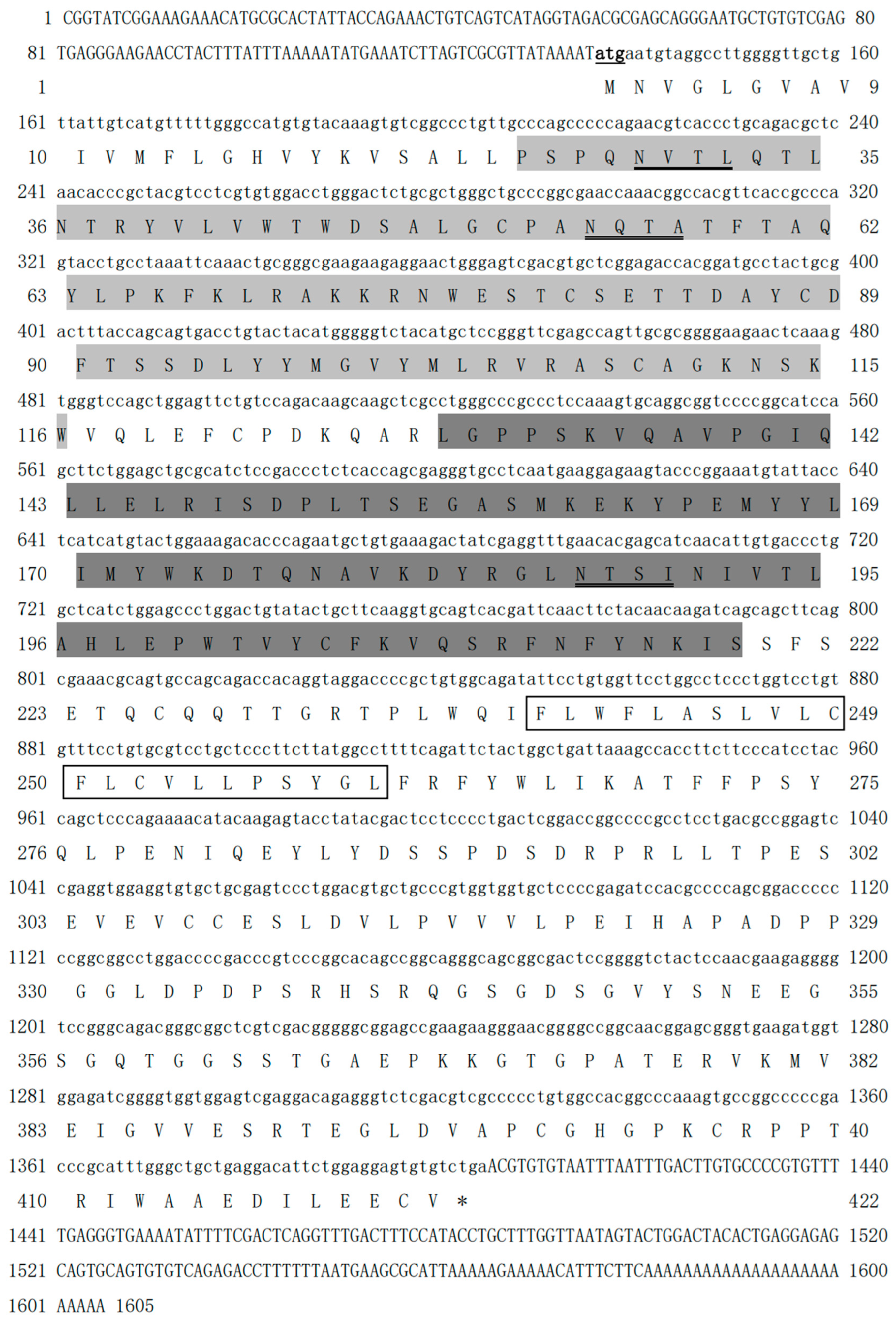
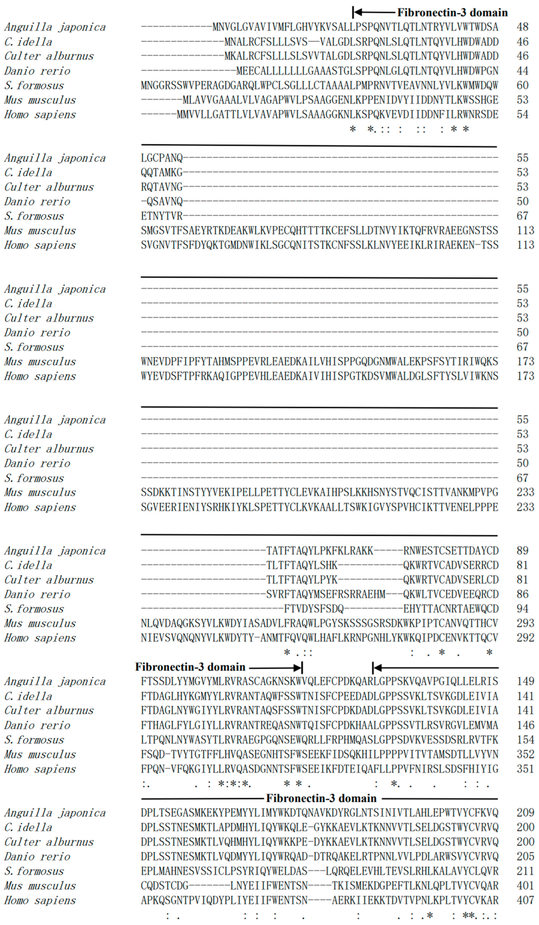
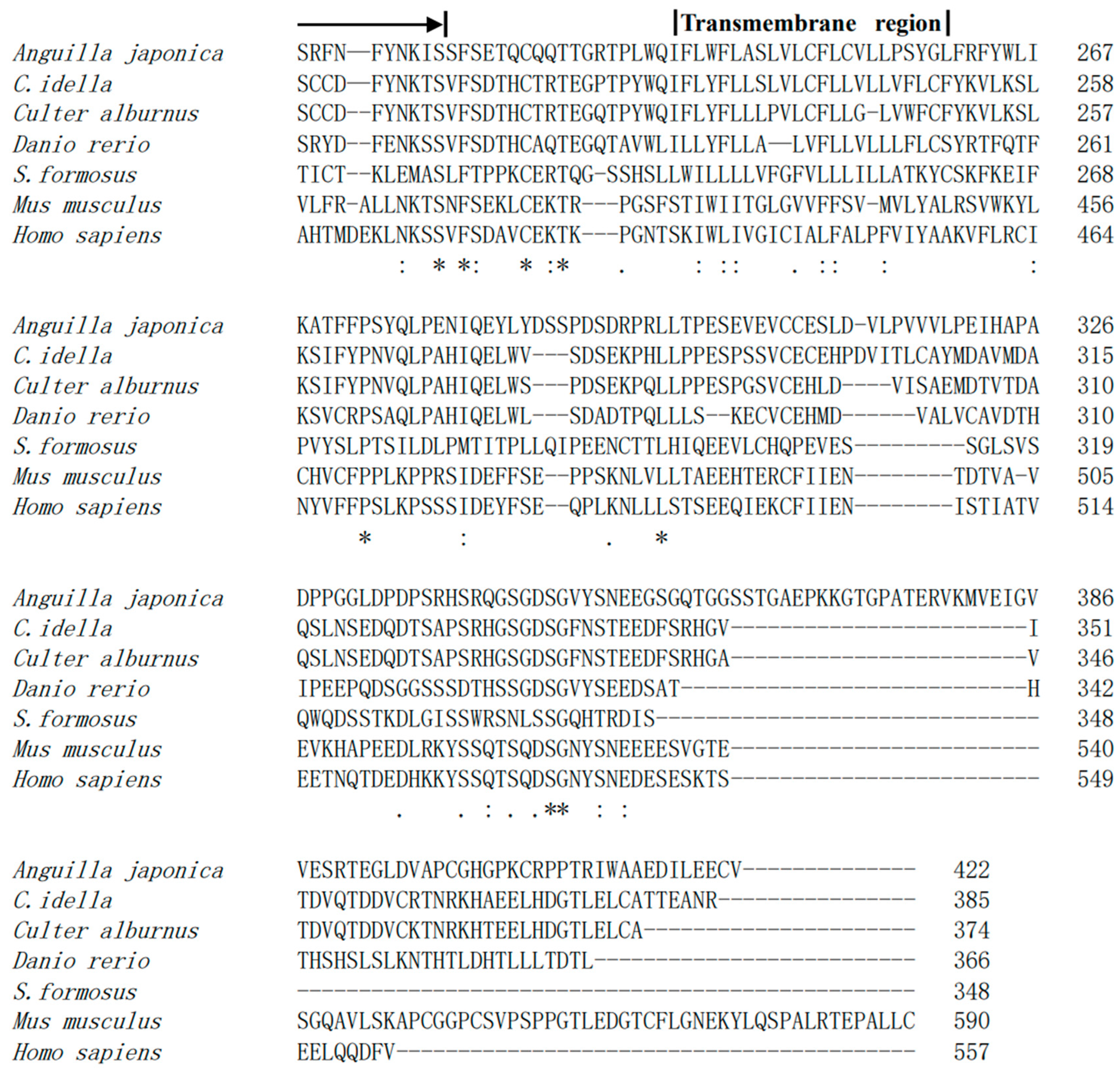
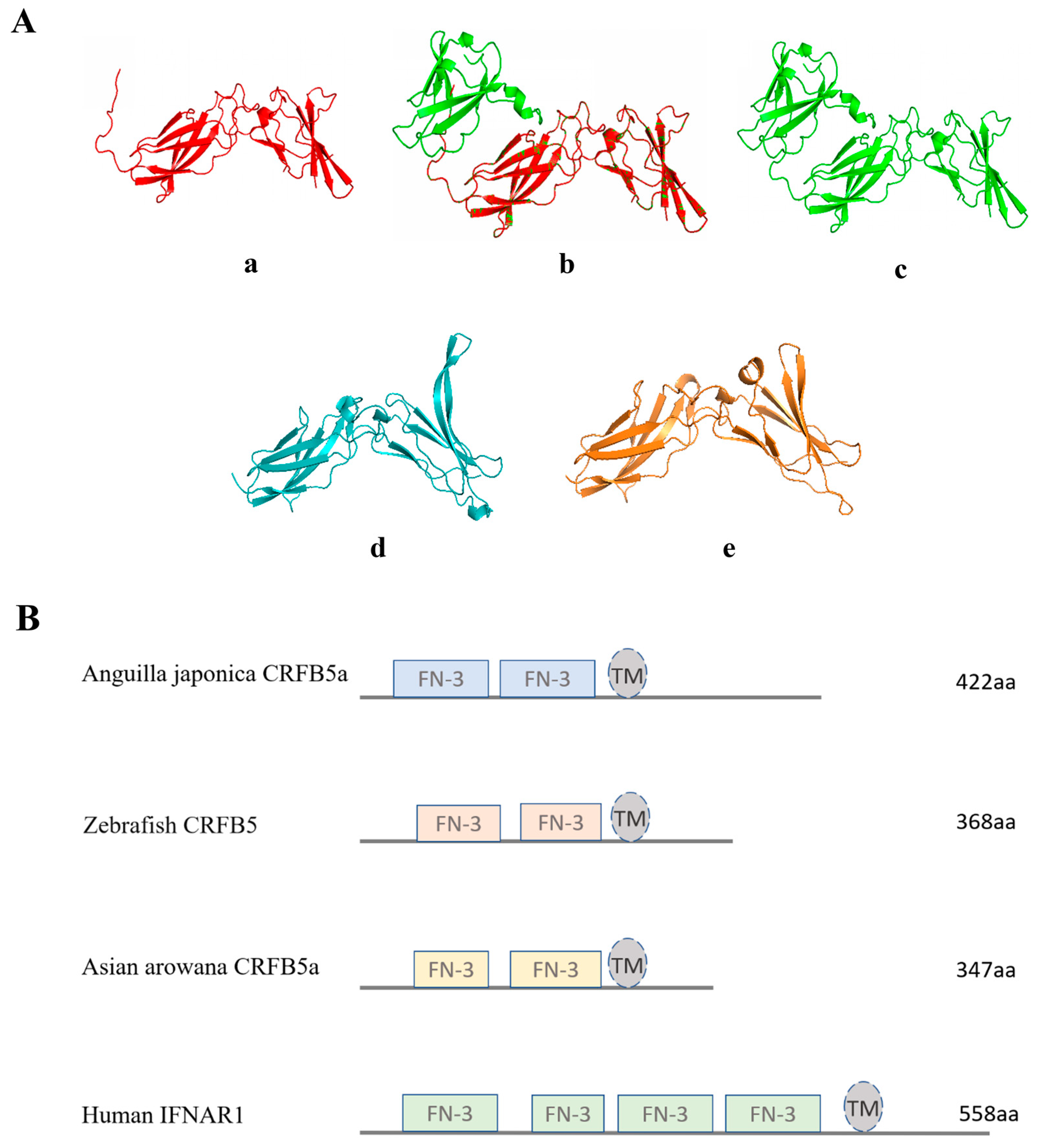
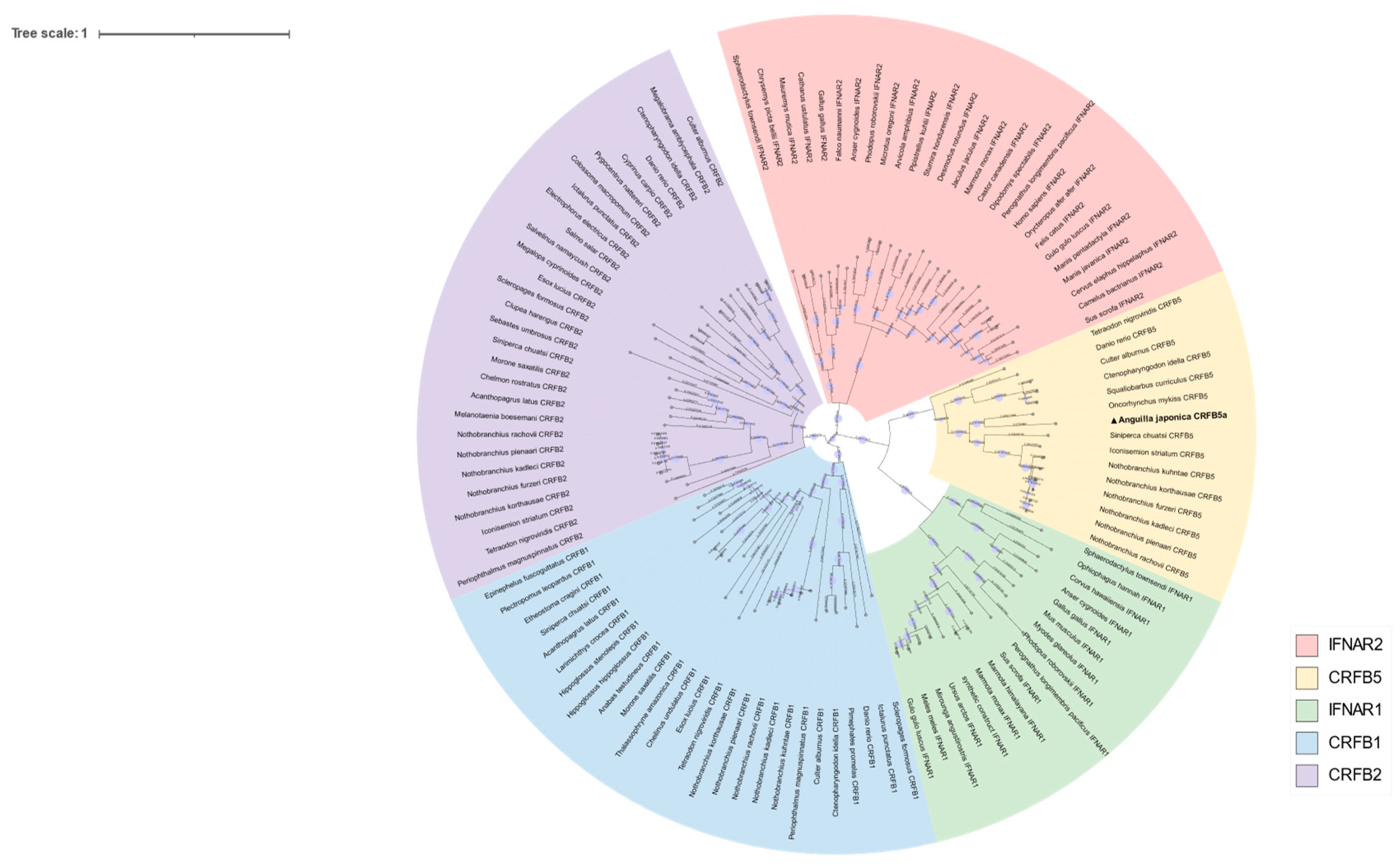
3.1. Sequence Analysis of CRFB5a from Japanese Eel

The cDNA of CRFB5a (GenBank registration number: MF537666) is 1605 bp and contains 135 bp of 5′ untranslated regions (UTR), 201 bp of 3′-UTR, and 1269 bp of ORF, which encoding 422 amino acids (aa) containing two fibronectin-3 domains (25–116 aa and 129–219 aa) and a transmembrane region (238–260 aa) (Figure 1). The fibronectin-3 domain and transmembrane region of AjCRFB5a were found to be preserved when it was compared to CRFB5 from zebrafish, Asian arowana, grass carp, topmouth culter, or IFNAR1 from mice and humans. (Figure 2). The three-dimensional structure of AjCRFB5a was similar to that of zebrafish and Asian arowana (Figure 3). Additionally, together with zebrafish CRFB5, AjCRFB5a, and other teleost CRFB5 clustered to one branch, whereas IFNAR1 and IFNAR2 from the mammals, birds, and reptiles or CRFB1 and CRFB2 from other bony fish formed another branch (Figure 4).

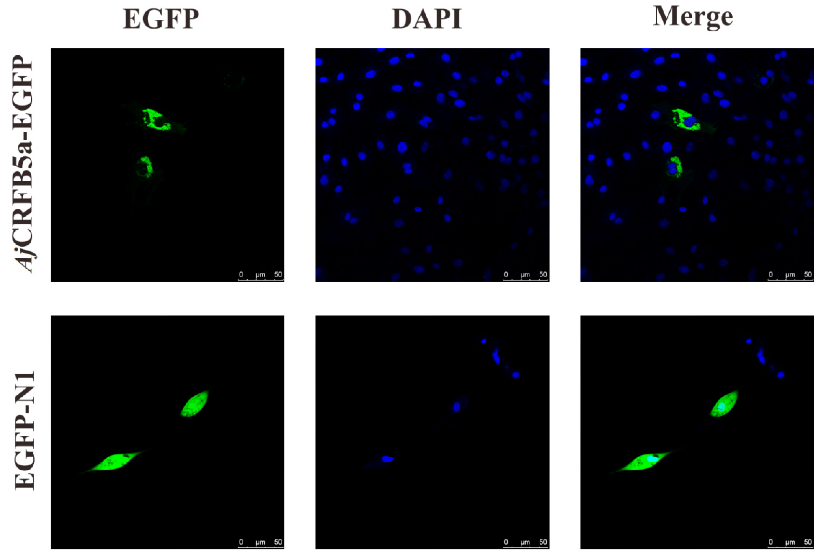
3.2. Subcellular Localization of AjCRFB5a

Fluorescence microscopy results showed that EGFP-AjCRFB5a proteins were distributed on the membranes with some of them in a condensed form in Japanese eel liver cells (Figure 5).

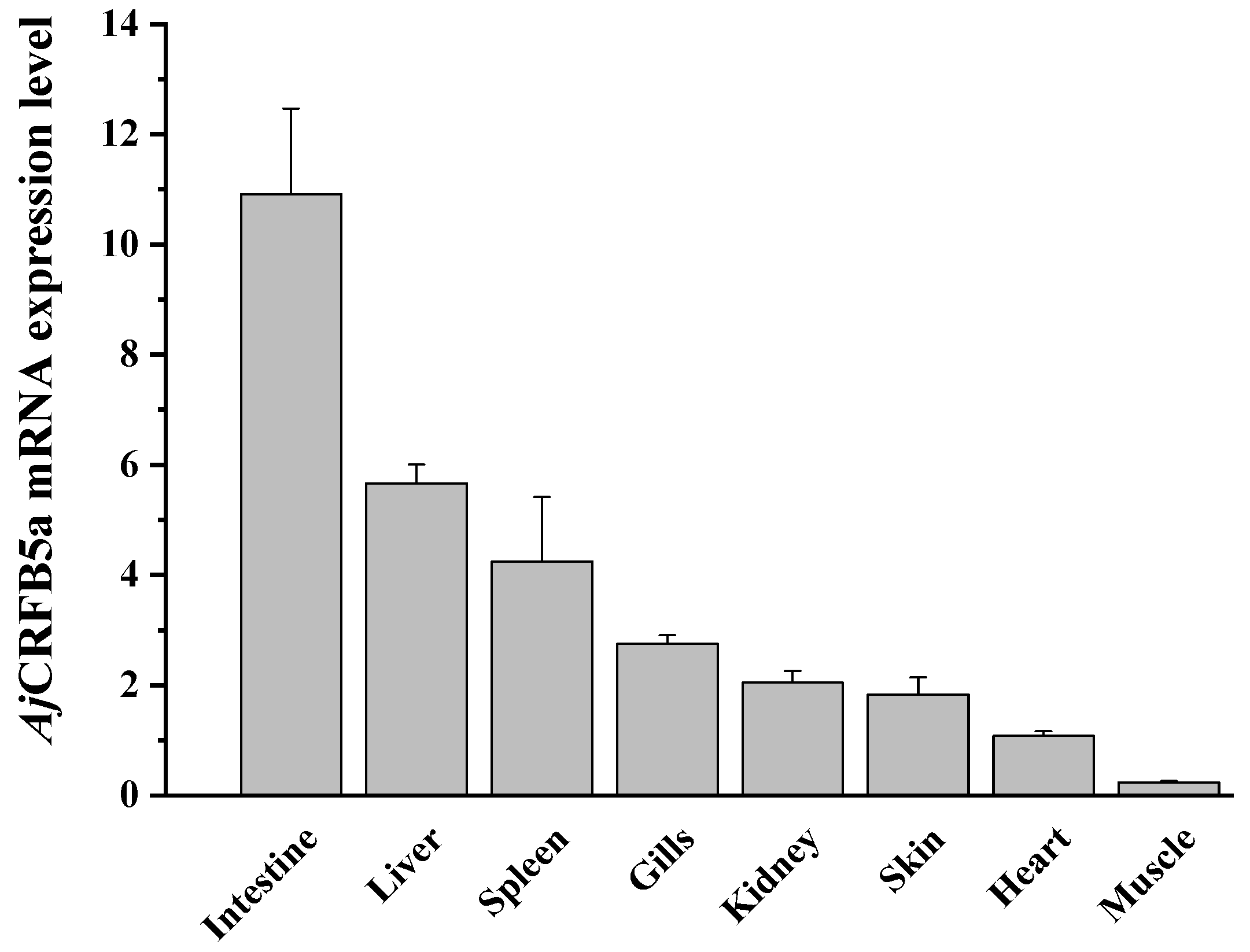
3.3. Tissue Expression of AjCRFB5a

The outcomes demonstrated that all identified organs and tissues expressed CRFB5a, with a higher expression level in the intestine and liver, followed by the spleen, gill, and kidney, and lower expression levels in the skin, heart, and muscle (Figure 6).

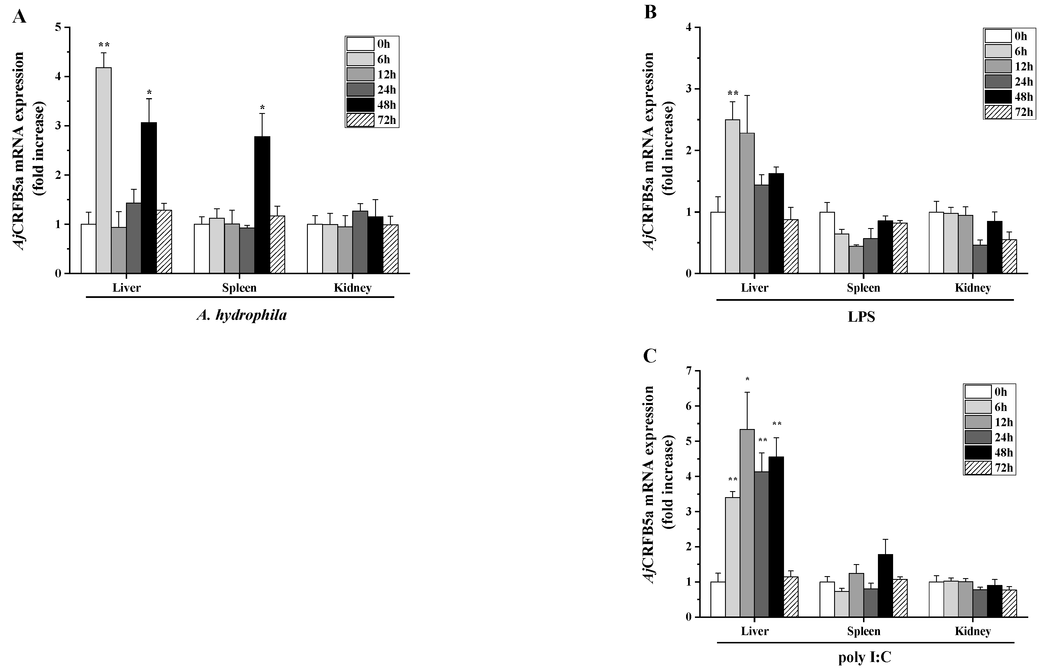
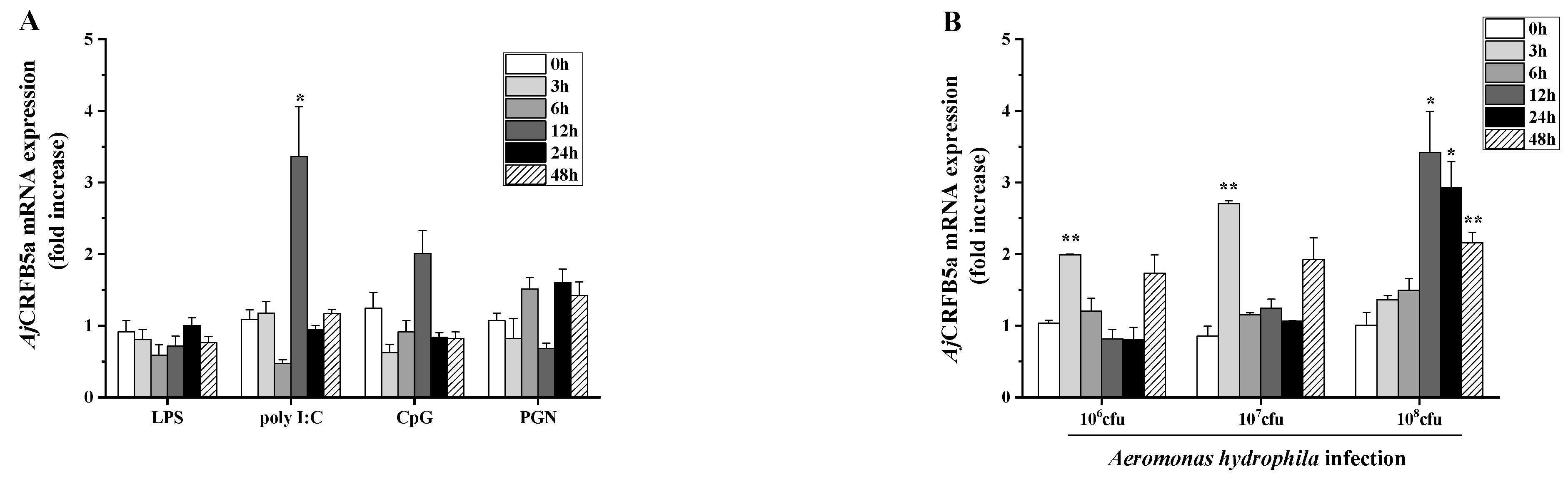
3.4. Gene Expression of AjCRFB5a In Vivo upon Stimulation

RT-qPCR analysis was used to identify the amounts of AjCRFB5a gene expression in the liver, spleen, and kidney of Japanese eels stimulated with LPS, poly I:C, and A. hydrophila. AjCRFB5a was increased following A. hydrophila infection, with 4.2 and 3.1 fold at 6 h and 48 h in the liver, and 2.8 fold at 48 h in the spleen (Figure 7A). Under LPS stimulation, AjCRFB5a was up-regulated 2.5 fold at 6 h in the liver, but no up-regulation of AjCRFB5a was detected in the kidney and spleen (Figure 7B). AjCRFB5a was also induced by poly I:C, with 3.4-, 5.3-, 4.1-, and 4.6 fold at 6 h, 12 h, 24 h, and 48 h in the liver, but no up-regulation of AjCRFB5a was detected in the kidney and spleen (Figure 7C).

3.5. Gene Expression of AjCRFB5a In Vitro after Stimulation

The expression of AjCRFB5a was up-regulated with 3.4-fold at 12 h upon poly I:C challenge, LPS, CpG, and PGN failed to induce CRFB5a gene (Figure 8A), or 2.0 and 2.7 fold at 3 h by the infection of 1 × 10⁶ cfu/mL, 1 × 10⁷ cfu/mL of A. hydrophila, and when the concentration reached 1 × 10⁸ cfu/mL, there was a 3.4-, 2.8-, and 2.2-fold increase at 12 h, 24 h, and 48 h, respectively (Figure 8B).

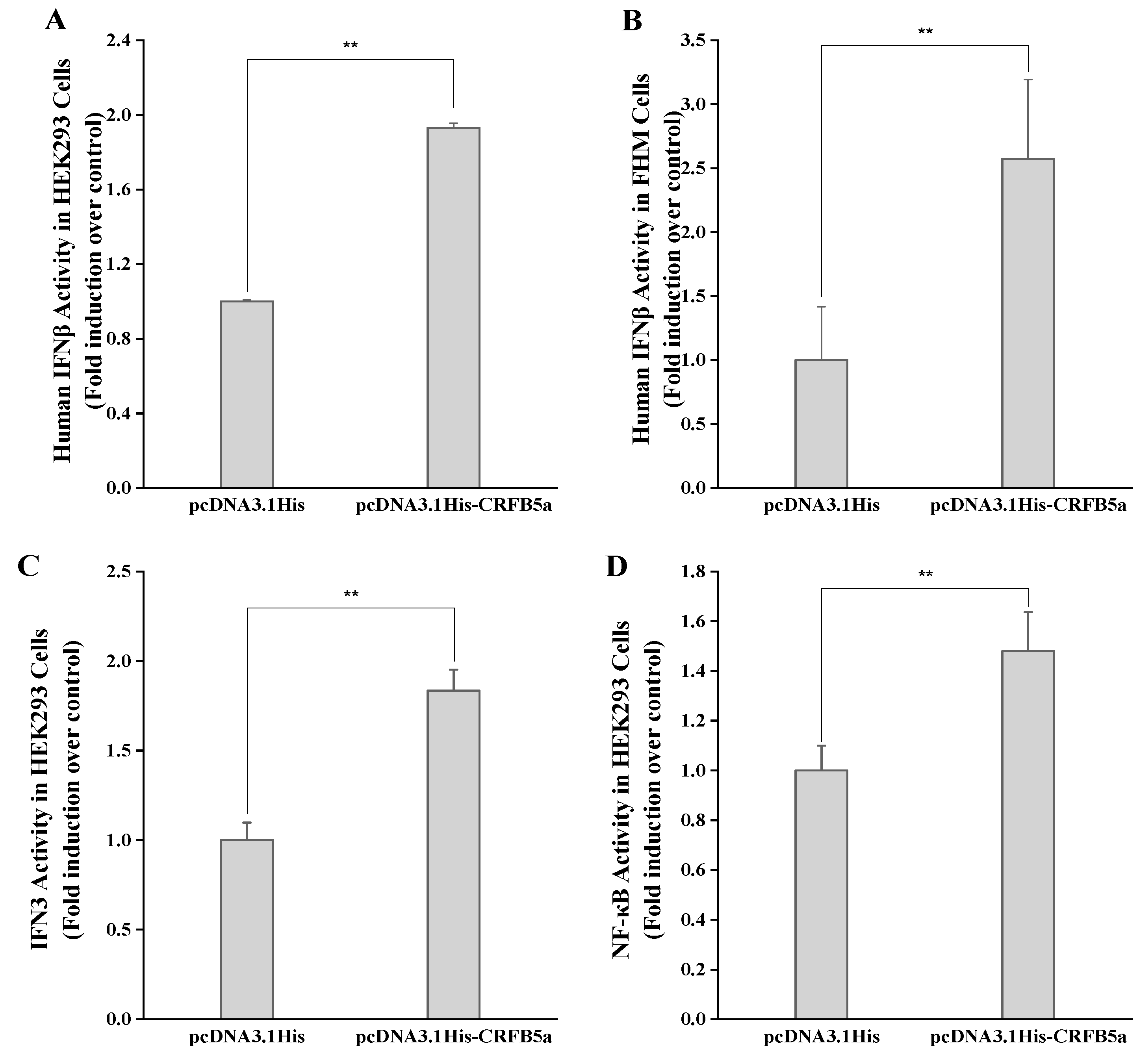
3.6. Dual Luciferase Activity of the AjCRFB5a Gene

To evaluate the effect of AjCRFB5a on the type I IFN and NF-κB signaling pathways, we co-transfected pcDNA3.1His-AjCRFB5a with human IFNβ, NF-κB luciferase reporter plasmids, and AjIFN3 promoter into HEK293 cells and FHM cells. The results showed that the activity of pcDNA3.1His-AjCRFB5a luciferase was significantly increased compared with pcDNA3.1His (Figure 9).

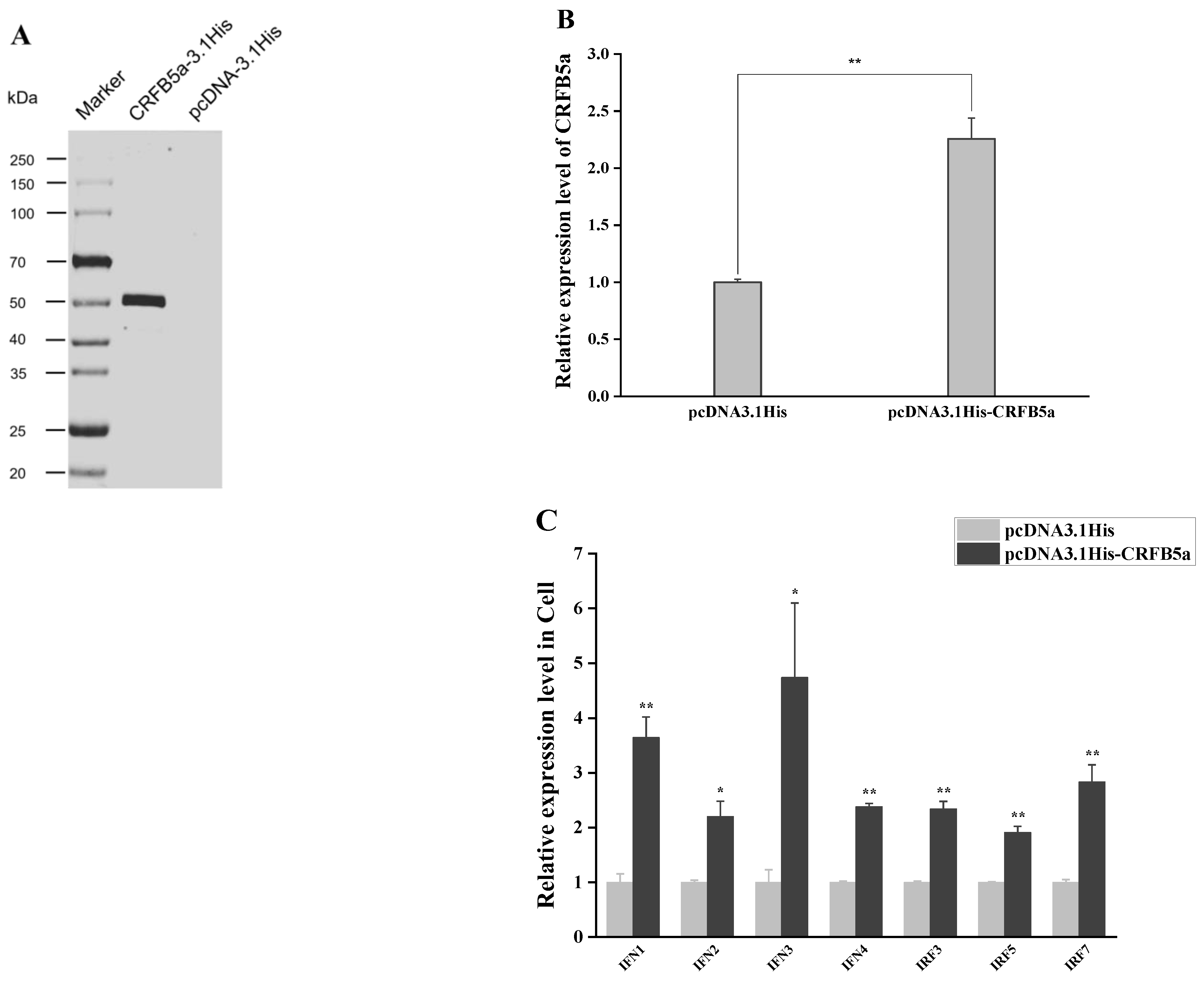
3.7. Induction of Immune-Related Genes under Overexpression of AjCRFB5a

The molecular weight of AjCRFB5a was calculated to be 47.24 kDa by the Novopro online tool, and Western blot analysis using Anti-His antibody showed that the molecular weight of the pcDNA3.1His-AjCRFB5a fusion protein was close to 50 kDa (Figure 10A). The expression profiles of AjCRFB5a and immune-related genes under overexpression of AjCRFB5a in Japanese eel liver cells were assayed using RT-qPCR. It was found that CRFB5a was successfully overexpressed in Japanese eel liver cells (Figure 10B). AjCRFB5a overexpression significantly enhanced the expression of immune-related genes in Japanese eel liver cells, including IFN1, IFN2, IFN3, IFN4, IRF3, IRF5, and IRF7 of type I IFN signaling pathway (Figure 10C).

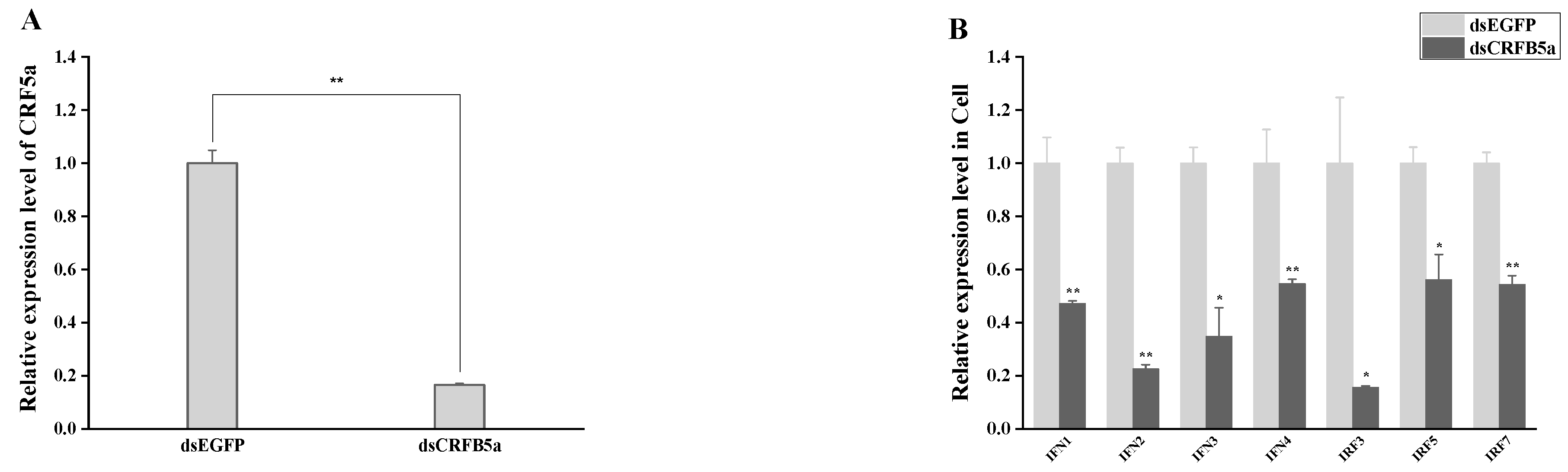
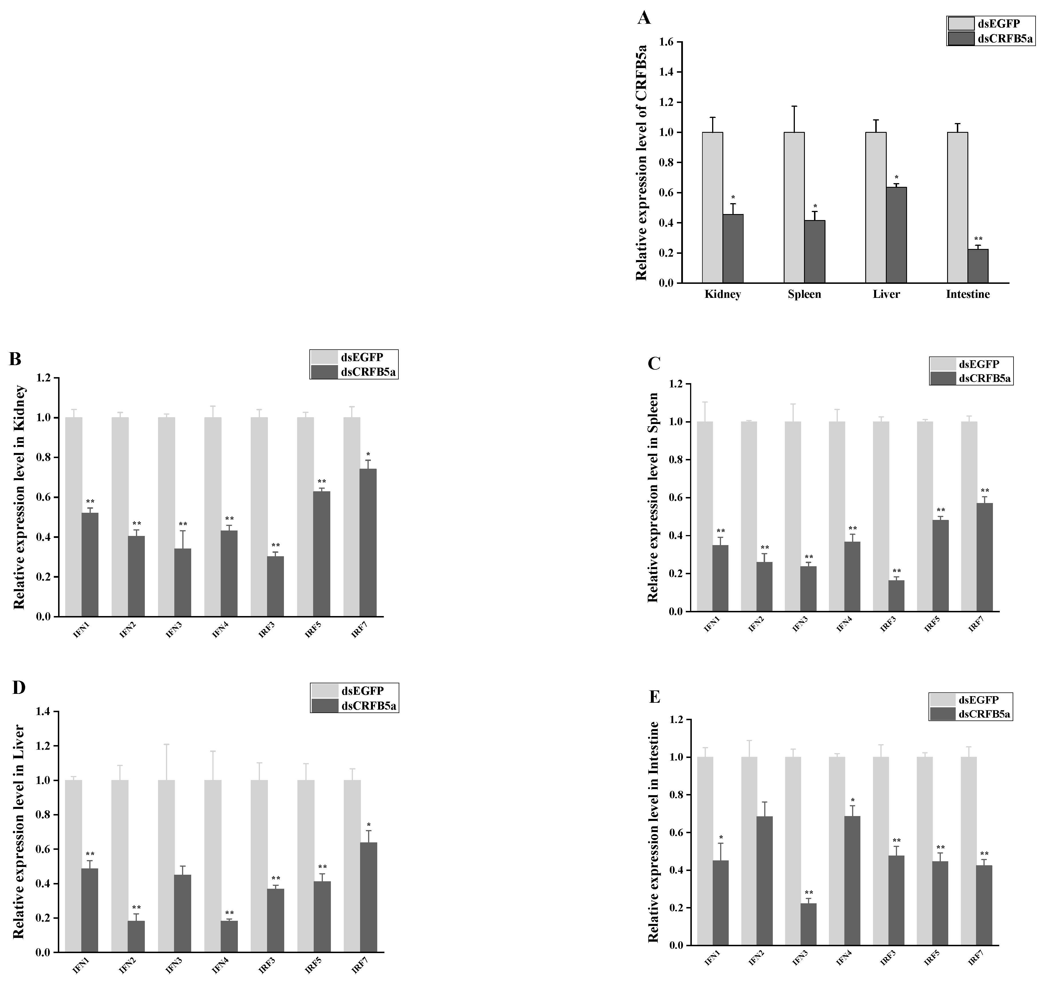
3.8. The effect of AjCRFB5a Gene Silencing on the Immune-Related Genes from Type I IFN Signaling Pathways

RT-qPCR analysis was used to assess the expression patterns of AjCRFB5a as well as immune-related genes in the liver, spleen, kidney, intestine, gills, and Japanese eel liver cells. The RT-qPCR results showed that the expression of AjCRFB5a in the liver cells of Japanese eel was also significantly inhibited (Figure 11A). Meanwhile, the expression of AjCRFB5a was significantly inhibited in the liver, spleen, kidney, and intestine (Figure 12A). Knocking down AjCRFB5a caused the expression of IFN1, IFN2, IFN3, IFN4, IRF3, IRF5, and IRF7 decreased to varying degrees in the kidney (Figure 12B), spleen (Figure 12C), liver (Figure 12D), and intestine (Figure 12E). All those genes were significantly down-regulated in Japanese eel liver cells following the inhibition of AjCRFB5a (Figure 11B).

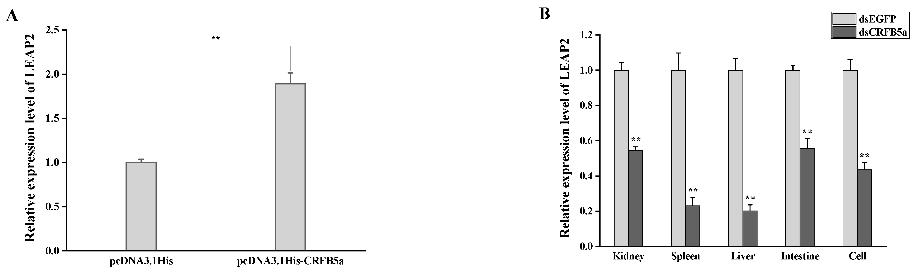
3.9. The Effect of AjCRFB5a on the Antimicrobial Peptide LEAP2

The expression profiles of the key antimicrobial peptide gene LEAP2 under AjCRFB5a overexpression and gene silencing were examined using RT-qPCR. The results revealed that the expression of the LEAP2 gene was significantly up-regulated after AjCRFB5a was overexpressed in Japanese eel liver cells (Figure 13A). In both in vitro and in vitro experiments, LEAP2 gene expression was significantly down-regulated after AjCRFB5a was knocked down in the kidney, spleen, liver, intestine, and Japanese eel liver cells (Figure 13B).

4. Discussion

In mammals, through binding with its receptors and triggering the JAK/STAT signaling cascade, type I IFN plays a key role in host antiviral immunity, ultimately leading to the production of Mx proteins and other antiviral proteins to prevent virus infection [6]. The immune function research of Type I IFN receptors in teleost fishes is now in the starting stage and is mainly involved in the recognition and interaction between CRFB and IFNs. However, whether fish CRFB is involved in the activation of known antiviral immune-related type I IFN pathways, or inflammatory signaling pathways including NF-κB and MAKP signaling pathways induced by viral or bacterial infections, remains largely unknown. In this study, we cloned a Japanese eel CRFB5a homolog, identified immune challenge-induced changes in its expression patterns in vivo and in vitro, and assessed the function of Japanese eel CRFB5a in regulating the activation of signaling pathways such as type I IFN, NF-κB, and MAPK signaling pathways by overexpressing or inhibiting AjCRFB5a using RNAi in vivo or in vitro. The current research offers a fresh understanding of CRFB5a’s multiple functions in vertebrates’ innate immune response.
According to multiple alignments and NCBI CDD analysis, a transmembrane region and two typical FN3 domains were found in CRFB5a showing a similar three-dimensional structure to zebrafish CRFB5, Asian arowana CRFB5a, and human IFNAR1 [18,20]. Together with the finding that overexpression of AjCRFB5a potently induced human IFN-β promoter activity in HEK293 cells, AjCRFB5a might possess the binding affinity with its corresponding IFNs to trigger immune-related signaling pathways similar to those mammalian IFNAR1. Unlike uniformly distributed EGFP in the entire cells, AjCRFB5a is mainly present in the membranes of Japanese eel liver cells, with no distribution in the nucleus, which is consistent with the findings in orange-spotted grouper [18]. The constitutive expression pattern of AjCRFB5a in Japanese eel was in line with the reports from Asian arowana, orange-spotted grouper, and Dabry’s sturgeon (Acipenser dabryanus) [18,20,29]. However, CRFB5 was found to be highly expressed in the liver of Japanese eel and Asian arowana, and lowly in the liver of orange-spotted grouper, which indicated that CRFB5 plays different roles in immune-related tissues due to the species difference [18,20].
The synthetic dsRNA poly I:C, recognized by TLR3, RIG-I, or MDA5 in the cytoplasm of all somatic cells [30], has been widely applied to mimic RNA viral infection to initiate the type I IFN signal transduction pathway [31]. Here, we discovered that poly I:C substantially increased the expression of AjCRFB5a in the liver, which was congruent with findings from Asian arowana [20]. The enhanced AjCRFB5a expression was also found in Japanese eel liver cells following stimulation with poly I: C, which is similar to the findings in grass carp kidney cells [32] and Dabry’s sturgeon primary spleen leukocytes [29]. Thus, these findings collectively suggest that AjCRFB5a might be involved in controlling host antiviral immune responses. In mammals, IFNAR1 and IFNAR2 were combined with IFN to activate the JAK/STAT signaling pathway, resulting in the expression of Mx proteins and other antiviral proteins [8,9]. Although the effect of type I IFN receptors on the activation of the immune-related signaling pathway remains unclear, recent studies documented that knocking down IFNAR1 in orange-spotted grouper caused a decreased number of nervous necrosis in grouper fin cells [18], and the complex of CRFB2 and CRFB5a in Asian arowana was found to combine with IFNb to enhance the gene expression of Mx and Vipein in EPC cells [20]. In order to answer the question of which mechanism is available for the elevated AjCRFB5a expression contributing to antiviral responses, we determined its potential roles in the regulation of IRF3/IRF7-mediated type I IFN by overexpression or RNAi of AjCRFB5a in vivo and in vitro. Our results showed that AjCRFB5a overexpression significantly enhanced the expression of IFN1, IFN2, IFN3, and IFN4, as well as transcriptional factors including IRF3, IR5, and IRF7. The generation of type I interferon (IFN) in mammals is induced by the phosphorylation of IRF3 and/or IRF7, which results in their dimerization and nuclear translocation [33]. Huang et al. [34] cloned four type I IFN (IFN1, 2, 3, 4) from Japanese eel and found that overexpression of IFN1-4 induced the luciferase activity of the Mx gene reporter. Hence, AjCRFB5a positively regulates host antiviral immune responses through the activation of IRF3 and IRF7-mediated type I IFN signaling pathways, resulting in the expression of Mx proteins or other antiviral proteins. Corresponding to the results of AjCRFB5a overexpression in vitro, knocking down the AjCRFB5a gene in the liver, spleen, and kidney of Japanese eel caused a significant decrease in expression of IFN1, IFN2, and IFN3, IFN4, IRF3, and IRF5, and IRF7 in all those tissues, which provide first-time evidence in vivo for fish CRFB involvement in the regulation of the type I IFN signaling pathway. Thus, the findings of overexpressing AjCRFB5a in vitro and knocking it down in vivo demonstrated that AjCRFB5a positively regulated the IRF3 or IRF7 depending on the type I IFN signaling pathway and played an important role in the antiviral immune response.
LPS is the major active component of endotoxins and performs important pathophysiological functions by activating the NF-κB signaling pathway [35]. LPS significantly increased AjCRFB5a expression in the liver of Japanese eels, which is consistent with the results from orange-spotted grouper [18] and suggested that AjCRFB5a might take part in the host antibacterial immune response. To clarify the role of AjCRFB5a in the inflammatory response against bacterial infection, we herein used a major aquatic pathogenic bacterium, A. hydrophila, to challenge Japanese eel in vivo and Japanese eel liver cells in vitro. A. hydrophila strongly induced the expression of AjCRFB5a in the Japanese eel liver and spleen, as well as the Japanese eel liver cells in vitro. This is direct proof that CRFB5 is involved in the anti-bacterial immune response of the host defense in teleost fishes, despite the fact that the data on the expression pattern of fish CRFB5 induced by bacterial infection is still limited [20]. NF-κB and AP-1 are well-known as the key elements in inflammatory responses that regulate the expression of inflammatory cytokines [35,36]. To further understand the regulatory mechanism mediated by the elevated AjCRFB5a expression upon host inflammatory response against pathogen invasion, the expression level of the important liver-expressed antimicrobial peptide LEAP2 was analyzed under the overexpression and RNAi of AjCRFB5a in vivo or in vitro. In Japanese eel liver cells, AjCRFB5a overexpression significantly up-regulated LEAP2 [37]. The results suggest that AjCRFB5a is able to produce antimicrobial immunity in the host by activating LEAP2.

5. Conclusions

In conclusion, the CRFB5a gene was identified in Japanese eels, and poly I:C stimulation and A. hydrophila infection can increase the gene’s expression. AjCRFB5a functions in the host immune response to bacterial infection by activating antimicrobial peptide LEAP2, and it works as a positive regulator of host antiviral immune responses by activating IRF3 and IRF7-mediated type I IFN signaling pathways.

Author Contributions

J.F. drafted the manuscript, designed and carried out a part of the experimental work; T.W. carried out a major part of the experimental work; P.L., P.C. and F.L. carried out a part of the experimental work; Y.W. and X.L. contributed to the discussion of this study and edited the manuscript. All authors have read and agreed to the published version of the manuscript.

Funding

This work was financially supported by the “Nature Science Foundation of Fujian Province” (No. 2020J01671, 2021J01830), “the Open Program of Key Laboratory of Cultivation and High-value Utilization of Marine Organisms in Fujian Province” (No. 2021fjscq03), “Engineering Research Center of the Modern Technology for Eel Industry, Ministry of Education, China” (No. RE202307, RE202208), and “Key Laboratory and Healthy Mariculture for the East China Sea, Ministry of Agriculture and Rural Affairs, China” (No. 2020ESHML02).

Institutional Review Board Statement

This study was conducted according to the guidelines of the Declaration of Helsinki, and approved by the Ethics Committee of the Animal Care and Use Committee of the Fisheries College of Jimei University (Animal Ethics No. 1067).

Informed Consent Statement

Not applicable.

Data Availability Statement

The datasets used or analyzed during the current study are available from the corresponding author upon reasonable request.

Conflicts of Interest

The authors declare no conflict of interest.

References

  1. Li, S.; Lu, L.-F.; Feng, H.; Wu, N.; Chen, D.-D.; Zhang, Y.-B.; Gui, J.-F.; Nie, P.; Zhang, Y.-A. IFN Regulatory Factor 10 Is a Negative Regulator of the IFN Responses in Fish. J. Immunol. 2014, 193, 1100–1109. [Google Scholar] [CrossRef] [PubMed]
  2. Pellefigues, C.; Tang, S.-C.; Schmidt, A.; White, R.F.; Lamiable, O.; Connor, L.M.; Ruedl, C.; Dobrucki, J.; Le Gros, G.; Ronchese, F. Toll-Like Receptor 4, but Not Neutrophil Extracellular Traps, Promote IFN Type I Expression to Enhance Th2 Responses to Nippostrongylus brasiliensis. Front. Immunol. 2017, 8, 1575. [Google Scholar] [CrossRef] [PubMed]
  3. Liu, F.; Bols, N.C.; Pham, P.H.; Secombes, C.J.; Zou, J. Evolution of IFN subgroups in bony fish—1: Group I-III IFN exist in early ray-finned fish, with group II IFN subgroups present in the Holostean spotted gar, Lepisosteus oculatus. Fish Shellfish Immunol. 2019, 95, 163–170. [Google Scholar] [CrossRef]
  4. Li, L.; Chen, S.N.; Laghari, Z.A.; Huo, H.J.; Hou, J.; Huang, L.; Li, N.; Nie, P. Myxovirus resistance (Mx) gene and its differential expression regulated by three type I and two type II IFNs in mandarin fish, Siniperca chuatsi. Dev. Comp. Immunol. 2020, 105, 103604. [Google Scholar] [CrossRef]
  5. Wei, L.; Zhang, R.; Zhang, J.; Li, J.; Kong, D.; Wang, Q.; Fang, J.; Wang, L. PRKAR2A deficiency protects mice from experimental colitis by increasing IFN-stimulated gene expression and modulating the intestinal microbiota. Mucosal Immunol. 2021, 14, 1282–1294. [Google Scholar] [CrossRef] [PubMed]
  6. Duncan, C.J.; Randall, R.E.; Hambleton, S. Genetic Lesions of Type I Interferon Signalling in Human Antiviral Immunity. Trends Genet. 2021, 37, 46–58. [Google Scholar] [CrossRef]
  7. Gui, J.; Zhao, B.; Lyu, K.; Tong, W.; Fuchs, S.Y. Downregulation of the IFNAR1 chain of type 1 interferon receptor contributes to the maintenance of the haematopoietic stem cells. Cancer Biol. Ther. 2017, 18, 534–543. [Google Scholar] [CrossRef]
  8. Walter, M.R. The Role of Structure in the Biology of Interferon Signaling. Front. Immunol. 2020, 11, 606489. [Google Scholar] [CrossRef]
  9. Langevin, C.; Boudinot, P.; Collet, B. IFN Signaling in Inflammation and Viral Infections: New Insights from Fish Models. Viruses 2019, 11, 302. [Google Scholar] [CrossRef]
  10. Liu, Y.; Du, H.; Wang, S.; Lv, Y.; Deng, H.; Chang, K.; Zhou, P.; Hu, C. Grass carp (Ctenopharyngodon idella) TNK1 modulates JAK-STAT signaling through phosphorylating STAT1. Dev. Comp. Immunol. 2021, 116, 103951. [Google Scholar] [CrossRef]
  11. Gong, X.-Y.; Zhang, Q.-M.; Gui, J.-F.; Zhang, Y.-B. SVCV infection triggers fish IFN response through RLR signaling pathway. Fish Shellfish Immunol. 2019, 86, 1058–1063. [Google Scholar] [CrossRef] [PubMed]
  12. Lutfalla, G.; Crollius, H.R.; Stange-Thomann, N.; Jaillon, O.; Mogensen, K.; Monneron, D. Comparative genomic analysis reveals independent expansion of a lineage-specific gene family in vertebrates: The class II cytokine receptors and their ligands in mammals and fish. BMC Genom. 2003, 4, 29. [Google Scholar] [CrossRef]
  13. Chen, Y.-M.; Kuo, C.-E.; Chen, G.-R.; Kao, Y.-T.; Zou, J.; Secombes, C.J.; Chen, T.-Y. Functional analysis of an orange-spotted grouper (Epinephelus coioides) interferon gene and characterisation of its expression in response to nodavirus infection. Dev. Comp. Immunol. 2014, 46, 117–128. [Google Scholar] [CrossRef] [PubMed]
  14. Gan, Z.; Cheng, J.; Chen, S.; Hou, J.; Li, N.; Xia, H.; Xia, L.; Lu, Y.; Nie, P. Identification and characterization of tilapia CRFB1, CRFB2 and CRFB5 reveals preferential receptor usage of three IFN subtypes in perciform fishes. Fish Shellfish Immunol. 2020, 107, 194–201. [Google Scholar] [CrossRef] [PubMed]
  15. Chen, H.; Liu, W.; Wang, B.; Mao, H.; Sun, Z.; Hou, Q.; Mi, Y.; Fan, L.; Hu, C. Cloning, identification of the two cytokine receptor family B subunits CRFB1 and CRFB5 from grass carp (Ctenopharyngodon idella). Fish Shellfish Immunol. 2015, 45, 211–220. [Google Scholar] [CrossRef]
  16. Sun, B.; Greiner-Tollersrud, L.; Koop, B.F.; Robertsen, B. Atlantic salmon possesses two clusters of type I interferon receptor genes on different chromosomes, which allows for a larger repertoire of interferon receptors than in zebrafish and mammals. Dev. Comp. Immunol. 2014, 47, 275–286. [Google Scholar] [CrossRef]
  17. Pang, A.N.; Chen, S.N.; Gan, Z.; Li, L.; Li, N.; Wang, S.; Sun, Z.; Liu, L.H.; Sun, Y.L.; Song, X.J.; et al. Identification of type II interferons and receptors in an osteoglossiform fish, the arapaima Arapaima gigas. Dev. Comp. Immunol. 2023, 139, 104589. [Google Scholar] [CrossRef]
  18. Tang, Z.Z.; Wang, T.-Y.; Chen, Y.-M.; Chen, T.-Y. Cloning and characterisation of type I interferon receptor 1 in orange-spotted grouper (Epinephelus coioides) for response to nodavirus infection. Fish Shellfish Immunol. 2020, 101, 302–311. [Google Scholar] [CrossRef]
  19. Stein, C.; Caccamo, M.; Laird, G.; Leptin, M. Conservation and divergence of gene families encoding components of innate immune response systems in zebrafish. Genome Biol. 2007, 8, R251. [Google Scholar] [CrossRef]
  20. Wang, S.; Chen, S.N.; Sun, Z.; Pang, A.N.; Wang, S.; Liu, L.H.; Liu, Y.; Nie, P. Four type I IFNs, IFNa1, IFNa2, IFNb, IFNc, and their receptor usage in an osteoglossomorph fish, the Asian arowana, Scleropages formosus. Fish Shellfish Immunol. 2021, 117, 70–81. [Google Scholar] [CrossRef]
  21. Liao, Z.; Wan, Q.; Su, J. Bioinformatics analysis of organizational and expressional characterizations of the IFNs, IRFs and CRFBs in grass carp Ctenopharyngodon idella. Dev. Comp. Immunol. 2016, 61, 97–106. [Google Scholar] [CrossRef]
  22. Wang, Z.; Xu, J.; Feng, J.; Wu, K.; Chen, K.; Jia, Z.; Zhu, X.; Huang, W.; Zhao, X.; Liu, Q.; et al. Structural and Functional Analyses of Type I IFNa Shed Light into Its Interaction with Multiple Receptors in Fish. Front. Immunol. 2022, 13, 862764. [Google Scholar] [CrossRef]
  23. Takeuchi, A.; Watanabe, S.; Yamamoto, S.; Miller, M.; Fukuba, T.; Miwa, T.; Okino, T.; Minamoto, T.; Tsukamoto, K. First use of oceanic environmental DNA to study the spawning ecology of the Japanese eel Anguilla japonica. Mar. Ecol. Prog. Ser. 2019, 609, 187–196. [Google Scholar] [CrossRef]
  24. Itakura, H.; Miyake, Y.; Kitagawa, T.; Kimura, S. Site fidelity, diel and seasonal activities of yellow-phase Japanese eels (Anguilla japonica) in a freshwater habitat as inferred from acoustic telemetry. Ecol. Freshw. Fish 2018, 27, 737–751. [Google Scholar] [CrossRef]
  25. Mizutani, T.; Sayama, Y.; Nakanishi, A.; Ochiai, H.; Sakai, K.; Wakabayashi, K.; Tanaka, N.; Miura, E.; Oba, M.; Kurane, I.; et al. Novel DNA virus isolated from samples showing endothelial cell necrosis in the Japanese eel, Anguilla japonica. Virology 2011, 412, 179–187. [Google Scholar] [CrossRef]
  26. Schulz, P.; Kaczorek, E.; Małaczewska, J.; Siwicki, A. Immunopathogenesis of Herpesviruses: Influence of Anguillid Herpes Virus 1 on Macrophage and Lymphocyte Activity in European eel (Anguilla anguilla). J. Comp. Pathol. 2016, 154, 97. [Google Scholar] [CrossRef]
  27. Feng, J.J.; Xu, Y.K.; Lin, P.; Wang, Y.L.; Zhang, Z.P.; Zou, P.F.; Peng, X.W. Fish IKK alpha from Japanese eel (Anguilla japonica) can activate NF-kappa B, AP1, and type I IFN signaling pathways. Fish Shellfish Immunol. 2020, 106, 982–992. [Google Scholar] [CrossRef] [PubMed]
  28. Wan, H.; Zhong, J.; Zhang, Z.; Zou, P.; Wang, Y. Comparative Transcriptome Reveals the Potential Modulation Mechanisms of Spfoxl-2 Affecting Ovarian Development of Scylla paramamosain. Mar. Biotechnol. 2022, 24, 125–135. [Google Scholar] [CrossRef]
  29. Luo, K.; Zhang, S.; Tang, D.; Xia, L.; Gao, W.; Tian, G.; Qi, Z.; Xu, Q.; Zhang, W. Analysis of the expression patterns of the cytokine receptor family B (CRFB) and interferon gamma receptor (IFNGR) in Dabry’s sturgeon (Acipenser dabryanus). Dev. Comp. Immunol. 2018, 84, 420–426. [Google Scholar]
  30. Kato, H.; Takeuchi, O.; Sato, S.; Yoneyama, M.; Yamamoto, M.; Matsui, K.; Uematsu, S.; Jung, A.; Kawai, T.; Ishii, K.J.; et al. Differential roles of MDA5 and RIG-I hel-icases in the recognition of RNA viruses. Nature 2006, 441, 101–105. [Google Scholar] [CrossRef]
  31. Zhang, J.; Sun, S.; Mao, Y.; Qiao, G.; Li, Q. Identification and analysis of differentially expressed microRNAs in gibel carp Carassius auratus gibelio responding to polyinosinic-polycytidylic acid (poly I:C) stimulation. Fish Shellfish Immunol. Rep. 2023, 4, 100083. [Google Scholar] [CrossRef]
  32. Li, L.; Chen, S.N.; Laghari, Z.A.; Huang, B.; Huo, H.J.; Li, N.; Nie, P. Receptor complex and signalling pathway of the two type II IFNs, IFN-gamma and IFN-gamma rel in mandarin fish or the so-called Chinese perch Siniperca chuatsi. Dev. Comp. Immunol. 2019, 97, 98–112. [Google Scholar] [CrossRef] [PubMed]
  33. Rathinam, V.A.; A Fitzgerald, K. Cytosolic surveillance and antiviral immunity. Curr. Opin. Virol. 2011, 1, 455–462. [Google Scholar] [CrossRef] [PubMed]
  34. Huang, B.; Wang, Z.; Liang, Y.; Zhai, S.; Huang, W.; Nie, P. Identification of four type I IFNs from Japanese eel with differential expression properties and Mx promoter inducibility. Dev. Comp. Immunol. 2019, 91, 62–71. [Google Scholar] [CrossRef]
  35. Zhang, Q.; Lenardo, M.J. Baltimore, 30 Years of NF-kappaB: A Blossoming of Relevance to Human Pathobiology. Cell 2017, 168, 37–57. [Google Scholar] [CrossRef] [PubMed]
  36. Yang, S.-H.; Sharrocks, A.D.; Whitmarsh, A.J. MAP kinase signalling cascades and transcriptional regulation. Gene 2013, 513, 1–13. [Google Scholar] [CrossRef]
  37. Duan, M.Z.; Huang, B.; Liang, Y.; Zhang, F.F.; Nie, P.; Huang, W.S. Molecular cloning and expression analysis of a liver expressed antimicrobial peptide-2 in Japanese eel, Anguilla japonica. Acta Hydrobiol. Sin. 2016, 40, 252–260. [Google Scholar]
Figure 1. Japanese eel AjCRFB5a gene cDNA and its deduced amino acid sequence. The nucleotide and amino acid sequences were numbered on the left. Light gray and dark gray represent fibronectin-3 domains, and the box represents the transmembrane region.
Animals 13 03157 g001
Figure 2. Multiple comparisons of IFNAR1 amino acid sequences of Japanese eel AjCRFB5a of other species. Structural domains are indicated by arrows, identical amino acid residues are denoted by “*”; similar amino acid residues are denoted by “: or .” for similar amino acid residues.
Animals 13 03157 g002aAnimals 13 03157 g002b
Figure 3. (A) Three-dimensional ribbon structure of IFN receptor (a) spatial structure simulation of Japanese eel AjCRFB5a (b) spatial structure simulation of IFN receptor superposition, (c) spatial structure simulation of human IFNAR1, (d) spatial structure simulation of zebrafish CRFB5, (e) spatial structure simulation of Asian arowana CRFB5a. (B) The predicted conserved functional domains of AjCRFB5a protein. FN-3 denotes the fibronectin type 3 structural domain, with TM as the transmembrane structural domain. The accession numbers are zebrafish (ABJ97310.1), Asian arowana (MW286829.1), and human (NP_000620.2).
Animals 13 03157 g003
Figure 4. AjCRFB5a’s phylogenetic tree with other CRFB5a proteins from different animals. The phylogram was built using the Neighbor-joining (NJ) method using the MEGA11 software (Version 11.0.13).
Animals 13 03157 g004
Figure 5. Subcellular localization of AjCRFB5a. EGFP-AjCRFB5a and EGFP-N1 empty vectors were electrotransfected into Japanese eel liver cells. The cells were stained with DAPI after 24 h of incubation and then examined and photographed under a confocal microscope.
Animals 13 03157 g005
Figure 6. Relative expression levels of AjCRFB5a gene in various tissues of healthy Japanese eel.
Animals 13 03157 g006
Figure 7. AjCRFB5a gene expression in vivo in response to immunological challenge. A. hydrophila (A), LPS (B), or poly I:C (C) were intraperitoneally injected into Japanese eels, respectively. RT-qPCR was used to calculate AjCRFB5a expression levels, and the fold increase was normalized to PBS control (n = 4). Statistical differences were marked with asterisks (*, p < 0.05; **, p < 0.01).
Animals 13 03157 g007
Figure 8. The levels of AjCRFB5a gene expression in Japanese eel liver cells after PAMP stimulation or after infection with A. hydrophila. Japanese eel liver cells were stimulated using LPS, poly I:C, CpG-DNA, PGN (A), and three different doses of A. hydrophila or PBS as a control (B). AjCRFB5a expression levels were determined using RT-qPCR, and the fold increase was standardized to PBS control (n = 4). Statistical differences were marked with asterisks (*, p < 0.05; **, p < 0.01).
Animals 13 03157 g008
Figure 9. Human IFNβ response is activated by ectopic expression of AjCRFB5a in HEK293 cells (A) and FHM cells (B); AjIFN3 (C) and NF-κB (D) response are activated by ectopic expression of AjCRFB5a in HEK293 cells. Cells cultured in 48-well plates with 1 × 10⁶ cells per well were transfected with 20 ng of pRL-TK reference plasmid, and 80 ng of luciferase reporter plasmid, plus 300 ng of pcDNA3.1His-AjCRFB5a, or empty pcDNA3.1His as a control. At 24 h post-transfection, cells were harvested to assay for luciferase activity. Statistical differences in expression levels between each sample and the pcDNA3.1His control are indicated by an asterisk (**, p < 0.01).
Animals 13 03157 g009
Figure 10. Expression pattern of immune-related genes after overexpression of CRFB5a. Japanese eel liver cells either received pcDNA3.1His-CRFB5a or pcDNA3.1His empty vector (control) during transfection. Western blot analysis using Anti-His antibody was performed to confirm the expression of pcDNA3.1His-AjCRFB5a and pcDNA3.1His fusion proteins (A). The expression levels of CRFB5a were detected by RT-qPCR (B). mRNA expression levels of IFN1, IFN2, IFN3, IFN4, IRF3, IRF5, IRF7 were detected by RT-qPCR (C). Statistical differences in expression levels between pcDNA3.1His-CRFB5a group and pcDNA3.1His empty vector control group are indicated by asterisks (*, p < 0.05; **, p < 0.01).
Animals 13 03157 g010
Figure 11. Immune-related gene expression patterns following in vitro AjCRFB5a gene silencing. By using RT-qPCR to measure AjCRFB5a’s mRNA expression levels in Japanese eel liver cells, the knockdown of the CRFB5a gene (A) was verified. The mRNA expression levels of IFN1, IFN2, IFN3, IFN4, IRF3, IRF5, IRF7 in the cells were detected by RT-qPCR (B). Statistical differences in expression levels between dsCRFB5a group and dsEGFP control are indicated by asterisks (*, p < 0.05; **, p < 0.01).
Animals 13 03157 g011
Figure 12. Immune-related gene expression patterns following in vivo AjCRFB5a gene silencing. To verify the knockdown of the AjCRFB5a gene, the levels of mRNA expression of AjCRFB5a in the kidney, spleen, liver, and intestine were found using RT-qPCR (A). RT-qPCR was used to identify the mRNA expression levels of IFN1, IFN2, IFN3, IFN4, IRF3, IRF5, IRF7 in the kidney (B), spleen (C), liver (D), and intestine (E). Statistical differences in expression levels between dsCRFB5a group and dsEGFP control group are indicated by asterisks (*, p < 0.05; **, p < 0.01).
Animals 13 03157 g012
Figure 13. Expression patterns of the LEAP2 gene following gene overexpression (A) and gene silencing (B). Statistical differences in expression levels between experimental and control group are indicated by asterisks (**, p < 0.01).
Animals 13 03157 g013
Table 1. Primers used for CRFB5a gene cloning and expression analysis.
Target GenePrimer Sequence
Specific primers for head-to-toe PCR
CRFB5a-ORF5′5′-ATGAATGTAGGCCTTGGGGTTG-3′
CRFB5a-ORF3′5′-GACACACTCCTCCAGAATGTCCTC-3′
Specific primers for 5′ RACE
CRFB5a-5-inner5′-GTGAAGGTGATGCTCTGATTGGC-3′
CRFB5a-5-outer5′-CGGAGGGGAAGAAGGTGGATTTGA-3′
Specific primers for RT-qPCR
5′real-CRFB5a5′-CGAGGTTTGAACACGAGCATC-3′
3′real-CRFB5a5′-TCTGCTGGCACTGCGTTTC-3′
5′real-IFN15′-CTTGCAGGTTGAGGAACGCAT-3′
3′real-IFN15′-GCATTCTTTCAGCTCCGAAGC-3′
5′real-IFN25′-CTCAGTGAGATGGGTGGAGA-3′
3′real-IFN25′-GCTCGCAGAAGAAACACATTTC-3′
5′real-IFN35′-TGACATCACGAGCAAATCTCAG-3′
3′real-IFN35′-CTCGAACACACTGCTCCAAGT-3′
5′real-IFN45′-GCTGGGCAGAGCGACATCATT-3′
3′real-IFN45′-AGAATCGTCACTCCCTGGCTT-3′
5′real-IRF35′-CCAGGCACACCAAGCGAGTTC-3′
3′real-IRF35′-ACCTCTTGAGGAATGTTGCTGTTCG-3′
5′real-IRF55′-CCAGGTGATTCCCGTCGTTGC-3′
3′real-IRF55′-CCAGGTGATTCCCGTCGTTGC-3′
5′real-IRF75′-AGATGCGTATGCGACCGATTGC-3′
3′real-IRF75′-ATCATCCTGCTGCTGGTTGTTCAG-3′
5′real-LEAP-25′-ACTGCCTGCGGTTTGGTGTTG-3′
3′real-LEAP-25′-GCTTGCTTCCCATGATCCTCCAC-3′
Specific primers for dsRNA preparation
5′dsCRFB5a5′-TGGGGTTGCTGTTATTGTCA-3′
3′dsCRFB5a5′-TGGGGTTGCTGTTATTGTCA-3′
5′dsCRFB5a-T75′-TAATACGACTCACTATAGGGTGGGGTTGCTGTTATTGTCA-3′
3′dsCRFB5a-T75′-TAATACGACTCACTATAGGGTGGGGTTGCTGTTATTGTCA-3′
5′dsEGFP5′-GGTGAACTTCAAGATCCGCC-3′
3′dsEGFP5′-CTTGTACAGCTCGTCCATGC-3′
5′dsEGFP-T75′-TAATACGACTCACTATAGGGGGTGAACTTCAAGATCCGCC-3′
3′dsEGFP-T75′-TAATACGACTCACTATAGGGCTTGTACAGCTCGTCCATGC-3′
Disclaimer/Publisher’s Note: The statements, opinions and data contained in all publications are solely those of the individual author(s) and contributor(s) and not of MDPI and/or the editor(s). MDPI and/or the editor(s) disclaim responsibility for any injury to people or property resulting from any ideas, methods, instructions or products referred to in the content.

