
Comparison of Skin Prick Tests (SPT), Intradermal Tests (IDT) and In Vitro Tests in the Characterization of Insect Bite Hypersensitivity (IBH) in a Population of Lusitano Horses: Contribution for Future Implementation of SPT in IBH Diagnosis
 and
and
Abstract
:Simple Summary
Abstract
1. Introduction
1.1. Relevance of the Study
1.2. Aim of the Study
2. Materials and Methods
2.1. Horses (Sample)
- Test Group (T)—30 IBH-affected horses presenting symptoms at the time of the tests;
- Control Group (C)—30 healthy horses.
2.1.1. Characterization of the C and T Horses
- (a)
- Ages
- (b)
- Sex
2.1.2. Inclusion Criteria for the Horses in the T Group
- Must be ≥1 year old;
- Living predominantly outdoors;
- Must present a seasonal pruritic dermatitis;
- The lesions must be no less than grade I (broken hair on the mane and/or base of the tail), from at least the previous equivalent season;
- No glucocorticoid or antihistamine therapy within the two weeks prior to the tests was allowed.
2.1.3. Exclusion Criteria for the Horses
- Horse breeds other than Lusitano;
- Gestational mares;
- Horses that presented other skin diseases;
- Horses that presented systemic signs of other diseases.
2.2. Skin Allergy Tests
2.2.1. Culicoides Allergens
2.2.2. Performing the Tests
2.2.3. Readings
2.3. Sulfidoleukotriene (sLT) Release Assay
2.4. IgE Serology by ELISA
2.5. Statistical Analysis
3. Results
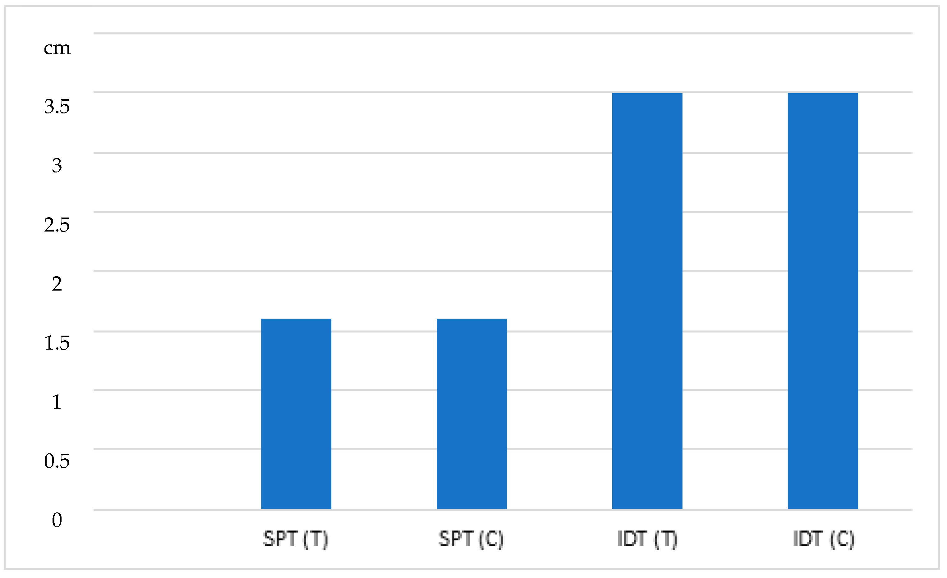
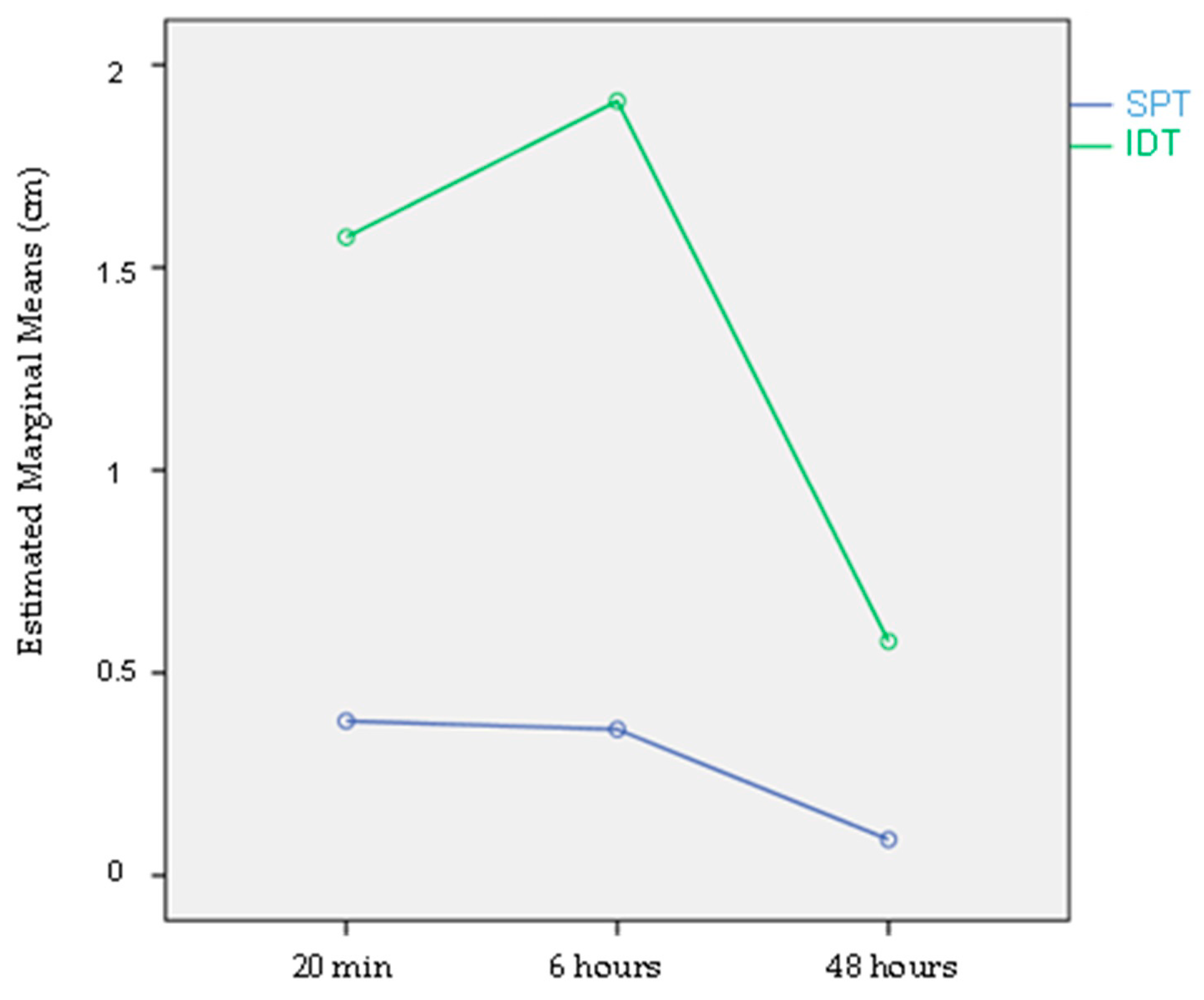
3.1. Skin Tests:
3.1.1. Skin Prick Test (SPT)
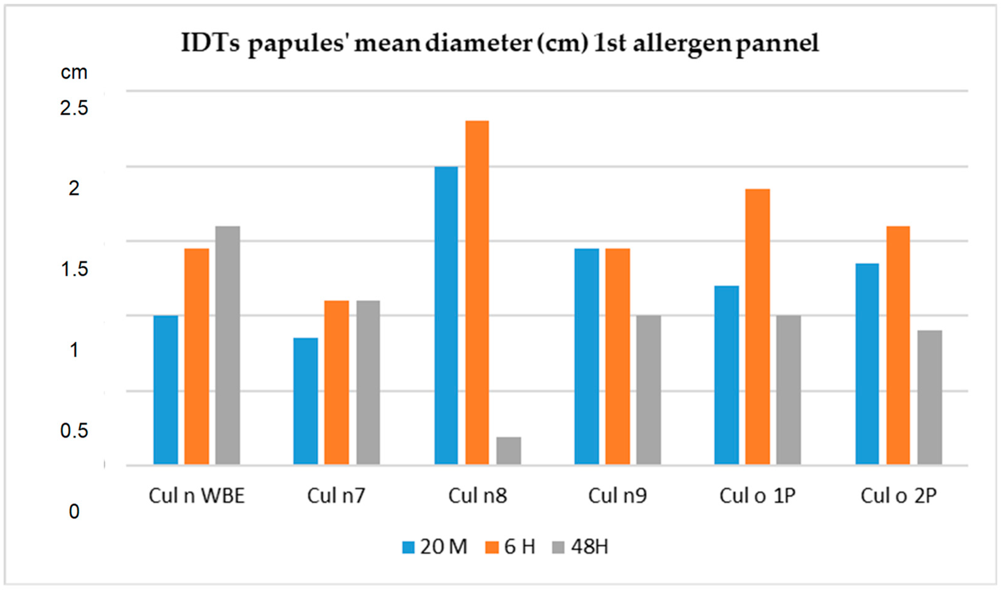
3.1.2. Intradermal Tests
3.2. sLT Release Assay
3.3. IgE Serology
4. Discussion
5. Conclusions
Author Contributions
Funding
Institutional Review Board Statement
Informed Consent Statement
Data Availability Statement
Acknowledgments
Conflicts of Interest
References
- Fadok, V.A. Update on equine allergies. Vet. Clin. N. Am. Equine Pract. 2013, 29, 541–550. [Google Scholar] [CrossRef]
- Wagner, B.; Miller WHJr Erb, H.N.; Lunn, D.P.; Antczak, D.F. Sensitization of skin mast cells with IgE antibodies to Culicoides allergens occurs frequently in clinically healthy horses. Vet. Immunol. Immunopathol. 2009, 132, 53–61. [Google Scholar] [CrossRef]
- Mueller, R.S.; Janda, J.; Jensen-Jarolim, E.; Rhyner, C.; Marti, E. Allergens in veterinary medicine. Allergy 2016, 71, 27–35. [Google Scholar] [CrossRef]
- Scott, D.W.; Miller, W.H. Skin immune system and allergic skin diseases. In Equine Dermatology, 2nd ed.; Elsevier: Amsterdam, The Netherlands, 2011; pp. 263–474. [Google Scholar]
- Jonsdottir, S.; Cvitas, I.; Svansson, V.; Fettelschloss-Gabriel, A.; Sigurbjorg, T.; Marti, E. New Strategies for Prevention and Treatment of Insect Bite Hypersensitivity in Horses. Vet. Dermatol. Curr. Dermatol. Rep. 2019, 8, 303–312. [Google Scholar] [CrossRef]
- Schaffartzik, A.; Hamza, E.; Janda, J.; Crameri, R.; Marti, E.; Rhyner, C. Equine insect bite hypersensitivity: What do we know? Vet. Immunol. Immunopathol. 2012, 147, 113–126. [Google Scholar] [CrossRef]
- Ramilo, D.W.; Diaz, S.; Pereira da Fonseca, I.; Delécolle, J.-C.; Wilson, A.; Meireles, J.; Lucientes, J.; Ribeiro, R.; Boinas, F. First Report of 13 Species of Culicoides (Diptera: Ceratopogonidae) in Mainland Portugal and Azores by Morphological and Molecular Characterization. PLoS ONE 2012, 7, e34896. [Google Scholar] [CrossRef]
- Pessoa, V.; Ramilo, D.W.; Pereira da Fonseca, I.; Ferreira, M.B.; Marti, E.; Tilley, P. Culicoides spp. found near Lusitano stud farms in mainland Portugal which may contribute for IBH studies. Vet. Parasit. Reg. Stud. Rep. 2020, 20, 100385. [Google Scholar] [CrossRef]
- Schaffartzik, A.; Marti, E.; Torsteinsdottir, S.; Mellor, P.S.; Crameri, R.; Rhyner, C. Selective cloning, characterization, and production of the Culicoides nubeculosus salivary gland allergen repertoire associated with equine insect bite hypersensitivity. Vet. Immunol. Immunopathol. 2011, 139, 200–209. [Google Scholar] [CrossRef]
- Van der Rijt, R.; van den Boom, R.; de Jongema, Y.; van Oldruitenborgh-Oosterbaan, M.M.S. Culicoides species attracted to horses with and without insect hypersensitivity. Vet. J. 2008, 178, 91–97. [Google Scholar] [CrossRef]
- Miller, J.E.; Mann, S.; Fettelschoss-Gabriel, A.; Wagner, B. Comparison of three clinical scoring systems for Culicoides hypersensitivity in a herd of Icelandic horses. Vet. Dermatol. 2019, 30, 536-e163. [Google Scholar] [CrossRef] [PubMed]
- Van Damme, C.; ven den Broek, J.; Sloet van Oldruitenborgh-Oosterbaan, M.M. Discrepancies in the bilateral IDT and serum tests in atopic horses. Vet. Dermatol. 2020, 31, 390-e104. [Google Scholar] [CrossRef]
- Wagner, B.; Miller, W.H.; Morgan, E.E.; Hillegas, J.M.; Erb, H.N.; Leibold, W.; Antzack, D.F. IgE and IgG antibodies in skin allergy of the horse. Vet. Res. 2006, 37, 813–825. [Google Scholar] [CrossRef]
- Anderson, G.S.; Belton, P.; Kleider, N. Hypersensitivity of horses in British Columbia to extracts of native and exotic species of Culicoides (Diptera: Ceratopogonidae). J. Med. Entomol. 1993, 30, 657–663. [Google Scholar] [CrossRef] [PubMed]
- Baselgia, S.; Doherr, M.G.; Mellor, P.; Torsteinsdottir, S.; Jermann, T.; Zurbriggen, A. Evaluation of an in vitro sulphidoleukotriene release test for diagnosis of insect bite hypersensitivity in horses. Equine Vet. J. 2006, 38, 40–46. [Google Scholar] [CrossRef]
- Langner, K.F.; Darpel, K.E.; Drolet, B.S.; Fischer, A.; Hampel, S.; Heselhaus, J.E. Comparison of cellular and humoral immunoassays for the assessment of summer eczema in horses. Vet. Immunol. Immunopathol. 2008, 122, 126–137. [Google Scholar] [CrossRef]
- Van der Meide, N.; Meulenbroeks, C.; van Altena, C.; Schurink, A.; Ducro, B.J.; Wagner, B. Culicoides obsoletus extract relevant for diagnostics of insect bite hypersensitivity in horses. Vet. Immunol. Immunopathol. 2012, 149, 245–254. [Google Scholar] [CrossRef]
- Marti, E.; Urwyler, A.; Neuenschwander, M.; Eicher, R.; Meier, D.; de Weck, A.L.; Gerber, H.; Lazary, S.; Dahinden, C.A. Sulfidoleukotriene generation from peripheral blood leukocytes of horses affected with insect bite dermal hypersensitivity. Vet. Immunol. Immunopathol. 1999, 71, 307–320. [Google Scholar] [CrossRef] [PubMed]
- Tilley PSales Luis, J.; Branco Ferreira, M. Comparison of skin prick tests with in vitro allergy tests in the characterization of horses with recurrent airway obstruction. J. Equine Vet. Sci. 2012, 32, 719–727. [Google Scholar] [CrossRef]
- Homburger, H. Methods in Laboratory Immunology. In Allergy: Principles and Practice I, 5th ed.; Middleton, E., Reed, C., Ellis, Eds.; CV Mosby: St Louis, MO, USA, 1998; pp. 417–429. [Google Scholar]
- Carrapatoso, I.; Cadinha, S.; Sanz, M.L. Basophil activation test in the study of food and drug hypersensitivity reactions. Rev. Port. Imunoalergol. 2005, 13, 153–164. [Google Scholar]
- Meulenbroeks, C.; van der Meide, N.; Willemse, T. Recombinant Culicoides obsoletus complex allergens stimulate antigenspecific T cells of insect bite hypersensitive Shetland ponies in vitro. Vet. Dermatol. 2015, 26, 467-e109. [Google Scholar] [CrossRef]
- Peeters, L.M.; Janssens, S.; Goddeeris, B.M. Evaluation of an IgE ELISA with Culicoides spp. extracts and recombinant salivary antigens for diagnosis of insect bite hypersensitivity in Warmblood horses. Vet. J. 2013, 198, 141–147. [Google Scholar] [CrossRef]
- Novotny, E.N.; White, S.J.; Wilson, A.D.; Stefánsdóttir, S.B.; Tijhaar, E.; Jonsdóttir, S.; Frey, R.; Reiche, D.; Rose, H.; Rhyner, C.; et al. Component-resolved microarray analysis of IgE sensitization profiles to Culicoides recombinant allergens in horses with insect bite hypersensitivity. Allergy 2021, 76, 1147–1157. [Google Scholar] [CrossRef] [PubMed]
- Cordeiro Raposo, A. O Filho do Vento, 5th ed.; Inapa: Lisbon, Portugal, 2002; ISBN 972-8387-06-7. [Google Scholar]
- Van der Meide, N.M.; Roders, N.; Sloet van Oldruitenborgh-Oosterbaan, M.M.; Schaap, P.J.; van Oers, M.M.; Leibold, W.; Savelkoul, H.F.; Tijhaar, E. Cloning and expression of candidate allergens from Culicoides obsoletus for diagnosis of insect bite hypersensitivity in horses. Vet. Immunol. Immunopathol. 2013, 153, 227–239. [Google Scholar] [CrossRef]
- Ziegler, A.; Hamza, E.; Jonsdottir, S.; Rhyner, C.; Wagner, B.; Scheupbach, G.; Svansson, V.; Torsteinsdottir, S.; Marti, E. Longitudinal analysis of allergen specific IgE and IgG subclasses as potential predictors of insect bite hypersensitivity following first exposure to Culicoides in Icelandic horses. Vet. Dermatol. 2018, 29, 51-e22. [Google Scholar] [CrossRef] [PubMed]
- Tahon, L.; Baselgia, S.; Gerber, V.; Doherr, M.G.; Straub, R.; Robinson, N.E.; Marti, E. In vitro allergy tests compared to intradermal testing in horses with recurrent airway obstruction. Vet. Immunol. Immunopathol. 2009, 127, 85–93. [Google Scholar] [CrossRef]
- Frey, R.; Bergvall, K.; Egenvall, A. Allergen-specific IgE in Icelandic horses with insect bite hypersensitivity and healthy controls, assessed by FcaR1a-basedserology. Vet. Immunol. Immunopathol. 2008, 126, 102–109. [Google Scholar] [CrossRef]
- Peeters, L.M.; Janssens, S.; Schaffartzik, A.; Marti, E.; Buys, N. Evaluation of IgE levels against Culicoides nubeculosus allergens in Belgian Warmblood horses. Commun. Agric. Appl. Biol. Sci. 2012, 77, 218–222. [Google Scholar]
- Curin, M.; Garib, V.; Valenta, R. Single recombinant and purified major allergens and peptides: How they are made and how they change allergy diagnosis and treatment. Ann. Allergy Asthma Immunol. 2016, 119, 201–209. [Google Scholar] [CrossRef]
- Arruda, L.K.; Barbosa, M.C.R.; Bardini, G.; Yang, A.C.; Genov, I.R.; Moreno, A.S. Recombinant allergens: Role in diagnosis and in allergen-specific immunotherapy. Braz. J. Allergy Immunol. 2013, 1, 211–218. [Google Scholar] [CrossRef]
- Chapman, M.D.; Smith, A.M.; Vailes, L.D.; Arruda, L.K.; Dhanaraj, V.; Pomés, A. Recombinant allergens for diagnosis and therapy of allergic disease. J. Allergy Clin. Immunol. 2000, 106, 409–418. [Google Scholar] [CrossRef]
- Jutel, M.; Solarewicz-Madejek, K.; Smolinska, S. Recombinant allergens: The present and the future. Hum. Vaccin. Immunother. 2012, 8, 1534–1543. [Google Scholar] [CrossRef]
- Linhart, B.; Valenta, R. Mechanisms underlying allergy vaccination with recombinant hypoallergenic allergen derivatives. Vaccine 2012, 30, 4328–4335. [Google Scholar] [CrossRef]
- Sloet van Oldruitenborgh-Oosterbaan, M.M.; van Poppel, M.; de Raat, I.J.; van den Boom, R.; Savelkoul, H.F.J. Intradermal testing of horses with and without insect bite hypersensitivity in the Netherlands using an extract of native Culicoides species. Vet. Dermatol. 2009, 20, 607–614. [Google Scholar] [CrossRef]
- Jose-Cunilleras, E.; Kohn, C.W.; Hillier, A.; Saville, W.J.; Lorch, G. Intradermal testing in healthy horses and horses with chronic obstructive pulmonary disease, recurrent urticaria, or allergic dermatitis. J. Am. Vet. Med. Assoc. 2001, 219, 1115–1121. [Google Scholar] [CrossRef]
- Lane, M.J.; Pucheu-Haston, C.M.; Kearney, M.T.; Woodward, M. Determination of irritant threshold concentrations of multiple tree, grass, weed and mould allergens for intradermal testing of horses residing in the southern USA. Vet. Dermatol. 2017, 28, 604-e147. [Google Scholar] [CrossRef]
- DeBoer, D.J.; Hillier, A. The ACVD task force on canine atopic dermatitis (XVI): Laboratory evaluation of dogs with atopic dermatitis with serum-based ‘allergy’ tests. Vet. Immunol. Immunopathol. 2001, 81, 277–287. [Google Scholar] [CrossRef]
- O’Driscoll, B.R.; Powell, G.; Chew, F.; Niven, R.M.; Miles, J.F.; Vyas, A.; Denning, D.W. Comparison of skin prick tests with specific serum immunoglobulin E in the diagnosis of fungal sensitization in patients with severe asthma. Clin. Exp. Allergy 2009, 39, 1677–1683. [Google Scholar] [CrossRef]
- Van der Haegen, A.; Griot-Wenk, M.E.; Welle, M.; Busato, A.; von Tscharner, C.; Zurbriggen, A.; Marti, E. Immunoglobulin E bearing cells in skin biopsies of horses with insect bite hypersensitivity. Equine Vet. J. 2001, 33, 699–706. [Google Scholar] [CrossRef]
- Wilson, A.D.; Harwood, L.J.; Björnsdottir, S.; Marti, E.; Day, M.J. Detection of IgG and IgE serum antibodies to Culicoides salivary gland antigens in horses with insect dermal hypersensitivity (sweet itch). Equine Vet.J. 2001, 33, 707–713. [Google Scholar] [CrossRef]
- Lomas, H.R.; Robinson, P.A. A Pilot Qualitative Investigation of Stakeholders’ Experiences and Opinions of Equine Insect Bite Hypersensitivity in England. Vet. Sci. 2018, 5, 3. [Google Scholar] [CrossRef]
- Heinzerling, L.; Mari, A.; Bergmann, K.C.; Bresciani, M.; Burbach, G.; Darsow, U.; Durham, S.; Fokkens, W.; Gjomarkaj, M.; Haahtela, T.; et al. The skin prick test—European standards. Clin. Transl. Allergy 2013, 3, 3. [Google Scholar] [CrossRef] [PubMed]
- Wood, R.A.; Phipatanakul, W.; Hamilton, R.G.; Eggleston, P.A. A comparison of skin prick tests, intradermal skin tests, and RASTs in the diagnosis of cat allergy. J. Allergy Clin. Immunol. 1999, 103, 773–779. [Google Scholar] [CrossRef] [PubMed]
- Jensen-Jarolim, E.; Einhorn, L.; Hermann, I.; Thalhamer, J.; Panakova, L. Pollen allergies in humans and their dogs, cats, and horses: Differences and similarities. Clin. Transl. Allergy 2015, 5, 15. [Google Scholar] [CrossRef]
- Fadok, V.A.; Greiner, E.C. Equine insect hypersensitivity: Skin test and biopsy results correlated with clinical data. Equine Vet. J. 1990, 22, 236–240. [Google Scholar] [CrossRef]
- Liccardi, G.; Dente, B.; Triggiani, M.; Russo, M.; Diamare, F.; Massari, A.; Pinzarrone, R.; D’Isanto, R.; Letizia, M.; D’Amato, M.; et al. A multicenter evaluation of the CARLA system for the measurement of specific IgE antibodies vs. other different methods and skin prick tests. J. Investig. Allergol. Clin. Immunol. 2002, 12, 235–241. [Google Scholar] [PubMed]
- Ricci, G.; Capelli, M.; Miniero, R.; Menna, G.; Zannarini, L.; Dillon, P.; Masi, M. Acomparison of different allergometric tests, skin prick test, Pharmacia UniCAP and ADVIA Centaur, for diagnosis of allergic diseases in children. Allergy 2003, 58, 38–45. [Google Scholar] [CrossRef] [PubMed]
- Crockard, A.D.; Ennis, M. Basophil histamine release tests in the diagnosis of allergy and asthma. Editor. Clin. Exp. Allergy 2001, 31, 345–350. [Google Scholar] [CrossRef] [PubMed]
- Crockard, A.D.; Ennis, M. Laboratory-based allergy diagnosis: Should we go with the flow? Editor. Clin. Exp. Allergy. 2001, 31, 975–977. [Google Scholar] [CrossRef] [PubMed]
- Lam, J.; Fettelschoss, V.; Olomski, F.; Rhiner, T.; Birkmann, K.; Kündig, T.M.; Fettelschoss Gabriel, A. Chronic Allergen Exposure Might Shift Allergic Mechanisms in Horses with Insect Bite Hypersensitivity. J. Clin. Immunol. Immunother. 2020, 6, 017. [Google Scholar] [CrossRef]
- Xu, Q.; Jiang, Q.; Yang, L.; Li, W.; Huang, N.; Yang, Y.; Ma, D.; Zhang, S.; Wang, Y.; Zhu, R. IgE and IgG4 Repertoire in Asymptomatic HDM-Sensitized and HDM-Induced Allergic Rhinitis Patients. Int. Arch. Allergy Immunol. 2021, 182, 1200–1211. [Google Scholar] [CrossRef] [PubMed]
- Wilkołek, P.; Szczepanik, M.; Sitkowski, W.; Adamek, L.; Pluta, M.; Taszkun, I.; Gołynski, M. A Comparison of Intradermal Skin Testing and Serum Insect Allergen-specific IgE Determination in Horses with Insect Bite Hypersensitivity from 2008 to 2016. J. Equine Vet. Sci. 2019, 75, 65–68. [Google Scholar] [CrossRef] [PubMed]
- De Weck, A.L.; Sanz, M.L. Flow cytometric cellular allergen stimulation test (FAST/Flow CAST). Technical and clinical evaluation of a new diagnostic test in allergy and pseudo-allergy. ACI Int. 2002, 14, 204–215. [Google Scholar]


| Allergen | Buffer | Skin Test (ST)/Serology (S) | References |
|---|---|---|---|
| Cul n 3 | PBS | S/ST | Schaffartzik et al., 2011 [9] |
| Cul o 1P | 20mM Tris 0.5M NaCl | S/ST | Peeters et al., 2013 [23] |
| Cul o 2P | 20mM Tris 0.5M NaCl | S/ST | Peeters et al., 2013 [23] |
| Cul o 3 | 20mM Tris 0.5M NaCl | S | Van der Meide et al., 2013 [26] |
| Cul o WBE | 0.9% NaCl | ST | Peeters et al., 2013 [23] |
| Cul n 1 | H2O | ST | Schaffartzik et al., 2011 [9] |
| Cul n 2 | PBS | ST | Schaffartzik et al., 2011 [9] |
| Cul n 3 | H2O | S/ST | Schaffartzik et al., 2011 [9] |
| Cul n 4 | H2O | S/ST | Schaffartzik et al., 2011 [9] |
| Cul n 5 | H2O | ST | Schaffartzik et al., 2011 [9] |
| Cul n 6 | H2O | ST | Schaffartzik et al., 2011 [9] |
| Cul n 7 | H2O | ST | Schaffartzik et al., 2011 [9] |
| Cul n 8 | H2O | ST | Schaffartzik et al., 2011 [9] |
| Cul n 9 | PBS | ST | Schaffartzik et al., 2011 [9] |
| Cul n 10 | H2O | S/ST | Schaffartzik et al., 2011 [9] |
| Cul n 11 | H2O | ST | Schaffartzik et al., 2011 [9] |
| Cul n WBE | 0.9% NaCl | ST | Ziegler et al. 2018, [27] |
| Cul n 4 Bar | PBS | ST | Jonsdottir et al., 2018 [5] |
| Culn 4 Bac | PBS | ST | Jonsdottir et al., 2018 [5] |
| Cul n 3 Bac | PBS | ST | Jonsdottir et al., 2018 [5] |
| Cul n 3 Bar | PBS | ST | Jonsdottir et al., 2018 [5] |
| Allergen (1st Panel) | Test | Group | Mean (cm) | Standard Deviation (cm) | 95% Confidence Limits | |
|---|---|---|---|---|---|---|
| Lower | Upper | |||||
| Cul n WBE | IDT * | C | 1.47 | 0.45 | 1.308 | 1.631 |
| T | 1.65 | 0.47 | 1.481 | 1.818 | ||
| SPT * | C | 0.33 | 0.42 | 0.179 | 0.480 | |
| T | 0.78 | 0.54 | 0.586 | 0.973 | ||
| Cul n 7 | IDT NS | C | 1.12 | 0.55 | 0.923 | 1.310 |
| T | 1.01 | 0.50 | 0.831 | 1.188 | ||
| SPT * | C | 0.21 | 0.4 | 0.066 | 0.353 | |
| T | 0.48 | 0.5 | 0.301 | 0.658 | ||
| Cul n 8 | IDT * | C | 0.87 | 0.57 | 0.666 | 1.074 |
| T | 0.97 | 0.77 | 0.694 | 1.245 | ||
| SPT * | C | 0.31 | 0.42 | 0.159 | 0.460 | |
| T | 0.39 | 0.45 | 0.228 | 0.551 | ||
| Cul n 9 | IDT * | C | 1.21 | 0.58 | 1.002 | 1.417 |
| T | 1.37 | 0.35 | 1.244 | 1.495 | ||
| SPT * | C | 0.32 | 0.4 | 0.176 | 0.463 | |
| T | 0.66 | 0.57 | 0.456 | 0.863 | ||
| Cul o 1P | IDT NS | C | 1.33 | 0.60 | 1.115 | 1.541 |
| T | 1.30 | 0.62 | 0.803 | 1.521 | ||
| SPT * | C | 0.30 | 0.40 | 0.156 | 0.443 | |
| T | 0.61 | 0.54 | 0.416 | 0.803 | ||
| Cul o 2P | IDT * | C | 1.21 | 0.73 | 0.948 | 1.471 |
| T | 1.26 | 0.72 | 1.002 | 1.517 | ||
| SPT * | C | 0.46 | 0.48 | 0.288 | 0.631 | |
| T | 0.69 | 0.45 | 0.529 | 0.851 | ||
| Allergen (2nd Panel) | Test | Group | Mean (cm) | Standard Deviation (cm) | 95% Confidence Limits | |
|---|---|---|---|---|---|---|
| Lower | Upper | |||||
| Cul o WBE | IDT | C | 1.46 | 0.33 | 1.195 | 1.724 |
| T | 1.53 | 0.33 | 1.265 | 1.794 | ||
| SPT | C | 0.13 | 0.33 | −0.134 | 0.394 | |
| T | 0.65 | 0.51 | 0.217 | 1.082 | ||
| Cul n 3 Bar | IDT | C | 0.39 | 0.22 | 0.213 | 0.566 |
| T | 1.56 | 0.29 | 1.327 | 1.792 | ||
| SPT | C | 0 | 0 | 0 | 0 | |
| T | 0.48 | 0.54 | 0.0479 | 0.912 | ||
| Cul n 4 Bac | IDT | C | 0.53 | 0.29 | 0.340 | 0.762 |
| T | 0.83 | 0.29 | 0.597 | 1.062 | ||
| SPT | C | 0.23 | 0.36 | −0.058 | 0.518 | |
| T | 0.27 | 0.47 | −0.106 | 0.646 | ||
| Z-Value to Test | Upper 1-Sided | 95% Confidence Limits | ||||
|---|---|---|---|---|---|---|
| Allergen | AUC | Standard Error | AUC > 0.5 | p-Value | Lower | Upper |
| Cul_n WBE | 0.6986 | 0.0835 | 2.377 | ≤0.01 | 0.4965 | 0.8288 |
| Cul_o WBE | 0.8923 | 0.0533 | 7.363 | ≤0.0001 | 0.7262 | 0.9600 |
| Specific IgE Levels (ELISA) | ROC Analysis (n = 59) | ||||||||
|---|---|---|---|---|---|---|---|---|---|
| Allergen | C (N = 29) | T (N = 30) | p Value (U-Test) | Significance | AUC | Standard Error | Z-Value to Test | Upper 1-Sided p-Value | 95% Confidence Limits |
| Median (Min–Max) | Median (Min–Max) | AUC > 0.5 | Lower/Upper | ||||||
| Cul n 3 | 0.106 (0.052–0.171) | 0.293 (0.152–0.454) | p ≤ 0.001 | Significant | 0.7621 | 0.0616 | 4.251 | ≤0.0001 | 0.612/0.859 |
| Cul n 4 | 0.016 (0–0.06) | 0.119 (0.02–0.18) | p ≤ 0.05 | Significant | 0.6707 | 0.0702 | 2.432 | ≤0.0175 | 0.509/0.786 |
| Cul n 10 | 0.035 (0–0.086) | 0.09 (0.07–0.195) | p ≤ 0.05 | Significant | 0.7063 | 0.0674 | 3.060 | ≤0.01 | 0.548/0.815 |
| Cul o 2 | 0.391 (0.323–0.444) | 0.573 (0.439–0.776) | p ≤ 0.05 | Significant | 0.6747 | 0.0714 | 2.446 | ≤0.01 | 0.509/0.791 |
| Cul o 1P | 0.201 (0.167–0.217) | 0.564 (0.26–0.82) | p ≤ 0.01 | Significant | 0.7730 | 0.0626 | 4.361 | ≤0.0001 | 0.495/0.778 |
| Cul o 2P | 0.216 (0.132–0.275) | 0.36 (0.223–0.467) | p ≤ 0.05 | Significant | 0.7184 | 0.0672 | 3.249 | ≤0.001 | 0.559/0.826 |
| Cul o 3 | 0.159 | 0.323 | p ≤ 0.05 | Significant | 0.659 | 0.0719 | 2.221 | ≤0.01 | 0.495/0.778 |
| (0.114–0.201) | (0.159–0.389) | ||||||||
Disclaimer/Publisher’s Note: The statements, opinions and data contained in all publications are solely those of the individual author(s) and contributor(s) and not of MDPI and/or the editor(s). MDPI and/or the editor(s) disclaim responsibility for any injury to people or property resulting from any ideas, methods, instructions or products referred to in the content. |
© 2023 by the authors. Licensee MDPI, Basel, Switzerland. This article is an open access article distributed under the terms and conditions of the Creative Commons Attribution (CC BY) license (https://creativecommons.org/licenses/by/4.0/).
Share and Cite
Pessoa, V.C.; Branco-Ferreira, M.; Jónsdóttir, S.; Marti, E.; Tilley, P. Comparison of Skin Prick Tests (SPT), Intradermal Tests (IDT) and In Vitro Tests in the Characterization of Insect Bite Hypersensitivity (IBH) in a Population of Lusitano Horses: Contribution for Future Implementation of SPT in IBH Diagnosis. Animals 2023, 13, 2733. https://doi.org/10.3390/ani13172733
Pessoa VC, Branco-Ferreira M, Jónsdóttir S, Marti E, Tilley P. Comparison of Skin Prick Tests (SPT), Intradermal Tests (IDT) and In Vitro Tests in the Characterization of Insect Bite Hypersensitivity (IBH) in a Population of Lusitano Horses: Contribution for Future Implementation of SPT in IBH Diagnosis. Animals. 2023; 13(17):2733. https://doi.org/10.3390/ani13172733
Chicago/Turabian StylePessoa, Vera Carvalho, Manuel Branco-Ferreira, Sigridur Jónsdóttir, Eliane Marti, and Paula Tilley. 2023. "Comparison of Skin Prick Tests (SPT), Intradermal Tests (IDT) and In Vitro Tests in the Characterization of Insect Bite Hypersensitivity (IBH) in a Population of Lusitano Horses: Contribution for Future Implementation of SPT in IBH Diagnosis" Animals 13, no. 17: 2733. https://doi.org/10.3390/ani13172733