Article

Comparison of Immune-Related Gene Expression in Two Chicken Breeds Following Infectious Bronchitis Virus Vaccination

by
Schwann Chuwatthanakhajorn
1,2,
Chi-Sheng Chang
3,
Kannan Ganapathy
4,
Pin-Chi Tang
1,5,*,† and
Chih-Feng Chen
1,5,6,*,†
1
Department of Animal Science, National Chung Hsing University, Taichung 402, Taiwan
2
Faculty of Veterinary Science, Mahidol University, Salaya, Nakhon Pathom 73170, Thailand
3
Department of Animal Science, Chinese Culture University, Taipei 111, Taiwan
4
Institute of Infection, Veterinary & Ecological Sciences (IVES), University of Liverpool, Neston CH64 7TE, UK
5
The iEGG and Animal Biotechnology Center, National Chung Hsing University, Taichung 402, Taiwan
6
Smart Sustainable New Agriculture Research Center (SMARTer), Taichung 402, Taiwan
*
Authors to whom correspondence should be addressed.
These authors contributed equally to this work.
Animals 2023, 13(10), 1642; https://doi.org/10.3390/ani13101642
Submission received: 2 March 2023 / Revised: 5 May 2023 / Accepted: 9 May 2023 / Published: 15 May 2023
(This article belongs to the Special Issue Genetic Analysis of Important Traits in Poultry)

Abstract

:

Simple Summary

Host genetics plays a significant role in the effectiveness of immune responses against pathogens. Disease severity for an individual or population is associated with dissimilarity in the levels of host gene expressions. In this study, the variations in the effects of host immune responsiveness between Taiwan Country and White Leghorn chicken breeds were investigated by next-generation sequencing. Overall, immune response-related genes between Taiwan Country chicken and White Leghorn chicken were expressed differently against live attenuated infectious bronchitis virus vaccination. The major histocompatibility complexes and cytokines were determined as significant players in determining the pattern and magnitude of immune responses following an infection. This study demonstrated the host genetic influences on the development of adaptive immune response between the Taiwan Country and the White Leghorn chickens after infectious bronchitis virus vaccination.

Abstract

This study aims to identify the immune-related genes and the corresponding biological pathways following infectious bronchitis virus vaccination in Taiwan Country and White Leghorn chicken breeds. Transcriptomic analyses of the spleen of these two breeds were conducted by next-generation sequencing. Compared to White Leghorn chicken, Taiwan Country chicken showed a significantly higher level of anti-infectious bronchitis virus (IBV) antibodies at 14 and 21 days pos vaccination. At 7 days post vaccination, in the Taiwan Country chicken, higher expression of mitogen-activated protein kinase 10, Major histocompatibility complex class 1, and V-set pre-B cell surrogate light chain 3 were found. In contrast, the White Leghorn chicken had a high expression of interleukin 4 induced 1, interleukin 6, and interleukin 22 receptor subunit alpha 2. These findings have highlighted the variations in immune induction between chickens with distinct genetic background and provided biological pathways and specific genes involved in immune responses against live attenuated IBV vaccine.

1. Introduction

Infectious bronchitis (IB) is a highly contagious disease in chickens, substantially affecting the poultry industry worldwide. This disease is caused by avian corona infectious bronchitis virus, a member of Gammacoronavirus [1]. Most clinical signs are related to the upper respiratory, urinary, and reproductive systems [2]. The mortality rate of IB infection ranges from 0% to 82%, depending on the viral strain, health status, and age of birds [3]. The virus primarily replicates in the trachea and causes ciliary stasis, leading to secondary bacterial infection, such as Escherichia coli and Mycoplasma gallisepticum infection [4]. This results in respiratory distress and poor growth performance [5]. In addition, the disease can cause significant loss due to a drop in egg production and poor egg quality [6,7].
Several studies have noted the influence of host genetic background on the immune response following inoculation of virulent or avirulent (vaccine) poultry viruses. Different breeds and sublines showed the variation of genes expression following infection, affecting the level of susceptibility to diseases [8,9,10,11]. The polymorphism of MHC genes was found to be particularly associated with resistant traits against pathogenic bacteria and viruses [12,13]. In addition, several genes were reported as disease-resistant genes in chicken, including Cyclophilin B (PPIB), MX Dynamin Like GTPase 1 (MX1), and 2’-5’-Oligoadenylate Synthetase Like (OASL) [14,15]. Vaccination studies also revealed the direct effect of host genetic background on variation of innate, mucosal, cellular, and humoral immune responses [16,17,18,19].
The spleen is an important organ in chickens, especially for development of both cellular and humoral immune responses [20]. The white pulp of the spleen contains ellipsoids, peri-ellipsoid sheaths (PESS), and lymph nodes, which are the main sites for initiating the humoral immune response by activating and differentiating B cells against blood-borne antigens [21]. This process leads to the secretion of large amounts of antibodies into the bloodstream and the development of memory B cells [22]. T lymphocytes are responsible for cell-mediated immune responses, and their activation occurs in the periarteriolar lymphoid sheaths (PELS), which allow for the monoclonal expansion and differentiation of naive T cells into effector T cells [23]. Gaining an understanding of the biological mechanisms underlying gene expression in the spleen could provide valuable insights into the effects of genetics on the development of immune responses in chickens.
Taiwan Country chicken (TCC) is a native breed in Taiwan, which was selected over 30 generations at National Chung Hsing university for prodigious characteristics including high growth rate and feasible adaptation. On the other hand, White Leghorn chicken (WLC) is a Mediterranean breed with high rate of egg production and outstanding feed efficiency. Previous studies have reported a variation in susceptibility between TCC and WLC responding to infectious diseases such as Newcastle disease, Marek’s disease, and Leucocytozoonosis [24,25,26]. The use and comparative analysis of these two genetically distinct breeds of chickens have the potential to elucidate the host genetic components that are associated with differential susceptibility to infections.
The aim of this study is to enhance our knowledge on the influence of host genetics on immune responses after IBV vaccination. RNA sequencing was used to cross-compare the immune-related genes of IBV-vaccinated Taiwan Country chickens (TCCs) and White Leghorn chickens (WLCs). The findings of this study would increase the availability of effective disease control strategies.

2. Materials and Methods

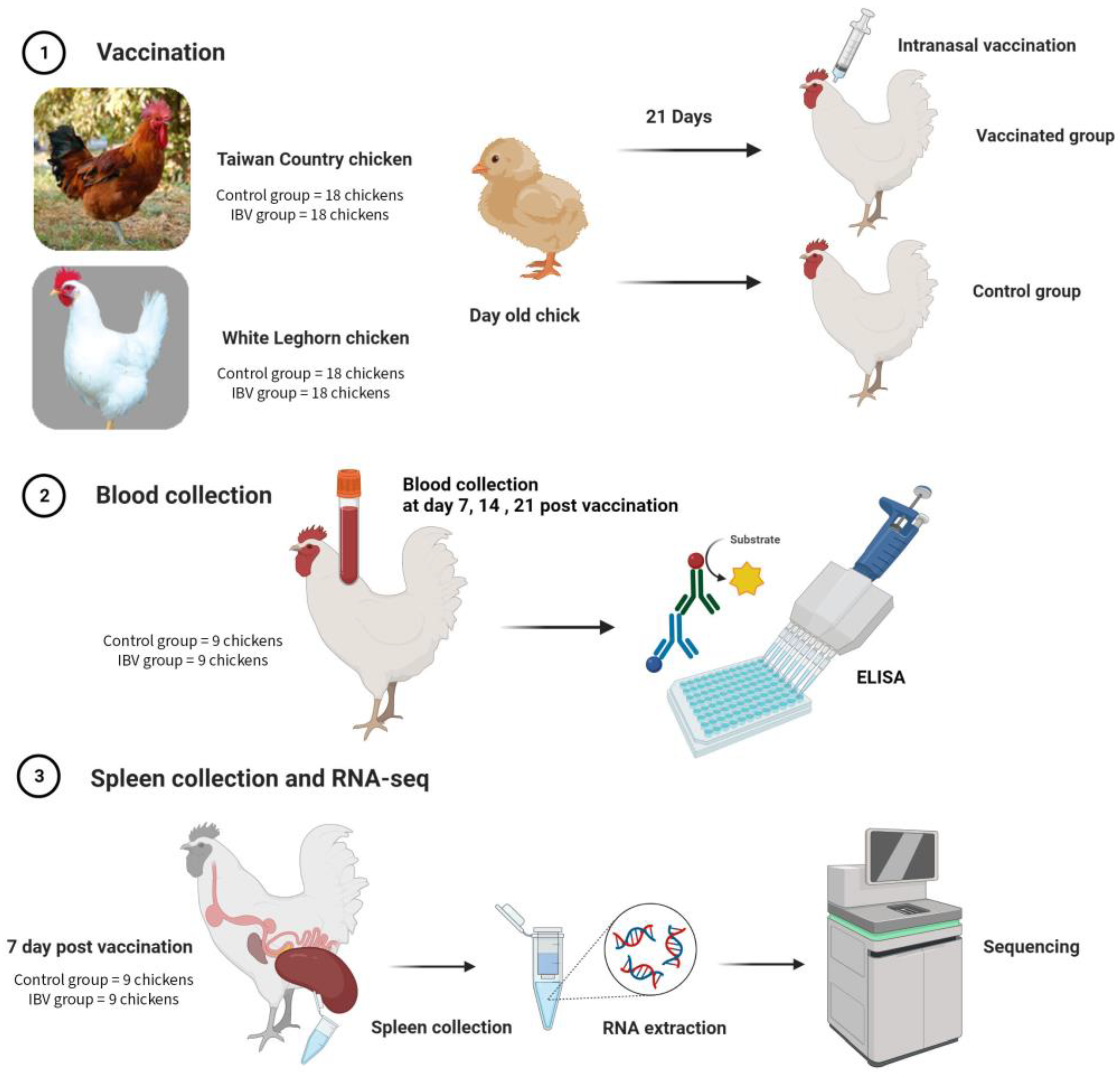
2.1. Animals and Tissue Sampling

In this experiment (Figure 1), B strain TCCs and WLCs were obtained from the National Chung Hsing University and Taiwan Hubbard GP Farm, respectively. Thirty-six day-old chicks of each type were obtained and raised in a disease-free facility with ad libitum feed and water. At 3 weeks old, the chickens of each breed were separated into two groups (18 each in the control and IBV groups). The control group received an IBV-free diluent intranasally, whereas the IBV group received live attenuated IBV, variant strain 4-91 (Nobilis IB 4-91, 7.2 log10 EID50, MSD Animal Health, Boxmeer, The Netherlands) intranasally.
Nine birds from each group of TCC and WLC were bled through the wing vein at 0 (before inoculation), 7, 14, and 21 days after the sham or IBV inoculations. After 2 h, the blood was centrifuged at 10,000 rpm for 10 min, and separated sera were stored in a freezer at −20 °C. At 7 days post vaccination, nine chickens of each group were humanely killed through cervical dislocation and spleens were collected. Each sample was transferred into a microcentrifuge tube filled with tissue storage reagent (RNAlater, Sigma-Aldrich, Singapore), and then directly frozen in a freezer at −80 °C.

2.2. Enzyme-Linked Immunosorbent Assay

All serum samples were tested for the antibody titer response to IBV by using the IB enzyme-linked immunosorbent assay kit (IDEXX IBV, Invitrogen, Taipei, Taiwan) following the manufacturer’s protocol. The optical density (OD) values were measured using a microplate spectrophotometer (Epoch, BioTek, Taipei, Taiwan). Antibody titers were determined following the manufacturer’s standard protocol using the sample-to-positive (S/P) ratio method and were reported with a positive cut-off value of 0.2.

2.3. RNA Extraction and cDNA Synthesis

Fifty milligrams of spleen from each sample was used for RNA extraction using total ribonucleic acid (RNA) isolation reagent (TRIzol Reagent, Invitrogen, Taipei, Taiwan) following the manufacturer’s protocol. The quality and concentration were determined using the microplate spectrophotometer (Epoch, BioTek, Taipei, Taiwan). Nine total RNA samples from each group were randomly pooled into three samples of equal amounts (three RNA pools in each of the three samples). In addition to using the total RNA for RNA sequencing, some RNAs were converted into complementary deoxyribonucleic acid (cDNA) by using the cDNA synthesis kit (RevertAid First Strand cDNA Synthesis kit, Thermo Scientific, Taipei, Taiwan) following the recommended protocol for quantitative real-time polymerase chain reaction (qPCR).

2.4. Library Construction and RNA Sequencing

RNAs extracted from spleen samples were analyzed through RNA Sequencing (RNA-Seq) with NGS, which was conducted by Biotools Co., Ltd. (New Taipei, Taiwan). RNA purity and quantification were checked using SimpliNano-Biochrom Spectrophotometers (Biochrom, Holliston, MA, USA). RNA degradation and integrity were monitored using Qsep 100 DNA/RNA Analyzer (BiOptic Inc., New Taipei, Taiwan). In total, 1 μL of total RNA per sample was used as input materials for RNA sample preparation. Sequencing libraries were generated using the KAPA mRNA HyperPrep Kit (KAPA Biosystems, Roche, Basel, Switzerland) following the manufacturer’s recommendations, and index codes were added to the attribute sequences of each sample. Briefly, messenger RNA (mRNA) was purified from the total RNA by using magnetic oligo-dT beads. Captured mRNA was fragmented through incubation at a high temperature in the presence of magnesium in the KAPA Fragment, Prime, and Elute Buffer. The first strand of cDNA was synthesized using random hexamer priming. A combination of second strand synthesis and A-tailing converted the cDNA–RNA hybrid into double-stranded cDNA (dscDNA), incorporated deoxyuridine triphosphate (dUTP) into the second cDNA strand, and added dAMP to the 3′ ends of the resulting dscDNA. The dsDNA adapter with 3′-dTMP overhangs were ligated to library insert fragments to generate library fragments carrying the adapters. To select cDNA fragments of 300–400 base pairs (bp), the library fragments were purified using the KAPA Pure Beads system (KAPA Biosystems, Roche, Basel, Switzerland). The library carrying the appropriate adapter sequences at both ends was amplified using KAPA HiFi HotStart ReadyMix (KAPA Biosystems, Roche, Basel, Switzerland) and library amplification primers. The strand marked with dUTP was not amplified, allowing strand-specific sequencing. Finally, PCR products were purified using the KAPA Pure Beads system, and library quality was assessed using the Qsep 100 DNA/RNA Analyzer (BiOptic Inc., New Taipei, Taiwan). All libraries were sequenced using the NovaSeq 6000 platform for paired-end 150 bp sequencing.

2.5. RNA-Seq Data Analysis and Go and KEGG Enrichment Analysis

In this study, the low-quality regions in the reads were removed using Trimmomatic based on the following criteria: THREADS:4; PHRED:33; ILLUMINACLIP: TruSeq3-PE. fa: 2:30:10; LEADING:3; TRAILING:3; SLIDINGWINDOW: 4:20 and MINLEN:36 [27]. The output files were further analyzed using FastQC [28] and MultiQC [29] to ensure the quality of the data. Clean reads were mapped to the Gallus gallus reference genome (GRCg7b) obtained from the National Center for Biotechnology Information by using HISAT2 software. Prior to the differential expression (DE) analysis, low-expression genes were excluded. Then, DE analysis between groups was performed using DESeq2 [30]. The p-values were corrected using the Benjamini–Hochberg procedure to control the false discovery rate (FDR). Transcripts that passed the cut-off criteria of |log2 (FoldChange)| > 1 and with a p-value < 0.05 were considered as differentially expressed genes (DEGs). Candidate genes were used to assess the biological pathway by using the Gene Ontology (GO) database and Kyoto Encyclopedia of Genes and Genomes (KEGG) database.

2.6. Quantitative Real-Time PCR Verification

The RNA-Seq results were validated through qPCR analysis with six selected genes, namely, interleukin 6 (IL-6), interleukin 22 (IL-22), C-C motif chemokine ligand 19 (CCL-19), C-X-C motif chemokine receptor 4 (CXCR4), C-X-C chemokine ligand 13-like 2 (CXCL13L2), and CD 34 molecule (CD34). These immune-related genes were selected based on their involvement in the GO and KEGG pathways and the availability of the sequences in the National Center for Biotechnology Information (NCBI) database. qPCR was performed using SYBR Green qPCR assays. The total volume of the reaction mixture was 10 µL, which comprised 5 µL of qPCR Master mix (PowerUP SYBR Green Master Mix, Appliedbiosystems, Taipei, Taiwan), 1 µL of cDNA from spleen extract, 3.8 µL of nuclease-free water, and 0.2 µL of forward and reverse primers (Supplementary Table S1). Then, each sample was placed into a qPCR analyzer (Step One Plus, Appliedbio systems, Taipei, Taiwan). In the analysis step, Step one software version 2.3 was used, followed by a qPCR cycle consisting of a Fast cycling mode (Primer Tm 60.0 °C) involving 40 cycles of denaturation (95.0 °C for 15 s), annealing (60.0 °C for 30 s), and elongation (60.0 °C for 30 s). In this study, we selected glyceraldehyde-3-phosphate dehydrogenase (GAPDH) as the endogenous gene due to its stable expression in our samples, and all qPCR reactions were performed in triplicate. Relative mRNA expression was calculated using the 2−ΔΔCt method [31].

2.7. Statistical Analysis

The differences in antibody titers against IBV between treatment and control groups in each breed were analyzed by t-test using the TTEST procedure. All statistical analyses were conducted using SAS software (SAS, 2012).

3. Results

3.1. Anti-IBV ELISA Titers

In this study, following the vaccination of the 3-week-old birds, there were no clinical signs. The mean anti-IBV ELISA titers in each breed of birds is presented in Table 1. Compared to WLCs, TCCs showed earlier and higher antibody production. Compared with the control group, the antibody titers of the vaccinated TCC and WLC were significantly higher at 7, 14, and 21 or 14 and 21 days post vaccination, respectively. When comparing the IBV-inoculated TCC and WLC birds, significantly higher antibody titers were observed in TCCs at 14 and 21 days post vaccination.

3.2. Splenic Transcriptome Sequencing

The results of all 12 splenic transcription data (68.12 Gb), with the GC content at approximately 44–49% and the percentage of Q30 at >93% (Supplementary Table S2), indicated that the data could be used for further analysis. In addition, the comparison efficiency of the total reads compared with the GRCg7b reference genome of the 12 samples was 93–95% (Supplementary Table S3), and the comparison efficiency of the percentage of read and the reference genome was approximately 87–93%.

3.3. Differentially Expressed Gene Profiling

To identify the effect of genetics in IBV vaccination, vaccinated and unvaccinated chickens of the same breed were compared (Table 2). In TCCs, 257 upregulated and 186 downregulated genes were found. Upregulated significant immune-related genes involved in the early immune response and B-cell activation were macrophage mannose receptor 1-like 2, mitogen-activated protein kinase 10 (MAPK10), transforming growth factor beta receptor 3, immunoglobulin superfamily DCC subclass member 4, MHC, and class I and V-set pre-B cell surrogate light chain 3. In contrast, WLCs showed 49 and 72 upregulated and downregulated genes, respectively. The upregulated genes in response to vaccination were the genes for interleukin 4 induced 1 (IL-4I1), IL-6, and interleukin 22 receptor subunit alpha 2 (IL22RA2).
In the unvaccinated groups between breeds (Table 3), 577 and 1118 genes were up-regulated and downregulated, respectively. TCCs highly expressed IL-4I1, IL-6, IL-22, C-X-C, motif chemokine ligand 14, and leukocyte immunoglobulin-like receptor subfamily B member 5. Upregulated genes in WLCs were the genes for interleukin 1 receptor-like 2, MAPK10, T cell-interacting, activating receptor on myeloid cells protein 1-like, and tumor necrosis factor (TNF) receptor superfamily member 19.
Regarding the vaccinated groups between breeds (Table 3), 470 and 416 genes were upregulated and downregulated, respectively. Immune-related genes highly expressed in TCCs were IL-8L1, IgG Fc-binding protein-like, c-type lectin domain family 2 member L-like, class I histocompatibility antigen F10 alpha chain-like, and MHC class II beta chain BLB1. In WLCs, highly upregulated genes were IL-6, IL-22 receptor subunit alpha 2, T cell surface glycoprotein CD8 alpha chain-like, granzyme K, MHC class I antigen YF5, and MHC class II beta chain BLB2.

3.4. GO and KEGG Databases of DEGs

To assess crucial biological processes, the pathways were analyzed according to GO and KEGG database. Significant pathways in each comparison from TCCs and WLCs are shown in Supplementary Tables S4–S7.
Regarding the comparison between treatment and control groups, the GO database of TCCs (Supplementary Table S4) revealed significant differences in terms of response to chemicals, response to hormones, and taxis. In addition, differences were found in terms of cytokine–cytokine receptor interaction, cytosolic DNA-sensing pathway, and influenza A in the KEGG pathway analysis (Supplementary Table S5).
On the other hand, the GO pathway analysis of WLCs (Supplementary Table S6) indicated the enriched pathway in term of IL-6 production, positive regulation of T cell proliferation, and positive regulation of B cell activation. Likewise, the KEGG pathway analysis (Supplementary Table S7) showed that the significant enriched pathways responding to vaccination in WLCs were those related to neuroactive ligand–receptor interaction, taurine and hypotaurine metabolism, phenylalanine metabolism, and cytokine–cytokine receptor interaction.
Comparing the vaccinated groups of the two breeds (Table 4), the significant GO enriched pathways in TCCs were those related to cell–cell adhesion through plasma-membrane adhesion molecules, cell differentiation, chemotaxis, and regulation of leukocyte migration. Meanwhile, significant GO pathways of WLCs were those involving cell proliferation, acute inflammatory response, cell differentiation, regulation of cell proliferation, and positive regulation of cell adhesion. From the KEGG pathway analysis (Table 5), B strain TCCs and WLCs were those related to cell adhesion molecules (CAMs), cytokine–cytokine receptor interaction, toll-like receptor (TLR) signaling pathway, salmonella infection, and influenza A.

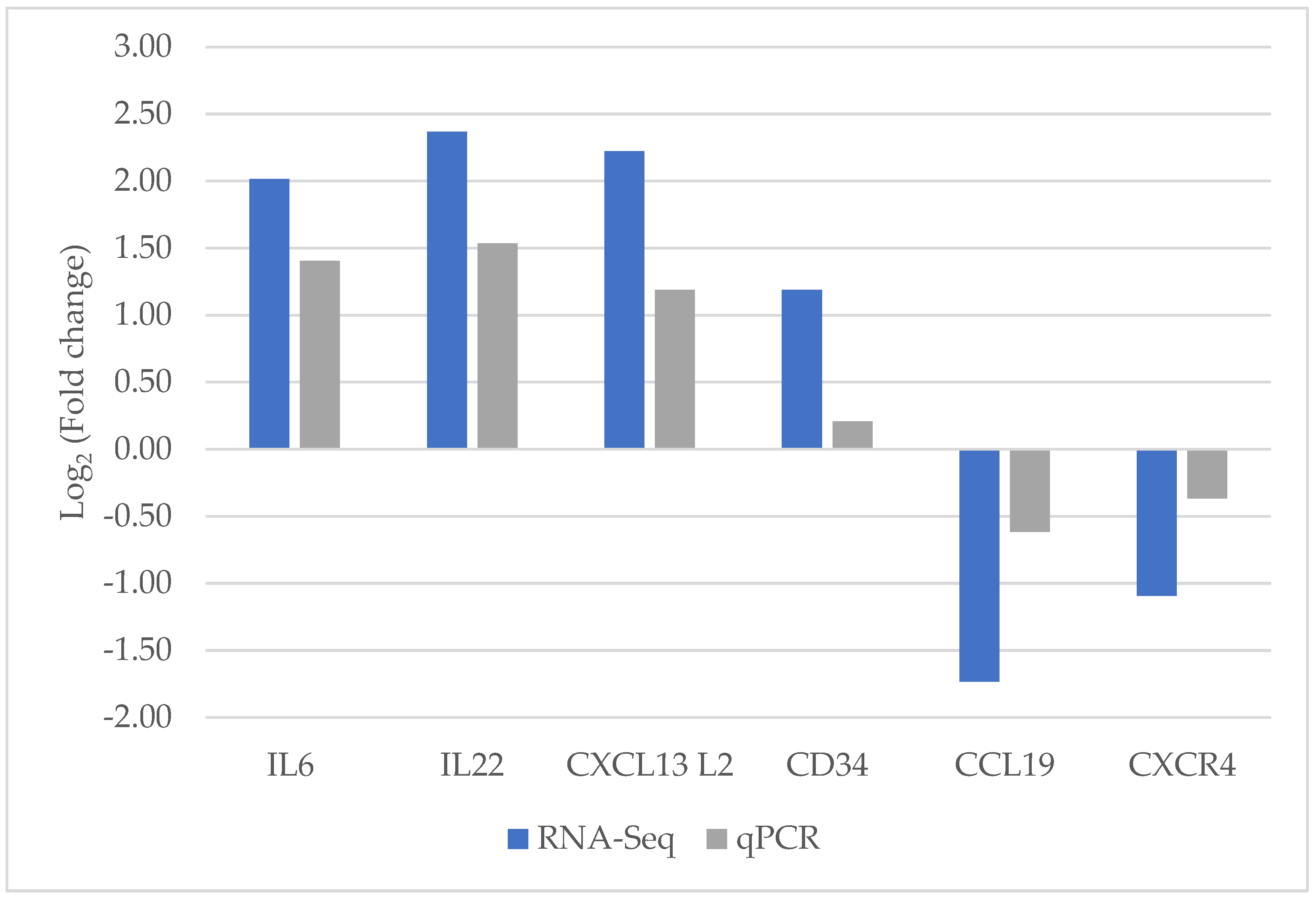
3.5. Validation Based on qPCR

In this study, significantly, DEGs related to innate and adaptive immune responses, including IL-6, IL-22, CCL-19, CXCR4, CXCL13L2, and CD34, were selected by using qPCR assay to confirm the creditability of the RNA-sequencing technique. The expression patterns of all DEGs from qPCR analysis were concomitant with those from RNA-Seq data analysis (Figure 2).

4. Discussion

Genetics play a crucial role in immune responses to infectious pathogens and vaccines [32]. Vigorous innate immune responses, rates of macrophage differentiation and activation, and MHC haplotype were well recognized as essential factors associated with disease resistance and susceptibility [33]. Between the breeds, variations in the MHC haplotype and immune-related genes define the outcomes of immune responses and survival ability. In this experiment, the comparison of transcriptomics between distinct genetic line chickens could provide better understanding on transcriptional responses in particular breeds and it revealed significant immune-related genes and molecular mechanisms against IBV vaccination between vaccinated and control groups, as well as between TCC and WLC.

4.1. Immune Responsiveness in Taiwan Country Chickens

From week 2 post vaccination, vaccinated TCCs showed higher antibody levels compared with the controls within 1 week after vaccination. At the same time, with the rise of systemic anti-IBV immunoglobulin, numerous genes involved in innate immune responses, B-cell accumulation and activation were highly expressed. The significant upregulated genes in TCCs responding to immunization were MAPK10, macrophage mannose receptor 1-like 2, Gallus gallus immunoglobulin-like receptor CHIR-B2-like, and V-set pre-B cell surrogate light chain 3. The Gallus gallus immunoglobulin-like receptor CHIR-B2-like is mainly expressed on B cells and its main function is inhibitory receptor-related with B cell proliferation process [34]. In addition, V-set pre-B cell surrogate light chain 3 has a higher expression response to IBV vaccination, which is significant in the B cell maturation and plays a crucial role in Pre-B cell receptor formation [35,36]. The high expression of these genes represented the accumulation of B cells within the spleen in response to vaccination compared with the control group.
GO and KEGG database analyses between vaccinated and control groups of TCCs (Supplementary Tables S4 and S5) showed significant pathways related to inflammation and pathogen recognition in response to vaccination. The remarkable enriched pathways from the KEGG database were those involving cytokine–cytokine receptor interaction, nucleotide oligomerization domain (NOD)-like receptor signaling pathway, and TLR signaling pathway. By identifying pathogen-associated molecular pattern (PAMP), NOD-like and Toll-like receptor signaling pathways are the important connector between innate and adaptive immune responses. Additionally, from the analysis from GO database, vaccinated TCCs were highly enriched in response to chemicals, response to hormones, taxis, and cellular response to peptide. These biological processes are essential mechanisms for the host to respond to external stimuli and initiate the adaptive immune response. In addition, hormone and taxis pathways are related to the attraction and accumulation of immune cells to the spleen, allowing T and B cell response to antigens in the circulatory system [37].

4.2. Immune Responsiveness in White Leghorn Chickens

WLCs showed only 49 upregulated genes and 72 downregulated genes in response to vaccination (Table 2). The significantly upregulated immune-related genes were involved in the innate immune response and activation of adaptive immune responses, including IL-6 and IL-4I1. IL-6 is a crucial multifunctional cytokine related to the proinflammatory process and the activation and differentiation of T and B lymphocytes. A study found that IL-6 is crucial for mice survival after infection with influenza through the optimization of T cell regulation and the migration and phagocytic activities of macrophages [38]. However, the presence of IL-6 and TGF-β are also related to immune tolerance by promoting the expression of regulatory T cells [39]. In human studies, IL-4I1 plays an important role in the limitation of side effects from adaptive immune response by inhibiting IFN -γ production and differentiation of effector T cells and turning naive T cells into regulatory T cells [40].
According to the GO and KEGG databases, the important GO pathways in WLCs (Supplementary Table S6) were those involving the regulation of IL-6 production, IL-6 production, positive regulation of T cell proliferation, and B cell activation. The main gene involved in these enriched pathways was IL-6. This evidence stated the important role of IL-6 in immune response modulation against IBV vaccination in WLCs. For the KEGG pathway analysis (Supplementary Table S7), the significant enriched pathways were involved in the inflammatory process, lymphocyte proliferation and differentiation, including cytokine–cytokine receptor interaction, transforming growth factor-beta signaling pathway, and phagosome.
From human study, the transforming growth factor-beta (TGF- β) signaling pathway plays a critical role on the divergent function of T cell. On the one hand, TGF-β plays a role in supporting the activation of functional T cells by promoting chemotaxis of immune cells. TGF-β can induce chemotaxis of CD4+ T cells towards the CXCL12 and promote the migration of antigen-presenting cells towards lymphatic vessels and lymph nodes [41,42]. On the other hand, TGF-β also controls the immune tolerance by inhibiting T-cell receptor signaling and promoting the differentiation and function of the regulatory T cells (Tregs) [39]. The highly expressed IL-4I1 and enriched transforming growth factor-beta signaling pathway in WLCs could limit the side effects from overstimulation of the adaptive immune response. However, lower levels of effective T cells could also reduce the host’s capability to eradicate the invasive pathogens. A previous study indicated that high expression of regulatory T cells is related to susceptible traits in chicken against Marek’s infection [43].

4.3. Differences of Immune Responsiveness between Breeds

To investigate the effect of genetics between breeds, the control groups between TCCs and WLCs were compared. TCCs had highly expressed cytokine-related genes involved in immunological tolerance, including IL-4I1, IL-6, and IL-22. As previously described, IL-4I1 plays a significant role in the limitation of immunopathology mediators by controlling the effector Th1 and Th17 cells [44]. Additionally, IL-22 is secreted by activated dendritic cell and Th 17 cells. It functions as a proinflammatory and regenerative factor and is crucial for the protection and regeneration of barrier organs such as the lungs and gastrointestinal system [45]. In addition, immunoglobulin superfamily member 1-like 6, MHC class I polypeptide-related sequence A, and class I histocompatibility antigen, F10 alpha chain-like were enriched. An immunoglobulin superfamily member, recognized as an adhesion molecule, plays a crucial role in the mediation of cell surface interaction and pathogen perception [46]. MHC class I polypeptide-related sequence A, class I histocompatibility antigen, and F10 alpha chain-like are related to the antigen presentation process through MHC class I.
The upregulated genes in unvaccinated WLCs were those related to T cell-interacting, activating receptor on myeloid cell protein 1-like, and nuclear factor of activated T cells (Table 3). T cell-interacting, activating receptor on myeloid cells protein 1 is a triggering receptor on macrophages and neutrophils related to TNF-α and IL-8 production against pathogen infection [47]. Likewise, the nuclear factor of activated T cells is a transcriptional factor that involves many normal body processes and is well known as a crucial player in T cell activation and the determination of the fate and function of the T cell population [48]. Moreover, WLCs also highly expressed interleukin 1 receptor-like 2 and MAPK10. The main function of interleukin 1 receptor-like 2 is inhibiting IL-1 activity, which can inhibit the excess production of proinflammatory cytokines.
Comparison between vaccinated groups of the two breeds (Table 3) revealed that the genes highly expressed in TCCs were those encoding IL-8L1, C-type lectin domain family 2 member L-like, IgG Fc-binding protein-like, C-type lectin domain family 2 member L-like, class I histocompatibility antigen, F10 alpha chain-like 3, class I histocompatibility antigen, F10 alpha chain-like, MHC class I polypeptide-related sequence A, and MHC class II beta chain BLB1 response to vaccination. The IL-8L1 plays a crucial role in neutrophil activation through the promotion of cell adhesion, transendothelial migration, and killing process. Furthermore, the function of the IgG Fc-binding protein is the modulation of the adaptive immune response through fusion with specific antigenic proteins [49]. Higher expression of the above genes in the TCCs in comparison to WLCs shows the role of these genes in activation of B and T cells in subsequent IBV vaccination.
Among the vaccinated groups, WLCs showed high expression of IL-6, IL-22 receptor subunit alpha 2, T cell surface glycoprotein CD8 alpha chain-like, granzyme K, MHC class I antigen YF5, and MHC class II beta chain BLB2. The high expression of these genes in WLCs relates to T cell development and activation process responding to IBV vaccination. The T cell surface glycoprotein CD8 alpha chain-like was related to CD8 T cell accumulation and T cell activation within the spleen. In addition, granzyme K was secreted by activated macrophage, natural killer cell, and cytotoxic T lymphocyte, which functionally promotes the cytotoxicity of invader and infected cells [50]. The higher expression of these genes compared with TCCs suggests that T cell development and activation were among the major immune processes in the spleen responding to vaccination in WLCs.
From the KEGG pathway analysis (Table 5), TCCs and WLCs shared several immune-related pathways responding to vaccination, including cell adhesion molecules (CAMs), cytokine–cytokine receptor interaction, toll-like receptor (TLR) signaling pathway, salmonella infection, and influenza A. Interestingly, the gene members in each pathway were expressed differently between the two breeds of chicken. The major gene members in TCCs were IL-8L1 and BLB1, whereas the main members in WLCs were IL-6 and BLB2. BLB1 and BLB2 are class II MHC genes that are highly polymorphic and responsible for transducing signals during the B cell activation process. The results in Table 1 show that TCCs had a higher capability to produce more immunoglobulin in response to IBV vaccination compared with WLCs. Based on this experiment, we could assume that the different expression of MHC class II between TCCs and WLCs might play a significant role on varying antibody production levels against IBV vaccination. However, further investigation is needed to confirm these findings.

5. Conclusions

This study provides a comprehensive understanding of immune related genes and biological pathways against IBV vaccination of TCCs and commercial WLCs. On day 7 post inoculation, the vaccinated TCCs group exhibited higher levels of antibody production compared to the control group. Additionally, a set of differentially expressed genes related to innate immune responses, as well as B cell proliferation and development, were highly expressed. These genes included MAPK10, macrophage mannose receptor 1-like 2, Gallus gallus immunoglobulin-like receptor CHIR-B2-like, and V-set pre-B cell surrogate light chain 3. On the contrary, the significant enriched immune-related genes in vaccinated WLCs were IL-6, IL-4I1, and IL22RA2, which related to proinflammatory process and the activation and differentiation of T and B lymphocytes. In addition, the data analysis revealed that significant biological pathways of both two breeds were related through cell adhesion molecules (CAMs), cytokine–cytokine receptor interaction, toll-like receptor (TLR) signaling pathway and influenza A against live attenuated IBV vaccination.

Supplementary Materials

The following supporting information can be downloaded at: https://www.mdpi.com/article/10.3390/ani13101642/s1, Table S1: Primer sequences used for quantitative real-time polymerase chain reaction analysis. Table S2: Sequencing data of Taiwan Country chicken and White Leghorn chicken of day 7 postvaccination. Table S3: Comparison of results from Taiwan Country chicken and White Leghorn chicken of day 7 postvaccination. Table S4: Gene ontology analysis of differentially expressed genes between vaccinated Taiwan Country chicken and unvaccinated Taiwan Country chicken (TCCVac VS TCCUnvac) at day 7 postvaccination. Table S5: Kyoto Encyclopedia Analysis of Genes and Genome pathway analysis of differentially expressed genes between vaccinated Taiwan Country chicken and unvaccinated Taiwan Country chicken (TCCVac VS TCCUnvac) at day 7 postvaccination. Table S6: Gene ontology analysis of differentially expressed genes between vaccinated White Leghorn chicken and unvaccinated White Leghorn chicken (WLCVac VS WLCUnvac) at day 7 postvaccination. Table S7: Kyoto Encyclopedia Analysis of Genes and Genome pathway analysis of differentially expressed genes between vaccinated White Leghorn chicken and unvaccinated White Leghorn chicken (WLCVac VS WLCUnvac) at day 7 postvaccination.

Author Contributions

Conceptualization, S.C., C.-F.C., P.-C.T., K.G. and C.-S.C.; methodology, S.C., C.-F.C., P.-C.T., K.G. and C.-S.C.; software, S.C. and C.-S.C.; validation, S.C. and P.-C.T.; formal analysis, S.C. and C.-S.C.; resources, C.-F.C. and P.-C.T.; writing—original draft preparation, S.C.; writing—review and editing, S.C., C.-F.C., P.-C.T., C.-S.C. and K.G.; supervision, C.-F.C., P.-C.T. and K.G.; project administration, C.-F.C. and P.-C.T.; funding acquisition, C.-F.C. and P.-C.T. All authors have read and agreed to the published version of the manuscript.

Funding

This research were funded by the Ministry of Science and Technology (MOST), Taiwan, grant number MOST 109-2321-B-005-019- and the Smart Sustainable New Agricultural Research Center (SMARTer), grant number NSTC 111-2634-F-005-001-.

Institutional Review Board Statement

The animal study protocol was approved by the Ethics Committee of National Chung Hsing University, Taiwan (IACUC No. 108-052R) and animal care was performed according to the guidelines of Institutional Animal Care and Use Committee (IACUC).

Informed Consent Statement

Not applicable.

Data Availability Statement

Data are available for fair use to support further scientific research upon request to the corresponding author.

Conflicts of Interest

The authors declare no conflict of interest.

References

  1. De Wit, J.J.; Cook, J.K.A. Spotlight on avian coronaviruses. Avian Pathol. 2020, 49, 313–316. [Google Scholar] [CrossRef] [PubMed]
  2. Ignjatović, J.; Sapats, S. Avian infectious bronchitis virus. Rev.—Off. Int. Epizoot. 2000, 19, 493–508. [Google Scholar] [CrossRef] [PubMed]
  3. Chhabra, R.; Forrester, A.; Lemiere, S.; Awad, F.; Chantrey, J.; Ganapathy, K. Mucosal, cellular, and humoral immune responses induced by different live infectious bronchitis virus vaccination regimes and protection conferred against infectious bronchitis virus Q1 strain. Clin. Vaccine Immunol. 2015, 22, 1050–1059. [Google Scholar] [CrossRef] [PubMed]
  4. Jackwood, M.W. Review of infectious bronchitis virus around the world. Avian Dis. 2012, 56, 634–641. [Google Scholar] [CrossRef] [PubMed]
  5. Roberts, J.R.; Chousalkar, K.K. Chapter 52—Infectious Bronchitis. In Egg Innovations and Strategies for Improvements; Hester, P.Y., Ed.; Academic Press: San Diego, CA, USA, 2017; pp. 561–570. [Google Scholar]
  6. Samiullah, S.; Roberts, J.; Chousalkar, K. Infectious bronchitis virus and brown shell colour: Australian strains of infectious bronchitis virus affect brown eggshell colour in commercial laying hens differently. Avian Pathol. 2016, 45, 552–558. [Google Scholar] [CrossRef]
  7. Hoerr, F.J. The pathology of infectious bronchitis. Avian Dis. 2021, 65, 600–611. [Google Scholar] [CrossRef]
  8. Iotsov, S.; Iotova, I.; Atanasov, K.; Nikolova, M.; Nikolov, N.D. Resistance of different breeds of laying poultry to Marek’s disease virus]. Vet. Med. Nauki 1983, 20, 3–8. [Google Scholar]
  9. Dunnington, E.A.; Larsen, C.T.; Gross, W.B.; Siegel, P.B. Antibody responses to combinations of antigens in white Leghorn chickens of different background genomes and major histocompatibility complex genotypes. Poult. Sci. 1992, 71, 1801–1806. [Google Scholar] [CrossRef]
  10. Kannaki, T.R.; Priyanka, E.; Haunshi, S. Research Note: Disease tolerance/resistance and host immune response to experimental infection with Pasteurella multocida A:1 isolate in Indian native Nicobari chicken breed. Poult. Sci. 2021, 100, 101268. [Google Scholar] [CrossRef]
  11. Khatab, S.A.; Hemeda, S.A.; El-Nahas, A.F.; Abd El Naby, W.S.H.; Hassan, S.; Alorabi, J.A.; Dawood, M.A.O. Intra- and inter-breed variation in immune response to acute and sub-chronic Salmonella infection and commercial immune-stimulant in two-layer breeds. Vet. Med. Sci. 2021, 7, 2353–2361. [Google Scholar] [CrossRef]
  12. Hedrick, P.W. Pathogen resistance and genetic variation at MHC loci. Evolution 2002, 56, 1902–1908. [Google Scholar] [CrossRef] [PubMed]
  13. Banat, G.R.; Tkalcic, S.; Dzielawa, J.A.; Jackwood, M.W.; Saggese, M.D.; Yates, L.; Kopulos, R.; Briles, W.E.; Collisson, E.W. Association of the chicken MHC B haplotypes with resistance to avian coronavirus. Dev. Comp. Immunol. 2013, 39, 430–437. [Google Scholar] [CrossRef] [PubMed]
  14. Smith, J.; Sadeyen, J.R.; Cavanagh, D.; Kaiser, P.; Burt, D.W. The early immune response to infection of chickens with Infectious Bronchitis Virus (IBV) in susceptible and resistant birds. BMC Vet. Res. 2015, 11, 256. [Google Scholar] [CrossRef] [PubMed]
  15. Deist, M.S.; Gallardo, R.A.; Bunn, D.A.; Dekkers, J.C.M.; Zhou, H.; Lamont, S.J. Resistant and susceptible chicken lines show distinctive responses to Newcastle disease virus infection in the lung transcriptome. BMC Genomics 2017, 18, 989. [Google Scholar] [CrossRef]
  16. Samina, I.; Brenner, J.; Peleg, B.A. Differences in protection between heavy and light breeds of chickens following vaccination with Newcastle disease vaccines—A survey of data, 1971 to 1990. Avian Pathol. 1992, 21, 693–697. [Google Scholar] [CrossRef] [PubMed]
  17. Chang, S.; Dunn, J.R.; Heidari, M.; Lee, L.F.; Song, J.; Ernst, C.W.; Ding, Z.; Bacon, L.D.; Zhang, H. Genetics and vaccine efficacy: Host genetic variation affecting Marek’s disease vaccine efficacy in White Leghorn chickens1. Poult. Sci. 2010, 89, 2083–2091. [Google Scholar] [CrossRef] [PubMed]
  18. Chang, C.S.; Tixier-Boichard, M.; Chazara, O.; Lee, Y.P.; Chen, C.F.; Chang, P.C.; Chen, J.W.; Bed’hom, B. Different immune responses to three different vaccines following H6N1 low pathogenic avian influenza virus challenge in Taiwanese local chicken breeds. BMC Proc. 2011, 5 (Suppl. 4), S33. [Google Scholar] [CrossRef] [PubMed]
  19. Mountford, J.; Gheyas, A.; Vervelde, L.; Smith, J. Genetic variation in chicken interferon signalling pathway genes in research lines showing differential viral resistance. Anim. Genet. 2022, 53, 640–656. [Google Scholar] [CrossRef]
  20. Lowenthal, J.W.; Bean, A.G.; Kogut, M.H. What’s so special about chicken immunology? Dev. Comp. Immunol. 2013, 41, 307–309. [Google Scholar] [CrossRef]
  21. Jeurissen, S.H. The role of various compartments in the chicken spleen during an antigen-specific humoral response. Immunology 1993, 80, 29–33. [Google Scholar]
  22. Zhang, Q.; Waqas, Y.; Yang, P.; Sun, X.; Liu, Y.; Ahmed, N.; Chen, B.; Li, Q.; Hu, L.; Huang, Y.; et al. Cytological study on the regulation of lymphocyte homing in the chicken spleen during LPS stimulation. Oncotarget 2017, 8, 7405–7419. [Google Scholar] [CrossRef] [PubMed]
  23. Zhang, Q.; Chen, B.; Yang, P.; Zhang, L.; Liu, Y.; Ullah, S.; Wu, L.; Waqas, Y.; Le, Y.; Chen, W.; et al. Identification and structural composition of the blood-spleen barrier in chickens. Vet. J. 2015, 204, 110–116. [Google Scholar] [CrossRef] [PubMed]
  24. Cheng, Y.S. Disease Resistance to Marek’s Disease in Country Chickens and White Leghorns. Master’s Thesis, National Chung Hsing University, Taichung, Taiwan, 1987. [Google Scholar]
  25. Chao, C.H.; Lee, Y.P. Immune responses to Newcastle disease virus vaccine and sheep erythrocytes in country chickens and White Leghorns. J. Chin. Soc. Anim. Sci. 1991, 20, 189–201. [Google Scholar]
  26. Chen, C.F.; Lee, Y.P. Comparison of disease resistance to leucocytozoonosis among country chickens, broilers and White Leghorns. J. Chin. Soc. Anim. Sci. 1991, 30, 305–316. [Google Scholar]
  27. Bolger, A.M.; Lohse, M.; Usadel, B. Trimmomatic: A flexible trimmer for Illumina sequence data. Bioinformatics 2014, 30, 2114–2120. [Google Scholar] [CrossRef]
  28. Andrews, S. FastQC: A Quality Control Tool for High Throughput Sequence Data. 2010. Available online: https://www.bioinformatics.babraham.ac.uk/projects/fastqc/ (accessed on 17 May 2018).
  29. Ewels, P.; Magnusson, M.; Lundin, S.; Käller, M. MultiQC: Summarize analysis results for multiple tools and samples in a single report. Bioinformatics 2016, 32, 3047–3048. [Google Scholar] [CrossRef]
  30. Love, M.I.; Huber, W.; Anders, S. Moderated estimation of fold change and dispersion for RNA-seq data with DESeq2. Genome Biol. 2014, 15, 550. [Google Scholar] [CrossRef]
  31. Livak, K.J.; Schmittgen, T.D. Analysis of relative gene expression data using real-time quantitative PCR and the 2(-Delta Delta C(T)) Method. Methods 2001, 25, 402–408. [Google Scholar] [CrossRef]
  32. Kaufman, J. The simple chicken major histocompatibility complex: Life and death in the face of pathogens and vaccines. Philos. Trans. R. Soc. Lond. Ser. B Biol. Sci. 2000, 355, 1077–1084. [Google Scholar] [CrossRef]
  33. Silva, A.P.D.; Gallardo, R.A. The chicken MHC: Insights into genetic resistance, immunity, and inflammation following infectious bronchitis virus infections. Vaccines 2020, 8, 637. [Google Scholar] [CrossRef]
  34. Viertlboeck, B.C.; Crooijmans, R.P.; Groenen, M.A.; Göbel, T.W. Chicken Ig-like receptor B2, a member of a multigene family, is mainly expressed on B lymphocytes, recruits both Src homology 2 domain containing protein tyrosine phosphatase (SHP)-1 and SHP-2, and inhibits proliferation. J. Immunol. 2004, 173, 7385–7393. [Google Scholar] [CrossRef] [PubMed]
  35. Mårtensson, I.L.; Ceredig, R. Review article: Role of the surrogate light chain and the pre-B-cell receptor in mouse B-cell development. Immunology 2000, 101, 435–441. [Google Scholar] [CrossRef]
  36. Rosnet, O.; Blanco-Betancourt, C.; Grivel, K.; Richter, K.; Schiff, C. Binding of free immunoglobulin light chains to VpreB3 inhibits their maturation and secretion in chicken B cells. J. Biol. Chem. 2004, 279, 10228–10236. [Google Scholar] [CrossRef] [PubMed]
  37. Dardenne, M.; Smaniotto, S.; de Mello-Coelho, V.; Villa-Verde, D.M.; Savino, W. Growth hormone modulates migration of developing T cells. Ann. N. Y. Acad. Sci. 2009, 1153, 1–5. [Google Scholar] [CrossRef]
  38. Velazquez-Salinas, L.; Verdugo-Rodriguez, A.; Rodriguez, L.L.; Borca, M.V. The role of interleukin 6 during viral infections. J. Immunol. 2019, 10, 1057. [Google Scholar] [CrossRef] [PubMed]
  39. Oh, S.A.; Li, M.O. TGF-β: Guardian of T cell function. J. Immunol. 2013, 191, 3973–3979. [Google Scholar] [CrossRef] [PubMed]
  40. Romagnani, S. IL4I1: Key immunoregulator at a crossroads of divergent T-cell functions. Eur. J. Immunol. 2016, 46, 2302–2305. [Google Scholar] [CrossRef]
  41. Li, M.O.; Wan, Y.Y.; Sanjabi, S.; Robertson, A.-K.L.; Flavell, R.A. Transforming growth factor-β regulation of immune responses. Annu. Rev. Immunol. 2006, 24, 99–146. [Google Scholar] [CrossRef]
  42. Franitza, S.; Kollet, O.; Brill, A.; Vaday, G.G.; Petit, I.; Lapidot, T.; Alon, R.; Lider, O. TGF-beta1 enhances SDF-1alpha-induced chemotaxis and homing of naive T cells by up-regulating CXCR4 expression and downstream cytoskeletal effector molecules. Eur. J. Immunol. 2002, 32, 193–202. [Google Scholar] [CrossRef]
  43. Gurung, A.; Kamble, N.; Kaufer, B.B.; Pathan, A.; Behboudi, S. Association of Marek’s disease induced immunosuppression with activation of a novel regulatory T cells in chickens. PLoS Pathog. 2017, 13, e1006745. [Google Scholar] [CrossRef]
  44. Molinier-Frenkel, V.; Prévost-Blondel, A.; Castellano, F. The IL4I1 Enzyme: A new player in the immunosuppressive tumor microenvironment. Cells 2019, 8, 757. [Google Scholar] [CrossRef]
  45. Eyerich, K.; Dimartino, V.; Cavani, A. IL-17 and IL-22 in immunity: Driving protection and pathology. Eur. J. Immunol. 2017, 47, 607–614. [Google Scholar] [CrossRef]
  46. Buck, C.A. Immunoglobulin superfamily: Structure, function and relationship to other receptor molecules. Semin. Cell Dev. Biol. 1992, 3, 179–188. [Google Scholar] [CrossRef]
  47. Tessarz, A.S.; Weiler, S.; Zanzinger, K.; Angelisová, P.; Horejsí, V.; Cerwenka, A. Non-T cell activation linker (NTAL) negatively regulates TREM-1/DAP12-induced inflammatory cytokine production in myeloid cells. J. Immunol. 2007, 178, 1991–1999. [Google Scholar] [CrossRef] [PubMed]
  48. Lee, J.U.; Kim, L.K.; Choi, J.M. Revisiting the concept of targeting NFAT to control T cell immunity and autoimmune diseases. Front. Immunol. 2018, 9, 2747. [Google Scholar] [CrossRef] [PubMed]
  49. Dong, W.; Zhang, H.; Huang, H.; Zhou, J.; Hu, L.; Lian, A.; Zhu, L.; Ma, N.; Yang, P.; Wei, K.; et al. Chicken IgY Fc linked to Bordetella avium ompA and Taishan Pinus massoniana pollen polysaccharide adjuvant enhances macrophage function and specific immune responses. Front. Microbiol. 2016, 7, 1708. [Google Scholar] [CrossRef]
  50. Bouwman, A.C.; van Daalen, K.R.; Crnko, S.; Ten Broeke, T.; Bovenschen, N. Intracellular and extracellular roles of granzyme K. Front. Immunol. 2021, 12, 677707. [Google Scholar] [CrossRef] [PubMed]
Figure 1. The sample collection and experimental processes of Taiwan Country chicken and White Leghorn chicken. Created with BioRender.com accessed on 9 April 2023.
Animals 13 01642 g001
Figure 2. Validation of selected genes between ribonucleic acid-sequencing and quantitative real-time polymerase chain reaction by Log2 Fold Change.
Animals 13 01642 g002
Table 1. Mean anti-IBV ELISA antibody titers [S (OD sample-OD negative control)/p (OD positive control-OD negative control) ratio] between the treatment and control groups of each breed of chickens.
DayTCCWLC
TreatmentControlTreatmentControl
Day 01.78 ± 0.701.37 ± 0.361.72 ± 0.331.44 ± 0.28
Day 73.83 a ± 1.151.04 b ± 0.131.68 ± 0.371.32 ± 0.37
Day 1431.40 a,x ± 6.551.04 b ± 0.377.90 a,y ± 1.360.88 b ± 0.21
Day 2138.00 a,x ± 5.730.93 b ± 0.358.79 a,y ± 1.431.02 b ± 0.17
a,b Mean ± SE within a breed for a given stage with different superscripts indicating different significance. x,y indicated significant differences between the breeds in a given stage (p < 0.05).
Table 2. List of significant immune-related differentially expressed genes between vaccinated and unvaccinated groups within a breed at day 7 post vaccination.
BreedUpregulated XDownregulated YImmune Related Geneslog2FoldChangep-Value
TCC257186Immunoglobulin superfamily DCC subclass member 4 X1.893.50 × 10−5
Transforming growth factor beta receptor 3 x1.434.82 × 10−4
Macrophage mannose receptor 1-like 2 X1.401.60 × 10−3
Mitogen-activated protein kinase 10 X1.805.88 × 10−3
Major histocompatibility complex, class I X2.495.66 × 10−3
V-set pre-B cell surrogate light chain 3 X2.497.56 × 10−4
Interleukin 6 Y−2.864.42 × 10−3
Interleukin 4 induced 1 Y−2.659.62 × 10−3
Interleukin 22 Y−2.543.50 × 10−3
WLC4972Interleukin 4 induced 1 X1.454.18 × 10−5
Interleukin 22 receptor subunit alpha 2 X1.268.11 × 10−4
Interleukin 6 X2.025.26 × 10−3
MHC class I polypeptide-related sequence A Y−1.569.59 × 10−6
Eosinophil peroxidase Y−1.364.65 × 10−4
Class I histocompatibility antigen, F10 alpha chain-like 3 Y−1.173.06 × 10−3
X Upregulated due to p-value ≤ 0.05; Y Downregulated due to p-value ≤ 0.05.
Table 3. The list of significant immune-related differentially expressed genes between Taiwan Country chicken and White Leghorn chicken at day 7 post vaccination according to the challenge status.
Challenge StatusBreedImmune Related Geneslog2FoldChangep-Value
TCCWLC
Unvaccinated5771118C-X-C, motif chemokine ligand 14 X6.464.74 × 10−7
Interleukin 22 X4.621.05 × 10−4
Interleukin 4 induced 1 X3.491.25 × 10−4
Interleukin 6 X3.201.39 × 10−4
Leukocyte immunoglobulin-like receptor subfamily B member 5 X4.982.63 × 10−3
Interleukin 1 receptor-like 2 Y−3.092.52 × 10−13
TNF receptor superfamily member 19 Y−1.583.14 × 10−8
T cell-interacting, activating receptor on myeloid cells protein 1-like Y−3.203.95 × 10−7
Mitogen-activated protein kinase 10 Y−1.823.49 × 10−03
Vaccinated470416Class I histocompatibility antigen, F10 alpha chain-like X6.824.78 × 10−83
C-type lectin domain family 2 member L-like X3.783.35 × 10−25
Major histocompatibility complex class II beta chain BLB1 X1.421.14 × 10−21
IgG Fc-binding protein-like X1.494.84 × 10−15
Interleukin 8-like 1 X1.541.03 × 10−3
Major histocompatibility complex class II beta chain BLB2 Y−1.071.61 × 10−13
T cell surface glycoprotein CD8 alpha chain-like Y−2.208.57 × 10−7
Granzyme K Y−1.071.00 × 10−7
MHC class I antigen YF5 Y−1.034.29 × 10−6
Interleukin 22 receptor subunit alpha 2 Y−1.519.32 × 10−5
Interleukin 6 Y−1.672.56 × 10−2
X Upregulated due to p-value ≤ 0.05; Y Downregulated due to p-value ≤ 0.05.
Table 4. Comparison of significant pathways from GO between the vaccinated groups of Taiwan Country chicken and White Leghorn chicken at day 7 post vaccination.
BreedDescriptionp-ValueCountGenes ID
TCCCell-cell adhesion via plasma-membrane adhesion molecules3.60 × 10−45CDH3/DSCAML1/FLRT3/NFASC/SDK2
Cell differentiation1.00 × 10−319ACE/ZP3/OLFM1/RNASE6/ACVR2B/BRINP1/
APOA1/DSCAML1/FLRT3/TLL1/RARB/MGP/
TAGLN/SERPINB10B/CSRP2/NFASC/SDK2/MSTN
Chemotaxis0.1643FLRT3/NFASC/MSTN
Regulation of leukocyte migration0.2141MSTN
Myeloid cell differentiation2.29 × 10−42ACE/SERPINB10B
WLCCell proliferation7.00 × 10−37NPPC/CDH13/ANKRD1/HPGDS/IL6/NTRK2/INHBA
Acute inflammatory response7.00 × 10−32FN1/IL6
Cell differentiation1.10 × 10−211NPPC/ANKRD1/VSIG1/TENM1/IL6/RPE65/THRB/NTRK2/INHBA/STMN2/BHLHE22
Regulation of cell proliferation1.10 × 10−26CDH13/ANKRD1/HPGDS/IL6/NTRK2/INHBA
Positive regulation of cell adhesion5.20 × 10−22CDH13/IL6
Cell-cell adhesion via plasma-membrane adhesion molecules5.20 × 10−22CDH13/TENM1
Table 5. Comparison of significant pathways from KEGG between the vaccinated groups of Taiwan Country chicken and White Leghorn chicken at day 7 post vaccination.
BreedDescriptionp-ValueCountSymbol
TCCCell adhesion molecules (CAMs)2.40 × 10⁻²⁶BLB1/CDH3/CDH1/NRXN1/NFASC/CNTNAP2
Cytokine-cytokine receptor interaction3.00 × 10−28GH/IL11RA/ACVR2B/IL8L1/BMP6/EDAR/CCR10/MSTN
Phagosome9.70 × 10−25BLB1/LOC425049/MMR1L1/ATP6V0D2/TUBA1C
NOD-like receptor signaling pathway0.2024IL8SL1/CAMP/GBP/GBP4L
Salmonella infection0.7201IL8L1
Influenza A0.7282BLB1/IL8L1
Toll-like receptor signaling pathway0.7931IL8L1
Herpes simplex virus 1 infection0.9411BLB1
WLCCytokine-cytokine receptor interaction6.70 × 10−25TNFSF15/IL6/IL18RAP/IL1RL2/INHBA
Cell adhesion molecules (CAMs)0.1483NCAM2/NRXN3/BLB2
NOD-like receptor signaling pathway0.4022IL6/CASR
Influenza A0.4482IL6/BLB2
Herpes simplex virus 1 infection0.4922IL6/BLB2
Salmonella infection0.5291IL6
Toll-like receptor signaling pathway0.6061IL6
Disclaimer/Publisher’s Note: The statements, opinions and data contained in all publications are solely those of the individual author(s) and contributor(s) and not of MDPI and/or the editor(s). MDPI and/or the editor(s) disclaim responsibility for any injury to people or property resulting from any ideas, methods, instructions or products referred to in the content.

