
Propagation of Babesia bigemina in Rabbit Model and Evaluation of Its Attenuation in Cross-Bred Calves
 ,
,  , and
, and 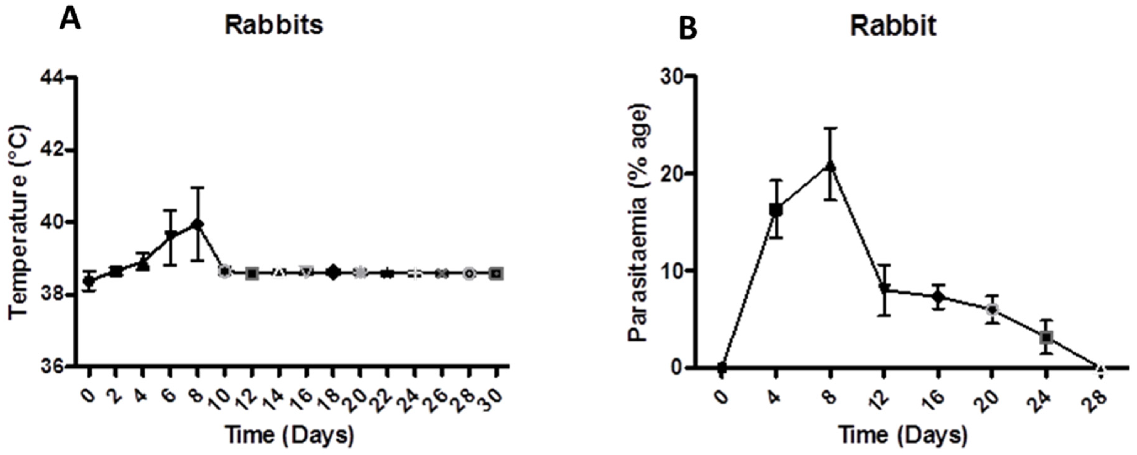
Abstract
:Simple Summary
Abstract
1. Introduction
2. Materials and Methods
2.1. Source of Parasite
2.2. Inoculation into Rabbit
2.3. Inoculation of Parasite into Calves
2.4. Statistical Analysis
3. Results
3.1. The Effect of Babesiosis on Body Temperature and Parasitaemia in Rabbits
3.2. The Effect of Babesiosis on Body Temperature, Parasitaemia, and Packed Cell Volume in Experimental Calves
4. Discussion
5. Conclusions
Author Contributions
Funding
Institutional Review Board Statement
Informed Consent Statement
Data Availability Statement
Acknowledgments
Conflicts of Interest
References
- Angus, B.M. The history of the cattle tick Boophilus microptus in Australia and achievements in its control. Int. J. Parasitol. 1996, 26, 1341–1355. [Google Scholar] [CrossRef]
- Ristic, M.; Levy, M. A new era of research toward solution of bovine babesiosis [Tick control, vaccines]. Bibliography 1981, 539–544. [Google Scholar]
- Babes, V. Sur l’hemoglobinurie bacterienne du boeuf. CR-Acad. Sci. 1888, 107, 692–694. [Google Scholar]
- Smith, T.; Kilborne, F.L. Investigations into the Nature, Causation, and Prevention of Texas or Southern Cattle Fever; US Department of Agriculture: Washington, DC, USA; Bureau of Animal Industry: Quezon City, Philippines, 1893. [Google Scholar]
- Mihalca, A.D.; Cozma, V.; Şuteu, E.; Marinculic, A.; Boireau, P. The quest for piroplasms: From Babeş and Smith to molecules. Sci. Parasitol. 2010, 11, 14–19. [Google Scholar]
- Starcovici, C. Bemerkungen über den durch Babes entdeckten Blutparasiten und die durch denselben hervorgebrachten Krankheiten, die seuchenhafte Hämoglobinurie des Rindes (Babes), das Texasfieber (Th. Smith) und der Carceag der Schafe (Babes). Zbl. Bakt. I. Abt. 1893, 14, 1–8. [Google Scholar]
- Schnittger, L.; Rodriguez, A.E.; Florin-Christensen, M.; Morrison, D.A. Babesia: A world emerging. Infect. Genet. Evol. 2012, 12, 1788–1809. [Google Scholar] [CrossRef] [PubMed]
- Bock, R.; Jackson, L.; De Vos, A.; Jorgensen, W. Babesiosis of cattle. Parasitology 2004, 129, S247–S269. [Google Scholar] [CrossRef]
- Böse, R.; Jorgensen, W.K.; Dalgliesh, R.J.; Friedhoff, K.T.; De Vos, A.J. Current state and future trends in the diagnosis of babesiosis. Vet. Parasitol. 1995, 57, 61–74. [Google Scholar] [CrossRef]
- Schnittger, A.R.; Tomazic, M.; Florin-Christensen, M. Current and prospective tools for the control of cattle-infecting Babesia parasites. In Protozoa: Biology, Classification and Role in Disease; Nova Publishers: Hauppauge, NY, USA, 2013; pp. 1–44. [Google Scholar]
- de Castro, J.J. Sustainable tick and tickborne disease control in livestock improvement in developing countries. Vet. Parasitol. 1997, 71, 77–97. [Google Scholar] [CrossRef]
- Callow, L. Protozoal and Rickettsial Diseases, Australian Bureau of Animal Health. Animal Health in Australia; Australian Government Publishing: Canberra, Australia, 1984; Volume 5, pp. 121–160.
- Jabbar, A.; Abbas, T.; Sandhu, Z.U.D.; Saddiqi, H.A.; Qamar, M.F.; Gasser, R.B. Tick-borne diseases of bovines in Pakistan: Major scope for future research and improved control. Parasit. Vect. 2015, 8, 1–13. [Google Scholar] [CrossRef] [PubMed]
- Mahoney, D.; Ross, D. Epizootiological factors in the control of bovine babesiosis. Aust. Vet. J. 1972, 48, 292–298. [Google Scholar] [CrossRef] [PubMed]
- Callow, L.; Rogers, R.; De Vos, A. Tick-borne diseases: Cattle-pathology and serology. In Australian Standard Diagnostic Techniques for Animal Diseases; CSIRO: Canberra, Australia, 1993; pp. 1–16. [Google Scholar]
- Johnston, L.; Trueman, K.; Pearson, R. Bovine babesiosis: Comparison of fluorescent antibody and giemsa staining in post-mortem diagnosis of infection. Aust. Vet. J. 1977, 53, 222–226. [Google Scholar] [CrossRef] [PubMed]
- Purnell, R.; Schroder, J. Herd prophylaxis of tick-borne diseases in susceptible cattle in South Africa. In Proceedings of the XIIIth World Congress on Diseases of Cattle, Durban, South Africa, 17–21 September 1984. [Google Scholar]
- Mendes, E.C.; Mendes, M.C.; Sato, M.E. Diagnosis of amitraz resistance in Brazilian populations of Rhipicephalus (Boophilus) microplus (Acari: Ixodidae) with larval immersion test. Exp. App. Acarol. 2013, 61, 357–369. [Google Scholar] [CrossRef] [PubMed]
- Taylor, R.; McHardy, N. Preliminary observations on the combined use of imidocarb and Babesia blood vaccine in cattle. J. S. Afr. Vet. Assoc. 1979, 50, 326–329. [Google Scholar]
- Purnell, R.; Lewis, D.; Young, E. Investigations on the prophylactic effect of treatment with imidocarb diproprionate on Babesia divergens infections in splenectomized calves. Br. Vet. J. 1980, 136, 452–456. [Google Scholar] [CrossRef]
- Roy-Smith, F. The prophylactic effects of imidocarb against Babesia argentina and Babesia bigemina infections of cattle. Aust. Vet. J. 1971, 47, 418–420. [Google Scholar] [CrossRef]
- Zintl, A.; Mulcahy, G.; Skerrett, H.E.; Taylor, S.M.; Gray, J.S. Babesia divergens, a bovine blood parasite of veterinary and zoonotic importance. Clin. Microbiol. Rev. 2003, 16, 622–636. [Google Scholar] [CrossRef]
- Callow, L.; Mellors, L.; McGregor, W. Reduction in virulence of Babesia bovis due to rapid passage in splenectomized cattle. Int. J. Parasitol. 1979, 9, 333–338. [Google Scholar] [CrossRef]
- Callow, L.; Dalgliesh, R.; De Vos, A. Development of effective living vaccines against bovine babesiosis—the longest field trial? Int. J. Parasitol. 1997, 27, 747–767. [Google Scholar] [CrossRef]
- Pipano, E. Live vaccine against hemoparasitic disease in livestock. Vet. Parasitol. 1995, 57, 213–231. [Google Scholar] [CrossRef]
- Levy, M.G.; Ristic, M. Babesia bovis: Continuous cultivation in a microaerophilous stationary phase culture. Science 1980, 207, 1218–1220. [Google Scholar] [CrossRef] [PubMed]
- Vega, C.A.; Buening, G.M.; Green, T.J.; Carson, C.A. In vitro cultivation of Babesia bigemina. Am. J. Vet. Res. 1985, 46, 416–420. [Google Scholar]
- Ramzan, M.S.; Rashid, M.I.; Akbar, H.; Avais, M.; Suleman, M. Theileria annulata: Its Propagation in Rabbits for the Attenuation of Piroplasms in Cross-Bred Calves. Animals 2022, 12, 813. [Google Scholar] [CrossRef] [PubMed]
- Rauf, U.; Suleman, M.; Abid, A.; Jamil, H.; Menghwar, H.; Durrani, A.Z.; Rashid, M.I.; Akbar, H. Humoral and cell-mediated immune response validation in calves after a live attenuated vaccine of Babesia bigemina. Pathogens 2020, 9, 936. [Google Scholar] [CrossRef]
- Khan, S.A.R.Z.A.M.I.N.; Khan, K.A.M.R.A.N.; Shah, S.U.; Ahmad, N.A.S.E.E.R. A preliminary assessment of rabbit farming and its scope in Khyber Pakhtunkhwa province of Pakistan. Sarhad. J. Agric. 2014, 30, 369–373. [Google Scholar] [CrossRef]
- Zaheer, H.; Rashid, I.; Akbar, H.; Farooq, R.K.; Ul Rehman, Z.; Abid, A.; Ali, S.; Akram, N.; Alvi, M.H. Expiry of a completely splenectomised calf in post-operative period due to mixed piroplasm infection: A case report. Annal. Parasitol. 2020, 66, 599–606. [Google Scholar]
- Zhou, M.; Cao, S.; Sevinc, F.; Sevinc, M.; Ceylan, O.; Moumouni, P.F.A.; Jirapattharasate, C.; Liu, M.; Wang, G.; Iguchi, A.; et al. Molecular detection and genetic identification of Babesia bigemina, Theileria annulata, Theileria orientalis and Anaplasma marginale in Turkey. Ticks. Tick-Borne. Dis. 2016, 7, 126–134. [Google Scholar] [CrossRef]
- Hagiwara, K.; Tsuji, M.; Ishihara, C.; Tajima, M.; Kurosawa, T.; Iwai, H.; Takahashi, K. Theileria sergenti infection in the Bo-RBC-SCID mouse model. Parasitol. Res. 1993, 79, 466–470. [Google Scholar] [CrossRef]
- Tsuji, M.; Hagiwara, K.; Takahashi, K.; Ishihara, C.; Azuma, I.; Siddiqui, W.A. Theileria sergenti proliferates in SCID mice with bovine erythrocyte transfusion. J. Parasitol. 1992, 78, 750–752. [Google Scholar] [CrossRef]
- Chaudhry, Z.I.; Suleman, M.; Younus, M.; Aslim, A. Molecular detection of Babesia bigemina and Babesia bovis in crossbred carrier cattle through PCR. Pak. J. Zool. 2010, 42, 201–204. [Google Scholar]
- Hashem, M.; Neamat-Allah, A.; Gheith, M.A. A study on bovine babesiosis and treatment with reference to hematobiochemical and molecular diagnosis. Slov. Vet. Res. 2018, 55, 165–173. [Google Scholar]
- Prism, G. Statistical Analysis Software GraphPad Version 6. Available online: https://www.graphpad.com/scientific-software/prism/ (accessed on 16 August 2022).
- Rogers, R.J.; Dimmock, C.K.; De Vos, A.J.; Rodwell, B.J. Bovine leucosis virus contamination of a vaccine produced in vivo against bovine babesiosis and anaplasmosis. Aust. Vet. J. 1988, 65, 285–287. [Google Scholar] [CrossRef]
- Todorovic, R.A.; Lopez, L.A.; Lopez, A.G.; Gonzalez, E.F. Bovine babesiosis and anaplasmosis: Control by premunition and chemoprophylaxis. Exp. Parasitol. 1975, 37, 92–104. [Google Scholar] [CrossRef]
- Abdel-Hamied, E.; Arafa, W.; Mahmoud, M. Oxidative stress, hemogram, hepatorenal function evaluation and molecular diagnosis of babesiosis in crossbred cows naturally infected with B. bigemina. Adv. Anim. Vet. Sci. 2020, 8, 1402–1409. [Google Scholar] [CrossRef]
- Sandhu, G.S.; Grewal, A.S.; Singh, A.; Kondal, J.K.; Singh, J.; Brar, R.S. Haematological and biochemical studies on experimental Theileria annulata infection in crossbred calves. Vet. Res. Comm. 1998, 22, 347–354. [Google Scholar] [CrossRef]
- Callow, L.; Pepper, P.M. Measurement of and correlations between fever, changes in the packed cell volume and parasitaemia in the evaluation of the susceptibility of cattle to infection with Babesia argentina. Aust. Vet. J. 1974, 50, 1–5. [Google Scholar] [CrossRef]
- Court, R.; Jackson, L.; Lee, R. Elevated anti-parasitic activity in peripheral blood monocytes and neutrophils of cattle infected with Babesia bovis. Int. J. Parasitol. 2001, 31, 29–37. [Google Scholar] [CrossRef]
- Salem, N.Y.; Yehia, S.G.; Farag, H.S.; Elkhiat, M.A. Clinical, hemato-biochemical alterations and oxidant–antioxidant biomarkers in Babesia-infected calves. Int. J. Vet. Sci. Med. 2016, 4, 17–22. [Google Scholar] [CrossRef] [Green Version]


Publisher’s Note: MDPI stays neutral with regard to jurisdictional claims in published maps and institutional affiliations. |
© 2022 by the authors. Licensee MDPI, Basel, Switzerland. This article is an open access article distributed under the terms and conditions of the Creative Commons Attribution (CC BY) license (https://creativecommons.org/licenses/by/4.0/).
Share and Cite
Ullah, N.; Ashraf, K.; Rehman, A.; Suleman, M.; Rashid, M.I. Propagation of Babesia bigemina in Rabbit Model and Evaluation of Its Attenuation in Cross-Bred Calves. Animals 2022, 12, 2287. https://doi.org/10.3390/ani12172287
Ullah N, Ashraf K, Rehman A, Suleman M, Rashid MI. Propagation of Babesia bigemina in Rabbit Model and Evaluation of Its Attenuation in Cross-Bred Calves. Animals. 2022; 12(17):2287. https://doi.org/10.3390/ani12172287
Chicago/Turabian StyleUllah, Naimat, Kamran Ashraf, Abdul Rehman, Muhammad Suleman, and Muhammad Imran Rashid. 2022. "Propagation of Babesia bigemina in Rabbit Model and Evaluation of Its Attenuation in Cross-Bred Calves" Animals 12, no. 17: 2287. https://doi.org/10.3390/ani12172287