Article

Expression and Characterization of the Spats1 Gene and Its Response to E2/MT Treatment in the Chinese Soft-Shelled Turtle (Pelodiscus sinensis)

1
Key Laboratory of Tropical and Subtropical Fishery Resource Application and Cultivation, Ministry of Agriculture and Rural Affairs, Pearl River Fisheries Research Institute, Chinese Academy of Fishery Sciences, Guangzhou 510380, China
2
Wuxi Fisheries College, Nanjing Agricultural University, Wuxi 214081, China
3
College of Fisheries and Life Science, Shanghai Ocean University, Shanghai 201306, China
*
Authors to whom correspondence should be addressed.
These authors contributed equally to this work.
Animals 2022, 12(14), 1858; https://doi.org/10.3390/ani12141858
Submission received: 7 June 2022 / Revised: 11 July 2022 / Accepted: 18 July 2022 / Published: 21 July 2022
(This article belongs to the Special Issue Mechanisms of Sex Determination and Reproduction in Aquatic Animals)

Abstract

:

Simple Summary

Spermatogenesis in Pelodiscus sinensis has distinct seasonal characteristics. Spats1 acts a key function in germ cell differentiation in mammals, yet its study in P. sinensis has not been reported. Here, we cloned the coding sequences (CDS) of Spats1 for the first time and characterized its expression in different tissues. In addition, the expression characteristics of Spats1 in the testis during different seasons and the expression pattern in embryos and adults after steroid hormone treatment were also investigated. The present research provides a preliminary study on the physiological regulation of Spats1 in P. sinensis, and lays the foundation for further systematic investigation of the molecular mechanisms of spermatogenesis and release in P. sinensis.

Abstract

Spats1 (spermatogenesis-associated, serinerich 1) has been characterized as a male-biased gene which acts an important role in the germ cell differentiation of mammals. Nevertheless, the function of Spats1 in the Chinese soft-shelled turtle (P. sinensis) has not yet been reported. To initially explore the expression of Spats1 in P. sinensis and its response to sex steroid treatment, we cloned the CDS of Spats1 for the first time and analyzed its expression profile in different tissues, including the testes in different seasons. The Spats1 cDNA fragment is 1201 base pairs (bp) in length and contains an open reading frame (ORF) of 849 bp, which codes for 283 amino acids. Spats1 mRNA was highly expressed in the testes (p < 0.01) and barely detectable in other tissues. In P. sinensis, the relative expression of Spats1 also responsive to seasonal changes in testis development. In summer (July) and autumn (October), Spats1 gene expression was significantly higher in the testes than in other seasons (p < 0.05). Spats1 mRNA was found to be specifically expressed in germ cells by chemical in situ hybridization (CISH), and it was mainly located in primary spermatocytes (Sc1), secondary spermatocytes (Sc2) and spermatozoa (St). Spats1 expression in embryos was not significantly changed after 17α-methyltestosterone (MT)and 17β-estradiol (E2) treatment. In adults, MT significantly induced Spats1 expression in male P. sinensis. However, the expression of Spats1 in testes was not responsive to E2 treatment. In addition, the expression of Spats1 in females was not affected by either MT or E2 treatment. These results imply that Spats1 is a male-specific expressed gene that is mainly regulated by MT and is closely linked to spermatogenesis and release in P. sinensis.

1. Introduction

Spermatogenesis is a complex physiological process that mainly includes spermatogonia cell differentiation and sperm formation [1]. Spermatogenesis occurs continuously in majority of mammals and birds without any relation to the season. However, spermatogenesis in reptiles is closely associated with the seasons [2], such as in wall lizard (Podarcis muralis) [3], Asian yellow pond turtles (Mauremys mutica) [4], and tropical rattlesnake (Crotalus durissus) [5]. P. sinensis is an important species of aquaculture which is broadly spread in Asia. Since the fast growth of turtle farming, the economic value of P. sinensis has also increased [6]. Phylogenetic analysis revealed that P. sinensis is a relatively ancient tetrapod, and the study of the morphological evolution and reproductive patterns of the species is of great scientific value [7,8]. In P. sinensis, spermatogenesis is a seasonally dependent physiological process which starts in the spring and proceeds through the summer and fall. Spermatogenesis occurs in May, and mature sperm is released at the end of October [9,10]. From December to the following March, P. sinensis enters hibernation, wherein sperm production is inactive [11]. Currently, studies on spermatogenesis of P. sinensis are mostly carried out by a histological examination [12], while less research has been conducted at the molecular level.
Sex hormones normally act as essential regulators in gonadal differentiation and sexual activity. In many species, the direction of sexual differentiation and the expression patterns of some genes related to sexual activity are modified by the treatment of sex steroid hormones [13,14]. In P. sinensis, sex steroid hormone treatment leads to sex reversal and affects the expression patterns of some genes that are closely related to sex differentiation. For example, significant masculinization of genetic female embryos and feminization of genetic male embryos were observed following aromatase inhibitor (AI) and E2 treatment. E2 could significantly downregulate the mRNA and protein expressions of doublesex-and-mab-3-related transcription factor 1 (Dmrt1) and SRY-box transcription factor 9 (Sox9), while AI exerted the opposite effect [15]. Although the trend of R-spondin 1 (Rspo1) expression was inconsistent across different periods of embryonic development, it was up- and down-regulated after exogenous estradiol and letrozole treatment, respectively [16].
Spermatogenesis is a complex process that involves a multiplicity of growth factors and cytokines [17]. Spats1 (spermatogenesis-associated serine-rich 1) is a gene that is tightly associated with spermatogenesis in mammals [18]. It was established that Spats1 is an evolutionarily conserved, testis-specific protein which is differentially expressed during rat male meiotic prophase [19], and the highest level of Spats1 expression was detected in primary spermatocytes among the different spermatogenic cell types [20]. Furthermore, Spats1 has been shown to be a highly phosphorylatable protein [21]. In addition, recent studies have identified Spats1 as a new molecular target in colorectal cancer [22]. However, no reports on the Spats1 in P. sinensis have been found.
Of interest, according to transcriptome data (Accession: PRJNA838782) of ovary and testis in the early development of P. sinensis [23], Spats1 was characterized as a male-specific expressed gene. In the present study, we obtained cDNA fragments of the Spats1 in P. sinensis, and explored the tissue distribution of Spats1 and seasonal patterns in testes for the first time. Significantly, this study also explored for the first time the response of Spats1 to E2 and MT treatment in the embryonic and adult gonads of P. sinensis. These findings contribute to the clarification of the physiological functions of Spats1 in P. sinensis, providing new viewpoints to investigate the regulatory mechanism of Spats1 on spermatogenesis in P. sinensis, and enriching the basic data for Spats1 in reptiles.

2. Materials and Methods

2.1. Animals and Ethics Statement

All experimental P. sinensis adults and embryos were obtained from Caixing Industrial Co. (Huizhou, China). All experimental procedures were performed in accordance with the regulations for animal care of the Pearl River Fisheries Research Institute (Guangzhou, China).

2.2. Collection of Samples

2.2.1. Cloning and Tissue Distribution Expression

A total of six P. sinensis were used for cloning and tissue distribution expression. Healthy three-year-old P. sinensis females and males (n = 3 each) were anesthetized with an intraperitoneal injection of 0.05% MS-222 (20 mg/kg, Sigma, St. Louis, MO, USA), and sacrificed. The heart, liver, spleen, kidney, brain, muscle, ovary and testis were sampled. Subsequently, these tissues were washed slightly with 1× PBS in diethylpyrocarbonate (DEPC) water, and stored in liquid nitrogen for total RNA extractions.

2.2.2. Seasonal Expression and In Situ Hybridization

A total of 36 male P. sinensis were used for exploring Spats1 expression in different seasons and localization in cells. Three healthy P. sinensis males ranging from 1 to 2 and 3-years-old were acquired each time in January, April, July, and October. Animals were anesthetized using 0.05% MS-222 (Sigma, St. Louis, MO, USA), and testes were collected and stored in liquid nitrogen for total RNA extractions. Another portion of the testis tissue was placed in 4% paraformaldehyde (PFA) at 4 °C for overnight fixation. Subsequently, it was dehydrated using a methanol gradient and finally kept in 100% methanol at −20 °C until use.

2.2.3. Hormone Treatment in Embryonic Development

A total of 300 fertilized eggs were collected, identified, and put into the incubator in the laboratory of the Pearl River Fisheries Research Institute during 12 h after spawning. According to the reasonable temperature range of incubation, referring to the previous studies [24,25,26,27], the incubation temperature was kept at 31 °C, and the humidity was held at 75% in this study. These 300 fertilized eggs were divided into three groups: the MT-treated group, the E2-treated group, and the control group. Referring to the research of Cai et al. [28], embryos were treated with E2 and MT. E2 and MT were solubilized in 95% ethanol at a concentration of 20 μg/μL, and 5 μL of the medication was applied to the eggshell topically in the adjacent areas of embryonic development at stages 15 and 16 (gonadal differentiation usually starts at Stage 17) [29]. The control group was processed with 5 μL of 95% ethanol. After hormone treatment, incubation conditions in the treatment and control groups remained at a temperature of 31 °C and 75% humidity. At stage 25, the gonads were rapidly isolated from the embryo (both kidneys and gonads) and then frozen and stored in liquid nitrogen for RNA extraction. DNA from other tissues was extracted with a kit (Mabio, Xiamen, China) and the gender of the embryos was determined through PCR amplification (primer ZHB1) [30].

2.2.4. Hormone Treatment in Adults

A total of 106 adult P. sinensis were used for hormone treatment, of which 53 were females and 53 were males. The gonads of three males and three females were randomly collected as a blank control group before hormone treatment. The remaining 50 females P. sinensis were equally divided into 2 groups (E2 treatment group, MT treatment group), and the remaining 50 males were also equally divided into 2 groups (E2 treatment group, MT treatment group), and injected with E2 and MT from the leg muscles at a concentration of 50 μg/μL and a dose of 10 mg/kg. The gonads of three females and three males were randomly collected from each group at 0 h, 6 h, 12 h, 24 h, 48 h, and 7 days after hormone treatment and placed in liquid nitrogen for total RNA extractions.

2.3. RNA Extraction and cDNA Synthesis

Total RNA from different tissues was extracted by RNA iso Plus (Takara, Beijing, China) according to the instructions. The quality of RNA was detected according to RNA electrophoresis (Bio-Rad, PowerPacTM, CA, USA) and NanoDrop 2000 Spectrophotometer (Thermo Fisher, NanoDropOne, MA, USA). Total RNA from different tissues was used as the template and the first-strand cDNA was synthesized following the protocol of the reverse transcription kit (Takara, Beijing, China).

2.4. Cloning and Sequencing

The primers (PsSpats1-1F/PsSpats1-1R) (Table 1) for Spats1 fragment amplification were designed based on RNA-Seq. The Spats1 was amplified with Taq Enzyme PCR (Accurate Biology, Changsha, China) according to the instructions. This step used approximately 2 μg of cDNA from the testes of P. sinensis with the primers PsSpats1-1F and PsSpats1-1R. The PCR conditions were 94 °C for 30 s, 35 cycles of 98 °C for 10 s, 58 °C for 30 s, 72 °C for 60 s, and 72 °C for 2 min, followed by cooling to 4 °C. Products were electrophoresed in 1.5% agarose gels. The target fragments were purified using a Universal DNA Purification Kit (TIANGEN, Beijing, China), and then ligated with the pMD19-T vector (Takara, China). Afterwards, the products were transformed into E. coli DH5α competent cells following the manual, and the recombinants were screened by blue–white color on LB plates containing ampicillin. Positive clones were identified by PCR. The PCR conditions were 94 °C for 30 s, 35 cycles of 98 °C for 10 s, 58 °C for 30 s, 72 °C for 60 s, and 72 °C for 2 min. The sequences of all product were verified at Tianyi Huiyuan (Guangzhou, China).

2.5. Structural Analysis

All amino acid sequences were obtained from NCBI GenBank and analyzed by DNAMAN 6.0. The segmentation of signal peptides is predicted with the SignalP-5.0 server program (http://www.cbs.dtu.dk//services/SignalP/index.php accessed on 6 June 2022). ClustalX 1.83 was used to perform amino acid multiple sequence alignment and the protein 3D model was derived by SWISS-MODEL server. A phylogenetic tree was modeled by maximum likelihood (ML) method of MEGA 7 and ClustalW (https://myhits.sib.swiss/cgi-bin/clustalw accessed on 6 June 2022) through amino acid sequences.

2.6. Cryostat Sections of the Gonad

The testes were collected in July from the three ages (1, 2, and 3 years) and fixed overnight at 4 °C in 4% PFA. The testes were immersed in 30% sugar-PBS buffer at 4 °C for 5 h after PBS washing. Samples were wrapped with O.T.C. (Tissue-Tek. Sakura Finetek USA, Inc., Torrance, CA, USA) and cut into 4-μm sections using a cryostat microtome (Leica, Wetzlar, Germany). The tissue sections were placed on slides and subsequently stored at −80 °C. Frozen tissue sections are mounted on frosted glass slides (4951 PLUS-001E, Thermo Fisher, MA, USA) and held at −80 °C till the next step of the experiment.

2.7. In Situ Hybridization

The amplification product gained from primers (PsISH Spats1-3F and PsISH Spats1-3R) (Table 1) designed based on the P. sinensis Spats1 cDNA sequence were ligated to the plasmid pMD19-T (Takara, Beijing, China). Plasmid extraction was performed on positive clones. The vector plasmid was linearized by restriction enzymes Apa I and Sac II, and precipitated overnight by addition of ethanol and sodium acetate. Wash the precipitate with 75% ethanol and subsequently solubilize it using DEPC water. Antisense and sense probes were synthesized with the digoxigenin RNA Labeling Kit (Roche, Basle, Switzerland), which uses T7 and Sp6 promoters. In situ hybridization was carried out following the protocol of the Reagent Kits (G3024) (Servicebio, Wuhan, China). BCIP/NBT chromogenic solution was used in Chemical in situ hybridization (CISH). Propidium iodide (PI) was used to stain nuclei. The sections were mounted with an anti-quenching mounting agent (Gold Antifade reagent, Invitrogen, Shanghai, China). Observations and photographs were taken using an Axio Observer Z1 microscope and an Axiocam 506 color imaging system (ZEISS, Oberkochen, Germany).

2.8. Semi-Quantitative PCR and Real-Time Quantitative PCR

Based on the Spats1 cDNA sequence of P. sinensis, a pair of specific primers PsSpats1-2F and PsSpats1-2R were designed for the semi-quantitative PCR performed (Table 1). The PCR condition was 35 cycles at 98 °C for 10 s, 58 °C for 30 s, and 72 °C for 60 s. The primer (Eef1a-F/Eef1a-R) (Table 1) was used as a control with a PCR amplification fragment of 252 bp. Separate amplification products with 1.5% agarose gel, stained with ethidium bromide, and photographed with the Alpha Innotech bioimaging system.
The qRT-PCR was performed using the same primers (PsSpats1-2F and PsSpats1-2R) (Table 1), and Eef1a was used as the internal reference gene. Follow the instructions of iTaq Universal SYBR Supermix (BIO-RAD, CA, USA) for qRT-PCR. The PCR volume was 20 μL, containing SYBR Supermix (10 μL), cDNA (1 μL, 100 ng), each primer (1 μL, 2 μM), and nuclease-free water (7 μL). Following PCR conditions were applied for all primers: 95 °C for 10 min; 40 cycles of 95 °C for 15 s, 55–60 °C for 15 s, and 72 °C for 15 s; melting curve analysis occurred at 95 °C for 15 s, 60 °C for 60 s, and 95 °C for 15 s. Three replicate experiments were analyzed for each sample. The expression level of Spats1 was normalized with the expression of Eef1a, then analyzed by the 2−∆Ct method for the expression level of Spats1.

2.9. Statistical Analysis

Normality tests were first performed to ensure that the t-test was applicable to the data analysis of this experiment, followed by Student’s t-test or one-way ANOVA using SPSS 25.0 software (SPSS Inc., Chicago, IL, USA). All values are presented as a mean ± SEM. p < 0.05 was considered statistically significant.

3. Results

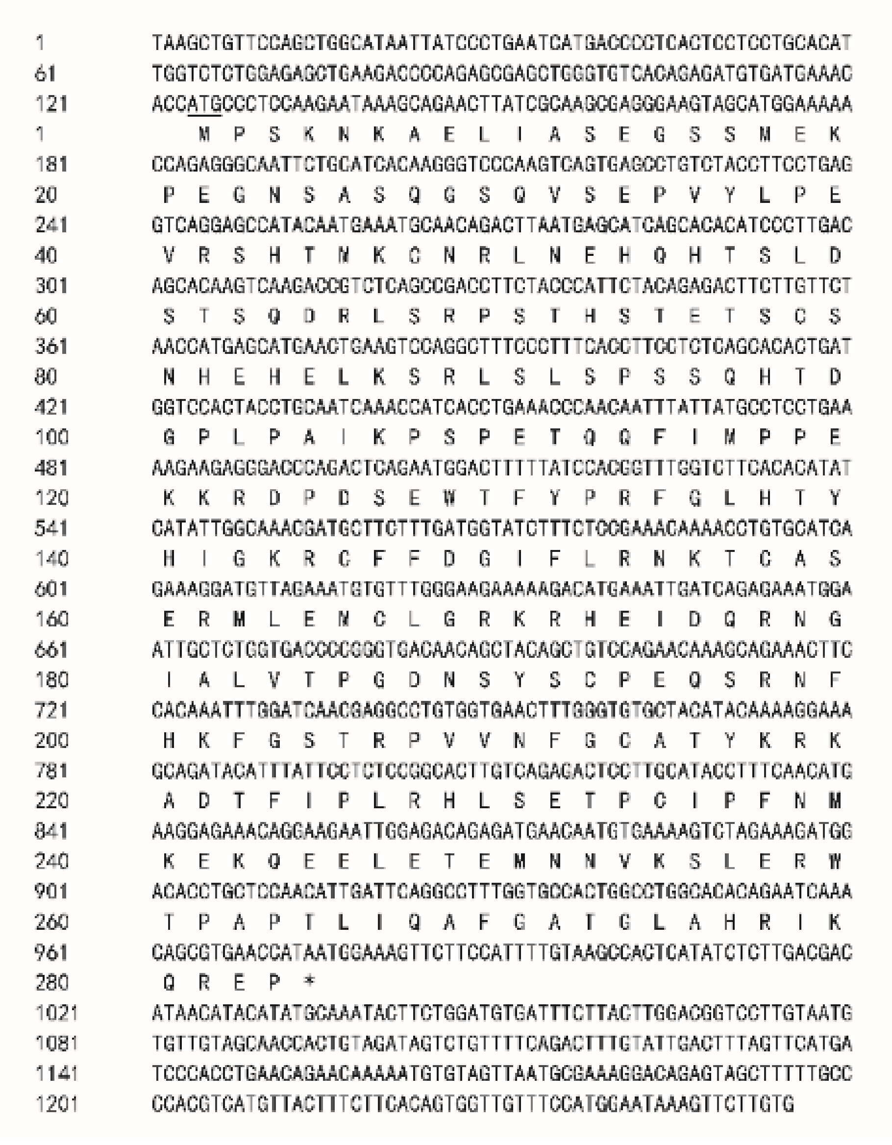
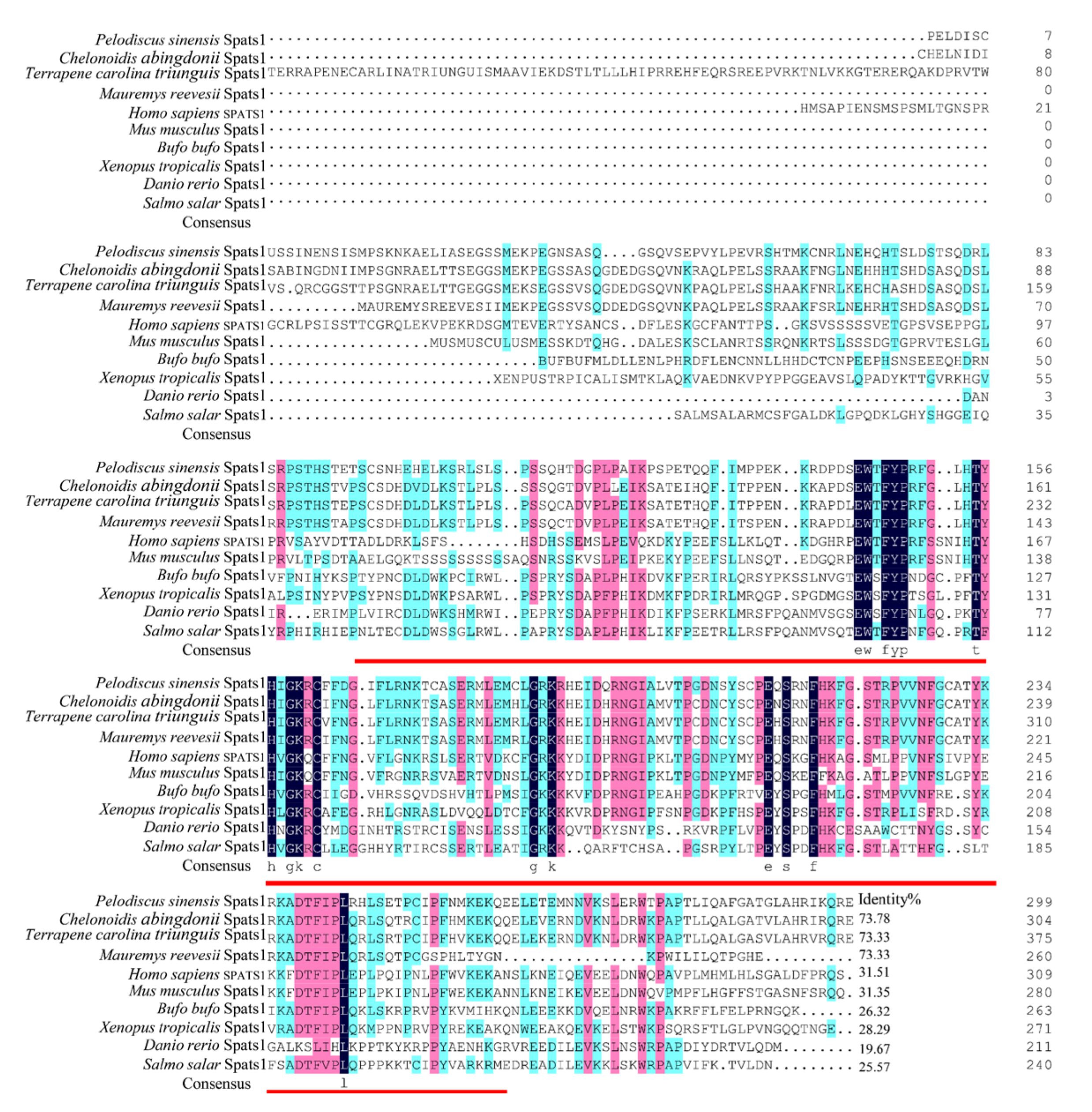
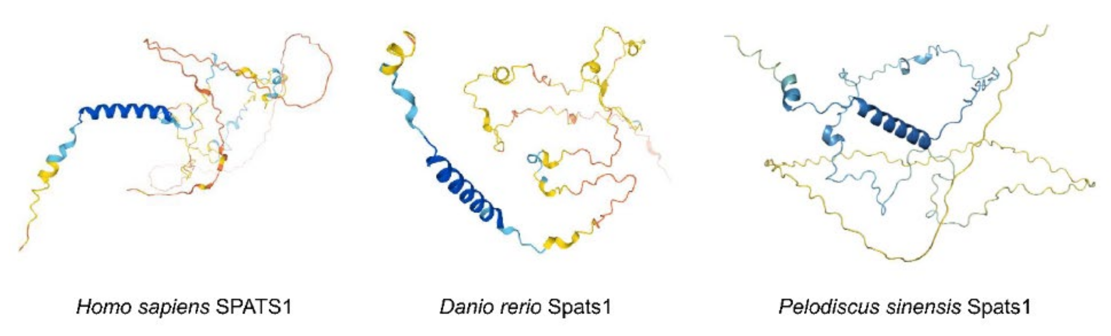
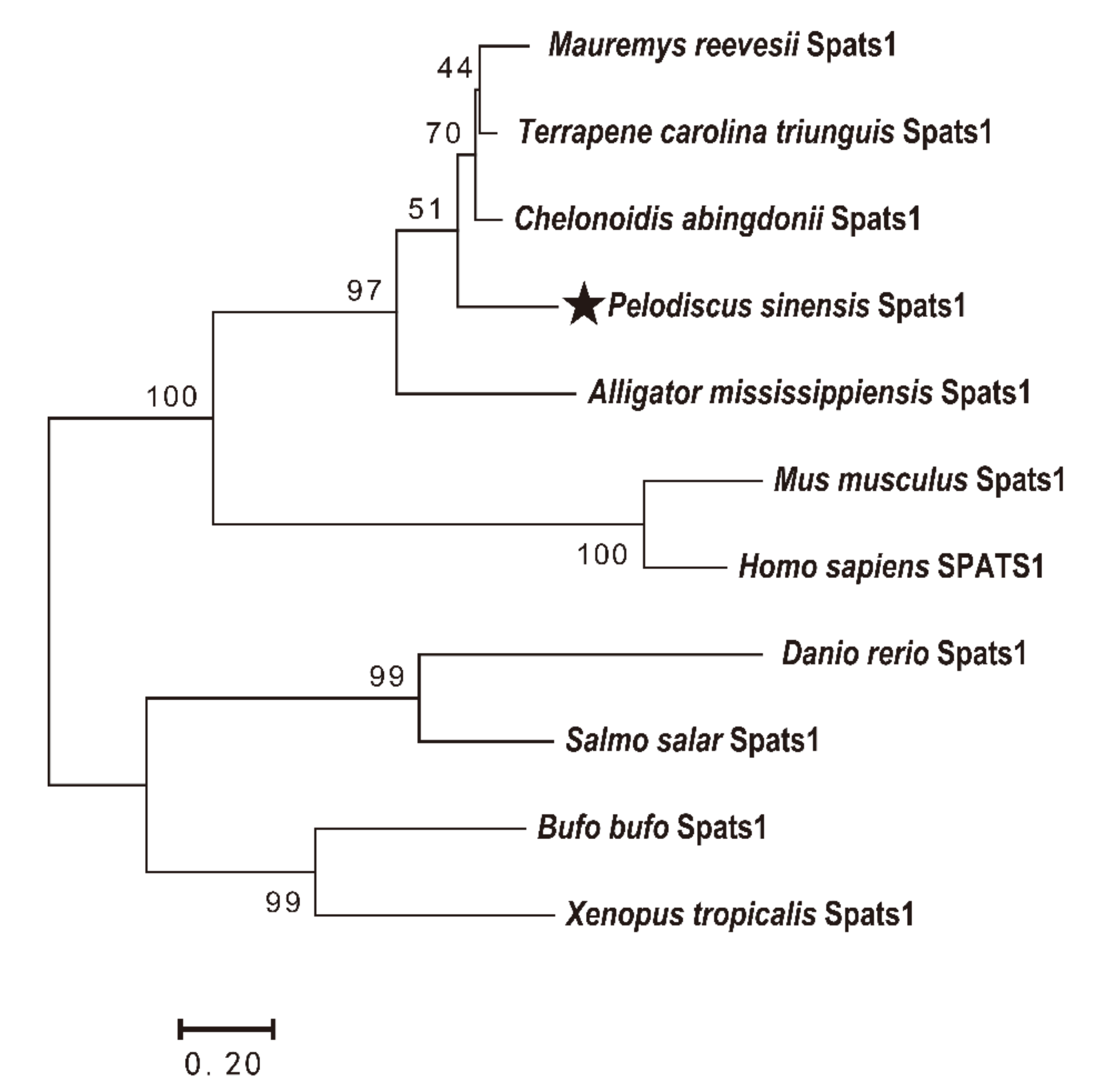
3.1. Cloning and Sequence Analyses

The Spats1 cDNA fragment is 1201 base pairs (bp) in length (Figure 1) and contains an open reading frame (ORF) of 849 bp, which encodes for a protein with 283 amino acids.
The inferred molecular weight of this protein is 32.15 kDa, and shares a 73.78% identity with Chelonoidis abingdonii Spats1 (XP_032655998.1), 73.33% with Terrapene carolina triunguis Spats1 (XP_029767743.1), and 73.33% with Mauremys reevesii Spats1 (XP_039386593.1). Yet, the sequence had low homology with those of mammals, fish, and amphibians, such as Homo sapiens SPATS1 (NP_001359010.1) (45.08%), Bufo bufo Spats1 (XP_040286913.1) (26.32%), and Danio rerio Spats1 (XP_017208226.1) (19.67%) (Figure 2).
The protein 3D structures of human (SPATS1) and zebrafish (Spats1) are similar in that they both begin with two α-helices at the C-terminus and are followed by a certain number of shorter α-helices interconnected by disordered loops. Although the N-terminal part of Spats1 in P. sinensis also starts with two α-helices, the number of subsequent short α-helices is smaller than that in both human and zebrafish (Figure 3). To better illustrate the evolutionary relationships of the Spats1 gene between different species, a phylogenetic tree was structured by maximum likelihood (ML) from the putative amino acid sequences of 11 species, inclusive of P. sinensis. The Spats1 proteins were classified into four clusters: mammalia, reptilia, amphibia, and teleosts; these clades were strongly supported by the bootstrap values (Figure 4). P. sinensis Spats1 was classified in the reptile cluster and was associated more strongly with Spats1 from turtles than from other species.

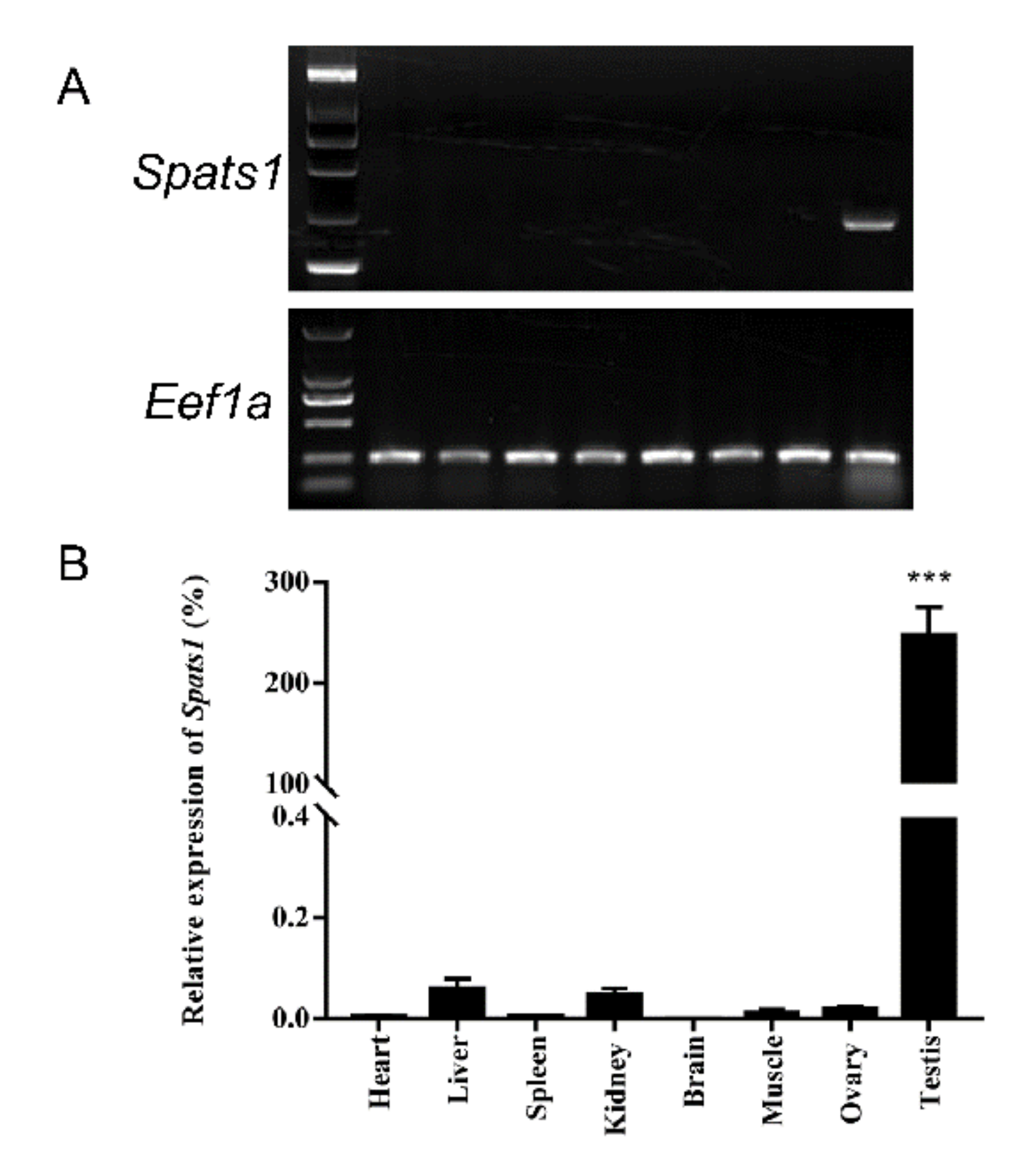
3.2. Tissue Distribution

Tissue distributions of Spats1 in three-year-old P. sinensis specimens were analyzed by Semi-Quantitative PCR and qRT-PCR, using Eef1a as a reference gene. Gel electrophoresis maps showed bright amplified band of Spats1 only in testis (Figure 5A). Spats1 was detected to highly expressed in testes via qRT-PCR (DF = 13, F = 682.449, p < 0.001), but rarely found in other tissues (Figure 5B). Hence, it was demonstrated that Spats1 mRNA was sex-specific expressed in the gonads and was only in the adult testes of P. sinensis.

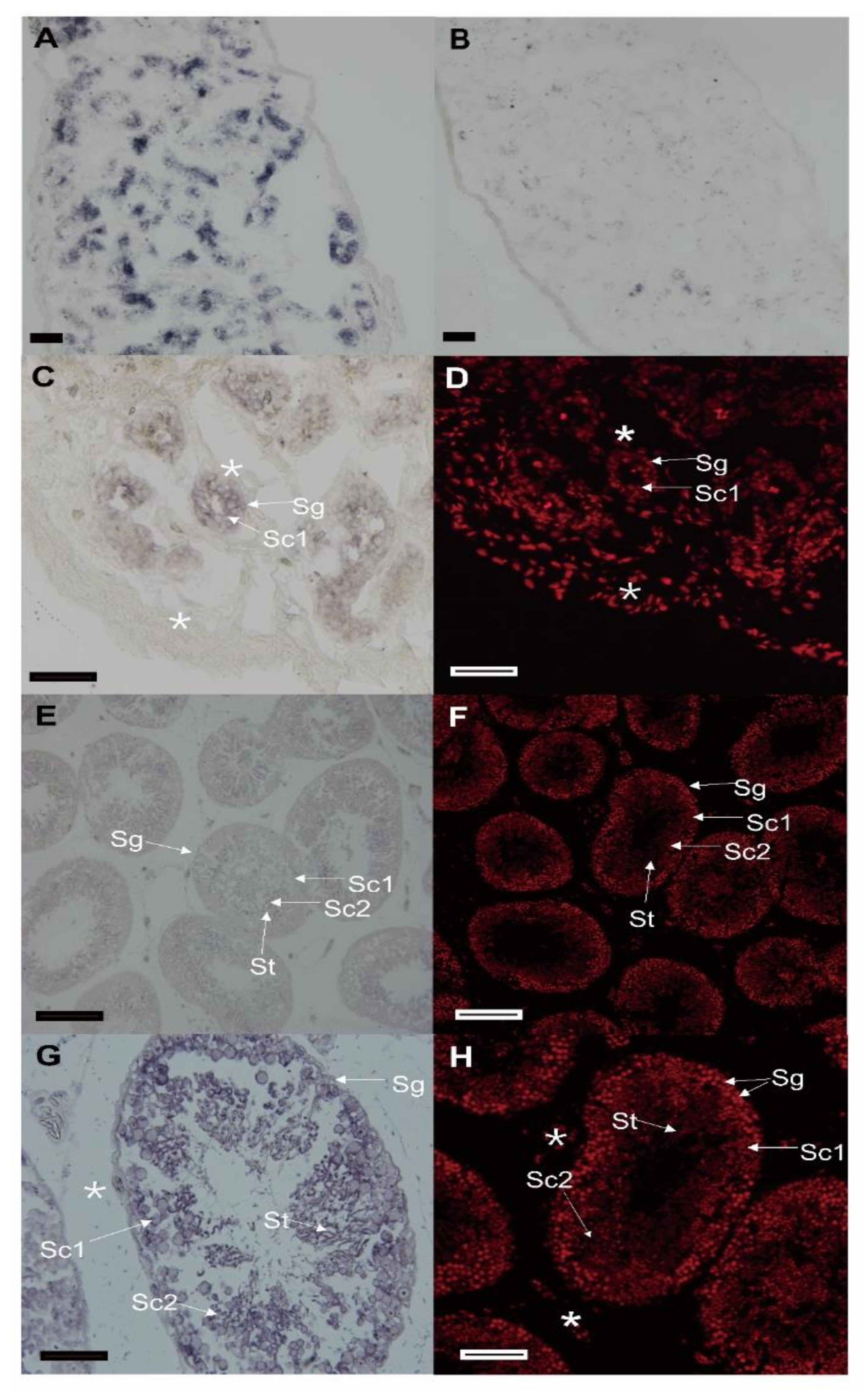
3.3. The Cellular Distribution of Spats1 mRNA in Testes

This is the first time that Spats1 mRNA was localized in the male germ cells of 1-, 2-, and three-year-old P. sinensis specimens. The CISH of the testis sections revealed that chemical signals of Spats1 transcripts were detected with the antisense probes (Figure 6A,C,E,G), while no signal was observed with sense probes (Figure 6B). The nucleus was stained with PI (Figure 6D,F,H). Spats1 mRNA was mainly distributed in primary spermatocytes (Sc1), secondary spermatocytes (Sc2), and spermatozoa (St). However, in spermatogonia (Sg) and somatic cells (*), Spats1 expression was weakly (Figure 6A,C,E,G).

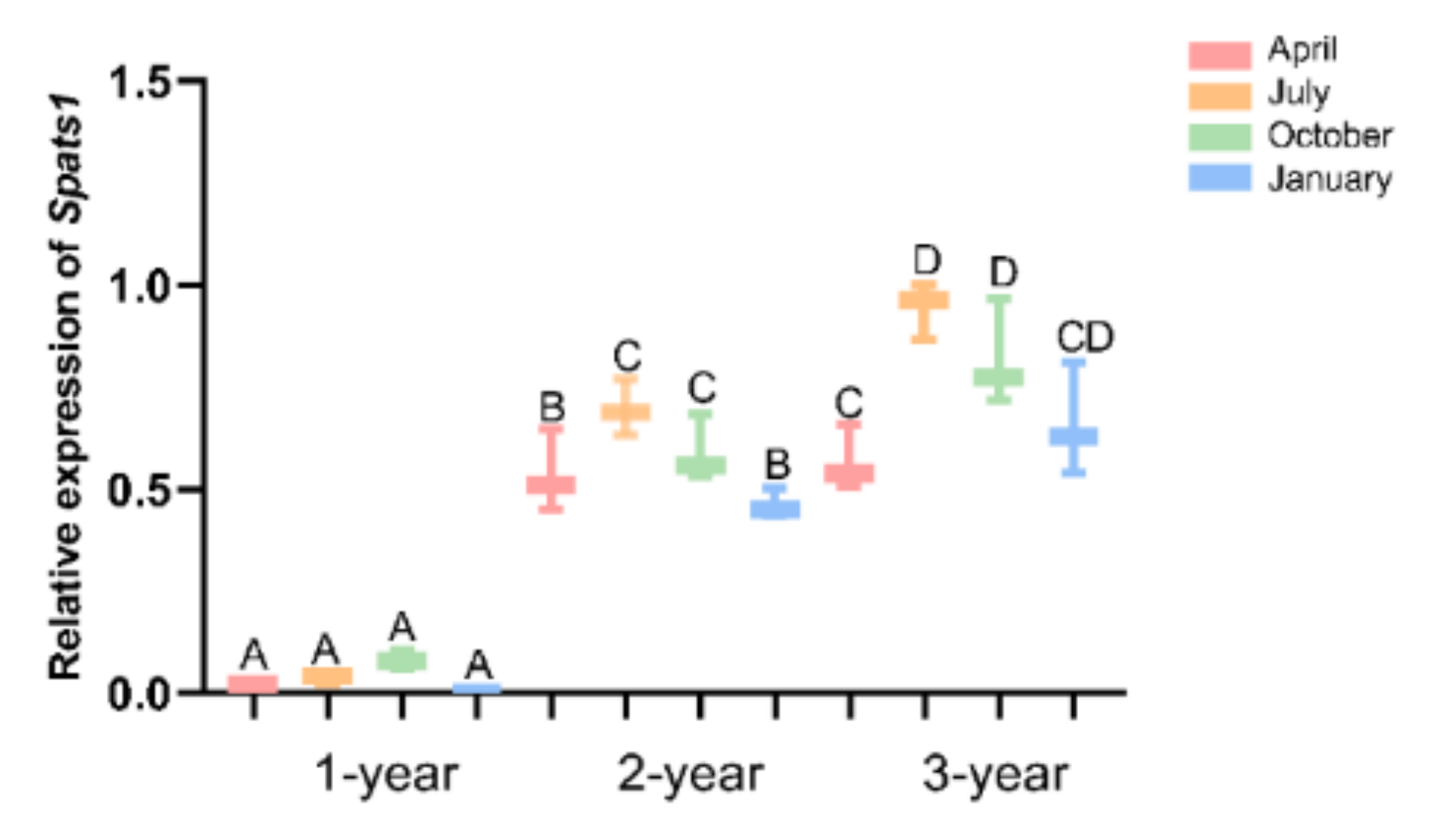
3.4. Expression Pattern of Spats1 mRNA in Testes of Different Seasons

In P. sinensis, spermatogenesis is accompanied by a distinct periodic rhythm. In this study, the expression pattern of the sex-bias gene Spats1 in P. sinensis testes was studied. qRT-PCR was used to test the relative expression levels of Spats1 mRNA in testes at different seasons. Overall, the expression of Spats1 in male P. sinensis appeared to increase with age. The expression of Spats1 was significantly lower in one-year-old males than in two- and three-year-old males in different seasons (DF = 24, F = 15.048, p < 0.01). Moreover, the expression of Spats1 in one-year-old male was not significantly different between seasons. In two- and three-year-old males, the expression pattern of Spats1 was significantly different across seasons (DF = 24, F = 15.048, p < 0.01). Spats1 expression was lowest in April and highest in July (DF = 24, F = 15.048, p < 0.01). Spats1 expression gradually decreased after July, but Spats1 expression was still significantly higher in October than in April (DF = 24, F = 15.048, p < 0.01) (Figure 7).

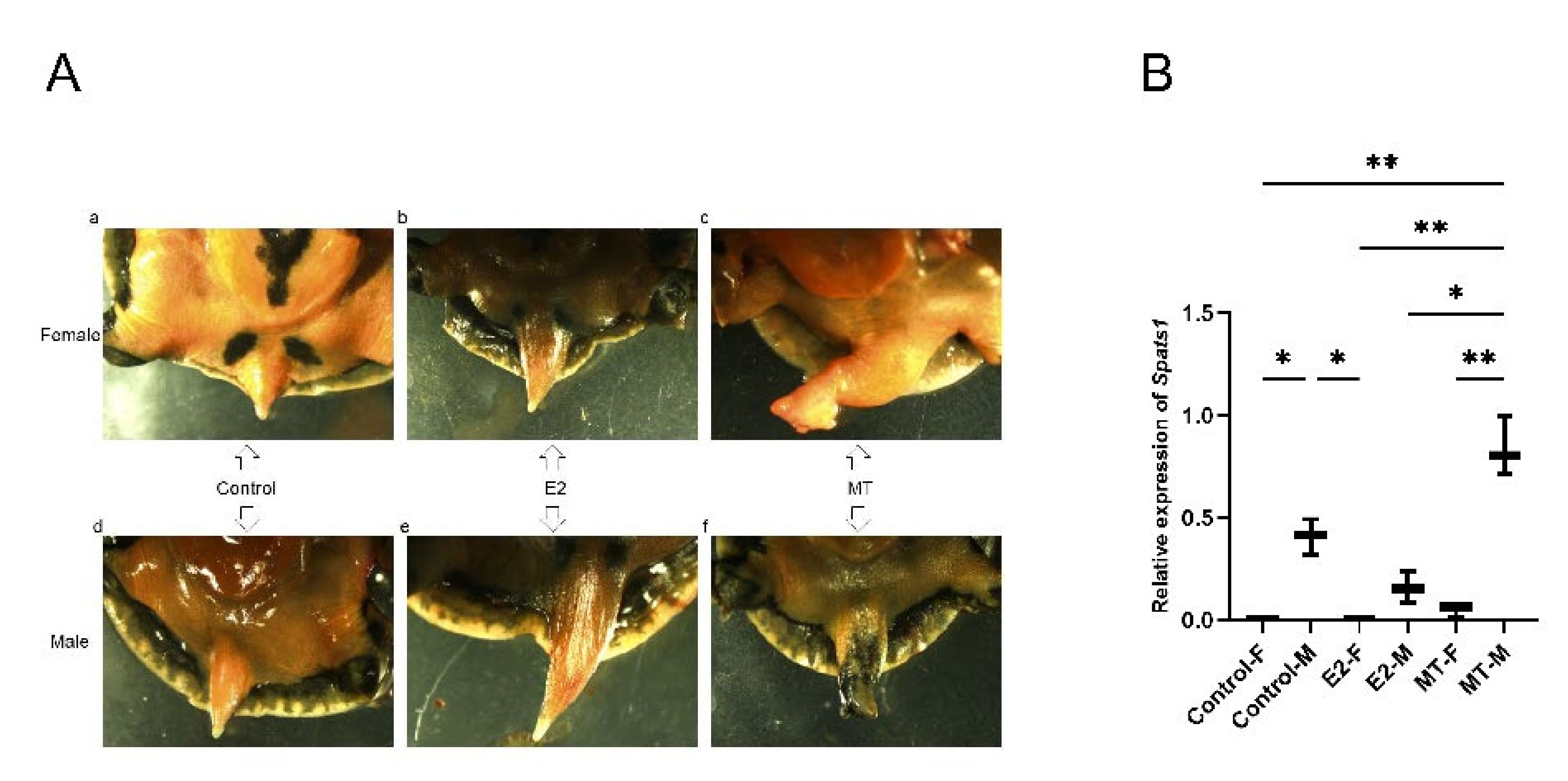
3.5. Effect of E2 and MT Treatment on Embryos

Compared to the untreated control, the tails of the newly hatched male and female P. sinensis turtles became more elongated after the E2 treatment (Figure 8A(b,e)). In contrast, the tails of MT-treated male and female P. sinensis became shorter and stouter (Figure 8A(c,f)). The qRT-PCR results showed that Spats1 expression was decreased in embryonic testes after E2 treatment and up-regulated after MT treatment compared to the control group, but none of the differences were significant. In addition, neither E2 nor MT treatment had a significant effect on Spats1 expression in female embryos (Figure 8B).

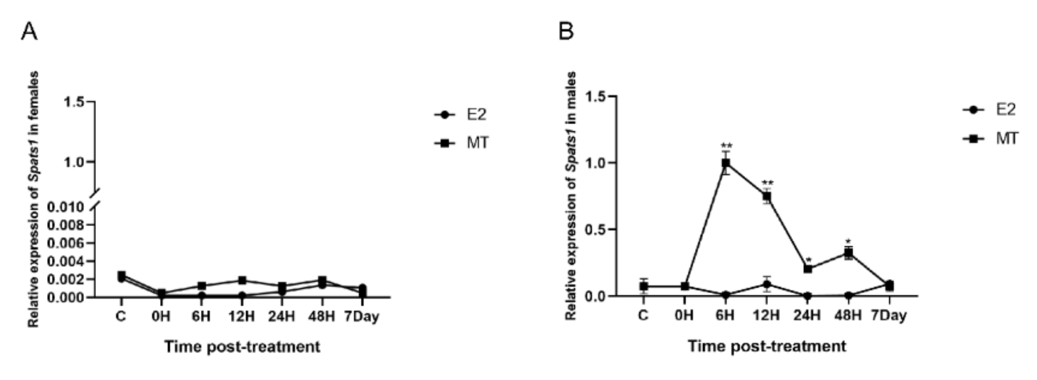
3.6. E2 and MT Treatment Were Performed on Adult Male and Female P. sinensis

E2 and MT treatments were performed on adult male and female P. sinensis, and it was found that both E2 and MT treatments could barely significantly affect Spats1 expression in female. At 6 h, 12 h, 24 h, 48 h, and even 7 days after E2 and MT treatment, Spats1 expression in females was always weak (Figure 9A). In contrast, in males, the expression of Sptas1 in testes increased significantly after MT treatment, peaked at 6 h, and then began to gradually decrease. However, at 12 h, its expression level remained significantly higher than that of the untreated controls (DF = 14, F = 60.518, p < 0.01). 24 and 48 h after MT treatment, the expression level of Spats1 in the testes continued to decrease, but was still significantly higher than the untreated controls (DF = 14, F = 60.518, p < 0.05). It was not until day 7 that the expression level of Spats1 decreased to the same level as the initial expression. Interestingly, the expression of Spats1 in the testes was almost unchanged after E2 treatment, either at 6 h, 12 h, 24 h, 48 h, or after 7 days (Figure 9B).

4. Discussion

In this study, the Spats1 cDNA was cloned and characterized. An 849 bp cDNA fragment of Spats1 encoding 283 amino acids was obtained. For protein 3D structures, although the N-terminal portion of Spats1 in P. sinensis begins with two α-helices, as in zebrafish and humans, the number of subsequent short α-helices is smaller than it is in either. The Spats1 3D structure in P. sinensis differs significantly from that of zebrafish and humans, suggesting that in P. sinensis may be evolutionarily more distant from humans and zebrafish. The results of protein 3D structures and multiple comparative analyses indicated the extensive homology of the P. sinensis Spats1 gene with Testudinidae animals. This implies that the protein function of Spats1 in turtles may be different from that of other species.
Of the tissues examined in this study, Spats1 mRNA expression was highest in the testis. Spats1 has shown a similar expression profile in various tissues in mice [20]. Interestingly, the distribution of Spats1 in different tissues was almost consistent with the pattern of Dmrt1 and anti-Müllerian hormone (Amh), which have been shown to be closely associated with testis differentiation in P. sinensis [15,31]. Similar to the gonadal structure of other vertebrates, the essential functional unit for sperm production in the testes of P. sinensis is the seminiferous tubule [32]. The arrangement of cells from the spermatogenic epithelium to the lumen is somatic cells, spermatogonia, primary spermatocytes, secondary spermatocytes, round spermatids, and sperm [33,34,35]. To obtain insight into the function of Spats1 in the development and differentiation of male germ cells, cellular localization of its transcripts in the testis was investigated. The obtained results revealed that Spats1 mRNA was mainly located in spermatocytes, secondary spermatocytes, and spermatozoa. In rats, Spats1 attains maximum levels during meiosis of the first spermatogenic wave, mostly in pachytene spermatocytes, whereas the signal was weaker in spermatogonia, Sertoli cells, and myoid cells [20]. Despite the fact that Spats1 is a testis-specific protein, the Spats1 loss-of-function mouse model analysis revealed that knockdown of Spats1 did not affect fertility in either males or females [36]. The testis-specific expression of Spats1 and the expression pattern in different germ cells imply that Spats1 may be closely associated with testicular differentiation and male germ cell differentiation in P. sinensis. In view of the differences in spermatogenesis mechanisms between reptiles and mammals, it remains to be investigated whether Spats1 has the same effects on fertility in P. sinensis as in rats.
Seasonally breeding animals undergoes a periodic fluctuation in gonad development. In male reptiles, spermatogenesis and steroidogenesis have a highly seasonality [37,38,39,40]. In this study, Spats1 expression in one-year-old males was very weak throughout the year, and Spats1 expression was highest in the testes at three years old. Testosterone expression increased with age, and the peak of testosterone occurred at the peak of spermatogenic activity [41,42]. This implies that the differential expression of Spats1 in different ages may be closely associated with the expression of testosterone. In addition, spermatogenesis of P. sinensis is initiated in the summer (June), and the mature spermatozoa are subsequently released from the epididymis in the autumn (October) [6,43]. In P. sinensis, the seminiferous epithelium is spermatogenically active in summer and autumn, but quiescent during the remaining part of the year [43]. The expression pattern of Spats1 in this experiment implies that Spats1 expression is seasonally influenced and closely related to spermatogenesis and release. Interestingly, a recent report on P. sinensis mentioned that the expression pattern of high-mobility group box 2 (Hmgb2) in the testes also strongly correlates with the season [44]. Therefore, we speculated that Spats1 regulates sperm production and release and might be affected by effective accumulative temperature [45].
Gonadal development and sexual activity are not only determined by the interactions of multiple genes but also by environmental factors, such as temperature or hormone exposure [46,47], and exposure to exogenous hormones during gonadal differentiation can alter the population gender ratio [48,49]. Steroid hormones are natural and induce sex change processes [50], among which E2 is one of the most potent estrogens impacting sex differentiation [51] and MT is one of the most potent androgens impacting sex differentiation [52]. For instance, E2 induces sex reversal in P. sinensis and the protandrous barramundi (Lates calcarifer) [53,54], and MT controls secondary sexual characteristics in Siniperca chuatsi as well [55]. The same phenomenon has been confirmed in mammals and fish [56]. P. sinensis embryo is an excellent model for studying gonadal development and reproduction since development takes place in the egg, and we can intervene directly in vitro. In this experiment, both male and female P. sinensis embryonic tails changed significantly after E2 and MT treatments compared to the untreated group, indicating that the sexual characteristics of P. sinensis were affected by E2 and MT. The treatment of embryos with hormones resulted in significant alterations in the expression of many genes [57,58]. The expression of some sexually dimorphic genes is significantly altered in the embryo after sex hormone treatment in P. sinensis, such as Dmrt1, Sox9 [15], and cytochrome p450 family 19 subfamily a (Cyp19a) [59]. However, in the present experiment, the expression of Spats1 in embryos was not significantly altered after E2 and MT treatment. In contrast, in adults, Spats1 expression was significantly increased in males after MT treatment, which is similar to the pattern in zebrafish, where the expression of gonadotrophin-releasing hormones 2 and 3 (gnrh2 and gnrh3) is upregulated after MT treatment [57]. The expression pattern of Spats1 in embryos and adults after sex hormone treatment suggests that Spats1 responds to sex endosteroid hormone regulation only in adults, and it is hypothesized that the gene may not be involved in sex differentiation in P. sinensis. Androgenic stimulation results not only in the production of seminal fluid, but also promotes the maturation of sperms [60]. In this experiment, the significant elevation of Spats1 expression after MT treated in adults, implying that Spats1 may be positively regulated by MT, which in turn promotes sperm maturation and release in P. sinensis. Studies have shown that estrogen is important for sperm activity and that estrogen treatment affects the expression of sex-related genes [61,62]. Yet, the expression of Spats1 in male P. sinensis changed weakly after E2 treatment in this experiment. This is different from the response pattern of other sexually dimorphic genes in adult P. sinensis after E2 and MT treatment, such as forkhead box L2 (Foxl2), whose expression was significantly altered in both males and females after either E2 or MT treatment [63]. This might be related to the concentration of estrogen treatment or that Spats1 is not located in the estrogen regulatory signaling pathway in P. sinensis. As for the fact that Spats1 expression in females has been very weak and almost unaffected by E2 and MT treatments, there are two possible reasons: inadequate concentration of hormone treatment, or the fact that Spats1 is not involved in female physiological activities at all. Thus, the specific regulation mechanism involved in this process needs to be further studied.

5. Conclusions

In summary, Spats1 is specifically expressed in the testis of P. sinensis with a seasonal expression pattern and is involved in spermatogenesis and release. Moreover, E2 and MT treatments revealed that Spats1 only responded to MT treatment both in embryos and in adults. Thus, additional research is necessary to investigate the issue in the future. This study enriches the data on Spats1 in P. sinensis and provides a reference for future studies on the molecular mechanisms of reproductive activity in this species and even other reptiles.

Author Contributions

Conceptualization, L.L. and J.Z. wrote the main manuscript text. C.C. and Y.W. prepared figures and tables. X.H. and X.L. are responsible for analysing the data. L.Y. and R.L. are responsible for data validation. C.W., H.C. and Y.L. preparation of experimental samples. X.Z. and W.L. are responsible for the experimental design. All authors reviewed the manuscript. All authors have read and agreed to the published version of the manuscript.

Funding

This work was funded by the National Key R&D Program of China (2018YFD0900201), the National Natural Science Foundation of China (32102792), The Project Supported by Guangdong Natural Science Foundation (2018A030313520), the Central Public-Interest Scientific Institution Basal Research Fund, CAFS (2020TD35), the National Freshwater Genetic Resource Center (NFGR-2022), the Guangdong Agricultural Research System (2019KJ150), and the China-ASEAN Maritime Cooperation Fund (CAMC-2018F).

Institutional Review Board Statement

The Animal Care and Ethics Committee of the Pearl River Fisheries Research Institute, Chinese Academy of Fishery Sciences (Guangzhou, China) approved this research (Approval Code: LAEC-PRFRI-20210148; Approval Date: 10 January 2021), and all experimental protocols and methods were performed in accordance with the relevant guidelines and regulations.

Informed Consent Statement

Not applicable.

Data Availability Statement

All datasets generated or analyzed during this study are included in the published article.

Conflicts of Interest

The authors declare no conflict of interest.

References

  1. Yang, T.; Yang, W.-X. The dynamics and regulation of microfilament during spermatogenesis. Gene 2020, 744, 144635. [Google Scholar] [CrossRef] [PubMed]
  2. Gribbins, K. Reptilian spermatogenesis: A histological and ultrastructural perspective. Spermatogenesis 2011, 1, 250–269. [Google Scholar] [CrossRef] [PubMed] [Green Version]
  3. Rosati, L.; Agnese, M.; Lorenzo, M.; Barra, T.; Valiante, S.; Prisco, M. Spermatogenesis and regulatory factors in the wall lizard Podarcis sicula. Gen. Comp. Endocrinol. 2020, 298, 113579. [Google Scholar] [CrossRef]
  4. Wang, Y.; Liu, X.; Li, W.; Zhao, J.; Liu, H.; Yu, L.; Zhu, X. Reproductive performance is associated with seasonal plasma reproductive hormone levels, steroidogenic enzymes and sex hormone receptor expression levels in cultured Asian yellow pond turtles (Mauremys mutica). Comp. Biochem. Physiol. Part B Biochem. Mol. Biol. 2021, 254, 110566. [Google Scholar] [CrossRef] [PubMed]
  5. Resende, F.C.d.; Avelar, G.F.d. The sexual segment of the kidney of a tropical rattlesnake, Crotalus durissus (Reptilia, Squamata, Viperidae), and its relationship to seasonal testicular and androgen cycles. J. Morphol. 2021, 282, 1402–1414. [Google Scholar] [CrossRef] [PubMed]
  6. Wang, P.; Li, X.; Wang, X.; Peng, N.; Luo, Z. Effects of dietary xanthophyll supplementation on growth performance, body color, carotenoids, and blood chemistry indices of chinese soft-shelled turtle Pelodiscus sinensis. N. Am. J. Aquac. 2020, 82, 394–404. [Google Scholar] [CrossRef]
  7. Gauthier, J.; Cannatella, D.C.; De Queiroz, K.; Kluge, A.G.; Rowe, T. Tetrapod phylogeny. Hierarchy Life 1989, 25, 337–353. [Google Scholar]
  8. Uno, Y.; Nishida, C.; Tarui, H.; Ishishita, S.; Takagi, C.; Nishimura, O.; Ishijima, J.; Ota, H.; Kosaka, A.; Matsubara, K.; et al. Inference of the protokaryotypes of amniotes and tetrapods and the evolutionary processes of microchromosomes from comparative gene mapping. PLoS ONE 2012, 7, e53027. [Google Scholar] [CrossRef]
  9. Chen, H.; Huang, Y.; Liu, T.; Haseeb, A.; Ahmed, N.; Zhang, L.; Bian, X.; Chen, Q. Characteristics of seasonal spermatogenesis in the soft-shelled turtle. Anim. Reprod. Sci. 2020, 214, 106307. [Google Scholar] [CrossRef]
  10. Zhang, L.; Han, X.; Li, M.; Bao, H.; Chen, Q. Spermiogenesis in soft-shelled turtle, Pelodiscus sinensis. Anat. Rec. Adv. Integr. Anat. Evol. Biol. 2007, 290, 1213–1222. [Google Scholar] [CrossRef]
  11. Bian, X.; Zhang, L.; Yang, L.; Yang, P.; Ullah, S.; Zhang, Q.; Chen, Q. Ultrastructure of epididymal epithelium and its interaction with the sperm in the soft-shelled turtle Pelodiscus sinensis. Micron 2013, 54, 65–74. [Google Scholar] [CrossRef] [PubMed]
  12. Viets, B.; Ewert, M.; Talent, L.; Nelson, C. Sex-determining mechanisms in squamate reptiles. J. Exp. Zool. 1994, 270, 45–56. [Google Scholar] [CrossRef]
  13. İnanan, B.E.; Acar, Ü. Comparison of milt and blood parameters including testosterone and gonadotropins levels of sex-reversed female rainbow trout (Oncorhynchus mykiss) by different steroid hormones. Aquac. Res. 2021, 52, 1876–1884. [Google Scholar] [CrossRef]
  14. Ruiz-García, A.; Roco, Á.S.; Bullejos, M. Sex differentiation in amphibians: Effect of temperature and its influence on sex reversal. Sex. Dev. 2021, 15, 157–167. [Google Scholar] [CrossRef] [PubMed]
  15. Wang, L.; Hu, T.; Mi, M.; Yang, K.; Qian, G.; Ge, C. The effects of estrogen on gonadal differentiation and expressions of DMRT1 and SOX9 in Pelodiscus sinensis. Acta Hydrobiol. Sin. 2014, 38, 467–473. [Google Scholar]
  16. Liang, H.; Meng, Y.; Cao, L.; Li, X.; Zou, G. Effect of exogenous hormones on R-spondin 1 (RSPO1) gene expression and embryo development in Pelodiscus sinensis. Reprod. Fertil. Dev. 2019, 31, 1425–1433. [Google Scholar] [CrossRef] [Green Version]
  17. Kopania, E.; Larson, E.; Callahan, C.; Keeble, S.; Good, J. Molecular evolution across mouse spermatogenesis. Mol. Biol. Evol. 2022, 39, msac023. [Google Scholar] [CrossRef]
  18. Geisinger, A.; Alsheimer, M.; Baier, A.; Benavente, R.; Wettstein, R. The mammalian gene pecanex 1 is differentially expressed during spermatogenesis. Biochim. Et Biophys. Acta (BBA)-Gene Struct. Expr. 2005, 1728, 34–43. [Google Scholar] [CrossRef]
  19. Geisinger, A.; Dos Santos, A.; Benavente, R.; Wettstein, R. Identification and characterization of Srsp1, a rat gene differentially expressed during spermatogenesis and coding for a serine stretch-containing protein. Cytogenet. Genome Res. 2002, 98, 249–254. [Google Scholar] [CrossRef]
  20. Capoano, C.A.; Wettstein, R.; Kun, A.; Geisinger, A. Spats 1 (Srsp1) is differentially expressed during testis development of the rat. Gene Expr. Patterns 2010, 10, 1–8. [Google Scholar] [CrossRef]
  21. da Cruz, I.; Rodríguez-Casuriaga, R.; Santiñaque, F.F.; Farías, J.; Curti, G.; Capoano, C.A.; Folle, G.A.; Benavente, R.; Sotelo-Silveira, J.R.; Geisinger, A. Transcriptome analysis of highly purified mouse spermatogenic cell populations: Gene expression signatures switch from meiotic-to postmeiotic-related processes at pachytene stage. BMC Genom. 2016, 17, 294. [Google Scholar] [CrossRef] [PubMed] [Green Version]
  22. Mouawad, N. SPATS1: New Molecular Target in Colorectal Cancer. Ph.D. Thesis, University of Pisa, Pisa, Italy, 2021. [Google Scholar]
  23. Zhu, J.; Lei, L.; Chen, C.; Wang, Y.; Liu, X.; Geng, L.; Li, R.; Chen, H.; Hong, X.; Yu, L.; et al. Whole-Transcriptome Analysis Identifies Gender Dimorphic Expressions of Mrnas and Non-Coding Rnas in Chinese Soft-Shell Turtle (Pelodiscus sinensis). Biology 2022, 11, 834. [Google Scholar] [CrossRef]
  24. Li, H.; Zhou, Z.; Wu, T.; Wu, Y.; Ji, X. Do fluctuations in incubation temperature affect hatchling quality in the Chinese soft-shelled turtle Pelodiscus sinensis? Aquaculture 2013, 406, 91–96. [Google Scholar] [CrossRef]
  25. Du, W.G.; Ji, X. The effects of incubation thermal environments on size, locomotor performance and early growth of hatchling soft-shelled turtles, Pelodiscus sinensis. J. Therm. Biol. 2003, 28, 279–286. [Google Scholar] [CrossRef]
  26. Zhang, Y.; Xiao, L.; Sun, W.; Li, P.; Zhou, Y.; Qian, G.; Ge, C. Knockdown of R-spondin1 leads to partial sex reversal in genetic female Chinese soft-shelled turtle Pelodiscus sinensis. Gen. Comp. Endocrinol. 2021, 309, 113788. [Google Scholar] [CrossRef]
  27. Zhang, Y.; Xiao, L.; Sun, W.; Li, P.; Zhou, Y.; Qian, G.; Ge, C. Characterization of the Pelodiscus sinensis polymeric immunoglobulin receptor (P. sinensis pIgR) and its response to LPS and Aeromonas sobria. Dev. Comp. Immunol. 2021, 121, 104072. [Google Scholar]
  28. Cai, H.; Sun, W.; Liu, H.; Bao, H.; Zhang, H.; Qian, G.; Ge, C. Molecular cloning and expression pattern of Amh and its preliminary functional analysis in male sexual differentiation in Pelodiscus sinensis. Sci. Sin. Vitae 2016, 46, 1423–1433. [Google Scholar]
  29. Tokita, M.; Kuratani, S. Normal embryonic stages of the Chinese softshelled turtle Pelodiscus sinensis (Trionychidae). Zool. Sci. 2001, 18, 705–715. [Google Scholar] [CrossRef] [Green Version]
  30. Li, W.; Zhu, X.; Xu, H.; Zhao, J.; Chen, C. A PCR Amplification Primer, Method and Kit for Rapid Identification of Genetic Traits in the Chinese Soft-shelled Turtle. China Patent CN108841945B, 5 May 2020. [Google Scholar]
  31. Sun, W.; Cai, H.; Zhang, G.; Zhang, H.; Bao, H.; Wang, L.; Ye, J.; Qian, G.; Ge, C. Dmrt1 is required for primary male sexual differentiation in Chinese soft-shelled turtle Pelodiscus sinensis. Sci. Rep. 2017, 7, 4433. [Google Scholar] [CrossRef] [Green Version]
  32. Cheng, C.Y.; Mruk, D.D. The biology of spermatogenesis: The past, present and future. Philos. Trans. R Soc. Lond. B Biol. Sci. 2020, 365, 1459–1463. [Google Scholar] [CrossRef] [Green Version]
  33. Sharma, R.; Agarwal, A. Spermatogenesis: An overview. Sperm Chromatin 2011, 19–44. [Google Scholar] [CrossRef]
  34. Johnson, L. Efficiency of spermatogenesis. Microsc. Res. Technol. 1995, 32, 385–422. [Google Scholar] [CrossRef] [PubMed]
  35. Saunders, P. Germ cell-somatic cell interactions during spermatogenesis. Reprod. Suppl. 2003, 61, 91–101. [Google Scholar] [CrossRef] [PubMed]
  36. Capoano, C.A.; Ortiz-Laquintana, L.A.; Rodríguez-Casuriaga, R.; Schlapp, G.; Meikle, M.N.; Mulet, A.P.; Crispo, M.; Benavente, R.; Geisinger, A. SPATS1 (spermatogenesis-associated, serine-rich 1) is not essential for spermatogenesis and fertility in mouse. PLoS ONE 2021, 16, e0251028. [Google Scholar] [CrossRef]
  37. Jiménez, R.; Burgos, M.; Barrionuevo, F.J. Circannual testis changes in seasonally breeding mammals. Sex. Dev. 2015, 9, 205–215. [Google Scholar] [CrossRef]
  38. Mahmoud, I.; Licht, P. Seasonal changes in gonadal activity and the effects of stress on reproductive hormones in the common snapping turtle, Chelydra serpentina. Gen. Comp. Endocrinol. 1997, 107, 359–372. [Google Scholar] [CrossRef]
  39. Ribeiro, Y.M.; de Matos, S.A.; Domingos, F.F.T.; Dos Santos, H.B.; Vieira, A.B.C.; Bazzoli, N.; Rizzo, E. Germ cell proliferation and apoptosis during testicular regression in a seasonal breeding fish kept in captivity. Tissue Cell 2017, 49, 664–671. [Google Scholar] [CrossRef]
  40. Lofts, B. Patterns of spermatogenesis and steroidogenesis in male reptiles. In Reproduction and Evolution; Calaby, J.H., Tyndale-Boscoe, C.H., Eds.; Australian Academic Science: Canberra, Australia, 1977; pp. 127–136. [Google Scholar]
  41. Radder, R.; Shanbhag, B.; Saidapur, S. Pattern of plasma sex steroid hormone levels during reproductive cycles of male and female tropical lizard, Calotes versicolor. Gen. Comp. Endocrinol. 2001, 124, 285–292. [Google Scholar] [CrossRef] [Green Version]
  42. Yu, W.; Li, H.; Zhang, G.; Kang, X.; Li, Q.; Cao, J.; Xu, E. Serum sex hormonal levels and gonadal development in broodstock soft-shelled turtle Trionyx sinensis in a green house. Fish. Sci. 2012, 31, 264–269. [Google Scholar]
  43. Zhang, L.; Han, X.K.; Qi, Y.Y.; Liu, Y.; Chen, Q.S. Seasonal effects on apoptosis and proliferation of germ cells in the testes of the Chinese soft-shelled turtle, Pelodiscus sinensis. Theriogenology 2008, 69, 1148–1158. [Google Scholar] [CrossRef]
  44. Li, W.; Zhu, J.; Lei, L.; Chen, C.; Liu, X.; Wang, Y.; Hong, X.; Xu, H.; Zhu, X. The Seasonal and Stage-Specific Expression Patterns of HMGB2 Suggest Its Key Role in Spermatogenesis in the Chinese Soft-Shelled Turtle (Pelodiscus sinensis). Biochem. Genet. 2022, 1–14. [Google Scholar] [CrossRef] [PubMed]
  45. Sukhan, Z.P.; Cho, Y.; Sharker, M.R.; Hossen, S.; Rha, S.J.; Kho, K.H. Effective accumulative temperature affects gonadal maturation by controlling expression of GnRH, GnRH receptor, serotonin receptor and APGWamide gene in Pacific abalone, Haliotis discus hannai during broodstock conditioning in hatcheries. J. Therm. Biol. 2021, 100, 103037. [Google Scholar] [CrossRef] [PubMed]
  46. Breitenbach, A.T.; Bowden, R.M.; Paitz, R.T. Effects of Constant and Fluctuating Temperatures on Gene Expression During Gonadal Development. Integr. Comp. Biol. 2022, icac011. [Google Scholar] [CrossRef] [PubMed]
  47. Di Lorenzo, M.; Mileo, A.; Laforgia, V.; De Falco, M.; Rosati, L. Alkyphenol Exposure Alters Steroidogenesis in Male Lizard Podarcis siculus. Animals 2021, 11, 1003. [Google Scholar] [CrossRef] [PubMed]
  48. Othman, R.; Ron, X.J.; Yao, H.; O’Bryant, P.; Rapp, D.; Pei, J.C.; Pei, J.C.; Wang, H.P. The effect of methyltestosterone (MT) on sex differentiation and growth in juvenile yellow perch (Perca flavescens). Fish Physiol. Biochem. 2022, 48, 161–171. [Google Scholar] [CrossRef]
  49. Cai, Y.; He, Z.; Zhang, Q.; Wang, X.; Zhang, S.; He, L.; Yang, D.; Zhang, M.; Yan, T. Oestradiol upregulates the expression of Cyp19a1a and the feminization ratio in Schizothorax prenanti. Aquac. Rep. 2022, 24, 101138. [Google Scholar] [CrossRef]
  50. Gardner, L.; Anderson, T.; Place, A.R.; Dixon, B.; Elizur, A. Sex change strategy and the aromatase genes. J. Steroid Biochem. Mol. Biol. 2005, 94, 395–404. [Google Scholar] [CrossRef]
  51. Jiang, Y.; Luo, H.; Hou, M.; Chen, J.; Tao, B.; Zhu, Z.; Song, Y.; Hu, W. Aromatase inhibitor induces sex reversal in the protogynous hermaphroditic rice field eel (Monopterus albus). Aquaculture 2022, 551, 737960. [Google Scholar] [CrossRef]
  52. Zhu, Q.; Han, C.; Liu, S.; Ouyang, H.; Liu, D.; Zhang, Z.; Huang, J.; Han, L.; Li, S.; Li, G. Development and gene expression analysis of gonad during 17α-methyltestosterone-induced sex reversal in mandarin fish (Siniperca chuatsi). Aquac. Rep. 2022, 23, 101049. [Google Scholar] [CrossRef]
  53. Li, P.; Guo, Y.; Jin, L.; Liang, X.; Chen, G.; Sun, W.; Xiao, L.; Qian, G.; Ge, C. ESR1 mediates estrogen-induced feminization of genetic male Chinese soft-shelled turtle. Biol. Reprod. 2022, ioac088. [Google Scholar] [CrossRef]
  54. Banh, Q.Q.; Guppy, J.L.; Domingos, J.A.; Budd, A.M.; Pinto, R.C.; Marc, A.F.; Jerry, D.R. Induction of precocious females in the protandrous barramundi (Lates calcarifer) through implants containing 17β-estradiol-effects on gonadal morphology, gene expression and DNA methylation of key sex genes. Aquaculture 2021, 539, 736601. [Google Scholar] [CrossRef]
  55. Weber, G.M.; Leeds, T.D.; Schneider, R.P. Sex reversal of female rainbow trout by immersion in 17α-methyltestosterone. Aquaculture 2020, 528, 735535. [Google Scholar] [CrossRef]
  56. Delbes, G.; Blázquez, M.; Fernandino, J.I.; Grigorova, P.; Hales, B.F.; Metcalfe, C.; Navarro-Martín, L.; Parent, L.; Robaire, B.; Rwigemera, A.; et al. Effects of endocrine disrupting chemicals on gonad development: Mechanistic insights from fish and mammals. Environ. Res. 2022, 204, 112040. [Google Scholar] [CrossRef] [PubMed]
  57. Spaan, K.; Haigis, A.C.; Weiss, J.; Legradi, J. Effects of 25 thyroid hormone disruptors on zebrafish embryos: A literature review of potential biomarkers. Sci. Total Environ. 2019, 656, 1238–1249. [Google Scholar] [CrossRef]
  58. Darras, V.M. The role of maternal thyroid hormones in avian embryonic development. Front. Endocrinol. 2019, 10, 66. [Google Scholar] [CrossRef]
  59. Liang, H.W.; Meng, Y.; Cao, L.H.; Li, X.; Zou, G.W. Expression and characterization of the cyp19a gene and its responses to estradiol/letrozole exposure in Chinese soft-shelled turtle (Pelodiscus sinensis). Mol. Reprod. Dev. 2019, 86, 480–490. [Google Scholar] [CrossRef]
  60. Dzyuba, V.; Cosson, J.; Papadaki, M.; Mylonas, C.C.; Steinbach, C.; Rodina, M.; Tučkova, V.; Linhart, O.; Shelton, W.; Gela, G. Influence of Environmental Temperature and Hormonal Stimulation on the In Vitro Sperm Maturation in Sterlet Acipenser ruthenus in Advance of the Spawning Season. Animals 2021, 11, 1417. [Google Scholar] [CrossRef]
  61. Dostalova, P.; Zatecka, E.; Dvorakova-Hortova, K. Of oestrogens and sperm: A review of the roles of oestrogens and oestrogen receptors in male reproduction. Int. J. Mol. Sci. 2017, 18, 904. [Google Scholar] [CrossRef] [Green Version]
  62. Komrsková, K. The Physiology and Reproductive Potential of Sperm. Ph.D. Thesis, Charles University, Prague, Czech Republic, 2021. [Google Scholar]
  63. Gao, L.L.; Diao, X.M.; Li, Y.; Zhai, X.L.; Zhou, C.L. Molecular cloning and expression of foxl2 gene induced by exogenous hormone in the pelodiscus sinensis. Acta Hydrobiol. Sin. 2019, 43, 7. [Google Scholar]
Figure 1. Nucleotide and predicted amino acid sequences of P. sinensis Spats1. The start codon (ATG) is underlined by a single line, and the stop codon (TAA) is represented by the asterisks.
Animals 12 01858 g001
Figure 2. Alignment of the amino acid sequences. The structural domain is underlined in red. GenBank accession numbers: Chelonoidis abingdonii Spats1 (XP_032655998.1), Terrapene carolina triunguis Spats1 (XP_029767743.1), Mauremys reevesii Spats1 (XP_039386593.1), Homo sapiens SPATS1 (NP_001359010.1), Bufo bufo Spats1 (XP_040286913.1), Danio rerio Spats1 (XP_017208226.1), Mus musculus Spats1 (NP_081925.2), Xenopus tropicalis Spats1 (XP_002936034.2), and Salmo salar Spats1 (XP_014000180.1). Black shading indicates the same amino acid sequence. Pink shading indicates conserved sequences with more than 75% of the listed polypeptides. The blue shading indicates the conserved sequence with more than half of the listed peptides.
Animals 12 01858 g002
Figure 3. The protein 3D structure models. Different colors represent different model confdence. Animals 12 01858 i001: Very high (Plddt > 90); Animals 12 01858 i002: Confident (90 > Plddt > 70); Animals 12 01858 i003: Low (70 > Plddt > 50); Animals 12 01858 i004: Very low (Plddt < 50).
Animals 12 01858 g003
Figure 4. Phylogenetic tree constructed through the maximum likelihood method. The scale bar shows the genetic distance. The number at each node indicates the bootstrap value obtained for 1000 replicates.
Animals 12 01858 g004
Figure 5. The distribution of Spats1 mRNA in different tissues. (A) Tissue distribution gel electrophoresis map. (B) Tissue distribution assay results using qRT-PCR. *** p < 0.001.
Animals 12 01858 g005
Figure 6. Localization of Spats1 mRNA in testes by chemical in situ hybridization (CISH). The signals were developed by alkaline phosphatase (AP) staining (purple). *: somatic cells, St: spermatozoa, Sc1: primary spermatocytes, Sc2: secondary spermatocytes, Sg: spermatogonia. (A) Detected signal by antisense probe in testis section. (B) No signal was detected by sense probe in testis section. (C,D) The testis sections of one-year-old specimen; (C) Localization of Spats1 in different cells; (D) Nucleus was stained with propidium iodide (PI). (E,F) The testis sections of a two-year-old specimen; (E) Localization of Spats1 in different cells; (F) Nucleus was stained with PI. (G,H) The testis sections of a three-year-old specimen; (G) Localization of Spats1 in different cells. H: Nucleus was stained with PI. Scale bars = 50 µm.
Animals 12 01858 g006
Figure 7. Expression of Spats1 in testes of different ages in different seasons. Different letters represent the significance of differences between different groups (p < 0.01).
Animals 12 01858 g007
Figure 8. (A) E2- and MT-treated embryonic tails. (a) Female controls; (b) E2-treated females; (c) MT-treated females; (d) Male controls; (e) E2-treated males; (f) MT-treated males. (B) Spats1 mRNA expression. Control-F: Control Female; Control-M: Control Male; E2-F: E2-treated females; E2-M: E2-treated males; MT-F: MT-treated females; MT-M: MT-treated males; Significant differences are represented by asterisks (* p < 0.05, ** p <0.01).
Animals 12 01858 g008
Figure 9. Relative expression of Spats1 gene in adults after E2 and MT treatment. (A) Effects of E2 and MT on transcript abundance of Spats1 gene in female adult P. sinensis. (B) Effects of E2 and MT on transcript abundance of Spats1 gene in male adult P. sinensis. * means significant differences (p < 0.05); ** means significant differences (p < 0.01).
Animals 12 01858 g009
Table 1. Primers used in experiments.
NameApplicationSequences (5′—3′)Amplification
Efficiency
Product
Size (bp)
PsSpats1-1FCloneCCCTGAATCATGACCCCTCA 1201
PsSpats1-1RCloneTCCATGGAAACAACCACTGTGAA
PsSpats1-2FSemi-Quantitative/qRT-PCRAGCGAGGGAAGTAGCAT98.25%251
Ps Spats1-2RSemi-Quantitative/qRT-PCRGGAAGGTGAAAGGGAAAA
Ps Spats1-3FCISHTAATACGACTCACTATAG
GGCGAGCCGACCTTCTACCCA
869
Ps Spats1-3RCISHATTTAGGTGACACTATAGAA
TACTCTCTGTCCTTTCGCATTA
Eef1a-FSemi-Quantitative/qRT-PCRACTCGTCCAACTGACAAGCCTC100.73%337
Eef1a-RSemi-Quantitative/qRT-PCRCACGGCGAACATCTTTCACAG
ZHB1-FSex identification PCRCTGGAAACAATATCATCGCCGAG 590
ZHB1-RSex identification PCRTGTGTGCCGTGCCTGCGA
Publisher’s Note: MDPI stays neutral with regard to jurisdictional claims in published maps and institutional affiliations.

