Article

Morphological Stasis in Time? A Triatoma brasiliensis brasiliensis Study Using Geometric Morphometrics in the Long Run

by
Letícia Paschoaletto
1,2,
Carolina Dale
1,
Vanessa Lima-Neiva
1,
Ana Laura Carbajal-de-la-Fuente
3,4,
Jader de Oliveira
5,
Hugo A. Benítez
6,7,* and
Jane Costa
1,*
1
Laboratório de Biodiversidade Entomológica, Instituto Oswaldo Cruz, Fiocruz, Rio de Janeiro 21040-361, Brazil
2
Programa de Pós-Graduação em Formação em Ciências para Professores, Universidade Federal do Rio de Janeiro, Rio de Janeiro 21941-617, Brazil
3
Consejo Nacional de Investigaciones Científicas y Técnicas (CONICET), Buenos Aires 1063, Argentina
4
Centro Nacional de Diagnóstico e Investigación en Endemo-Epidemias (CeNDIE), Administración Nacional de Laboratorios e Institutos de Salud (ANLIS)—Ministerio de Salud y Desarrollo Social de la Nación, Buenos Aires 1063, Argentina
5
Laboratório de Entomologia em Saúde Pública, Departamento de Epidemiologia, Faculdade de Saúde Pública, Universidade de São Paulo, São Paulo 05508-070, Brazil
6
Laboratorio de Ecología y Morfometría Evolutiva, Centro de Investigación de Estudios Avanzados del Maule, Universidad Católica del Maule, Talca 3466706, Chile
7
Centro de Investigación en Recursos Naturales y Sustentabilidad (CIRENYS), Universidad Bernardo O’Higgins, Avenida Viel 1497, Santiago 8370993, Chile
*
Authors to whom correspondence should be addressed.
Animals 2022, 12(11), 1362; https://doi.org/10.3390/ani12111362
Submission received: 6 April 2022 / Revised: 14 May 2022 / Accepted: 23 May 2022 / Published: 26 May 2022
(This article belongs to the Special Issue Geometric Morphometrics Applied to Biological Structures)

Abstract

:

Simple Summary

Triatomines are vector insects capable of transmitting the protozoan that causes Chagas disease, thus representing a health risk in several countries, especially in Central and South America. Triatoma brasiliensis brasiliensis, the main triatomine vector in northeastern Brazil, needs frequent monitoring as it is able to colonize various natural and artificial ecotopes as well as to infest domiciles. This research uses geometric morphometrics as a tool to evaluate changes in the morphology and analyze a large temporal dataset of 102 years of collections of T. b. brasiliensis. Finding novelty results which indicate a morphological stasis in the morphology.

Abstract

Triatoma brasiliensis brasiliensis Neiva, 1911 is one of the most important vectors of Chagas disease in the Brazilian semiarid regions in the north-east. The risk imposed by T. b. brasiliensis to the human populations, due to frequent invasions and/or colonization of the domiciles, demands constant monitoring and control actions as well as an understanding of its evolutionary process. In this context, the following research studies the pattern of shape adaptation over time using a large dataset from 102 years of specimen collections in order to identify the morphological plasticity of this vector in Brazil. This dataset was analyzed using geometric morphometrics tools and the timescale was divided into eight different groups, containing specimens from 1912 to 2014. Geometric morphometrics analysis showed an interesting morphological stasis in the wing shape of T. b. brasiliensis, which allowed us to understand the high capacity of adaptation to changes in climate condition through time, and the invasive status which Triatoma species have around the world. Moreover, these results showed novel findings as an interesting phenotypic pattern, with no modifications in more than 100 years, leading us to understand the shape evolution in Triatominae as a vector species of diseases.

1. Introduction

Chagas disease (CD) is caused by the protozoan Trypanossoma cruzi (Chagas, 1909) (Trypanosomatida, Trypanosomatidae), and is transmitted to humans mainly through hematophagous insects of the Triatominae subfamily. These insects suck human blood and defecate during or shortly after the blood repasture, leaving infective forms of T. cruzi that enter the host through skin wounds or mucosal membranes. Besides this form of vectorial transmission, other mechanisms are possible, such as blood transfusion, organ donation, oral ingestion (of food contaminated with the parasite), vertical transmission (mother to child during pregnancy), and as a result of laboratory accidents [1]. In this context, Chagas disease or American Trypanosomiasis remains as one of the most important and yet neglected diseases in the world [2,3].
Currently, there are more than 155 triatomine species, all considered to be potential vectors of T. cruzi [4,5]. In Brazil, a diverse fauna of triatomines is recorded, many of them composing species complexes. Triatoma brasiliensis complex, for example, is a monophyletic group comprising two subspecies and six species: Triatoma brasiliensis brasiliensis Neiva, 1911, Triatoma petrocchiae Pinto & Barreto 1925, Triatoma melanica Neiva & Lent 1941, Triatoma brasiliensis macromelasoma Galvão, 1956, Triatoma bahiensis Sherlock & Serafim, 1967, Triatoma lenti Sherlock & Serafim, 1967, Triatoma sherlocki Papa, Jurberg, Carcavallo, Cerqueira & Barata, 2002, and Triatoma juazeirensis Costa & Felix, 2007. Occurring in 12 Brazilian states, this complex is quite spread out in the country and colonizes mainly the caatinga and cerrado biomes [6,7,8,9]. The cerrado biome has strong climatic seasonality, and is characterized by alternating between rainy periods, during spring and summer, and dry periods, in autumn and winter. Its vegetation comprises forest, savanna and grassland formations [10]. The caatinga biome, on the other hand, is composed of low trees and shrubs with xerophytic characteristics, in addition to presenting a rainfall regime with great water deficit throughout the year [11].
Triatoma brasiliensis brasiliensis is one of the most important vectors of Chagas disease in the semiarid regions in Brazil [6,7,12] and occurs in six states: Ceará, Maranhão, Paraíba, Pernamb uco, Piauí, and Rio Grande do Norte. This species displays eclectic feeding behavior and can inhabit different ecotopes, besides presenting one of the highest rates of T. cruzi infection and high rates of intradomiciliary infestation [7,8,13,14,15,16,17,18]. The epidemiologic importance of T. b. brasiliensis has motivated a variety of studies, placing it as one of the most studied species of triatomines. The substantial amount of information available on its morphological, behavioral, biological, and eco-epidemiologic aspects has been providing subsides to improve the strategies of Chagas disease control programs in endemic areas [7,8,12,18,19,20,21,22].
Geometric morphometrics (GM) have been applied to the study of triatomines for a wide variety of approaches showing resolution, precision, and convergence with molecular tools [22,23,24]. Dujardin et al. [25,26] used GM analyses to differentiate domestic and wild populations of Triatoma infestans from Bolivia and to detect, through changes in sexual dimorphism of Rhodnius and Triatoma species, the transition from wild habitats to artificial ones, respectively. Lunardi et al. [27] applied this technique to compare specimens of Triatoma williami fed with different food sources in the Amazon, Brazil. Later, different peridomestic habitats of T. infestans in Bolivia were analyzed by the power of GM [28,29]. For T. b. brasiliensis, several evolutionary aspects were explored by means of GM revealing, for the first time, the homoploidal hybrid speciation process in the triatomine group [30].
The risk posed by T. b. brasiliensis to the human populations, due to constant invasions and/or colonizations, emphasizes the necessity of studies on its morphological variability to evaluate possible morphological changes over time, which could also be related to eventual evolutionary trends. The objective of this study is to broaden the available knowledge of this vector by seeking population characteristics that could clarify whether there were phenotypic changes possibly correlated to its genetics over time. This piece of information is of utmost importance to the understanding of the evolutionary processes of T. b. brasiliensis as it will add relevant data for the monitoring of infestations and aspects of its vectorial potential. This will assist in providing strategic plans for the control measures.

2. Materials and Methods

2.1. Origin and Identification of T. b. brasiliensis Specimens

This study was conducted with specimens of T. b. brasiliensis deposited in two entomological collections: a) the Entomological Collection of Instituto Oswaldo Cruz (CEIOC) and b) the Collection of Triatomines of Instituto Oswaldo Cruz (CTIOC), both in Brazil.
The specimens used come from different localities in six states in northeastern Brazil. Identification of the specimens was confirmed according to Lent & Wygodzinsky [31]; Costa et al. [6] and Dale et al. [9].
A total of 111 specimens of T. b. brasiliensis, 46 males and 65 females, were analyzed. Of these, 40 were from CEIOC and came from field captures carried out in 1996 and 2002, in the states of Rio Grande do Norte and Paraíba, respectively. The remaining 71 were deposited in the CTIOC and were collected in different periods and states (Table 1). The insects were separated according to the year of capture and then grouped, which culminated in the formation of eight groups for the analysis, comprising specimens from 1912 to 2014 (Table 1).
Ten specimens (five males and five females) of T. infestans collected in the municipality of Santa Rosa, Rio Grande do Sul, Brazil, during 2014, were used as an external group, in the final analyses performed.

2.2. Wings Preparation

The left hemelytron of both sexes was used. Those hemelytra that did not show the defined number of anatomical reference points were excluded. All wings suitable for the analysis were classified according to sex, year of collection, and geographical origin, and were photographed using a Nikon Coolpix 990 camera (Tokyo, Japan).

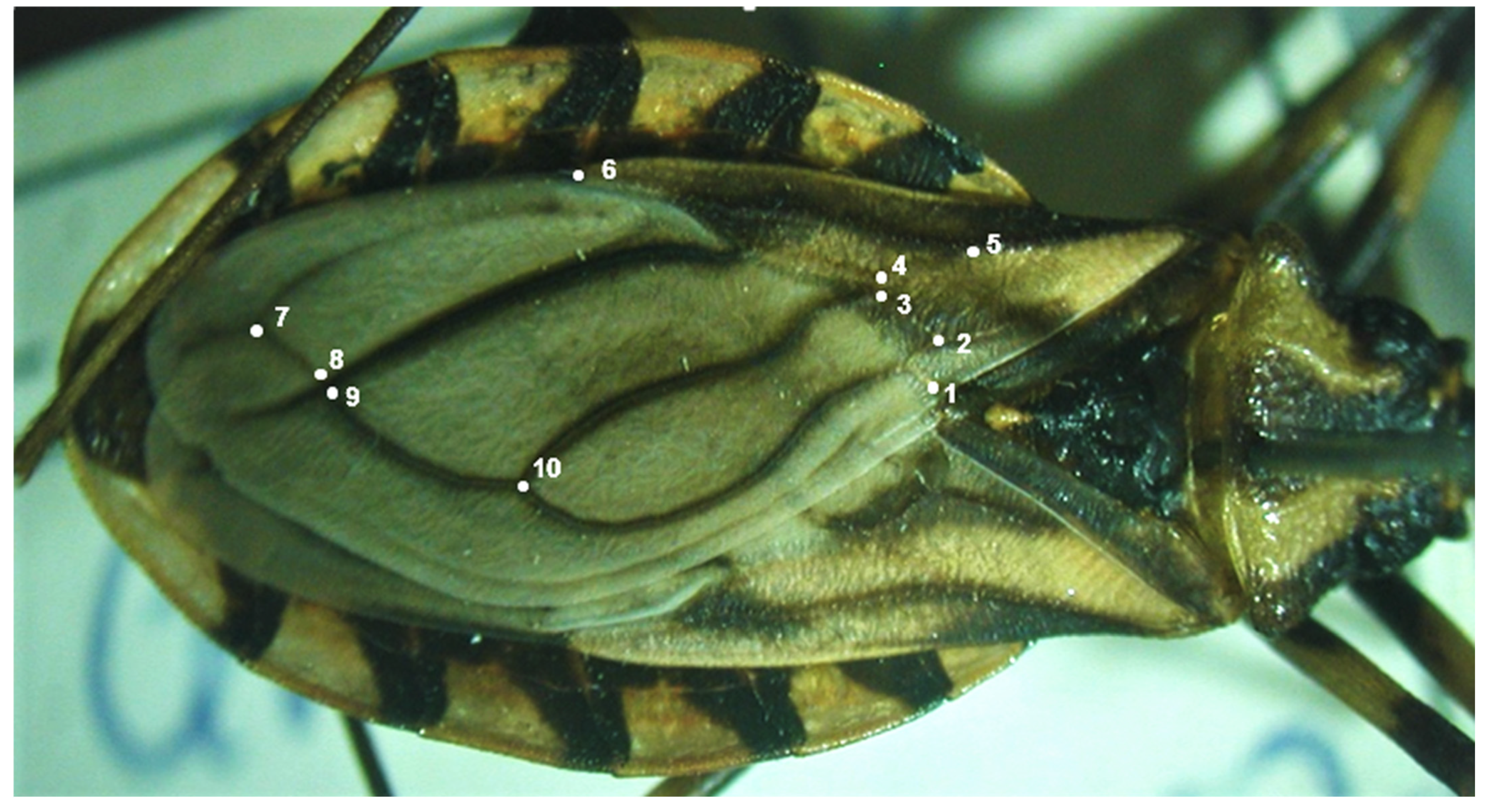
2.3. Data Collection

Ten landmarks were selected and identified from digital photos of each wing:1. Intersection of Pcu and Pcu + first anal vein; 2. Intersection of Cu and Cu–postocubitus (Cu–Pcu); 3. Intersection of Cu and M–Cu; 4. Intersection of media and cubitus (M–Cu); 5. Bifurcation of the radius (R) and median (M) veins; 6. Membrane portion on radius vein; 7. First intersection of R + M and Pcu (postocubitus); 8 Second intersection of R + M and Pcu (postocubitus); 9. Intersection of M and extension of Cu–Pcu veins; 10. Intersection of Pcu and Cu.
These points were determined according to Oliveira et al. [23] (Figure 1). The landmarks were digitized by means of the software TPSDig2 v2.31 [32,33].

2.4. Geometric Morphometrics Analyses

The main method for GM analysis is the Procrustes superimposition, which removes mathematical information produced by size, position, and orientation [34,35]. This analysis was performed using TPSRelw v1.7 [36]. Additionally, the measurement error was analyzed and found to be negligible. Fruciano [37] indicates that measurement errors in GM can inflate the amount of variance and, since morphological analyses are based on the amount of “explained” relative to “residual” variance, the misposition of landmarks could influence the results.
TPSRelw shows two types of shape deformation: the first is the average shape, and the second one is obtained by the partial and relative wraps and denotes the shape changes of each individual. This process is also called principal component analysis, which represents the shape space of possible variation of every specimen [38,39].
The Lambda test of Wilks evaluated the multivariate differences between the groups [40]. To identify the variation among the groups, a canonical variate analysis (CVA) was performed using the shape data categorized by chronological order. With the results of the CVA, average distances were extracted and an unweighted pair group method with arithmetic mean (UPGMA) dendrogram was built to identify the relationships among the groups studied. All these analyses were performed using the software JMP (SAS Institute Inc., Cary, NC, USA version 3.2.2).
The centroid size measurements were used to reduce the size of the wings to a single variable [41] and the measurements and statistical significance among groups were tested by means of the analysis of variance (ANOVA), whereas the Tukey test was applied for the analysis between pairs.

3. Results

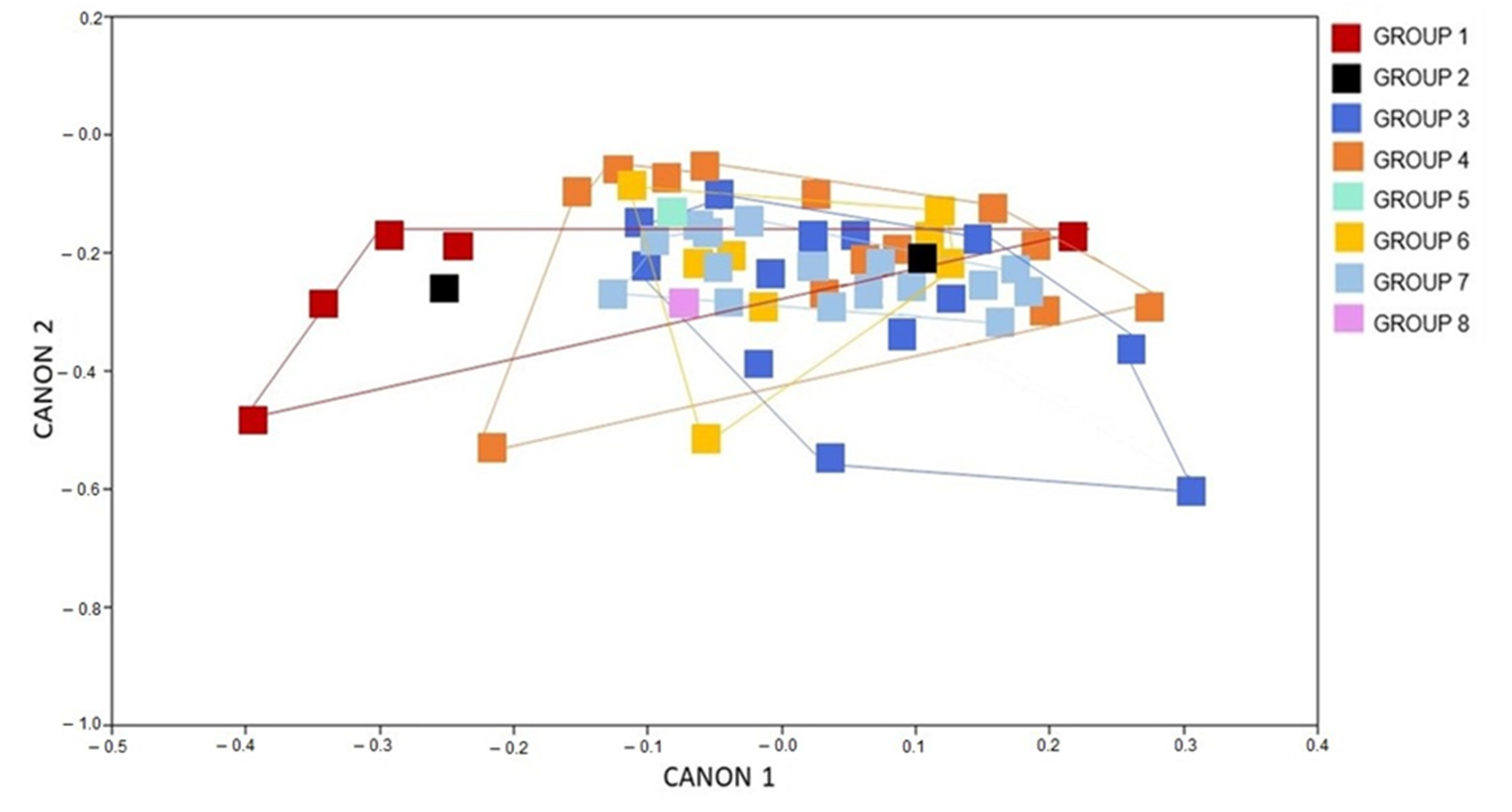
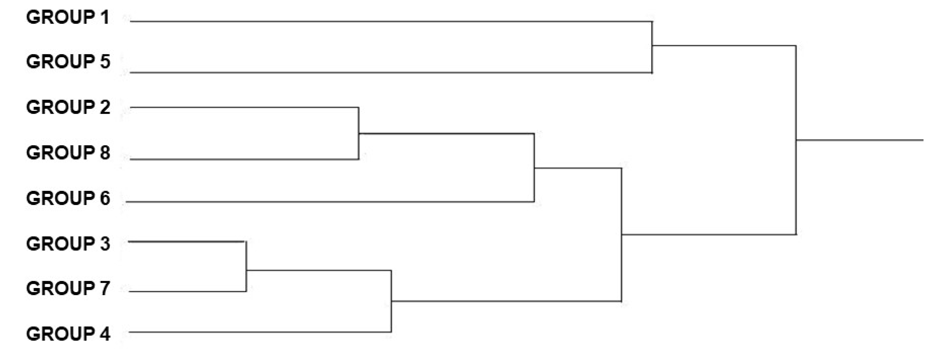
3.1. Morphometric Analysis

3.1.1. Analysis of the Females Wings

The CVA of the female specimens of T. b. brasiliensis shows a big overlap among the groups studied, meaning that the left-wing shape is not largely different among chronological groups (Figure 2).
The cluster analysis (Figure 3), considering the average of Mahalanobis distances among specimens, generated a dendrogram that shows all the overlapping groups, indicating that there has been no change over the years. A larger cluster that included more groups showed greater similarity in wing shape between groups.
The dendrogram (Figure 3), which shows the proximity among the populations, demonstrates that the specimens from group 8 are separated from the others, being the most differentiated group.
There is greater proximity between groups 1 and 5, 3 and 6, and between groups 4 and 7 (Figure 3).

3.1.2. Analysis of the Males Wings

The results observed in the CVA referring to the male populations corroborate the results obtained for females: an overlap of geometric shapes suggests that there was no phenotypic change determined by the chronological groups (Figure 4).
The dendrogram (Figure 5) shows that there are more similarities among specimens in groups 1 and 5, 2 and 8, and 3 and 7.

3.1.3. Analysis of the Males and Females Wing Shape

A third analysis was carried out, now with male and female populations of T. b. brasiliensis together. The populations are structured in overlapping areas, suggesting that they remain unchanged in their phenotypes (Figure 6).
The dendrogram (Figure 7) showing the proximity among these groups determined a closer proximity for the groups 1 and 3, 2 and 5, 4 and 6, and between groups 7 and 8 (Figure 7).
An additional analysis was carried out among all the groups studied and an external group, composed of 10 specimens of T. infestans collected in Rio Grande do Sul, to verify the possible level of morphological convergence of the T. b. brasiliensis groups (Figure 8). The result of the analysis ratifies the previous ones, in which all groups overlap.
The CVA below shows the external group (T. infestans) separated from the other groups of T. b. brasiliensis.

4. Discussion

The following research found that geometric morphometrics tools were not powerful enough to detect differences in wing morphology on a timescale of more than 100 years of T. b. brasiliensis. The analysis applied to the wing shape showed similar phenotypes around the whole set of landmarks for the eight groups of years. Morphological stasis founded in this data has been observed in highly invasive species such as Drosophila spp., in which there is an evolutionary trend to keep the shape morphology in order to keep their invasive behavior [42,43,44].
In the triatomine group, the GM analyses have been applied in a wide variety of approaches, allowing for the understanding of the speciation process and morphological differentiation. Gumiel et al. [45] used GM in taxonomic studies, resulting in the classification of Triatoma melanosoma as a junior synonym of T. infestans. Furthermore, in a study by Schachter-Broide et al. [46], GM was used to analyze T. infestans wings and, from the simultaneous analysis of both size and shape, it was possible to clarify the direction and elapsed time of the insect dispersion, establishing the relevance of this type of study to corelate morphological heterogeneities to the reinfestation patterns. Nattero et al. [47] concluded that the shape of the wings could be considered a reasonably good phenotypic marker, as it made it possible to distinguish the four species of the sordida subcomplex through GM analyses.
In the T. brasiliensis species complex, several applications of GM have revealed important aspects of its evolutionary history [30] and systematics [14,22]. Costa et al. [30] and Dujardin et al. [48] highlight the fact that triatomines can present a phenotype change due to environmental pressures, and this is a gradual process shown to parallel evolutionary relationships among closely related species. In the case of the members of the monophyletic group T. brasiliensis complex, it was suggested that the speciation process, also revealing morphological changes between T. b. brasiliensis and T. melanica, could have taken 5.2 million years, supposedly in the early Pliocene [49]. In this study, the analysis of the 111 T. b. brasiliensis specimens, from different states in the north-east of Brazil, from 1912 to 2014, showed no changes in the wing phenotype. A recent study based on molecular and morphological markers showed similar results in populations of T. b. brasiliensis from Rio Grande do Norte and Paraíba [22]. It is important to stress that the specimens here studied presented the typical color pattern of the T. b. brasiliensis according to previous descriptions in the literature [6,9,31].
Gurgel-Gonçalves et al. [50] combined GM and niche modeling to characterize two closely related species, T. sordida and T. garciabesi, obtaining satisfactory results in their studies. This approach emphasizes the power of GM to characterize morphologically cryptic species. Therefore, the separation of T. infestans as the outgroup from all the other T. b. brasiliensis groups corroborates previous studies in indicating that GM is an important tool for systematics and evolutionary relationships analyses and lends support in identifying the morphological stability of the T. b. brasiliensis over the last 100 years.
It is worth mentioning that the Northeast region is composed of four geographical areas: Meio-norte (mid-north), Sertão (hinterland), the semiarid Agreste region, and Zona da Mata (forest area). The Sertão is the largest, spreading over eight northeastern states—Piauí, Ceará, Rio Grande do Norte, Paraíba, Pernambuco, Alagoas, Bahia, and Sergipe. This vast area with a semi-arid climate is covered by a typical kind of vegetation known as caatinga. An ecological niche modeling was carried out to evaluate the climate and verify possible climatic environmental changes [7]. It was observed that this area presents climate stability without indication of possible changes for the next 50 years. This geographic–climatic stability could also be a factor influencing the morphological stability observed in T. b. brasiliensis.
A similar analysis has been performed with specimens of T. infestans with a time frame of over 70 years [51]. Regarding this species, which specifically occupied domiciliary ecotopes (in Brazil), where high climatic stability is also expected, no significant changes were observed in the different populations over the years. Although the dispersion history of T. infestans is quite distinct from that of T. brasiliensis, both species apparently occupy stable ecological spaces [7,52]. Triatoma infestans exclusively occupies the intradomicile in Brazil, whereas T. b. brasiliensis occupies several natural and artificial ecotopes. Both species are in different ecological spaces but in stable conditions of occupation.
In a recent study by Vilaseca et al. [29], GM was able to detect morphological variation of T. infestans specimens that inhabited two geographically distinct environments in Bolivia. This approach allowed for a better understanding of the biological adaptation of this species since the results showed the influence of different environmental factors on the size and shape of this species.

5. Conclusions

This research presents a novelty finding in which no differences were observed between multiple populations of the vector of Chagas disease, T. b. brasiliensis, for 102 years. Several reasons could explain this morphological stasis, nevertheless, the invasive force and the protozoan vector’s capacity to be spread could be explored in future studies. It is important to notice that according to the distributional potential analysis, no external forces have been favoring changes in this species morphology given the climatic and geographic stability in the main area infested by T. b. brasiliensis, in the north-east region of Brazil. In consonance with the GM results, T. b. brasiliensis is going to continue to be characterized by a specific and stable phenotype, which facilitates its identification by the technicians of the public health services responsible for the monitoring activities in the infested areas in Brazil. Triatoma b. brasiliensis is a native species, occurs in natural and artificial ecotopes and, consequently, cannot be eliminated by the control programs. It also presents a broad geographic distribution and is considered a relevant T. cruzi vector; therefore, T. b. brasiliensis is going to continuously impose a risk of Chagas disease transmission to the human populations, demanding constant monitoring and control actions.

Author Contributions

Conceptualization: J.C.; Methodology: L.P., C.D., V.L.-N., H.A.B. and J.C.; Formal Analisys: L.P., C.D. and J.C.; Curation Date: L.P.; C.D., J.C.; Original Draft Preparation: L.P., C.D. and J.C.; Writing—Review, Visualization and Editing: L.P., C.D., V.L.-N., A.L.C.-d.-l.-F., J.d.O.; H.A.B. and J.C.; Supervision: C.D. and J.C.; Project Administration J.C.; Funding Acquisition: J.C. All authors have read and agreed to the published version of the manuscript.

Funding

National Council for Scientific and Technological Development—CNPq for J.C Process # 303363/2017-7.

Institutional Review Board Statement

Ethical review and approval were waived for this study, because Triatoma brasiliensis brasiliensis is not a protected or endangered species in Brazil and is also not included in the CITES program of the International Union for Conservation of Nature.

Informed Consent Statement

Not applicable.

Data Availability Statement

All insects used in this study are deposited in the entomological collections of Instituto Oswaldo Cruz-Fiocruz.

Acknowledgments

To Jean Pierre Dujardin for reviewing the manuscript. To Angie Vasconcellos for the English version.

Conflicts of Interest

The authors declare no conflict of interest. The funders had no role in the design of the study; in the collection, analyses, or interpretation of data; in the writing of the manuscript; or in the decision to publish the results.

References

  1. Silveira, A.C. Os novos desafios e perspectivas futuras do controle. Rev. Soc. Bras. Med. Trop. 2011, 44, 122–124. (In Portuguese) [Google Scholar] [CrossRef] [PubMed] [Green Version]
  2. Stanaway, J.D.; Roth, G. The burden of Chagas disease: Estimates and challenges. Glob. Heart 2015, 10, 139–144. [Google Scholar] [CrossRef] [PubMed]
  3. WHO. World Health Organization. Chagas Disease (American Trypanosomiasis). 2021. Available online: https://www.who.int/news-room/facts-in-pictures/detail/chagas-disease (accessed on 11 January 2022).
  4. Costa, J.; Dale, C.; Galvão, C.; Almeida, C.E.; Dujardin, J.P. Do the new triatomine species pose new challenges or strategies for monitoring Chagas disease? An overview from 1979–2021. Mem. Inst. Oswaldo Cruz 2021, 116, 1–10. [Google Scholar] [CrossRef] [PubMed]
  5. Alevi, K.C.C.; de Oliveira, J.; da Silva Rocha, D.; Galvão, C. Trends in Taxonomy of Chagas Disease Vectors (Hemiptera, Reduviidae, Triatominae): From Linnaean to Integrative Taxonomy. Pathogens 2021, 10, 1627. [Google Scholar] [CrossRef]
  6. Costa, J.; Correia, N.C.; Lima-Neiva, V.; Gonçalves, T.C.M.; Felix, M. Revalidation and redescription of Triatoma brasiliensis macromelasoma Galvão, 1956 and an identification key for the Triatoma brasiliensis complex (Hemiptera: Reduviidae: Triatominae). Mem. Inst. Oswaldo Cruz 2013, 108, 785–789. [Google Scholar] [CrossRef]
  7. Costa, J.; Dornak, L.L.; Almeida, C.E.; Peterson, A.T. Distributional potential of the Triatoma brasiliensis species complex at present and under scenarios of future climate conditions. Parasit Vectors 2014, 7, 238. [Google Scholar] [CrossRef]
  8. Costa, J.; Lima-Neiva, V.; Almeida, C.E.O. complexo Triatoma brasiliensis (Hemiptera, Reduviidae, Triatominae) como modelo de estudo: Uma abordagem multidisciplinar e ecoepidemiológica. Atual. Med. Trop. Vetores 2020, 6, 99–121. (In Portuguese) [Google Scholar] [CrossRef]
  9. Dale, C.; Almeida, C.E.; Mendonça, V.J.; Oliveira, J.; da Rosa, J.A.; Galvão, C.; Costa, J. An updated and illustrated dichotomous key for the Chagas disease vectors of Triatoma brasiliensis species complex and their epidemiologic importance. ZooKeys 2018, 805, 33–43. [Google Scholar] [CrossRef]
  10. Nascimento, D.; Novais, G. Clima do Cerrado: Dinâmica atmosférica e características, variabilidades e tipologias climáticas. Élisée Rev. Geografia UEG 2020, 9, e922021. (In Portuguese) [Google Scholar]
  11. Moreira, J.N. Caracterização da vegetação de Caatinga e da dieta de novilhos no Sertão de Pernambuco. Pesqui Agropecu 2006, 41, 1643–1651. (In Portuguese) [Google Scholar] [CrossRef] [Green Version]
  12. Costa, J.; Almeida, C.E.; Dotson, E.M.; Lins, A.; Vinhaes, M.; Silveira, A.C.; Beard, C.B. The epidemiologic importance of Triatoma brasiliensis as a Chagas disease vector in Brazil: A revision of domiciliary captures during 1993–1999. Mem. Inst. Oswaldo Cruz 2003, 98, 443–449. [Google Scholar] [CrossRef] [PubMed]
  13. Silveira, A.C. Situação do controle da transmissão vetorial da doença de Chagas nas Américas. Cad. Saúde Pública 2000, 16, 35–42. (In Portuguese) [Google Scholar] [CrossRef] [PubMed]
  14. Almeida, C.E.; Faucher, L.; Lavina, M.; Costa, J.; Harry, M. Molecular Individual-Based Approach on Triatoma brasiliensis: Inferences on Triatomine Foci, Trypanosoma cruzi Natural Infection Prevalence, Parasite Diversity and Feeding Sources. PLoS Negl. Trop. Dis. 2016, 10, e0004447. [Google Scholar] [CrossRef] [PubMed] [Green Version]
  15. Lilioso, M.; Folly-Ramos, E.; Rocha, F.L.; Rabinovich, J.; Capdevielle-Dulac, C.; Harry, M. High Triatoma brasiliensis densities and Trypanosoma cruzi prevalence in domestic and peridomestic habitats in the state of Rio Grande do Norte, Brazil: The source for Chagas disease outbreaks? J. Trop. Med. Hyg. 2017, 96, 1456–1459. [Google Scholar] [CrossRef] [PubMed]
  16. Bezerra, C.M.; Belisário, C.J.; Pessoa, G.C.D.; Barezani, C.P.; Rosa, A.C.L.; Ferreira, F.C.; Ramos, A.N., Jr.; Gurtler, R.E.; Diotaiuti, L. Microsatellite variation revealed panmictic pattern for Triatoma brasiliensis (Triatominae: Reduviidae) in rural northeastern Brazil: The control measures implications. BMC Genom. 2020, 21, 92. [Google Scholar] [CrossRef]
  17. Lilioso, M.; Pires-Silva, D.; von Hertwig, M.F.F.; Oliveira, J.; da Rosa, J.A.; Vilela, R.V.; Folly-Ramos, E.; Almeida, C.E. Triatoma petrocchiae (Hemiptera, Reduviidae, Triatominae): A Chagas disease vector of T. brasiliensis species complex associated to reptiles. Infect. Genet. Evol. 2020, 82, 104307. [Google Scholar] [CrossRef]
  18. Lima-Neiva, V.; Toma, H.K.; Aguiar, L.M.A.; Lopes, C.M.; Dias, L.P.; Gonçalves, T.C.M.; Costa, J. The connection between Trypanosoma cruzi transmission cycles by Triatoma brasiliensis brasiliensis: A threat to human health in an area susceptible to desertification in the Seridó, Rio Grande do Norte, Brazil. PLoS Negl. Trop. Dis. 2021, 15, e0009919. [Google Scholar] [CrossRef]
  19. Costa, J.; Lorenzo, M. Biology, diversity and strategies for the monitoring and control of triatomines—Chagas disease vectors. Mem. Inst. Oswaldo Cruz 2009, 1, 46–51. [Google Scholar] [CrossRef] [Green Version]
  20. Silveira, A.C.; Dias, J.C.P. O controle da transmissão vetorial. Rev. Soc. Bras. Med. Trop. 2011, 44, 52–63. (In Portuguese) [Google Scholar] [CrossRef]
  21. Lima-Oliveira, T.M.; Fontes, F.V.H.M.; Lilioso, M.; Pires-Silva, D.; Teixeira, M.M.G.; Meza, J.G.B.; Harry, M.; Fileé, J.; Costa, J.; Valença-Barbosa, C.; et al. Molecular eco-epidemiology on the sympatric Chagas disease vectors Triatoma brasiliensis and Triatoma petrocchiae: Ecotopes, genetic variation, natural infection prevalence by trypanosomatids and parasite genotyping. Acta Trop. 2020, 201, 105188. [Google Scholar] [CrossRef]
  22. Kamimura, E.H.; Viana, M.C.; Lilioso, M.; Fontes, F.H.M.; Pires-Silva, D.; Valença-Barbosa, C.; Carbajal-de-la-Fuente, A.L.; Folly-Ramos, E.; Solferin, V.N.; Thyssen, P.J.; et al. Drivers of molecular and morphometric variation in Triatoma brasiliensis (Hemiptera: Triatominae): The resolution of geometric morphometrics for populational structuring on a microgeographical scale. Parasit Vectors 2020, 13, 455. [Google Scholar] [CrossRef] [PubMed]
  23. Oliveira, J.; Marcet, P.L.; Takiya, D.M.; Mendonça, V.J.; Belintani, T.; Bargues, M.D.; Mateo, L.; Chagas, V.; Folly-Ramos, E.; Cordeiro-Estrela, P.; et al. Combined phylogenetic and morphometric information to delimit and unify the Triatoma brasiliensis species complex and the brasiliensis subcomplex. Acta Trop. 2017, 170, 140–148. [Google Scholar] [CrossRef] [PubMed]
  24. Belintani, T.; Oliveira, J.; Pinotti, H.; Silva, L.A.; Alevi, K.C.C.; Galvão, C.; Rosa, J.A. Phylogenetic and phenotypic relationships of the Triatoma sordida subcomplex (Hemiptera: Reduviidae: Triatominae). Acta Trop. 2020, 212, 105679. [Google Scholar] [CrossRef] [PubMed]
  25. Dujardin, J.P.; Bermudez, H.; Asine, C.; Schofield, C.J.; Tibayrenc, M. Metric differences between silvatic and domestic Triatoma infestans (Heteroptera- Reduviidae) in Bolivia. J. Med. Entomol. 1997, 34, 544–551. [Google Scholar] [CrossRef]
  26. Dujardin, J.P.; Chávez, T.; Moreno, J.M.; Machane, M.; Noireau, F.; Schofield, C.J. Comparison of isoenzyme electrophoresis and morphometric analysis for phylogenetic reconstruction of the Rhodniini (Hemiptera: Reduvidae: Triatominae). J. Med. Entomol. 1999, 36, 653–659. [Google Scholar] [CrossRef]
  27. Lunardi, R.R.; Benítez, H.A.; Câmara, T.P.; Gomes, L.P.; Arrais-Silva, W.W. Head shape variation in response to diet in Triatoma williami (Hemiptera, Reduviidae: Triatominae), a possible Chagas disease vector of legal Amazônia. Zoo. Anz. 2017, 267, 187–193. [Google Scholar] [CrossRef]
  28. Vilaseca, C.; Méndez, M.A.; Pinto, C.F.; Benítez, H.A. Assessment of patterns of geometric shape variation in Triatoma infestans Hemiptera: Reduviidae: A first report in populations from Bolivia. Insects 2020, 11, 274. [Google Scholar] [CrossRef]
  29. Vilaseca, C.; Méndez, M.A.; Pinto, C.F.; Lemic, D.; Benítez, H.A. Unraveling the Morphological Variation of Triatoma infestans in the Peridomestic Habitats of Chuquisaca Bolivia: A Geometric Morphometric Approach. Insects 2021, 12, 185. [Google Scholar] [CrossRef]
  30. Costa, J.; Peterson, A.T.; Dujardin, J.P. Morphological evidence suggests homoploid hybridization as a possible mode of speciation in the Triatominae (Hemiptera, Heteroptera, Reduviidae). Infect. Genet. Evol. 2009, 9, 263–270. [Google Scholar] [CrossRef]
  31. Lent, H.; Wygodzinsky, P.W. Revision of the Triatominae (Hemiptera, Reduviidae), and their significance as vectors of Chagas’ disease. Bull. Am. Mus. Nat. Hist. 1979, 163, 3. [Google Scholar]
  32. Rohlf, J. On Applications of Geometric Morphometrics to Studies of Ontogeny and Phylogeny. Syst. Biol. 1998, 47, 147–158. [Google Scholar] [CrossRef] [PubMed] [Green Version]
  33. Rohlf, F.J. Tpsdig, Digitize Landmarks and Outlines, Version 2.17; Department of Ecology and Evolution: State University of New York at Stony Brook, NY, USA, 2013. [Google Scholar]
  34. Rohlf, F.J.; Slice, D. Extensions of the Procustes methods for the optimal superimposition of landmarks. Syst. Zool. 1990, 39, 40–59. [Google Scholar] [CrossRef] [Green Version]
  35. Dryden, I.L.; Mardia, K.V. Statistical Shape Analysis; John Wiley and Sons: Hoboken, NJ, USA, 1998; Volume 4. [Google Scholar]
  36. Rohlf, J. General Biology; The University of Chicago Press: Chicago, IL, USA, 2010; p. 85. [Google Scholar]
  37. Fruciano, C. Measurement error in geometric morphometrics. Dev. Genes Evol. 2016, 226, 139–158. [Google Scholar] [CrossRef] [PubMed]
  38. Moraes, D.A. A Morfometria Geométrica e a “Revolução na Morfometria”: Localizando mudanças na forma dos organismos. Bioletim 2003, 3, 14–20. (In Portuguese) [Google Scholar]
  39. Villalobos-Leiva, A.; Benítez, H.A. Morfometría Geométrica y sus Nuevas Aplicaciones en Ecología y Biología Evolutiva: Parte 2. Int. J. Morphol. 2020, 38, 1818–1836. (In Spanish) [Google Scholar] [CrossRef]
  40. Hair, J.F.; Tatham, R.L.; Anderson, R.E.; Black, W. Multivariate Data Analysis. Prentice Hall 1998, 5, 768. [Google Scholar]
  41. Bookstein, F.L. Morphometric Tools for Landmark Data: Geometry and Biology; Cambridge University Press: Cambridge, UK, 1991; p. 435. [Google Scholar]
  42. Hansen, T.F.; Houle, D. Evolvability, Stabilizing Selection, and the Problem of Stasis. In Phenotypic Integration: Studying the Ecology and Evolution of Complex Phenotypes; Oxford University Press: Oxford, UK, 2004; pp. 130–150. [Google Scholar]
  43. Houle, D.; Bolstad, G.H.; van der Linde, K.; Hansen, T.F. Mutation predicts 40 million years of fly wing evolution. Nature 2017, 548, 447–450. [Google Scholar] [CrossRef]
  44. Benitez, H.A.; Püschel, T.; Suazo, M. Drosophila wing integration and modularity: A multi-level approach to understand the history of morphological structures. Biology 2022, 11, 567. [Google Scholar] [CrossRef]
  45. Gumiel, M.S.; Catalá, F.; Noireau, A.; Rojas, D.A.; García, A.; Dujardin, J.P. Wing geometry in Triatoma infestans (Klug) and T. melanosoma Martinez, Olmedo & Carcavallo (Hemiptera: Reduviidae). Syst. Entomol. 2003, 28, 2–6. [Google Scholar] [CrossRef]
  46. Schachter-Broide, J.; Gürtler, R.E.; Kitron, U.; Dujardin, J.P. Temporal variations of wing size and shape of Triatoma infestans (Hemiptera: Reduviidae) populations from northwestern Argentina using geometric morphometry. J. Med. Entomol. 2009, 46, 994–1000. [Google Scholar] [CrossRef] [Green Version]
  47. Nattero, J.; Piccinali, R.V.; Lopes, C.M.; Hernández, M.L.; Abrahan, L.; Lobbia, P.A.; Rodríguez, C.S.; Carbajal-de-la-Fuente, A.L. Morphometric variability among the species of the Sordida subcomplex (Hemiptera: Reduviidae: Triatominae): Evidence for differentiation across the distribution range of Triatoma sordida. Parasit Vectors 2017, 10, 412. [Google Scholar] [CrossRef] [PubMed] [Green Version]
  48. Dujardin, J.P.; Costa, J.; Bustamante, D.; Jaramillo, N.; Catalá, S. Deciphering morphology in Triatominae: The evolutionary signals. Acta Trop. 2009, 110, 101–111. [Google Scholar] [CrossRef] [PubMed]
  49. Monteiro, F.A.; Donnelly, M.J.; Beard, C.B.; Costa, J. Nested clade and phylogeographic analyses of the Chagas disease vector Triatoma brasiliensis in Northeast Brazil. Mol. Phylogenet. Evol. 2004, 32, 46–56. [Google Scholar] [CrossRef] [PubMed]
  50. Gurgel-Gonçalves, R.; Ferreira, J.B.C.; Rosa, A.F.; Bar, M.E.; Galvão, C. Geometric morphometrics and ecological niche modelling for delimitation of near-sibling triatomine species. Med. Vet. Entomol. 2011, 25, 84–93. [Google Scholar] [CrossRef]
  51. Paschoaletto, L.; Lima-Neiva, V.; Lopes, C.M.; Dale, C.; Mallet, J.R.S.; La Fuente, A.L.C.A.; Costa, J. 72 years of temporal analysis through geometric morphometrics detects phenotypic variation in populations of Triatoma infestans (Klug, 1834). Acta Biol. Par. 2020, 49, 1–17. [Google Scholar] [CrossRef]
  52. Forattini, O.P. Biogeography, origin, and distribution of triatominae domiciliarity in Brazil. Rev. Saúde Pública 2006, 40, 964–998. [Google Scholar] [CrossRef] [Green Version]
Figure 1. Image of the left wing of a male of Triatoma brasiliensis brasiliensis Neiva, 1911 showing the selected 10 landmarks: 1. Intersection of Pcu and Pcu (postocubitus) + first anal vein; 2. Intersection of Cu (cubitus) and Cu–postocubitus (Cu–Pcu); 3. Intersection of Cu and M–Cu; 4. Intersection of media and cubitus (M–Cu); 5. Bifurcation of the radius (R) and median (M) veins; 6. Membrane portion on radius vein; 7. First intersection of R + M and Pcu (postocubitus); 8 Second intersection of R + M and Pcu; 9. Intersection of M and extension of Cu–Pcu veins; 10. Intersection of Pcu and Cu.
Animals 12 01362 g001
Figure 2. Canonical variate analysis showing the wing shape influence of the of Triatoma brasiliensis brasiliensis females from various states and years (1912–2014) Canon represents the canonical variate dimension.
Animals 12 01362 g002
Figure 3. UPGMA dendrogram of the Triatoma brasiliensis brasiliensis females, from various states and years (1912–2014) showing the phylogenetic distances among the groups. Groups: 1, 3, 4, 5, 6, 7, 8.
Animals 12 01362 g003
Figure 4. Canonical variate analysis showing the wing shape influence of Triatoma brasiliensis brasiliensis males from several states and years (1912–2014). Groups: 1, 2, 3, 4, 5, 6, 7, 8 Canon represents the canonical variate dimension.
Animals 12 01362 g004
Figure 5. UPGMA Dendrogram of the Triatoma brasiliensis brasiliensis males, from various states and years (1912–2014), showing the phylogenetic distances between the groups.
Animals 12 01362 g005
Figure 6. Canonical variate analysis showing the conformation influence in males and females of Triatoma brasiliensis brasiliensis from different states and years (1912–2014). among the groups Canon represents the canonical variate dimension.
Animals 12 01362 g006
Figure 7. UPGMA dendrogram of the male and female populations of Triatoma brasiliensis brasiliensis from different states and years (1912–2014), showing the phylogenetic distances among the groups.
Animals 12 01362 g007
Figure 8. Canonical variate analysis showing the conformation influence in males and females of Triatoma brasiliensis brasiliensis from several states and years (1912–2014). Groups: 1, 2, 3, 4, 5, 6, 7, 8, and an external group T. infestans Canon represents the canonical variate dimension.
Animals 12 01362 g008
Table 1. Localities and years of the left-wing samples of Triatoma brasiliensis brasiliensis used for this study. Availability of samples were arranged chronologically, abbreviation are defined as CEIOC: the Entomological Collection of Instituto Oswaldo Cruz and CTIOC: the Collection of Triatomines of Instituto Oswaldo Cruz.
GroupsYearCollectionStateCityMalesFemalesTotal
Group 11912CTIOCPiauíFloriano516
Group 21922CTIOCCearáQuixada102
1929Rio Grande do NorteAcary10
Group 31940CTIOCCearáRussas12619
1941PernambucoMangabeira10
Group 41950CTIOCParaíbaSanta Lúcia12722
1955Santa Lúcia12
Group 51977CTIOCPiauíBrejo Seco134
Group 61996CEIOC
CTIOC
CTIOC
Rio Grande do NorteSerra Negra do Norte, Caicó4524
1998PiauiBrejo Seco22
1999PiauiBrejo Seco29
Group 72002CEIOCParaíbaLivramento, São José da Lagoa da Tapada, São José das Piranhas, São Francisco da Prata161531
Group 82014CTIOCCearáSanta Quitéria123
TOTAL 111
Publisher’s Note: MDPI stays neutral with regard to jurisdictional claims in published maps and institutional affiliations.

