
Leptospira Seroprevalence in Colombian Dairy Herds

 , , and
Abstract
:Simple Summary
Abstract
1. Introduction
2. Materials and Methods
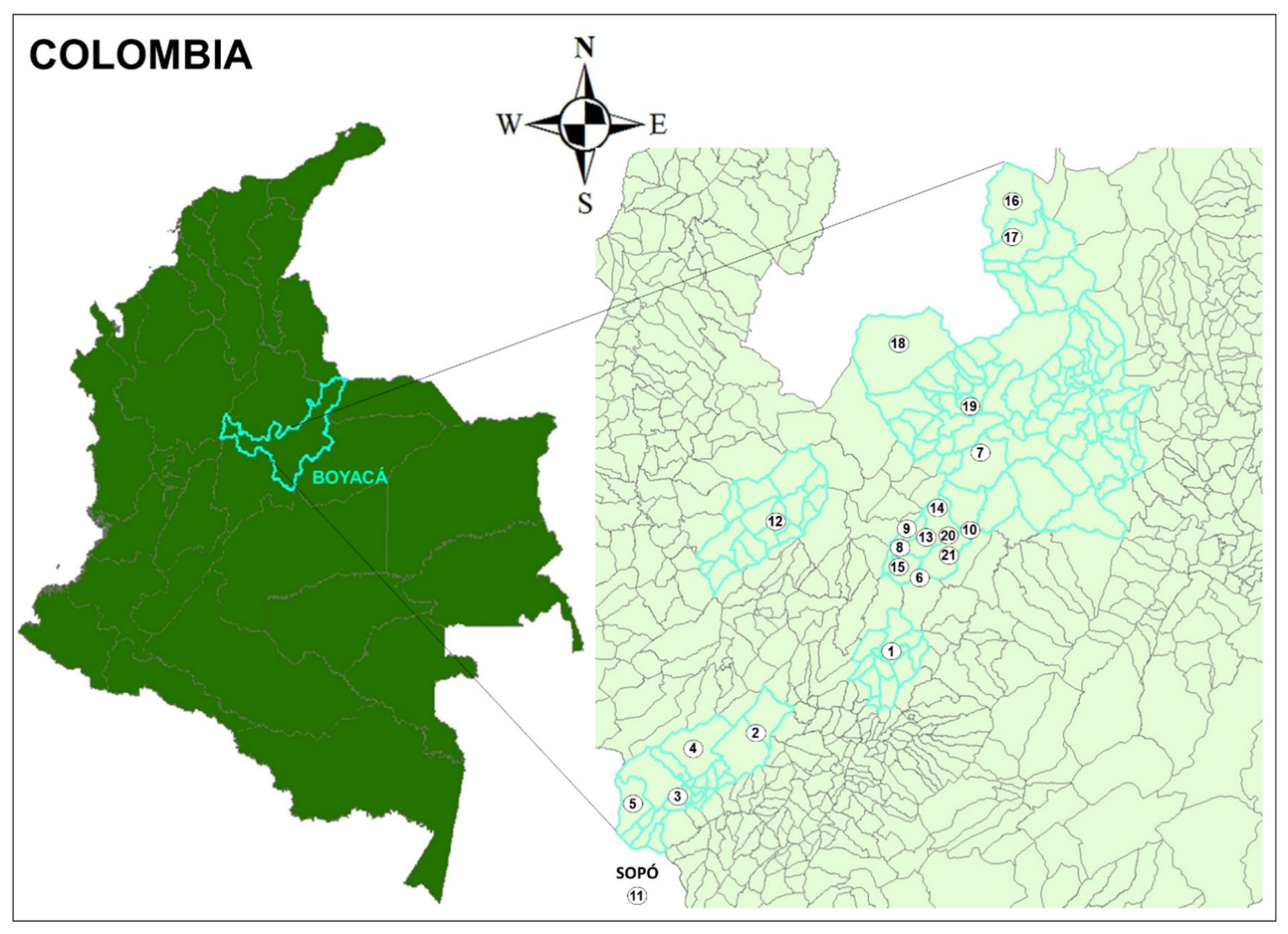
2.1. Study Sites
2.2. Sampled Population
2.3. Serology
2.4. Statistical Analysis
3. Results
4. Discussion
5. Conclusions
Author Contributions
Funding
Institutional Review Board Statement
Informed Consent Statement
Conflicts of Interest
References
- Hartskeerl, R.A.; Collares-Pereira, M.; Ellis, W.A. Emergence, Control and Re-Emerging Leptospirosis: Dynamics of Infection in the Changing World. Clin. Microbiol. Infect. 2011, 17, 494–501. [Google Scholar] [CrossRef] [Green Version]
- Dufour, B.; Moutou, F.; Hattenberger, A.M.; Rodhain, F. Global Change: Impact, Management, Risk Approach and Health Measures—The Case of Europe. Rev. Off. Int. Epizoot. 2008, 27, 529–550. [Google Scholar] [CrossRef]
- Petrakovsky, J.; Bianchi, A.; Fisun, H.; Nájera-Aguilar, P.; Pereira, M.M. Animal Leptospirosis in Latin America and the Caribbean Countries: Reported Outbreaks and Literature Review (2002–2014). Int J. Environ. Res. Public Health 2014, 11, 10770–10789. [Google Scholar] [CrossRef] [Green Version]
- Loureiro, A.P.; Hamond, C.; Lilenbaum, W. Leptospirosis in Horses. Vet. Rec. 2013, 172, 479–480. [Google Scholar] [CrossRef]
- Mazeri, S.; Scolamacchia, F.; Handel, I.G.; Morgan, K.L.; Tanya, V.N.; Bronsvoort, B.M. deC Risk Factor Analysis for Antibodies to Brucella, Leptospira and C. Burnetii among Cattle in the Adamawa Region of Cameroon: A Cross-Sectional Study. Trop. Anim. Health Prod. 2013, 45, 617–623. [Google Scholar] [CrossRef] [PubMed]
- Schoonman, L.; Swai, E.S. Herd- and Animal-Level Risk Factors for Bovine Leptospirosis in Tanga Region of Tanzania. Trop. Anim. Health Prod. 2010, 42, 1565–1572. [Google Scholar] [CrossRef] [PubMed]
- Sebek, Z.; Sixl, W.; Valova, M.; Marth, E.; Köck, M.; Reinthaler, F.F. Serological Investigations for Leptospirosis in Humans in Columbia. Geogr. Med. Suppl. 1989, 3, 51–60. [Google Scholar]
- Szyfres, B. Leptospirosis as an Animal and Public Health Problem in Latin America and the Caribbean Area. Bull. Pan Am. Health Organ. 1976, 10, 110–125. [Google Scholar]
- Agudelo-Flórez, P.; Restrepo-Jaramillo, B.N.; Arboleda-Naranjo, M. [Leptospirosis in Uraba, Antioquia, Colombia: A seroepidemiological and risk factor survey in the urban population]. Cad. Saude Publica 2007, 23, 2094–2102. [Google Scholar] [CrossRef] [Green Version]
- Góngora, A.; Parra, J.; Aponte, L.; Gómez, L. [Seroprevalence of Leptospira spp in population groups of Villavicencio, Colombia]. Rev. Salud Publica 2008, 10, 269–278. [Google Scholar] [CrossRef] [Green Version]
- Pérez-García, J.; Agudelo-Flórez, P.; Parra-Henao, G.J.; Ochoa, J.E.; Arboleda, M. Incidence and Underreporting of Leptospirosis Comparing Three Diagnostic Methods in the Endemic Region of Urabá, Colombia. Biomedica 2019, 39, 150–162. [Google Scholar] [CrossRef] [PubMed]
- Da Silva Pinto, P.; Libonati, H.; Penna, B.; Lilenbauma, W. A Systematic Review on the Microscopic Agglutination Test Seroepidemiology of Bovine Leptospirosis in Latin America. Trop. Anim. Health Prod. 2016, 48, 239–248. [Google Scholar] [CrossRef]
- Grooms, D.L. Reproductive Losses Caused by Bovine Viral Diarrhea Virus and Leptospirosis. Theriogenology 2006, 66, 624–628. [Google Scholar] [CrossRef]
- Levett, P.N. Leptospirosis. Clin. Microbiol. Rev. 2001, 14, 296–326. [Google Scholar] [CrossRef] [Green Version]
- Alonso-Andicoberry, C.; García-Peña, F.J.; Pereira-Bueno, J.; Costas, E.; Ortega-Mora, L.M. Herd-Level Risk Factors Associated with Leptospira Spp. Seroprevalence in Dairy and Beef Cattle in Spain. Prev. Vet. Med. 2001, 52, 109–117. [Google Scholar] [CrossRef]
- Lilenbaum, W.; Souza, G.N. Factors Associated with Bovine Leptospirosis in Rio de Janeiro, Brazil. Res. Vet. Sci. 2003, 75, 249–251. [Google Scholar] [CrossRef]
- Segura-Correa, V.M.; Solis-Calderon, J.J.; Segura-Correa, J.C. Seroprevalence of and Risk Factors for Leptospiral Antibodies among Cattle in the State of Yucatan, Mexico. Trop. Anim. Health Prod. 2003, 35, 293–299. [Google Scholar] [CrossRef] [PubMed]
- Ensuncho-Hoyos, C.; Rodríguez-Rodríguez, V.; Pérez-Doria, A.; Vergara, O.; Calderón-Rangel, A. Epidemiology Behavior of Leptospirosis in Ciénaga de Oro, Córdoba (Colombia). Trop. Anim. Health Prod. 2017, 49, 1345–1351. [Google Scholar] [CrossRef]
- Ochoa, J.E.; Sánchez, A.; Ruiz, I. [Epidemiology of leptospirosis in a livestock production area of the Andes]. Rev. Panam. Salud Publica 2000, 7, 325–331. [Google Scholar] [CrossRef] [Green Version]
- León, A.G.Z. Factores de riesgo asociados a leptospirosis en hatos bovinos de Pereira, 2002–2005. Investig. Andin. 2009, 11, 108–117. [Google Scholar] [CrossRef]
- Atlas Interactivo-Climatológico-IDEAM. Available online: http://atlas.ideam.gov.co/visorAtlasClimatologico.html (accessed on 6 March 2020).
- Instituto Colombiano Agropecuario-ICA. Available online: https://www.ica.gov.co/areas/pecuaria/servicios/epidemiologia-veterinaria/censos-2016/censo-2017.aspx (accessed on 2 February 2021).
- Vera, E.; Taddei, S.; Cavirani, S.; Schiavi, J.; Angelone, M.; Cabassi, C.S.; Schiano, E.; Quintavalla, F. Leptospira Seroprevalence in Bardigiano Horses in Northern Italy. Animals 2019, 10, 23. [Google Scholar] [CrossRef] [PubMed] [Green Version]
- Cochran William, G. Sampling Techniques, 3rd Edition | Wiley. Available online: https://www.wiley.com/en-us/Sampling+Techniques%2C+3rd+Edition-p-9780471162407 (accessed on 10 February 2021).
- Sousa, V.D.; Zauszniewski, J.A.; Musil, C.M. How to Determine Whether a Convenience Sample Represents the Population. Appl. Nurs. Res. 2004, 17, 130–133. [Google Scholar] [PubMed]
- OIE-World Organisation for Animal Health. Available online: http://www.oie.int/fileadmin/Home/eng/Health_standards/tahm/3.01.12_LEPTO.pdf (accessed on 20 March 2019).
- Ryan, E.G.; Leonard, N.; O’Grady, L.; Doherty, M.L.; More, S.J. Herd-Level Risk Factors Associated with Leptospira Hardjo Seroprevalence in Beef/Suckler Herds in the Republic of Ireland. Ir. Vet. J. 2012, 65, 6. [Google Scholar] [CrossRef] [Green Version]
- Lilenbaum, W.; Santos, M.R. Effect of Management Systems on the Prevalence of Bovine Leptospirosis. Vet. Rec. 1996, 138, 570–571. [Google Scholar] [CrossRef] [PubMed]
- Olivera, M.; Chaparro, J.J.; Chaparro, Y.; Piedrahita, D.; Fernández-Silva, J.; Londoño, J.; Palacio, L.G.; Ramírez-Vásquez, N.; Villar, D.; Olivera, M.; et al. Cross-Sectional Study of 13 Leptospira Serovars in Cows in a Colombian Dairy Region. Rev. Colomb. Cienc. Pecu. 2018, 31, 10–16. [Google Scholar] [CrossRef]
- Figueiredo da Costa, D.; Cristiny Rodrigues Silva, M.L.; Martins, G.; Medeiros Dantas, A.F.; Almeida de Melo, M.; Santos de Azevedo, S.; Lilenbaum, W.; Alves, C.J. Susceptibility among Breeds of Sheep Experimentally Infected with Leptospira Interrogans Pomona Serogroup. Microb. Pathog. 2018, 122, 79–83. [Google Scholar] [CrossRef] [PubMed]
- Dos Santos, J.P.; Lima-Ribeiro, A.M.C.; Oliveira, P.R.; dos Santos, M.P.; Ferreira, A.; Medeiros, A.A.; Tavares, T.C.F. Seroprevalence and Risk Factors for Leptospirosis in Goats in Uberlândia, Minas Gerais, Brazil. Trop. Anim. Health Prod. 2012, 44, 101–106. [Google Scholar] [CrossRef] [PubMed]
- Otte, M.J.; Ravenborg, T.; Hüttner, K. A Pilot Study of Elevated Abortion and Stillbirth Ratios in Cattle in the Foothills of the Eastern Plains of Colombia. Prev. Vet. Med. 1995, 22, 103–113. [Google Scholar] [CrossRef]
- Levett, P.N. Usefulness of Serologic Analysis as a Predictor of the Infecting Serovar in Patients with Severe Leptospirosis. Clin. Infect. Dis. 2003, 36, 447–452. [Google Scholar] [CrossRef]
- Smythe, L.D.; Wuthiekanun, V.; Chierakul, W.; Suputtamongkol, Y.; Tiengrim, S.; Dohnt, M.F.; Symonds, M.L.; Slack, A.T.; Apiwattanaporn, A.; Chueasuwanchai, S.; et al. The Microscopic Agglutination Test (MAT) Is an Unreliable Predictor of Infecting Leptospira Serovar in Thailand. Am. J. Trop. Med. Hyg. 2009, 81, 695–697. [Google Scholar] [CrossRef] [Green Version]
- Blanco, R.M.; dos Santos, L.F.; Galloway, R.L.; Romero, E.C. Is the Microagglutination Test (MAT) Good for Predicting the Infecting Serogroup for Leptospirosis in Brazil? Comp. Immunol. Microbiol. Infect. Dis. 2016, 44, 34–36. [Google Scholar] [CrossRef]
- Chirathaworn, C.; Inwattana, R.; Poovorawan, Y.; Suwancharoen, D. Interpretation of Microscopic Agglutination Test for Leptospirosis Diagnosis and Seroprevalence. Asian Pac. J. Trop. Biomed. 2014, 4, S162–S164. [Google Scholar] [CrossRef] [Green Version]
- Turner, L.H. Leptospirosis. II. Serology. Trans. R Soc. Trop. Med. Hyg. 1968, 62, 880–899. [Google Scholar] [CrossRef]
- Alexander, A.D.; Evans, L.B.; Toussaint, A.J.; Marchwicki, R.H.; McCRUMB, F.R. Leptospirosis in Malaya. II. Antigenic Analysis of 110 Leptospiral Strains and Other Serologic Studies. Am. J. Trop. Med. Hyg. 1957, 6, 871–889. [Google Scholar] [CrossRef] [PubMed]
- Kmety, E. [Paradox reactions and their significance in serodiagnosis of some types of leptospirosis]. Zentralbl. Bakteriol. Orig. 1958, 170, 597–608. [Google Scholar] [PubMed]
- Dupouey, J.; Faucher, B.; Edouard, S.; Richet, H.; Kodjo, A.; Drancourt, M.; Davoust, B. Human Leptospirosis: An Emerging Risk in Europe? Comp. Immunol. Microbiol. Infect. Dis. 2014, 37, 77–83. [Google Scholar] [CrossRef] [PubMed]
- Shearer, K.E.; Harte, M.J.; Ojkic, D.; Delay, J.; Campbell, D. Detection of Leptospira Spp. in Wildlife Reservoir Hosts in Ontario through Comparison of Immunohistochemical and Polymerase Chain Reaction Genotyping Methods. Can. Vet. J. 2014, 55, 240–248. [Google Scholar]
- Deháquiz, M.J.E.; Bermúdez, C.L.T.; Rodríguez, C.L.F. Environmental Management Model for Small Dairies in the Industrial Corridor of Boyaca (Colombia). Agron. Colomb. 2012, 30, 141–148. [Google Scholar]


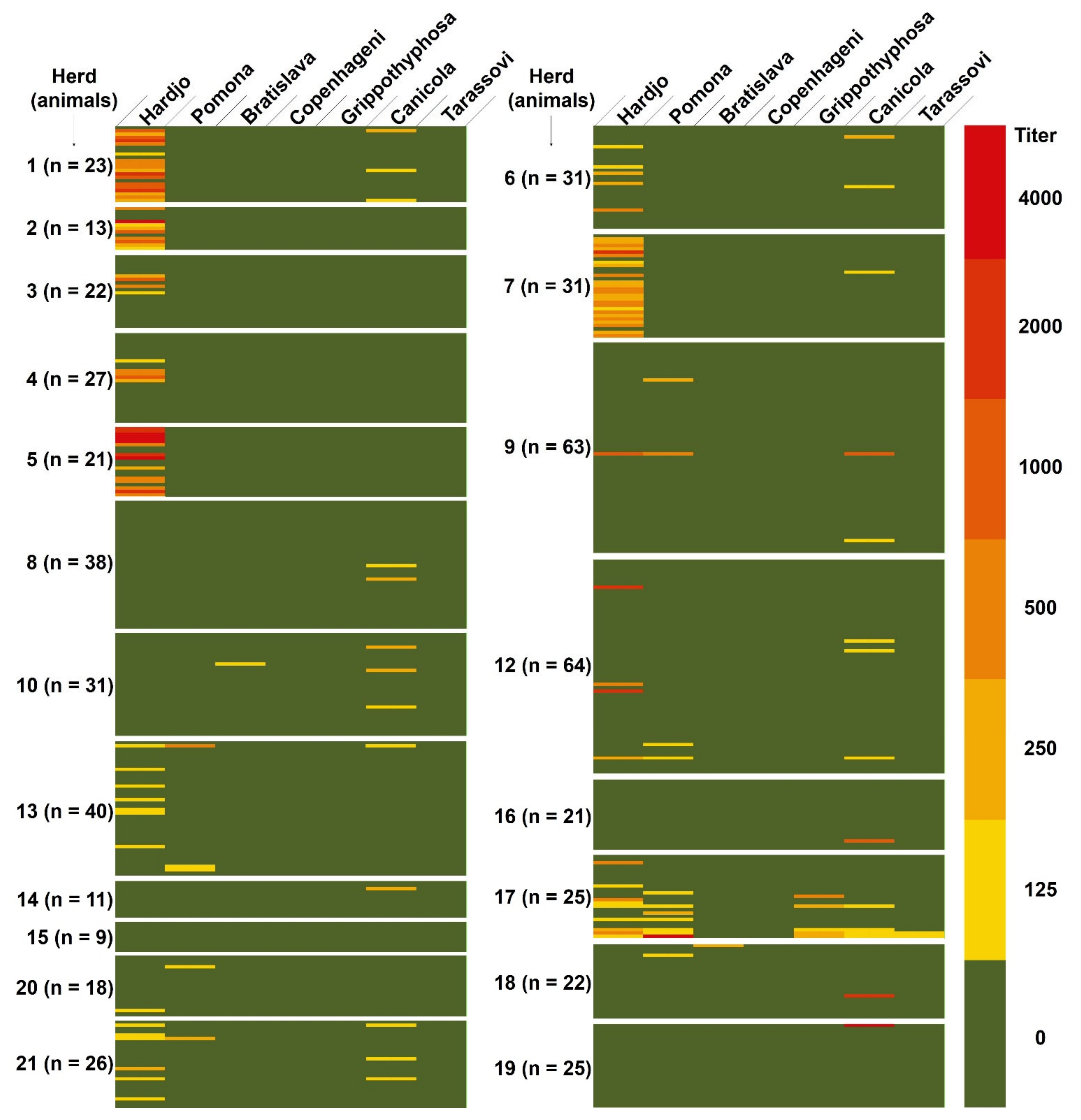
| Herd | Municipality | N. of Animals Tested/Total | Vaccination against Leptospira spp. | Presence of Abortions | Presence of other Animal Species a | Milking Mode | Herd Modernization |
|---|---|---|---|---|---|---|---|
| 1 | Soracá | 23/23 | NO | YES | C, D, H | manual | low |
| 2 | Ventaquemada | 13/13 | NO | YES | D | mechanical | medium |
| 3 | Ventaquemada | 22/22 | NO | NO | D | mechanical | medium |
| 4 | Ventaquemada | 27/27 | NO | NO | C, D | mechanical | medium |
| 5 | Ventaquemada | 21/21 | NO | NO | D | mechanical | medium |
| 6 | Oicatá | 31/31 | NO | NO | D | mechanical | medium |
| 7 | Oicatá | 31/31 | NO | NO | C, D, H | mechanical | medium |
| 8 | Oicatá | 38/38 | NO | YES | D | mechanical | medium |
| 9 | Oicatá | 63/63 | NO | YES | D, Po | mechanical | medium |
| 10 | Oicatá | 31/31 | NO | YES | D | mechanical | medium |
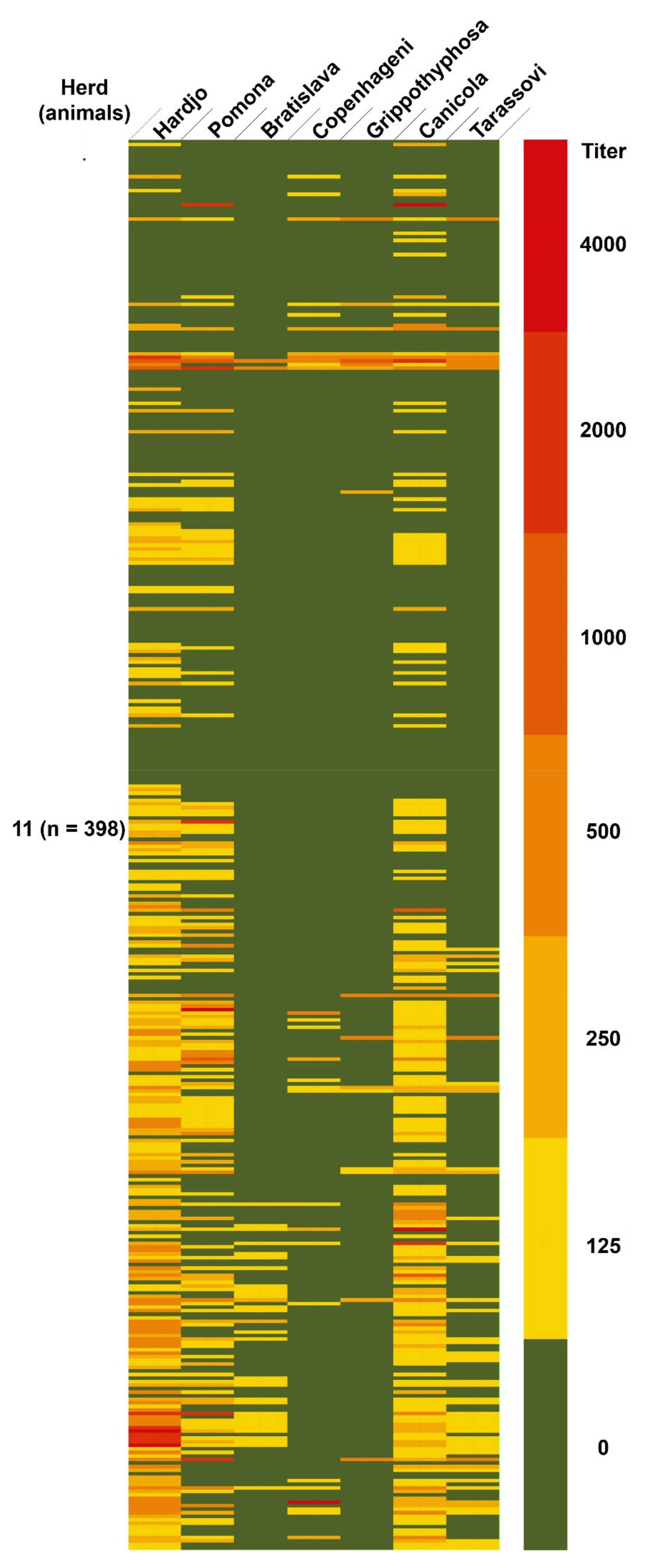
| 11 | Sopó | 398/700 | YES | YES (high) | C, D, H | mechanical | high |
| 12 | Chiquiza | 64/64 | NO | NO | D | manual | low |
| 13 | Oicatá | 40/40 | NO | YES | D, Pi | manual | low |
| 14 | Oicatá | 11/11 | NO | YES | D | mechanical | medium |
| 15 | Oicatá | 9/9 | NO | YES | D | mechanical | medium |
| 16 | Paipa | 21/21 | NO | NO | D | manual | low |
| 17 | Paipa | 25/25 | NO | YES | D | mechanical | medium |
| 18 | Sotaquira | 22/22 | NO | YES | D, Po | mechanical | medium |
| 19 | Tuta | 25/25 | NO | NO | C, D, H | mechanical | medium |
| 20 | Oicatá | 18/18 | NO | NO | D | manual | low |
| 21 | Oicatá | 26/26 | NO | NO | C, D, H | mechanical | medium |
| Serovar | Animals from Unvaccinated Herds Pos/All Animals (%) | Animals from Vaccinated Herd Pos/All Animals (%) | Animals from all Herds Pos/All Animals (%) |
|---|---|---|---|
| Bratislava | 2/561 (0.4) | 34/398 (8.5) | 36/959 (3.8) |
| Canicola | 28/561 (5.0) | 204/398 (51.3) | 232/959 (24.2) |
| Copenhageni | 0/561 (0) | 28/398 (7.0) | 28/959 (2.9) |
| Grippotyphosa | 5/561 (0.9) | 17/398 (4.3) | 22/959 (2.3) |
| Hardjo | 108/561 (19.3) | 228/398 (57.3) | 336/959 (35.0) |
| Pomona | 17/561 (3.0) | 144/398 (36.2) | 161/959 (16.8) |
| Tarassovi | 2/561 (0.4) | 57/398 (14.3) | 59/959 (6.2) |
| Number of Serovars to Which the Animal Is Positive | Animals from Unvaccinated Herds Animals/All Positive Animals (%) | Animals from Vaccinated Herd Animals/All Positive Animals (%) | Animals from all Herds Animals/All Positive Animals (%) |
|---|---|---|---|
| 1 | 121/135 (89.6) | 42/248 (16.9) | 163/383 (42.6) |
| 2 | 7/135 (5.2) | 53/248 (21.4) | 60/383 (15.7) |
| 3 | 3/135 (2.2) | 95/248 (38.3) | 98/383 (25.6) |
| 4 | 2/135 (1.5) | 23/248 (9.3) | 25/383 (6.5) |
| 5 | 2/135 (1.5) | 25/248 (10.1) | 27/383 (7.0) |
| 6 | 0/135 (0) | 8/248 (3.2) | 8/383 (2.1) |
| 7 | 0/135 (0) | 2/248 (0.8) | 2/383 (0.5) |
| Herd | AB a | C | IC | G | H | P | T | Total |
|---|---|---|---|---|---|---|---|---|
| 1 | 0/23 (0) b | 3/23 (13.0) | 0/23 (0) | 0/23 (0) | 18/23 (78.3) | 0/23 (0) | 0/23 (0) | 18/23 (78.3) |
| 2 | 0/13 (0) | 0/13 (0) | 0/13 (0) | 0/13 (0) | 9/13 (69.2) | 0/13 (0) | 0/13 (0) | 9/13 (69.2) |
| 3 | 0/22 (0) | 0/22 (0) | 0/22 (0) | 0/22 (0) | 4/22 (18.2) | 0/22 (0) | 0/22 (0) | 4/22 (18.2) |
| 4 | 0/27 (0) | 0/27 (0) | 0/27 (0) | 0/27 (0) | 5/27 (18.5) | 0/27 (0) | 0/27 (0) | 5/27 (18.5) |
| 5 | 0/21 (0) | 0/21 (0) | 0/21 (0) | 0/21 (0) | 14/21 (66.7) | 0/21 (0) | 0/21 (0) | 14/21 (66.7) |
| 6 | 0/31 (0) | 2/31 (6.5) | 0/31 (0) | 0/31 (0) | 5/31 (16.1) | 0/31 (0) | 0/31 (0) | 7/31 (22.6) |
| 7 | 0/31 (0) | 1/31 (3.2) | 0/31 (0) | 0/31 (0) | 25/31 (80.6) | 0/31 (0) | 0/31 (0) | 26/31 (83.9) |
| 8 | 0/38 (0) | 2/38 (5.3) | 0/38 (0) | 0/38 (0) | 0/38 (0) | 0/38 (0) | 0/38 (0) | 2/38 (5.3) |
| 9 | 0/63 (0) | 2/63 (3.2) | 0/63 (0) | 0/63 (0) | 1/63 (1.6) | 2/63 (3.2) | 0/63 (0) | 3/63 (4.8) |
| 10 | 1/31 (3.2) | 3/31 (9.7) | 0/31 (0) | 0/31 (0) | 0/31 (0) | 0/31 (0) | 0/31 (0) | 4/31 (12.9) |
| 11 | 34/398 (8.5) | 204/398 (51.3) | 28/398 (7.0) | 17/398 (4.3) | 228/398 (57.3) | 144//398 (36.2) | 57/398 (14.3) | 248/398 (62.3) |
| [6.7–10.3] c | [48–54.6] | [5.3–8.7] | [3–5.6] | [54–60.6] | [33–39.4] | [12–16.6] | [59.1–65.5] | |
| 12 | 0/64 (0) | 3/64 (4.7) | 0/64 (0) | 0/64 (0) | 4/64 (6.3) | 2/64 (3.1) | 0/64 (0) | /64 (10.9) |
| 13 | 0/40 (0) | 1/40 (2.5) | 0/40 (0) | 0/40 (0) | 7/40 (17.5) | 3/40 (7.5) | 0/40 (0) | 9/40 (22.5) |
| 14 | 0/11 (0) | 1/11 (9.1) | 0/11 (0) | 0/11 (0) | 0/11 (0) | 0/11 (0) | 0/11 (0) | 1/11 (9.1) |
| 15 | 0/9 (0) | 0/9 (0) | 0/9 (0) | 0/9 (0) | 0/9 (0) | 0/9 (0) | 0/9 (0) | 0/9 (0) |
| 16 | 0/21 (0) | 1/21 (4.8) | 0/21 (0) | 0/21 (0) | 0/21 (0) | 0/21 (0) | 0/21 (0) | 1/21 (4.8) |
| 17 | 0/25 (0) | 4/25 (16.0) | 0/25 (0) | 5/25 (20.0) | 9/25 (36.0) | 7/25 (28.0) | 2/25 (8.0) | 12/25 (48.0) |
| 18 | 1/22 (4.5) | 1/22 (4.5) | 0/22 (0) | 0/22 (0) | 0/22 (0) | 1/22 (4.5) | 0/22 (0) | 3/22 (13.6) |
| 19 | 0/25 (0) | 1/25 (4.0) | 0/25 (0) | 0/25 (0) | 0/25 (0) | 0/25 (0) | 0/25 (0) | 1/25 (4.0) |
| 20 | 0/18 (0) | 0/18 (0) | 0/18 (0) | 0/18 (0) | 1/18 (5.6) | 1/18 (5.6) | 0/18 (0) | 2/18 (11.1) |
| 21 | 0/26 (0) | 3/26 (11.5) | 0/26 (0) | 0/26 (0) | 6/26 (23.1) | 1/26 (3.8) | 0/26 (0) | 7/26 (26.9) |
Publisher’s Note: MDPI stays neutral with regard to jurisdictional claims in published maps and institutional affiliations. |
© 2021 by the authors. Licensee MDPI, Basel, Switzerland. This article is an open access article distributed under the terms and conditions of the Creative Commons Attribution (CC BY) license (http://creativecommons.org/licenses/by/4.0/).
Share and Cite
Taddei, S.; Moreno, G.; Cabassi, C.S.; Schiano, E.; Spadini, C.; Cavirani, S. Leptospira Seroprevalence in Colombian Dairy Herds. Animals 2021, 11, 785. https://doi.org/10.3390/ani11030785
Taddei S, Moreno G, Cabassi CS, Schiano E, Spadini C, Cavirani S. Leptospira Seroprevalence in Colombian Dairy Herds. Animals. 2021; 11(3):785. https://doi.org/10.3390/ani11030785
Chicago/Turabian StyleTaddei, Simone, Giovanni Moreno, Clotilde Silvia Cabassi, Emiliana Schiano, Costanza Spadini, and Sandro Cavirani. 2021. "Leptospira Seroprevalence in Colombian Dairy Herds" Animals 11, no. 3: 785. https://doi.org/10.3390/ani11030785