Communication

US3 Serine/Threonine Protein Kinase from MDV-1, MDV-2, and HVT Differentially Regulate Viral Gene Expression and Replication

1
Department of Veterinary Pathobiology, College of Veterinary Medicine & Biomedical Sciences, Texas A&M University, College Station, TX 77843, USA
2
Department of Veterinary Integrative Biosciences, College of Veterinary Medicine & Biomedical Sciences, Texas A&M University, College Station, TX 77843, USA
*
Authors to whom correspondence should be addressed.
Microorganisms 2021, 9(4), 785; https://doi.org/10.3390/microorganisms9040785
Submission received: 18 March 2021 / Revised: 6 April 2021 / Accepted: 8 April 2021 / Published: 9 April 2021
(This article belongs to the Special Issue Marek’s Disease Virus)

Abstract

:
Gallid alphaherpesvirus 2 (GaHV-2), commonly known as Marek’s disease virus type 1 (MDV-1), is an oncogenic avian alphaherpesvirus, and along with its close relatives—Gallid alphaherpesvirus 3 (GaHV-3) or MDV-2 and Meleagrid alphaherpesvirus 1 (MeHV-1) or turkey herpesvirus (HVT)—belongs to the Mardivirus genus. We and others previously showed that MDV-1 US3 protein kinase plays an important role in viral replication and pathogenesis, which could be partially compensated by MDV-2 and HVT US3. In this study, we further studied the differential roles of MDV-1, MDV-2 and HVT US3 in regulating viral gene expression and replication. Our results showed that MDV-2 and HVT US3 could differentially compensate MDV-1 US3 regulation of viral gene expression in vitro. MDV-2 and HVT US3 could also partially rescue the replication deficiency of MDV-1 US3 null virus in the spleen and thymus, as determined by immunohistochemistry analysis of MDV-1 pp38 protein. Importantly, using immunohistochemistry and dual immunofluorescence assays, we found that MDV-2 US3, but not HVT US3, fully compensated MDV-1 US3 regulation of MDV-1 replication in bursal B lymphocytes. In conclusion, our study provides the first comparative analysis of US3 from MDV-1, MDV-2 and HVT in regulating viral gene expression in cell culture and MDV-1 replication in lymphocytes.

1. Introduction

Marek’s disease (MD) is a lymphoproliferative disease of chickens, caused by an oncogenic avian alphaherepsvirus, Gallid alphaherpesvirus 2 [GaHV-2], commonly known as Marek’s disease virus type 1 (MDV-1). Susceptible chickens become infected with MDV-1 through the respiratory route by inhaling dander shed by infected chickens. MDV-1 infection exhibits distinct lytic and latent phases where various immune cells are involved [1]. During the early stage of MDV-1 infection, it is widely accepted that the infected lung phagocytic cells, such as macrophage, transfer the virus to B lymphocytes, which further infect activated T lymphocytes [2]. However, a recent study, using a knockout chicken line where B lymphocytes maturation and antibody production were abrogated, showed that B lymphocytes are dispensable for MDV-1 replication and oncogenesis [3]. Therefore, the mechanisms of MDV-1 infection and its interplay with the host immune system remain to be explored.
US3 is a conserved serine/threonine protein kinase encoded by alphaherpesviruses, which has been shown to be important for virus genome replication, nucleocapsid nuclear egress, apoptosis inhibition, transcription regulation, and immune evasion [4]. Alphaherpesvirus encoded US3 protein kinase contains a conserved kinase activity domain that includes an ATP-binding site and a catalytic active site, which is essential for its kinase activity [4]. The substrates of herpes simplex virus type 1 (HSV-1) US3 include viral proteins, such as glycoprotein B (gB), nuclear phosphoprotein UL31, and membrane phosphoprotein UL34, and cellular proteins, such as cAMP response element-binding protein (CREB), histone deacetylase 1 (HDAC1), HDAC2, p65, and programmed cell death protein 4 (PDCD4) [5,6,7,8,9,10]. US3 of MDV-1 was first described in 1993 and was shown not to be essential for virus replication in vitro [11]. Later, Schumacher et al. demonstrated that MDV-1 US3 shared the functions of other alphaherpesviruses-encoded US3 orthologs in regulating virion morphogenesis, apoptosis and host cytoskeleton structure [12,13]. They also showed that US3 interacts with phosphorylates MDV-1 pp38 [13], a viral protein important for MDV replication in B lymphocytes [14,15]. Recently, we identified more MDV-1 US3 substrates, including MDV-1 Meq oncoprotein, cellular CREB and HDAC1 and 2, and characterized the role of MDV-1 US3-mediated phosphorylation in regulating cellular and viral gene expression, protein interactions and virus replication [16,17]. In addition, we found that MDV-1 US3 disrupts the promyelocytic leukemia protein nuclear bodies (PML-NBs) in a US3 kinase activity and host proteasomal pathway-dependent manner similar to HSV-2 US3 [18].

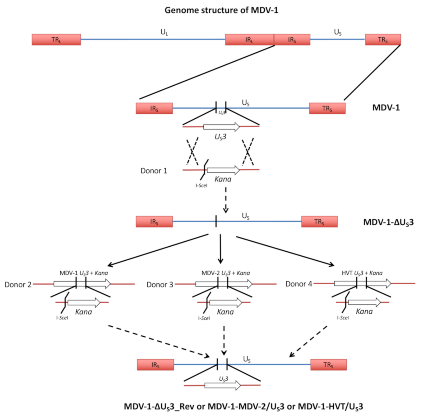
2. Materials and Methods

2.1. Viruses and Cell Culture

All viruses used in this study were generated previously using a very virulent plus strain, 686, of MDV-1 [16,17]. Chicken embryonic fibroblasts (CEF) were maintained at 37 °C in the presence of 5% CO2 in Leibowitz–McCoy (LM, 1:1) medium supplemented with 5% newborn calf serum.

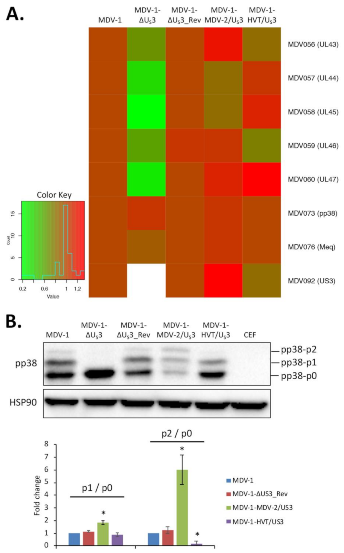
2.2. Quantitative Polymerase Chain Reaction (qPCR) and Western Blot

CEF seeded on 60 mm dishes were infected with 300 plaque-forming units (PFU) of different viruses or remained uninfected. Seven days later, cells were harvested for RNA, DNA and protein isolation as described previously [16].
All qPCR were performed in a CFX96 Real time PCR Detection System using iTaq Universal SYBR Green Supermix, and the results were analyzed using the 2−ΔΔCT method.
Proteins extracted from infected and uninfected CEF were subjected to sodium dodecyl sulphate-polyacrylamide gel electrophoresis (SDS-PAGE), followed by Western blot analysis with antibodies to MDV pp38 and chicken HSP90 protein. The density of each protein band was quantified using Image J software.
Western blot and qPCR were performed in two independent experiments and the statistical differences analyzed using t-test.

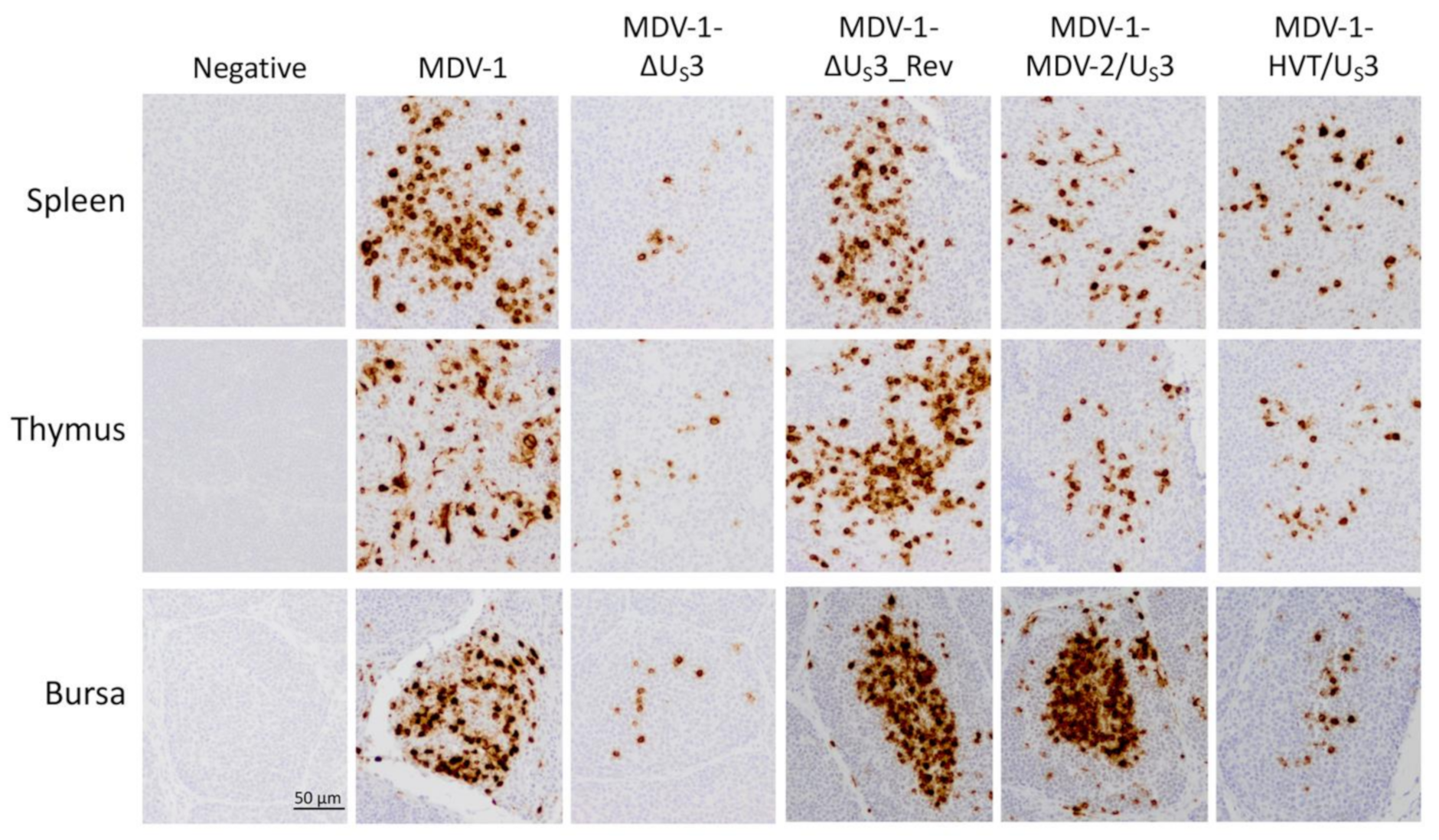
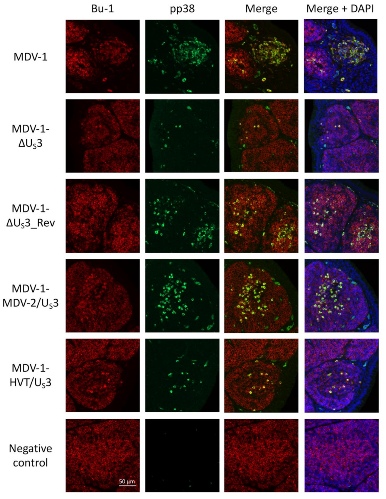
2.3. Immunohistochemistry (IHC) and Immunofluorescence Assay (IFA)

One-day-old specific-pathogen-free (SPF) chickens were inoculated subcutaneously with 2000 PFU of viruses or remained uninoculated as described previously [17].
IHC of lymphoid organ sections. At six days post-inoculation, lymphoid organs (spleen, thymus, and bursa) were collected from virus-inoculated or uninoculated (negative control) chickens, fixed in 10% neutral buffered formalin solution for 48 h and stored in 70% ethanol until used. Tissue sections were prepared and subjected to immunostaining with anti-MDV-1 pp38 monoclonal antibody and VECTASTAIN ABC kit according to the manufacturer’s instructions, using DAB (3,3’-diaminobenzidine) as the substrate. A representative image of three independent samples from each group is presented.
IFA of bursa sections. Bursa tissue sections, prepared from virus-inoculated or uninoculated (negative control) chickens, were subjected to IFA. After blocking with 5% bovine serum albumin (BSA), sections were incubated with mouse anti-pp38 antibody for 1 h at room temperature, followed by another hour incubation with Alexa Flour 488 conjugated goat anti-mouse antibody. After three washes with phosphate-buffered saline (PBS), sections were stained with Alexa Flour 647 conjugated mouse anti-chicken Bu-1 antibody (B lymphocyte marker) for 1 h at room temperature. 4′,6-diamidino-2-phenylindole (DAPI) was used to stain the cell nuclei. A representative image of three independent samples from each group is presented.

3. Results and Discussion

Using chimeric MDVs (Figure 1), we recently reported that US3 from MDV-2 and HVT, close relatives of MDV-1, could partially compensate the function of MDV-1 US3 in virus replication and pathogenesis [17]. In this study, we further determined the role of MDV-1, MDV-2 and HVT US3 in regulating viral gene expression in cell culture and virus replication in lymphoid organs. The construction of US3 deletion (MDV-1-ΔUS3), revertant (MDV-1-ΔUS3_Rev) and chimeric MDVs (MDV-1-MDV-2/US3, chimeric MDV-1 expressing MDV-2 US3 and MDV-1-HVT/US3, chimeric MDV-1 expressing HVT US3) is briefly outlined in Figure 1, and has been described in detail previously [16,17]. Here, we first analyzed the expression of a cluster of MDV-1 genes (MDV056 to MDV060) encoded by the unique long (UL) region of MDV-1, which we previously showed to be down-regulated in MDV-1 US3 null virus-infected chicken embryonic fibroblasts (CEF) [16]. Our results show that, compared to parental MDV-1, the expression of MDV056 to MDV060 was lower in MDV-1-ΔUS3-infected CEF, and was fully restored in the revertant virus and differentially compensated by expression of MDV-2 and HVT US3 in chimeric viruses; on the other hand, the expression of pp38 and meq was not affected (Figure 2A). These results suggest that US3 from MDV-1, MDV-2, and HVT differentially regulates viral gene expression. Western blot analysis of pp38 protein, a known substrate of MDV-1 US3, showed that MDV-2 and HVT US3 could compensate the ability of MDV-1 US3 to phosphorylate pp38, resulting in two additional higher molecular weight pp38 protein species (Figure 2B, pp38-p1 and pp38-p2). Interestingly, MDV-2 US3 exhibited a stronger potential to phosphorylate pp38, as the relative amount of phosphorylated pp38 form (Figure 2B, bottom graph), especially the p2 to p0 ratio (p2/p0), in MDV-1-MDV-2/US3-infected cells was significantly greater than that in parental MDV-1-, MDV-1-ΔUS3_Rev-, or MDV-1-HVT/US3-infected cells.
We have previously shown that deletion of MDV-1 US3 causes the replication deficiency of MDV-1-ΔUS3 in splenocytes (determined by virus genome copy number), which could be partially rescued by MDV-2 and HVT US3 [16,17]. In addition, we showed that MDV-2 and HVT US3 may play different roles in the replication of chimeric MDVs in B and T lymphocytes as the inoculation of MDV-1-MDV-2/US3 and MDV-1-HVT/US3 resulted in different levels of bursa and thymus atrophy (Table 1). In this study, we further analyzed the role of US3 from MDV-1, MDV-2, and HVT in regulating virus replication in lymphoid organs using histological methods. Tissue sections prepared from lymphoid organs (spleen, thymus, and bursa), collected from virus-inoculated or uninoculated chickens at day six post-inoculation, were subjected to immunohistochemistry (IHC) analysis of pp38 antigen expression. Our results show that there was a smaller number of cells expressing pp38 in the spleen of MDV-1-ΔUS3-inoculated chickens compared to parental and revertant viruses, which was partially restored in MDV-1-MDV-2/US3 and MDV-1-HVT/US3-inoculated chickens (Figure 3, spleen). Similar results were observed in the thymus, where MDV-2 and HVT US3 partially increased the number of pp38-expressing cells (Figure 3, thymus). Interestingly, the pp38 expression pattern in the bursa of chickens inoculated with MDV-1-MDV-2/US3 was similar to parental and revertant MDV-1 and there were clearly more cells expressing pp38 than in the bursa of MDV-1-ΔUS3- and MDV-1-HVT/US3-inoculated chickens (Figure 3, bursa). These bursa and thymus histological results are consistent with our previous report that infection with MDV-1-MDV-2/US3 resulted in bursa atrophy comparable to parental and revertant viruses, while inoculation of MDV-1-ΔUS3 and MDV-1-HVT/US3 did not induce bursa atrophy, and inoculation of MDV-1-ΔUS3, MDV-1-MDV-2/US3 and MDV-1-HVT/US3 all resulted in a similar level of mild thymus atrophy (Table 1). Taken together, these results suggest that US3 from MDV-1, MDV-2, and HVT may play different roles in the replication of MDV-1 in B and T lymphocytes.
To specifically study the role of US3 from MDV-1, MDV-2, and HVT in regulating virus replication in B lymphocytes, we performed sequential dual immunofluorescence assay (IFA) using Bu-1, a B lymphocyte marker, and pp38, an MDV marker, in bursa sections collected from virus-inoculated or uninoculated chickens at day six post-inoculation. Since only a mouse anti-chicken Bu-1 monoclonal antibody was available, bursa sections were incubated sequentially with mouse anti-pp38 monoclonal antibody, Alexa Flour 488 conjugated goat anti-mouse antibody, and Alexa Flour 647 conjugated mouse anti-chicken Bu-1 monoclonal antibody each for 1 h at room temperature, with three phosphate-buffered saline (PBS) washes after incubation with each antibody. The merged IFA image shows that the majority of pp38 expressing cells are B lymphocytes located in the bursa medulla of chickens inoculated with parental MDV-1 (Figure 4, MDV-1). Consistent with the IHC results in Figure 3, a similar pattern of B lymphocytes expressing pp38 was observed in the bursa of parental, revertant and MDV-1-MDV-2/US3-inoculated chickens, and there were more pp38-expressing B lymphocytes than in the bursa of MDV-1-ΔUS3- and MDV-1-HVT/US3-inoculated chickens (Figure 4). These results suggest that MDV-2 US3, but not HVT US3, could fully compensate the role of MDV-1 US3 in regulating the replication of MDV-1 in B lymphocytes. Overall, this study demonstrates that US3 from MDV-1, MDV-2 and HVT differentially regulate virus replication in lymphoid organs.
Using an MDV quantitative polymerase chain reaction (qPCR) array, we previously determined that 34 MDV-1 genes are differentially regulated by deleting MDV-1 US3 [16]. Out of these gene, we noticed that a cluster of UL genes, including MDV056 (UL43, probable membrane protein), MDV057 (UL44, virion membrane glycoprotein C), MDV058 (UL45, envelope/membrane protein), MDV059 (UL46, tegument phosphoprotein) and MDV060 (UL47, tegument phosphoprotein), are highly down-regulated in MDV-1 US3 null virus [16]. We and others have shown that MDV-1 US3 is important for MDV-1 genome replication and plaque-forming efficacy [12,13,16], and that MDV-2 and HVT US3 could partially compensate the role of MDV-1 US3 in these processes [17]. The downregulation of genes encoding membrane or tegument proteins (e.g., MDV056 to MDV060) may be one of the reasons contributing to the growth deficiency of MDV-1 US3 null virus. Our results here show that the expression of MDV056 to MDV060 was differentially compensated in CEF infected with MDV-1-MDV-2/US3 and MDV-1-HVT/US3 (Figure 2A), which could explain, at least partially, the role of MDV-2 and HVT US3 in rescuing the growth deficiency of MDV-1 US3 null virus in vitro. Furthermore, our IHC results show that the smaller number of cells expressing pp38 in the spleen and thymus of chickens inoculated with MDV-1-ΔUS3 was partially rescued by MDV-2 and HVT US3, and MDV-2 US3, but not HVT US3, could fully rescue the number of cells expressing pp38 in the bursa (Figure 3). These results provide the histological evidence for our previous report on genome copy number [17] and lymphoid organ atrophy induced by the inoculation of MDV-1-MDV-2/US3 and MDV-1-HVT/US3 (Table 1). Notably, for the first time, we demonstrate that MDV-1 US3 is important for the replication of MDV-1 in B lymphocytes using dual IFA; in particular, MDV-2 US3, but not HVT US3, could fully compensate MDV-1 US3 to rescue the replication deficiency of MDV-1 US3 null virus in B lymphocytes (Figure 4), even though both MDV-2 and HVT US3 exhibit a similar level (59% and 60%, respectively) of amino acid sequence identity to MDV-1 US3 (Figure S1). In addition, by comparing the amino acid sequence, we noticed that ~100 amino acids at the amino-terminus of US3 from MDV-1, MDV-2, and HVT are highly variable (Figure S1), which might contribute the differential functions observed for US3 from MDV-1, MDV-2, and HVT. Another interesting result we noticed is that both MDV-2 and HVT US3 could fully compensate the replication of MDV-1 US3 null virus in feather follicle epithelium (FFE) [17], suggesting that the replication of MDV-1 in lymphocytes and the epithelium is regulated differently. Even though the mechanisms involved remain to be explored, these results emphasize the diverse functions of US3 from MDV-1, MDV-2, and HVT. The mechanisms of MDV US3 regulation of virus replication and gene expression will be the focus of future studies.
Recent studies have challenged the widely accepted dogma of MDV-1 infection in immune cells [3]. The limited availability of commercial antibodies for chicken immune cell markers restricts the study of the interplay between MDV-1 infection and the host immune system. The development of more antibodies specific for chicken cell markers and the use of sequential dual IFA strategies described here and elsewhere opens up the possibility to precisely analyzing MDV-1 infection in specific cell types, which would help elucidate the life cycle of MDV-1 in chickens.

Supplementary Materials

The following is available online at https://www.mdpi.com/article/10.3390/microorganisms9040785/s1, Figure S1: Multiple sequence alignment of alphaherpesvirus US3 protein kinase.

Author Contributions

Y.L. designed and performed the experiments, analyzed the data and drafted the manuscript. M.A.-M. helped with tissue sample collection. X.F. and Q.L. helped with immunohistochemistry and immunofluorescence assays. B.L. and S.M.R. oversaw the project and edited the manuscript. All authors have read and agreed to the published version of the manuscript.

Funding

This work was supported by Texas A&M University and USDA funding.

Institutional Review Board Statement

All animal experiments were conducted following protocols approved by the Texas A&M University Institutional Animal Care and Use Committee (IACUC). (Protocol number 2019-0192 and date of approval 08/15/2019).

Informed Consent Statement

Not applicable.

Data Availability Statement

Data is contained within the article or supplementary material.

Acknowledgments

We thank Klaus Osterrieder for providing BAC reagents necessary for construction of recombinant viruses.

Conflicts of Interest

All authors declare that there are no conflict of interest.

References

  1. Boodhoo, N.; Gurung, A.; Sharif, S.; Behboudi, S. Marek’s disease in chickens: A review with focus on immunology. Vet. Res. 2016, 47, 119. [Google Scholar] [CrossRef] [PubMed] [Green Version]
  2. Osterrieder, N.; Kamil, J.P.; Schumacher, D.; Tischer, B.K.; Trapp, S. Marek’s disease virus: From miasma to model. Nat. Rev. Microbiol. 2006, 4, 283–294. [Google Scholar] [CrossRef] [PubMed]
  3. Bertzbach, L.D.; Laparidou, M.; Hartle, S.; Etches, R.J.; Kaspers, B.; Schusser, B.; Kaufer, B.B. Unraveling the role of B cells in the pathogenesis of an oncogenic avian herpesvirus. Proc. Natl. Acad. Sci. USA 2018, 115, 11603–11607. [Google Scholar] [CrossRef] [PubMed] [Green Version]
  4. Deruelle, M.J.; Favoreel, H.W. Keep it in the subfamily: The conserved alphaherpesvirus US3 protein kinase. J. Gen. Virol. 2011, 92, 18–30. [Google Scholar] [CrossRef] [PubMed]
  5. Kato, A.; Arii, J.; Shiratori, I.; Akashi, H.; Arase, H.; Kawaguchi, Y. Herpes simplex virus 1 protein kinase Us3 phosphorylates viral envelope glycoprotein B and regulates its expression on the cell surface. J. Virol. 2009, 83, 250–261. [Google Scholar] [CrossRef] [PubMed] [Green Version]
  6. Kato, A.; Yamamoto, M.; Ohno, T.; Tanaka, M.; Sata, T.; Nishiyama, Y.; Kawaguchi, Y. Herpes simplex virus 1-encoded protein kinase UL13 phosphorylates viral Us3 protein kinase and regulates nuclear localization of viral envelopment factors UL34 and UL31. J. Virol. 2006, 80, 1476–1486. [Google Scholar] [CrossRef] [PubMed] [Green Version]
  7. Wang, X.; Patenode, C.; Roizman, B. US3 protein kinase of HSV-1 cycles between the cytoplasm and nucleus and interacts with programmed cell death protein 4 (PDCD4) to block apoptosis. Proc. Natl. Acad. Sci. USA 2011, 108, 14632–14636. [Google Scholar] [CrossRef] [PubMed] [Green Version]
  8. Walters, M.S.; Kinchington, P.R.; Banfield, B.W.; Silverstein, S. Hyperphosphorylation of histone deacetylase 2 by alphaherpesvirus US3 kinases. J. Virol. 2010, 84, 9666–9676. [Google Scholar] [CrossRef] [Green Version]
  9. Wang, K.; Ni, L.; Wang, S.; Zheng, C. Herpes simplex virus 1 protein kinase US3 hyperphosphorylates p65/RelA and dampens NF-kappaB activation. J. Virol. 2014, 88, 7941–7951. [Google Scholar] [CrossRef] [PubMed] [Green Version]
  10. Wu, H.; Li, T.; Zeng, M.; Peng, T. Herpes simplex virus type 1 infection activates the Epstein-Barr virus replicative cycle via a CREB-dependent mechanism. Cell Microbiol. 2012, 14, 546–559. [Google Scholar] [CrossRef] [PubMed]
  11. Sakaguchi, M.; Urakawa, T.; Hirayama, Y.; Miki, N.; Yamamoto, M.; Zhu, G.S.; Hirai, K. Marek’s disease virus protein kinase gene identified within the short unique region of the viral genome is not essential for viral replication in cell culture and vaccine-induced immunity in chickens. Virology 1993, 195, 140–148. [Google Scholar] [CrossRef] [PubMed]
  12. Schumacher, D.; Tischer, B.K.; Trapp, S.; Osterrieder, N. The protein encoded by the US3 orthologue of Marek’s disease virus is required for efficient de-envelopment of perinuclear virions and involved in actin stress fiber breakdown. J. Virol. 2005, 79, 3987–3997. [Google Scholar] [CrossRef] [PubMed] [Green Version]
  13. Schumacher, D.; McKinney, C.; Kaufer, B.B.; Osterrieder, N. Enzymatically inactive U(S)3 protein kinase of Marek’s disease virus (MDV) is capable of depolymerizing F-actin but results in accumulation of virions in perinuclear invaginations and reduced virus growth. Virology 2018, 375, 37–47. [Google Scholar] [CrossRef] [PubMed] [Green Version]
  14. Reddy, S.M.; Lupiani, B.; Gimeno, I.M.; Silva, R.F.; Lee, L.F.; Witter, R.L. Rescue of a pathogenic Marek’s disease virus with overlapping cosmid DNAs: Use of a pp38 mutant to validate the technology for the study of gene function. Proc. Natl. Acad. Sci. USA 2002, 99, 7054–7059. [Google Scholar] [CrossRef] [PubMed] [Green Version]
  15. Gimeno, I.M.; Witter, R.L.; Hunt, H.D.; Reddy, S.M.; Lee, L.F.; Silva, R.F. The pp38 gene of Marek’s disease virus (MDV) is necessary for cytolytic infection of B cells and maintenance of the transformed state but not for cytolytic infection of the feather follicle epithelium and horizontal spread of MDV. J. Virol. 2005, 79, 4545–4549. [Google Scholar] [CrossRef] [PubMed] [Green Version]
  16. Liao, Y.; Lupiani, B.; Bajwa, K.; Khan, O.A.; Izumiya, Y.; Reddy, S.M. Role of Marek’s Disease Virus (MDV)-Encoded US3 Serine/Threonine Protein Kinase in Regulating MDV Meq and Cellular CREB Phosphorylation. J. Virol. 2020, 94, e00892-20. [Google Scholar] [CrossRef] [PubMed]
  17. Liao, Y.; Lupiani, B.; Ai-Mahmood, M.; Reddy, S.M. Marek’s disease virus US3 protein kinase phosphorylates chicken HDAC 1 and 2 and regulates viral replication and pathogenesis. PLoS Pathog. 2021, 17, e1009307. [Google Scholar] [CrossRef] [PubMed]
  18. Liao, Y.; Lupiani, B.; Reddy, S.M. Manipulation of Promyelocytic Leukemia Protein Nuclear Bodies by Marek’s Disease Virus Encoded US3 Protein Kinase. Microorganisms 2021, 9, 685. [Google Scholar] [CrossRef] [PubMed]
Figure 1. Construction of US3 deletion, revertant and chimeric MDVs. MDV-1 genome consists of unique long and short regions (UL, US) and inverted repeat long and short regions (TRL, IRL, TRS, IRS). US3 was deleted from a bacterial artificial chromosome (BAC) containing the genome MDV-1, strain 686, using a two-step Red-mediated recombination method (MDV-1-ΔUS3 BAC). The US3 deletion BAC was used as a backbone to construct MDV-1-ΔUS3_Rev (revertant), MDV-1-MDV-2/US3 (chimeric MDV-1 expressing MDV-2 US3), and MDV-1-HVT/US3 (chimeric MDV-1 expressing HVT US3) BACs. All BACs were transfected into chicken embryonic fibroblasts (CEF) to produce recombinant viruses.
Microorganisms 09 00785 g001
Figure 2. US3 of MDV-1, MDV-2, and HVT differentially regulate viral gene expression. Chicken embryonic fibroblasts (CEF) seeded on 60 mm dishes were infected with 300 plaque-forming units (PFU) of the different viruses or remained uninfected as negative control. Seven days later, cells were harvested for RNA, DNA and protein isolation. (A) Gene expression analysis. The extracted genomic DNA was used to determine MDV-1 genome copy number. Total RNA extracted from infected CEF were subjected to cDNA synthesis, followed by quantitative polymerase chain reaction (qPCR) to measure the expression of the indicated MDV-1 genes. The data were analyzed by the 2−ΔΔCT method using chicken GAPDH as the internal control and normalized to MDV-1 genome copy number. The relative average fold changes of each gene from two independent experiments are presented as heat map, where red indicates upregulation and green indicates downregulation. (B) Analysis of pp38 protein expression. Proteins extracted from infected and uninfected CEF were subjected to sodium dodecyl sulphate-polyacrylamide gel electrophoresis (SDS-PAGE), followed by Western blot analysis with antibodies to MDV pp38 and chicken HSP90 protein, as loading control (upper panel). Phosphorylated pp38 proteins (pp38-p1 and pp38-p2) were quantified with Image J, normalized to unmodified pp38 protein (pp38-p0) and presented as average fold changes, from two independent experiments, of p1 to p0 (bottom graph, left) and p2 to p0 (bottom graph, right) ratios relative to the value derived from parental MDV-1-infected cells. Error bars represent standard deviation (SD). The statistical difference between values derived from parental MDV-1 infected cells and those from other groups was analyzed using a t-test. *: p < 0.05.
Microorganisms 09 00785 g002
Figure 3. US3 of MDV-1, MDV-2, and HVT differentially regulate virus replication in lymphoid organs. At six days post-inoculation, lymphoid organs (spleen, thymus, and bursa) were collected from virus-inoculated or uninoculated (negative control) chickens, fixed in 10% neutral buffered formalin solution for 48 h, and stored in 70% ethanol until used. Tissue sections were prepared and subjected to immunostaining with anti-pp38 monoclonal antibody and VECTASTAIN ABC kit according to the manufacturer’s instructions, using DAB (3,3’-diaminobenzidine) as the substrate. A representative image of three independent samples from each group is presented; the scale bar is the same for every panel (scale bar = 50 μm).
Microorganisms 09 00785 g003
Figure 4. US3 of MDV-1, MDV-2, and HVT differentially regulate virus replication in bursal B lymphocytes. Bursa tissue sections, prepared from virus-inoculated or uninoculated (negative control) chickens, were subjected to immunofluorescence assay. After blocking, sections were incubated with mouse anti-pp38 antibody for 1 h at room temperature, followed by another hour incubation with Alexa Flour 488 conjugated goat anti-mouse antibody. After three washes with phosphate-buffered saline (PBS), sections were stained with Alexa Flour 647 conjugated mouse anti-chicken Bu-1 antibody (B lymphocyte marker) for 1 h at room temperature. 4′,6-diamidino-2-phenylindole (DAPI) was used to stain cell nuclei. A representative image of three independent samples from each group is presented; the scale bar is the same for every panel (scale bar = 50 μm).
Microorganisms 09 00785 g004
Table 1. Lymphoid organs to body weight ratio of inoculated and negative control chickens on day 14 post-inoculation.
VirusBursa/Body Weight × 100Thymus/Body Weight × 100
Negative0.422 ± 0.100 a0.741 ± 0.098 a
MDV-10.213 ± 0.079 b0.283 ± 0.113 c
MDV-1-ΔUS30.419 ± 0.057 a0.495 ± 0.106 b
MDV-1-ΔUS3_Rev0.141 ± 0.072 b0.204 ± 0.050 c
MDV-1-MDV-2/US30.188 ± 0.078 b0.426 ± 0.152 b
MDV-1-HVT/US30.391 ± 0.084 a0.429 ± 0.091 b
On day 14 post-inoculation, five chickens from each group were euthanized and the weights of their lymphoid organs (bursa and thymus) and bodies were measured. Data were presented as a ratio of the bursa or thymus to body weight multiplied by 100. The value in the table represents the average ratio derived from five chickens ± standard deviation. Indices with different letter superscripts are statistically different (p < 0.05). Table 1 is adapted from Figure 2B of “Marek’s disease virus US3 protein kinase phosphorylates chicken HDAC 1 and 2 and regulates viral replication and pathogenesis” by Liao Y, Lupiani B, AI-Mahmood M, Reddy SM (2021). PLoS Pathog 17(2): e1009307. Copyright 2021 by Liao et al.
Publisher’s Note: MDPI stays neutral with regard to jurisdictional claims in published maps and institutional affiliations.

