Article

Adhesion Properties, Biofilm Forming Potential, and Susceptibility to Disinfectants of Contaminant Wine Yeasts

Faculty of Bioscience and Technology for Food, Agriculture and Environment, University of Teramo, Via R. Balzarini 1, 64100 Teramo, Italy
*
Author to whom correspondence should be addressed.
Microorganisms 2021, 9(3), 654; https://doi.org/10.3390/microorganisms9030654
Submission received: 24 February 2021 / Revised: 15 March 2021 / Accepted: 18 March 2021 / Published: 22 March 2021
(This article belongs to the Special Issue Microbial Dynamics in Wine Production)

Abstract

:
In this study, yeasts isolated from filter membranes used for the quality control of bottled wines were identified and tested for their resistance to some cleaning agents and potassium metabisulphite, adhesion to polystyrene and stainless-steel surfaces, and formation of a thin round biofilm, referred to as a MAT. A total of 40 strains were identified by rRNA internal transcribed spacer (ITS) restriction analysis and sequence analysis of D1/D2 domain of 26S rRNA gene. Strains belong to Pichia manshurica (12), Pichia kudriavzevii (9), Pichia membranifaciens (1), Candida sojae (6), Candida parapsilosis (3), Candida sonorensis (1), Lodderomyces elongisporus (2), Sporopachydermia lactativora (3), and Clavispora lusitaniae (3) species. Regarding the adhesion properties, differences were observed among species. Yeasts preferred planktonic state when tested on polystyrene plates. On stainless-steel supports, adhered cells reached values of about 6 log CFU/mL. MAT structures were formed only by yeasts belonging to the Pichia genus. Yeast species showed different resistance to sanitizers, with peracetic acid being the most effective and active at low concentrations, with minimum inhibitory concentration (MIC) values ranging from 0.08% (v/v) to 1% (v/v). C. parapsilosis was the most sensible species. Data could be exploited to develop sustainable strategies to reduce wine contamination and establish tailored sanitizing procedures.

1. Introduction

The grapevine (Vitis vinifera) phyllosphere and wine musts harbor a complex microbiome, including yeasts, filamentous fungi, and bacteria that modulate grapevine health, growth, and wine fermentation [1,2]. During wine fermentation, these microbes interact among them during alcoholic (AF) and malolactic (MLF) fermentations. This dynamic interaction has great influence on the nutritional, hygienic, safety, and organoleptic characteristics of the final product. In particular, fermentative yeasts (e.g., Saccharomyces cerevisiae) and lactic acid bacteria (LAB, predominantly Oenococcus oeni) modulate the flavor and aroma of wine [3]. However, apart from S. cerevisiae, recognized as the main fermentative agent, other yeast species, known as non-Saccharomyces yeasts, such as Hanseniaspora/Kloeckera, Pichia, Candida, or Metschnikowia, release metabolites, which influence the chemical environment and/or fermentation process and are implicated in early stages of the AF [4]. These microorganisms could originate from the vineyard soil [5,6], air, precipitation (rainfall, hail, snow), be transported by animal vectors (bees, insects, and birds) [7,8,9], and be resident in nearby native forests [6,10]. Some of these wine-related non-Saccharomyces yeasts (e.g., Brettanomyces bruxellensis, Pichia manshurica) can spoil wine (e.g., volatile acidity and phenolic odors) [11,12]. Wine spoilage yeasts mainly belong to the following genera: Dekkera/Brettanomyces, Candida, Hanseniaspora, Pichia, Metschnikowia, Saccharomycodes, Schizosaccharomyces, and Zygosaccharomyces [13], which can cause film formation in bulk wines, cloudiness, sediment formation, and gas production in bottled wines, and off-flavor production during all processing and storing stages [14]. Saccharomyces cerevisiae can also be regarded as a spoilage microorganism, when associated with the refermentation of bottled wines [12,14]. According to previously published studies, the production environment harbors yeast strains, including spoilage ones, that are the most adapted to the processing environments and to the ecological niches. Moreover, the modes of contamination are often specific to each processing facility. Therefore, it is essential to study each manufacturing plant to determine the origin of the spoilage yeasts. The adhesion properties and biofilm-forming ability of spoilage yeasts have been recently reported for B. bruxellensis [15], and P. manschurica from organic wines [16]. The susceptibility of planktonic and biofilm yeast cells to different disinfectants and cleaning procedures were reported for B. bruxellensis [17], Zygosaccharomyces rouxii [18], and Pichia pastoris [19]. However, no data are available for other wine spoilage/contaminant yeasts. One of the main challenges of the wine industry is to identify new spoilage species and determine their physiological characteristics and ability to resist sanitization procedures. The financial burden generated by the use of ineffective and excess amounts of disinfectants may be reduced by choosing an appropriate disinfectant according to the dominant species of microorganism. Environmental pollution from the use of excessive quantities of chemicals may similarly be avoided. Therefore, in the present study, yeasts isolated from filter membranes used for the quality control of bottled wines were tested for their ability to adhere on abiotic surfaces and to form MAT structures (MATs). MATs are complex multicellular structures composed of yeast form cells [20]. The ability to form this kind of biofilm has been tested for some commercial wine yeast strains since it could suggest the ability of tested strains to adhere to surfaces of the wine environment, grape berries, and grapevine and to establish associations with other microbes, thereby affecting microbial dynamics during fermentation [21]. Finally, strains were tested for their resistance to potassium metabisulphite—since it shows a well-known anti-fungal activity and, together with membrane filtration, represents the main currently applied control action—and some cleaning agents. In particular, sodium hydroxide/sodium hypochlorite-based detergent and peracetic acid-based sanitizing agent were tested since they are generally used in cellars to reduce/prevent wine spoilage.

2. Materials and Methods

2.1. Origin of Samples

Yeasts were isolated from Biosart® 100 Monitors filter membranes (0.45 μm) (Sartorius, Monza, Italy). These filters are generally used for the quality control of bottled wines. The samples were obtained from the following cultivars: Pinot, Cerasuolo, Passerina, Montepulciano, Pecorino, Trebbiano, Merlot Cabernet, and Merlot. All colonies present on the filters were isolated and streaked on YPD medium (yeast extract 1% w/v, peptone 2% w/v, glucose 2% w/v, and agar 2% w/v; Oxoid, Milan, Italy). Plates were incubated at 30 °C for 48 h. Colonies were purified by repetitive streaking passages in YPD medium. All strains were stored at −80 °C in YPD broth supplemented with glycerol (20% v/v final concentration) (Sigma-Aldrich Srl., Milan, Italy) or on YPD agar at 4 °C for short-term storage. They belong to the Culture Collection of Microbial Biotechnology Laboratory (Faculty of BioScience and Technology for Food, Agriculture and Environment, University of Teramo).

2.2. Yeasts Identification

Genomic DNAs were extracted using the InstaGene Matrix Kit (Bio-Rad, Milan, Italy) according to the manufacturer’s instructions. The 5.8S internal transcribed spacer (ITS) rRNA region was amplified in a Bio-Rad thermocycler (MyCycler, Bio-Rad) using primers ITS1 (5′ TCCGTAGGTGAACCTGCGG 3′) and ITS4 (5′ TCCTCCGCTTATTGATATGC 3′), as previously described [22]. Amplicons were digested with the following restriction enzymes: CfoI, HaeIII, and HinfI [22]. Species identification was confirmed via sequencing D1/D2 domain of 26S rRNA gene, employing NL1 (5′- GCATATCAATAAGCGGAGGAAAAG -3′) and NL4 (5′- GGTCCGTGTTTCAAGACGG -3′) primer pairs [23]. Amplified fragments were purified using ExoSAP-IT Express PCR Cleanup (Thermo Fisher, Monza, Italy) according to the manufacturer’s instructions and sent to BMR Genomics (Padua University, Padua, Italy) for sequencing. The obtained sequences were compared to those available in the GenBank database (http://www.ncbi.nml.nih.gov/BLAST, accessed on 24 February 2021) and those of the Ribosomal Database Project (http://rdp.cme.msu.edu/index.jsp, accessed on 24 February 2021) to determine the closest known relative species on the basis of 26S rRNA gene homology [24].

2.3. Adhesion Properties of Yeasts

The adhesion properties of yeasts were tested on polystyrene and on stainless-steel surfaces. Yeasts were inoculated in six-well polystyrene plates in YPD medium and incubated at 30 °C for two weeks. Plate counts of planktonic and sessile cells were performed according to Perpetuini et al. [16]. Yeasts were also inoculated on stainless-steel supports according to Tomičić and Raspor [25]. Unattached cells were removed, while sessile cells were detached through several passages of rinsing and pipetting with saline solution (NaCl 0.85% w/v) and recovered. Viable count on YPD medium was carried out. All analyses were performed in triplicate only on viable and cultivable cells.

2.4. MAT Formation

The multicellular growth pattern (MAT formation) of yeast strains was tested as described by Reynold and Fink [20]. Yeast cells were inoculated in the center of YPD soft agar plates (agar 0.3% w/v) using a toothpick and incubated at 25 °C for 15 days. Plates containing 2% (w/v) agar were used as negative controls. The diameter was evaluated by photographing the plates and analyzing the photos with ImageJ software (http://imagej.nih.gov, accessed on 24 February 2021) according to Schneider et al. [26]. All tests were performed in triplicate.

2.5. Determination of the Minimum Inhibitory Concentrations (MICs)

Commercial cleaning agents commonly used for the hygienic practices in wineries as well as potassium metabisulphite were tested against the isolated yeasts. The agents were diluted to various concentrations within the in-use range: sodium hydroxide/sodium hypochlorite-based detergent (Enoclin Cloractiv L—SH) 0.05–5% (v/v) (L’Enotecnica, Nizza Monferrato, Italy); peracetic acid-based sanitizing agent (Enoclin Peracetic—PA) (L’Enotecnica) 0.02–10% (v/v); potassium metabisulphite 5–1600 ppm (L’Enotecnica). The minimum inhibitory concentrations (MICs) were evaluated according to Tristezza et al. [27]. Assays were performed in triplicate using the microtiter dilutions method. The MIC was defined as the lowest concentration of the disinfectant agent that prevents visible growth, by production of turbidity or pellet, after an incubation period of 48 h. Pichia manshurica PED 141-1 strain was used as control [16].

2.6. Statistical Analysis

Data were analyzed by means of Prism 7.0 program (GraphPad Software Inc., La Jolla, CA, USA) using t-test. A level of p < 0.05 was considered statistically significant.

3. Results and Discussion

3.1. Yeast Identification

A total of 40 yeasts were isolated from filter membranes. Restriction profile comparisons between isolates and published strains allowed assigning the isolates to Pichia manshurica (12), Pichia kudriavzevii (9), Pichia membranifaciens (1), Candida sojae (6), Candida parapsilosis (3), Candida sonorensis (1), Sporopachydermia lactativora (3), Lodderomyces elongisporus (2), and Clavispora lusitaniae (3) (Table S1). The D1/D2 domain of the 26S rRNA gene was sequenced and subsequently compared with sequences available in the GenBank database and the Ribosomal Database Project to confirm identification results. All the sequences obtained displayed similarity values ranging from 99% to 100% to reference sequences, confirming the identity of isolates (Table S1). P. manshurica is common in several fermented foods, and it is usually found in the early stages of spontaneous fermentation of alcoholic beverages, where it was firstly isolated [28]. It has also been found in spoiled and organic wines, suggesting that it is well adapted to wine environment and could be considered a contaminant in wineries [29]. This species is associated with off-flavors, off-odors [11], and biogenic amines production [30] and showed considerable hydrophobicity and biofilm formation on polystyrene [16]. P. kudriavzevii can be found in various fermented beverages (e.g., wine, beer, cereal-based beverage), soil, and fruits (e.g., mango pulp) [31,32]. It is a non-conventional yeast that is able to resist organic acids [33]. It can be considered a spoilage yeast in some food products, such as kimchi, where it is involved in the production of surface biofilm, off-odors, and texture softening [34,35]. Moreover, some strains isolated from nuruk—a Korean microbial starter for fermented products—showed high thermotolerance and ethanol production [36]. Among Pichia isolates, a strain of P. membranifaciens was also found. It can spoil wine and other fermented foods with the production of biofilm on the surface of wines and undesirable volatile compounds such as volatiles phenols [29,37]. It has also been previously studied for biofilm formation on stainless steel in filler implants in breweries [38]. Yeasts belonging to the Candida genus were identified as C. sojae, C. parapsilosis, and C. sonorensis. C. sojae and C. parapsilosis are placed in the Lodderomyces–Spathaspora clade [34] and are phylogenetically similar to Lodderomyces elongisporus, which was also found in the samples analyzed in this study. C. sojae was first isolated from effluents of a soybean extraction process [39] and it has a strong connection with C. tropicalis and C. albicans species, which are considered to be pathogenic, but differs from these species because of its inability to ferment maltose and because it has a low maximum growth temperature (below 40 °C). C. parapsilosis has been previously found in wine environments, probably originating from damaged grapes or soil [40,41]. A strain of C. sonorensis was found, probably brought by soil contamination because it is an asexual yeast species found only in the decaying tissue of cacti [42]. It is able to ferment glucose to ethanol, it is relatively tolerant to low pH environments, and has simple nutritional requirements [42,43]. Three strains of Spor. lactativora were isolated. This yeast was previously isolated from reverse osmosis filtration membranes, even after being subjected to a cleaning process, where it was able to form biofilm communities along with other filamentous fungi (Magnusiomyces spicifer and Saprochaete clavata) and Gram-negative bacteria [37,44]. Finally, three strains of Cl. lusitaniae were identified. This yeast has been previously isolated in grape and apple must, citrus fruits, and orange juice [45,46,47,48]. Species generally associated with wine spoilage such as Brettanomyces/Dekkera, Zygosaccharomyces spp., and Saccharomyces spp. were not identified [14].

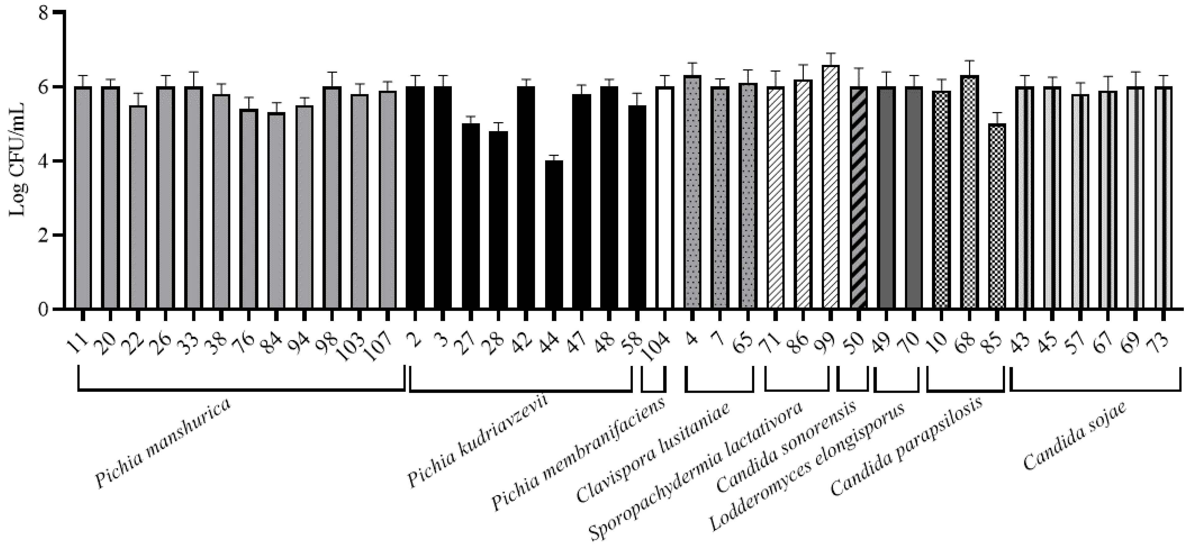
3.2. Yeast Adhesion to Abiotic Surfaces
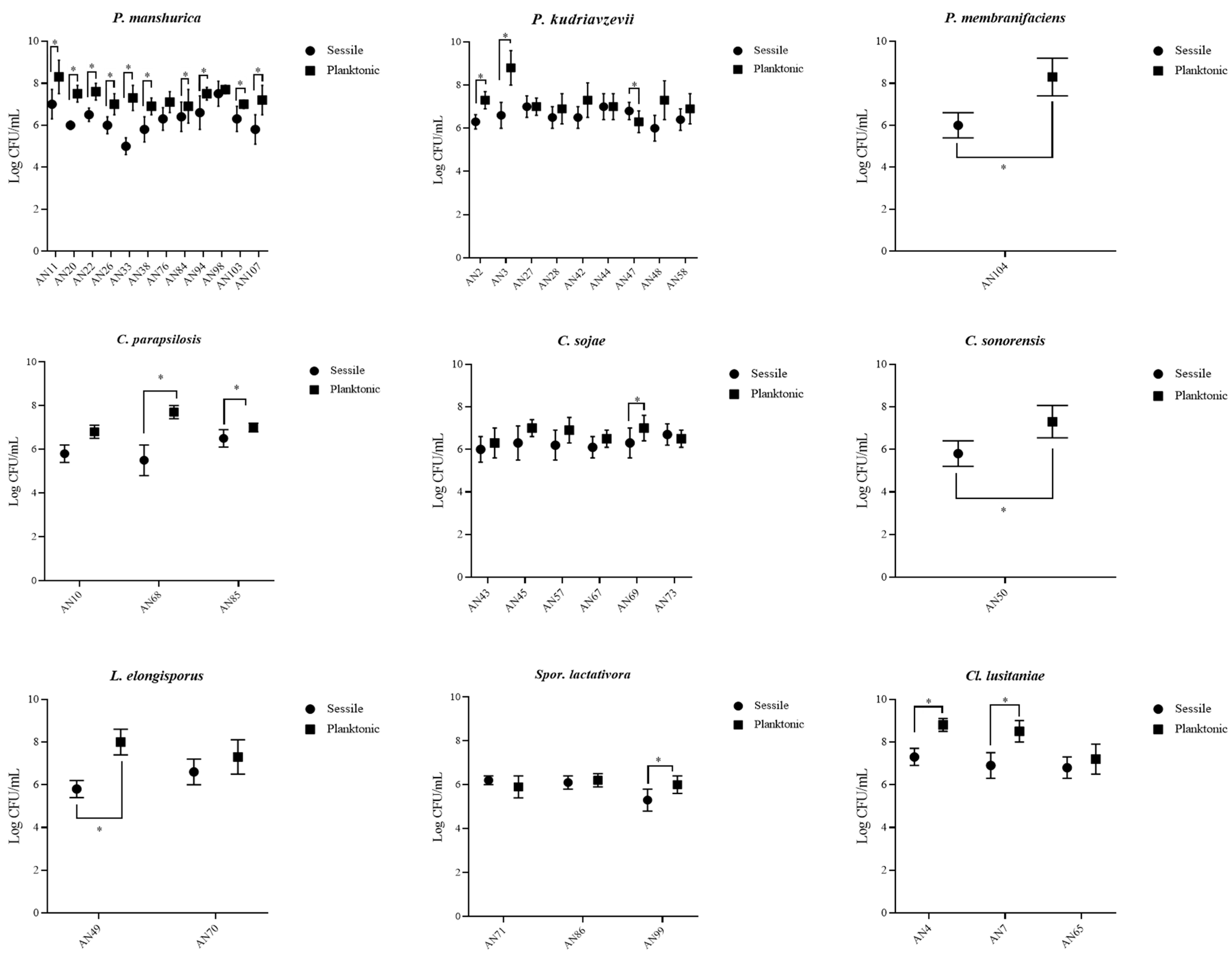
Strains were tested for their ability to adhere on polystyrene and stainless-steel surfaces. The attention was focused on these materials since they are widespread in cellars (e.g., stainless-steel tanks, packaging for bottles, working surfaces, etc.). Differences were observed between species. On polystyrene plates, the planktonic state was favored (Figure 1). Intra- and inter-species differences were observed for planktonic and sessile cell counts. In general, planktonic cell counts were higher than sessile ones, with few exceptions (AN43, AN47, and AN71). For instance, P. manshurica sessile cells ranged from 5 log CFU/mL (AN33) to 7.5 log CFU/mL (AN98), while planktonic ones ranged from 6.9 log CFU/mL (AN84) to 8.3 log CFU/mL (AN11) (Figure 2). On stainless-steel surfaces, strains adhered with values of about 6 log CFU/mL. The lowest values were observed for three strains of P. kudriavzevii (AN27, AN28, and AN44) (Figure 2). Our data concerning P. membranifaciens are in agreement with Storgårds et al. [38], who showed that P. membranifaciens—as well as other yeast species such as Candida krusei, Rhodotorula mucilaginosa, Wickerhamomyces anomalus (ex P. anomala), and S. cerevisiae—formed biofilms on abiotic surfaces of brewery bottling plants. Biofilms of P. membranifaciens have been also reported in a rotating biological contactor (RBC) for the treatment of wine-cellar effluent [49]. Tomičić et al. [50] showed that yeasts can adhere to wooden surfaces. This ability is influenced by wood type, disinfectant, relative humidity, and temperature [50], but not by wood surface roughness [51]. The ability to form biofilm on wooden matrixes could represent a problem for wine quality since the spoilage metabolism of some yeast species such as P. manshurica could be maintained during wine aging [11]. The ability of P. manshurica to form biofilm on abiotic surfaces has been reported by our group and is mainly related to the hydrophobic nature of its surface [16]. However, it is important to underline that microbial attachment is often a two-step process. The first step is related to physico-chemical interactions, which are important to help the cells to approach the contact surface and attach loosely to it. The adhesion to abiotic surfaces is the first step of biofilm formation, and it is necessary to evaluate the biofilm-forming ability of these strains. Surface proteins/adhesins are involved in the second step since they support the cells to stick firmly to the surface [52]. Adhesion abilities have been described for the first time in P. kudriavzevii strains. As for the other members of the Pichia genus, this capacity is probably explained by their aerobic nature and fast growth [53]. The adhesion properties of Spor. lactativora have also been described by Vitzilaiou et al. [44]. These authors found that this species colonized the reverse osmosis membrane filtration elements from a whey water filtration unit. The majority of studies concerning the genus Candida have been performed on C. albicans. This species is able to colonize both abiotic and biotic surfaces through the expression of specific adhesins called Als [54]. Recently, Valotteau et al. [55] revealed the ability of Candida glabrata to adhere on abiotic surfaces thanks to Epa proteins (Epa1, Epa6, and Epa7), which contribute to both hydrophilic and hydrophobic interaction.

3.3. MATs Formation

MAT structures are considered an elaborate multicellular biofilm related to the sliding motility. According to Recht et al. [56], sliding motility is defined as a form of surface motility “produced by the expansive forces of the growing bacterial population in combination with cell surface properties that favour reduced friction between the cells and the substrate”. Only strains belonging to the Pichia genus were able to form MAT structures on semi-solid agar. MATs presented a central hub made of networks of cables and radial spokes with more or less jagged edges (Figure S1). Radial spokes were particularly evident in the MATs formed by P. membranifaciens AN104 strain. This structure was also less smooth and had more jagged edges than the others. All P. kudriavzevii strains as well as the P. membranifaciens strain showed structures covering all agar surface with a diameter of about 8.5 ± 0.44 cm, with the only exception being P. kudriavzevii AN3 strain, which had a diameter of 7.63 ± 0.32 cm. Intraspecies differences were especially detected for P. manshurica strains. The diameters ranged from 3.63 ± 0.45 cm (AN76) to 8.5 ± 0.61 cm (AN98). In particular, AN11, AN26, and AN33 strains showed values higher than 5 cm, while AN22 was 6.7 ± 0.38 cm, AN103 and AN94 were about 7 cm, AN76 and AN84 were more than 3 cm, while the others were about 8 cm.
MATs formation was previously described for P. manshurica by Perpetuini et al. [16], while it is reported for the first time in this study in P. kudriavzevii and P. membranifaciens. This phenomenon has been also described in other yeast species such as Debaryomyces hansenii and Kluyveromyces marxianus [57,58]. However, it is well characterized only in S. cerevisiae [20,59,60]. In this species, the ability to form biofilm and MAT structures is associated with the expression of specific adhesins such as Flo11p. Thirteen different structures of Flo11p have been described [61], and in some cases the Flo11 domain is present in double or triple copies, such as in some non-Saccharomyces species (e.g., Kluyveromyces lactis, Cl. lusitaniae, and C. parapsilosis) [62,63]. Moreover, Perpetuini et al. [58] showed that orthologs of FLO11 and STE12 genes were overexpressed in MAT structures formed by dairy K. marxianus strains. Some studies have indicated that in Candida spp. many ALS genes are regulated by orthologues of the pathways known to regulate adhesion in S. cerevisiae [64,65,66,67]. Moreover, in C. albicans, six transcriptional regulators (Efg1, Tec1, Bcr1, Ndt80, Brg1, and Rob1) involved in biofilm formation have been described [68], while in C. glabrata, lectin-like adhesins include EPA gene products [69]. However, the presence of these adhesins is not sufficient to explain MAT structures formation. This phenomenon appears to be quite complex and related to strain ploidy: increased ploidy reduced MATs formation, with a tetraploid strain showing almost no MAT formation [20]. Moreover, additional pathways acting in a Flo11-independent manner have been described. Sarode et al. [70] described this kind of pathway, referring to it as the biofilm pathway. It involves the class E vacuolar protein sorting (vps) components of the multivesicular body pathway. In 2014, the same authors identified a cell wall signaling protein (Wsc1p) impacting MATs formation [60]. Obtained data suggest that the Pichia genus could probably present in its genome specific adhesins and tailored metabolic pathways involved in MATs formation.

3.4. Determination of the Minimum Inhibitory Concentrations (MICs)

The main source of microbial contamination during food production may be the processing plant itself, caused by unsuccessful hygiene measures, the ability of yeasts to withstand a stressful environment, and/or the inefficacy of disinfectants [71]. Therefore, it is important to identify new contaminant/spoilage species and determine their physiological characteristics and ability to resist sanitization procedures. For this reason, strains were tested for their resistance to PA, SH, and potassium metabisulphite. The MICs are reported in Table 1. Yeast species showed MIC differences, suggesting that the resistance is species-dependent, and strains within the species had different behaviors. Similar results have been reported in other studies [72,73,74]. PA was the most effective sanitizer, being active at low concentrations. In fact, MIC values ranged from 0.08% to 1%, with C. parapsilosis having the most sensible strains. SH detergent was more effective at lower concentrations than the recommended in-use concentration. In fact, strains showed MICs ranging from 0.4% to 2%. Regarding potassium metabisulphite, MICs varied from 80 ppm to 160 ppm. Spor. lactativora was the most sensitive species to the tested compounds, exhibiting the lowest MIC, followed by L. elongisporus, Cl. Lusitaniae, and C. parapsilosis, which were sensitive to PA, but slightly resistant to SH and potassium metabisulphite. Pichia spp. showed the highest resistance to potassium metabisulphite and presented similar MIC values for PA and SH compared to other species. C. sojae and C. sonorensis exhibited good resistance to SH but limited resistance to PA and potassium metabisulphite. PA and SH detergents were effective against all species tested, which is in agreement with previous studies [16,27]. P. manshurica strains had a resistance to PA similar to values reported by Perpetuini et al. [16] with only two strains (33 and 76) showing a higher resistance. Obtained MICs for SH were lower than those reported by the same authors. In fact, a strain showed values of MIC of 5%, which is equal to the maximum recommended in-use concentration, while the other strains presented values ranging from 1% to 3%. Winniczuk and Parrish [75] reported the efficacy of hypochlorite, peracetic acid, phosphoric acid, and anionic compounds against yeast strains isolated from orange juice. The efficiency of potassium metabisulphite is species- and strain-dependent and can produce growth retardation or cell killing [76]. As expected, the values of MIC were low, which is in agreement with other authors [16,77].

4. Conclusions

The selective pressure exerted by the environment during wine production reduces the natural yeast diversity to a limited number of well-adapted species. However, contaminant yeasts can develop, and an understanding of the origin and routes of contamination of these species can contribute to a more effective control of production processes. This study revealed novel information on the diversity and preservative resistance of yeasts encountered in the wine environment. A strategy to control yeast contamination should be the examination of the production lines to identify possible points in the processes where wines could be exposed to yeast contamination. Further studies will be focused on P. manshurica in order to better characterize its ability to form biofilm using different media (e.g., wine) and to identify the genes involved in its adhesion. This information will be useful to establish a link between the genomic background and phenotypic traits of adherent spoilage yeasts. Additional studies are also necessary to evaluate the susceptibility of yeasts to common and novel disinfectants in the planktonic and biofilm state. A close examination of the biofilm-forming capacity of strains will be performed. In fact, the application of disinfectants in the winery is important to remove the adhered cells in order to prevent biofilms formation since they are difficult to eradicate. These results could be useful to develop new strategies to decrease wine contamination and to stress the importance of accurate sanitizing procedures designed ad hoc to eradicate resistant populations. This kind of study is useful to develop new strategies to eliminate/reduce wine contamination and/or control yeast growth and thus reduce food waste. Moreover, better knowledge about contaminant yeasts provides a fundamental tool to implement and improve the HACCP (Hazard Analysis and Critical Control Points) systems.

Supplementary Materials

The following are available online at https://www.mdpi.com/2076-2607/9/3/654/s1: Table S1: Yeasts origin and identification. Figure S1: MATs formation on soft agar by tested strains at 25 °C after 7 days.

Author Contributions

Conceptualization: G.P. and R.T.; methodology: A.P.R. and N.B.; software: N.B.; validation: R.T.; investigation: A.P.R. and N.B. resources: R.T. and G.A.; data curation: G.P., N.B., and A.P.R.; writing—original draft preparation: G.P. and R.T.; writing—review and editing: G.P. and R.T.; visualization: R.T; supervision: R.T.; project administration: R.T.; funding acquisition: R.T. All authors have read and agreed to the published version of the manuscript.

Funding

This research was funded by University of Teramo FARDIB-2019 and by Progetto DM61315 “Sviluppo della qualità delle produzioni vitivinicole abruzzesi”.

Institutional Review Board Statement

Not applicable.

Informed Consent Statement

Not applicable.

Data Availability Statement

Data is contained within the article or Supplementary Material.

Acknowledgments

The authors are grateful to Fabrizia Tittarelli for her kind cooperation and Citra Vini S.C. for providing samples.

Conflicts of Interest

The authors declare no conflict of interest.

References

  1. Barata, A.; Malfeito-Ferreira, M.; Loureiro, V. The Microbial Ecology of Wine Grape Berries. Int. J. Food Microbiol. 2012, 153, 243–259. [Google Scholar] [CrossRef] [PubMed]
  2. Gilbert, J.A.; van der Lelie, D.; Zarraonaindia, I. Microbial Terroir for Wine Grapes. Proc. Natl. Acad. Sci. USA 2014, 111, 5–6. [Google Scholar] [CrossRef] [PubMed] [Green Version]
  3. Swiegers, J.H.; Bartowsky, E.J.; Henschke, P.A.; Pretorius, I. Yeast and Bacterial Modulation of Wine Aroma and Flavour. Aust. J. Grape Wine Res. 2005, 11, 139–173. [Google Scholar] [CrossRef]
  4. Fleet, G.H.; Lafon-Lafourcade, S.; Ribéreau-Gayon, P. Evolution of Yeasts and Lactic Acid Bacteria during Fermentation and Storage of Bordeaux Wines. Appl. Environ. Microbiol. 1984, 48, 1034–1038. [Google Scholar] [CrossRef] [PubMed] [Green Version]
  5. Zarraonaindia, I.; Owens, S.M.; Weisenhorn, P.; West, K.; Hampton-Marcell, J.; Lax, S.; Bokulich, N.A.; Mills, D.A.; Martin, G.; Taghavi, S. The Soil Microbiome Influences Grapevine-Associated Microbiota. MBio 2015, 6. [Google Scholar] [CrossRef] [Green Version]
  6. Morrison-Whittle, P.; Goddard, M.R. From Vineyard to Winery: A Source Map of Microbial Diversity Driving Wine Fermentation. Environ. Microbiol. 2018, 20, 75–84. [Google Scholar] [CrossRef] [Green Version]
  7. Goddard, M.R.; Anfang, N.; Tang, R.; Gardner, R.C.; Jun, C. A Distinct Population of Saccharomyces cerevisiae in New Zealand: Evidence for Local Dispersal by Insects and Human-Aided Global Dispersal in Oak Barrels. Environ. Microbiol. 2010, 12, 63–73. [Google Scholar] [CrossRef]
  8. Stefanini, I.; Dapporto, L.; Legras, J.-L.; Calabretta, A.; Di Paola, M.; De Filippo, C.; Viola, R.; Capretti, P.; Polsinelli, M.; Turillazzi, S. Role of Social Wasps in Saccharomyces cerevisiae Ecology and Evolution. Proc. Natl. Acad. Sci. USA 2012, 109, 13398–13403. [Google Scholar] [CrossRef] [Green Version]
  9. Lam, S.S.; Howell, K.S. Drosophila-Associated Yeast Species in Vineyard Ecosystems. FEMS Microbiol. Lett. 2015, 362, fnv170. [Google Scholar] [CrossRef] [Green Version]
  10. Knight, S.; Goddard, M.R. Quantifying Separation and Similarity in a Saccharomyces cerevisiae Metapopulation. ISME J. 2015, 9, 361–370. [Google Scholar] [CrossRef] [PubMed] [Green Version]
  11. Perpetuini, G.; Tittarelli, F.; Battistelli, N.; Suzzi, G.; Tofalo, R. Contribution of Pichia manshurica Strains to Aroma Profile of Organic Wines. Eur. Food Res. Technol. 2020, 246, 1405–1417. [Google Scholar] [CrossRef]
  12. Hernández, A.; Pérez-Nevado, F.; Ruiz-Moyano, S.; Serradilla, M.J.; Villalobos, M.C.; Martín, A.; Córdoba, M.G. Spoilage Yeasts: What Are the Sources of Contamination of Foods and Beverages? Int. J. Food Microbiol. 2018, 286, 98–110. [Google Scholar] [CrossRef] [PubMed]
  13. Enrique, M.; Marcos, J.F.; Yuste, M.; Martínez, M.; Vallés, S.; Manzanares, P. Antimicrobial Action of Synthetic Peptides towards Wine Spoilage Yeasts. Int. J. Food Microbiol. 2007, 118, 318–325. [Google Scholar] [CrossRef]
  14. Loureiro, V.; Malfeito-Ferreira, M. Spoilage Yeasts in the Wine Industry. Int. J. Food Microbiol. 2003, 86, 23–50. [Google Scholar] [CrossRef]
  15. Dimopoulou, M.; Renault, M.; Dols-Lafargue, M.; Albertin, W.; Herry, J.-M.; Bellon-Fontaine, M.-N.; Masneuf-Pomarede, I. Microbiological, Biochemical, Physicochemical Surface Properties and Biofilm Forming Ability of Brettanomyces bruxellensis. Ann. Microbiol. 2019, 69, 1217–1225. [Google Scholar] [CrossRef]
  16. Perpetuini, G.; Tittarelli, F.; Schirone, M.; Di Gianvito, P.; Corsetti, A.; Arfelli, G.; Suzzi, G.; Tofalo, R. Adhesion Properties and Surface Hydrophobicity of Pichia manshurica Strains Isolated from Organic Wines. LWT 2018, 87, 385–392. [Google Scholar] [CrossRef]
  17. Pinto, L.; Baruzzi, F.; Cocolin, L.; Malfeito-Ferreira, M. Emerging Technologies to Control Brettanomyces spp. in Wine: Recent Advances and Future Trends. Trends Food Sci. Technol. 2020, 99, 88–100. [Google Scholar] [CrossRef]
  18. Frisón, L.N.; Chiericatti, C.A.; Aríngoli, E.E.; Basílico, J.C.; Basílico, M.Z. Effect of Different Sanitizers against Zygosaccharomyces rouxii. J. Food Sci. Technol. 2015, 52, 4619–4624. [Google Scholar] [CrossRef] [Green Version]
  19. Zhao, L.; Zhang, Y.; Yang, H. Efficacy of Low Concentration Neutralised Electrolysed Water and Ultrasound Combination for Inactivating Escherichia coli ATCC 25922, Pichia pastoris GS115 and Aureobasidium pullulans 2012 on Stainless Steel Coupons. Food Control 2017, 73, 889–899. [Google Scholar] [CrossRef]
  20. Reynolds, T.B.; Fink, G.R. Bakers’ Yeast, a Model for Fungal Biofilm Formation. Science 2001, 291, 878–881. [Google Scholar] [CrossRef]
  21. Tek, E.L.; Sundstrom, J.F.; Gardner, J.M.; Oliver, S.G.; Jiranek, V. Evaluation of the Ability of Commercial Wine Yeasts to Form Biofilms (Mats) and Adhere to Plastic: Implications for the Microbiota of the Winery Environment. FEMS Microbiol. Ecol. 2018, 94, fix188. [Google Scholar] [CrossRef] [PubMed] [Green Version]
  22. Tofalo, R.; Chaves-López, C.; Di Fabio, F.; Schirone, M.; Felis, G.E.; Torriani, S.; Paparella, A.; Suzzi, G. Molecular Identification and Osmotolerant Profile of Wine Yeasts That Ferment a High Sugar Grape Must. Int. J. Food Microbiol. 2009, 130, 179–187. [Google Scholar] [CrossRef] [PubMed]
  23. Kurtzman, C.P.; Robnett, C.J. Identification and Phylogeny of Ascomycetous Yeasts from Analysis of Nuclear Large Subunit (26S) Ribosomal DNA Partial Sequences. Antonie Van Leeuwenhoek 1998, 73, 331–371. [Google Scholar] [CrossRef] [PubMed]
  24. Altschul, S.F.; Madden, T.L.; Schäffer, A.A.; Zhang, J.; Zhang, Z.; Miller, W.; Lipman, D.J. Gapped BLAST and PSI-BLAST: A New Generation of Protein Database Search Programs. Nucleic Acids Res. 1997, 25, 3389–3402. [Google Scholar] [CrossRef] [Green Version]
  25. Tomičić, R.; Raspor, P. Influence of Growth Conditions on Adhesion of Yeast Candida spp. and Pichia spp. to Stainless Steel Surfaces. Food Microbiol. 2017, 65, 179–184. [Google Scholar] [CrossRef] [PubMed]
  26. Schneider, C.A.; Rasband, W.S.; Eliceiri, K.W. NIH Image to ImageJ: 25 Years of Image Analysis. Nat. Methods 2012, 9, 671–675. [Google Scholar] [CrossRef]
  27. Tristezza, M.; Lourenço, A.; Barata, A.; Brito, L.; Malfeito-Ferreira, M.; Loureiro, V. Susceptibility of Wine Spoilage Yeasts and Bacteria in the Planktonic State and in Biofilms to Disinfectants. Ann. Microbiol. 2010, 60, 549–556. [Google Scholar] [CrossRef]
  28. Mikata, K.; Ueda-Nishimura, K. Reclassification of Pichia membranifaciens Sensu Kurtzman. Antonie Van Leeuwenhoek 2000, 77, 159–171. [Google Scholar] [CrossRef]
  29. Saez, J.S.; Lopes, C.A.; Kirs, V.E.; Sangorrín, M. Production of Volatile Phenols by Pichia manshurica and Pichia membranifaciens Isolated from Spoiled Wines and Cellar Environment in Patagonia. Food Microbiol. 2011, 28, 503–509. [Google Scholar] [CrossRef]
  30. Tristezza, M.; Vetrano, C.; Bleve, G.; Spano, G.; Capozzi, V.; Logrieco, A.; Mita, G.; Grieco, F. Biodiversity and Safety Aspects of Yeast Strains Characterized from Vineyards and Spontaneous Fermentations in the Apulia Region, Italy. Food Microbiol. 2013, 36, 335–342. [Google Scholar] [CrossRef]
  31. Del Mónaco, S.M.; Barda, N.B.; Rubio, N.C.; Caballero, A.C. Selection and Characterization of a Patagonian Pichia kudriavzevii for Wine Deacidification. J. Appl. Microbiol. 2014, 117, 451–464. [Google Scholar] [CrossRef]
  32. Dandi, N.D.; Dandi, B.N.; Chaudhari, A.B. Bioprospecting of Thermo- and Osmo-Tolerant Fungi from Mango Pulp–Peel Compost for Bioethanol Production. Antonie Van Leeuwenhoek 2013, 103, 723–736. [Google Scholar] [CrossRef]
  33. Sun, W.; Vila-Santa, A.; Liu, N.; Prozorov, T.; Xie, D.; Faria, N.T.; Ferreira, F.C.; Mira, N.P.; Shao, Z. Metabolic Engineering of an Acid-Tolerant Yeast Strain Pichia kudriavzevii for Itaconic Acid Production. Metab. Eng. Commun. 2020, 10, e00124. [Google Scholar] [CrossRef]
  34. Kurtzman, C.; Fell, J.W.; Boekhout, T. The Yeasts: A Taxonomic Study; Elsevier: Amsterdam, The Netherlands, 2011. [Google Scholar]
  35. Moon, S.H.; Chang, M.; Kim, H.Y.; Chang, H.C. Pichia kudriavzevii Is the Major Yeast Involved in Film-Formation, off-Odor Production, and Texture-Softening in over-Ripened Kimchi. Food Sci. Biotechnol. 2014, 23, 489–497. [Google Scholar] [CrossRef]
  36. Choi, D.-H.; Park, E.-H.; Kim, M.-D. Isolation of Thermotolerant Yeast Pichia kudriavzevii from Nuruk. Food Sci. Biotechnol. 2017, 26, 1357–1362. [Google Scholar] [CrossRef] [PubMed]
  37. Zara, G.; Budroni, M.; Mannazzu, I.; Fancello, F.; Zara, S. Yeast Biofilm in Food Realms: Occurrence and Control. World J. Microbiol. Biotechnol. 2020, 36, 1–10. [Google Scholar] [CrossRef] [PubMed]
  38. Storgårds, E.; Tapani, K.; Hartwall, P.; Saleva, R.; Suihko, M.L. Microbial Attachment and Biofilm Formation in Brewery Bottling Plants. J. Am. Soc. Brew. Chem. 2006, 64, 8–15. [Google Scholar] [CrossRef]
  39. Nakase, T.; Suzuki, M.; Takashima, M.; Miyakawa, Y.; Kagaya, K.; Fukazawa, Y.; Komagata, K. Candida sojae, a New Species of Yeast Isolated from an Extraction Process of Water-Soluble Substances of Defatted Soybean Flakes. J. Gen. Appl. Microbiol. 1994, 40, 161–169. [Google Scholar] [CrossRef] [Green Version]
  40. Fleet, G.H. Yeast Interactions and Wine Flavour. Int. J. Food Microbiol. 2003, 86, 11–22. [Google Scholar] [CrossRef]
  41. González, S.S.; Barrio, E.; Querol, A. Molecular Identification and Characterization of Wine Yeasts Isolated from Tenerife (Canary Island, Spain). J. Appl. Microbiol. 2007, 102, 1018–1025. [Google Scholar] [CrossRef]
  42. Ganter, P.F.; Cardinali, G.; Giammaria, M.; Quarles, B. Correlations among Measures of Phenotypic and Genetic Variation within an Oligotrophic Asexual Yeast, Candida sonorensis, Collected from Opuntia. FEMS Yeast Res. 2004, 4, 527–540. [Google Scholar] [CrossRef] [Green Version]
  43. Ilmén, M.; Koivuranta, K.; Ruohonen, L.; Rajgarhia, V.; Suominen, P.; Penttilä, M. Production of L-Lactic Acid by the Yeast Candida sonorensis Expressing Heterologous Bacterial and Fungal Lactate Dehydrogenases. Microb. Cell Factories 2013, 12, 53. [Google Scholar] [CrossRef] [PubMed] [Green Version]
  44. Vitzilaiou, E.; Stoica, I.M.; Knøchel, S. Microbial Biofilm Communities on Reverse Osmosis Membranes in Whey Water Processing before and after Cleaning. J. Membr. Sci. 2019, 587, 117174. [Google Scholar] [CrossRef]
  45. Pietrowski, G.A.M.; Bittencourt, J.V.M.; Brandão, L.R.; Rosa, C.A.; Alberti, A.; Nogueira, A. Identification and Selection of Non-Saccharomyces Strains Isolate from Brazilian Apple Must. Ciênc. Rural 2018, 48. [Google Scholar] [CrossRef]
  46. Mestre Furlani, M.V.; Maturano, Y.P.; Combina, M.; Mercado, L.A.; Toro, M.E.; Vazquez, F. Selection of Non-Saccharomyces Yeasts to Be Used in Grape Musts with High Alcoholic Potential: A Strategy to Obtain Wines with Reduced Ethanol Content. FEMS Yeast Res. 2017, 17. [Google Scholar] [CrossRef] [Green Version]
  47. Pereyra, M.M.; Díaz, M.A.; Meinhardt, F.; Dib, J.R. Effect of Stress Factors Associated with Postharvest Citrus Conditions on the Viability and Biocontrol Activity of Clavispora lusitaniae Strain 146. PLoS ONE 2020, 15, e0239432. [Google Scholar] [CrossRef] [PubMed]
  48. Mingorance-Cazorla, L.; Clemente-Jiménez, J.M.; Martínez-Rodríguez, S.; Las Heras-Vázquez, F.J.; Rodríguez-Vico, F. Contribution of Different Natural Yeasts to the Aroma of Two Alcoholic Beverages. World J. Microbiol. Biotechnol. 2003, 19, 297–304. [Google Scholar] [CrossRef]
  49. Coetzee, G.; Malandra, L.; Wolfaardt, G.M.; Viljoen-Bloom, M. Dynamics of a Microbial Biofilm in a Rotating Biological Contactor for the Treatment of Winery Effluent. Water SA 2004, 30, 407–412. [Google Scholar] [CrossRef] [Green Version]
  50. Tomičić, R.; Tomičić, Z.; Thaler, N.; Humar, M.; Raspor, P. Factors Influencing Adhesion of Bacteria Escherichia coli, Pseudomonas aeruginosa, Staphylococcus aureus and Yeast Pichia membranifaciens to Wooden Surfaces. Wood Sci. Technol. 2020, 54, 1663–1676. [Google Scholar] [CrossRef]
  51. Tomičić, R.; Tomičić, Z.; Raspor, P. Adhesion of Candida spp. and Pichia spp. to Wooden Surfaces. Food Technol. Biotechnol. 2017, 55, 138–142. [Google Scholar] [CrossRef]
  52. Alberti-Segui, C.; Morales, A.J.; Xing, H.; Kessler, M.M.; Willins, D.A.; Weinstock, K.G.; Cottarel, G.; Fechtel, K.; Rogers, B. Identification of Potential Cell-Surface Proteins in Candida albicans and Investigation of the Role of a Putative Cell-Surface Glycosidase in Adhesion and Virulence. Yeast 2004, 21, 285–302. [Google Scholar] [CrossRef]
  53. Malfeito-Ferreira, M. Yeasts and Wine Off-Flavours: A Technological Perspective. Ann. Microbiol. 2011, 61, 95–102. [Google Scholar] [CrossRef]
  54. Biniarz, P.; Baranowska, G.; Feder-Kubis, J.; Krasowska, A. The Lipopeptides Pseudofactin II and Surfactin Effectively Decrease Candida albicans Adhesion and Hydrophobicity. Antonie Van Leeuwenhoek 2015, 108, 343–353. [Google Scholar] [CrossRef] [Green Version]
  55. Valotteau, C.; Prystopiuk, V.; Cormack, B.P.; Dufrêne, Y.F. Atomic Force Microscopy Demonstrates That Candida glabrata Uses Three Epa Proteins To Mediate Adhesion to Abiotic Surfaces. Msphere 2019, 4. [Google Scholar] [CrossRef] [PubMed] [Green Version]
  56. Recht, J.; Martínez, A.; Torello, S.; Kolter, R. Genetic Analysis of Sliding Motility in Mycobacterium smegmatis. J. Bacteriol. 2000, 182, 4348–4351. [Google Scholar] [CrossRef] [PubMed] [Green Version]
  57. Gori, K.; Knudsen, P.B.; Nielsen, K.F.; Arneborg, N.; Jespersen, L. Alcohol-Based Quorum Sensing Plays a Role in Adhesion and Sliding Motility of the Yeast Debaryomyces hansenii. FEMS Yeast Res. 2011, 11, 643–652. [Google Scholar] [CrossRef] [PubMed] [Green Version]
  58. Perpetuini, G.; Tittarelli, F.; Suzzi, G.; Tofalo, R. Cell Wall Surface Properties of Kluyveromyces marxianus Strains from Dairy-Products. Front. Microbiol. 2019, 10, 79. [Google Scholar] [CrossRef] [PubMed] [Green Version]
  59. Tofalo, R.; Perpetuini, G.; Fasoli, G.; Schirone, M.; Corsetti, A.; Suzzi, G. Biodiversity Study of Wine Yeasts Belonging to the “Terroir” of Montepulciano d’Abruzzo “Colline Teramane” Revealed Saccharomyces cerevisiae Strains Exhibiting Atypical and Unique 5.8 S-ITS Restriction Patterns. Food Microbiol. 2014, 39, 7–12. [Google Scholar] [CrossRef]
  60. Sarode, N.; Davis, S.E.; Tams, R.N.; Reynolds, T.B. The Wsc1p Cell Wall Signaling Protein Controls Biofilm (Mat) Formation Independently of Flo11p in Saccharomyces cerevisiae. G3 Genes Genomes Genet. 2014, 4, 199–207. [Google Scholar] [CrossRef] [Green Version]
  61. Goossens, K.; Willaert, R. Flocculation Protein Structure and Cell–Cell Adhesion Mechanism in Saccharomyces cerevisiae. Biotechnol. Lett. 2010, 32, 1571–1585. [Google Scholar] [CrossRef] [Green Version]
  62. Lo, W.-S.; Dranginis, A.M. FLO11, a Yeast Gene Related to the STA Genes, Encodes a Novel Cell Surface Flocculin. J. Bacteriol. 1996, 178, 7144–7151. [Google Scholar] [CrossRef] [PubMed] [Green Version]
  63. Willaert, R.G. Adhesins of Yeasts: Protein Structure and Interactions. J. Fungi 2018, 4, 119. [Google Scholar] [CrossRef] [PubMed] [Green Version]
  64. Lengeler, K.B.; Davidson, R.C.; D’souza, C.; Harashima, T.; Shen, W.-C.; Wang, P.; Pan, X.; Waugh, M.; Heitman, J. Signal Transduction Cascades Regulating Fungal Development and Virulence. Microbiol. Mol. Biol. Rev. 2000, 64, 746–785. [Google Scholar] [CrossRef] [Green Version]
  65. Liu, H. Transcriptional Control of Dimorphism in Candida albicans. Curr. Opin. Microbiol. 2001, 4, 728–735. [Google Scholar] [CrossRef]
  66. Kaur, R.; Domergue, R.; Zupancic, M.L.; Cormack, B.P. A Yeast by Any Other Name: Candida glabrata and Its Interaction with the Host. Curr. Opin. Microbiol. 2005, 8, 378–384. [Google Scholar] [CrossRef]
  67. Maidan, M.M.; De Rop, L.; Serneels, J.; Exler, S.; Rupp, S.; Tournu, H.; Thevelein, J.M.; Van Dijck, P. The G Protein-Coupled Receptor Gpr1 and the Gα Protein Gpa2 Act through the CAMP-Protein Kinase A Pathway to Induce Morphogenesis in Candida albicans. Mol. Biol. Cell 2005, 16, 1971–1986. [Google Scholar] [CrossRef] [Green Version]
  68. Nobile, C.J.; Fox, E.P.; Nett, J.E.; Sorrells, T.R.; Mitrovich, Q.M.; Hernday, A.D.; Tuch, B.B.; Andes, D.R.; Johnson, A.D. A Recently Evolved Transcriptional Network Controls Biofilm Development in Candida albicans. Cell 2012, 148, 126–138. [Google Scholar] [CrossRef] [Green Version]
  69. Cormack, B.P.; Ghori, N.; Falkow, S. An Adhesin of the Yeast Pathogen Candida glabrata Mediating Adherence to Human Epithelial Cells. Science 1999, 285, 578–582. [Google Scholar] [CrossRef]
  70. Sarode, N.; Miracle, B.; Peng, X.; Ryan, O.; Reynolds, T.B. Vacuolar Protein Sorting Genes Regulate Mat Formation in Saccharomyces cerevisiae by Flo11p-Dependent and-Independent Mechanisms. Eukaryot. Cell 2011, 10, 1516–1526. [Google Scholar] [CrossRef] [Green Version]
  71. Bokulich, N.A.; Mills, D.A. Facility-Specific “House” Microbiome Drives Microbial Landscapes of Artisan Cheesemaking Plants. Appl. Environ. Microbiol. 2013, 79, 5214–5223. [Google Scholar] [CrossRef] [Green Version]
  72. Garnier, L.; Valence, F.; Pawtowski, A.; Auhustsinava-Galerne, L.; Frotté, N.; Baroncelli, R.; Deniel, F.; Coton, E.; Mounier, J. Diversity of Spoilage Fungi Associated with Various French Dairy Products. Int. J. Food Microbiol. 2017, 241, 191–197. [Google Scholar] [CrossRef] [PubMed]
  73. Resa, C.P.O.; Jagus, R.J.; Gerschenson, L.N. Natamycin Efficiency for Controlling Yeast Growth in Models Systems and on Cheese Surfaces. Food Control 2014, 35, 101–108. [Google Scholar] [CrossRef]
  74. Romero-Gil, V.; García-García, P.; Garrido-Fernández, A.; Arroyo-López, F.N. Susceptibility and Resistance of Lactic Acid Bacteria and Yeasts against Preservatives with Potential Application in Table Olives. Food Microbiol. 2016, 54, 72–79. [Google Scholar] [CrossRef] [Green Version]
  75. Winniczuk, P.P.; Parish, M.E. Minimum Inhibitory Concentrations of Antimicrobials against Micro-Organisms Related to Citrus Juice. Food Microbiol. 1997, 14, 373–381. [Google Scholar] [CrossRef]
  76. Divol, B.; du Toit, M.; Duckitt, E. Surviving in the Presence of Sulphur Dioxide: Strategies Developed by Wine Yeasts. Appl. Microbiol. Biotechnol. 2012, 95, 601–613. [Google Scholar] [CrossRef] [PubMed]
  77. Erten, H. Relations between Elevated Temperatures and Fermentation Behaviour of Kloeckera apiculata and Saccharomyces cerevisiae Associated with Winemaking in Mixed Cultures. World J. Microbiol. Biotechnol. 2002, 18, 377–382. [Google Scholar] [CrossRef]
Figure 1. Viable yeast cell counts in planktonic and sessile states on polystyrene surface. * p < 0.05.
Microorganisms 09 00654 g001
Figure 2. Viable yeast cell counts on steel surface.
Microorganisms 09 00654 g002
Table 1. Minimum inhibitory concentrations (MICs) of cleaning agents and potassium metabisulphite.
StrainsSpeciesPA
(0.05–10%)
SH
(0.5–5%)
Potassium Metabisulphite
(5–1600 ppm)
AN11P. manshurica0.21140
AN20, AN22, AN380.21.2160
AN260.21.2140
AN33, AN760.41.2160
AN84, AN1030.21.2150
AN940.21160
AN98, AN1070.21.2160
PED 141-1 0.255150
AN2, AN3P. kudriavzevii12160
AN27, AN280.81.2120
AN420.61.2140
AN440.61.280
AN47, AN48, AN5812160
AN4Cl. lusitaniae12120
AN70.51.2100
AN6512130
AN71Spor. lactativora0.20.8120
AN860.50.480
AN990.10.6100
AN10, AN68, AN85C. parapsilosis0.081120
AN430.12130
AN450.12120
AN57C. sojae0.22110
AN67, AN730.8280
AN6912120
AN104P. membranifaciens0.61.5140
AN50C. sonorensis12130
AN49, AN70L. elongisporus0.61.5100
Publisher’s Note: MDPI stays neutral with regard to jurisdictional claims in published maps and institutional affiliations.

