Article

Effect of 17β-Estradiol, Progesterone, and Tamoxifen on Neurons Infected with Toxoplasma gondii In Vitro

by
María de la Luz Galván Ramírez
1,*,
Judith Marcela Dueñas-Jiménez
2,
Adrián Fernando Gutiérrez-Maldonado
3 and
Laura Rocío Rodríguez Pérez
1
1
Departamento de Microbiología y Patología, Centro Universitario de Ciencias de la Salud, Universidad de Guadalajara, Guadalajara 44340, Mexico
2
Laboratorio de Neurofisiología, Departamento de Fisiología, Centro Universitario de Ciencias de la Salud, Universidad de Guadalajara, Sierra Mojada 950, Col. Independencia, Guadalajara 44340, Mexico
3
Coordinación Carreras Agronegocios y Agrobiotecnología, Centro Universitario de la Ciénega, Universidad de Guadalajara, Av. Universidad, no.1115, col. Lindavista, Ocotlán 47810, Mexico
*
Author to whom correspondence should be addressed.
Microorganisms 2021, 9(10), 2174; https://doi.org/10.3390/microorganisms9102174
Submission received: 7 September 2021 / Revised: 13 October 2021 / Accepted: 14 October 2021 / Published: 19 October 2021

Abstract

:
Toxoplasma gondii (T. gondii) is the causal agent of toxoplasmosis, which produces damage in the central nervous system (CNS). Toxoplasma–CNS interaction is critical for the development of disease symptoms. T. gondii can form cysts in the CNS; however, neurons are more resistant to this infection than astrocytes. The probable mechanism for neuron resistance is a permanent state of neurons in the interface, avoiding the replication of intracellular parasites. Steroids regulate the formation of Toxoplasma cysts in mice brains. 17β-estradiol and progesterone also participate in the control of Toxoplasma infection in glial cells in vitro. The aim of this study was to evaluate the effects of 17β-estradiol, progesterone, and their specific agonists–antagonists on Toxoplasma infection in neurons in vitro. Neurons cultured were pretreated for 48 h with 17β-estradiol or progesterone at 10, 20, 40, 80, or 160 nM/mL or tamoxifen 1 μM/mL plus 17β-estradiol at 10, 20, 40, 80, and 160 nM/mL. In other conditions, the neurons were pretreated during 48 h with 4,4′,4″-(4-propyl-[1H] pyrozole-1,3,5-triyl) trisphenol or 23-bis(4-hydroxyphenyl) propionitrile at 1 nM/mL, and mifepristone 1 µM/mL plus progesterone at 10, 20, 40, 80, and 160 nM/mL. Neurons were infected with 5000 tachyzoites of the T. gondii strain RH. The effect of 17β estradiol, progesterone, their agonists, or antagonists on Toxoplasma infection in neurons was evaluated at 24 and 48 h by immunocytochemistry. T. gondii replication was measured with the 3-(4,5-dimethylthiazol-2-yl)-2,5-diphenyltetrazolium bromide reduction assay. 17β-Estradiol alone or plus tamoxifen reduced infected neurons (50%) compared to the control at 48 h. Progesterone plus estradiol decreased the number of intracellular parasites at 48 h of treatment compared to the control (p < 0.001). 4,4′,4″-(4-propyl-[1H] pyrozole-1,3,5-triyl) trisphenol and 23-bis(4-hydroxyphenyl) propionitrile reduced infected neurons at 48 h of treatment significantly compared to the control (p < 0.05 and p < 0.001, respectively). The Toxoplasma infection process was decreased by the effect of 17β-estradiol alone or combined with tamoxifen or progesterone in neurons in vitro. These results suggest the essential participation of progesterone and estradiol and their classical receptors in the regulation of T. gondii neuron infection.

1. Introduction

Toxoplasma gondii is an intracellular obligate protozoan. It is the causal agent of toxoplasmosis, which induces damage in the central nervous system. Its success as an invasive organism is that it possesses a great capacity to migrate transepithelial [1]. When Toxoplasma gondii reaches essential organs, such as the brain, it causes cerebral toxoplasmosis. Neurons are the main objective of Toxoplasma gondii at the central nervous system (CNS) in vivo, and this parasite–neuron interaction promotes the establishment of chronic CNS infection [2]. Toxoplasma gondii can maintain itself during its entire lifespan within the CNS in a bradyzoite stage encysted in neurons [3,4,5]. Furthermore, neurons derived from stem cells can also develop cysts of Toxoplasma [6]. However, Toxoplasma division in neurons is not an active process compared to glial cells [7]. In congenital toxoplasmosis, neurologic forms can cause hydrocephalus and microcephaly. The classic triad is hydrocephalus, ventricular dilation, and intracerebral calcifications. Brain toxoplasmosis too can course with meningoencephalitis, psychiatric complications, personality changes, decreased IQ, and psychomotor performance, and it is also associated with schizophrenia [8,9].
On the other hand, there are sex differences between female and male BALB/K mouse infected with Toxoplasma. Females exhibit higher necrotic lesions, mortality, and parasite burden than males [10,11]. The administration of testosterone to females reduced parasite burdens and lesions [12]. Furthermore, dehydroepiandrosterone (DHEA) can reduce the passive invasion and viability of extracellular Toxoplasma tachyzoites in vitro [13]. These results suggest the participation of an essential steroid hormone in Toxoplasma gondii infection.
17β-estradiol (E2). 17β-estradiol belongs to a group of hormones synthesized in the CNS derived from cholesterol. The main functions of E2 in the CNS are activating genomic effects typically related with classical estrogen receptors (ERα, ERβ). The non-genomic response of E2 is through membrane receptors (GPERs) that generate fast cellular signaling pathways, inducing the activity of secondary messengers and kinase proteins [14,15,16,17,18,19,20].
Agonists of E2 (PPT and DPN). Among the estrogen receptor (ER) agonists, there is a selective ligand, such as 4,4′,4″-(4-propyl-[1H]) pyrazole-1,3,5-triyl) trisphenol (PPT), which is specific to the alpha estradiol receptor (ERα). There, too, is a selective agonist for the 17-beta estradiol receptor (ERβ): 23-bis(4-hydroxyphenyl) propionitrile (DPN) [21]. Transcription of ERs is stimulated by PPT and DPN, explaining their action mechanisms [22]. DPN possesses a greater affinity for ERβ, with a 100-fold greater preference towards ERβ, being 30–50-fold more potent as an agonist for ERβ than for ERα. PPT exhibits high affinity for ERα, with a 400-fold greater preference for ERα, being 1000-fold more potent as an agonist for ERα than ERβ; thus, it is used to evaluate the biological role of ERα [23].
Tamoxifen (Tam) is the prototype of a class of pharmaceuticals, known as selective estrogen receptor modulators (SERMs), that are known exert estrogenic and anti-estrogenic effects on various tissues. This pharmaceutical drug competes with 17β-estradiol for binding to ER, with an affinity 100–1000-fold greater than that of estradiol. The ER–tamoxifen complex binds to DNA; however, estrogens’ agonist and antagonist messages act through the presentation of promoting elements dependent on the cell type [21,24,25,26].
Progesterone (P4) is synthesized within the CNS, both in neurons and glial cells; thus, it can be considered a neurosteroid [27,28]. The specific receptors of progesterone include progesterone receptor A (PRA) and progesterone receptor B (PRB) [26] as well as several progesterone membrane receptors. The neurons and glial cells synthesize progesterone as a neurosteroid [27,28].
Mifepristone (RU 486) was the first progesterone antagonist. In the presence of progesterone, mifepristone (Mif) acts as a competitive antagonist of PRA. In the absence of progesterone, Mif acts as a partial antagonist of PRB [29,30,31,32]. On the other hand, the progesterone and the antagonist RU 486 can modulate the cells’ autophagy and is strongly associated with cellular survival [33,34,35].
17β-estradiol and progesterone in neurons. In 1986, Pung and Luster demonstrated that in ovariectomized rats, there were variations in susceptibility to infection when hormone levels are re-established [36,37]. Despite the significant damage caused by the T. gondii parasite in the CNS, there are few studies on 17β-estradiol’s and progesterone’s (P4) effects on cerebral toxoplasmosis [38].
Recently our research group demonstrated that T. gondii induces the expression of ERα and ERβ and lowers prolactin receptor (PRLR) in THP-1 monocytes cells. Progesterone decreases PRLR and ERβ and increases ERα; E2 decreases PRLR, and prolactin (PRL) reduces ERα and ERβ expression [39]. However, the effect of estradiol and progesterone on cultured neurons infected with Toxoplasma is unknown. This study aimed to determine the role of 17β-estradiol and progesterone as well as agonists (i.e., DPN and PPT) and antagonists (i.e., tamoxifen and mifepristone) on neurons infected by Toxoplasma gondii in vitro.

2. Material and Methods

2.1. Ethical Aspects

This study was carried out from January 2019 to March 2020 in strict accordance with the recommendations of Official Mexican Standards NOM-067. The protocol was approved with registration number CI-07618. All animal experiments were approved by the biosafety, research, and ethics committees of the University Center of Health Sciences of the University of Guadalajara.

2.2. Parasites

We used tachyzoites of the virulent RH strain of Toxoplasma gondii for our experiments. They were obtained by intraperitoneal (i.p.) passage in Swiss–Webster female mice of six-week-olds, injected with 100,000 tachyzoites. After three days, parasites were harvested from peritoneal exudates to infect neurons. The concentration of tachyzoites was determined by counting in a hemocytometer using a light microscope [39].

2.3. Cortical Neuron Culture

For the culture of neurons, we sacrificed one to three newborn rats not more than four days old. We used the brain cortex. The brain tissue was obtained by craniotomy and mechanically dissociated in OPTI-MEM 51985 medium (Invitrogen, Carlsbad, CA, USA) supplemented with 10% horse serum (HS) and a 1% solution of penicillin–streptomycin. Cells were counted using a 0.3% Trypan Blue solution to determine the percentage of survival. The cells were plated (50,000) onto 0.1% poly-L-lysine pretreated coverslips placed in 24-well plates and maintained at 37 °C in a 95% air and 5% CO2 atmosphere. The medium was changed every two days until obtaining 90% confluence.

2.4. Neuron Separation

After seven or eight days of culture, 90% of confluent cells were separated (oligodendrocytes, microglia, and astrocytes). Then neurons were incubated in Neurobasal® medium-plus B27 supplement plus cytosine arabinoside 5 µM for 24 h. The medium was replaced every three days. The neurons obtained had 95% purity.

2.5. Experimental Treatments in Neurons In Vitro

We add to neurons 10, 20, 40, 80, or 160 nM/mL of E2 or P4 for 48 h or the combination of E2 and P4 at a concentration of 160 nM/mL. Tam was utilized at a concentration of 1 μM/mL plus E2 10, 20, 40, 80, or 160 nM/mL. PPT or DPN was administrated to the cultured neurons at a concentration of 1 nM/mL. Mif was added at a dose of 1 µM/mL plus P4 at doses of 10, 20, 40, 80, and 160 nM/mL; then, T. gondii tachyzoites were added (5 × 10³ parasites per well), and they were allowed to infect the neurons for periods of 24 and 48 h. Each hormone dose was evaluated independently in three replications, repeating the experiment three times. For the immunocytochemical experiment, infected and uninfected cells were fixed with 3.7% paraformaldehyde, for 5 min at room temperature and stored at 4 °C.

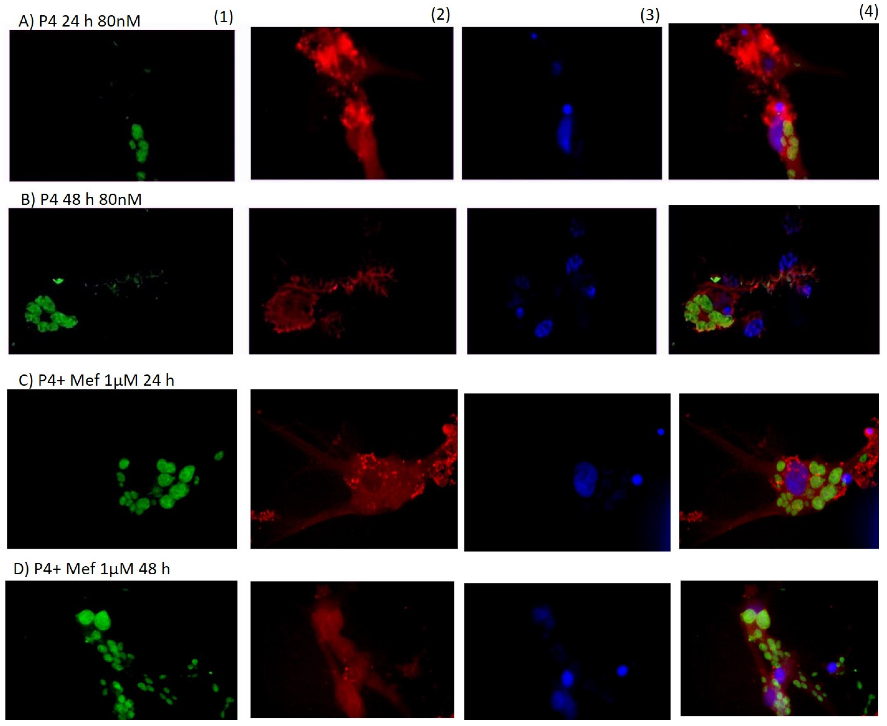
2.6. Immunocytochemical Method

We detected T. gondii infection in cultured neurons by immunocytochemistry. Neurons were washed with phosphate-buffered solution (0.1 M PBS), and they were permeabilized with Triton X-100 in PBS (TPBS) for 1 h. Next, the neurons were incubated with mouse anti-β-Tubulin (β-Tubulin) antibody (1:450, MAB1637); Millipore, Carpinteria, CA, USA) and rabbit anti-Toxoplasma antibody (1:1500; GenWay Biotech, San Diego, CA, USA) diluted and incubated at 4 °C for 16 h in a humidity chamber. Then, cells were incubated with Alexa Fluor 594 antibody-labeled anti-mouse IgG (1:1000, Abcam 150116) and Alexa Flour 488 antibody-labeled anti-rabbit IgG (1:1000, Abcam 150073) for one hour at room temperature under conditions of darkness in a humidity chamber. Finally, the cells were washed twice with PBS and incubated for 5 min with a nucleic acid stain (DAPI, Invitrogen) diluted 1:3000 in PBS.

2.7. Microscopic Analysis

The neurons were quantified a total of nine times for each hormone dose and their agonists and antagonists. We counted one hundred cells for each dose: the number of infected neurons was evaluated using digital images from an Olympus IX71 microscope (40× magnification) employing Image-Pro Plus ver.6.0 software (Media Cybernetics 2.6) to merge the images.

2.8. T. gondii Replication

The enzyme mitochondrial succinate dehydrogenase develops the MTT Tetrazolium reduction assay in live cells. Only viable and early apoptotic cells can reduce the tetrazolium salt MTT (yellow), resulting in the formation of water-insoluble formazan crystals (purple); dead cells will therefore retain the yellow color of the medium [40,41,42].
T. gondii replication in infected and treated neurons was evaluated by the MTT assay. We used 96-well plates to culture neurons (5 × 10⁴ cells/1.7 cm2) using the method described previously. Forty microliters of MTT solution (5 mg/mL; Sigma, St. Louis, MO, USA) were added for two h and incubated at 37 °C in 5% CO2 and 95% air. Subsequently, we added 100 µL of 50% dimethylformamide and 20% sodium dodecyl sulfate (SDS) to dissolve the formazan crystals, and the solution was incubated overnight. Absorbance was measured at 570 nm in an enzyme-linked immunosorbent assay (ELISA) plate reader (Biolab-System,Los Angeles, CA, USA). The percentage of replication was calculated from the optical density (OD) of each well as follows: ((mean OD of treated, infected neurons in well with drug)/(mean OD of non-treated and infected neurons)) × 100. Results are presented as the mean viability ± standard error of the mean (SEM).

2.9. Cytotoxicity Assay

To evaluate the cytotoxicity of neurons, we used the MTT assay. Progesterone and 17B-estradiol, along with their antagonists or agonists, were added to the neurons in vitro. The neurons were treated with E2 or P4 at 10, 20, 40, 80, and 160 nM/mL doses. In another assay, we added to the neurons E2 plus P4 at 160 nM/mL or E2 at 10, 20, 40, 80, and 160 nM/mL plus one µM/mL tamoxifen. We add progesterone at doses of 10, 20, 40, 80, and 160 nM/mL plus mifepristone at a 1 µM/mL concentration. PPT or DPN was added to the cells at a 1 nM/mL dose for 48 h.

2.10. Statistical Analysis
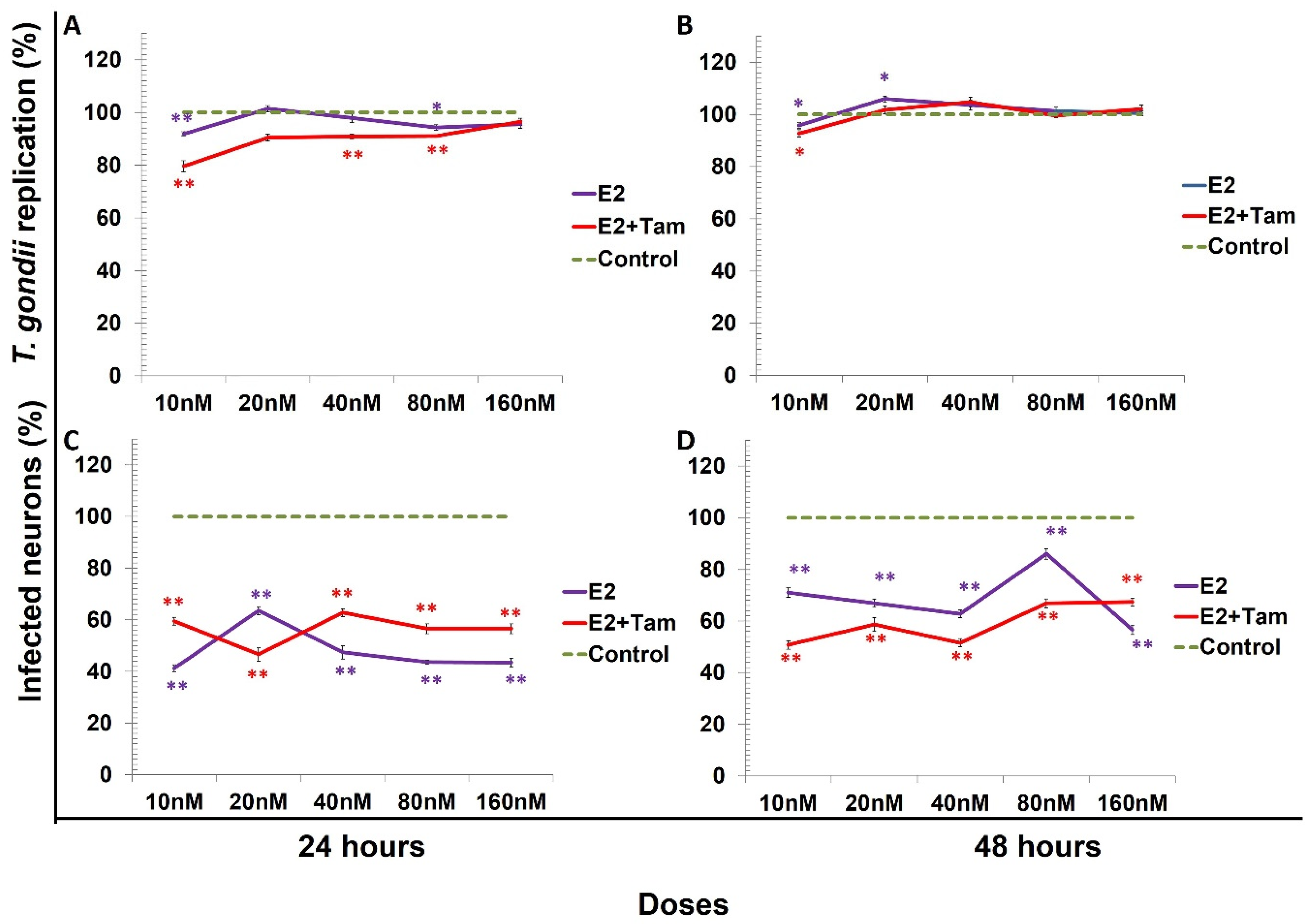
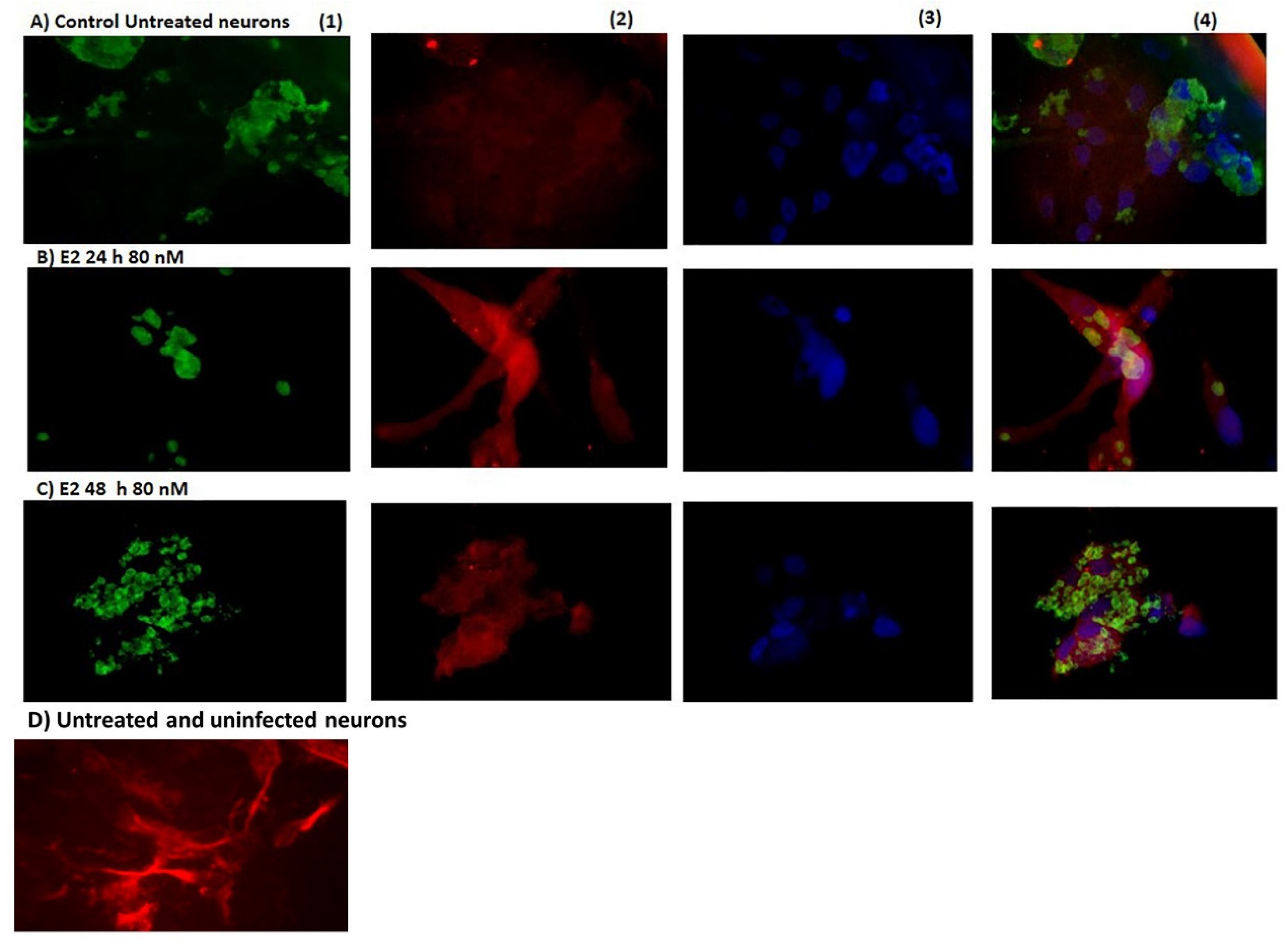
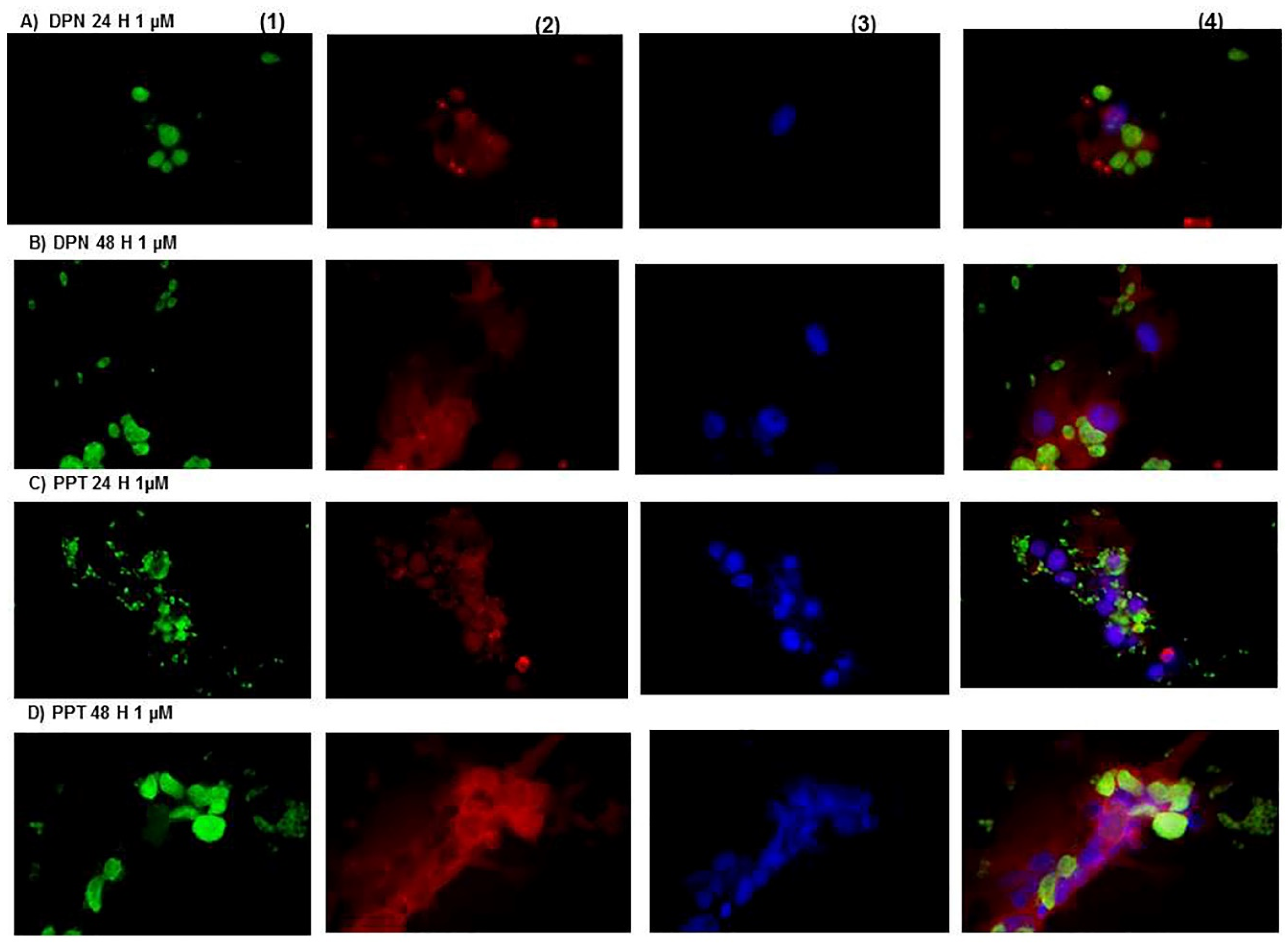
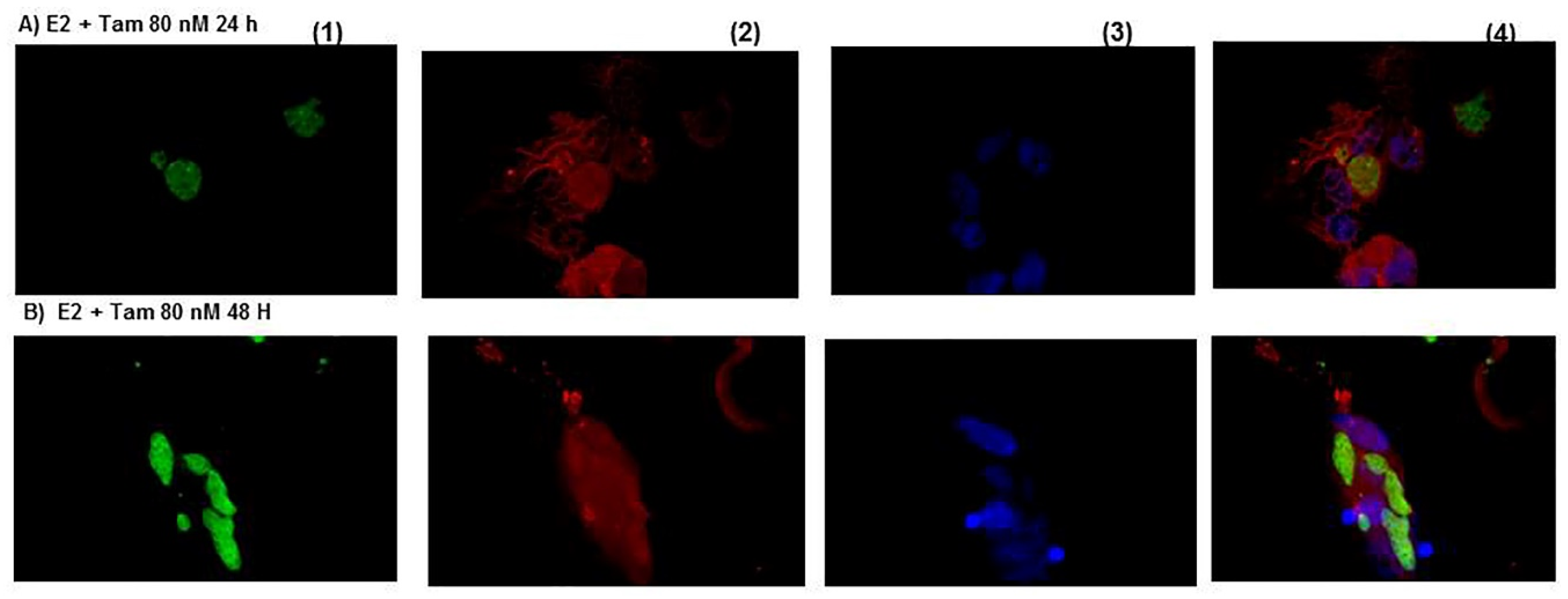
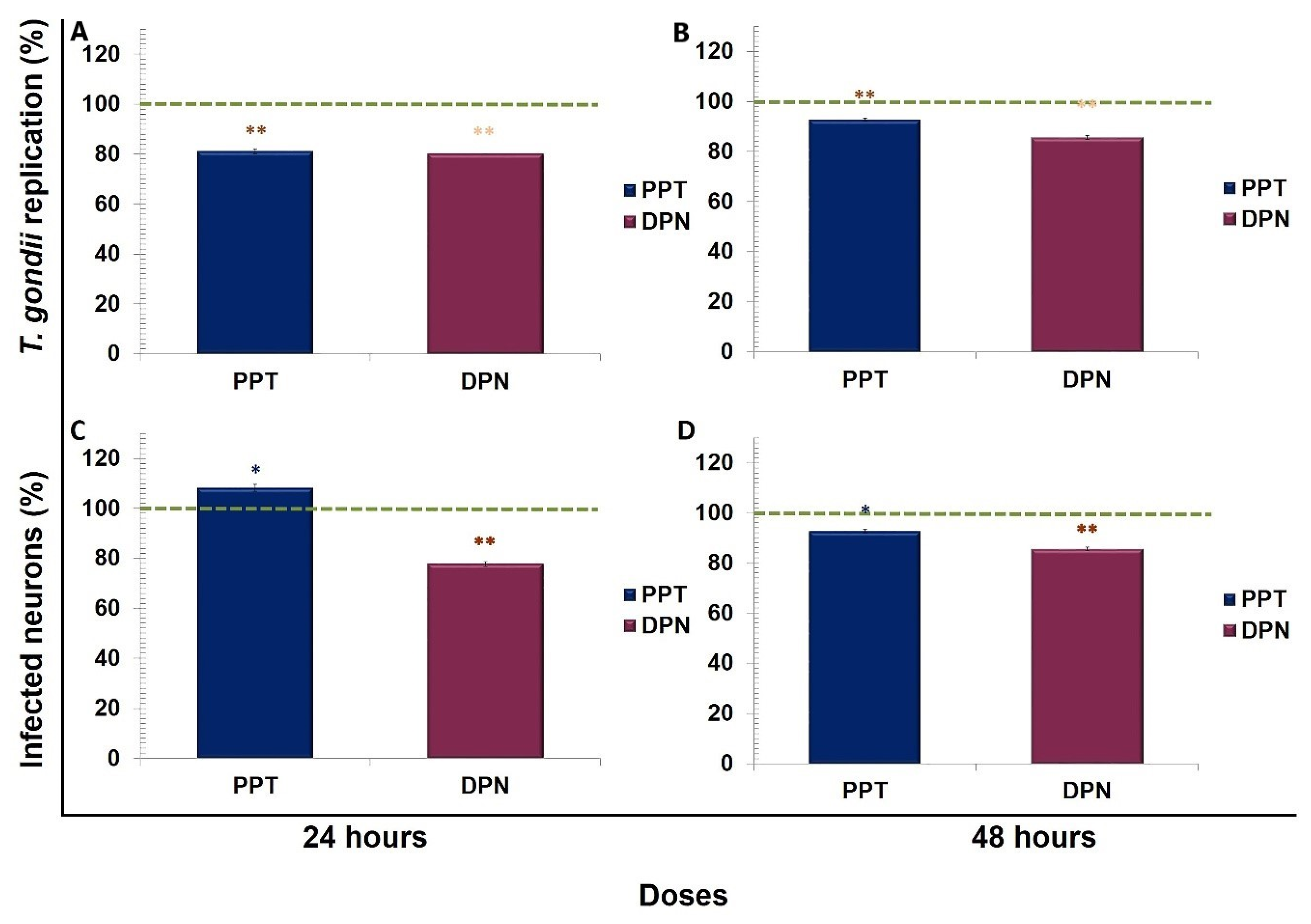
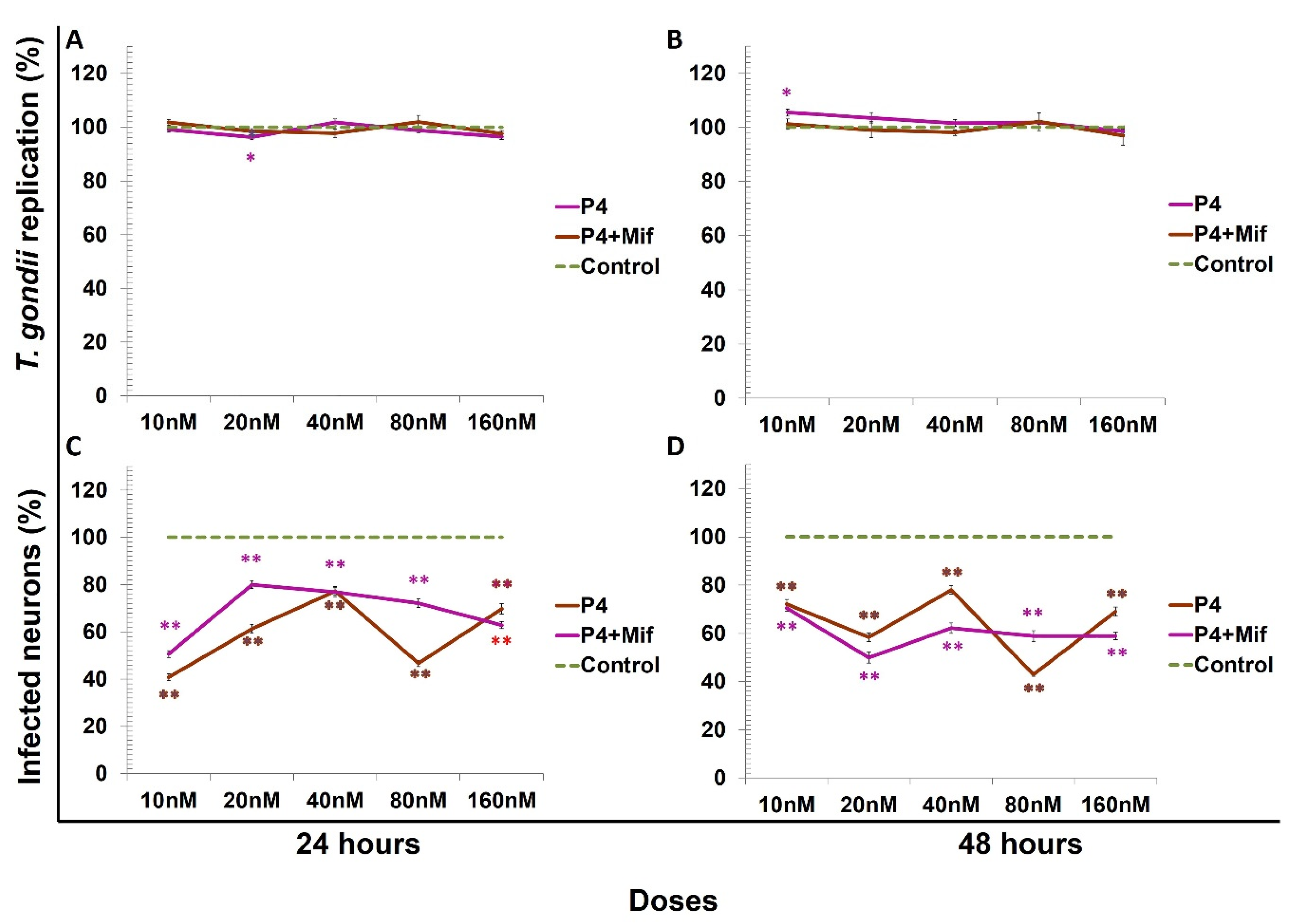
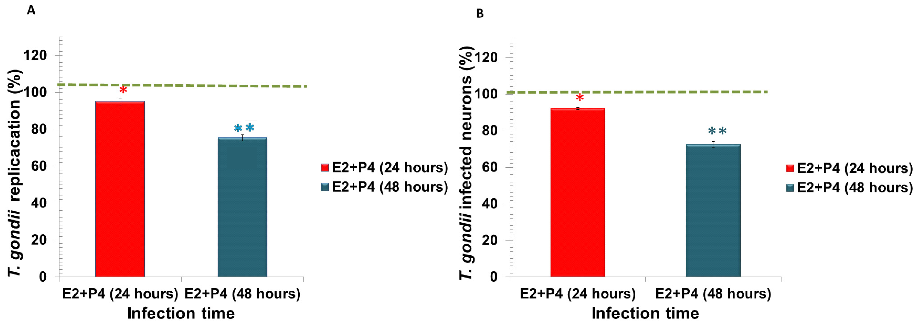
Quantitative variables are represented as the mean ± SEM. All the experimental groups were analyzed by analysis of variance (ANOVA) and a Dunnett T3 post hoc test. The control group consisted of uninfected cells that were 100% viable.

3. Results

Step and Displacement Analysis
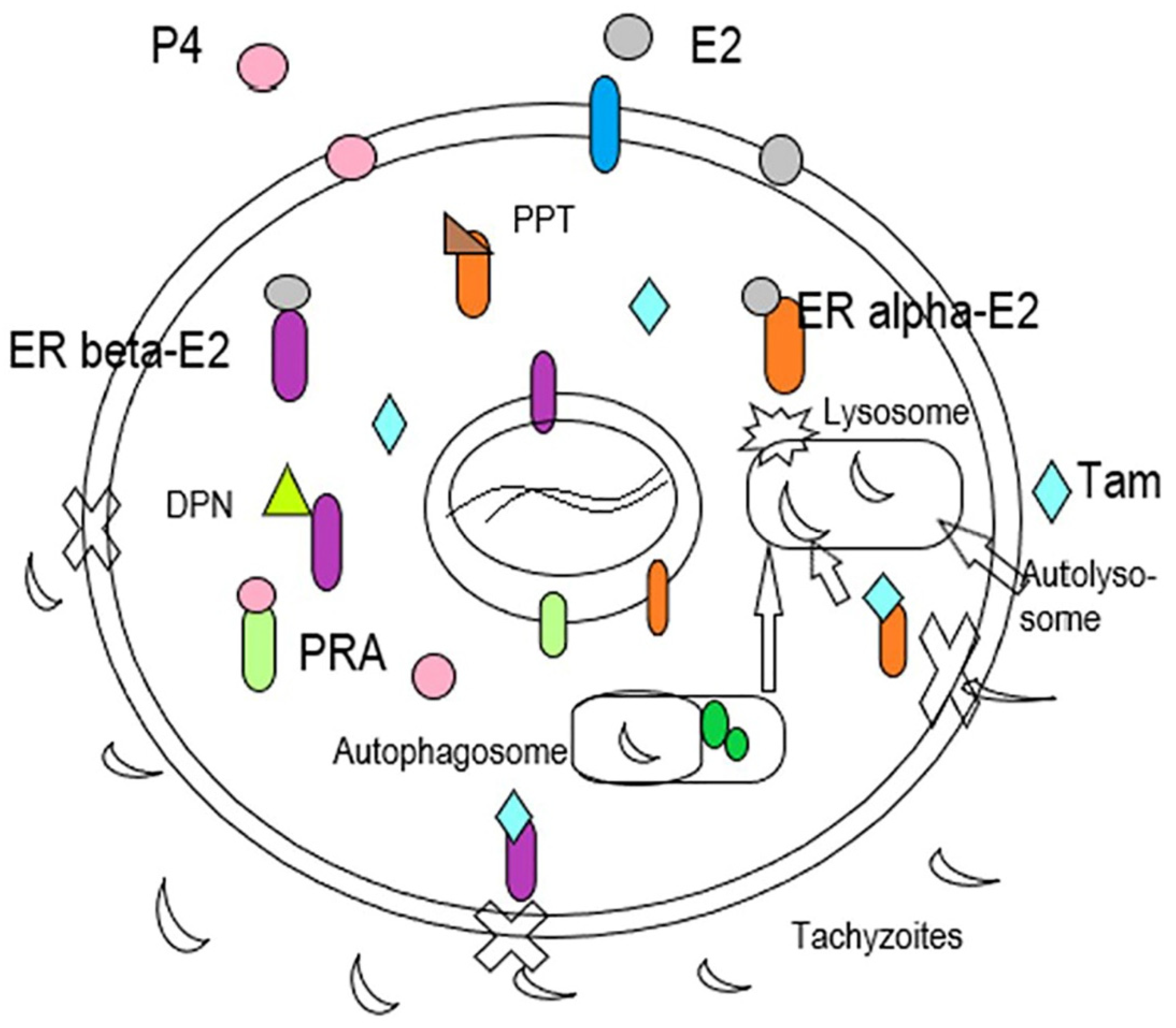
All data were compared vs. the control (100%). At 24 h, 17β-estradiol reduced approximately 10% T. gondii replication at doses of 10 and 80 nM, with a statistically significant difference (p < 0.001 and p < 0.005, respectively). The combination of E2 (10 nM) plus Tam reduced T. gondii replication by 20% with a statistically significant difference of (p < 0.001). We observed a reduction in T. gondii replication of 10% only with E2 at 10 nM and with E2 plus tamoxifen at a similar percentage with a significant difference compared to the control (p < 0.05) at 48 h (Figure 1A,B). The percentage of infected neurons treated with E2 was markedly reduced (58%) at 10 nM at 24 h. The combination of E2 at 10 nM plus Tam at one µM/mL decreased 50% the percentage of infected neurons at 48 h with a statistically significant difference of p < 0.001 (Figure 1C,D).
Figure 2 shows the morphology of infected neurons with Toxoplasma gondii. In (A) the positive control (untreated and infected neurons), Toxoplasma was in an aggregate form, and the neurons present were damaged. In (B), the structure of neurons infected by Toxoplasma and treated with E2 (80 nM) was more preserved than the control at 24 h. In (C), the neurons treated with E2 (80 nM) showed the highest infection by Toxoplasma gondii at 48 h.
In Figure 3A,B, the neurons infected with Toxoplasma gondii under the effect of DPN (1 nM/mL) at 24 and 48 h exhibited parasites intra- and extracellularly. In (C) and (D), the neurons in the presence of PPT (1 nM/mL) at 24 h and 48 h were more infected concerning DPN. The neurons were damaged, and a remarkable percent of them were destroyed.
Figure 4A,B show neurons in the presence of E2 plus tamoxifen exhibiting a minor intracellular infection compared to E2 or PPT at 48 h. However, the neurons disintegrated and their cytoplasmic membrane was broken at 24 h of treatment.
PPT and DPN decreased T. gondii replication by approximately 20% compared to the control with a statistically significant difference of (p < 0.001) at 24 and 48 h p.i. (Figure 5A,B). The agonist of ERβ DPN reduced the percentage of infected neurons at 24 and 48 h (22% and 16%, respectively) versus the control with a statistically significant difference of (p < 0.001) (Figure 5C,D).
Toxoplasma gondii replication decreased by progesterone, only at a dose of 20 nM at 24 h. The combination of progesterone plus Mif (1 µM/mL) did not affect T. gondii replication (Figure 6A,B). There was a significant reduction in the percent of infected neurons by progesterone or by its combination with Mif (p < 0.001) both at 24 and at 48 h p.i. (Figure 6 C,D).
Figure 7A,B, show that the neurons treated with progesterone at 24 and 48 h had more damage by Toxoplasma than E2 plus Tam or PPT or DPN. In Figure 7C,D, neurons infected and treated with progesterone and Mifepristone were better preserved than those treated only with progesterone.
The effects of E2 plus P4 on Toxoplasma gondii replication and the percentage of infected neurons are shown in Figure 8A,B. The pretreatments reduced the percentage of replication and neurons infected with E2 plus P4 (25–30%) at 24 and 48 h with a statistically significant difference of * p < 0.05 and ** p < 0.001, respectively.

4. Discussion

To our knowledge, this is a pioneer study in the analysis of the effect of hormonal steroids (i.e., 17β-estradiol and progesterone) as well as its agonists and antagonists regarding Toxoplasma gondii infection in cultured cortex neurons.
In brain mice, Toxoplasma gondii tachyzoites invade the microglia and the neurons [42,43]. In primary cultures of human fetal cells, T. gondii tachyzoites were replicated in astrocytes and neurons; the highest T. gondii infection was observed in astrocytes [44,45]. In the present study, neurons treated with E2 at a physiological dose (10 nM) decreased T. gondii replication at 24 h p.i. The highest concentration of E2 (160 nM) did not control the Toxoplasma infection. A saturation process could be taking place in the estradiol receptors. However, the percentage of infected neurons decreased at all doses of estradiol utilized in this study. 17β-Estradiol regulates the invasion of Toxoplasma gondii into neurons and may avoid the entry of parasites. However, a greater number of studies are necessary to corroborate this hypothesis. Tamoxifen in combination with estradiol reduced T. gondii replication in neurons even with a lower E2 dose; maybe the mechanism involves preferment binding of tamoxifen to ERβ compared to ERα, similar to what occurs with the DPN agonist. Tamoxifen, as a SERM member, competes with estradiol: however, its affinity for classical estrogen receptors is minor to E2, and it could act as a partial agonist [26,46]. Pung and Luster 1986 [36] demonstrated that tamoxifen could reduce Toxoplasma gondii cysts in mouse brain. In this study, we observed a reduction in Toxoplasma tachyzoites inside neurons by tamoxifen; this effect was similar in astrocytes infected by Toxoplasma gondii [47]. On the other hand, it has been reported that tamoxifen induces autophagy in infected cells, and this may be another mechanism involved in reducing Toxoplasma gondii burden in neurons [48,49].
The interaction of E2 and Tam also decreased the percentage of infected neurons. Our results are similar to those found by Zhang in 2017, who showed that tamoxifen reduces the number of parasites by approximately 50% in host cells [50].
The mechanism by which tamoxifen affects Toxoplasma infection is unknown. However, in Figure 9, we discuss the possible interactions of sex hormones with T. gondii infections.
Observe that tamoxifen can induce autophagy and the steroid hormones can avoid the entrance of parasites to neurons through the membrane. The mechanism of this last process is unknown. Figure 9 shows the interactions between hormones, and their receptors are shown on its specific receptor. Tamoxifen binds to ERβ (blue and purple), and it is possible that this complex, induces the formation of an autophagosome. With the presence of proteins (in green) necessary for this process, in consequence, it will active a lysosome and will form an autolysosome. Progesterone (P4, pink), receptor of progesterone (PRA, light green), E2 (estradiol, gray), estradiol receptor beta (ER-beta-E2, gray and purple), selective agonist for receptor β of E2, 23-bis(4-hydroxyphenyl) propionitrile (DPN, green triangle). Estrogen receptor α bind to estradiol (ER-alfa-E2, orange and gray), selective agonist of receptor α, E2 4,4′,4″-(4-propyl-[1H] pyrozole-1,3,5-triyl) trisphenol (PPT, brown triangle ), selective estrogen receptor modulator, tamoxifen (Tam, blue rhombus).
In the present study, DPN produced an essential reduction in Toxoplasma gondii infection in neurons in vitro. The response of the neurons to this agonist maybe is due to fact of its high specificity for ERβ, affording better neuronal protection [51,52,53,54,55].
Progesterone did not reduce Toxoplasma gondii replication despite; it decreased the percentage of infected neurons. Progesterone could modulate the number of their receptors in neurons in vitro [55]. In current studies of our group of work, we reported that progesterone could reduce ERβ and increase ERα in THP1 cells, maybe in neurons could be a similar regulation and thus modulate the parasite invasion [39].
The antagonist of progesterone, Mif, did not revert the action of progesterone on T. gondii replication at any of the experimental dosages; this could be due to the dose and time of exposure. However, there are no previous studies in this regard; thus, it is imperative to acquire a more significant number of investigations on this topic. The combination of progesterone and Mif exerted a lesser effect on Toxoplasma infected neurons compared with progesterone alone. One explanation could be a change in progesterone membrane receptors as well as in their affinity [13,30,31,32].
The combination of 17β-estradiol and progesterone significantly reduced Toxoplasma gondii replication and the percentage of infected neurons; this may be due to the major expression of classical estrogen receptors [39], E2 plus progesterone modulating the viability of extracellular parasites and the passive invasion to neurons. This result is similar to host cells infected with tachyzoites treated with dehydroepiandrosterone (DHEA) [13,56,57].

5. Conclusions

E2 and tamoxifen as well as PPT and DPN regulated Toxoplasma gondii infection in neurons.
The neuronal response to these agonists is clear evidence of the essential participation of classical estrogen receptors in the process of Toxoplasma gondii infection.
The combination of E2 and P4 decreased Toxoplasma gondii infected neurons. This action could be considered as a neuro-protector effect on T. gondii infection.

Author Contributions

Conceived and designed the experiments, M.d.l.L.G.R. and J.M.D.-J.; Performed the experiments, analyzed the images, and created the database: A.F.G.-M. and L.R.R.P.; Contributed reagents/materials, M.d.l.L.G.R. and J.M.D.-J.; Wrote the paper: M.d.l.L.G.R. and J.M.D.-J. All authors have read and agreed to the published version of the manuscript.

Funding

This work was partially supported by the Program to Support the Improvement of the Production Conditions of the SNI and SNCA Members (PRO-SNI) of the University of Guadalajara. Number: 227303/2019.

Institutional Review Board Statement

Not applicable.

Informed Consent Statement

Not applicable.

Data Availability Statement

Not applicable.

Conflicts of Interest

The authors declare no conflict of interest.

References

  1. Barragan, A.; Hitziger, N. Transepithelial Migration by Toxoplasma. Subcell Biochem. 2008, 47, 198–207. [Google Scholar] [CrossRef]
  2. Cabral, C.M.; Tuladhar, S.; Dietrich, H.K.; Nguyen, E.; Macdonald, W.R.; Trivedi, T.; Devineni, A.; Koshy, A.A. Neurons are the Primary Target Cell for the Brain-Tropic Intracellular Parasite Toxoplasma gondii. PLoS Pathog. 2016, 12, e1005447. [Google Scholar] [CrossRef] [PubMed]
  3. Ferguson, D.J.; Hutchison, W.M. The host-parasite relationship of Toxoplasma gondii in the brains of chronically infected mice. Virchows Arch. A Pathol. Anat Histopathol. 1987, 411, 39–43. [Google Scholar] [CrossRef]
  4. Suzuki, T.; Schirra, F.; Richards, S.M.; Jensen, R.V.; Sullivan, D.A. Estrogen and Progesterone Control of Gene Expression in the Mouse Meibomian Gland. Investig. Ophthalmol. Vis. Sci. 2008, 49, 1797–1808. [Google Scholar] [CrossRef]
  5. Parlog, A.; Schlüter, D.; Dunay, I.R. Toxoplasma gondii-induced neuronal alterations. Parasite Immunol. 2015, 37, 159–170. [Google Scholar] [CrossRef] [PubMed]
  6. Tanakaa, N.; Ashoura, D.; Dratzb, E.; Halonen, S. Use of human induced pluripotent stem cell-derived neurons as a model for Cerebral Toxoplasmosis. Microbes Infect. 2015, 18, 496–504. [Google Scholar] [CrossRef] [PubMed]
  7. Creuzet, C.; Robert, F.; Roisin, M.P.; Van Tan, H.; Benes, C.; Dupouy-Camet, J.; Fagard, R. Neurons in primary culture are less efficiently infected by Toxoplasma gondii than glial cells. Parasitol. Res. 1998, 84, 25–30. [Google Scholar] [CrossRef] [PubMed]
  8. Gomez-Marin, J.E. Manifestaciones clínicas de la toxoplasmosis humana. In Toxoplasmosis Humna; Galván-Ramírez, M., Flores, M., Eds.; Ecorfan: Ecatepec, Mexico, 2017; pp. 139–162. [Google Scholar]
  9. Galván-Ramírez, M.d.l.L.; Navarro Machuca, G.; Covarrubias Castillo, S.A.; Benavides González, J.C.; Rodríguez Pérez, L.R.; Dueñas Jiménez, S.H.; Dueñas Jiménez, J.M. Toxoplasmosis Is More Frequent in Schizophrenia Patients Than in the General Population in Mexico and Is Not Associated with More Severe Course of Schizophrenia Measured with the Brief Psychiatric Rating Scale. Pathogens 2021, 10, 820. [Google Scholar] [CrossRef]
  10. Roberts, C.W.; Cruickshank, S.M.; Alexander, J. Sex-determined resistance to Toxoplasma gondii is associated with temporal differences in cytokine production. Infect. Immun. 1995, 63, 2549–2555. [Google Scholar] [CrossRef] [PubMed] [Green Version]
  11. Walker, W.; Roberts, C.W.; Ferguson, D.J.; Jebbari, H.; Alexander, J. Innate immunity to Toxoplasma gondii is influenced by gender and is associated with differences in interleukin-12 and gamma interferon production. Infect. Immun. 1997, 65, 1119–1121. [Google Scholar] [CrossRef] [Green Version]
  12. Liesenfeld, O.; Nguyen, T.A.; Pharke, C.; Suzuki, Y. Importance of gender and sex hormones in regulation of susceptibility of the small intestine to peroral infection with Toxoplasma gondii tissue cysts. J. Parasitol. 2001, 87, 1491–1493. [Google Scholar] [CrossRef]
  13. Muñiz-Hernández, S.; Luna-Nophal, A.; León, C.T.G.; Domínguez-Ramírez, L.; Patrón-Soberano, O.A.; Nava-Castro, K.E.; Ostoa-Saloma, P.; Morales-Montor, J. Dehydroepiandrosterone Effect on Toxoplasma gondii: Molecular Mechanisms Associated to Parasite Death. Microorganisms 2021, 9, 513. [Google Scholar] [CrossRef] [PubMed]
  14. Garcia-Segura, L.M.; Melcangi, R.C. Steroids and glial cell function. Glia 2006, 54, 485–498. [Google Scholar] [CrossRef] [Green Version]
  15. Shao, R.; Cao, S.; Wang, X.; Feng, Y.; Billig, H. The elusive and controversial roles of estrogen and progesterone receptors in human endometriosis. Am. J. Transl. Res. 2014, 6, 104–113. [Google Scholar] [PubMed]
  16. Sa, Q.; Ochiai, E.; Tiwari, A.; Perkins, S.; Mullins, J.; Gehman, M.; Huckle, W.R.; Eyestone, W.H.; Saunders, T.L.; Shelton, B.J.; et al. Cutting Edge: IFN-γ Produced by Brain-Resident Cells Is Crucial to Control Cerebral Infection with Toxoplasma gondii. J. Immunol. 2015, 195, 796–800. [Google Scholar] [CrossRef] [PubMed] [Green Version]
  17. Godinjak, Z.; Bilalovic, N. Estrogen and Progesterone Receptors in Endometrium in Women with Unexplained Infertility. Mater. Socio Medica 2014, 26, 51–52. [Google Scholar] [CrossRef] [Green Version]
  18. Verthelyi, D. Sex hormones as immunomodulators in health and disease. Int. Immunopharmacol. 2001, 1, 983–993. [Google Scholar] [CrossRef]
  19. McEwen, B.S.; Akama, K.T.; Spencer-Segal, J.L.; Milner, T.A.; Waters, E.M. Estrogen effects on the brain: Actions beyond the hypothalamus via novel mechanisms. Behav. Neurosci. 2012, 126, 4–16. [Google Scholar] [CrossRef] [Green Version]
  20. Roque, C.; Baltazar, G. G protein-coupled estrogen receptor 1 (GPER) activation triggers different signaling pathways on neurons and astrocytes. Neural Regen. Res. 2019, 14, 2069–2070. [Google Scholar] [CrossRef]
  21. Garrido-Gracia, J.C.; Gordon, A.; Bellido, C.; Aguilar, R.; Barranco, I.; Millán, Y.; de Las Mulas, J.M.; Sánchez-Criado, J.E. The integrated action of estrogen receptor isoforms and sites with progesterone receptor in the gonadotrope modulates LH secretion: Evidence from tamoxifen-treated ovariectomized rats. J. Endocrinol. 2007, 193, 107–119. [Google Scholar] [CrossRef] [PubMed]
  22. Gottfried-Blackmore, A.; Croft, G.; McEwen, B.S.; Bulloch, K. Transcriptional activity of estrogen receptors ERalpha and ERbeta in the EtC.1 cerebellar granule cell line. Brain Res. 2007, 1186, 41–47. [Google Scholar] [CrossRef] [PubMed]
  23. Harrington, W.R.; Sheng, S.; Barnett, D.H.; Petz, L.N.; Katzenellenbogen, J.A.; Katzenellenbogen, B.S. Activities of estrogen receptor alpha- and beta-selective ligands at diverse estrogen responsive gene sites mediating transactivation or transrepression. Mol. Cell. Endocrinol. 2003, 206, 13–22. [Google Scholar] [CrossRef]
  24. Franco-Rodríguez, N.E.; Dueñas-Jiménez, J.M.; De la Torre Valdovinos, B.; López Ruiz, J.R.; Hernández Hernández, L.; Dueñas Jiménez, S.H. Tamoxifen favoured the rat sensorial cortex regeneration after a penetrating brain injury. Brain Res. Bull. 2013, 98, 64–75. [Google Scholar] [CrossRef] [PubMed]
  25. Lee, E.-S.Y.; Yin, Z.; Milatovic, D.; Jiang, H.; Aschner, M. Estrogen and Tamoxifen Protect against Mn-Induced Toxicity in Rat Cortical Primary Cultures of Neurons and Astrocytes. Toxicol. Sci. 2009, 110, 156–167. [Google Scholar] [CrossRef] [Green Version]
  26. Dávila, J.T.; Garrán, A.D.T. Selective estrogen receptors modulators (SERMs): Biochemistry, pharmacology, and clinical use in gynecology. Ginecol. Obstet. Méx. 2005, 73, 424–435. [Google Scholar]
  27. Melcangi, R.C.; Giatti, S.; Calabrese, D.; Pesaresi, M.; Cermenati, G.; Mitro, N.; Viviani, B.; Garcia-Segura, L.M.; Caruso, D. Levels and actions of progesterone and its metabolites in the nervous system during physiological and pathological conditions. Prog. Neurobiol. 2014, 113, 56–69. [Google Scholar] [CrossRef] [PubMed]
  28. Diep, C.H.; Daniel, A.R.; Mauro, L.J.; Knutson, T.P.; Lange, C.A. Progesterone action in breast, uterine, and ovarian cancers. J. Mol. Endocrinol. 2015, 54, R31–R53. [Google Scholar] [CrossRef] [Green Version]
  29. Cadepond, F.; Ulmann, A.; Baulieu, E.E. RU486 (MIFEPRISTONE): Mechanisms of Action and Clinical Uses. Annu. Rev. Med. 1997, 48, 129–156. [Google Scholar] [CrossRef] [Green Version]
  30. Behl, C.; Trapp, T.; Skutella, T.; Holsboer, F. Protection against oxidative stress-induced neuronal cell death—A novel role for RU486. Eur. J. Neurosci. 1997, 9, 912–920. [Google Scholar] [CrossRef]
  31. Khan, J.A.; Tikad, A.; Fay, M.; Hamze, A.; Fagart, J.; Chabbert-Buffet, N.; Meduri, G.; Amazit, L.; Brion, J.-D.; Alami, M.; et al. A New Strategy for Selective Targeting of Progesterone Receptor with Passive Antagonists. Mol. Endocrinol. 2013, 27, 909–924. [Google Scholar] [CrossRef] [Green Version]
  32. Bouchard, P.; Chabbert-Buffet, N.; Fauser, B.C. Selective progesterone receptor modulators in reproductive medicine: Pharmacology, clinical efficacy and safety. Fertil. Steril. 2011, 96, 1175–1189. [Google Scholar] [CrossRef]
  33. Choi, S.; Shin, H.; Song, H.; Lim, H.J. Suppression of autophagic activation in the mouse uterus by estrogen and progesterone. J. Endocrinol. 2014, 221, 39–50. [Google Scholar] [CrossRef] [PubMed]
  34. Spitz, I.M. Progesterone receptor antagonists. Curr. Opin. Investig. Drugs 2016, 7, 882–890. [Google Scholar]
  35. Chabbert-Buffet, N.; Meduri, G.; Bouchard, P.; Spitz, I.M. Selective progesterone receptor modulators and progesterone antagonists: Mechanisms of action and clinical applications. Hum. Reprod. Updat. 2005, 11, 293–307. [Google Scholar] [CrossRef] [PubMed] [Green Version]
  36. Pung, O.J.; Luster, M.I. Toxoplasma gondii: Decreased resistance to infection in mice due to estrogen. Exp. Parasitol. 1986, 61, 48–56. [Google Scholar] [CrossRef]
  37. Jones, L.A.; Jean-Paul, A.; Henriquez, F.L.; Lyons, R.E.; Nickdel, M.B.; Carter, K.C. Toll-like receptor-4-mediated macrophage activation is differentially regulated by progesterone via the glucocorticoid and progesterone receptors. Immunology 2008, 125, 59–69. [Google Scholar] [CrossRef] [PubMed]
  38. Galván-Ramírez, M.d.l.L.; Gutiérrez-Maldonado, A.F.; Verduzco-Grijalva, F.; Jiménez, J.M.D. The role of hormones on Toxoplasma gondii infection: A systematic review. Front. Microbiol. 2014, 5, 503. [Google Scholar] [CrossRef]
  39. Galván-Ramírez, M.d.l.L.; De Arellano, A.R.; Rodríguez-Pérez, L.R.; Lopez-Pulido, E.I.; Muñoz-Valle, J.F.; Pereira-Suárez, A.L. Hormonal modulation of Toxoplasma gondii infection: Regulation of hormonal receptors and cytokine production in THP-1 cells. Exp. Parasitol. 2019, 204, 107721. [Google Scholar] [CrossRef]
  40. Galván-Ramírez, M.d.l.L.; Jiménez, J.M.D.; Pérez, L.R.R.; Troyo-Sanroman, R.; Ramírez-Herrera, M.A.; García-Iglesias, T. Effect of Nitaxozanide and Pyrimethamine on Astrocytes Infected by Toxoplasma gondii In Vitro. Arch. Med. Res. 2013, 44, 415–421. [Google Scholar] [CrossRef]
  41. Mossman, T. Rapid colorimetric assay for cellular growth and survival: Application to proliferation and cytotoxicity assays. J. Immunol. Methods 1983, 65, 55–63. [Google Scholar] [CrossRef]
  42. Anand, N.; Kanwar, K.R.; Dubey, M.L.; Vahishta, R.K.; Sehgal, R.; Verma, A.K. Effect of lactoferrin protein on red blood cells and macrophages: Mechanism of parasite–host interaction. Drug Des. Dev. Ther. 2015, 9, 3821–3835. [Google Scholar] [CrossRef] [Green Version]
  43. Dzitko, K.; Gatkowska, J.; Płociński, P.; Dziadek, B.; Długońska, H. The effect of prolactin (PRL) on the growth of Toxoplasma gondii tachyzoites in vitro. Parasitol. Res. 2010, 107, 199–204. [Google Scholar] [CrossRef]
  44. Courret, N.; Darche, S.; Sonigo, P.; Milon, G.; Buzoni-Gâtel, D.; Tardieux, I. CD11c- and CD11b-expressing mouse leukocytes transport single Toxoplasma gondii tachyzoites to the brain. Blood 2006, 107, 309–316. [Google Scholar] [CrossRef] [Green Version]
  45. Contreras-Ochoa, C.O.; Lagunas-Martínez, A.; Belkind-Gerson, J.; Díaz-Chávez, J.; Correa, D. Toxoplasma gondii invasion and replication within neonate mouse astrocytes and changes in apoptosis related molecules. Exp. Parasitol. 2013, 134, 256–265. [Google Scholar] [CrossRef] [PubMed]
  46. Teppa, G.A.D.; Terán, D.J. New biochemical and molecular aspects of estrogen’s action. Ginecol. Obstet. Méx. 2005, 73, 436–442. [Google Scholar]
  47. Halonen, S.K.; Lyman, W.D.; Chiu, F.C. Growth and Development of Toxoplasma gondii Human Neurons and Astrocytes. J. Neuropathol. Exp. Neurol. 1996, 55, 1150–1156. [Google Scholar] [CrossRef] [PubMed] [Green Version]
  48. Galván-Ramírez, M.L.; Gutiérrez-Maldonado, A.F.; Dueñas-Jiménez, J.M.; Rodríguez-Pérez, L.R.; Troyo-Sanromán, R. Effect of 17β-estradiol and progesterone on astrocytes infected with Toxoplasma. J. Adv. Parasitol. 2016, 3, 22–31. [Google Scholar] [CrossRef] [Green Version]
  49. Dittmar, A.J.; Drozda, A.A.; Blader, I.J. Drug Repurposing Screening Identifies Novel Compounds That Effectively Inhibit Toxoplasma gondii Growth. mSphere 2016, 1, e00042-15. [Google Scholar] [CrossRef] [Green Version]
  50. Zhang, X.; Liu, J.; Li, M.; Fu, Y.; Zhang, T.; Han, Q.; Liu, Q. Role of an estradiol regulatory factor-hydroxysteroid dehydrogenase (HSD) in Toxoplasma gondii infection and pathogenicity. J. Steroid Biochem. Mol. Biol. 2017, 174, 176–182. [Google Scholar] [CrossRef] [PubMed]
  51. Garcia-Segura, L.M.; Azcoitia, I.; DonCarlos, L.L. Neuroprotection by estradiol. Prog. Neurobiol. 2001, 63, 29–60. [Google Scholar] [CrossRef] [Green Version]
  52. Garcia-Segura, L.M.; Dueñas, M.; Busiguina, S.; Naftolin, F.; Chowen, J.A. Gonadal hormone regulation of neuronal-glial interactions in the developing neuroendocrine hypothalamus. J. Steroid Biochem. Mol. Biol. 1995, 53, 293–298. [Google Scholar] [CrossRef]
  53. Garcia-Segura, L.M.; Sanz, A.; Mendez, P. Cross-Talk between IGF-I and Estradiol in the Brain: Focus on Neuroprotection. Neuroendocrinology 2006, 84, 275–279. [Google Scholar] [CrossRef]
  54. Spampinato, S.F.; Molinaro, G.; Merlo, S.; Iacovelli, L.; Caraci, F.; Battaglia, G.; Nicoletti, F.; Bruno, V.; Sortino, M.A. Estrogen Receptors and Type 1 Metabotropic Glutamate Receptors Are Interdependent in Protecting Cortical Neurons against β-Amyloid Toxicity. Mol. Pharmacol. 2012, 81, 12–20. [Google Scholar] [CrossRef] [Green Version]
  55. Micevych, P.; Sinchak, K. Estradiol regulation of progesterone synthesis in the brain. Mol. Cell. Endocrinol. 2008, 290, 44–50. [Google Scholar] [CrossRef] [PubMed] [Green Version]
  56. Baudry, M.; Bi, X.; Aguirre, C. Progesterone–estrogen interactions in synaptic plasticity and neuroprotection. Neuroscience 2013, 239, 280–294. [Google Scholar] [CrossRef] [PubMed] [Green Version]
  57. Fagard, R.; Van Tan, H.; Creuzet, C.; Pelloux, H. Differential Development of Toxoplasma gondii in Neural Cells. Parasitol. Today 1999, 15, 504–507. [Google Scholar] [CrossRef]
Figure 1. Toxoplasma gondii replication in neurons, representing the percentage of the control value. Untreated neurons (control; dashed line), neurons pretreated with 17β-estradiol (E2) (purple line), and neurons treated with E2 + tamoxifen (red line). 17β-Estradiol reduced Toxoplasma gondii replication at doses of 10 and 80 nM at 24 h. However, at 48 h, the replication was reduced only at the 10 nM dose and increased at 20 nM with a statistically significant difference of * p < 0.05. (A,B) The percentage of infected neurons was reduced by E2 and by the combination of tamoxifen at 24 and 48 h with statistically significant differences in respect to the control, ** p < 0.001; (C,D) exhibit these changes.
Microorganisms 09 02174 g001
Figure 2. Representative photographs of Toxoplasma gondii infection in cultured neurons detected by immunocytochemistry. The parasite is shown in green; the neurons are presented in red. The neuron’s nucleus is in blue. The merged are depicted in the last column’s photographs. (A) The control group at 24 h showed the untreated and infected neurons (T. gondii tachyzoites RH strain). The neurons were destroyed almost completely by Toxoplasma gondii. (B) Observe that there was substantial damage to the neurons in the 17-β estradiol (E2)-treated group; however, some neuronal branches were conserved. (C) In the neurons treated with 17-β estradiol, the parasite changed its structure. (D) Negative control neurons, uninfected and untreated. The neuron exhibited some dendrites and did not show signs of damage. (40× magnification).
Microorganisms 09 02174 g002
Figure 3. (A,B) (DPN 1µM, 24 and 48 h): The parasite destroyed the neurons treated with the agonist of ER β or DPN, in consequence, a significant number of the parasites were present at 24 and 48 h. (C,D) (PPT 1 µM, 24 and 48 h, respectively): The neurons treated with PPT had a more significant infection than those with DPN; however, the neurons were better preserved. (40× magnification).
Microorganisms 09 02174 g003
Figure 4. Representative photographs of Toxoplasma gondii infection in cultured neurons detected by immunocytochemistry. The parasite is in green, the neurons are presented in red, and the neuron’s nuclei are in blue. The merged are shown in the last column’s photographs. (A,B). Neurons treated with 17-β estradiol plus tamoxifen were destroyed and were less infected at 24 h than at 48 h compared to the untreated control. (40× magnification).
Microorganisms 09 02174 g004
Figure 5. Percentage of Toxoplasma gondii replication at 24 h and 48 h. Percentage of T. gondii-infected neurons at 24 and 48 h after treatment with PPT (navy blue bar) or DPN (burgundy bar). (A,B) T. gondii replication was reduced by PPT and DPN with respect to the control (100%), with statistically significant differences of (p < 0.001). (C) At 24 h, the percentage of infected neurons treated with PPT was significantly increased, compared with the control (p < 0.05); however, DPN reduced this percentage significantly (p < 0.001). (D) At 48 h, the percentage of infected neurons was reduced by both agonists (i.e., PPT and DPN) with statistical significance compared with the control at * p < 0.05 and at ** p < 0.001, respectively.
Microorganisms 09 02174 g005
Figure 6. Toxoplasma gondii replication in neurons. Replication is expressed as the percentage of the control value. Untreated neurons (control; dashed line), neurons pretreated with progesterone (P4) (brown line), and neurons treated with P4 + mifepristone (fuchsia line). Progesterone did not regulate Toxoplasma infection, except at 20 nM dose by 24 h. However, at 48 h at a 10 nM dose, this replication increased with statistical significance: * p < 0.05 (A,B). (C,D) The percentage of infected neurons was reduced by progesterone combined with Mif at 24 and 48 h with a statistically significant difference of ** p < 0.001.
Microorganisms 09 02174 g006
Figure 7. (A,B) Neurons infected with tachyzoites of T. gondii and treated for 24 and 48 h with progesterone at 80 nM. In (C,D) the neurons treated with progesterone plus mifepristone at 24 and 48 h exhibit many tachyzoites; however, the neurons present were better preserved compared to progesterone alone. (40× magnification).
Microorganisms 09 02174 g007
Figure 8. Percentage of Toxoplasma gondii replication treated with 17β-estradiol combined with progesterone E2 + P4 (red and blue bars, respectively) at 24 and 48 h. (A) Percentage of T. gondii in neurons decreased with E2 + P4, compared with the control, at 48 h p.i., ** p < 0.001. (B) Percentage of infected neurons treated with E2 + P4 was reduced compared with the untreated control at 24 and 48 h, with a significant difference of * p < 0.05 and ** p < 0.001, respectively.
Microorganisms 09 02174 g008
Figure 9. The interactions among sex hormones (i.e., estradiol, progesterone) and tamoxifen (Tam) with the tachyzoites of Toxoplasma gondii.
Microorganisms 09 02174 g009
Publisher’s Note: MDPI stays neutral with regard to jurisdictional claims in published maps and institutional affiliations.

