Article

Lanthanide-Dependent Methanol and Formaldehyde Oxidation in Methylobacterium aquaticum Strain 22A

1
Institute of Plant Science and Resources, Okayama University, Okayama 710-0046, Japan
2
Advanced Low Carbon Technology Research and Development Program, Japan Science and Technology Agency, Tokyo 102-0076, Japan
3
RIKEN Center for Sustainable Resource Science, Kanagawa 230-0045, Japan
4
Department of Biochemistry, Faculty of Science, Okayama University of Science, Okayama 700-8530, Japan
5
The United Graduate School of Agricultural Science, Gifu University, Gifu 501-1193, Japan
6
The Graduate School of Natural Sciences and Technologies, Gifu University, Gifu 501-1193, Japan
*
Author to whom correspondence should be addressed.
Microorganisms 2020, 8(6), 822; https://doi.org/10.3390/microorganisms8060822
Submission received: 5 May 2020 / Revised: 26 May 2020 / Accepted: 28 May 2020 / Published: 30 May 2020
(This article belongs to the Special Issue Microbial One-Carbon Metabolism of Natural and Engineered Systems)

Abstract

:
Lanthanides (Ln) are an essential cofactor for XoxF-type methanol dehydrogenases (MDHs) in Gram-negative methylotrophs. The Ln3+ dependency of XoxF has expanded knowledge and raised new questions in methylotrophy, including the differences in characteristics of XoxF-type MDHs, their regulation, and the methylotrophic metabolism including formaldehyde oxidation. In this study, we genetically identified one set of Ln3+- and Ca2+-dependent MDHs (XoxF1 and MxaFI), that are involved in methylotrophy, and an ExaF-type Ln3+-dependent ethanol dehydrogenase, among six MDH-like genes in Methylobacterium aquaticum strain 22A. We also identified the causative mutations in MxbD, a sensor kinase necessary for mxaF expression and xoxF1 repression, for suppressive phenotypes in xoxF1 mutants defective in methanol growth even in the absence of Ln3+. Furthermore, we examined the phenotypes of a series of formaldehyde oxidation-pathway mutants (fae1, fae2, mch in the tetrahydromethanopterin (H4MPT) pathway and hgd in the glutathione-dependent formaldehyde dehydrogenase (GSH) pathway). We found that MxaF produces formaldehyde to a toxic level in the absence of the formaldehyde oxidation pathways and that either XoxF1 or ExaF can oxidize formaldehyde to alleviate formaldehyde toxicity in vivo. Furthermore, the GSH pathway has a supportive role for the net formaldehyde oxidation in addition to the H4MPT pathway that has primary importance. Studies on methylotrophy in Methylobacterium species have a long history, and this study provides further insights into genetic and physiological diversity and the differences in methylotrophy within the plant-colonizing methylotrophs.

1. Introduction

Methanol is the most abundant single-carbon compound produced by plants in pectin metabolism, primarily during leaf expansion, and is emitted through the stomata [1]. It can be metabolized by methylotrophic bacteria as a sole carbon and energy source [2,3]. Methylotrophs play an important role as methanol consumers in the phyllosphere and have received significant research attention [4,5,6]. Methylobacterium extorquens strain AM1, recently reclassified as Methylorubrum extorquens strain AM1 [7] (hereafter referred to as strain AM1) has long served as a model organism of Gram-negative methylotrophs for biochemical and physiological studies of methanol metabolism [8,9]. The metabolic process of strain AM1 begins with the oxidation of methanol into formaldehyde in the periplasm using pyrroloquinoline quinone (PQQ)-dependent methanol dehydrogenases (MDHs) [2,10]. Formaldehyde is then oxidized into formate within the cytoplasm through the tetrahydromethanopterin (H4MPT) pathway. This pathway is catalyzed by formaldehyde-activating enzyme (Fae), methylene-H4MPT dehydrogenases MtdA and MtdB, methenyl-H4MPT cyclohydrolase (Mch), and formyltransferase/hydrolase complex (Fhc) [11,12]. Formate is then finally oxidized to CO2 by formate dehydrogenases (Fdh) [11,13] or converted into methylene-tetrahydrofolate via the tetrahydrofolate (H4F) pathway for assimilation [12,14] (Figure 1).
PQQ-dependent MDHs can be classified into two types: MxaFI and XoxF. MxaFI is a heterotetrameric (α2β2), calcium (Ca2+)-dependent MDH [15], whereas XoxF is a dimeric lanthanide (Ln3+)-dependent MDH [16,17,18,19]. The recent findings regarding the Ln3+ dependency of XoxF have expanded our knowledge of methylotrophy and generated new questions. First, the wider distribution of xoxF in many bacterial genomes led to the discovery of new methylotrophs of novel genera [20,21,22] and the discovery that certain known microorganisms are methylotrophs [23,24]. Second, bacteria containing both MDHs, including strain AM1, exhibit a phenomenon called the “lanthanide switch”. XoxF1 expression is induced in the presence of Ln3+, whereas MxaFI is expressed when Ln3+ is absent [25,26,27]. In strain AM1, the lanthanide switch is controlled by a two-component regulation system (TCRS) consisting of MxbD (sensor) and MxbM (regulator), which is responsible for the expression of mxaF and the repression of xoxF1 in the absence of Ln3+ [27,28,29]. The ligand of MxbD is currently unknown but has been suggested to be XoxF1 [27,30], because mxaF expression is dependent on the presence of xoxF1 in addition to mxbDM and mxcQE (encoding another set of TCRS). Third, a set of genes encoding the TonB-dependent receptor and the ABC transporter, which are now classified as the lut (lanthanide utilization and transport) genes, is essential for Ln3+-dependent methylotrophy in strain AM1 and also in M. extorquens strain PA1 [30,31]. These findings suggest that Ln3+ is incorporated into the cytosol; the observation of the phosphate-salt of Ln3+ in the cytosol supports this possibility [31]. Fourth, in addition to XoxF1, another Ln3+-dependent MDH homolog named ExaF participates in methylotrophy in strain AM1. ExaF oxidizes ethanol and acetaldehyde as well as methanol and formaldehyde [32]. Finally, the XoxF proteins encoded in many bacterial genomes can be categorized into five major groups (groups XoxF1–XoxF5, [33]). XoxF-type MDHs have been characterized from various species including Methylacidiphilum infernorum, Paracoccus denitrificans, Methylotenera mobilis [34], Methylacidiphilum fumariolicum SolV [18], and Methylomirabilis oxyfera [35].
Methylobacterium aquaticum strain 22A (hereafter referred to as strain 22A) was isolated from the moss Racomitrium japonicum [36] with which it engages in beneficial symbiotic interaction [37]. Our transcriptome (RNA-seq) analysis of strain 22A showed distinct gene regulation affected by the presence of Ln3+ [38]. First, among the six MDH-like genes encoded in the genome, the expression of the mxa cluster was downregulated and that of the xox cluster was upregulated; a gene (Maq22A_c07235) encoding a putative ExaF-type alcohol dehydrogenase (ADH) was also upregulated. Second, the genes for formaldehyde oxidation (H4MPT pathway and glutathione-dependent formaldehyde dehydrogenase, GSH pathway) were downregulated. Third, strain 22A cells grown in the presence of La3+ showed reduced production of formaldehyde through methanol oxidation compared to those grown in the absence of La3+. These observations raise important questions regarding the metabolic enzymes in the methylotrophic pathways.
Based on the recent reclassification of Methylobacterium species, strain AM1 and strain 22A differ at the genus level, prompting us to study the methylotrophy in strain 22A in greater depth as another model for methylotrophy. There are several differences between the two strains: while strain AM1 has two functional XoxF genes (XoxF1 and XoxF2) [27], the second XoxF gene in strain 22A seems to be a pseudogene with low transcription [38]. In addition to the H4MPT pathway, strain 22A contains genes for the GSH pathway that are absent from strain AM1. In the GSH pathway, formaldehyde is conjugated with glutathione to generate S-hydroxymethyl glutathione by Gfa (glutathione-dependent formaldehyde-activating enzyme). Then, it is converted into formate by Hgd (S-hydroxymethyl glutathione dehydrogenase) and Fgh (S-formylglutathione hydrolase), generating NADH. In addition, it has been suggested that direct oxidation of methanol to formate by XoxF takes place in Methylacidiphilum fumariolicum strain SolV [18], and this direct oxidation is reported to occur in an XoxF-type-specific way [33]. Another study has suggested that in strain AM1, the H4MPT pathway is not downregulated with La3+, and that in vivo formaldehyde oxidation by XoxF does not occur. However, it was also shown that ExaF can alleviate formaldehyde toxicity in an fae mutant strain, indicating in vivo formaldehyde oxidation [19].
Based on this background, we tried to answer the following questions: 1) Which MDH-like proteins encoded in the strain 22A genome participates in methylotrophy? 2) Is the GSH pathway functional in strain 22A as an additional route for formaldehyde oxidation? 3) Does XoxF or ExaF directly oxidize methanol into formate, and, if they do, are the formaldehyde oxidation pathways still necessary? The results of this work will increase our understanding of methylotrophy in Methylobacterium and related bacteria.

2. Materials and Methods

2.1. Strains and Culture Conditions

M. aquaticum strain 22A (FERM-BP11078) [37] was used throughout this study. Strain 22A was grown on R2A medium or mineral medium (MM) [39] containing either 0.5% methanol, 0.5% succinate, or both. Escherichia coli DH5α and S17-1 were grown on LB medium. Kanamycin (25 mg/L) and LaCl3 (30 μM) were added when necessary. For growth experiments, strain 22A and its derivatives were grown in 200 μL medium prepared in 96-well plates, which were rotary-shaken at 300 rpm at 28 °C. Growth was monitored by measuring optical density at 600 nm using a microplate reader (PowerScan HT, Sumitomo Dainippon Pharma, Osaka, Japan). MeOH-La and MeOH+La conditions refer to MM medium containing 0.5% methanol in the absence and presence of 30 μM LaCl3, respectively.

2.2. Construction of Mutant Strains

ΔmxaF, ΔxoxF1, and ΔmxaFΔxoxF1 were generated in our previous study [38]. Gene deletion mutants for xoxF2 (Maq22A_c27990), adh4 (Maq22A_1p32165), exaF (Maq22A_c07235), adh6 (Maq22A_1p30675), and mxbD (Maq22A_c05310) were generated using the allele-replacement vector pK18mobSacB as previously reported [38,39]. In brief, each 1 kb upstream and downstream region of the target gene was PCR-amplified and cloned in tandem into the vector, using the primers listed in Table S1 and an In-Fusion Cloning kit (Takara Bio Co., Shiga, Japan). The vectors were introduced into strain 22A and its derivatives by conjugation using E. coli S17-1. Single-crossover mutants were selected by kanamycin resistance, and double-crossover mutants were selected by 10% sucrose resistance. PCR diagnosis using Up_F and Down_R primers (Table S1) was carried out as previously described [39].
We also generated re-mxaF, in which MxaF was left intact while the other five MDH-like genes were deleted. Similarly, re-xoxF1, re-adh4, re-exaF, and re-adh6 were generated. re-0 refers to a mutant in which all six MDH-like genes were deleted. ΔxoxF1 could grow slowly on methanol only in the presence of La3+, whereas re-mxaF could not grow irrespective of La3+. We isolated suppression mutants (ΔxoxF1Sup and re-mxaFSup) from ΔxoxF1 and re-mxaF, respectively; these exhibited recovered growth on methanol in the absence of La3+.
To determine the phenotypes of the formaldehyde oxidation-pathway mutants, a series of gene deletion mutants of formaldehyde oxidation enzyme genes, including fae1 (Maq22A_c16490), fae2 (Maq22A_1p31155), mch (Maq22A_c16475), and hgd (Maq22A_c21490), were generated in the wild-type (WT) and its derivatives.

2.3. Genome Resequencing of ΔxoxF1Sup and re-mxaFSup

The genomic DNA of ΔxoxF1Sup and re-mxaFSup was isolated using a Wizard Genomic DNA purification kit (Promega, Tokyo, Japan) and outsourced for genome resequencing with HiSeq 2000 (paired-end, 100 bp, total ca. 20 million reads) by Macrogen Japan Co. (Kyoto, Japan). The data were mapped to six replicons of strain 22A genome sequences using BWA [40] at Maser (https://cell-innovation.nig.ac.jp). The generated BAM files were visualized with BamViewer [41]. The mutations were called with a samtools mpileup command [42].

2.4. Expression Vector Construction

pQF was a kind gift from Dr. J. Vorholt (Addgene plasmid #48095, ETH Zurich, Switzerland) [43]. We replaced the tetAR (tetracycline-resistance genes) locus of the vector with a kanamycin-resistance gene (Km) as follows. The Km was PCR-amplified with KmGeneF2 and KmGeneR2 primers using pK18mobSacB as a template. The portion of pQF except tetAR was amplified with pQF-KmF3 and pQF-KmR3. The resultant PCR products were combined using an In-Fusion Cloning kit to generate pQFKm. A PCR-generated mxbD ORF of strain 22A was introduced into the HindIII and BamHI-digested vector using an In-Fusion Cloning kit. In these constructs, the N-terminal 3xFLAG sequence in the vector was eliminated, but the C-terminal sequence remained.
Meanwhile, the tetAR locus of the pCM130 vector (Addgene plasmid #45828, [44]) was also replaced with Km (pCM130 and pQF share the same vector backbone) to generate pCM130Km. The XylE and the TrrnB transcription terminator were eliminated by PCR and self-cyclization using pCM130KmC-F and pCM130Km-R primers to generate pCM130KmC, which contains an EcoRI site for general cloning. The xoxF1 of strain 22A containing its promoter (ca. 1.2 kb upstream region of xoxF1, containing a part of gloB ORF) was PCR-amplified and cloned into the EcoRI site of the pCM130kmC. The 3′-primers contained His-Tag sequences. The resultant plasmid pCM130kmC-xoxF1-His was transformed into ΔxoxF1.

2.5. qPCR

Total RNA was purified from strain 22A and its derivatives using Trizol (Sigma, MO, USA). The RNA samples were treated with Promega RQ DNase I (Promega, Tokyo, Japan). cDNA synthesis was done using ReverTra Ace (Toyobo, Osaka, Japan) and a random hexamer primer. qPCR was done using the CFX Connect Real-Time PCR detection system (Bio-Rad, CA, USA), a Thunderbird SYBR green kit (Toyobo, Osaka, Japan), and primers designed for qPCR (Table S1). The thermal program was as follows: 95 °C for 1 min, followed by 45 cycles of 95 °C for 15 s and 60 °C for 30 s. A dissociation curve analysis was then conducted. The PCR-generated amplicons of each target region using the genome as a template were serially diluted and used for the generation of standard curves. Data acquisition and analysis were carried out with CFX Manager ver 3.1 (Bio-Rad, CA, USA). The expression level of each target gene was evaluated as a relative expression against rpoC (Maq22A_c27065)—the expression level of which was stable [38].

2.6. Purification of Recombinant XoxF1

ΔxoxF1 (pCM130kmC-xoxF1-His) was grown in the presence of 30 μM LaCl3. The cells in mid- to late-log phase (OD600 0.5 to 0.8) were collected by centrifugation, suspended in 50 mM potassium phosphate buffer (KPB, pH 7.0), and disrupted (4800 rpm for 30 s, 5 times) with a bead beater (Mini-BeadBeater model 3110BX, BioSpec Products, Inc., OK, USA). The homogenate was centrifuged at 20,400× g at 4 °C for 10 min. The supernatant was used as a cell extract and loaded onto the Ni-NTA column (3.5 mL, COSMOGEL His-accept, Nacalai Tesque, Kyoto, Japan) pre-equilibrated with buffer A (100 mM Tris-HCl pH 9.0, 150 mM NaCl, and 5 mM Imidazole). The column was washed with 20 mL buffer A and the protein was eluted with elution buffer (buffer A containing 250 mM Imidazole), 4 mL per fraction. The fractions were then desalted and concentrated with a 30 kDa-cutoff centrifugal filter tube (Amicon Ultra-15, Merck Millipore, Cork, Ireland).

2.7. MDH Activity and Enzyme Kinetics

The mixture, which consisted of 158 μL of 100 mM Tris-HCl pH 9.0, 2 μL of 1.5 M ammonium chloride, 10 μL of 6.6 mM phenazine methosulfate (PMS), 10 μL of 1 mM dichlorophenol indophenol (DCPIP), and 10 μL of enzyme solution (0.1 mg/mL), was plated in 96-well plates and incubated at 30 °C for 30 min, as previously described [38]. Methanol (10 μL of 20 mM) was added to start the reaction. Formaldehyde (prepared by autoclaving paraformaldehyde solution at 121 °C, 15 min) was also used as a substrate. The decrease in absorbance at 600 nm was monitored using a PowerScan HT (Sumitomo Dainippon Pharma, Osaka, Japan) microplate reader. The activity was calculated with the molar extinction coefficient for DCPIP at 600 nm of 19,000 M−1·cm−1 and then multiplied by 1.62 to convert the readings to a 1-cm light path. One unit of activity was defined as the enzyme amount that catalyzed the reduction of 1 μmol DCPIP per minute. The protein concentration was measured according to the Bradford method [45] using bovine serum albumin as the standard. The enzyme kinetics of XoxF1 were studied with varied concentrations of methanol and formaldehyde. The resultant data were fitted to non-linear regression and kinetic parameters were calculated according to the Michaelis–Menten equation.

2.8. S-hydroxymethyl Glutathione Dehydrogenase Assay

The reaction mixture consisted of 100 μL of 120 mM sodium phosphate (pH 7), 50 μL of 100 mM formaldehyde, and 10 μL of 100 mM glutathione [46]. The mixture was incubated at 25 °C for 10 min. NAD (5 μL of 40 mM NAD) and an appropriate amount of enzyme was added to start the reaction. The activity was measured at 340 nm (εNADH = 6220 M−1·cm−1). One unit of enzyme is defined as the amount that catalyzed the formation of 1 μmol of NADH per minute.

2.9. Statistical Analysis

Prism 6 (GraphPad Software, Inc., CA, USA) was used for statistical analysis.

3. Results

3.1. Phylogenetic Analysis MDH-Like Proteins of M. aquaticum Strain 22A

The M. aquaticum strain 22A genome encodes six MDH-like genes. As previously reported in our transcriptomic and phenotypic analysis [38], mxaF encodes Ca2+-dependent MDH, xoxF1 encodes Ln3+-dependent MDH, and another xoxF1-like gene (xoxF2) seems to be a pseudogene. exaF (Maq22A_c07235) responded to La3+ and seemed to be an ortholog of exaF found in strain AM1 [47]. PQQ-ADHs are categorized into nine types and PQQ-MDHs are classified into five subgroups (groups XoxF1–XoxF5) [33]. As shown in Figure 2, a phylogenetic analysis revealed that XoxF1 (strain 22A) is close to XoxF1 (strain AM1) and XoxF2 (strain AM1); XoxF2 (strain 22A) is close to XoxF1 of M. nodulans; MxaF (strain 22A) can be classified among the MxaF proteins from various methylotrophs; ExaF (strain 22A) is somewhat distant from ExaF (strain AM1, PQQ-ADH type 2b) and can be categorized as PQQ-ADH type 6b; and Adh4 (Maq22A_1p32165) and Adh6 (Maq22A_1p30675) can be categorized as PQQ-ADH type 6a. To understand which genes are involved in methylotrophy in strain 22A, we generated single-gene remaining mutants to determine their growth on methanol.

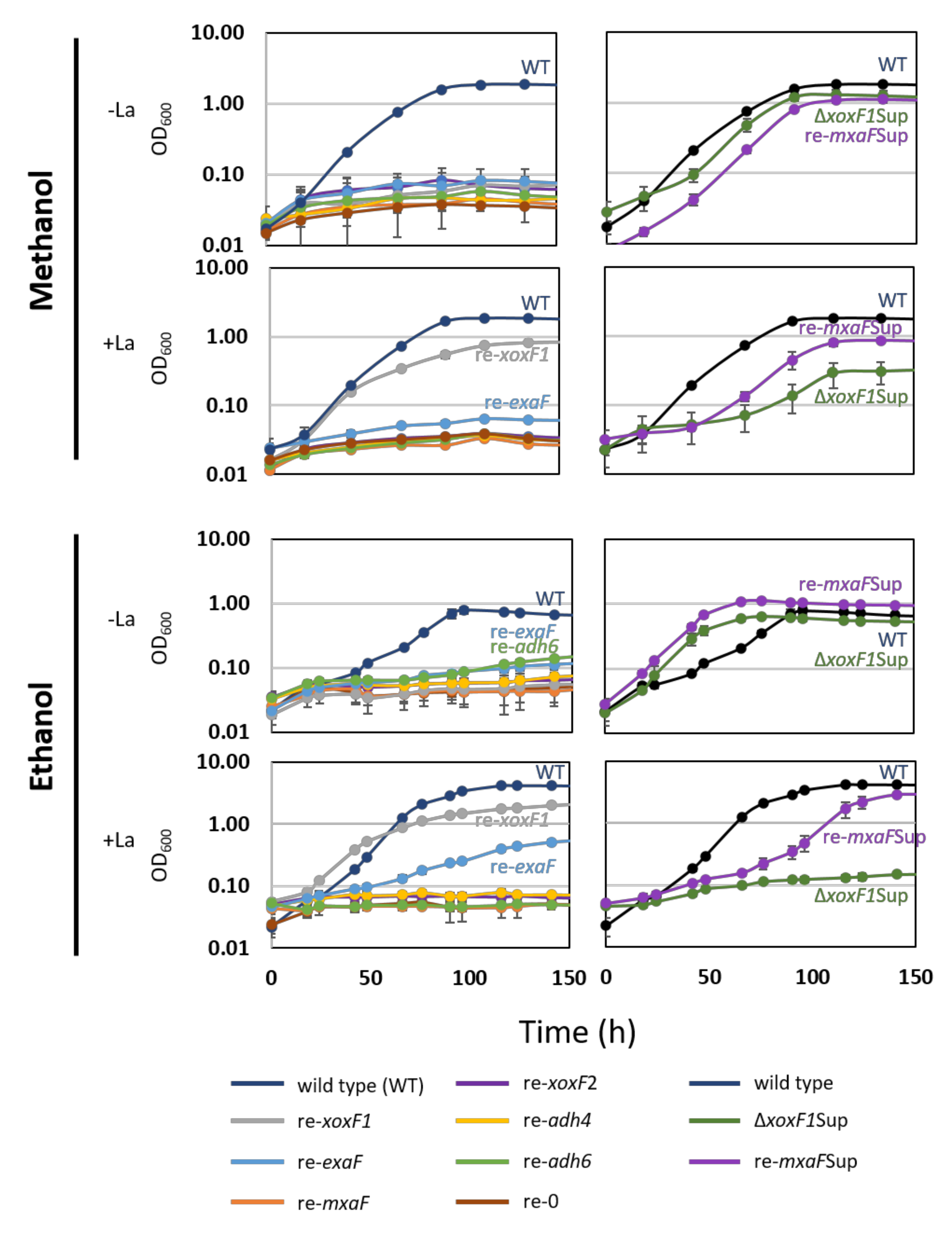
3.2. The Phenotype of MDH-Like Gene Mutants

To determine the roles of each MDH-like gene in methanol and ethanol growth, single-gene-remaining mutants of the six MDH-like genes were successfully generated, and their growth on methanol and ethanol in the presence and absence of La3+ was tested (Figure 3).
re-mxaF could not grow on methanol irrespective to the presence of La3+. re-xoxF1 could grow only in the presence of La3+, with lower growth yield compared to the wild-type. The specific growth rate (SGR) and yield of re-exaF and re-0 growth on methanol were not statistically significantly different (p = 0.999 for both growth rate and yield, technical triplicates, data not shown), suggesting that ExaF does not contribute to methanol growth. re-mxaFSup and ΔxoxF1Sup showed similar results, but slower growth on methanol compared to the wild-type in the absence of La3+. Their growth in the presence of La3+ resulted in slower growth and lower yield compared to the wild-type.
For ethanol growth, in the presence of La3+, re-xoxF1 could grow, while re-exaF grew slowly with a low yield. Based on these results, we could conclude that in MM+La condition XoxF1 mainly supports the growth. The suppression mutants have an MxaF—the regulation of which is independent of the presence of xoxF1.

3.3. Mutations Detected in the Suppression Mutants

The mutation sites in re-mxaFSup and ΔxoxF1Sup were identified through genome resequencing (Table S2). We found independent mutations in mxbD (Maq22A_c05310). In ΔxoxF1Sup, A761G caused N254S mutation. In re-mxaFSup, the deletion of 12 nucleotides at positions 613–624 caused a loss of 205LLLH. These positions are located within and close to the HAMP domain (positions 215–262) of MxbD, respectively.

3.4. ΔmxbD Phenotype

To prove that the mutations in mxbD were the cause of the suppression, we generated ΔmxbD. ΔmxbD showed no growth in MeOH-La conditions (Figure 4A); in MeOH+La conditions, it showed slower growth than the wild-type. The expression levels of mxaF and xoxF1 in the wild-type and ΔmxbD were quantified by qPCR (0.5% succinate was supplemented for ΔmxbD in the absence of La3+) (Figure 4B). The wild-type showed upregulated mxaF and xoxF1 expression in the absence and presence of La3+, respectively, showing that the Ln3+-switch is operating. In ΔmxbD, mxaF expression was repressed whereas xoxF1 expression was upregulated irrespective of La3+. These results suggest that mxbD is required for mxaF expression and that its absence releases xoxF1 repression.
To reproduce the suppression mutant phenotype, ΔxoxF1ΔmxbD was generated, and each mxbD from the wild-type (mxbD_w), ΔxoxF1Sup (mxbDxoxF1Sup), and re-mxaFSup (mxbD_re-mxaFSup) was introduced into ΔxoxF1ΔmxbD, using pQFKm as an expression vector. The mutant containing the control plasmid (pQFKm) did not grow on methanol (Figure 4C). The mutant complemented with wild-type mxbD did not grow as well since its genotype was the same as that of ΔxoxF1. The mutants complemented with the mutated mxbD showed growth in MeOH-La conditions. These results proved that the suppression phenotype was due to the mutations in mxbD and not to the other mutations detected by genome resequencing. The mutations commonly found in ΔxoxF1Sup and re-mxaFSup (13 sites) might be due to errors of the original genome sequence, which were not confirmed in this study (Table S2).

3.5. The Recombinant XoxF1-His Oxidizes Methanol and Formaldehyde

To characterize the functions of XoxF1 from strain 22A, an expression vector based on pCM130KmC was designed and constructed: pCM130kmC-xoxF1-His was introduced into ΔxoxF1. ΔxoxF1 could grow only slowly on methanol in the presence of La3+, but the transformant could grow well, suggesting that the introduced xoxF1 complemented the phenotype and that the His-tagged XoxF1 is catalytically functional in vitro.
The apparent monomeric molecular mass of the purified His-tagged XoxF1 (XoxF1-His) was ca. 60 kDa, as revealed by SDS-PAGE (Figure 5). The metal content of the purified XoxF1-His was determined to be 0.58 La3+ per monomer protein according to ICP-MS analysis. The enzyme kinetics of XoxF1-His were determined using methanol and formaldehyde as substrates. The kinetic constants (Km values were 0.03 and 0.34 mM, Vmax were 1.19 and 1.24 U·mg−1 for methanol and formaldehyde, respectively) indicated that XoxF1 can oxidize both methanol and formaldehyde in vitro. The kinetic parameters were within similar ranges reported for strain AM1 [19] and Bradyrhizobium diazoefficiens USDA110 [50].

3.6. hgd Encodes S-hydroxymethylglutathione Dehydrogenase

The GSH pathway in strain 22A is catalyzed by Gfa (Maq22A_1p37180) as the first step [51], which is expressed at a low level [38], followed by Hgd and other enzymes. As a pathway that oxidizes formaldehyde into formate, it is metabolically redundant to the H4MPT pathway. To determine the importance and contribution of the GSH pathway to formaldehyde oxidation, we measured the specific enzyme activity of Hgd in the cell-free extract of the cells grown on methanol plus succinate as a supplementary carbon source in the absence and presence of La3+ (Figure 6).
The wild-type, and Δfae1Δfae2 in the wild-type, ΔmxaF, and ΔxoxF1Sup background were used for the assay. The wild-type showed ca. 0.1 U/mg Hgd activity in both conditions. Δhgd did not show any activity, suggesting that hgd indeed encodes S-hydroxymethylglutathione dehydrogenase. Δfae1Δfae2 showed increased Hgd activity in the absence of La3+. Interestingly, ΔxoxF1SupΔfae1Δfae2 showed four times higher activity than the wild-type. However, ΔmxaFΔfae1Δfae2 showed almost no change in Hgd activity. These results suggested that Hgd activity is regulated depending on formaldehyde level in the cell, which was considered to increase in Δfae1Δfae2 mutants.

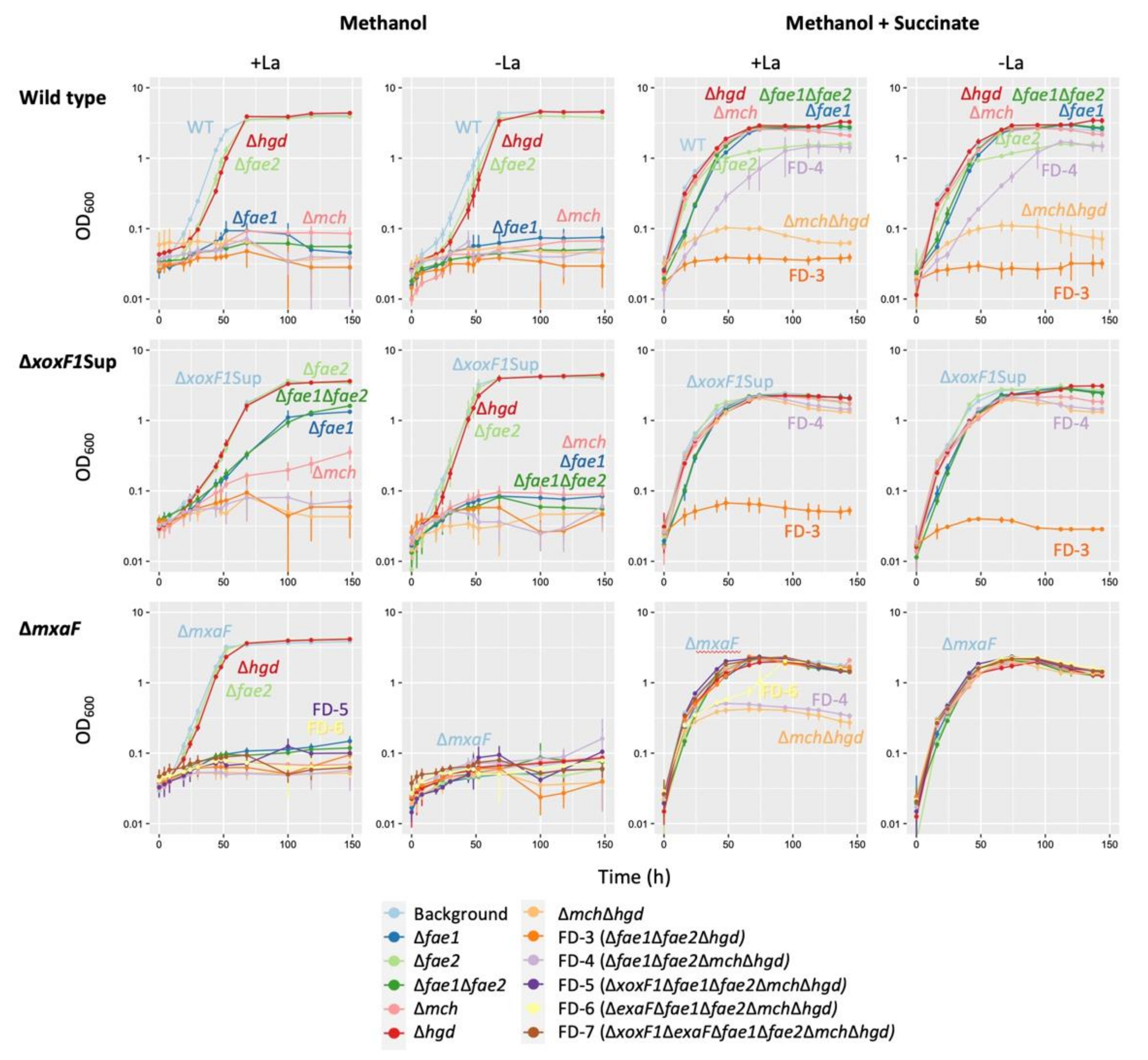
3.7. Characterization of the Mutants in Formaldehyde Oxidation Pathways

From the results above, we decided to use the following MDH mutants as genetic backgrounds to further study the formaldehyde oxidation pathways: ΔxoxF1Sup, which can grow moderately under MeOH-La conditions and slowly under MeOH+La conditions, with growth mainly dependent on mxaF, while its expression is de-repressed by the mutation in mxbD; and ΔmxaF, which uses mainly XoxF1 to grow on methanol only in the presence of La3+. We then generated single and multiple gene deletion mutants of the genes involved in formaldehyde oxidation, namely, fae1, fae2, and mch in the H4MPT pathway and hgd in the GSH pathway, of the wild-type, ΔxoxF1Sup, and ΔmxaF. They were grown in liquid MM containing methanol or methanol plus succinate in the presence and absence of La3+. The growth phenotypes and SGRs of the mutants are shown in Figure 7 and Table S3, respectively.
In the wild-type background, only Δhgd and Δfae2 grew on methanol irrespective to La3+ with lower growth rates compared to the wild-type, suggesting that Hgd and Fae2 participate in the methylotrophy in the wild-type. Other mutants in the H4MPT pathway (fae1 and mch) did not grow at all, suggesting the primary importance of the H4MPT pathway for the wild-type. In ΔxoxF1Sup background, interestingly, Δfae1, Δfae1Δfae2, and Δmch could grow slowly only in the presence of La3+. Because methylotrophic growth depending on ExaF was almost negligible (Figure 3), their growths were considered to be due to the de-repressed MxaF and GSH pathway. In the ΔmxaF background, only Δfae2 and Δhgd showed comparable growth to the background in the presence of La3+. These growths were considered to be dependent mainly on XoxF1-mediated methanol oxidation and the H4MPT pathway. FD-5 (ΔxoxF1Δfae1Δfae2ΔmchΔhgd) and FD-6 (ΔexaFΔfae1Δfae2ΔmchΔhgd) could not grow on methanol as a sole carbon source. Therefore, NADH generation in formaldehyde oxidation pathways is necessary for the growth, even if XoxF1 or ExaF oxidize methanol to formate.
We then used methanol plus succinate to discriminate the reason for the growing inability of the mutants on methanol; metabolic capacity or formaldehyde toxicity. In the wild-type background, Δfae1, Δfae2, Δfae1Δfae2, Δmch and Δhgd could grow irrespective to La3+, with growth rates comparable to the wild-type. Δfae2 showed decreased growth yield compared to the wild-type, again suggesting its participation in methylotrophy. FD-3 (Δfae1Δfae2Δhgd) could not grow, irrespective of La3+. This result suggested the involvement of the GSH pathway for formaldehyde detoxification. Interestingly, FD-4 (Δfae1Δfae2ΔmchΔhgd) could grow slowly, suggesting that additional mch deletion in FD-3 alleviates formaldehyde toxicity, and possibly that methanol oxidation (formaldehyde generation) occurred slowly in FD-4. In ΔxoxF1Sup background, only FD-3 could not grow whereas FD-4 could grow, again implying that no formaldehyde toxicity occurs in FD-4. In the ΔmxaF background in the presence of La3+, ΔmchΔhgd and FD-4 showed low-yield growth, suggesting that complete defect of formaldehyde oxidation pathways caused formaldehyde toxicity. This toxicity is less severe than that that occurred in ΔmchΔhgd and FD-3 in the wild-type background and ΔxoxF1Sup FD-3 that were unable to grow. This difference was due to the presence of mxaF, suggesting that MxaF produces formaldehyde to a toxic level, and XoxF1 also does so at a lower level. FD-5 (FD-4 plus ΔxoxF1) did not show toxicity, whereas FD-4 did, suggesting that XoxF1 is indeed causing formaldehyde toxicity in the absence of the formaldehyde oxidation pathways. FD-6 (FD-4 plus ΔexaF) showed a biphasic growth, suggesting that XoxF1 causes formaldehyde toxicity to some extent but later degrades that formaldehyde. Because FD-5 grew normally, formaldehyde toxicity by ExaF did not occur. Based on the results above, XoxF1 produces formaldehyde in vivo but does not oxidize it efficiently. On the other hand, ExaF has shown to oxidize formaldehyde in vivo.

4. Discussion

4.1. Identification of PQQ-ADH Genes for Methylotrophy in Strain 22A

Through phenotyping of MDH-like gene mutants (Figure 2 and Figure 3), we concluded that strain 22A possesses a set of MDHs (MxaF and XoxF1); ExaF-type ADH that supports Ln3+-dependent growth on ethanol. XoxF2 (strain 22A) did not support methylotrophic growth, therefore xoxF2 is a pseudogene or involved in other alcohol metabolism, which is different from the orthologous gene xoxF2 in strain AM1. Methylotrophic bacteria usually possess multiple XoxF-type genes besides MxaF. XoxF5-type is commonly found in alpha-, beta-, and gammaproteobacteria [52]. XoxF4-type is found in Methylophilaceae [52]. XoxF1-, XoxF2-, and XoxF3-types are limited to specific taxa [33,53]. Additionally, strains of Burkholderiales contain MDH2 that oxidizes both methanol and ethanol [54]. The PQQ-ADH type 6a family, to which two proteins associated with strain 22A (Adh4 and Adh6) belong, has not been characterized. The substrate specificity of these ADHs should be investigated to reveal the ability for the utilization of different alcohols in strain 22A.

4.2. The Mutation in MxbD de-repress mxaF

The genomes of the suppression mutants contain mutations in the HAMP domain of MxbD (Table S2). ΔmxbD was unable to grow in the absence of La3+ and showed no expression of mxaF and high xoxF1. Therefore, MxbD is necessary for mxaF expression but not for xoxF1 (Figure 4). Similar suppression mutants have been isolated from M. extorquens strain PA1 [30] and P. putida KT2440 [55]. The mutations found in the HAMP domain may confer signaling to the intracellular domain in the absence of ligand binding [56,57]. The overall regulation system for MDHs depending on the availability of substrates and metals, has not yet been clarified. It should be noted that ΔxoxF1Sup could be useful in further genetic studies as a mutant that has functional mxaF even with deleted xoxF1.

4.3. Formaldehyde Oxidation by XoxF1 and ExaF

We have confirmed that the purified recombinant XoxF1 oxidizes formaldehyde in vitro (Figure 5). In vitro activity of XoxF toward formaldehyde has also been shown in Methylacidiphilum fumariolicum SolV [18], M. extorquens AM1 [3,47], and Bradyrhizobium diazoefficiens USDA110 [50]. To determine whether this also occurs in vivo, we generated a series of formaldehyde oxidation pathway deficient mutants. The absence of formaldehyde toxicity in FD-3 in the ΔmxaF background (Figure 7) suggests XoxF1 and partly ExaF alleviate formaldehyde toxicity in vivo, implying that they oxidize formaldehyde in vivo. However, the biphasic growth of ΔmxaF FD-6 shows that formaldehyde oxidation by XoxF1 in vivo is not efficient. We previously showed that the GSH and H4MPT pathways in strain 22A were downregulated in the presence of La3+, yet cellular formaldehyde degradation activity remained high while formaldehyde production decreased [38]. These results indicated that although XoxF1 and ExaF have been shown to individually oxidize formaldehyde in vivo, ExaF may have the primary role in formaldehyde oxidation in the presence of La3+ when both enzymes are available. However, mutants lacking both of the formaldehyde oxidation pathways were unable to sustain their growth on methanol due to the lack of NADH generation [58].

4.4. The Role of the GSH Pathway

The GSH pathway for formaldehyde oxidation is considered “the most common reaction” in many organisms, including plants, mammals, and yeasts [59]. This pathway is used in methylotrophic bacteria such as Paracoccus denitrificans and Rhodobacter sphaeroides [33,51]. Some bacteria contain both GSH and H4MPT pathways, such as Burkholderia phymatum and B. fungorum LB400 [33,60]. In the Methylobacterium group, the genes for the pathway are found primarily in clade C1 (including M. aquaticum) and occasionally in clade A [7]. This patchy gene conservation within the genus is of interest, and its role in methylotrophy in the Methylobacterium group has not been investigated. We confirmed that hgd indeed encodes S-hydroxymethylglutathione-dehydrogenase (Figure 6). Δhgd in the wild-type background showed slower growth on methanol (Figure 7). In ΔxoxF1Sup background, whereas Δfae1Δfae2 could grow on methanol slowly, FD-3 could not grow, suggesting that the GSH pathway plays a role in formaldehyde oxidation. The introduction of flhA (GSH-and NAD-dependent formaldehyde dehydrogenase) and fghA (S-formyl-GSH hydrolase) from P. denitrificans complemented methanol growth in the H4MPT pathway deficient mutant of strain AM1 [11], suggesting that its role is metabolically exchangeable with that of the H4MPT pathway. Hgd is reported to have 1.1 U/mg activity (methanol conditions) in P. denitrificans [51], whereas it has ca. 0.1 U/mg activity in strain 22A. Taking these findings together, we conclude that the GSH pathway plays only a supportive role in the net formaldehyde oxidation in strain 22A. The patchy distribution of the pathway in Methylobacterium and related species may suggest the diminished importance of the GSH pathway in H4MPT pathway containing species.

4.5. The Role of H4MPT Pathway in Strain 22A

Strain AM1 fae mutant was unable to grow in methanol medium unless succinate was added, showing that fae is essential for methanol growth and that its absence causes formaldehyde toxicity [11,13], even in the presence of Ln3+ [19,47]. The substrate of Mch, methenyl-H4MPT, inhibits MtdA (NADP-dependent methylene-H4F dehydrogenase) in the H4F pathway, and thus controls the assimilation/dissimilation rate of formate [61]. Therefore, Mch is indispensable in strain AM1.
In strain 22A, Fae1 and Mch are also found to be essential for the wild-type to grow on methanol. However, Δfae1, Δfae1Δfae2, and Δmch in the ΔxoxF1Sup background could grow on methanol in the presence of La3+. We concluded that the H4MPT pathway is not essential when the methylotrophic growth is dependent only on de-repressed MxaF in strain 22A with a GSH pathway in the presence of La3+.

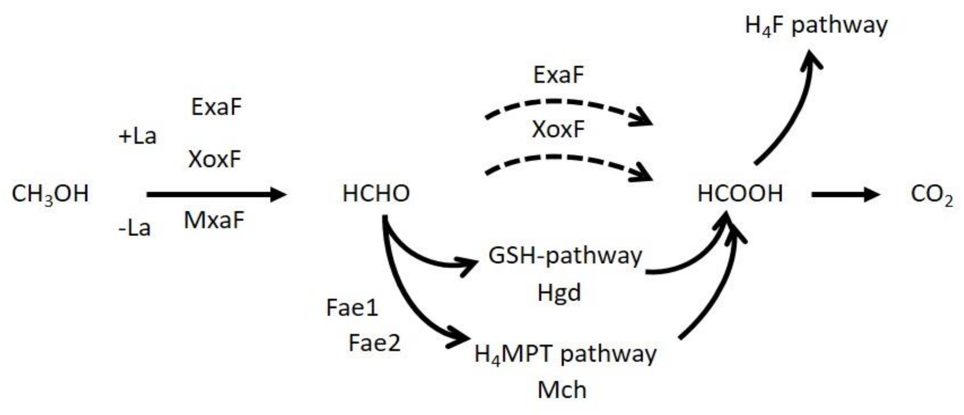
4.6. Overall Methanol Metabolism in Strain 22A

Through this study, we first revealed that among the six MDH-like proteins in strain 22A, two MDHs (MxaF and XoxF1) are involved in methanol oxidation. Second, MxbD is necessary for mxaF expression and xoxF1 repression, and a suppression mutant ΔxoxF1Sup enabled us to use it as a MxaF-expressing mutant without xoxF1. Third, the GSH pathway is functional and plays a role in formaldehyde oxidation and the generation of NADH; due to the low expression, however, its role is supportive for the net formaldehyde oxidation in addition to the H4MPT pathway that holds primary importance. Fourth, XoxF1 is capable of formaldehyde oxidation in vivo and in vitro and alleviates formaldehyde toxicity in formaldehyde oxidation-pathway mutants but in the absence of the NADH-producing pathways, it cannot solely support methanol growth. ExaF is involved in Ln3+-dependent ethanol growth and formaldehyde detoxification. In a natural environment, these redundant oxidation pathways catalyzed by different MDHs and formaldehyde oxidation pathways would enable cells to control the carbon flux in the redundant pathways, depending on the availability of Ln3+, the intracellular level of formaldehyde, and the required NADH level.

Supplementary Materials

The following are available online at https://www.mdpi.com/2076-2607/8/6/822/s1: Table S1, Primers used in this study; Table S2, Mutations detected in the suppression mutants by genome resequencing; Table S3, Specific growth rates (SGRs) and cell yield of various formaldehyde oxidation mutants grown on methanol and methanol plus succinate in the presence and absence of La3+.

Author Contributions

Conceptualization, methodology, P.Y., R.M., T.N. and A.T.; investigation, P.Y., Y.N., S.H., Y.F., T.I. and S.M.; writing—original draft preparation, P.Y.; writing—review and editing, all authors; formal analysis and visualization, P.Y. and A.T.; supervision and funding acquisition, A.T. All authors have read and agreed to the published version of the manuscript.

Funding

This work was supported in part by MEXT KAKENHI (No. 15H04476 and 18H02129), the Japan Science and Technology Agency (JST) A-Step feasibility study stage (#AS251Z00826N), and the Ohara Foundation for Agricultural Science.

Acknowledgments

We are grateful to Mrs. S. Rikiishi (IPSR, Okayama Univ.) for ICP analysis.

Conflicts of Interest

The authors declare no conflict of interest. The funders had no role in the design of the study; in the collection, analyses, or interpretation of data; in the writing of the manuscript, or in the decision to publish the results.

References

  1. Fall, R.; Benson, A.A. Leaf methanol the simplest natural product from plants. Trends Plant Sci. 1996, 1, 296–301. [Google Scholar] [CrossRef]
  2. Anthony, C. The Biochemistry of Methylotrophs; Academic Press: London, UK, 1982. [Google Scholar]
  3. Schmidt, S.; Christen, P.; Kiefer, P.; Vorholt, J.A. Functional investigation of methanol dehydrogenase-like protein XoxF in Methylobacterium extorquens AM1. Microbiology 2010, 156, 2575–2586. [Google Scholar] [CrossRef] [PubMed] [Green Version]
  4. Vorholt, J.A. Microbial life in the phyllosphere. Nat. Rev. Microbiol. 2012, 10, 828–840. [Google Scholar] [CrossRef] [PubMed]
  5. Knief, C.; Delmotte, N.; Chaffron, S.; Stark, M.; Innerebner, G.; Wassmann, R.; von Mering, C.; Vorholt, J.A. Metaproteogenomic analysis of microbial communities in the phyllosphere and rhizosphere of rice. ISME J. 2012, 6, 1378–1390. [Google Scholar] [CrossRef] [Green Version]
  6. Iguchi, H.; Yurimoto, H.; Sakai, Y. Interactions of methylotrophs with plants and other heterotrophic bacteria. Microorganisms 2015, 3, 137–151. [Google Scholar] [CrossRef]
  7. Green, P.N.; Ardley, J.K. Review of the genus Methylobacterium and closely related organisms: A proposal that some Methylobacterium species be reclassified into a new genus, Methylorubrum gen. Nov. Int. J. Syst. Evol. Microbiol. 2018, 68, 2727–2748. [Google Scholar] [CrossRef]
  8. Chistoserdova, L.; Chen, S.-W.; Lapidus, A.; Lidstrom, M.E. Methylotrophy in Methylobacterium extorquens AM1 from a genomic point of view. J. Bacteriol. 2003, 185, 2980–2987. [Google Scholar] [CrossRef] [Green Version]
  9. Anthony, C. How half a century of research was required to understand bacterial growth on C1 and C2 compounds; the story of the serine cycle and the ethylmalonyl-CoA pathway. Sci. Prog. 2011, 94, 109–137. [Google Scholar] [CrossRef]
  10. Anthony, C.; Williams, P. The structure and mechanism of methanol dehydrogenase. BBA-Proteins Proteom. 2003, 1647, 18–23. [Google Scholar] [CrossRef]
  11. Marx, C.J.; Chistoserdova, L.; Lidstrom, M.E. Formaldehyde-detoxifying role of the tetrahydromethanopterin-linked pathway in Methylobacterium extorquens AM1. J. Bacteriol. 2003, 185, 7160–7168. [Google Scholar] [CrossRef] [Green Version]
  12. Peyraud, R.; Schneider, K.; Kiefer, P.; Massou, S.; Vorholt, J.A.; Portais, J.-C. Genome-scale reconstruction and system level investigation of the metabolic network of Methylobacterium extorquens AM1. BMC Syst. Biol. 2011, 5, 189. [Google Scholar] [CrossRef] [PubMed] [Green Version]
  13. Vorholt, J.A.; Marx, C.J.; Lidstrom, M.E.; Thauer, R.K. Novel formaldehyde-activating enzyme in Methylobacterium extorquens AM1 required for growth on methanol. J. Bacteriol. 2000, 182, 6645–6650. [Google Scholar] [CrossRef] [PubMed] [Green Version]
  14. Crowther, G.J.; Kosaly, G.; Lidstrom, M.E. Formate as the main branch point for methylotrophic metabolism in Methylobacterium extorquens AM1. J. Bacteriol. 2008, 190, 5057–5062. [Google Scholar] [CrossRef] [PubMed] [Green Version]
  15. Ghosh, M.; Anthony, C.; Harlos, K.; Goodwin, M.G.; Blake, C. The refined structure of the quinoprotein methanol dehydrogenase from Methylobacterium extorquens at 1.94 Å. Structure 1995, 3, 177–187. [Google Scholar] [CrossRef] [Green Version]
  16. Hibi, Y.; Asai, K.; Arafuka, H.; Hamajima, M.; Iwama, T.; Kawai, K. Molecular structure of La3+-induced methanol dehydrogenase-like protein in Methylobacterium radiotolerans. J. Biosci. Bioeng. 2011, 111, 547–549. [Google Scholar] [CrossRef]
  17. Nakagawa, T.; Mitsui, R.; Tani, A.; Sasa, K.; Tashiro, S.; Iwama, T.; Hayakawa, T.; Kawai, K. A catalytic role of XoxF1 as La3+-dependent methanol dehydrogenase in Methylobacterium extorquens strain AM1. PLoS ONE 2012, 7, e50480. [Google Scholar] [CrossRef] [Green Version]
  18. Pol, A.; Barends, T.R.M.; Dietl, A.; Khadem, A.F.; Eygensteyn, J.; Jetten, M.S.M.; Op den Camp, H.J.M. Rare earth metals are essential for methanotrophic life in volcanic mudpots. Environ. Microbiol. 2014, 16, 255–264. [Google Scholar] [CrossRef]
  19. Good, N.M.; Moore, R.S.; Suriano, C.J.; Martinez-Gomez, N.C. Contrasting in vitro and in vivo methanol oxidation activities of lanthanide-dependent alcohol dehydrogenases XoxF1 and ExaF from Methylobacterium extorquens AM1. Sci. Rep. 2019, 9, 1–12. [Google Scholar] [CrossRef] [Green Version]
  20. Lv, H.; Masuda, S.; Fujitani, Y.; Sahin, N.; Tani, A. Oharaeibacter diazotrophicus gen. Nov., sp. Nov., a diazotrophic and facultatively methylotrophic bacterium, isolated from rice rhizosphere. Int. J. Syst. Evol. Microbiol. 2017, 67, 576–582. [Google Scholar] [CrossRef]
  21. Lv, H.; Sahin, N.; Tani, A. Isolation and genomic characterization of Novimethylophilus kurashikiensis gen. Nov. Sp. Nov., a new lanthanide-dependent methylotrophic species of methylophilaceae. Environ. Microbiol. 2018, 20, 1204–1223. [Google Scholar] [CrossRef]
  22. Lv, H.; Tani, A. Genomic characterization of methylotrophy of Oharaeibacter diazotrophicus strain SM30T. J. Biosci. Bioeng. 2018, 126, 667–675. [Google Scholar] [CrossRef] [PubMed]
  23. Fitriyanto, N.A.; Fushimi, M.; Matsunaga, M.; Pertiwiningrum, A.; Iwama, T.; Kawai, K. Molecular structure and gene analysis of Ce3+-induced methanol dehydrogenase of Bradyrhizobium sp. MAFF211645. J. Biosci. Bioeng. 2011, 111, 613–617. [Google Scholar] [CrossRef]
  24. Howat, A.M.; Vollmers, J.; Taubert, M.; Grob, C.; Dixon, J.L.; Todd, J.D.; Chen, Y.; Kaster, A.-K.; Murell, J.C. Comparative genomics and mutational analysis reveals a novel XoxF-utilizing methylotroph in the Roseobacter group isolated from the marine environment. Front. Microbiol. 2018, 9. [Google Scholar] [CrossRef] [PubMed]
  25. Farhan Ul Haque, M.; Kalidass, B.; Bandow, N.; Turpin, E.A.; DiSpirito, A.A.; Semrau, J.D. Cerium regulates expression of alternative methanol dehydrogenase in Methylosinus trichosporium OB3b. Appl. Environ. Microb. 2015, 81, 7546–7552. [Google Scholar] [CrossRef] [PubMed] [Green Version]
  26. Chu, F.; Lidstrom, M.E. XoxF acts as the predominant methanol dehydrogenase in the type I methanotroph Methylomicrobium buryatense. J. Bacteriol. 2016, 198, 1317–1325. [Google Scholar] [CrossRef] [Green Version]
  27. Vu, H.N.; Subuyuj, G.A.; Vijayakumar, S.; Good, N.M.; Martinez-Gomez, N.C.; Skovran, E. Lanthanide-dependent regulation of methanol oxidation systems in Methylobacterium extorquens AM1 and their contribution to methanol growth. J. Bacteriol. 2016, 198, 1250–1259. [Google Scholar] [CrossRef] [Green Version]
  28. Skovran, E.; Palmer, A.D.; Rountree, A.M.; Good, N.M.; Lidstrom, M.E. XoxF is required for expression of methanol dehydrogenase in Methylobacterium extorquens AM1. J. Bacteriol. 2011, 193, 6032–6038. [Google Scholar] [CrossRef] [Green Version]
  29. Zheng, Y.; Huang, J.; Zhao, F.; Chistoserdova, L. Physiological effect of XoxG(4) on lanthanide-dependent methanotrophy. mBio 2018, 9, e02430-17. [Google Scholar] [CrossRef] [Green Version]
  30. Ochsner, A.M.; Hemmerle, L.; Vonderach, T.; Nüssli, R.; Bortfeld-Miller, M.; Hattendorf, B.; Vorholt, J.A. Use of rare-earth elements in the phyllosphere colonizer Methylobacterium extorquens PA1. Mol. Microbiol. 2019, 111, 1152–1166. [Google Scholar] [CrossRef] [Green Version]
  31. Roszczenko-Jasińska, P.; Vu, H.N.; Subuyuj, G.A.; Crisostomo, R.V.; Cai, J.; Raghuraman, C.; Ayala, E.M.; Clippard, E.J.; Lien, N.F.; Ngo, R.T.; et al. Lanthanide transport, storage, and beyond: Genes and processes contributing to XoxF function in Methylorubrum extorquens AM1. bioRxiv 2019. [Google Scholar] [CrossRef]
  32. Good, N.M.; Vu, H.N.; Suriano, C.J.; Subuyuj, G.A.; Skovran, E.; Martinez-Gomez, N.C. Pyrroloquinoline quinone ethanol dehydrogenase in Methylobacterium extorquens AM1 extends lanthanide-dependent metabolism to multicarbon substrates. J. Bacteriol. 2016, 198, 3109–3118. [Google Scholar] [CrossRef] [PubMed] [Green Version]
  33. Keltjens, J.T.; Pol, A.; Reimann, J.; Op den Camp, H.J.M. PQQ-dependent methanol dehydrogenases: Rare-earth elements make a difference. Appl. Microbiol. Biotechnol. 2014, 98, 6163–6183. [Google Scholar] [CrossRef] [PubMed]
  34. Chistoserdova, L. Modularity of methylotrophy, revisited. Environ. Microbiol. 2011, 13, 2603–2622. [Google Scholar] [CrossRef] [PubMed]
  35. Wu, M.L.; Wessels, H.J.C.T.; Pol, A.; Op den Camp, H.J.M.; Jetten, M.S.M.; van Niftrik, L.; Keltjens, J.T. XoxF-type methanol dehydrogenase from the anaerobic methanotroph “Candidatus methylomirabilis oxyfera”. Appl. Environ. Microbiol. 2015, 81, 1442–1451. [Google Scholar] [CrossRef] [PubMed] [Green Version]
  36. Tani, A.; Akita, M.; Murase, H.; Kimbara, K. Culturable bacteria in hydroponic cultures of moss Racomitrium japonicum and their potential as biofertilizers for moss production. J. Biosci. Bioeng. 2011, 112, 32–39. [Google Scholar] [CrossRef] [PubMed]
  37. Tani, A.; Takai, Y.; Suzukawa, I.; Akita, M.; Murase, H.; Kimbara, K. Practical application of methanol-mediated mutualistic symbiosis between Methylobacterium species and a roof greening moss, Racomitrium japonicum. PLoS ONE 2012, 7, e33800. [Google Scholar] [CrossRef] [Green Version]
  38. Masuda, S.; Suzuki, Y.; Fujitani, Y.; Mitsui, R.; Nakagawa, T.; Shintani, M.; Tani, A. Lanthanide-dependent regulation of methylotrophy in Methylobacterium aquaticum strain 22A. mSphere 2018, 3. [Google Scholar] [CrossRef] [Green Version]
  39. Alamgir, K.M.; Masuda, S.; Fujitani, Y.; Fukuda, F.; Tani, A. Production of ergothioneine by Methylobacterium species. Front. Microbiol. 2015, 6. [Google Scholar] [CrossRef] [Green Version]
  40. Li, H.; Durbin, R. Fast and accurate short read alignment with burrows-wheeler transform. Bioinformatics 2009, 25, 1754–1760. [Google Scholar] [CrossRef] [Green Version]
  41. Carver, T.; Harris, S.R.; Otto, T.D.; Berriman, M.; Parkhill, J.; McQuillan, J.A. BamView: Visualizing and interpretation of next-generation sequencing read alignments. Brief. Bioinform. 2012, 14, 203–212. [Google Scholar] [CrossRef] [Green Version]
  42. Li, H.; Handsaker, B.; Wysoker, A.; Fennell, T.; Ruan, J.; Homer, N.; Marth, G.; Abecasis, G.; Durbin, R. 1000 Genome Project Data Processing Subgroup. The sequence alignment/map format and SAMtools. Bioinformatics 2009, 25, 2078–2079. [Google Scholar] [CrossRef] [PubMed] [Green Version]
  43. Kaczmarczyk, A.; Vorholt, J.A.; Francez-Charlot, A. Cumate-inducible gene expression system for sphingomonads and other alphaproteobacteria. Appl. Environ. Microbiol. 2013, 79, 6795–6802. [Google Scholar] [CrossRef] [PubMed] [Green Version]
  44. Marx, C.J.; Lidstrom, M.E. Development of improved versatile broad-host-range vectors for use in methylotrophs and other gram-negative bacteria. Microbiology 2001, 147, 2065–2075. [Google Scholar] [CrossRef] [PubMed] [Green Version]
  45. Bradford, M. A rapid and sensitive method for the quantitation of microgram quantities of protein utilizing the principle of protein-dye binding. Anal. Biochem. 1976, 72, 248–254. [Google Scholar] [CrossRef]
  46. Uotila, L.; Koivusalo, M. [42] formaldehyde dehydrogenase. In Methods in Enzymology; Elsevier: Amsterdam, The Netherlands, 1981; pp. 314–320. [Google Scholar] [CrossRef]
  47. Good, N.M.; Walser, O.N.; Moore, R.S.; Suriano, C.J.; Huff, A.F.; Martínez-Gómez, N.C. Investigation of lanthanide-dependent methylotrophy uncovers complementary roles for alcohol dehydrogenase enzymes. bioRxiv 2018. [Google Scholar] [CrossRef]
  48. Jones, D.T.; Taylor, W.R.; Thornton, J.M. The rapid generation of mutation data matrices from protein sequences. Bioinformatics 1992, 8, 275–282. [Google Scholar] [CrossRef]
  49. Kumar, S.; Stecher, G.; Tamura, K. MEGA7: Molecular evolutionary genetics analysis version 7.0 for bigger datasets. Mol. Biol. Evol. 2016, 33, 1870–1874. [Google Scholar] [CrossRef] [Green Version]
  50. Wang, L.; Suganuma, S.; Hibino, A.; Mitsui, R.; Tani, A.; Matsumoto, T.; Ebihara, A.; Fitriyanto, N.A.; Pertiwiningrum, A.; Shimada, M.; et al. Lanthanide-dependent methanol dehydrogenase from the legume symbiotic nitrogen-fixing bacterium Bradyrhizobium diazoefficiens strain USDA110. Enzyme Microb. Technol. 2019, 130, 109371. [Google Scholar] [CrossRef]
  51. Goenrich, M.; Bartoschek, S.; Hagemeier, C.H.; Griesinger, C.; Vorholt, J.A. A glutathione-dependent formaldehyde-activating enzyme (Gfa) from Paracoccus denitrificans detected and purified via two-dimensional proton exchange NMR spectroscopy. J. Biol. Chem. 2002, 277, 3069–3072. [Google Scholar] [CrossRef] [Green Version]
  52. Huang, J.; Yu, Z.; Chistoserdova, L. Lanthanide-dependent methanol dehydrogenases of XoxF4 and XoxF5 clades are differentially distributed among methylotrophic bacteria and they reveal different biochemical properties. Front. Microbiol. 2018, 9. [Google Scholar] [CrossRef]
  53. Chistoserdova, L. C1 transfer enzymes and coenzymes linking methylotrophic bacteria and methanogenic archaea. Science 1998, 281, 99–102. [Google Scholar] [CrossRef] [PubMed]
  54. Kalyuzhnaya, M.G.; Hristova, K.R.; Lidstrom, M.E.; Chistoserdova, L. Characterization of a novel methanol dehydrogenase in representatives of Burkholderiales: Implications for environmental detection of methylotrophy and evidence for convergent evolution. J. Bacteriol. 2008, 190, 3817–3823. [Google Scholar] [CrossRef] [Green Version]
  55. Wehrmann, M.; Berthelot, C.; Billard, P.; Klebensberger, J. The PedS2/PedR2 two-component system is crucial for the rare earth element switch in Pseudomonas putida KT2440. mSphere 2018, 3. [Google Scholar] [CrossRef] [PubMed] [Green Version]
  56. Aravind, L.; Ponting, C.P. The cytoplasmic helical linker domain of receptor histidine kinase and methyl-accepting proteins is common to many prokaryotic signalling proteins. FEMS Microbiol. Lett. 1999, 176, 111–116. [Google Scholar] [CrossRef] [PubMed]
  57. Matamouros, S.; Hager, K.R.; Miller, S.I. HAMP domain rotation and tilting movements associated with signal transduction in the PhoQ sensor kinase. mBio 2015, 6. [Google Scholar] [CrossRef]
  58. Šmejkalová, H.; Erb, T.J.; Fuchs, G. Methanol assimilation in Methylobacterium extorquens AM1: Demonstration of all enzymes and their regulation. PLoS ONE 2010, 5, e13001. [Google Scholar] [CrossRef] [PubMed] [Green Version]
  59. Yurimoto, H.; Kato, N.; Sakai, Y. Assimilation, dissimilation, and detoxification of formaldehyde, a central metabolic intermediate of methylotrophic metabolism. Chem. Rec. 2005, 5, 367–375. [Google Scholar] [CrossRef]
  60. Marx, C.J.; Miller, J.A.; Chistoserdova, L.; Lidstrom, M.E. Multiple formaldehyde oxidation/detoxification pathways in Burkholderia fungorum LB400. J. Bacteriol. 2004, 186, 2173–2178. [Google Scholar] [CrossRef] [Green Version]
  61. Martinez-Gomez, N.C.; Good, N.M.; Lidstrom, M.E. Methenyl-dephosphotetrahydromethanopterin is a regulatory signal for acclimation to changes in substrate availability in Methylobacterium extorquens AM1. J. Bacteriol. 2015, 197, 2020–2026. [Google Scholar] [CrossRef] [Green Version]
Figure 1. A schematic of the methylotrophy pathway. Dashed lines indicate possible direct oxidations.
Microorganisms 08 00822 g001
Figure 2. Molecular phylogenetic analysis of MDH-like proteins found in strain 22A (in boldface) and other related sequences. The evolutionary history was inferred by using the maximum likelihood method based on the Jones-Taylor-Thornton (JTT) matrix-based model [48]. The tree is drawn to scale. Evolutionary analyses were conducted in MEGA7 [49]. Bar, average number of amino acid substitutions per site.
Microorganisms 08 00822 g002
Figure 3. Left panel, growth of single-gene-remaining mutants of MDH-like genes and a mutant without any MDH-like gene (re-0). Right panel, growth of the suppression mutants. Cultivation was done in 96-well plates under shaking at 300 rpm. The results are presented as the average ± SD (technical triplicates).
Microorganisms 08 00822 g003
Figure 4. (A) Growth of strain 22A wild-type and ΔmxbD on (left panel) succinate and (right panel) methanol in the presence/absence of La3+. Circles, wild-type (WT); squares, ΔmxbD; filled symbols, in the presence of La3+; open symbols, in the absence of La3+. The results are presented as the average ± SD (technical triplicates). (B) Quantification of mxaF and xoxF1 expression in strain 22A wild-type and ΔmxbD, grown on methanol in the presence/absence of La3+. ΔmxbD in +La condition was grown on methanol plus succinate. The results are presented as the average ± SD (biological triplicates). The compact letter display indicates statistically significant differences (p < 0.05, ANOVA and Tukey’s multiple comparison test) among the means for each gene. (C) Growth of ΔxoxF1ΔmxbD complemented by wild-type and mutated mxbD on methanol in the presence/absence of La3+. Open circle, vector control; closed circle, ΔxoxF1ΔmxbD (mxbD-wt); squares, ΔxoxF1ΔmxbD (mxbD-re-mxaFSup); triangles, ΔxoxF1ΔmxbD (mxbDxoxF1Sup). The results are presented as the average ± SD (technical triplicates).
Microorganisms 08 00822 g004
Figure 5. (A) Purification of XoxF1-His expressed in strain 22A. SDS-PAGE of crude cell extract, flow-through fraction through Ni-NTA column, and purified MDH in eluate fractions. An arrow indicates the purified XoxF1-His. (B) The activity of XoxF1-His against methanol and formaldehyde of varying concentrations. The data were used to calculate kinetic parameters. The results are presented as the average ± SD (technical triplicates).
Microorganisms 08 00822 g005
Figure 6. Hgd activity in the cell-free extracts of the wild-type, Δfae1Δfae2, ΔxoxF1SupΔfae1Δfae2, ΔmxaFΔfae1Δfae2, and Δhgd, grown on methanol plus succinate in the (A) presence and (B) absence of La3+. Filled bar, +La3+; open bar, -La3+; n.d., not detected. The results are presented as the average ± SD (biological triplicates). Statistical tests were done with ANOVA and Tukey’s multiple comparison test independently for each dataset (presence/absence of La3+), and p values for the comparisons with the wild-type data are shown.
Microorganisms 08 00822 g006
Figure 7. Growth of various formaldehyde oxidation-pathway mutants constructed in the wild-type, ΔxoxF1Sup, and ΔmxaF backgrounds, in methanol and methanol plus succinate liquid medium in the presence and absence of La3+. FD-3 (Δfae1Δfae2Δhgd), FD-4 (Δfae1Δfae2ΔmchΔhgd), FD-5 (ΔxoxF1Δfae1Δfae2ΔmchΔhgd), FD-6 (ΔexaFΔfae1Δfae2ΔmchΔhgd), and FD-7 (ΔxoxF1ΔexaFΔfae1Δfae2ΔmchΔhgd) correspond to all three genetic backgrounds, except that FD-5 to FD-7 were constructed only for the mxaF background. The results are presented as the average ± SD (technical triplicates).
Microorganisms 08 00822 g007

