Article

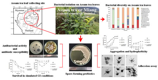
Culturable Bacterial Community on Leaves of Assam Tea (Camellia sinensis var. assamica) in Thailand and Human Probiotic Potential of Isolated Bacillus spp.

by
Patthanasak Rungsirivanich
1,2,
Witsanu Supandee
3,
Wirapong Futui
1,2,
Vipanee Chumsai-Na-Ayudhya
1,2,
Chaowarin Yodsombat
4 and
Narumol Thongwai
1,5,*
1
Department of Biology, Faculty of Science, Chiang Mai University, Chiang Mai 50200, Thailand
2
Graduate School, Chiang Mai University, Chiang Mai 50200, Thailand
3
Darunsikkhalai School, King Mongkut’s University of Technology Thonburi, Bangkok 10150, Thailand
4
Production and Water Quality Control Division, Provincial Waterworks Authority Region 2, Saraburi 18000, Thailand
5
Research Center in Bioresources for Agriculture, Industry and Medicine, Chiang Mai University, Chiang Mai 50200, Thailand
*
Author to whom correspondence should be addressed.
Microorganisms 2020, 8(10), 1585; https://doi.org/10.3390/microorganisms8101585
Submission received: 7 September 2020 / Revised: 10 October 2020 / Accepted: 13 October 2020 / Published: 14 October 2020
(This article belongs to the Special Issue Microbial Isolation and Characterization)

Abstract

:
Assam tea plants (Camellia sinensis var. assamica) or Miang are found in plantations and forests of Northern Thailand. Leaf fermentation has been performed for centuries, but little information is available about their associated microbial community. One hundred and fifty-seven bacterial isolates were isolated from 62 Assam tea leaf samples collected from 6 provinces of Northern Thailand and classified within the phyla of Firmicutes, Actinobacteria, and Proteobacteria. Phayao and Phrae provinces exhibited the highest and the lowest bacterial diversities, respectively. The bacterial community structural pattern demonstrated significant differences between the west and the east sides. Since some Bacillus spp. have been reported to be involved in fermented Miang, Bacillus spp. isolated in this study were chosen for further elucidation. Bacillus siamensis ML122-2 exhibited a growth inhibitory effect against Staphylococcus aureus ATCC 25923 and MRSA DMST 20625, and the highest survival ability in simulated gastric and intestinal fluids (32.3 and 99.7%, respectively), autoaggregation (93.2%), cell surface hydrophobicity (50.0%), and bacterial adherence with Vero cells (75.8% of the control Lactiplantibacillus plantarum FM03-1). This B. siamensis ML122-2 is a promising probiotic to be used in the food industry and seems to have potential antibacterial properties relevant for the treatment of antibiotic-resistant infections.

Graphical Abstract

1. Introduction

Assam tea (Camellia sinensis var. assamica) or “Miang”, as called by Northern Thai people, is a perennial native plant from the highlands of Northern Thailand, i.e., Chiang Mai, Chiang Rai, Lampang, Nan, Phayao, and Phrae provinces, and neighbor countries [1,2]. “Fermented Miang” production has been inherited from generation to generation for centuries [3]. The microbiome of fermented Assam tea leaves studied by several researchers has revealed the involvement of naturally-occurring lactic acid bacteria (LAB), including Enterococcus calmelliae, Lacticaseibacillus casei, Lacticaseibacillus camelliae, Lacticaseibacillus pantheris, Lacticaseibacillus thailandensis, Lactiplantibacillus pentosus, Lactiplantibacillus plantarum, Leuconostoc mesenteroides, Lentilactobacillus buchneri, Levilactobacillus brevis, Pediococcus siamensis, Streptococcus faecalis, and Streptococcus lactis [1,4,5]. Hence, fermented Assam tea leaves are rich in probiotics providing health benefits besides its catechin, catechin derivatives, and other bioactive compounds.
Microbial diversity in tea plants and its influence on the fermentation process has been studied by several authors, indicating that the process is driven by the interaction between fungal and bacterial diversity, community composition, and environmental factors (climate, relative humidity, soil pH, and soil structure) [2,6,7,8,9,10].
The bacterial community on Assam tea leaves from the tea gardens in Nan province of Thailand revealed by Rungsirivanich et al. [11] consisted of the genera Bacillus, Floricoccus, Kocuria, Lysinibacillus, Micrococcus, and Staphylococcus. Additionally, Bacillus licheniformis, Bacillus siamensis, and Bacillus tequilensis were pointed as important elements for biological control during leaf fermentation by inhibiting the growth of food spoilage and pathogenic microbes [12]. In this gut microbiome era, sources of probiotics are intensely studied, and the Assam tea plant and its products have proved to be notable.
For decades, LAB have been widely recognized as probiotics, the live microbes conferring health benefits on the host when adequate amounts are administered [13]. Probiotic consumption presents several benefits such as prevention of diarrhea and bowel inflammation that leads to cancer. Besides that, probiotics also help to decrease cholesterol levels and improve lactose intolerance as well as to enhance the utilization of nutrients [14]. Recently, Bacillus spp. have been studied as spore-forming probiotics due to the presence of endospores that enhance tolerance to the extreme conditions of the gastrointestinal tract [15]. Previous studies also showed that some species of Bacillus participate in antimicrobial peptide production such as bacteriocin-like inhibitory substance (BLIS) [16,17].
This study aimed to assess the bacterial diversity on Assam tea leaves collected from six provinces in Northern Thailand, namely Chiang Mai, Chiang Rai, Lampang, Phayao, Phrae, and Nan provinces, and to investigate the probiotic properties of Bacillus spp. isolated from Assam tea leaf surfaces. The obtained results will provide insightful information regarding the role of these bacteria on Assam tea leaves as well as their probiotic potential.

2. Materials and Methods

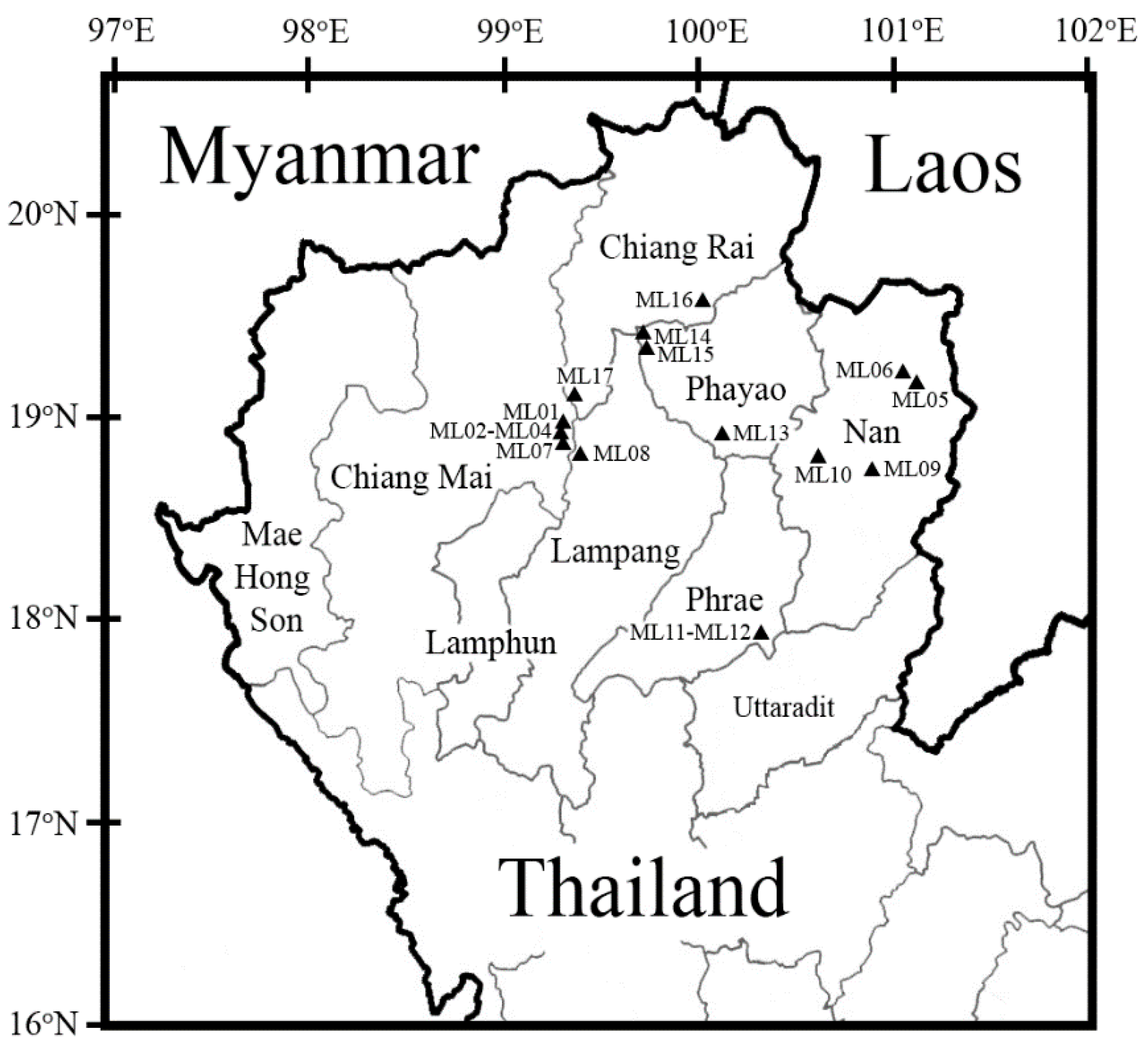
2.1. Sampling Site and Collection of Assam Tea Leaves

Sixty two fresh Assam tea leaf samples were collected during the time period of 7 July 2015 to 18 November 2018 at altitudes of 243–1278 m above sea level from 17 sampling sites (Figure 1 and Table S1) in Northern Thailand, including a province on the west side (Chiang Mai (codes ML01-ML04 in Doi Saket and ML07 in Mae On districts)) and five provinces on the east side (Chiang Rai (codes ML16 in Mae Loi and ML17 in Wiang Papao districts), Lampang (code ML08 in Mueang Pan district), Phayao (code ML13 in Dok Khamtai and ML14–ML15 in Mae Chai districts), Phrae (codes ML11–ML12 in Mueang Phrae district), and Nan (codes ML05-ML06 in Pua, ML09 in Phu Phiang, and ML10 in Mueang Nan districts)). The collection of Assam tea leaves was performed as described by Levetin and Dorsey [18] with slight modifications. A 4 cm2 (2 cm × 2 cm) area on the surface of fresh leaves was swabbed using a moistened sterile cotton swab. Bacterial isolation was performed as soon as possible within a day.

2.2. Isolation of Bacteria

A serial dilution (1 × 101–1 × 10²) of each swabbed sample was conducted using NaCl, 0.85% (w/v). The spread plate technique was subsequently performed on tryptic soy agar (TSA) (Merck™, Darmstadt, Germany) and de Man, Rogosa, and Sharpe (MRS) agar (Merck™) containing 0.004% (w/v) bromocresol purple. The culture plates were incubated at 37 °C for 24–48 h. All bacterial colonies on TSA showing different morphologies and the yellow bacterial colonies on MRS agar were further re-streaked to obtain pure cultures [5,12]. These bacterial isolates were kept at −20 °C until used.

2.3. Bacterial Genomic DNA Extraction

The chromosomal DNA of each bacterial isolate was extracted according to the method described by Pitcher et al. [19] with slight modifications. Briefly, each isolate was grown in tryptic soy broth (TSB) at 37 °C for 24–48 h. One milliliter of culture broth was centrifuged for 5 min at 6000 rpm at 4 °C. The supernatant was removed, and the pellet was resuspended in 1 mL of 1X TNE buffer (10 mM Tris, 1 mM EDTA, 0.1 M NaCl, pH 7.4). The mixture was then centrifuged for 5 min at 6000 rpm at 4 °C. Three hundred microliters of 1X TNE containing 2% (v/v) Triton X-100 were added to each pellet and mixed for 2 min using a vortex mixer. Subsequently, 300 µL of a solution containing phenol:chloroform:isoamyl alcohol (25:24:1) was added and mixed for 2 min, followed by centrifugation for 10 min at 12,000 rpm at 4 °C. The aqueous phase was transferred to a new microtube and mixed with 500 µL of ethanol at 95% (v/v). Each tube was stored at –20 °C overnight and spun at 12,000 rpm at 4 °C for 15 min. The supernatant was slowly removed and the pellet was dried at room temperature and resuspended in 30–50 µL in sterile distilled water. The DNA on each tube was kept at −20 °C.

2.4. 16S rRNA Gene Amplification

Amplification of the bacterial 16S rRNA gene was performed by polymerase chain reaction (PCR) in a thermal cycler (Labcycler, SensoQuest, Germany). The universal bacterial primers, 27F (5′-AGA GTT TGA TCM TGG CTC AG-3′) and 1492R (5′-TAC GGY TAC CTT GTT ACG ACT T-3′) were used [20]. The PCR reactions were held at 94 °C for 5 min for DNA denaturation, followed by 35 cycles of denaturation at 94 °C for 30 s, annealing at 56 °C for 30 s, extension at 72 °C for 1 min, and an elongation step at 72 °C for 5 min to ensure complete amplification. PCR samples were temporarily stored at 4 °C. Amplicons were electrophoresed on agarose gel, 0.8% (w/v), supplemented with 1X nucleic acid staining solution (RedSafe®, iNtRON Biotechnology, Inc., Seongnam-Si, Korea) for 50 min at 95 V and 300 mA in 1X Tris-acetate-EDTA (TAE) buffer (40 mM Tris-acetate, 1 mM EDTA, pH 8.0) using the electrophoretic gel system (EC320, Minicell Primo, CA, USA) at room temperature. The marker used was a 1 kb DNA ladder (RBC Bioscience, New Taipei, Taiwan). The gel documentation system (SynGene, Frederick, MD, USA) was used for gel visualization under UV light. The PCR products were purified and sequenced by DNA sequencing services (First BASE Laboratories Sdn Bhd., Selangor, Malaysia). The 16S rRNA gene sequence homologies were retrieved from the National Center for Biotechnology Information (NCBI) GenBank (https://www.ncbi.nlm.nih.gov/) and EzBioCloud databases (https://www.ezbiocloud.net/). The sequence data were aligned, and the phylogenetic tree was constructed by a neighbor-joining method [21] with the MEGA 7 program [22]. The bootstrap values of 1000 replicates were performed [23] with the Tamura–Nei model [24]. Amplicon sequences were deposited in the GenBank.

2.5. Probiotic Property Characterization of Bacillus Strains

2.5.1. Antibacterial Activity Investigation

The pathogenic bacteria consisting of Bacillus cereus TISTR 687, Escherichia coli ATCC 25922, E. coli O157:H7 DMST 12743, methicillin resistant Staphylococcus aureus DMST 20625, Salmonella Typhi DMST 22842, Shigella dysenteriae DMST 1511, Staphylococcus aureus ATCC 25923, and Vibrio cholerae DMST 2873 were used for determination of antibacterial activity. The Bacillus strains were cultured in TSB at 37 °C overnight. The supernatant was filtered through a 0.45 μm nylon syringe filter before use. Thirty-three microliters of the Bacillus culture filtrate were used to evaluate the antimicrobial properties against pathogenic bacteria using the agar well diffusion method [25].

2.5.2. Antibiotic Susceptibility Assay

Antibiotic discs consisting of amikacin (30 μg), amoxyclav (30 μg), ampicillin (10 μg), ampicillin/sulbactum (10 μg/10 μg), cefixime (5 μg), ceftriaxone (30 μg), cefuroxime (30 μg), cefuroxime axetil (30 μg), cefotaxime (30 μg), cefoxitin (30 μg), co-trimoxazole (25 μg), gentamicin (10 μg), meropenem (10 μg), ofloxacin (5 μg), tetracycline (30 μg), and ticarcillin/clavulanic acid (75/10 μg) were purchased from Himedia, India. The susceptibility test of Bacillus strains was done as described by the Clinical Laboratory Standards Institute (CLSI) [26].

2.5.3. Survival Evaluation in Simulated Gastric and Intestinal Fluids

The fluids resembling human gastric and intestinal fluids were prepared according to the method of Huang and Adams [27]. The Bacillus isolates were grown in TSB at 37 °C for 18–24 h before harvesting by centrifugation for 5 min at 6000 rpm at 4 °C. Then, the cell pellets were washed twice and resuspended in 1X PBS (~1 × 10⁹ CFU/mL). Then, the Bacillus cell suspension was inoculated into simulated gastric fluid (TSB supplemented with pepsin, 3 mg/mL, pH 2.5) to obtain about 1 × 10⁸ CFU/mL of the initial cell concentration. The mixture was incubated at 37 °C and 50 rpm in a shaker incubator. Viable cell counts were performed on TSA plates at 0, 1, 2, 3, and 4 h after incubation. In parallel, Bacillus isolates were inoculated into the simulated intestinal fluids (TSB added with pancreatin, 1 mg/mL, and bile salt, 0.3% (w/v), pH 8.0). Viable bacterial cell counts were done on TSA plates at 0, 1, 2, 4, 6, 12, and 24 h after incubation. The percentage of survival rate was calculated.

2.5.4. Cellular Autoaggregation Assay

Autoaggregation was assessed following the modified protocol of Valeriano et al. [28]. To harvest the Bacillus grown cells, centrifugation at 6000 rpm 4 °C for 5 min was conducted. The cells were resuspended in 1X PBS to obtain the turbidity of 0.1 at 600 nm (ODi) and left standing undisturbed at room temperature. The absorbance of the upper suspension fluid was tested at 600 nm (ODt) at 1, 2, 4, 6, 12, 24, 36, and 48 h. The auto-aggregation percentage was estimated in accordance with the following equation: Autoaggregation (%) = (1 − (ODt/ODi)) × 100.

2.5.5. Cell Surface Hydrophobicity Assay

Cellular hydrophobicity was elucidated by measuring the ability of bacterial cells to adhere to hydrocarbon [15]. The cells of Bacillus after cultivation at 37 °C for 18–24 h were harvested by centrifugation. The cell pellet was adjusted equivalent to 0.1 at 600 nm (Ainitial) (approximately 1 × 10⁸ CFU/mL) in 1X PBS. The resuspended cells and xylene (3 mL of each) were added into the tube and mixed well with a vortex mixer. The mixture was allowed to stand without disturbance for 30 min at room temperature to allow separation of organic and aqueous phases. Measurement of the aqueous phase final absorbance (Afinal) was conducted. The control used was Lactobacillus acidophilus TISTR 2365. The surface hydrophobicity percentage was calculated as the following: surface hydrophobicity (%) = ((Aintial − Afinal)/Aintial) × 100.

2.5.6. In Vitro Bacterial Adhesion Assay

Cell adhesion was carried out by using the Vero cell line, a kidney epithelial cell. The assay was performed according to the modified method of Llanco et al. [29]. The Vero cells were cultivated in 6-well plates with Dulbecco’s modified Eagle medium (DMEM, Gibco®, Loughborough, UK) containing 10% (v/v) fetal bovine serum (Capricorn Scientific GmbH, Ebsdorfergrund, Germany) and 1% (v/v) penicillin/streptomycin (Caisson Laboratories, Inc., Smithfield, UT, USA), and incubated at 37 °C in a CO2 incubator for 48 h. Afterwards, the harvested cells were adjusted equivalent to 1 × 10⁵ cell/well prior to incubation overnight at 37 °C in a CO2 incubator, and then triple washed with 1X PBS. The cell pellets of Bacillus strain were resuspended in DMEM without antibiotics before addition into the well (1 × 10⁸ CFU/well final concentration). The plate was incubated for 1 h and washed three times with 1X PBS. After that, the Vero cells were fixed in absolute methanol for 5 min prior to staining with Giemsa stain for 15 min. Control strains were Escherichia coli ATCC 25922 (known as normal gut flora) and the well-known probiotics Lactobacillus acidophilus TISTR 2365 and Lactiplantibacillus plantarum FM03-1 (GenBank accession no. MF599378). Bacterial adhesion characteristics were observed under a compound microscope, and the percentage of adhesion was elucidated

2.6. Statistical Analysis

Alpha diversity was conducted to analyze the complexity of family diversity on Assam tea leaves through four indices including Shannon’s index [30], Pielou’s evenness index [31], Simpson’s index [32], and species richness [33]. To examine the relationship between relative abundances of bacterial families and provinces, the number of an individual family found divided by the total number of families found in the province was multiplied by 100, and the histogram was constructed using Microsoft Excel software (version 1909) [7]. Principal coordinate analysis (PCoA) was performed using PAST software (version 3.25) [34]. A one-way analysis of variance (ANOVA) was performed to investigate statistical significance of antibacterial activity, survival in simulated gastrointestinal fluids, cellular autoaggregation, cell surface hydrophobicity, and bacterial adhesion assay using SPSS 22.0 with Duncan’s multiple range tests. p < 0.05 was considered to indicate significant difference.

3. Results

3.1. Bacterial Isolation from Assam Tea Leaves and 16S rRNA Gene Identification

Viable bacterial cells were obtained from 62 fresh Assam tea leaf surfaces collected in the 17 sampling sites (Figure 1; Table S1). The TSA grown colonies provided 156 bacterial isolates. A maximum of seven different colony morphologies were found in samples collected from Thep Sadej subdistrict, Doi Saket district, Chiang Mai province (code ML041; 18°55′17.81″ N, 99°19′53.91″ E). The number of bacterial isolates from each province were 39 = 39 > 30 > 20 > 14 = 14 obtained from Chiang Mai = Nan > Phayao > Chiang Rai > Lampang = Phrae provinces, respectively. Meanwhile, only one bacterial isolate with acid producing capacity but catalase negative was found on MRS agar from the sample collected in Sritoi subdistrict, Mae Chai district, Phayao province (code ML151; 19°21′13.31″ N, 99°42′48.01″ E;) (Figure 2A and Table S1).
All 157 bacterial isolates were found within three phyla (Actinobacteria, Firmicutes, and Proteobacteria), 15 families (Bacillaceae, Cellulomonadaceae, Corynebacteriaceae, Enterobacteriaceae, Erwiniaceae, Leuconostocaceae, Microbacteriaceae, Micrococcaceae, Moraxellaceae, Paenibacillaceae, Planococcaceae, Pseudomonadaceae, Sphingomonadaceae, Staphylococcaceae, and Streptococcaceae), and 20 genera (Acinetobacter, Anoxybacillus, Bacillus, Brevibacillus, Cellulomonas, Corynebacterium, Curtobacterium, Enterobacter, Floricoccus, Kocuria, Lysinibacillus, Macrococcus, Microbacterium, Micrococcus, Pantoea, Pseudomonas, Solibacillus, Sphingomonas, Staphylococcus, and Weissella) (Figure 2B and Figure S1).
The family Bacillaceae comprised the largest number of members (60 isolates). Interestingly, only two bacterial families, Bacillaceae and Staphylococcaceae, could be found in all provinces. Moreover, Phayao province displayed the highest number of bacterial families (10) including Bacillaceae, Enterobacteriaceae, Erwiniaceae, Leuconostocaceae, Microbacteriaceae, Micrococcaceae, Planococcaceae, Pseudomonadaceae, Sphingomonadaceae, and Staphylococcaceae, with 16S rRNA gene sequence similarity between 93.2 and 100.0%. For the MRS grown isolate, it was identified as a member of the family Leuconostocaceae, with 16S rRNA gene sequence similarity of 99.9% (Table S2).

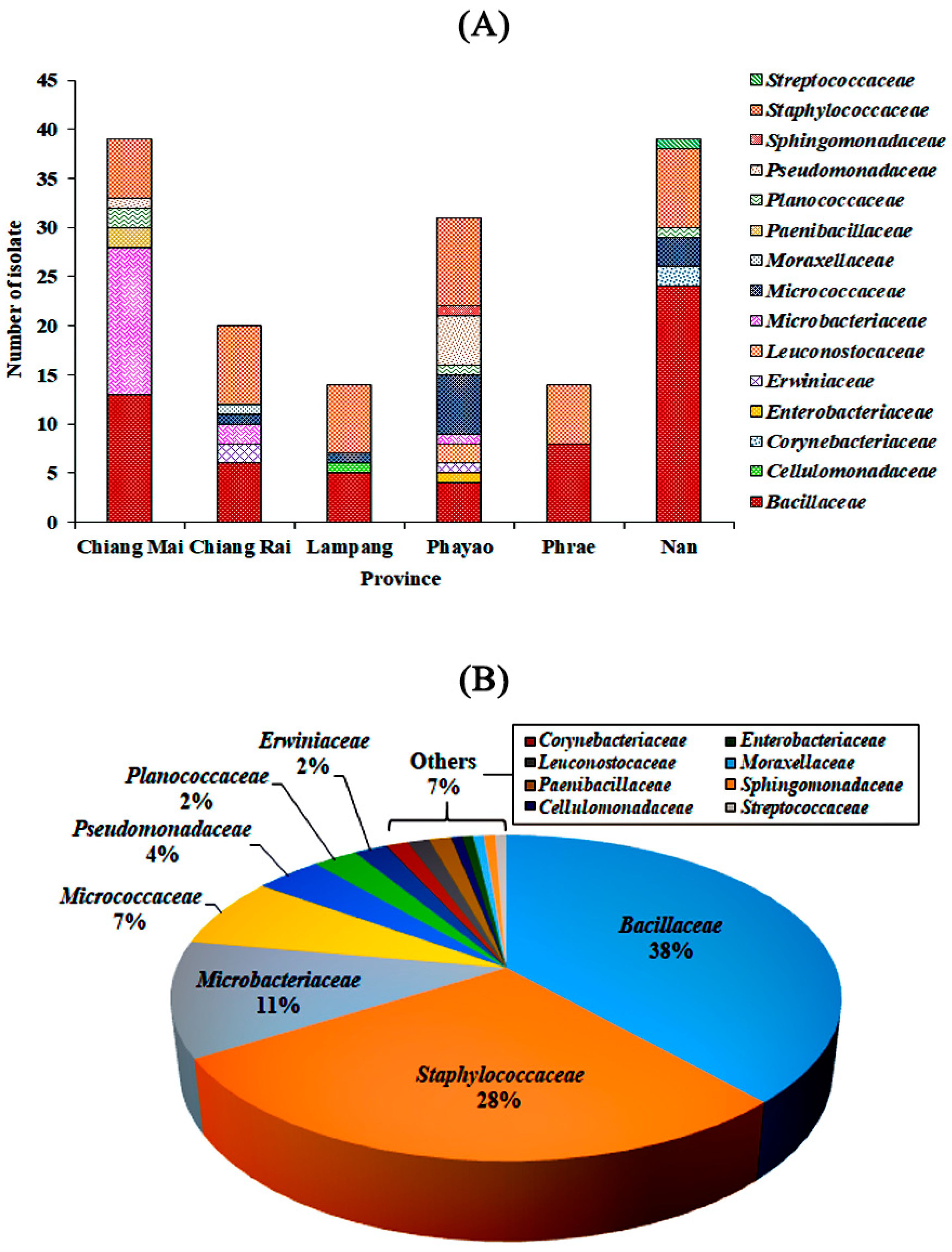
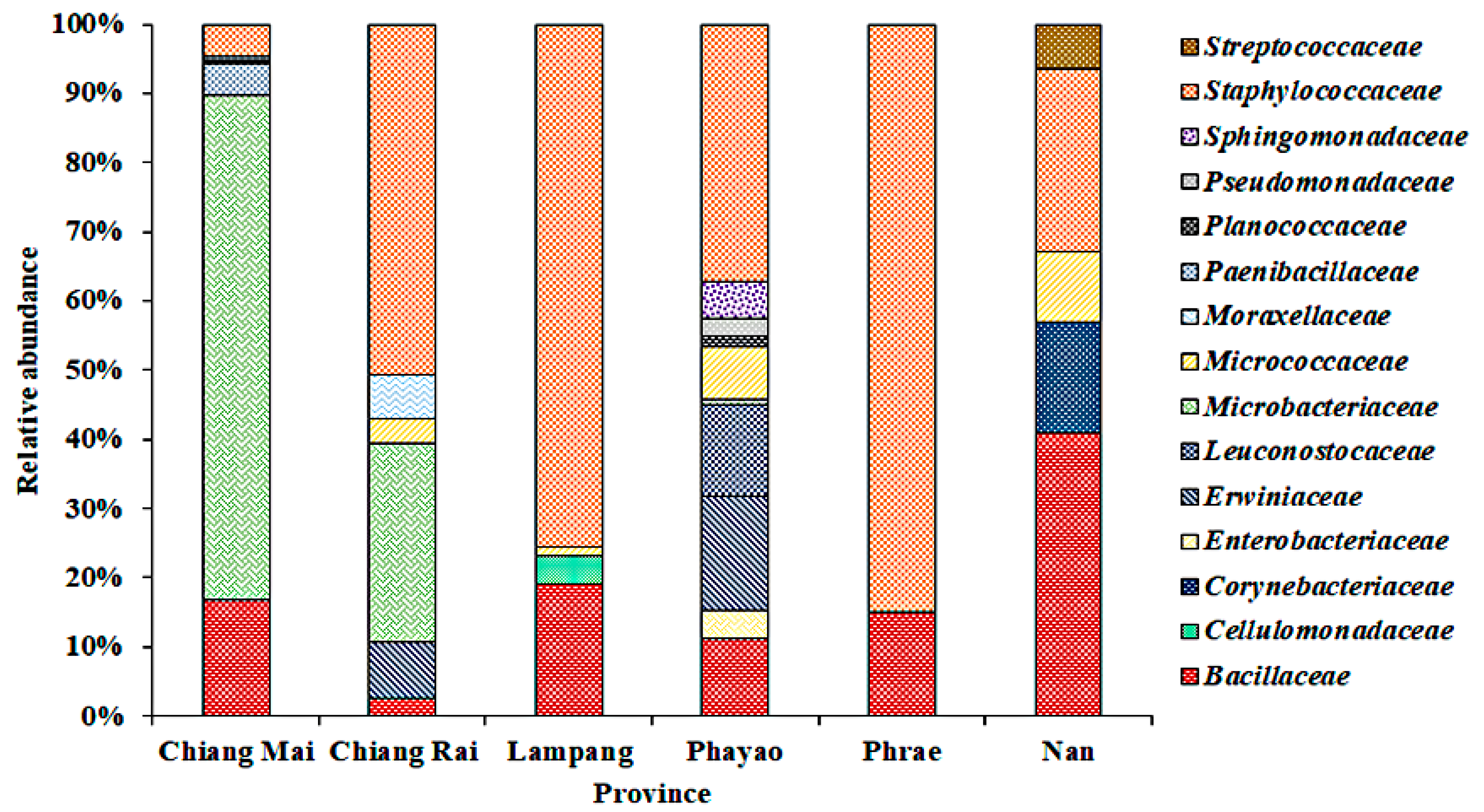
3.2. Bacterial Community on Assam Tea Leaf Surface

The relative abundance of bacterial families on Assam tea leaves from each province is presented in Figure 3. Leaves of Assam tea collected from Phayao province demonstrated the highest diversity with 10 bacterial families and 30 different isolates, while Assam tea leaves obtained from Phrae province had the lowest bacterial diversity with 14 different isolates belonging to two families, Bacillaceae and Staphylococcaceae.
Interestingly, bacteria from the Bacillaceae and Staphylococcaceae families were found in all provinces, accounting for 2.7–40.9 and 4.7–84.9% of the relative abundance, respectively. The dominant bacterial families on Assam tea leaf surfaces of Chiang Mai and Nan provinces were Microbacteriaceae (72.8%) and Bacillaceae (40.9%), respectively, while the family Staphylococcaceae was dominant in Chiang Rai, Lampang, Phayao, and Phrae provinces with a relative abundance ranging between 37.2 and 84.9%.
When focusing on bacterial communities from leaves collected in the Nan province in March 2016 (code ML051-ML063) and March 2018 (code ML064–ML067), the data obtained suggested that the families Micrococcaceae, Planococcaceae, and Streptococcaceae found in 2016 were completely replaced by the Bacillaceae and Staphylococcaceae families in 2018. Likewise, in the Chiang Mai province, the families Microbacteriaceae, Paenibacillaceae, Planococcaceae, and Pseudomonadaceae found in 2015 (codes ML011–ML041) were replaced by the Bacillaceae and Staphylococcaceae families in 2016 (code ML071).

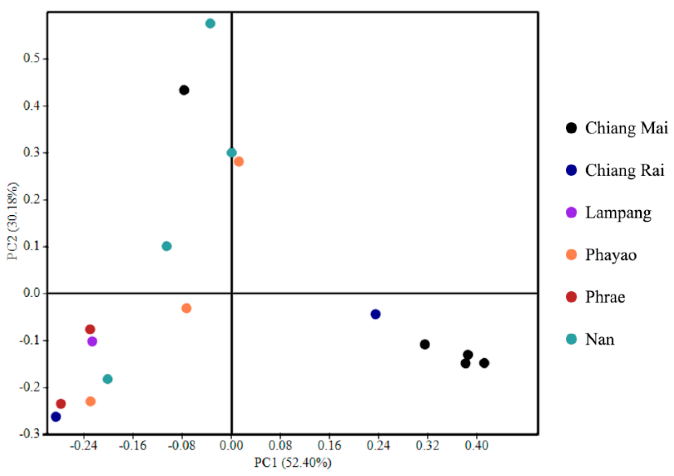
3.3. Bacterial Alpha Diversity, Richness, and Community Structure

The Shannon (H’) diversity index ranged from 0.89 to 1.49. The H’ value of Phayao province presented the highest diversity. The Simpson’s (D) indices ranged between 0.51 and 0.71. Three provinces, including Chiang Mai, Chiang Rai, and Phrae provinces, had a similar D value of 0.53, and, consistent with H’, Phayao province presented the highest diversity (0.71). Likewise, Phayao province showed the highest index value of evenness (J’ = 0.77), while the lowest value was found in Nan and Chiang Rai provinces (J’ = 0.56). The species richness (R) in all provinces ranged from 18.73 to 69.84. Lampang province had the highest R value (69.84), whereas Phrae province showed the lowest value (18.73) (Table 1).
The beta diversity analysis and the separation pattern of bacterial communities were shown by PCoA, suggesting that most bacterial communities from Chiang Mai province were explicitly separated from Lampang, Phayao, Phrae, and Nan provinces. Additionally, the bacterial communities of Chiang Rai and Lampang presented overlap between two main groups (Figure 4).

3.4. Probiotic Property Determination of Bacillus Strains

3.4.1. Antibacterial Activity

The culture filtrates of all isolates belonging to a family Bacillaceae (60 isolates) were tested for their antibacterial activity against test pathogenic bacteria. B. clausii ML062-2 was the only isolate promoting growth inhibition of E. coli O157:H7 (7.2 ± 0.1 mm). B. subtilis ML066-3 and all B. licheniformis strains presented antimicrobial activity against B. cereus with diameters of inhibitory clear zones ranging from 7.3 ± 0.3 to 11.3 ± 0.4 mm. Three strains, namely B. subtilis ML066-3, B. licheniformis ML075-1, and B. siamensis ML122-2 inhibited S. aureus growth (8.0 to 9.0 ± 0.0 mm). All B. siamensis strains tested positive against MRSA (9.3–12.0 mm). Note that only B. siamensis ML122-2 could inhibit both S. aureus and MRSA (Table 2). Based on the data mentioned above, B. licheniformis ML075-1 and B. siamensis ML122-2 were selected for further probiotic potential evaluation.

3.4.2. Antibiotic Susceptibility

B. licheniformis ML075-1 and B. siamensis ML122-2 were sensitive to all 16 tested antibiotics with the zone of inhibition ranging from 15.6 to 43.9 mm. Meropenem presented the widest clear zone for strain ML075-1 (41.5 mm) and ML122-2 (43.9 mm). L. acidophilus TISTR 2365 was susceptible to 13 antibiotics (9.9–49.0 mm of clear zone) except amikacin, co-trimoxazole, and ofloxacin (data not shown) [11].

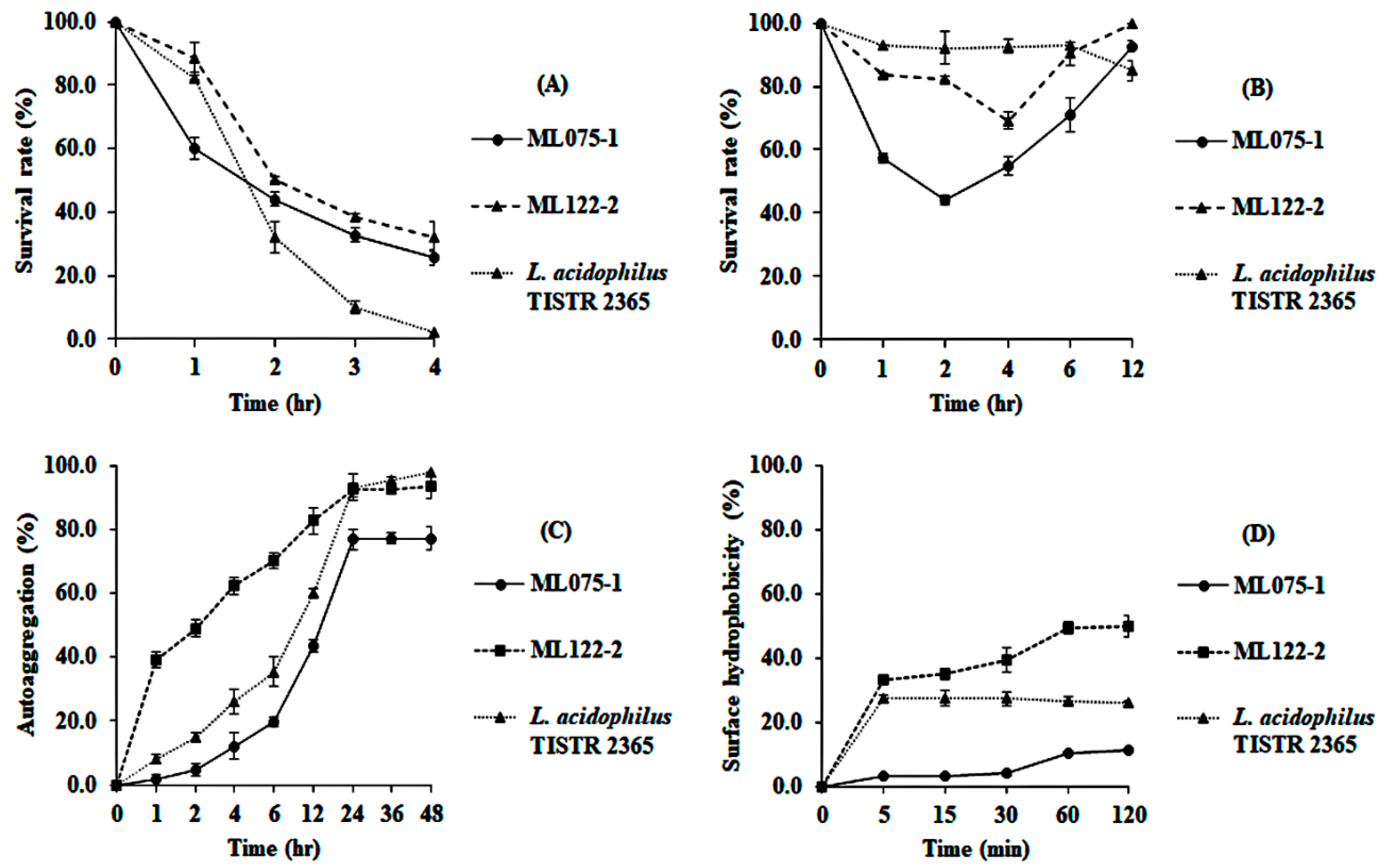
3.4.3. Survival in Gastrointestinal Tract Conditions, Autoaggregation, and Cell Surface Hydrophobicity Investigation

B. siamensis ML122-2 revealed resistance to gastric fluid up to 2 h of incubation (50.4% survival rate), while B. licheniformis ML075-1 and L. acidophilus TISTR 2365 expressed 44.0 and 32.2% survival rates, respectively, for the same incubation period. B. siamensis ML122-2 and B. licheniformis ML075-1 showed resistance to the intestinal fluid for 6 h with survival rates of 90.3 and 70.9%, respectively (p < 0.05), while the survival rate of L. acidophilus TISTR 2365 was 92.7% for the same period of incubation. Fascinatingly, the survival rate of B. licheniformis ML075-1 and B. siamensis ML122-2 in intestinal fluid reached the lowest value at 2 and 4 h of incubation, respectively, and increased after further incubation for 4 and 6 h, respectively (Figure 5A,B).
For autoaggregation ability, B. siamensis ML122-2 presented the highest percentage of autoaggregation at 1 h after incubation (39.1%), followed by L. acidophilus TISTR 2365 and B. licheniformis ML075-1 (8.2 and 1.6%, respectively), which were significantly different (p < 0.05). After incubation for 24 h, B. siamensis ML122-2 and L. acidophilus TISTR 2365 had an autoaggregation percentage of 92.4 and 93.2%, respectively (p > 0.05), while B. licheniformis ML075-1 demonstrated 76.8% (p < 0.05). Additionally, B. siamensis ML122-2 showed significantly (p < 0.05) higher cell surface hydrophobicity than L. acidophilus TISTR 2365 and B. licheniformis ML075-1 at 30 min of incubation (39.6, 27.5 and 3.9%, respectively) (Figure 5C,D).

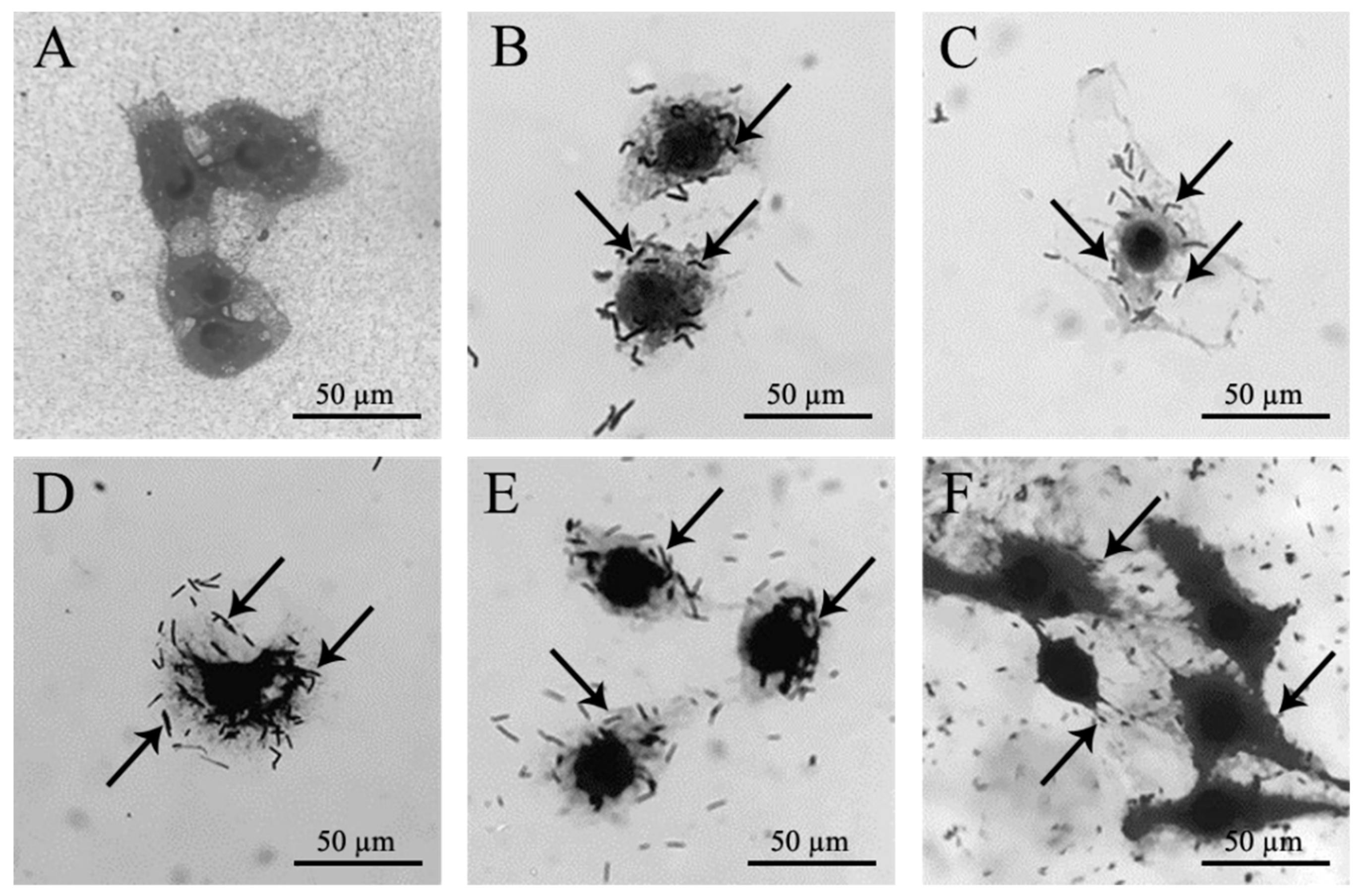
3.4.4. Bacterial Adhesion

B. licheniformis ML075-1 and B. siamensis ML122-2 presented adhesion ability on Vero cells of 52.6 and 75.8%, respectively, when using L. plantarum FM03-1 as a positive control (100% adhesion). In relation to L. acidophilus TISTR 2365, B. siamensis ML122-2 (19.5%) displayed a higher adhesion on Vero cells than B. licheniformis ML075-1 (15.4%). Meanwhile, E. coli ATCC 25922 (a normal intestinal flora strain) exhibited 127.2 and 493.6% adhesion when compared with L. acidophilus TISTR 2365 and L. plantarum FM03-1, respectively (Table 3 and Figure 6).

4. Discussion

Assam tea plants or Camellia sinensis var. assamica are widely found in the highlands of Northern Thailand at altitudes over 200 m above sea level. The microbial composition of Assam tea leaves in Thailand first described by Rungsirivanich et al. [11] revealed the presence of culturable bacteria from the genera Bacillus, Floricoccus, Kocuria, Lysinibacillus, Micrococcus, and Staphylococcus. To gain further insight into the bacterial community of Assam tea leaves and understand their role in fermentation, as well as to identify beneficial bacteria for healthy food development and biological control, a larger area of sampling sites with various environmental conditions (altitude, soil, and climate) was explored in this study, covering six provinces (Chiang Mai, Chiang Rai, Lampang, Phayao, Phrae, and Nan) in the upper northern region of Thailand. Through molecular taxonomic markers, the isolated bacteria were classified within 15 families and 20 genera, some of which were adapted to extreme environments. For example, the genera Anoxybacillus and Lysinibacillus are reported as thermophilic [35,36], which is related to the formation of endospores. Most strains of the genus Cellulomonas have been described as cellulolytic bacteria, which can be found in cellulose-rich habitats [37]. On the other hand, Pantoea spp. Curtobacterium spp., Microbacterium spp., Sphingomonas spp. Lysinibacillus spp., Solibacillus spp., Kocuria spp., and Pseudomonas spp. have been grouped as endophytic bacteria implicated in plant growth promotion with some species producing antimicrobial substances [38,39,40,41,42,43,44], which may be involved in growth enhancement and biological control in Assam tea orchards [7]. The genera Acinetobacter and Brevibacillus have been reported to be found not only in the environment (involved in bioremediation [45] and biogeochemical cycles [46]) but also in animals and humans [47,48]. The Weissella and Floricoccus genera are grouped as lactic acid bacteria (LAB) [49,50]. Several reports reveal that some LAB strains can produce bacteriocin or bacteriocin-like inhibitory substances that play a role in inhibition of pathogenic microbes and in biological control. Moreover, LAB are also important in fermentation processes, and some species present probiotic properties [51]. Bacillus spp. have been widely found in various environments. Some species of Bacillus are pathogenic (e.g., B. cereus), probiotics (e.g., B. clausii, B. subtilis, B. licheniformis), endophytic (with the ability to produce the plant hormone indole-3-acetic acid or IAA; e.g., B. altitudinis, B. paramycoides, B. tequilensis), and antimicrobial (e.g., B. niacin, B. mobilis) [52,53,54,55,56]. Staphylococcus spp., Macrococcus spp., and Micrococcus spp. have been reported as normal flora on human and animal skins, and opportunistic pathogens such as S. epidermidis, S. xylosus, Macrococcus canis, and Micrococcus luteus [57,58,59] may lead to bloodstream infections. Besides that, Corynebacterium aurimucosum is also reported to be the cause of urinary tract infection in humans [60], while Enterobacter hormaechei is described as the causal agent of opportunistic infections in urinary and respiratory tracts [61]. The genus Pantoea has been generally found in plant surfaces as well as human feces. Some species have also been reported as human pathogens leading to bacteremia [62]. The existence of mammal microbial flora on Assam tea leaves indicates the close relationship between human, animals, and Assam tea plants. Assam tea orchards in the study area exist both in the forests and in the area around tea farmers’ houses. Possibly, the human and animal flora spread to the plants via their activities, but how these bacteria affect the tea plants is not known.
The microbial community structure has been suggested to be associated with temperature, mineralization, hydrocarbon content, soil organic matter, and moisture [63,64]. In this study, the family Microbacteriaceae and Staphylococcaceae were explicitly dominant in Chiang Mai and Phrae provinces with 72.8 and 84.9% relative abundance, respectively. Moreover, the elevation above sea level is also considered to affect microbial biodiversity [65]. Although, the number of bacterial isolates identified within the family Bacillaceae was higher than that of Staphylococcaceae (38 and 28%, respectively), the Staphylococcaceae family presented higher relative abundance in Chiang Rai, Lampang, Phayao, and Phrae provinces than did the family Bacillaceae. By interview and observation, the collecting sites in these four provinces were quite inactive in terms of tea leaf plucking for fermented Miang production during the sample collecting period. Hence, the tea plants were undisturbed, which allowed certain bacteria to establish, thrive, and succeed in this microecosystem. The active Assam tea orchards displayed higher numbers of bacterial species, possibly due to the more turns of tea leaves which were often taken off. Moreover, altitude and temperature seemed to be unrelated to the occurrence.
The microbes found on Assam tea leaf surfaces revealed an abundance of Actinobacteria, Firmicutes, and Proteobacteria phyla, which have been commonly found in the environment including soils, leaves, stems, and roots, as well as skin of human and animals [66]. In this study, all diversity indices indicated that Phayao province had the highest bacterial biodiversity, while Phrae province had the lowest. Interestingly, the family Micrococcaceae was not found only in Chiang Mai and Phrae provinces, which clearly displayed low diversity. The absence of the family Micrococcaceae in Chiang Mai and Phrae may be due to the geographical location (Figure 1). The microbial biodiversity clearly indicated the difference of bacteria existing on Assam tea leaves between two areas (referred to as the west and the east sides of Northern Thailand). The west (Mae Hong Son, Chiang Mai and Lamphun provinces) and the east (Chiang Rai, Lampang Phayao, Phrae, and Nan provinces) sides of Northern Thailand can be separated by western Phi Pan Nam range or known as Khun Tan range. Moreover, the difference of bacterial communities may be related to flow through rivers, especially the Wang River, which runs from Chiang Rai, upper Northern Thailand, passes through Lampang to Tak provinces, and lower Northern Thailand. These data support the various processes of Assam tea leaf fermentation and microbes in fermented Miang found in both areas, which have been reported by Kanpiengjai et al. [2], Khanongnuch et al. [3], and Chaikaew et al. [5].
In general, slow shifts from the original states of any microbial communities are demonstrated when continuous cultivation has been performed for a long time [7]. In this study, bacterial communities on Assam tea leaves at Nan province in March 2016 and at Chiang Mai province in 2015 were completely replaced by the Bacillaceae and Staphylococcaceae families within 2 years. This occurrence may be due to the involvement of human activity, wildfire, and deforestation in the study area and surroundings.
“Fermented Miang” or fermented Assam tea leaves made by the local wisdom of people in the areas of Northern Thailand, the Lao People’s Democratic Republic, and the Republic of the Union of Myanmar for centuries has been unpopular with the new generation for many reasons, such as its physical appearance and old-fashioned packaging. However, the hidden market share has been noticed recently. The use of probiotics in fermented Assam tea leaves has attracted people with healthy lifestyles. Hence, information on bacterial communities would benefit further product development and elucidation of properties in the field of the gut microbiome or related subjects.
A previous study described the probiotic properties and utilization of Bacillus [67]. In fermented Miang, B. tequilensis, B. siamensis, B. megaterium, B. toyonensis, and B. aryabhattai presented great probiotic potential [68]. Some Bacillus strains mentioned above could be also found in this study, suggesting a persistence of these bacteria from fresh to fermented leaves.
B. licheniformis ML075-1 and B. siamensis ML122-2 demonstrated antagonistic activity against pathogenic bacteria, especially against B. cereus TISTR 687 and MRSA DMST 20625, respectively. Previous studies reported that Bacillus spp. can produce antimicrobial substances such as peptides, bacteriocins, or BLIS [16]. Interestingly, B. siamensis ML122-2 could inhibit growth of MRSA DMST 20625, a cause of resistance to many antibiotics. Culture broth pH seemed not to be responsible for the antagonistic activity of these isolates (data not shown). In the future, its antimicrobial active principle should be further elucidated.
In this study, B. licheniformis ML075-1 and B. siamensis ML122-2 were susceptible to all tested antibiotics. This susceptibility results may ensure that when these Bacillus isolates are used as probiotics, the chance that an antibiotic resistance gene will be transferred to any recipient bacteria in the intestine is scarce [15].
Acid and base tolerance are among the most important requirements for probiotics potential due to their need to survive in the stomach and intestines [69]. B. siamensis ML122-2 demonstrated higher survival rate in the gastric fluid than B. licheniformis ML075-1 and L. acidophilus TISTR 2365, respectively. The previous study by Kristoffersen et al. [70] indicated that B. cereus ATCC 14579 spores were germinated in the distal parts of the small intestine, which contains lower amounts of bile salt. In this study, the alkaline condition in simulated intestinal fluid was tested against Bacillus and was consistent with the previous study in that at the start of the experiment where high concentration of bile salt was present, Bacillus strain count was reduced, with an increasing rate after incubation for four (B. licheniformis ML075-1) and six (B. siamensis ML122-2) hours. This might be because the bacteria adapted to the test fluid or produced some metabolites to neutralize the fluid.
The adhesion ability of probiotics has been considered to be a critical prerequisite for bacterial colonization on the host gut [69]. In this study, B. siamensis ML122-2 demonstrated autoaggregation (higher than 90%), high hydrophobicity, and high adherence ability to Vero cells, therefore presenting the highest probiotics potential among the identified bacteria. In the future, it will be worth further investigating the potential of these species for the treatment of antibiotic-resistant infections. Furthermore, B. siamensis ML122-2 may provide a functional ingredient useful in synbiotic production, which will be investigated in the near future.

5. Conclusions

Assam tea leaves are rich in microbial biodiversity. These microbial communities may play a major role in biological control and Assam tea leaf fermentation. The microbiodiversity on Assam tea leaves presented a stabilized community representing good sources of probiotics. Particularly, B. siamensis ML122-2 isolated from Assam tea leaf surfaces demonstrated high probiotic potential. It represents antibacterial activity, especially MRSA; hence, its antimicrobial substance should be elucidated in the future.

Supplementary Materials

The following are available online at https://www.mdpi.com/2076-2607/8/10/1585/s1: Figure S1, Phylogenetic relationships of some bacterial isolates (bold) isolated from Assam tea leaves in Northern Thailand with their closest species and related taxa based on 16S rRNA gene sequence analysis. The branching pattern was generated by the neighbor-joining method. Bootstrap values (expressed as percentages of 1000 replications). Bar, 0.05 substitutions per nucleotide position. Saccharolobus caldissimus JCM 32116T (GenBank accession no. LC275065) is presented as outgroup sequence. Table S1, Assam tea leaf collecting site from different regions in Northern Thailand. The data presented number of Assam tea plants, locations, altitude, bacterial cell count, number of isolates per sample, and number of species per sample. Table S2, Classification of bacteria isolated from Assam tea leaf surfaces compared with the type strain and the similarity (%) of 16S rRNA gene sequence.

Author Contributions

Conceptualization, P.R. and N.T.; methodology, P.R., W.S., W.F., V.C.-N.-A., C.Y. and N.T.; formal analysis and investigation, P.R. and N.T.; data curation, P.R.; writing—original draft preparation, P.R. and N.T.; funding acquisition, N.T. All authors have read and agreed to the published version of the manuscript.

Funding

This research was funded by the National Research Council of Thailand, grant number PHD60I0089; Tea Gallery Group (Thailand) Co., Ltd.; Amazing Tea Limited Partnership; and the scholarship for Teaching Assistant and Research Assistant (TA/RA) awarded by the Graduate School, Chiang Mai University, Thailand.

Acknowledgments

This research was partially supported by the Research Center in Bioresources for Agriculture, Industry, and Medicine, Chiang Mai University; and the Microbiology Section, Department of Biology, Faculty of Science, Chiang Mai University, Thailand.

Conflicts of Interest

The authors declare no conflict of interest. The funders had no role in the design of the study; in the collection, analyses, or interpretation of data; in the writing of the manuscript; or in the decision to publish the results.

References

  1. Okada, S.; Daengsubha, W.; Uchimura, T.A.I.; Ohara, N.; Kozaki, M. Flora of lactic acid bacteria in miang produced in northern Thailand. J. Gen. Appl. Microbiol. 1986, 32, 57–65. [Google Scholar] [CrossRef]
  2. Kanpiengjai, A.; Chui-Chai, N.; Chaikaew, S.; Khanongnuch, C. Distribution of tannin-’tolerant yeasts isolated from Miang, a traditional fermented tea leaf (Camellia sinensis var. assamica) in northern Thailand. Int. J. Food Microbiol. 2016, 238, 121–131. [Google Scholar] [CrossRef] [PubMed]
  3. Khanongnuch, C.; Unban, K.; Kanpiengjai, A.; Saenjum, C. Recent research advances and ethno-botanical history of miang, a traditional fermented tea (Camellia sinensis var. assamica) of northern Thailand. J. Ethn. Foods 2017, 4, 135–144. [Google Scholar] [CrossRef]
  4. Tanasupawat, S.; Pakdeeto, A.; Thawai, C.; Yukphan, P.; Okada, S. Identification of lactic acid bacteria from fermented tea leaves (miang) in Thailand and proposals of Lactobacillus thailandensis sp. nov., Lactobacillus camelliae sp. nov., and Pediococcus siamensis sp. nov. J. Gen. Appl. Microbiol. 2007, 53, 7–15. [Google Scholar] [CrossRef] [PubMed] [Green Version]
  5. Chaikaew, S.; Baipong, S.; Sone, T.; Kanpiengjai, A.; Chui-chai, N.; Asano, K.; Khanongnuch, C. Diversity of lactic acid bacteria from Miang, a traditional fermented tea leaf in northern Thailand and their tannin-tolerant ability in tea extract. J. Microbiol. 2017, 55, 720–729. [Google Scholar] [CrossRef] [PubMed]
  6. Han, W.; Kemmitt, S.J.; Brookes, P.C. Soil microbial biomass and activity in Chinese tea gardens of varying stand age and productivity. Soil Biol. Biochem. 2007, 39, 1468–1478. [Google Scholar] [CrossRef]
  7. Li, Y.C.; Li, Z.; Li, Z.W.; Jiang, Y.H.; Weng, B.Q.; Lin, W.X. Variations of rhizosphere bacterial communities in tea (Camellia sinensis L.) continuous cropping soil by high-throughput pyrosequencing approach. J. Appl. Microbiol. 2016, 121, 787–799. [Google Scholar] [CrossRef]
  8. Zhang, Y.; Skaar, I.; Sulyok, M.; Liu, X.; Rao, M.; Taylor, J.W. The microbiome and metabolites in fermented Pu-erh tea as revealed by high-throughput sequencing and quantitative multiplex metabolite analysis. PLoS ONE 2016, 11, e0157847. [Google Scholar] [CrossRef]
  9. Nancib, A.; Nancib, N.; Boudrant, J. Production of lactic acid from date juice extract with free cells of single and mixed cultures of Lactobacillus casei and Lactococcus lactis. World J. Microbiol. Biotechnol. 2009, 25, 1423–1429. [Google Scholar] [CrossRef]
  10. Cavicchioli, R.; Ripple, W.J.; Timmis, K.N.; Azam, F.; Bakken, L.R.; Baylis, M.; Behrenfeld, M.J.; Boetius, A.; Boyd, P.W.; Classen, A.T.; et al. Scientists’ warning to humanity: Microorganisms and climate change. Nat. Rev. Microbiol. 2019, 17, 569–586. [Google Scholar] [CrossRef] [Green Version]
  11. Rungsirivanich, P.; Inta, A.; Tragoolpua, Y.; Thongwai, N. Partial rpoB gene sequencing identification and probiotic potential of Floricoccus penangensis ML061-4 isolated from Assam tea (Camellia sinensis var. assamica). Sci. Rep. 2019, 9, 16561. [Google Scholar] [CrossRef] [PubMed]
  12. Rungsirivanich, P.; Thongwai, N. Antibacterial activity and tannin tolerance of Bacillus spp. isolated from leaves of Miang (Camellia sinensis (L.) Kuntze var. assamica (J.W. Mast.) Kitam.). Int. J. Biosci. Biochem. Bioinform. 2020, 10, 26–33. [Google Scholar] [CrossRef] [Green Version]
  13. FAO/WHO. Report of a Joint Food and Agriculture Organization of the United Nations and World Health Organization (FAO/WHO) expert consultation on guidelines for the evaluation of probiotics in food. Available online: http://www.who.int/foodsafety/fs_management/en/probiotic_guidelines.pdf (accessed on 5 August 2002).
  14. Angmo, K.; Kumari, A.; Savitri; Bhalla, T.C. Probiotic characterization of lactic acid bacteria isolated from fermented foods and beverage of Ladakh. LWT Food Sci. Technol. 2016, 66, 428–435. [Google Scholar] [CrossRef]
  15. Meidong, R.; Doolgindachbaporn, S.; Jamjan, W.; Sakai, K.; Tashiro, Y.; Okugawa, Y.; Tongpim, S. A novel probiotic Bacillus siamensis B44v isolated from Thai pickled vegetables (Phak-dong) for potential use as a feed supplement in aquaculture. J. Gen. Appl. Microbiol. 2017, 63, 246–253. [Google Scholar] [CrossRef] [PubMed] [Green Version]
  16. Abriouel, H.; Franz, C.M.A.P.; Omar, N.B.; Gálvez, A. Diversity and applications of Bacillus bacteriocins. FEMS Microbiol. Rev. 2011, 35, 201–232. [Google Scholar] [CrossRef] [Green Version]
  17. Zendo, T. Screening and characterization of novel bacteriocins from lactic acid bacteria. Biosci. Biotechnol. Biochem. 2013, 77, 893–899. [Google Scholar] [CrossRef]
  18. Levetin, E.; Dorsey, K. Contribution of leaf surface fungi to the air spora. Aerobiologia 2006, 22, 3–12. [Google Scholar] [CrossRef]
  19. Pitcher, D.G.; Saunders, N.A.; Owen, R.J. Rapid extraction of bacterial genomic DNA with guanidium thiocyanate. Lett. Appl. Microbiol. 1989, 8, 151–156. [Google Scholar] [CrossRef]
  20. Weisburg, W.G.; Barns, S.M.; Pelletier, D.A.; Lane, D.J. 16S ribosomal DNA amplification for phylogenetic study. J. Bacteriol. 1991, 173, 697–703. [Google Scholar] [CrossRef] [Green Version]
  21. Saitou, N.; Nei, M. The neighbor-joining method: A new method for reconstructing phylogenetic trees. Mol. Biol. Evol. 1987, 4, 406–425. [Google Scholar] [CrossRef]
  22. Kumar, S.; Stecher, G.; Tamura, K. MEGA7: Molecular evolutionary genetics analysis version 7.0 for bigger dataset. Mol. Biol. Evol. 2016, 33, 1870–1874. [Google Scholar] [CrossRef] [PubMed] [Green Version]
  23. Felsenstein, J. Confidence limits on phylogenies: An approach using the bootstrap. Evolution 1985, 39, 783–791. [Google Scholar] [CrossRef] [PubMed]
  24. Tamura, K.; Nei, M. Estimation of the number of nucleotide substitutions in the control region of mitochondrial DNA in humans and chimpanzees. Mol. Biol. Evol. 1993, 10, 512–526. [Google Scholar] [CrossRef] [PubMed]
  25. Bonev, B.; Hooper, J.; Parisot, J. Principles of assessing bacterial susceptibility to antibiotics using the agar diffusion method. J. Antimicrob. Chemo. 2008, 61, 1295–1301. [Google Scholar] [CrossRef] [PubMed] [Green Version]
  26. Clinical Laboratory Standards Institute. Performance standards of antimicrobial disc susceptibility tests. CLSI 2012, 32, 3. [Google Scholar]
  27. Huang, Y.; Adams, M.C. In vitro assessment of the upper gastrointestinal tolerance of potential probiotic dairy propionibacteria. Int. J. Food Microbiol. 2004, 91, 253–260. [Google Scholar] [CrossRef] [PubMed]
  28. Valeriano, V.D.; Parungao-Balolong, M.M.; Kang, D.K. In vitro evaluation of the mucin-adhesion ability and probiotic potential of Lactobacillus mucosae LM1. J. Appl. Microbiol. 2014, 117, 485–497. [Google Scholar] [CrossRef] [PubMed]
  29. Llanco, L.A.; Nakano, V.; de Moraes, C.T.P.; Piazza, R.M.F.; Avila-Campos, M.J. Adhesion and invasion of Clostridium perfringens type A into epithelial cells. Braz. J. Microbiol. 2017, 48, 764–768. [Google Scholar] [CrossRef] [PubMed]
  30. Spellerberg, I.F.; Fedor, P.J. A tribute to Claude Shannon (1916-2001) and a plea for more rigorous use of species richness, species diversity and the ‘Shannon-Wiener’Index. Glob. Ecol. Biogeogr. 2003, 12, 177–179. [Google Scholar] [CrossRef] [Green Version]
  31. Pielou, E.C. The measurement of diversity in different types of biological collections. J. Theor. Biol. 1966, 13, 131–144. [Google Scholar] [CrossRef]
  32. Simpson, E.H. Measurement of diversity. Nature 1949, 163, 688. [Google Scholar] [CrossRef]
  33. Colwell, R.K. Biodiversity: Concepts, Patterns and Measurement. In The Princeton Guide to Ecology; Simon, A.L., Ed.; Princeton University Press: Princeton, NJ, USA, 2009; pp. 257–263. [Google Scholar]
  34. Yeh, H.Y.; Line, J.E.; Hinton, A., Jr.; Gao, Y.; Zhuang, H. The effect of rosemary extract and cold plasma treatments on bacterial community diversity in poultry ground meats. Heliyon 2019, 5, e02719. [Google Scholar] [CrossRef] [PubMed] [Green Version]
  35. Cihan, A.C. Taxonomic classification of Anoxybacillus isolates from geothermal regions in Turkey by 16S rRNA gene sequences and ARDRA, ITS-PCR, Rep-PCR analyses. Pol. J. Microbiol. 2013, 62, 149–163. [Google Scholar] [CrossRef] [PubMed]
  36. Wenzler, E.; Kamboj, K.; Balada-Llasat, J.M. Severe sepsis secondary to persistent Lysinibacillus sphaericus, Lysinibacillus fusiformis and Paenibacillus amylolyticus bacteremia. Int. J. Infect. Dis. 2015, 35, 93–95. [Google Scholar] [CrossRef] [PubMed] [Green Version]
  37. Lagier, J.C.; Ramasamy, D.; Rivet, R.; Raoult, D.; Fournier, P.E. Non contiguous-finished genome sequence and description of Cellulomonas massiliensis sp. nov. Stand. Genomic Sci. 2012, 7, 258–270. [Google Scholar] [CrossRef] [PubMed] [Green Version]
  38. Kim, H.; Nishiyama, M.; Kunito, T.; Senoo, K.; Kawahara, K.; Murakami, K.; Oyaizu, H. High population of Sphingomonas species on plant surface. J. Appl. Microbiol. 2004, 85, 731–736. [Google Scholar] [CrossRef]
  39. Bafana, A. Diversity and metabolic potential of culturable root-associated bacteria from Origanum vulgare in sub-Himalayan region. World J. Microbiol. Biotechnol. 2013, 29, 63–74. [Google Scholar] [CrossRef]
  40. Walterson, A.M.; Stavrinides, J. Pantoea: Insights into a highly versatile and diverse genus within the Enterobacteriaceae. FEMS Microbiol. Rev. 2015, 39, 968–984. [Google Scholar] [CrossRef] [PubMed] [Green Version]
  41. Undabarrena, A.; Beltrametti, F.; Claverías, F.P.; González, M.; Moore, E.R.B.; Seeger, M.; Cámara, B. Exploring the diversity and antimicrobial potential of marine Actinobacteria from the Comau Fjord in Northern Patagonia, Chile. Front. Microbiol. 2016, 7, 1135. [Google Scholar] [CrossRef]
  42. Naureen, Z.; Rehman, N.U.; Hussain, H.; Hussain, J.; Gilani, S.A.; Housni, S.K.A.; Mabood, F.; Khan, A.L.; Farooq, S.; Abbas, G.; et al. Exploring the potentials of Lysinibacillus sphaericus ZA9 for plant growth promotion and biocontrol activities against phytopathogenic fungi. Front. Microbiol. 2017, 8, 1477. [Google Scholar] [CrossRef]
  43. Hussain, A.; Amna; Kamran, M.A.; Javed, M.T.; Hayet, K.; Farooq, M.A.; Ali, N.; Ali, M.; Manghwar, H.; Jan, F.; et al. Individual and combinatorial application of Kocuria rhizophila and citric acid on phytoextraction of multi-metal contaminated soils by Glycine max L. Environ. Exp. Bot. 2019, 159, 23–33. [Google Scholar] [CrossRef]
  44. Qessaoui, R.; Bouharroud, R.; Furze, J.N.; El Aalaoui, M.; Akroud, H.; Amarraque, A.; Van Vaerenbergh, J.; Tahzima, R.; Mayad, E.H.; Chebli, B. Applications of new rhizobacteria Pseudomonas isolates in agroecology via fundamental processes complementing plant growth. Sci. Rep. 2019, 9, 12832. [Google Scholar] [CrossRef] [PubMed] [Green Version]
  45. Uniyal, S.; Paliwal, R.; Verma, M.; Sharma, R.K.; Rai, J.P. Isolation and characterization of fipronil degrading Acinetobacter calcoaceticus and Acinetobacter oleivorans from rhizospheric zone of Zea mays. Bull. Environ. Contam. Toxicol. 2016, 96, 833–838. [Google Scholar] [CrossRef] [PubMed]
  46. Chebbi, A.; Mhiri, N.; Rezgui, F.; Ammar, N.; Maalej, A.; Sayadi, S.; Chamkha, M. Biodegradation of malodorous thiols by a Brevibacillus sp. strain isolated from a Tunisian phosphate factory. FEMS Microbiol. Lett. 2015, 362, fnv097. [Google Scholar] [CrossRef] [PubMed] [Green Version]
  47. Panda, A.K.; Bisht, S.S.; DeMondal, S.; Kumar, N.S.; Gurusubramanian, G.; Panigrahi, A.K. Brevibacillus as a biological tool: A short review. Antonie van Leeuwenhoek 2014, 105, 623–639. [Google Scholar] [CrossRef] [PubMed]
  48. Atrouni, A.A.; Joly-Guillou, M.; Hamze, M.; Kempf, M. Reservoirs of non-baumannii Acinetobacter species. Front. Microbiol. 2016, 7, 49. [Google Scholar] [CrossRef] [PubMed] [Green Version]
  49. Fusco, V.; Quero, G.M.; Cho, G.S.; Kabisch, J.; Meske, D.; Neve, H.; Bockelmann, W.; Franz, C.M.A.P. The genus Weissella: Taxonomy, ecology and biotechnological potential. Front. Microbiol. 2015, 6, 155. [Google Scholar] [CrossRef] [Green Version]
  50. Chuah, L.O.; Yap, K.P.; Shamila-Syuhada, A.K.; Thong, K.L.; Ahmad, R.; Liong, M.T.; Rusul, G. Floricoccus tropicus gen. nov., sp. nov. and Floricoccus penangensis sp. nov. isolated from fresh flowers of durian tree and hibiscus. Int. J. Syst. Evol. Microbiol. 2017, 67, 4979–4985. [Google Scholar] [CrossRef] [Green Version]
  51. Papadimitriou, K.; Alegría, A.; Bron, P.A.; De Angelis, M.; Gobbetti, M.; Kleerebezem, M.; Lemos, J.A.; Linares, D.M.; Ross, P.; Stanton, C.; et al. Stress physiology of lactic acid bacteria. Microbiol. Mol. Biol. Rev. 2016, 80, 837–890. [Google Scholar] [CrossRef] [Green Version]
  52. Harvey, Z.H.; Snider, M.J. Draft genome sequence of the nicotinate-metabolizing soil bacterium Bacillus niacini DSM 2923. Genome Announc. 2014, 2, e01251-14. [Google Scholar] [CrossRef] [Green Version]
  53. Elshaghabee, F.M.F.; Rokana, N.; Gulhane, R.D.; Sharma, C.; Panwar, H. Bacillus as potential probiotics: Status, concerns, and future perspectives. Front. Microbiol. 2017, 8, 1490. [Google Scholar] [CrossRef] [PubMed] [Green Version]
  54. Li, H.; Guan, Y.; Dong, Y.; Zhao, L.; Rong, S.; Chen, W.; Lv, M.; Xu, H.; Gao, X.; Chen, R.; et al. Isolation and evaluation of endophytic Bacillus tequilensis GYLH001 with potential application for biological control of Magnaporthe oryzae. PLoS ONE 2018, 13, e0203505. [Google Scholar] [CrossRef] [PubMed] [Green Version]
  55. Korobov, V.V.; Zhurenko, E.I.; Zharikova, N.V.; Iasakov, T.R.; Markusheva, T.V. Application of the new degrader strain Bacillus mobilis 34T for soil treatment from 2,4,5-trichlorophenoxyacetic acid. Moscow Univ. Biol. Sci. Bull. 2019, 74, 154–157. [Google Scholar] [CrossRef]
  56. Saran, A.; Imperato, V.; Fernandez, L.; Gkorezis, P.; D’Haen, J.; Merini, L.J.; Vangronsveld, J.; Thijs, S. Phytostabilization of polluted military soil supported by bioaugmentation with PGP-trace element tolerant bacteria isolated from Helianthus petiolaris. Agronomy 2020, 10, 204. [Google Scholar] [CrossRef] [Green Version]
  57. Coates, R.; Moran, J.; Horsburgh, M.J. Staphylococci: Colonizers and pathogens of human skin. Future Microbiol. 2014, 9, 75–91. [Google Scholar] [CrossRef]
  58. Ma, A.P.Y.; Jiang, J.; Tun, H.M.; Mauroo, N.F.; Yuen, C.S.; Leung, F.C.C. Complete genome sequence of Staphylococcus xylosus HKUOPL8, a potential opportunistic pathogen of mammals. Genome Announc. 2014, 2, e00653-14. [Google Scholar] [CrossRef] [Green Version]
  59. Brawand, S.G.; Cotting, K.; Gómez-Sanz, E.; Collaud, A.; Thomann, A.; Brodard, I.; Rodriguez-Campos, S.; Strauss, C.; Perreten, V. Macrococcus canis sp. nov., a skin bacterium associated with infections in dogs. Int. J. Syst. Evol. Microbiol. 2017, 67, 621–626. [Google Scholar] [CrossRef]
  60. Lo, S.; Thiam, I.; Fall, B.; Ba-Diallo, A.; Diallo, O.F.; Diagne, R.; Dia, M.L.; Ka, R.; Sarr, A.M.; Sow, A.I. Urinary tract infection with Corynebacterium aurimucosum after urethroplasty stricture of the urethra: A case report. J. Med. Case Rep. 2015, 9, 156. [Google Scholar] [CrossRef] [Green Version]
  61. Monahan, L.G.; DeMaere, M.Z.; Cummins, M.L.; Djordjevic, S.P.; Chowdhury, P.R.; Darling, A.E. High contiguity genome sequence of a multidrug-resistant hospital isolate of Enterobacter hormaechei. Gut Pathog. 2019, 11, 3. [Google Scholar] [CrossRef]
  62. Asai, N.; Koizumi, Y.; Yamada, A.; Sakanashi, D.; Watanabe, H.; Kato, H.; Shiota, A.; Hagihara, M.; Suematsu, H.; Yamagishi, Y.; et al. Pantoea dispersa bacteremia in an immunocompetent patient: A case report and review of the literature. J. Med. Case Rep. 2019, 13, 33. [Google Scholar] [CrossRef] [Green Version]
  63. Polyakova, O.; Billor, N. Impact of deciduous tree species on litterfall quality, decomposition rates and nutrient circulation in pine stands. For. Ecol. Manag. 2007, 253, 11–18. [Google Scholar] [CrossRef]
  64. Xiao, M.; Sun, S.S.; Zhang, Z.Z.; Wang, J.M.; Qiu, L.W.; Sun, H.Y.; Song, Z.Z.; Zhang, B.Y.; Gao, D.L.; Zhang, G.Q.; et al. Analysis of bacterial diversity in two oil blocks from two low-permeability reservoirs with high salinities. Sci. Rep. 2016, 6, 19600. [Google Scholar] [CrossRef] [PubMed] [Green Version]
  65. Siles, J.A.; Cajthaml, T.; Minerbi, S.; Margesin, R. Effect of altitude and season on microbial activity, abundance and community structure in Alpine forest soils. FEMS Microbiol. Ecol. 2016, 92, fiw008. [Google Scholar] [CrossRef] [PubMed] [Green Version]
  66. Malele, I.; Nyingilili, H.; Lyaruu, E.; Tauzin, M.; Ollivier, B.B.; Cayol, J.L.; Fardeau, M.L.; Geiger, A. Bacterial diversity obtained by culturable approaches in the gut of Glossina pallidipes population from a non sleeping sickness focus in Tanzania: Preliminary results. BMC Microbiol. 2018, 18, 164. [Google Scholar] [CrossRef] [PubMed] [Green Version]
  67. Jeżewska-Frąckowiak, J.; Żebrowska, J.; Czajkowska, E.; Jasińska, J.; Pęksa, M.; Jędrzejczak, G.; Skowron, P.M. Identification of bacterial species in probiotic consortiums in selected commercial cleaning preparations. Acta Biochim. Pol. 2019, 66, 215–222. [Google Scholar] [CrossRef] [PubMed]
  68. Unban, K.; Kochasee, P.; Shetty, K.; Khanongnuch, C. Tannin-tolerant and extracellular tannase producing Bacillus isolated from traditional fermented tea leaves and their probiotic functional properties. Foods 2020, 9, 490. [Google Scholar] [CrossRef]
  69. Amin, F.A.Z.; Sabri, S.; Ismail, M.; Chan, K.W.; Ismail, N.; Esa, N.M.; Lila, M.A.M.; Zawawi, N. Probiotic properties of Bacillus strains isolated from stingless bee (Heterotrigona itama) honey collected across Malaysia. Int. J. Environ. Res. Public Health 2020, 17, 278. [Google Scholar] [CrossRef] [Green Version]
  70. Kristoffersen, S.M.; Ravnum, S.; Tourasse, N.J.; Økstad, O.A.; Kolstø, A.; Davies, W. Low concentrations of bile salts induce stress responses and reduce motility in Bacillus cereus ATCC 14570. J. Bacteriol. 2007, 189, 5302–5313. [Google Scholar] [CrossRef] [PubMed] [Green Version]
Figure 1. The areas indicating the Assam tea plant sampling sites in Northern Thailand including Chiang Mai, Chiang Rai, Lampang, Phayao, Phrae, and Nan provinces.
Microorganisms 08 01585 g001
Figure 2. Bacterial families of all bacterial isolates obtained from Assam tea leaves collected from six provinces of Northern Thailand. (A) Number of bacterial isolates from each family in each province, (B) proportion of each bacterial family among the 157 isolates obtained.
Microorganisms 08 01585 g002
Figure 3. Relative abundance of different bacterial families found on Assam tea leaves collected from Chiang Mai, Chiang Rai, Lampang, Phayao, Phrae, and Nan provinces.
Microorganisms 08 01585 g003
Figure 4. Principal coordinate analysis (PCoA) of bacterial communities on Assam tea leaves of Chiang Mai, Chiang Rai, Lampang, Phayao, Phrae, and Nan provinces.
Microorganisms 08 01585 g004
Figure 5. The percentage survival of Bacillus spp. and L. acidophilus TISTR 2365 in gastric (A) and intestinal (B) fluids. Autoaggregation (C) and cell surface hydrophobicity (D) abilities of strain ML075-1, ML122-2, and TISTR 2365. Data are presented as mean ± standard deviation. The experiments were done in triplicate.
Microorganisms 08 01585 g005
Figure 6. Bacterial adhesion of B. licheniformis ML075-1 (B), B. siamensis ML122-2 (C), L. acidophilus TISTR 2365 (D), L. plantarum FM03-1 (E) and E. coli ATCC 25922 (F) on Vero cells. (A) Vero cell control.
Microorganisms 08 01585 g006
Table 1. Estimated number of observed diversity and richness in each province.
ProvinceH’DJ’R
Chiang Mai0.900.530.6926.31
Chiang Rai1.200.530.5658.39
Lampang1.390.670.6369.84
Phayao1.490.710.7746.74
Phrae0.890.530.7318.73
Nan1.010.510.5632.30
Abbreviations: H’, Shannon’s index; D, Simpson’s index; J’, Pielou’s evenness index; R, species richness.
Table 2. Antibacterial activity of Bacillus culture filtrates. Gentamicin, 0.1 and 50 (for MRSA) mg/mL were used as positive controls. Data were expressed as mean ± standard deviation of three independent experiments. The difference letters were considered statistically significant at p < 0.05.
Bacillus StrainZone of Inhibition (mm)
B. cereus
TISTR 687
E. coli O157:H7 DMST 12743S. aureus
ATCC 25923
MRSA
DMST 20625
B. clausii ML062-20 e7.2 ± 0.1 b0 d0 d
B. subtilis ML066-37.3 ± 0.3 d0 c9.0 ± 0.0 b0 d
B. licheniformis ML071-19.3 ± 0.4 c0 c0 d0 d
B. licheniformis ML073-19.8 ± 0.4 c0 c0 d0 d
B. licheniformis ML075-111.3 ± 0.4 b0 c9.0 ± 0.0 b0 d
B. licheniformis ML076-211.0 ± 0.0 b0 c0 d0 d
B. siamensis ML122-20 e0 c8.0 ± 0.0 c12.0 ± 0.0 a
B. siamensis ML123-10 e0 c0 d9.3 ± 0.4 c
B. siamensis ML124-10 e0 c0 d11.3 ± 0.4 b
Gentamicin 16.0 ± 0.8 a11.6 ± 0.4 a14.9 ± 0.2 a12.1 ± 0.2 a
Table 3. Bacterial adherence of Bacillus strains and E. coli ATCC 25922 to Vero cells when compared with Lactobacillus acidophilus and Lactiplantibacillus plantarum. Data are presented as mean ± standard deviation (n = 4). The different letters represent statistically significant difference (p < 0.05).
Bacterial StrainAdhesion Ability (%)
L. acidophilus TISTR 2365L. plantarum FM03-1
L. acidophilus TISTR 2365100 bNot applied
L. plantarum FM03-1Not applied100 b
B. licheniformis ML075-115.4 ± 3.3 c52.6 ± 1.6 d
B. siamensis ML122-219.5 ± 1.9 c75.8 ± 7.4 c
E. coli ATCC 25922127.2 ± 8.2 a493.6 ± 31.8 a
Publisher’s Note: MDPI stays neutral with regard to jurisdictional claims in published maps and institutional affiliations.

