Article

Paenibacillus lutrae sp. nov., A Chitinolytic Species Isolated from A River Otter in Castril Natural Park, Granada, Spain

by
Miguel Rodríguez
1,
José Carlos Reina
1,
Victoria Béjar
1,2 and
Inmaculada Llamas
1,2,*
1
Department of Microbiology, Faculty of Pharmacy, University of Granada, 18071 Granada, Spain
2
Institute of Biotechnology, Biomedical Research Center (CIBM), University of Granada, 18100 Granada, Spain
*
Author to whom correspondence should be addressed.
Microorganisms 2019, 7(12), 637; https://doi.org/10.3390/microorganisms7120637
Submission received: 5 November 2019 / Revised: 26 November 2019 / Accepted: 29 November 2019 / Published: 2 December 2019
(This article belongs to the Special Issue Microbial Isolation and Characterization)

Abstract

:
A highly chitinolytic facultative anaerobic, chemoheterotrophic, endospore-forming, Gram-stain-positive, rod-shaped bacterial strain N10T was isolated from the feces of a river otter in the Castril Natural Park (Granada, Spain). It is a slightly halophilic, motile, catalase-, oxidase-, ACC deaminase- and C4 and C8 lipase-positive strain. It is aerobic, respiratory and has a fermentative metabolism using oxygen as an electron acceptor, produces acids from glucose and can fix nitrogen. Phylogenetic analysis of the 16S rRNA gene sequence, multilocus sequence analysis (MLSA) of 16S rRNA, gyrB, recA and rpoB, as well as phylogenomic analyses indicate that strain N10T is a novel species of the genus Paenibacillus, with the highest 16S rRNA sequence similarity (95.4%) to P. chitinolyticus LMG 18047T and <95% similarity to other species of the genus Paenibacillus. Digital DNA–DNA hybridization (dDDH) and average nucleotide identity (ANIb) were 21.1% and <75%, respectively. Its major cellular fatty acids were anteiso-C15:0, C16:0, and iso-C15:0. G + C content ranged between 45%–50%. Using 16S rRNA phylogenetic and in silico phylogenomic analyses, together with chemotaxonomic and phenotypic data, we demonstrate that type strain N10T (= CECT 9541T =LMG 30535T) is a novel species of genus Paenibacillus and the name Paenibacillus lutrae sp. nov. is proposed.

1. Introduction

Chitins, the second most abundant polymers after cellulose, are widely found in nature. They are present in arthropod, nematode, and mollusc integuments, in insect gut linings, fungal cell walls, some algae and in protozoan cysts [1]. Given their wide distribution, chitin degraders could be used effectively to combat pathogenic organisms, of which chitin is an essential component. Chitin can be degraded by hydrolytic enzymes, called chitinases (EC 3.2.1.14), which are produced by several viruses, as well as prokaryote and eukaryote organisms [2,3].
The principal bacterial groups capable of degrading chitin belong to the genera Cytophaga, Flavobacterium [4,5], and Actinomyces [6,7]. Other groups, which are also active chitin degraders, include species of the genera Bacillus, Paenibacillus, Serratia, Sporosarcina, Exiguobacterium [6,7,8,9,10], Aeromonas [11], Pseudomonas [12], Stenotrophomonas [13], Streptomyces, Arthrobacter and Micromonospora [14,15,16,17,18,19].
Chitin-degrading bacteria mainly inhabit marine habitats, where they account for approximately 1% of all prokaryotes [20,21]. These bacteria can also be isolated from soil and other habitats such as the digestive system of certain mammals and insects [22,23] with chitin digestion is proven to require bacterial symbiosis. The biotechnological applications of chitinolytic microorganisms and their enzymes include chitin bioconversion in fertilizer compounds; in insecticides, nematicides, and fungicides used in agricultural pest control; as well as for biodegradable materials, such as contact lenses, artificial skin, and sutures, in human health care [24,25,26,27].
Paenibacillus species were originally assigned to the genus Bacillus, first described in 1872, and the type species was B. subtilis. A phylogenetic analysis of Bacillus species in 1992 based on 16S rRNA sequencing segregated the species into at least five distinct clusters, one of which was reassigned to the novel genus Paenibacillus [28]. Members of this genus, currently composed of over two hundred species, have the following characteristics: ellipsoidal spores in swollen sporangia, G + C content ranging from 39 to 54 mol %, 12-methyltetradecanoic acid (anteiso-C15:0) as the major cellular fatty acid, and menaquinone-7 (MK-7) as the principal isoprenoid quinone. The chitinolytic species are P. alvei [29], P. chitinolyticus [30,31], P. dendritiformis [32], P. elgii [33], P. larvae [34], P. macerans [35,36], P. mucilaginosus [37], P. pasadenensis [38], P. pulvifaciens [39], P. timonensis [40], and P. xylanilyticus [41].
Our research group was interested in selecting new chitin-degrading bacteria with potential biotechnological applications. Given that river otters are known to contain Firmicutes as the dominant group in their intestinal microbiota [42] and that river crabs are part of their diet, we analyzed the microbiota present in the feces of river otters living in the Castril Natural Park in Granada, Spain. In this study, we describe a new chitin-degrading species isolated from the feces of a river otter and demonstrate that the intestinal microbiota of crustacean-eating otters is a good source of bacteria with biotechnological potential as chitinase producers.

2. Material and Methods

2.1. Bacterial Strains and Growth Conditions

Strain N10T was isolated from the feces of a river otter in Castril Natural Park, Granada, Spain (37°52′00′′ N 2°45′58′′ W). One gram of feces was suspended in 0.9% (w/v) saline solution and heated to 80 °C for 20 min to select sporulated bacteria. 0.1 mL of the sample was then plated on Luria–Bertani (LB) agar medium and incubated at 28 °C for 7 days. The different isolated colonies were subsequently plated and purified on the same medium. Paenibacillus polymyxa CECT 155T and Paenibacillus chitinolyticus LMG 18047T were used as reference strains for comparative purposes. All strains were maintained and cultivated in LB medium at 28 °C unless otherwise stated.

2.2. Phenotypic Characterization

Minimal standards for describing new taxa of aerobic, endospore-forming bacteria as recommended by Logan et al. [43] were used in the description of type species Paenibacillus. The morphology, size, and pigmentation of colonies were observed on LB agar medium after 48 h of incubation at 28 °C. Cell morphology, flagella type, size, spore morphology and position in the sporangia were determined by transmission electron microscopy (TEM) after negatively staining cells with 2% (w/v) uranyl acetate. Motility was observed using log phase culture according to the hanging drop method. Oxidase activity [44] and catalase were also determined.
Growth range and optimum growth were determined in LB broth medium at different NaCl concentrations ranging from 0% to 10% (w/v) in 1.0 intervals at pH 7. The pH growth range and optimum were determined in LB broth medium. pH was adjusted from 4 to 11 in 1.0 pH unit intervals, using the following buffer systems: 0.1 M citric acid/0.1 M sodium citrate (pH 4.0–5.0,); 0.1 M KH2PO4/0.1 M NaOH (pH 6.0–8.0); 0.1 M NaHCO3/0.1 M Na2CO3 (pH 9.0–10.0); 0.2 M KH2PO4/0.1 M NaOH (pH 11.0) [45]. Optical density at 600 nm was monitored using a spectrophotometer to determine bacterial growth. The temperature range and optimum for growth were determined at 15, 22, 28, 37, and 45 °C on LB agar plates. Anaerobic growth capacity was evaluated on LB agar medium by incubation in hermetic jars using the Gas Pak anaerobic system (BBL) to generate an anaerobic atmosphere over a one-week period. H2S production was tested in liquid LB medium supplemented with 0.01% (w/v) L-cysteine using a strip of paper impregnated with lead acetate and placed in the neck of the tube as an indicator [46]. To assay nitrate reduction, 0.2% (w/v) KNO3 was added to the LB medium. Nitrite was detected with α-naphthylamine/sulfanilic acid reagents, and gas production was detected in inverted Durham tubes [47]. Indole production was determined using peptone broth as the growth medium [48], while the methyl-red and Voges–Proskauer tests used peptone supplemented with 0.5% w/v glucose [49]. The use of citrate as sole carbon source was tested in Simmons medium [50].
Hydrolysis of chitin, casein, cellulose, DNA, Tweens® 20 and 80; and production of acid and alkaline phosphatase, lecithinase, lipase, and hemolysine; and the liquefaction of gelatine were also performed [50]. Growth on MacConkey agar medium was assessed after 7 days of incubation. Nitrogen-fixing capacity was tested in Burk medium by following the protocol described by Stella and Suhaimi [51]. The 1-aminocyclopropane-1-carboxylic acid (ACC) deaminase enzyme was detected according to the protocol described by Poonguzhali et al. [52]. Siderophore production was tested using the chromeazurol sulphonate (CAS) method [53]. Other biochemical characteristics were analyzed using API 50CH, API 20NE and API ZYM, according to the manufacturer’s instructions.
The diffusion agar method was used to assay antimicrobial susceptibility [54]. The following antimicrobial discs were tested: ampicillin 10 μg, chloramphenicol 30 μg, kanamycin 30 μg, nalidixic acid 30 μg, rifampicin 2 μg, streptomycin 10 μg, gentamicin 10 μg, neomycin 30 μg, novobiocin 30 μg, penicillin G 10 μg, tetracyclin 30 μg, and trimethoprim/sulfamethoxazole 1.25/23.75 μg.

2.3. Chemotaxonomic Analysis

The cellular fatty acids, polar lipids, respiratory quinones and the diamino acid of the cell-wall peptidoglycan were analyzed at the German Collection of Microorganisms and Cell Cultures (DSMZ) according to the protocol described by Tindall et al. [55], Tindall [56,57] and Schumann [58], respectively. For these analyses, the cell mass of N10T strain, P. chitinolyticus LMG 18047T and P. polymyxa CECT 155T, was obtained after growing 24 h at 28 °C in TSB medium.

2.4. Phylogenetic 16S rRNA Gene Analysis

Genomic DNA was isolated by using the X-DNA purification kit (Xtrem Biotech S.L., Granada, Spain). The 16S rRNA gene was amplified with the aid of universal bacterial primers 16F27 and 16R1488. The PCR product was purified and cloned into the pGEM®-T vector (Promega, Wisconsin, USA). Direct sequencing of PCR-amplified DNA was determined using an ABI PRISM DyeTerminator Cycle Sequencing Ready Reaction kit (Perkin-Elmer, Massachusetts, USA) and an ABI PRISM 377 sequencer (Perkin-Elmer) according to the manufacturer’s instructions. The DNA sequence obtained was compared to reference 16S rRNA gene sequences available in GenBank and EMBL databases obtained from the NCBI Genome Database using BLASTN software [59] and the EzBioCloud server (https://www.ezbiocould.net) [60]. The phylogenetic analysis was carried out using Molecular Evolutionary Genetics Analysis (MEGA) software version X [61], following multiple data alignments by CLUSTAL OMEGA [62]. Distances and clustering were determined according to the neighbor-joining and maximum-parsimony methods. Cluster stability was ascertained by bootstrap analysis (1000 replications). The GenBank/EMBL/DDBJ accession number for the 16S rRNA sequence of Paenibacillus lutrae N10T is MG831947.

2.5. G+C Content

DNA was purified according to Marmur’s method [63]. G+C content of Paenibacillus spp. was determined by the thermal denaturalization (Tm) method using Escherichia coli NCTC 9001 as control [64].

2.6. Genome Sequencing and Assembly

DNA of strain N10T, previously extracted according to the protocol described by Marmur [63], was sequenced using the Illumina Hi-Seq platform at the STAB VIDA facility (Caparica, Portugal) with 2 × 150-bp paired-end reads. The reads, which were processed by BBDuk (https://sourceforge.net/projects/bbmap/) to remove adapters and low-quality bases and reads, were then assembled using SPAdes software v. 3.11.1 [65] and the contigs were blasted against the nr/nt database to remove those contigs belonging to contaminants.

2.7. In Silico ANI and DDH

Average nucleotide identity based on BLAST (ANIb) and MUMmer (ANIm) algorithms was determined with the aid of JSpeciesWS software [66], while digital DNA–DNA hybridization (dDDH) was calculated using the BLAST+ algorithm on the DSMZ Genome-to-Genome Distance Calculator (GGDC 2.1) platform [67]. The results presented in this study are based on the recommended Formula 2 (identities/HSP length), which, being independent of genome length, is robustly protected against the use of incomplete draft genomes. OrthoANI was similarly calculated using OrthoANI software [68].

2.8. Analysis of Multilocus Sequences and Core Orthologous Genes

A phylogenetic tree was built by aligning the concatenated sequences of the 16S rRNA, gyrB, recA, and rpoB genes obtained with the aid of CLUSTAL OMEGA [62]. For strain N10T, these genes were extracted from the annotation made with Rapid Annotation Subsystem Technology (RAST) software [69], whereas for the rest of the species, the sequences were obtained from their publicly available genome deposited in NCBI. Finally, the phylogenetic tree was constructed using iQ-TREE 1.6.12 [70] according to the maximum likelihood method, taking into account the partitions of genes and codons [71,72], with Bacillus subtilis subsp. subtilis ATCC 6051T as the outgroup. A core genome analysis of strain N10T and the five closest related bacteria based on their 16s rRNA percentage of similarity for which their genome was available was also performed using Bacterial Pan Genome Analysis (BPGA) software [73] with the default parameters. After obtaining the core of these six bacterial genomes, all protein orthologs belonging to the core genome were concatenated and aligned by MAFFT [74]. A phylogenomic tree of the core genes of the species was then constructed using MEGA X software according to the neighbor-joining method.

3. Results and Discussion

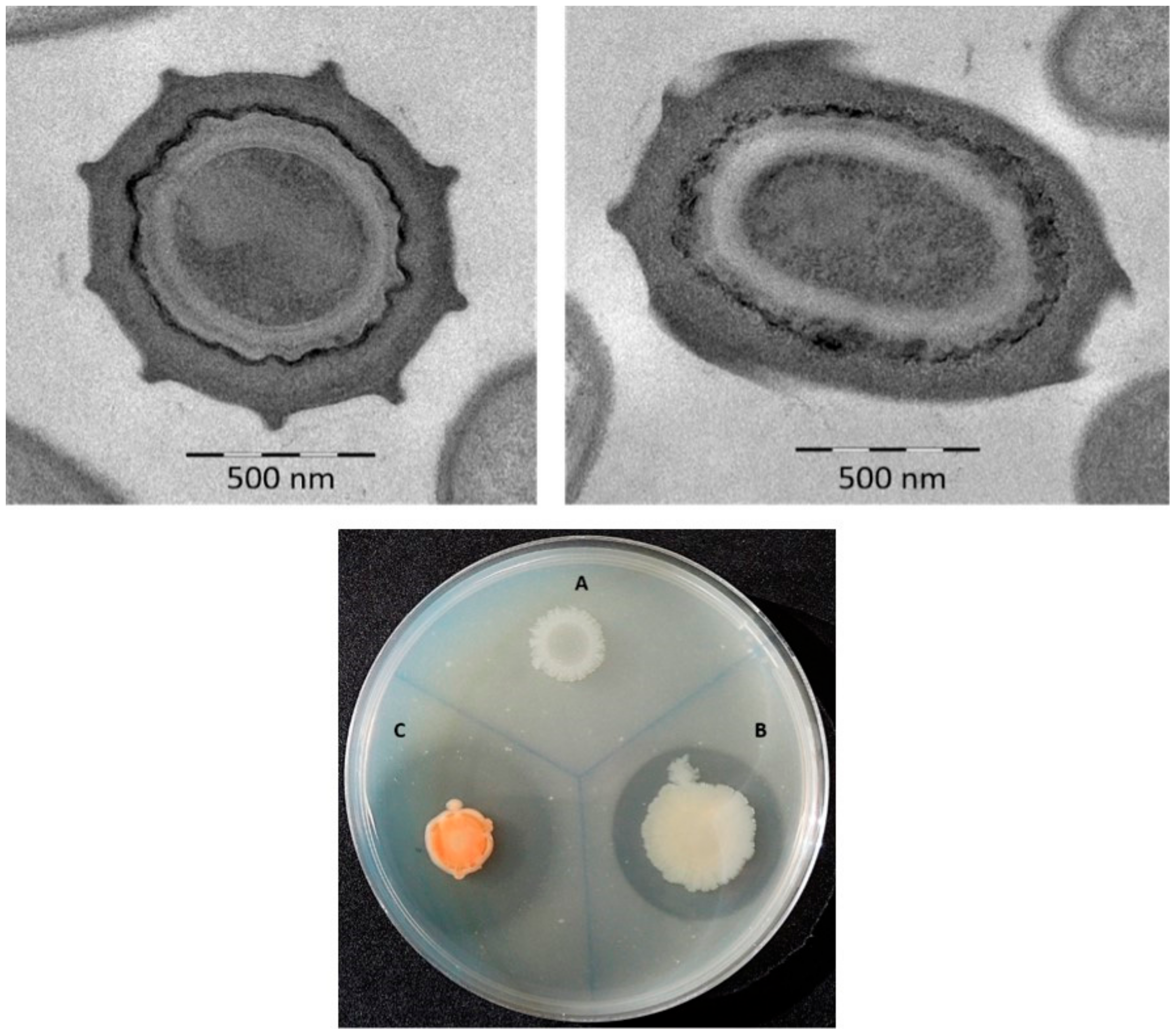
3.1. Phenotypic Characterization

Given its chitinolytic activity, strain N10T was selected from among the eleven sporulated strains isolated from the river otter feces and then characterized taxonomically. Its colonies on LB medium had a circular (diameter 1.0–2.0 mm) and convex morphology with a brilliant pink pigmentation after 48 h of incubation. Cells were found to be rod-shaped (2.2 × 0.8 µm), motile by peritrichous flagella and Gram-, catalase- and oxidase-positive. Strain N10T was slightly halophilic and produced oval endospores (1.2 × 0.8 µm) in swollen sporangia (Figure 1).
Strain N10T was found to be a facultative anaerobe, with no differences between aerobic and anaerobic growth being detected. It grew in a 15 to 37 °C temperature range, with 28 °C as optimum, in a pH range of 7 to 10, with 8 pH as optimum. The strain, which grew in the presence of 0% to 3% (w/v) concentrations of NaCl, with optimum growth at 1% (w/v) NaCl, was slightly halophilic.
Strain N10T was found to produce H2S from L-cystein; to hydrolyze chitin (Figure 1) and Tween® 20, but not Tween® 80, gelatine, casein, starch, DNA or lecithovitellin; to fix N2; and to degrade 1-aminocyclopropane-1-carboxilic acid (ACC). The tests for methyl red, indole, Voges–Proskauer, citrate, hemolysis, alkaline phosphatase, and aerobic nitrate reduction to nitrite, were negative. Its phenotypic characteristics are shown in Table 1 and the species description section. The differential characteristics of strain N10T with respect to the most closely related species, P. chitinolyticus (LMG 18047T) and with the type strain of genus P. polymyxa (CECT 155T) are also shown in Table 1. The strain N10T mainly differs in relation to the following features: pink coloration of the colonies, the slight requirements of NaCl, the inability to reduce aerobically nitrate, produce acids from glucose and many other sugar compounds and enzymes such as acid phosphatase, β-galactosidase or β-glucosidase.

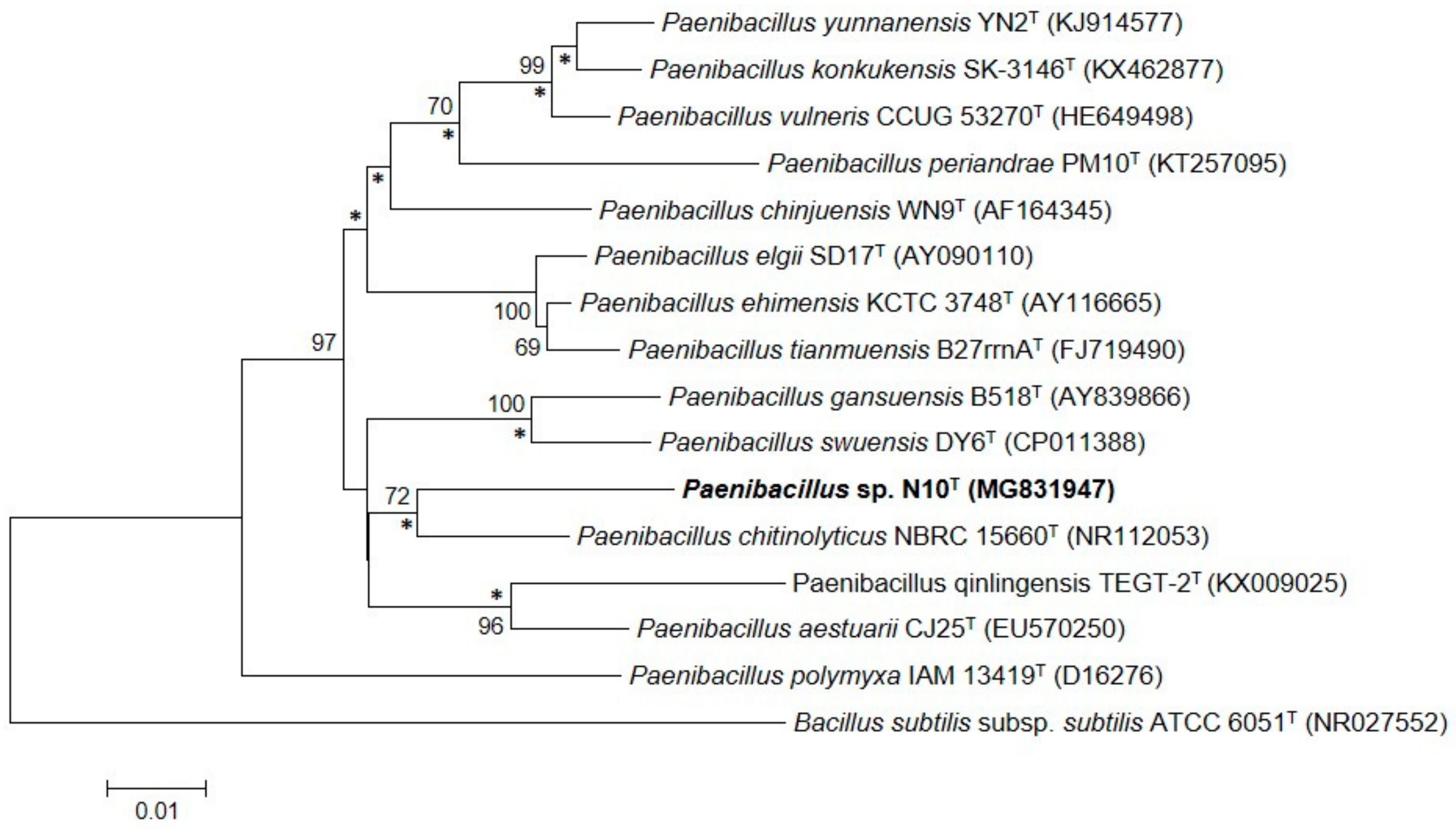
3.2. Phylogenetic Analysis Based on the 16S rRNA Gene Sequence

Sequencing of the cloned 16S rRNA gene of strain N10T resulted in a virtually complete 1520 bp-long sequence. Phylogenetic analysis of the 16S rRNA gene sequences of strain N10T and other 100 related strains revealed that strain N10T is a member of the genus Paenibacillus (Figure S1). It showed the highest sequence similarity to P. chitinolyticus LMG 18047T (95.4%) and less than 95% similarity to other species of genus Paenibacillus. The phylogenetic tree constructed using the neighbor-joining algorithm indicates that strain N10T forms a cluster with the P. chitinolyticus species, which showed the highest sequence similarity (Figure 2 and Figure S1). Using maximum-likelihood and maximum-parsimony algorithms, the Paenibacillus species was found to have the same phylogenetic distribution (Figures S2 and S3).

3.3. Analysis of Cellular Fatty Acids, Polar Lipids, Isoprenoid Quinones and Peptidoglycan Structure

The fatty acid profile of strain N10T, which showed a predominance of anteiso-C15:0 (56.95%), C16:0 (11.69%), and iso-C15:0 (8.46%), was similar from that of the most closely related strains and the type strain of the genus (Table 2). The major isoprenoid quinone of strain N10T was found to be MK-7, while diphosphatidylglycerols (DPGs), phosphatidylglycerol (PG), phosphatidylethanolamine (PE), and phosphoaminolipids (PNL) were the main polar lipids, features also commonly found in genus Paenibacillus [75]. The results of two-dimensional thin-layer chromatography (TLC) analysis of polar lipids are shown in the Supplementary Materials (Figure S4). The diamino acid of the cell-wall peptidoglycan for N10T strain and P. chitinolyticus LMG 18047T was meso-diamino-pimelic acid (type A1γ), as found in other members of Paenibacillus genus.

3.4. G+C Content

The G+C content of strain N10T, determined by the melting temperature (Tm) method, was in the 45–48 mol% range, while an in silico analysis of its draft genome G+C content produced a value of 49.8 mol%. These percentages fall within the broad 39 to 54 mol% range commonly accepted for members of genus Paenibacillus [28].

3.5. Whole-Genome Sequencing and Assembly

The over 5.54 Mbp-long draft genome of strain N10T, after manual curation, resulted in 48 contigs, while the 6.43 Mbp-long draft genome of P. chitinolyticus LMG 18047T in the public databases contains 32 contigs. After assembling the whole genome of strain N10T, the sequence was tested using Quality Assessment Tool for Genome Assemblies (QUAST) software. The results indicate that the whole-genome sequence was of sufficient quality, with an N50 value of 286655, an L50 value of 5, and 380X coverage. PGAP annotation [76] showed a total of 4718 protein-coding genes (PCGs), 3771 of which were assigned to a functional COG category in the EggNOG database [77,78]; categories K (transcription) and G (carbohydrate transport and metabolism) were the most abundant, with seven chitinase genes being found in the latter category. Strain N10T presented 117 RNAs and 8 16S rRNA gene copies. Nevertheless, only one of them was complete, whereas the rest of the copies corresponded to partial sequences. This genome sequence, which was deposited in the GenBank/EMBL/DDBJ database under accession number RHLK00000000, was used for further analysis.

3.6. ANI and dDDH Calculations

The average nucleotide identities (ANIs), based on BLAST (ANIb) and MUMmer (ANIm), for strain N10T and related Paenibacillus species, are shown in Table S1. The ANIb and ANIm values between strain N10T and the most closely related P. chitinolyticus LMG 18047T were 74.4% and 84.6%, respectively. In all cases, the ANI values for strain N10T and all species studied were lower than the cut-off for species demarcation (95%–96%) proposed by Richter and Rosselló-Móra [79,80]. Although not clear genus delimitation has been elucidated based on ANI values [81], Pérez-Cataluña et al. [74] consider that 68% could be the genus ANI threshold value. The ANIb between N10T and P. chitinolyticus LMG 18047T is over this value, reinforcing that strain N10T belongs to the Paenibacillus genus. As expected, according to Lee et al. [68], the OrthoANI value between N10T strain and P. chitinolyticus was slightly higher (75.6%) than the ANIb value (74.4%) (Table S2).
A comparison of the whole-genome sequences of the species analyzed showed digital DNA–DNA hybridization (dDDH) similarity of less than 32% (Table S2). The dDDH similarity between strain N10T and P. chitinolyticus LMG 18047T was 21.1%, a percentage that supports species delineation, as the established cut-off value for dDDH is 70% [82]. This was also the case for dDDH similarity between N10T and P. polymyxa, and the closest related species P. elgii, P. vulneris and P. qinlingensis, with percentages of 31.4%, 19.9%, 21.6% and 19.3%, respectively.

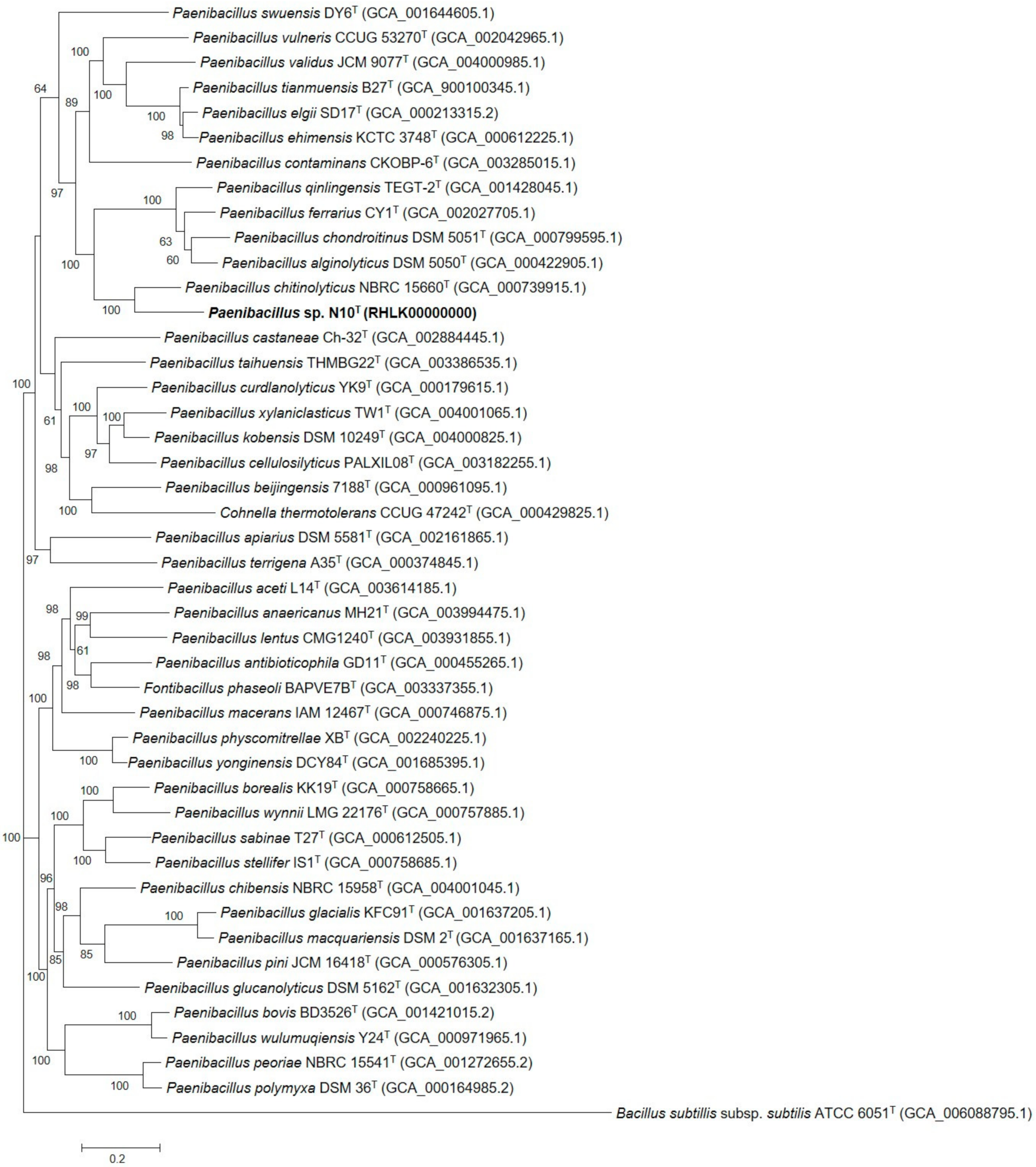
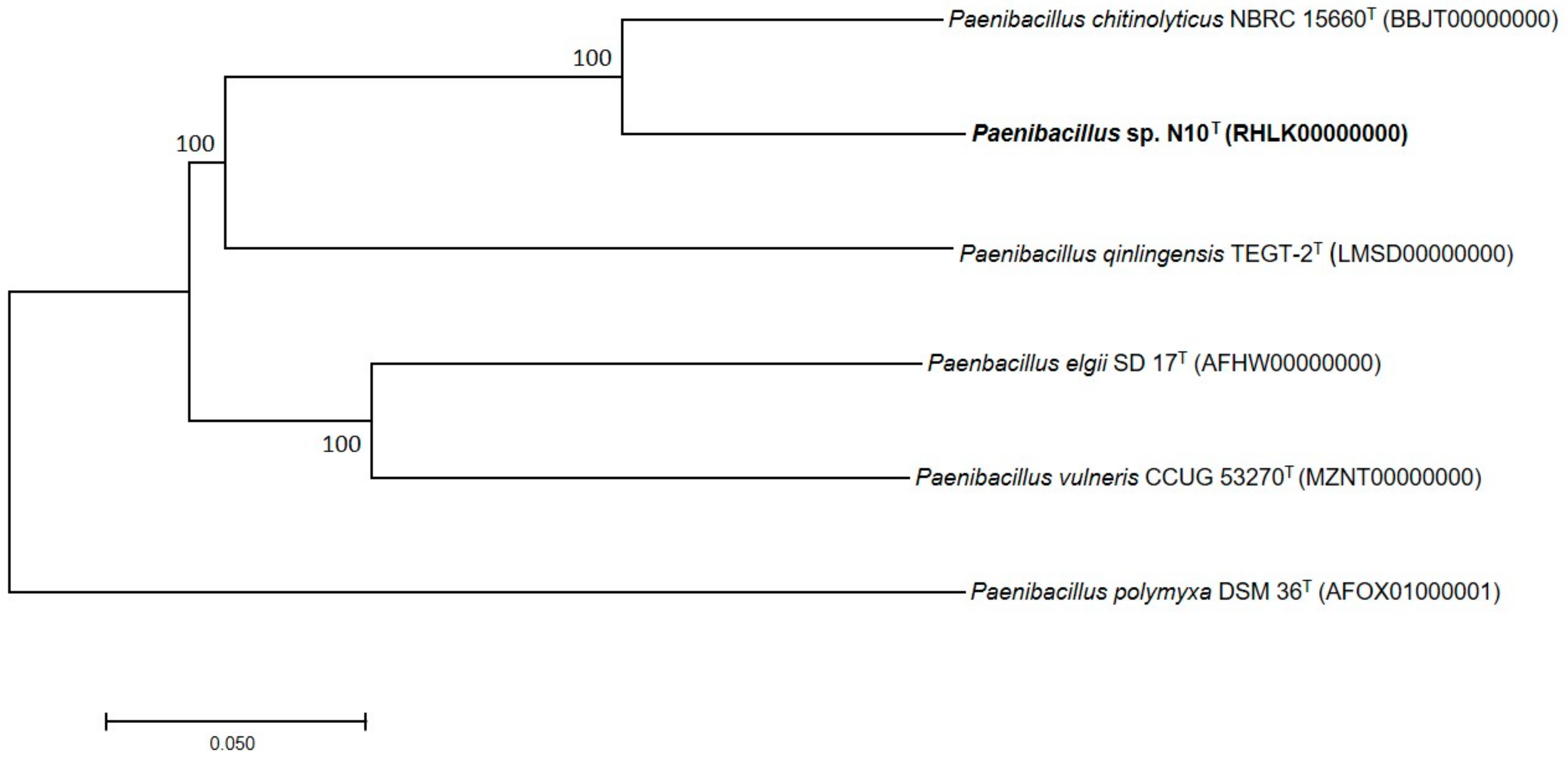
3.7. Phylogenetic Analysis of Multilocus Sequences and Core Orthologous Proteins

For a more robust analysis, a phylogenetic tree was built by aligning the concatenated sequences of the 16S rRNA and the three housekeeping gyrB, recA, and rpoB genes resulting in an alignment of 8269 nt. The 43 type strains out of the 102 closest species for which these sequences were available and correctly annotated, along with N10T and Bacillus subtilis subsp. subtilis ATCC 6051T as the outgroup, were used for this analysis. The resulting phylogenetic tree constructed according to the maximum likelihood method (Figure 3) confirms that strain N10T belongs to the genus Paenibacillus in accordance with the result obtained in the previous phylogenetic analysis using 16S rRNA sequences. The tree was carried out taking into account the partitions and codons of each gene.
A phylogenetic tree reconstruction based on the concatenated alignment of the 1078 core orthologous proteins confirmed that strain N10T and the five closest related bacteria have the same phylogenetic distribution (Figure 4), thus corroborating our previous results.

4. Conclusions

The polyphasic taxonomic study, which included a phenotypic and phylogenetic 16S rRNA, gyrB, recA, and rpoB gene analysis, as well as chemotaxonomic and genomic analyses, showed that strain N10T isolated from river otter (L. lutra) feces constitutes a novel species (proposed name: Paenibacillus lutrae sp. nov.) within genus Paenibacillus, with type strain N10T (= CECT 9541T = LMG 30535T). The description of Paenibacillus lutrae sp. nov. is summarized in Appendix A.

Supplementary Materials

The following are available online at https://www.mdpi.com/2076-2607/7/12/637/s1, Figure S1: Phylogenetic position of strain N10T based on the neighbour-joining algorithm of the 16S rRNA gene sequence and its relationship with other 100 related species. The GenBank/EMBL/DDBJ accession number of each sequence is shown in parenthesis. Bootstrap values are expressed as percentages of 1000 replications, and those greater than 60% are shown at branch points. Bar shows sequence divergence. Bar, 0.015 substitutions per nucleotide position. Figure S2: Molecular phylogenetic analysis of the 16S rRNA sequence according to the maximum likelihood method. Evolutionary history was inferred using the aforementioned method based on the Jukes-Cantor model. The tree with the highest log likelihood (-5308.40532.87) is shown. Bootstrap values are expressed as percentages of 1000 replications, and those over 60% are shown at branch points. The Bacillus subtilis subsp. subtilis ATCC 6051T sequence was used as the outgroup. Figure S3: Molecular phylogenetic analysis of the 16S rRNA sequence using the maximum parsimony method. The most parsimonious tree (length = 592) is shown. The consistency, retention and composite indices are 0.501114, 0.554672 and 0.344796 (0.277954), respectively, for all sites and parsimony-informative sites (in parentheses). Bootstrap values are expressed as percentages of 1000 replications, and those over 60% are shown at branch points. The Bacillus subtilis subsp. subtilis ATCC 6051T sequence was used as the outgroup. Figure S4. Polar lipid profile of strain N10T determined after two-dimensional TLC using molybdatophosphoric acid. AL: aminolipid; DPG: diphosphatidylglycerol; L: lipid; PE: phosphatidylethanolamine; PG: phosphatidylglycerol; PL: phospholipid; PNL: phosphoaminolipid. Table S1. ANIb and ANIm (in brackets) values among the genomes of strain N10T (1) and the most related species of Paenibacillus genus: P. chitinolyticus LMG18047T (2), P. polymyxa CECT155T (3), P. elgii SD17T (4), P. vulneris CCUG 53270T (5) and P. qinlingensis TEGT-2T (6). Table S2. Genome sequence similarity between N10T strain and genome sequences of closely related type strains with available genomes.

Author Contributions

M.R., J.C.R., V.B. and I.L. conceived and supervised the study; V.B. and I.L. designed the experiments; MR performed the experiments; J.C.R. took over genomic analysis; M.R., J.C.R. and I.L. analysed the data, prepared the figures and wrote the manuscript; M.R., J.C.R., V.B. and I.L. edited the manuscript and reviewed the literature.

Funding

This study was supported by a grant from the Spanish Ministerio de Educación y Ciencia (AGL-2015-68806-R) and from the Plan Andaluz de Investigación (Research Group BIO 188). Miguel A. Rodríguez is supported by a University of Granada Programme (Empleo Garantía Juvenil). José C. Reina is supported by an FPU fellowship from the Spanish Ministerio de Educación, Cultura y Deporte (FPU15/01717).

Acknowledgments

The authors wish to thank Francisco Rodríguez Vicario for providing samples from the Castril Natural Park and Michael O’Shea for proofreading the manuscript.

Conflicts of Interest

The authors declare no conflict of interest.

Appendix A

Description of Paenibacillus lutrae sp. nov

Paenibacillus lutrae (lu’trae. L. fem. gen. n. lutrae pertaining to the Lutra lutra otter from which the bacterium was isolated).
Paenibacillus lutrae sp. nov. is a motile, straight, Gram-positive, 2.2 × 0.8 µm rod-shaped bacteria which forms oval endospores in swollen sporangia. The colonies of P. lutrae on LB medium are a pink color after growing for 48 h at 28 °C. Cells growth pattern is uniform in a liquid medium. They are capable of growing in the presence of 0% to 3% (w/v) NaCl concentrations, with 1% (w/v) as optimum. The cells grow in a temperature range of 15 to 37 °C, with optimum growth at 28 °C, and a pH range of 7 to 10, with pH 8 as optimum. P. lutrae is a chemoorganotrophic and anaerobic facultative microorganism. Under aerobic conditions, nitrate is not reduced. Indole and methyl-red tests are negative, and catalase and oxidase are negative. Acids are not produced from adonitol, d-celobiose, d-fructose, d-galactose, myo-inositol, lactose, d-mannitol, d-mannose, d-melezitose, l-rhamnose, sucrose, d-salicilin, d-sorbitol, sorbose or d-trehalose. Tween 20 is hydrolyzed but, starch, casein, lecithovitellin, gelatine, Tween 80, urea, blood, or phosphates are not. H2S is produced from L-cysteine but not butanediol from glucose (Voges–Proskauer negative). ONPG is negative. No growth occurs on MacConkey agar. They are susceptible to ampicillin (10 μg), chloramphenicol (30 μg), gentamycin (10 μg), neomycin (30 μg), novobiocin (30 μg), penicillin G (10 μg), nalidixic acid (30 μg), rifampicin (2 μg) and tetracycline (30 μg). They are resistant to streptomycin (10 μg), kanamycin (30 μg) and trimetroprim/sulphametoxazol (1.25/23.75 μg).
The principal fatty acids of P. lutrae are anteiso-C15:0 (56.95%), C16:0 (11.69%), and iso-C15:0 (8.46%), and the main isoprenoid quinone is MK-7. The principal polar lipids are diphosphatidylglycerols (DPGs), phosphatidylglycerol (PG), phosphatidylethanolamine (PE), and phosphoaminolipids (PNL). The diamino acid of the cell-wall peptidoglycan was meso-diamino-pimelic acid (type A1γ). DNA G+C content was in the 45–48 mol% range according to the Tm method and 49.8 mol% according to in silico determination.
Type strain N10T (= CECT 9541T = LMG 30535T) was isolated from the feces of a river otter in Castril Natural Park in Granada (Spain).
The GenBank/EMBL/DDBJ accession number for the 16S rRNA sequence of Paenibacillus lutrae N10T is MG831947, and the complete genome is deposited under the accession number RHLK00000000.

References

  1. Flach, J.; Pilet, P.-E.; Jollès, P. What’s new in chitinase research? Experientia 1992, 48, 701–716. [Google Scholar] [CrossRef] [PubMed]
  2. Kramer, K.J.; Muthukrishnan, S. Insect chitinases: Molecular biology and potential use as biopesticides. Insect Biochem. Mol. Biol. 1997, 27, 887–900. [Google Scholar] [CrossRef]
  3. Gooday, G.W. Diversity of roles for chitinases in nature. In Chitin and Chitosan—The Versatile Environmentally Friendly Modern Materials; Zakaria, M.B., Muda, W.M.W., Abdullah, M.P., Eds.; Penerbit Universiti Kebangsaan Malasya: Bangi, Malasia, 1995; Volume 3, pp. 191–202. [Google Scholar]
  4. Cottrell, M.T.; Kirchman, D.L. Natural assemblages of marine proteobacteria and members of the Cytophaga-Flavobacter cluster consuming low-and high-molecular-weight dissolved organic matter. Appl. Environ. Microbiol. 2000, 66, 1692–1697. [Google Scholar] [CrossRef] [PubMed]
  5. Beier, S.; Bertilsson, S. Uncoupling of chitinase activity and uptake of hydrolysis products in freshwater bacterioplankton. Limnol. Oceanogr. 2011, 56, 1179–1188. [Google Scholar] [CrossRef]
  6. Reyes-Ramírez, A.; Escudero-Abarca, B.; Aguilar-Uscanga, G.; Hayward-Jones, P.; Barboza-Corona, J.E. Antifungal activity of Bacillus thuringiensis chitinase and its potential for the biocontrol of phytopathogenic fungi in soybean seeds. J. Food Sci. 2004, 69, M131–M134. [Google Scholar] [CrossRef]
  7. Huang, C.J.; Wang, T.K.; Chung, S.C.; Chen, C.Y. Identification of an antifungal chitinase from a potential biocontrol agent, Bacillus cereus 28-9. J. Biochem. Mol. Biol. 2005, 38, 82–88. [Google Scholar] [CrossRef] [PubMed]
  8. Aktuganov, G.E.; Melent’ev, A.I.; Galimzyanova, N.F.; Shirokov, A.V. The study of mycolytic properties of aerobic spore-forming bacteria producing extracellular chitinases. Microbiology 2008, 77, 700–709. [Google Scholar] [CrossRef]
  9. Singh, A.K.; Ghodke, I.; Chhatpar, H.S. Pesticide tolerance of Paenibacillus sp. D1 and its chitinase. J. Environ. Manag. 2009, 91, 358–362. [Google Scholar] [CrossRef]
  10. Terahara, T.; Ikeda, S.; Noritake, C.; Minamisawa, K.; Ando, K.; Tsuneda, S.; Harayama, S. Molecular diversity of bacterial chitinases in arable soils and the effects of environmental factors on the chitinolytic bacterial community. Soil Biol. Biochem. 2009, 41, 473–480. [Google Scholar] [CrossRef]
  11. Brzezinska, M.S.; Donderski, W. Chitinolytic bacteria in two lakes of different trophic status. Pol. J. Ecol. 2006, 54, 295–301. [Google Scholar]
  12. Haas, D.; Défago, G. Biological control of soil-borne pathogens by fluorescent pseudomonads. Nat. Rev. Microbiol. 2005, 3, 307–319. [Google Scholar] [CrossRef] [PubMed]
  13. Someya, N.; Ikeda, S.; Morohoshi, T.; Tsujimoto, M.N.; Yoshida, T.; Sawada, H.; Ikeda, T.; Tsuchiya, K. Diversity of culturable chitinolytic bacteria from rhizospheres of agronomic plants in Japan. Microbes Environ. 2009, 26, 7–14. [Google Scholar] [CrossRef] [PubMed]
  14. Spiegel, Y.; Chet, I.; Cohn, E. Use of chitin for controlling plant plant-parasitic nematodes. Plant Soil 1987, 98, 337–345. [Google Scholar] [CrossRef]
  15. Vionis, A.P.; Niemeyer, F.; Karagouni, A.D.; Schrempf, H. Production and processing of a 59-kilodalton exochitinase during growth of Streptomyces lividans carrying pCHIO12 in soil microcosms amended with crab or fungal chitin. Appl. Environ. Microbiol. 1996, 62, 1774–1780. [Google Scholar]
  16. Williamson, N.; Brian, P.; Wellington, E. Molecular detection of bacterial and streptomycete chitinases in the environment. Antonie Van Leeuwenhoek 2000, 78, 315–321. [Google Scholar] [CrossRef]
  17. Gomes, R.C.; Semêdo, L.T.; Soares, R.M.; Alviano, C.S.; Linhares, L.F.; Coelho, R.R. Chitinolytic activity of actinomycetes from a cerrado soil and their potential in biocontrol. Lett. Appl. Microbiol. 2000, 30, 146–150. [Google Scholar] [CrossRef]
  18. Gomes, R.; Semêdo, L.; Soares, R.; Linhares, L.; Ulhoa, C.; Alviano, C.; Coelho, R. Purification of a thermostable endochitinase from Streptomyces RC1071 isolated from a cerrado soil and its antagonism against phytopathogenic fungi. J. Appl. Microbiol. 2001, 90, 653–661. [Google Scholar] [CrossRef]
  19. Yasir, M.; Aslam, Z.; Kim, S.W.; Lee, S.W.; Jeon, C.O.; Chung, Y.R. Bacterial community composition and chitinase gene diversity of vermicompost with antifungal activity. Bioresour. Technol. 2009, 100, 4396–4403. [Google Scholar] [CrossRef]
  20. Cottrell, M.T.; Moore, J.A.; Kirchman, D.L. Chitinases from uncultured marine microorganisms. Appl. Environ. Microbiol. 1999, 65, 2553–2557. [Google Scholar]
  21. Beier, S.; Jones, C.M.; Mohit, V.; Hallin, S.; Bertilsson, S. Global phylogeography of chitinase genes in aquatic metagenomes. Appl. Environ. Microbiol. 2011, 77, 1101–1106. [Google Scholar] [CrossRef]
  22. Eski, A.; Demir, I.; Gullu, M.; Demirbag, Z. Biodiversity and pathogenicity of bacteria associated with the gut microbiota of beet armyworm, Spodoptera exigua Hübner (Lepidoptera: Noctuidae). Microb. Pathog. 2018, 121, 350–358. [Google Scholar] [CrossRef] [PubMed]
  23. Pasari, N.; Gupta, M.; Eqbal, D.; Yazdani, S.S. Genome analysis of Paenibacillus polymyxa A18 gives insights into the features associated with its adaptation to the termite gut environment. Sci. Rep. 2019, 9, 6091. [Google Scholar] [CrossRef] [PubMed]
  24. Beygmoradi, A.; Homaei, A. Marine chitinolytic enzymes, a biotechnological treasure hidden in the ocean? Appl. Microbiol. Biotechnol. 2018, 102, 9937–9948. [Google Scholar] [CrossRef] [PubMed]
  25. Chen, L.; Wei, Y.; Shi, M.; Li, Z.; Zhang, S.-H. An archaeal chitinase with a secondary capacity for catalyzing cellulose and its biotechnological applications in shell and straw degradation. Front. Microbiol. 2019, 10, 1253. [Google Scholar] [CrossRef]
  26. Chen, Q.; Peng, D. Nematode chitin and application. Adv. Exp. Med. Biol. 2019, 1142, 209–219. [Google Scholar] [CrossRef]
  27. Zhou, J.; Chen, J.; Xu, N.; Zhang, A.; Chen, K.; Xin, F.; Zhang, W.; Ma, J.; Fang, Y.; Jiang, M.; et al. The broad-specificity chitinases: Their origin, characterization, and potential application. Appl. Microbiol. Biotechnol. 2019, 103, 3289–3295. [Google Scholar] [CrossRef]
  28. Ash, C.; Priest, F.G.; Collins, M.D. Molecular identification of rRNA group 3 bacilli (Ash, Farrow, Wallbanks and Collins) using a PCR probe test. Antonie van Leeuwenhoek 1993, 64, 253–260. [Google Scholar] [CrossRef]
  29. Djukic, M.; Becker, D.; Poehlein, A.; Voget, S.; Daniel, R. Genome sequence of Paenibacillus alvei DSM 29, a secondary invader during European foulbrood outbreaks. J. Bacteriol. 2012, 194, 6365. [Google Scholar] [CrossRef]
  30. Kuroshima, K.-I.; Sakane, T.; Takata, R.; Yokota, A. Bacillus ehimensis sp. nov. and Bacillus chitinolyticus sp. nov., new chitinolytic members of the genus Bacillus. Int. J. Syst. Evol. Microbiol. 1996, 46, 76–80. [Google Scholar] [CrossRef]
  31. Lee, J.S.; Pyun, Y.R.; Bae, K.S. Transfer of Bacillus ehimensis and Bacillus chitinolyticus to the genus Paenibacillus with emended descriptions of Paenibacillus ehimensis comb. nov. and Paenibacillus chitinolyticus comb. nov. Int. J. Syst. Evol. Microbiol. 2004, 54, 929–933. [Google Scholar] [CrossRef]
  32. Sun, H.; Mao, X. Discovery and characterization of a novel chitosanase from Paenibacillus dendritiformis by phylogeny-based enzymatic product specificity prediction. J. Agric. Food Chem. 2018, 66, 4645–4651. [Google Scholar] [CrossRef] [PubMed]
  33. Kim, Y.H.; Park, S.K.; Hur, J.Y.; Kim, Y.C. Purification and characterization of a major extracellular chitinase from a biocontrol bacterium, Paenibacillus elgii HOA73. Plant. Pathol. J. 2017, 33, 318–328. [Google Scholar] [CrossRef]
  34. García-González, E.; Poppinga, L.; Fünfhaus, A.; Hertlein, G.; Hedtke, K.; Jakubowska, A.; Genersch, E. Paenibacillus larvae chitin-degrading protein PlCBP49 is a key virulence factor in American Foulbrood of honey bees. PLoS Pathog. 2014, 10, e1004284. [Google Scholar] [CrossRef] [PubMed]
  35. Chung, Y.R.; Kim, C.H.; Hwang, I.; Chun, J. Paenibacillus koreensis sp. nov., a new species that produces an iturin-like antifungal compound. Int. J. Syst. Evol. Microbiol. 2000, 50, 1495–1500. [Google Scholar] [CrossRef]
  36. Doan, C.T.; Tran, T.N.; Nguyen, V.B. Reclamation of marine chitinous materials for chitosanase production via microbial conversion by Paenibacillus macerans. Mar. Drugs 2018, 16, 429. [Google Scholar] [CrossRef] [PubMed] [Green Version]
  37. Doan, C.T.; Tran, T.N.; Nguyen, V.B. Production of a thermostable chitosanase from shrimp heads via Paenibacillus mucilaginosus TKU032 conversion and its application in the preparation of bioactive chitosan oligosaccharides. Mar. Drugs 2019, 17, 217. [Google Scholar] [CrossRef] [PubMed] [Green Version]
  38. Passera, A.; Marcolungo, L.; Casati, P. Hybrid genome assembly and annotation of Paenibacillus pasadenensis strain R16 reveals insights on endophytic life style and antifungal activity. PLoS ONE 2018, 13, e0189993. [Google Scholar] [CrossRef] [Green Version]
  39. Cody, R.M. Distribution of chitinase and chitobiase in Bacillus. Curr. Microbiol. 1989, 19, 201–205. [Google Scholar] [CrossRef]
  40. Yahiaoui, M.; Laribi-Habchi, H.; Bouacem, K.; Asmani, K.L.; Mechri, S.; Harir, M.; Bendif, H.; Aissani-El Fertas, R.; Jaouadi, B. Purification and biochemical characterization of a new organic solvent-tolerant chitinase from Paenibacillus timonensis strain LK-DZ15 isolated from the Djurdjura Mountains in Kabylia, Algeria. Carbohydr. Res. 2019, 483, 107747. [Google Scholar] [CrossRef]
  41. Liao, W.; Liu, P.; Liao, W.; Miao, L. Complete genome of the chitin-degrading bacterium, Paenibacillus xylanilyticus W4. Genome Biol. Evol. 2019, 11, 3252–3255. [Google Scholar] [CrossRef]
  42. An, C.; Okamoto, Y.; Xu, S.; Eo, K.Y.; Kimura, J.; Yamamoto, N. Comparison of fecal microbiota of three captive carnivore species inhabiting Korea. J. Vet. Med. Sci. 2017, 79, 542–546. [Google Scholar] [CrossRef] [PubMed] [Green Version]
  43. Logan, N.A.; Berge, O.; Bishop, A.H.; Busse, H.-J.; De Vos, P.; Fritze, D.; Heyndrickx, M.; Kämpfer, P.; Rabinovitch, L.; Salkinoja-Salonen, M.S.; et al. Proposed minimal standards for describing new taxa of aerobic, endospore-forming bacteria. Int. J. Syst. Evol. Microbiol. 2009, 59, 2114–2121. [Google Scholar] [CrossRef] [PubMed] [Green Version]
  44. Kovacs, N. Identification of Pseudomonas pyocyanea by the oxidase reaction. Nature 1956, 178, 703. [Google Scholar] [CrossRef] [PubMed]
  45. Xie, Q.Y.; Lin, H.P.; Li, L.; Brown, R.; Goodfellow, M.; Deng, Z.; Hong, K. Verrucosispora wenchangensis sp. nov., isolated from mangrove soil. Antonie Van Leeuwenhoek 2012, 102, 1–7. [Google Scholar] [CrossRef] [PubMed]
  46. Clarke, P.H. Hydrogen sulphide production by bacteria. J. Gen. Microbiol. 1953, 8, 397–407. [Google Scholar] [CrossRef] [PubMed] [Green Version]
  47. Skerman, V.B.D. A Guide to the Identification of the Genera of Bacteria; The Williams & Wilkins, Co.: Baltimore, MD, USA, 1961; p. 251. [Google Scholar]
  48. Kovacs, N. Eine vereinfachte methode zum nachweis der indolbildung durch bakterien. Immunforsch 1928, 55, 311–315. [Google Scholar]
  49. Barritt, M.M. The intensification of the Voges-Proskauer reaction by the addition of α-naphthol. J. Pathol. Bacteriol. 1936, 42, 441–454. [Google Scholar] [CrossRef]
  50. Uttley, A.; Collins, C. Theory and practice of bacterial identification. In Cowan and Steel’s Manual for the Identification of Medical Bacteria, 3rd ed.; Barrow, G.I., Feltham, R.K.A., Eds.; Cambridge University Press: Cambridge, UK, 1993; pp. 46–49. [Google Scholar]
  51. Matthews, S.; Suhaimi, M. Selection of suitable growth medium for free-living diazotrophs isolated from compost. J. Trop. Agric. Food Sci. 2010, 38, 211–219. [Google Scholar]
  52. Poonguzhali, S.; Madhaiyan, M.; Sa, T. Cultivation-dependent characterization of rhizobacterial communities from field grown Chinese cabbage Brassica campestris ssp. pekinensis and screening of traits for potential plant growth promotion. Plant Soil 2006, 286, 167–180. [Google Scholar] [CrossRef]
  53. Alexander, D.B.; Zuberer, D.A. Use of chrome azurol S reagents to evaluate siderophore production by rhizosphere bacteria. Biol. Fert. Soils 1991, 12, 39–45. [Google Scholar] [CrossRef]
  54. Bauer, A.W.; Kirby, W.M.; Sherris, J.C.; Turck, M. Antibiotic susceptibility testing by a standardized single disk method. Am. J. Clin. Pathol. 1966, 45, 493–496. [Google Scholar] [CrossRef] [PubMed]
  55. Tindall, B.J.; Sikorski, J.; Smibert, R.A.; Krieg, N.R. Phenotypic characterization and the principles of comparative systematics. In Methods for General and Molecular Microbiology, 3rd ed.; Reddy, C., Beveridge, T., Breznak, J., Marzluf, G., Schmidt, T., Snyder, L.A., Eds.; ASM Press: Washington, DC, USA, 2007; pp. 330–393. [Google Scholar]
  56. Tindall, B.J. A comparative study of the lipid composition of Halobacterium saccharovorum from various sources. Syst. Appl. Microbiol. 1990, 13, 128–130. [Google Scholar] [CrossRef]
  57. Tindall, B.J. Lipid composition of Halobacterium lacusprofundi. FEMS Microbiol. Lett. 1990, 66, 199–202. [Google Scholar] [CrossRef]
  58. Schumann, P. Peptidoglycan structure. Method. Microbiol. 2011, 38, 101–129. [Google Scholar] [CrossRef]
  59. Altschul, S.F.; Gish, W.; Miller, W.; Myers, E.W.; Lipman, D.J. Basic local alignment search tool. J. Mol. Biol. 1990, 215, 403–410. [Google Scholar] [CrossRef]
  60. Yoon, S.H.; Ha, S.M.; Kwon, S.; Lim, J.; Kim, Y.; Seo, H.; Chun, J. Introducing EzBioCloud: A taxonomically united database of 16S rRNA gene sequences and whole-genome assemblies. Int. J. Syst. Evol. Microbiol. 2017, 67, 1613–1617. [Google Scholar] [CrossRef]
  61. Kumar, S.; Stecher, G.; Li, M.; Knyaz, C.; Tamura, K. MEGA X: Molecular Evolutionary Genetics Analysis across Computing Platforms. Mol. Biol. Evol. 2018, 35, 1547–1549. [Google Scholar] [CrossRef]
  62. Sievers, F.; Wilm, A.; Dineen, D.; Gibson, T.J.; Karplus, K.; Li, W.; López, R.; McWilliam, H.; Remmert, M.; Soding, J.; et al. Fast, scalable generation of high-quality protein multiple sequence alignments using Clustal Omega. Mol. Syst. Biol. 2011, 7, 539. [Google Scholar] [CrossRef]
  63. Marmur, J. A procedure for the isolation of deoxyribonucleic acid from micro-organisms. J. Mol. Biol. 1961, 3, 208–218. [Google Scholar] [CrossRef]
  64. Owen, R.; Pitcher, D. Current methods for estimating DNA base composition and levels of DNA-DNA hybridization. In Chemicals Methods in Bacterial Systematics; Goodfellow, M., Minnikin, D.E., Eds.; Academic Press: London, UK, 1985; Volume 20, pp. 67–93. [Google Scholar]
  65. Nurk, S.; Bankevich, A.; Antipov, D.; Gurevich, A.A.; Korobeynikov, A.; Lapidus, A.; Prjibelski, A.D.; Pyshkin, A.; Sirotkin, A.; Sirotkin, Y.; et al. Assembling single-cell genomes and mini-metagenomes from chimeric MDA products. J. Comput. Biol. 2013, 20, 714–737. [Google Scholar] [CrossRef] [Green Version]
  66. Richter, M.; Rosselló-Móra, R.; Oliver Glöckner, F.; Peplies, J. JSpeciesWS: A web server for prokaryotic species circumscription based on pairwise genome comparison. Bioinformatics 2015, 32, 929–931. [Google Scholar] [CrossRef] [PubMed]
  67. Meier-Kolthoff, J.P.; Auch, A.F.; Klenk, H.P.; Göker, M. Genome sequence-based species delimitation with confidence intervals and improved distance functions. BMC Bioinform. 2013, 14, 60. [Google Scholar] [CrossRef] [Green Version]
  68. Lee, I.; Kim, Y.O.; Park, S.C.; Chun, J. OrthoANI: An improved algorithm and software for calculating average nucleotide identity. Int. J. Syst. Evol. Microbiol. 2016, 66, 1100–1103. [Google Scholar] [CrossRef] [PubMed]
  69. Aziz, R.K.; Bartels, D.; Best, A.A.; DeJongh, M.; Disz, T.; Edwards, R.A.; Formsma, K.; Gerdes, S.; Glass, E.M.; Kubal, M.; et al. The RAST Server: Rapid annotations using subsystems technology. BMC Genom. 2008, 9, 75. [Google Scholar] [CrossRef] [PubMed] [Green Version]
  70. Nguyen, L.T.; Schmidt, H.A.; von Haeseler, A.; Minh, B.Q. IQ-TREE: A fast and effective stochastic algorithm for estimating maximum-likelihood phylogenies. Mol. Biol. Evol. 2015, 32, 268–274. [Google Scholar] [CrossRef]
  71. Chernomor, O.; von Haeseler, A.; Minh, B.Q. Terrace aware data structure for phylogenomic inference from supermatrices. Syst. Biol. 2016, 65, 997–1008. [Google Scholar] [CrossRef] [Green Version]
  72. Kainer, D.; Lanfear, R. The effects of partitioning on phylogenetic inference. Mol. Biol. Evol. 2015, 32, 1611–1627. [Google Scholar] [CrossRef] [Green Version]
  73. Chaudhari, N.M.; Gupta, V.K.; Dutta, C. BPGA-an ultra-fast pan-genome analysis pipeline. Sci. Rep. 2016, 6, 24373. [Google Scholar] [CrossRef] [Green Version]
  74. Katoh, K.; Standley, D.M. MAFFT multiple sequence alignment software version 7: Improvements in performance and usability. Mol. Biol. Evol. 2013, 30, 772–780. [Google Scholar] [CrossRef] [Green Version]
  75. Yao, R.; Wang, R.; Wang, D.; Su, J.; Zheng, S.; Wang, G. Paenibacillus selenitireducens sp. nov., a selenite-reducing bacterium isolated from a selenium mineral soil. Int. J. Syst. Evol. Microbiol. 2014, 64, 805–811. [Google Scholar] [CrossRef]
  76. Tatusova, T.; DiCuccio, M.; Badretdin, A.; Chetvernin, V.; Nawrocki, E.P.; Zaslavsky, L.; Lomsadze, A.; Pruitt, K.D.; Borodovsky, M.; Ostell, J. NCBI prokaryotic genome annotation pipeline. Nucleic Acids Res. 2016, 44, 6614–6624. [Google Scholar] [CrossRef] [PubMed]
  77. Huerta-Cepas, J.; Szklarczyk, D.; Forslund, K.; Cook, H.; Heller, D.; Walter, M.C.; Rattei, T.; Mende, D.R.; Sunagawa, S.; Kuhn, M.; et al. eggNOG 4.5: A hierarchical orthology framework with improved functional annotations for eukaryotic, prokaryotic and viral sequences. Nucleic Acids Res. 2016, 44, D286–D293. [Google Scholar] [CrossRef] [PubMed] [Green Version]
  78. Huerta-Cepas, J.; Forslund, K.; Coelho, L.P.; Szklarczyk, D.; Jensen, L.J.; von Mering, C.; Bork, P. Fast genome-wide functional annotation through orthology assignment by eggNOG-Mapper. Mol. Biol. Evol. 2017, 34, 2115–2122. [Google Scholar] [CrossRef] [PubMed] [Green Version]
  79. Richter, M.; Rosselló-Móra, R. Shifting the genomic gold standard for the prokaryotic species definition. Proc. Natl. Acad. Sci. USA 2009, 106, 19126–19131. [Google Scholar] [CrossRef] [Green Version]
  80. Kim, M.; Oh, H.S.; Park, S.C.; Chun, J. Towards a taxonomic coherence between average nucleotide identity and 16S rRNA gene sequence similarity for species demarcation of prokaryotes. Int. J. Syst. Evol. Microbiol. 2014, 64, 346–351. [Google Scholar] [CrossRef]
  81. Pérez-Cataluña, A.; Salas-Massó, N.; Diéguez, A.L.; Balboa, S.; Lema, A.; Romalde, J.L.; Figueras, M.J. Revisiting the taxonomy of the genus Arcobacter: Getting order from the chaos. Front. Microbiol. 2018, 9, 2077. [Google Scholar] [CrossRef] [PubMed]
  82. Goris, J.; Konstantinidis, K.T.; Klappenbach, J.A.; Coenye, T.; Vandamme, P.; Tiedje, J.M. DNA-DNA hybridization values and their relationship to whole-genome sequence similarities. Int. J. Syst. Evol. Microbiol. 2007, 57, 81–91. [Google Scholar] [CrossRef] [Green Version]
Figure 1. Transmission electron micrographs of oval-shaped endospores of strain N10T (upper images) and chitinolytic activity (lower image). P. polymyxa CECT 155T (A), P. chitinolitycus LMG 18047T (B), and strain N10T (C) on LB medium supplemented with 1% (w/v) colloidal chitin.
Microorganisms 07 00637 g001
Figure 2. Phylogenetic position of strain N10T based on the neighbor-joining algorithm of the 16S rRNA gene sequence and its relationship with other related species. The GenBank/EMBL/DDBJ accession number of each sequence is shown in parenthesis. Bootstrap values are expressed as percentages of 1000 replications, and those greater than 60% are shown at branch points. Bar shows sequence divergence. Bar, 0.01 substitutions per nucleotide position. Asterisks indicate that the corresponding nodes were also obtained in the trees generated with the maximum-likelihood and maximum-parsimony algorithms.
Microorganisms 07 00637 g002
Figure 3. Maximum-likelihood tree based on a comparison of the concatenated 16S rRNA, gyrB, recA, and rpoB gene sequences, showing the phylogenetic position of strain N10T and its relationship with the 43 closely related species of genus Paenibacillus. Bootstrap values are expressed as percentages of 1000 replications, and those greater than 60% are shown at branch points. Bar, 0.2 substitutions per nucleotide position.
Microorganisms 07 00637 g003
Figure 4. Tree constructed according to the neighbor-joining method based on 1078 core orthologous proteins of strain N10T and five additional most closely related species of genus Paenibacillus. Bootstrap values are expressed as percentages of 1000 replications, and those over 60% are shown at branch points. Bar, 0.05 substitutions per nucleotide position.
Microorganisms 07 00637 g004
Table 1. Differential characteristics between N10T strain with respect to its closest relative and the type strain of the genus Paenibacillus.
Characteristic123
Colony PigmentationPinkCreamCream-White
NaCl range (%) (w/v)0–30–40–5
NaCl optimum (%) (w/v)100
pH range7–106–96–9
pH optimum877
Oxidase+v-
Methyl red--+
Voges-Proskauer--+
Aerobic nitrate reduction-++
Hydrolysis of:
Gelatine-v-
Chitin++-
Starch--+
Casein-v+
Tween 80--+
ACC deaminase++-
Assimilation of:
l-arabinose--+
d-mannose-++
d-mannitol--+
N-acetyl-glucosamine-+-
Potassium gluconate-++
Enzymes:
Alkaline phosphatase-+-
Acid phosphatase-++
α-galactosidase--+
β-galactosidase-++
β-glucosidase-++
Acids from carbohydrates:
Glycerol-++
Galactose-++
Manose-++
Amygdaline-++
Arbutin-++
Salicin-++
Cellobiose-++
Lactose-++
Melibiose-++
Sucrose-++
Trehalose-++
Gentiobiose-++
d-turanose-++
Antimicrobial susceptibility:
AmpicilinSIS
CloramphenicolSIS
Kanamycin RRS
Nalidixic acidSRS
RifampicinSSR
Trimethoprim/sulfamethoxazoleRSS
DNA G + C content (Tm) (mol %)45–4851–5343–46
DNA G + C content (in silico) (mol %)49.852.445.5
Strains: 1, N10T strain; 2, P. chitinolitycus LMG 18047T; 3, P. polymyxa CECT 155T. +, positive; -, negative; v, variable; S, susceptible; I, intermediate; R, resistant. All strains were rods, Gram-positive, growth in anaerobic conditions and produce oval spores. All data shown in this table derive from this study. All strains were negative for indole, citrate, cellulase, DNase, lecithinase, hemolysis, and siderophore production and did not grow on MacConkey agar, but were positive for catalase, H2S production, nitrogen-fixing and Tween® 20 hydrolysis. The three strains analyzed were negative for arginine dihydrolase (ADH), urease, gelatinase, capric acid, malic acid, adipic acid and phenylacetic acid assimilations, C14 lipase, leucine arylamidase, valine arylamidase, cysteine arylamidase, trypsin, α-chymotrypsin, β-glucuronidase, N-acetyl-β-glucosaminidase, α-mannosidase, and α-fucosidase. All strains showed positive results for esculin, d-glucose and d-maltose assimilations, C4 and C8 lipases, α-glucosidase, and naphthol-AS-BI-phosphohydrolase. All strains were positive for acid production from esculin, glucose, glycogen, d-maltose, 1-methyl-α-d-glucopyranoside, starch, and D-ribose. By contrast, they were negative for acid production from d-adonitol, d-arabinose, d- and l-arabitol, dulcitol, erythritol, d- and l-fucose, inositol, 1-methyl-α-d-mannopyranoside, potassium gluconate, potassium 2-ketogluconate, and potassium 5-ketogluconate, d-sorbitol, l-sorbose, d-tagatose, xylitol and d- and l-xylose. All strains were sensitive to gentamicin, neomycin, novobiocin, penicillin G, and tetracycline, but were resistant to streptomycin.
Table 2. Cellular fatty acids content of N10T and related species of the genus. Strains: 1, N10T; 2, P. chitinolyticus LMG 18047T; 3, P. polymyxa CECT 155T. -, not detected or lower than 1% of total composition.
Cellular Fatty Acids123
Straight-chain fatty acids:
C14:02.502.49-
C15:01.17--
C16:011.6925.888.39
Branched-chain fatty acids:
iso-c14:01.86-3.16
iso-c15:0 8.466.263.44
anteiso-c15:0 56.9549.2047.79
iso-c16:05.982.3418.80
iso-c17:02.634.733.38
anteiso-c17:05.826.7612.24
Unsaturated fatty acids:
C16:1 ω11c1.59--

