Article

Modeling the Characteristic Residues of Chlorophyll f Synthase (ChlF) from Halomicronema hongdechloris to Determine Its Reaction Mechanism

School of Life and Environmental Sciences, University of Sydney, Sydney, NSW 2006, Australia
*
Author to whom correspondence should be addressed.
Current address: School of Biotechnology and Biomolecular Sciences, University of New South Wales, Sydney, NSW 2052, Australia.
Microorganisms 2023, 11(9), 2305; https://doi.org/10.3390/microorganisms11092305
Submission received: 29 August 2023 / Revised: 7 September 2023 / Accepted: 7 September 2023 / Published: 13 September 2023
(This article belongs to the Special Issue Phototrophic Bacteria 2.0)

Abstract

:
Photosystem II (PSII) is a quinone-utilizing photosynthetic system that converts light energy into chemical energy and catalyzes water splitting. PsbA (D1) and PsbD (D2) are the core subunits of the reaction center that provide most of the ligands to redox-active cofactors and exhibit photooxidoreductase activities that convert quinone and water into quinol and dioxygen. The performed analysis explored the putative uncoupled electron transfer pathways surrounding P680+ induced by far-red light (FRL) based on photosystem II (PSII) complexes containing substituted D1 subunits in Halomicronema hongdechloris. Chlorophyll f-synthase (ChlF) is a D1 protein paralog. Modeling PSII-ChlF complexes determined several key protein motifs of ChlF. The PSII complexes included a dysfunctional Mn4CaO5 cluster where ChlF replaced the D1 protein. We propose the mechanism of chlorophyll f synthesis from chlorophyll a via free radical chemistry in an oxygenated environment created by over-excited pheophytin a and an inactive water splitting reaction owing to an uncoupled Mn4CaO5 cluster in PSII-ChlF complexes. The role of ChlF in the formation of an inactive PSII reaction center is under debate, and putative mechanisms of chlorophyll f biosynthesis are discussed.

1. Introduction

Photosynthesis is a fundamental process that converts light energy into chemical energy. Oxygenic phototrophs use two photosystems (PSI and PSII) that constitute the Z-scheme and the associated generation of assimilatory power. Photosystems are multisubunit complexes in the thylakoid membrane, and PSII absorbs and transforms light energy into chemical energy, resulting in water splitting and oxygen evolution. PsbA (D1) and PsbD (D2) are the core subunits of PSII that exhibit photooxidoreductase activities and convert plastoquinone and water into plastoquinol (QH2) and dioxygen (O2). The Mn4CaO5 cluster is the oxygen evolution center of PSII and is directly coordinated with multiple conserved residues of D1 and PsbC (CP43)-glutamic acid-354 (E354), further stabilized through second coordination spheres supported by conserved residues of D1, D2, and CP43 [1,2,3]. The tyrosine-161 (Y161) and histidine-190 (H190) residues of D1 work in tandem to transfer electrons to the reaction center Chl a (P680), wherein Y161 transfers electrons and H190 acts as a proton acceptor [4,5]. Charge separation involves the transfer of an electron from P680, which then forms P680+, to the primary acceptor (plastoquinone, Q) via an intermediary acceptor, pheophytin (Pheo a).
Amino acid residues surrounding the Mn4CaO5 cluster affect the PSII oxygen evolving capacity [6,7]. For example, a N181A/S mutation of the D1 protein results in slower O-O bond formation during the water-splitting reaction in Synechocystis sp. PCC 6803 [8]. A D170A/S substitution of D1 poisons oxygen evolution capacity, while a D170E mutation is complementary with little effect on activity [9]. Additionally, the substitutions N87A/S, F117L, D170AES, and E189D in D1 result in impaired water-splitting activity and possibly reduced PSII stability [5].
Cyanobacteria typically possess multiple psbA gene copies with different expression levels that mostly reflect varying environmental conditions [1,10,11,12]. For example, Synechocystis sp. PCC 6803 has three psbA genes, with psbA1 upregulated in high light, psbA2 constitutively expressed [13], and psbA3 expressed during low oxygen “microaerophilic” conditions. A general name of psbA′ is given to “microaerophilic” psbA genes since other cyanobacteria have a comparable strategy [14].
Dark expression of sentinel psbA (belonging to the “rogue” D1 group) blocked oxygen-evolving PSII activity through an uncleavable C-terminus and altered amino acid residues surrounding the Mn4CaO5 cluster in the nitrogen-fixing cyanobacterium Cyanothece [1,15,16]. Recently, a D1 variant was renamed “chlorophyll f synthase (ChlF)”. It is a far-red light (FRL)-induced paralog of D1 in Chl f-producing cyanobacteria [17]. The ChlF-encoding gene participates in the far-red light-induced photoacclimation (FaRLiP) gene cluster, and it is only transcribed under FRL conditions. It is unknown how ChlF catalyzes the formation of chlorophyll f (Chl f) from Chl a, although molecular oxygen is required [18]. The ChlF-encoding gene from Chroococcidiopsis thermalis PCC 7203 (AFY86562) was used to replace the D1 subunit-encoding gene in the PSII complex of Synechocystis sp. PCC 6803 that resulted in up to ~0.1% Chl f of total chlorophyll when the transformant was grown photoheterotrophically with 5 mM glucose at a light intensity of 10–30 μmol photons m−2 s−1 [19]. ChlF is a D1 paralog that lacks many of the conserved amino acid residues surrounding the Mn4CaO5 cluster [1]. Those conserved amino acids in ChlF may play important roles in controlling the assembly of an unconventional oxygen-evolving center if ChlF substitutes for D1 in PSII complexes.
Chl f has a 2-formyl in the C2-position of Chl a responsible for bulk red-shifted absorption and possesses the most red-shifted absorption in chlorophylls [20]. Chl f enables cyanobacteria to utilize habitats in low white light (WL) and high FRL conditions [21,22,23,24,25,26]. Far-red light acclimation is also reported in endolithic cyanobacteria [22,27,28]. Chl f is present in PSI and PSII when ChlF is heterologously expressed in Synechocystis sp. PCC 6803 and Synechococcus sp. PCC 7002 [19,29]. Chl f is detected in isolated PSI and PSII systems induced by FRL conditions in the following Chl f-producing cyanobacteria: Chroococcidiopsis thermalis, Synechococcus sp. PCC 7335, and Halomicronema hongdechloris [19,29,30,31,32], although quite a few other cyanobacteria have been reported to produce Chl f [22,23,24,25,26]. Absorption spectra of naturally occurring Chl f-containing photosystems are more red-shifted than those of heterologously produced Chl f-containing PS complexes, owing to a relatively higher Chl f content [29,32].
There are five psbA genes in the H. hongdechloris genome. In particular, psbA2 (XM38_018880) codes for WL-D1 (or typical D1) protein in H. hongdechloris in WL conditions [33]. In turn, psbA3 (XM38_020870) and psbA4 (XM38_037330) are expressed in WL conditions but at a lower level than psbA2 [33]. H. hongdechloris has paralogous genes encoding FRL-D2 (XM38_010820), FRL-CP47 (XM38_010800), FRL-CP43 (XM38_010810), FRL-PsbH (XM38_010780), FRL-PsbO (XM38_010880), and FRL-PsbV (XM38_010890) subunits of PSII in FRL-grown cells. In addition, FRL-D1 (XM38_010770) and ChlF (XM38_010900) encoding genes in the FaRLiP gene cluster are upregulated in FRL conditions [33].
The Mn4CaO5 cluster is the center of the water-splitting catalytic region and is directly coordinated with D1 and CP43 in PSII [34]. Cells grown in FRL conditions have a lower O2-evolving rate than those grown under WL conditions [35]. ChlF from H. hongdechloris has over 70% conserved residues compared with typical D1 proteins. This supports the idea that ChlF may replace the WL-D1 subunit and exhibit similar stability in the PSII complexes [33]. A specific Q127 and D128 substitution in ChlF (the QD motif) is critical for activity and might be involved in binding the newly synthesized Chl f [19]. However, it is unclear whether these amino acid residues are directly involved in Chl f biosynthesis.
This report used published data on cyanobacterial PSII structure to characterize the functional motifs of ChlF from H. hongdechloris at the primary and 3D-structural modeling levels. The new insights into the functional mechanisms of ChlF and Chl f biosynthesis should allow for a better understanding of the various functions of D1 paralogs.

2. Materials and Methods

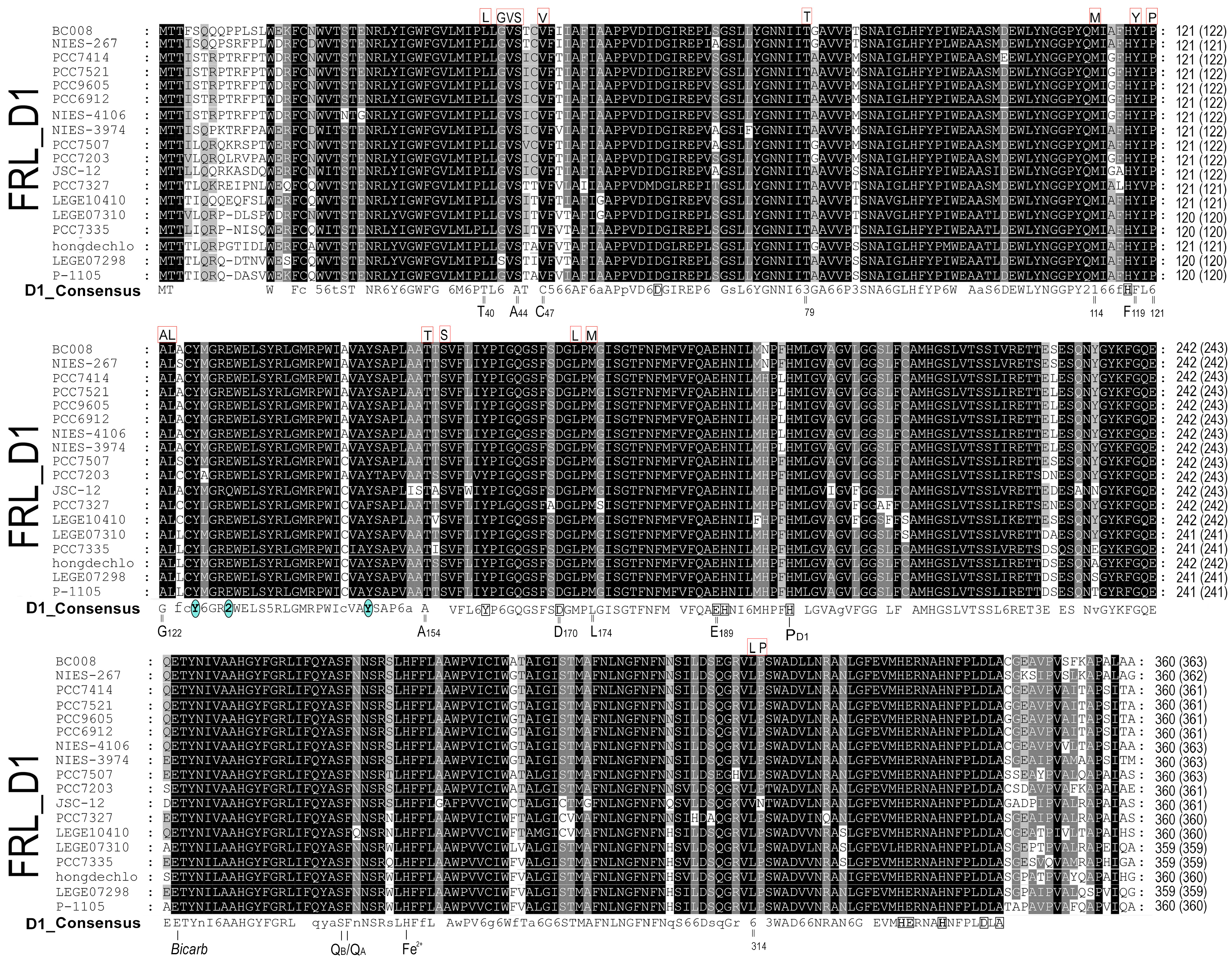
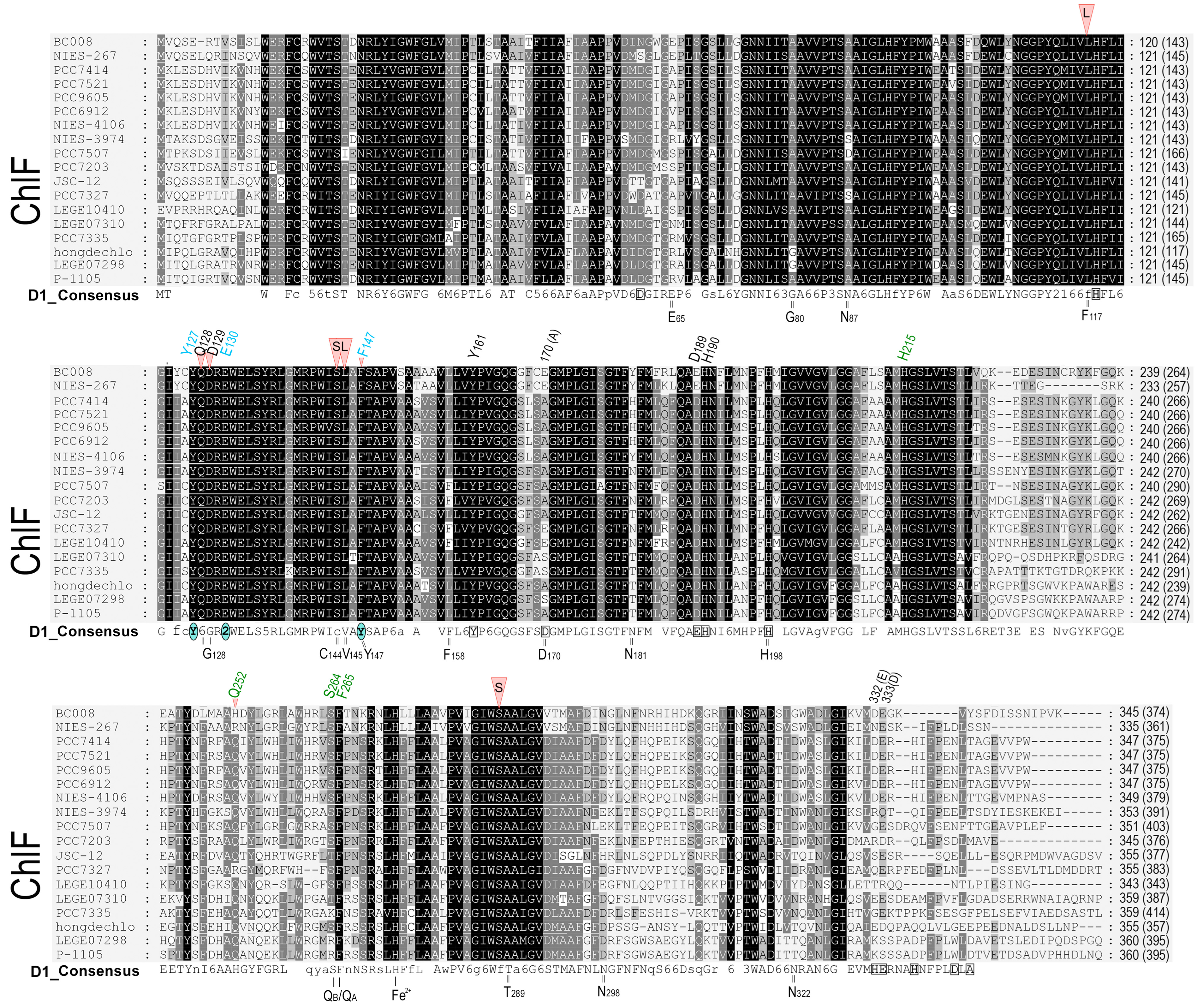
2.1. D1 Protein Sequence Alignment

The FaRLiP gene cluster is used as a biomarker of Chl f-producing cyanobacteria. Eighteen cyanobacterial genomes possessing the FaRLiP cluster were selected and obtained from GenBank (NCBI) and the Integrated Microbial Genomes of the DOE joint Genome Institute (Table S1). All putative ChlF and D1 sequences from 18 selected cyanobacterial genomes were retrieved, with a total of 100 D1 paralogs. Identical copies were removed, and 83 sequences (Table S1) were used for sequence comparison. Sequence alignment was generated using Muscle in MEGA 11 and manually refined by predicting the PDB files from each D1 protein group using the SWISS-MODEL online service from ExPasy. The H. hongdechloris D2 protein was used as an outgroup.

2.2. The D1 Sequence Comparison

The D1 sequences were retrieved from cyanobacteria containing the FaRLiP gene clusters as described in Wang et al. 2022 [36] and then aligned based on the published D1 tree [11,12]. The subset of D1 homologs (including ChlF sequences) from 18 selected species of cyanobacteria containing the FaRLiP gene cluster were re-aligned with the typical D1 consensus sequence and refined manually according to conserved motifs and predicted secondary structures. The grouped D1 sequences were verified using neighbor-joining (NJ) phylogenetic analysis. The NJ distance tree was determined after 1000 replications using the JTT (Jones, Taylor, and Thornton) model after the deletion of gaps [37].

2.3. Structure Modeling and Comparison

Three-dimensional structural modeling was performed using Chimera version 1.14 [38], https://www.cgl.ucsf.edu/chimera/download.html (accessed on 1 March 2020). The PSII structural data from Thermosynechococcus vestitus BP-1 (also named Thermostichus vulcanus) (PDB 5TIS and PDB 3WU2) (non-Chl f-producing cyanobacteria) were used as templates to simulate structural models of D1 paralogs from H. hongdechloris. The following Uniprot data were predicted: A0A1Z3HIN9, H. hongdechloris ChlF (XM38_010900); A0A1Z3HJ62, H. hongdechloris FRL-D1 (XM38_010770); A0A1Z3HKW5, H. hongdechloris WL-D1 (XM38_018880); and P51765, T. vulcanus D1. Note that Uniprot P51765 has a R279P substitution compared with Uniprot P0A444 (D1 of T. vulcanus). All stimulated structure models were minimized and optimized after adding H-bonds and the protonation states of conserved charged amino acid residues, such as histidine (H) and glutamic acid (E). All predicted models were refined using the Swissprot database (https://swissmodel.expasy.org, accessed on 1 March 2020) against the template file (5TIS.pdb). SWISS-MODEL allows the production of a 3D model of heteromeric complexes [39], such as FRL-PSII complexes stimulated using PSII subunits from the FaRLiP cluster (FRL-PSII subunits). The individual 3D models of ChlF, FRL-D1, and WL-D1 of H. hongdechloris were used in place of D1 from T. vulcanus (PDB 5TIS). The heterodimers of D1/CP43/CP47/D2 using WL-PSII and FRL-PSII subunits from H. hongdechloris were predicted against the PDB 5TIS template.
Hydrogen atoms and hydrogen bonds are not observable in most crystal structures. However, many cofactors in PSII, such as Pheo a and the plastoquinones QA and QB, interact with the D1 protein via hydrogen bonds (H-bonds). Therefore, hydrogen bonding is important to determine the physical structure parameters. The following criteria are needed to form H-bonds in a predicted protein structure: (1) donor atom−Acceptor atom <3.9 Å; (2) Hydrogen−Acceptor atom <2.5 Å; (3) donor atom−Hydrogen atom−Acceptor atom >90.0°; (4) adjacent atom to acceptor A′−Acceptor atom−Donor atom >90.0°; and (5) adjacent atom to acceptor A′−Acceptor atom−Hydrogen atom >90.0° as discussed in Torshin et al. [40] and McDonald and Thornton [41]. H-bond analysis was predicted and verified using the tools in Chimera.

3. Results

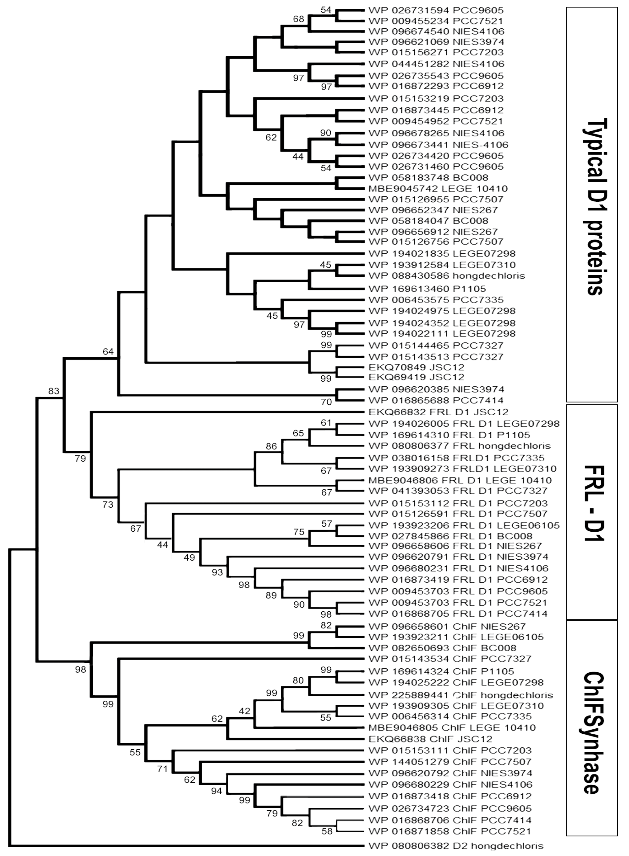
3.1. Alignment and Phylogenetic Relationship of D1 Paralogs in Chl f-Producing Cyanobacteria

There are 19 published genomes containing the FaRLiP gene cluster. Eighteen of them are complete or incomplete whole genomes, and most are labeled as ‘permanent drafts’ (Table S1). D1-encoding genes and their paralogs were retrieved from GenBank (NCBI) and the Integrated Microbial Genomes of the DOE joint Genome Institute and verified after BlastP comparison and annotation.
The D1 homologs can be divided into three main groups: WL-D1 (Typical D1), FRL-D1, and FRL-ChlF (Figure 1). According to protein alignments, FRL-D1 proteins possess all essential conserved motifs of the D1 Protein and contribute to functional Chl a/f-binding PSII under FRL conditions, including the high-affinity Mn-binding sites of D170 and E189 (Figure 2). The similar structures and conserved motifs among 18 FRL-D1 sequences can be defined as L40, G42, V43, S44, V47, T79, M114, Y119, P121, A122, L123, T154, S156, L172, M174, L314, and P315 (Figure 2).
ChlF forms a distinct branch from FRL-D1 and other D1 groups (Figure 1). The similar residues among ChlF synthase sequences can be characterized as: L117, Q127, D128, S144, L145, F147, Q 252, and S285 (Figure 3), including the previously reported QD motif at sites 127 and 128 (Q152 and D153 of ChlF from H. hongdechloris) [19]. The sequence comparison confirmed that the H252Q substitution is dominant in ChlF sequences, and only two cases of H252R substitutions were observed in Pleurocapsa sp. PCC 7327 and Calothrix parasitica NIES-267 (Figure 3). Interestingly, there is almost no overlap between the specific FRL-D1 motifs and ChlF sequences (Figure 2 and Figure 3). ChlF demonstrated significant changes in the conserved residues surrounding the Mn4CaO5 cluster, although the most significant changes are not consistent in ChlF sequences, as highlighted in Figure 3. For example, the conserved residue D170 in the typical D1 sequence is A, S, and E in ChlF (Figure 3). Campbell et al. reported that Mn2+ was oxidized and stabilized as Mn4+ in a D170E mutant, resulting in a photoactivating PSII that produces oxidizing manganese (Mn4+) but without O2 evolution [42,43]. The substitutions N87A/S and D170AES are noticed in ChlF alignment, and those changes in D1 impair oxygen evolution activities [5]. Thus, the replacement of D1 with the ChlF paralog may cause the loss of oxygen evolution.

3.2. The Modeling of D1 Paralogs from H. hongdechloris

ChlF shares over 70% of its residues with D1 proteins. This allows for the generation of a reliable 3D model using structural templates of known PSII structures in cyanobacteria. PDB 3WU2 from T. vulcanus was initially used as the template for modeling ChlF. ChlF interaction with other PSII subunits pertain to FRL-regulated structural reassembling subunits, especially FRL-D2 and FRL-CP43. The initial modeling trials without added H-atoms generated poor individual models of ChlF, FRL-D1, and WL-D1 (typical D1) since the estimated overlap (3.5 Å) scores were well below 0.7 (Table S2). This score refers to the proportion of Cα atoms within 3.5 Å of the proposed modeling structures. The modeling of H. hongdechloris WL-D1 was relatively well matched against 3WU2-D1, including the positions of all functional motifs. Further, FRL-CP43 modeling was improved after adding H-atoms to the initial model (Table S2). The modeling results were further improved by excluding a certain number of amino acids at the N- or C-termini that do not align with the structural template. The estimated overlap scores were significantly improved to well above 0.7 after using the truncated sequences. The estimated overlap (3.5 Å) score of the truncated ChlF models was similar and above 0.9, with model three showing the highest score of 0.994 (Table S2).

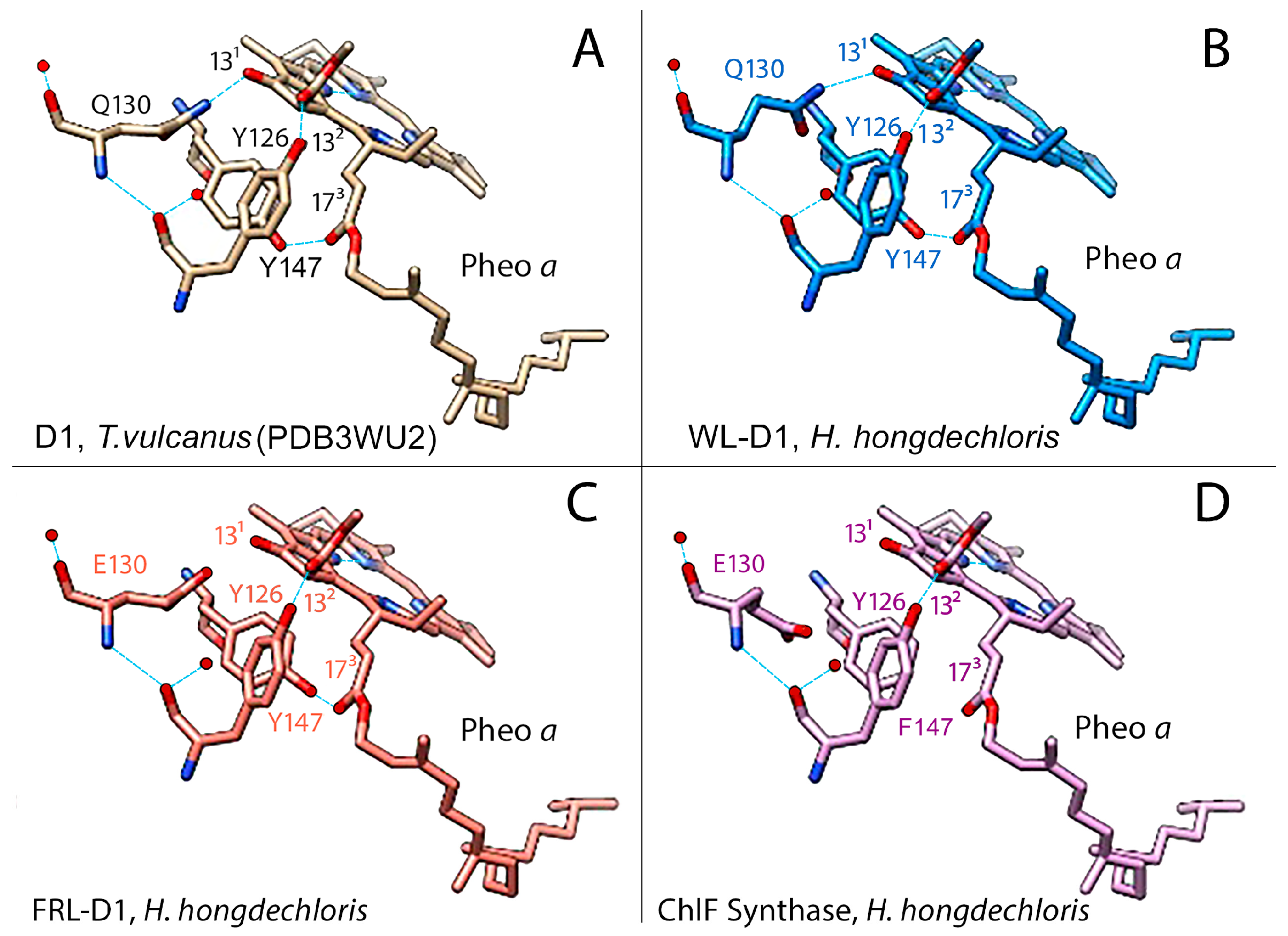
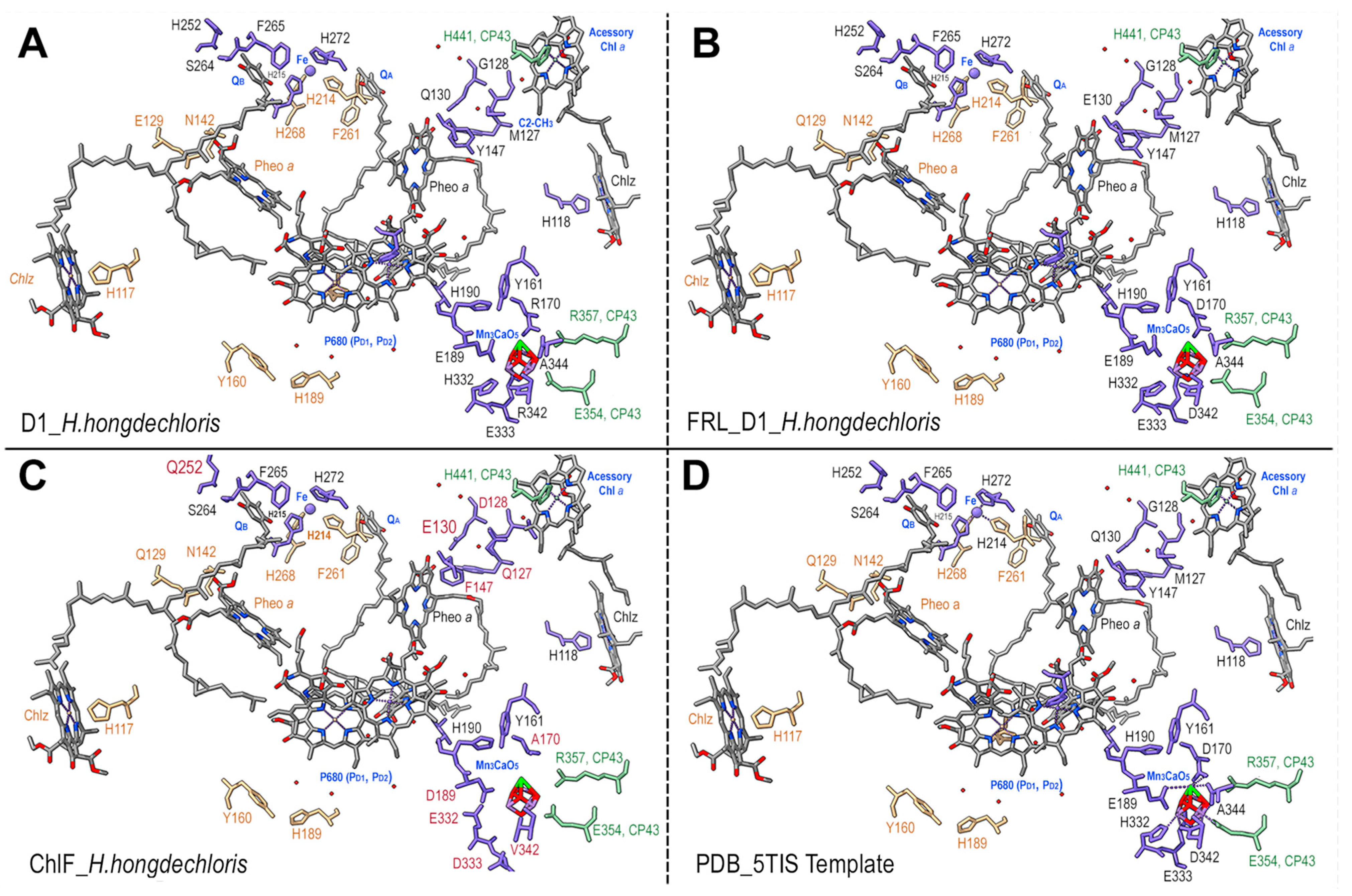
3.3. Pheophytin a Binding Environment

Both FRL-D1 and ChlF have conserved E130 sites (Figure 2 and Figure 3). The strength of the hydrogen bond between this residue and Pheo a could be changed. The structures were further modeled by SWISS-MODEL against the PDB 5TIS template from T. vulcanus [44]. The FRL-D1 model retained H-bonds with Pheo a and had a similar distance to the PDB 5TIS template, although the substitution of E130 (protonated at OE2) provided a stronger (shorter) H-bond than Q130 in the template (Figure 4). Hence, FRL-D1 demonstrated a comparable Pheo a binding profile to that in highlight acclimated cyanobacteria or plant PSII systems [45,46].
ChlF modeling following the template placement of ligands generated H-bonds when E130 was selectively protonated at OE2; however, H-bonds were absent in non-protonated E130 and protonated E130-OE1 situations (Figure 4). ChlF retains the Y126 H-bond with C132 of Pheo a to provide a comparable H-bond distance to the template. However, the Y147F substitution in ChlF results in a loss of the H-bond, Y147-O•••H•••O2-C173-Pheo a (Figure 4 and Figure S1, and Table S3). This contributes to the lessened interaction with Pheo a, although the impact of the missing H-bond with C173 Pheo a is unknown. The potential change in orientation of Pheo a due to the loss of an H-bond at C173 Pheo a enhanced the strength of the H-bond with E130 in ChlF (Figure S1). Using flourescence relaxation measurement, Rappaport et al. observed acceleration of the S2QA recombination process in the Q130E mutant [45]. Thus, E130 in FRL-D1 and ChlF could decrease the recombination rate of 1[P680+/Pheo a] to P*680 in order to acclimate to changed light environments.

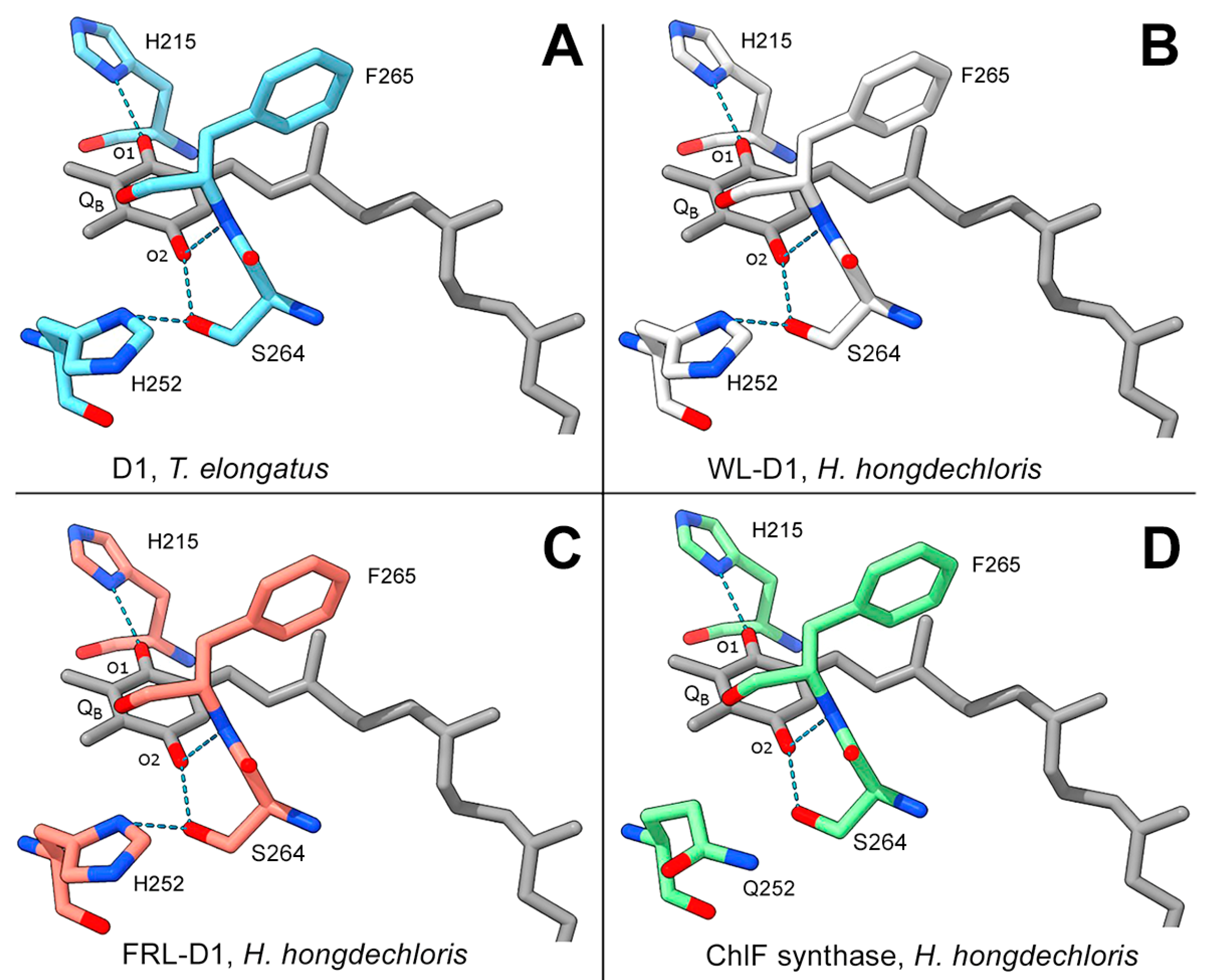
3.4. QB Binding Environment

The typical D1 subunit has three H-bonds with two keto groups of QB: H215-N-H•••O=keto QBO1, S264-O-H•••O=keto QBO2, and F265N-H•••O=keto QBO2. Additionally, S264 forms an H-bond with H252, H252-N-H•••O-S264 (Figure 5). Modeling of FRL-D1 and WL-D1 of H. hongdechloris simulated the H-bonds of H252-N-H•••O-S264 and the QB coupling environments were potentially unchanged (Figure 5). H252 protonation supports the protonation of QB to form QBH2, an important electron transfer towards oxygen-evolving activities [47]. However, ChlF has a H252Q substitution that is known to affect electron transfer (Figure 5D). The lack of an H-bond between Q252 and S264 inhibits the putative protonation of QB and conversion of QBH2 [2], and mutant H252Q exhibitsed rapid electron transfer from QA to QB [48]. The common substitution of H252Q (Figure 5D) implies that there is a more flexible structure in this region because H-bond formation should precede protonation to preserve some of the H-bonds supporting QB.

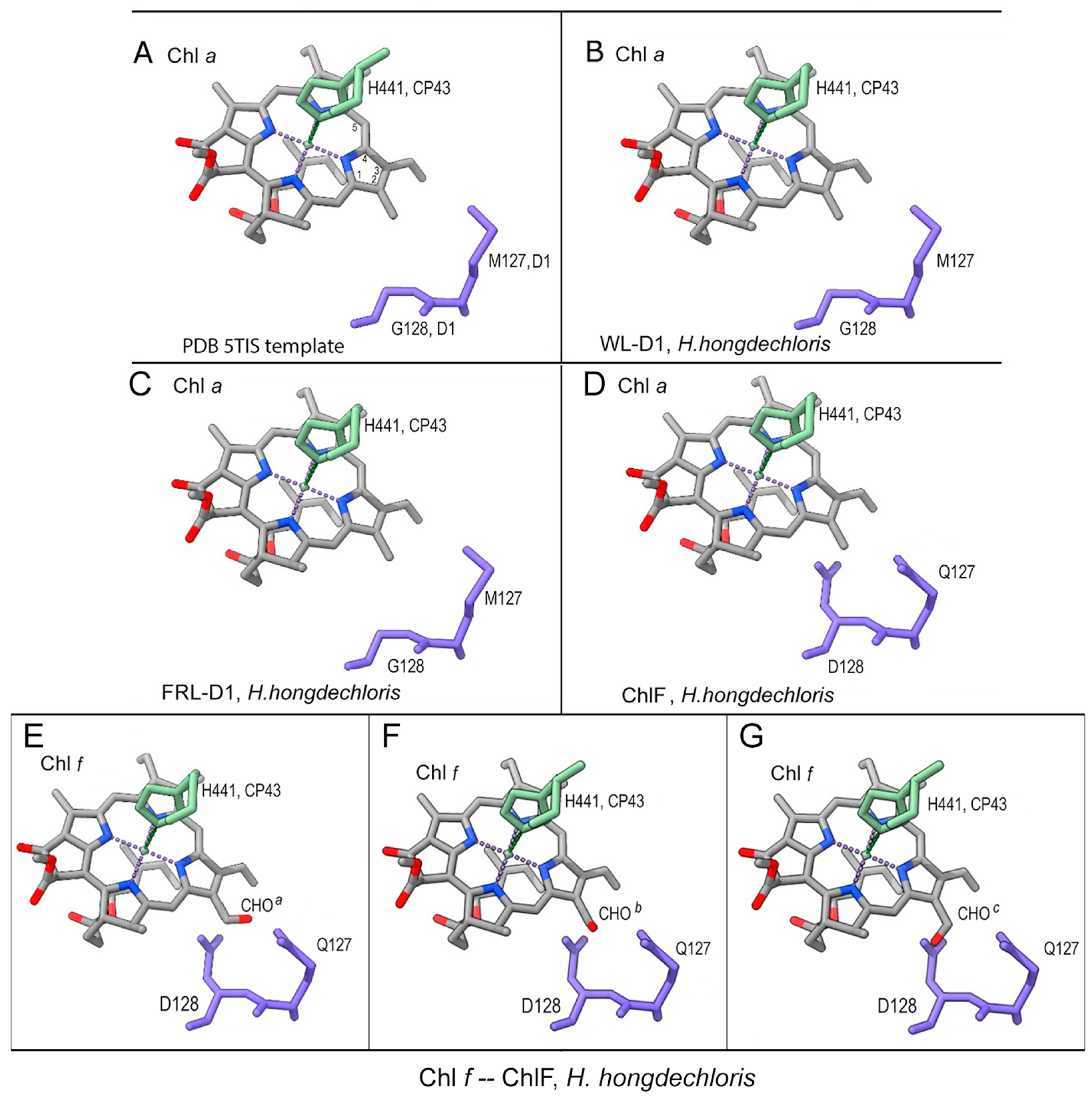
3.5. Putative Chl f-Binding Region of ChlF-PSII Complexes

The specific QD motif in ChlF probably plays a biochemical role in the synthesis and binding of Chl f, which is at the position of CP43 binding to Chl a through the axial ligand, H441 [19]. ChlF was modeled with multiple rotamers to test the acceptable placement of Q127 or/and D128 substitutions. There were clashes with the original placement of D128 and the Chl a/Chl f atoms. Position-c of the three putative spatial orientations of the formyl group of Chl f (designated C2-CHOa/b/c) showed obvious clashes, while positions-a and -b only showed clashes with the C2 and C3 sub-groups of Chl a (Figure 6). Potential H-bonds were formed with D128-OE2 of ChlF after manually placing Chl f in the position of Chl a and confirmed after minimization and refinement of the model (Figure S2). However, no H-bonds were formed between the C2-formyl group of Chl f and protonated D128 at OE1 or when D128 was unprotonated (Figure S2). This was further supported by the minimized modeling structure of the ChlF-PSII complex. Therefore, protonated D128 at the OE2 position is the optimized situation in the modeled ChlF structure.
Further minimization steps included: (1) 2-formyl modification of Chl a-H441 of CP43 (CLA506.C); (2) H-bond formation; (3) protonation and H-atom addition of E130 OE2 and D128 OE2; (4) movement of Chl f into a favorable position for H-bond formation with D128 OE2; and (5) the formation of a H-bond between D128 OE2 and Chl f. The refined and minimized structure proposed that the ideal placement occurred in rotamer model seven if Chl f replaced Chl a in the region (Figure S2).

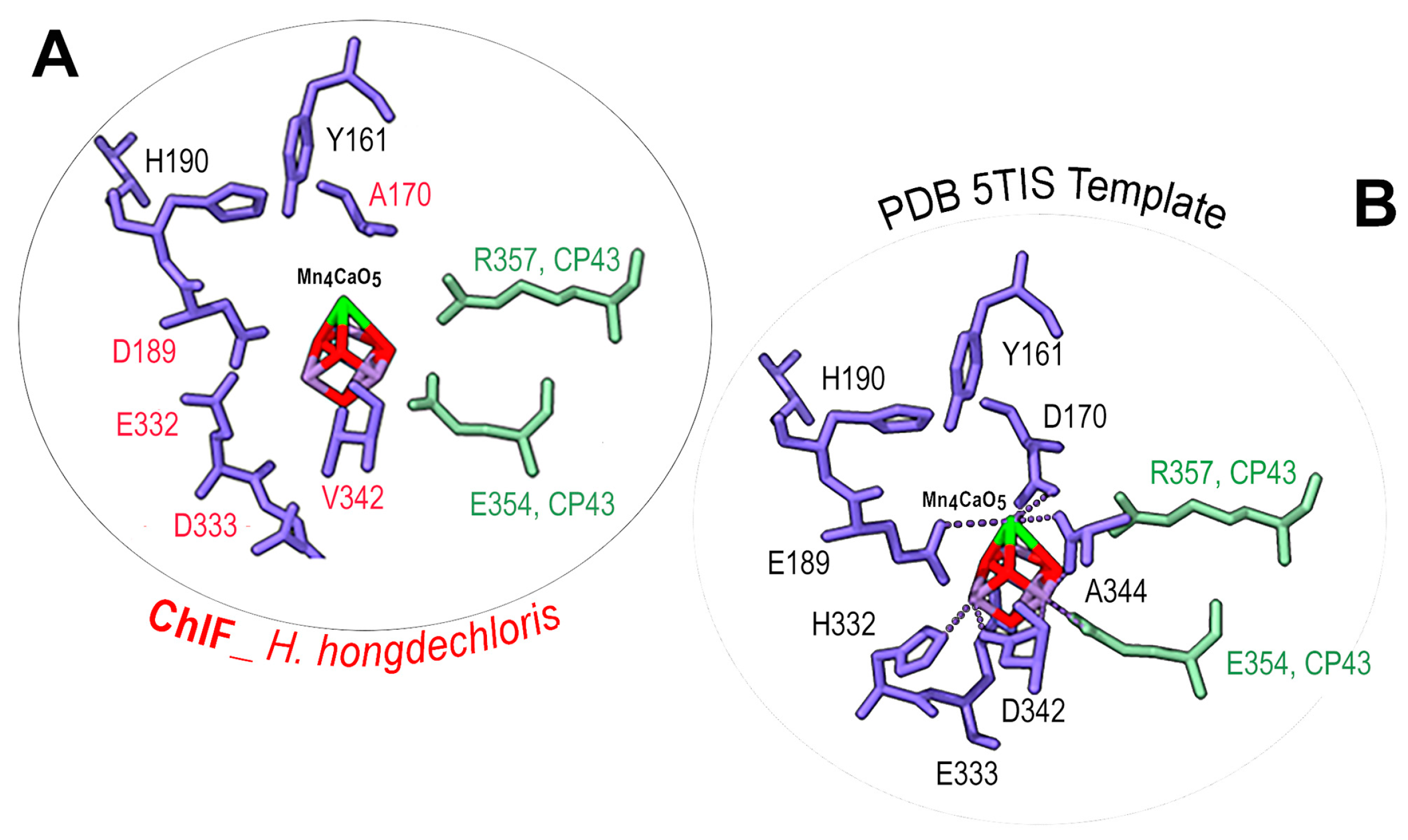
3.6. Mn4CaO5 Cluster Modeling

There is no consensus sequence for the Mn4CaO5 cluster following the alignment of ChlF sequences, despite some organisms retaining some of the typical amino acids (Figure 3). The significant changes in ChlF at the sites coordinating the Mn4CaO5 cluster (Figure 3) suggest that water splitting in a PSII-ChlF complex is impossible owing to the uncoupled Mn4CaO5 cluster. Multiple subunits of the H. hongdechloris PSII complexes were replaced with subunits of FRL-D1/D2/CP43/CP47 and/or ChlF/D2/CP43/CP47 and modeled using SWISS-MODEL.
The detailed environment surrounding the Mn4CaO5 cluster was demonstrated in Figure 7. The PSII-ChlF model showed conserved axial ligands H118 (D1), H198 (D1), H117 (D2), and H197 (D2), together with H-bonds with Pheo a, QB, and the conserved Y161/H190 pair. Additionally, the presence of two conserved amino acid residues, D/E189 (Mn1, Ca) of ChlF and E354 of CP43 (Mn2 and Mn3), suggests an Mn4CaO5 cluster may still be able to assemble or be present in the PSII-ChlF complex in an unconventional way (Figure 8). E189 provides a ligand to Mn in the metal cluster, but D189 from ChlF in H. hongdechloris showed no H-bond ligand to the metal cluster (Figure 8). D189 represents the conserved site in 16 ChlF sequences, except ChlF from Mastigocoleus testarum BC008 and Calothrix parasitica NIES-267, supporting the dysfunctional oxygen evolution center in PSII-ChlF complexes (Figure 3). The D1 mutant of E189D was reported to abolish water oxidation [4].
Additionally, D170 involves the light-driven one electron oxidation of Mn2+ to Mn3+, and it is A170 from H. hongdechloris ChlF (Figure 8). The residues H332, E333, and D342 at the C-terminal of the typical D1 sequence are important for assembly of the Mn4CaO5 cluster, and they are E332, D333, and V342 in the H. hongdechloris ChlF sequence (Figure 3). According to the model, the ligands with the Mn4CaO5 cluster are lacking in the ChlF sequence, indicating that these residue substitutions could lead to an altered function and efficiency of PSII oxygen evolution. The E333D mutant in Synechocystis PCC 6803 showed no photosynthetic oxygen evolution, and the H332E mutant appeared to induce rapid charge recombination between QA and Yzox [49]. Therefore, the water splitting process is presumed deficient, and ChlF probably has an alternative structure and function due to the several amino acid substitutions that are critical for coordinating the Mn4CaO5 cluster.

4. Discussion

The genes encoding FRL-D1 and ChlF are in the FaRLiP cluster under the same regulatory mechanism [33]. The different functions of D1 paralogs (FRL-D1 and ChlF) support the hypothesis of two different forms of PSII complexes assembled under FRL conditions. One of the substituted FRL-PSII complexes contains FRL-D1 and other FRL-PSII subunits. FRL-D1 proteins possess all essential conserved motifs of the D1 protein and agree well with the published FRL-PSII cryo-EM structure [50], although the alignment of 18 FRL-D1 sequences showed variations at the positions of L124 with A/S/C and L206 with three species having F206 at the position (Figure 2). The presence of the critical residue coupling of the oxygen-evolving Mn4CaO5 cluster in FRL-D1 suggests a functional Chl a/f-binding PSII under FRL conditions as PSII-FRL-D1 complexes (Figure 2). Therefore, the FRL-PSII complex conducts PSII-performing charge separation and water-splitting reactions under FRL conditions, which is consistent with the recent structure of PSII complexes acclimated to FRL conditions [50,51,52]. Meanwhile, the FRL-PSII complex (PSII-ChlF) containing the ChlF subunit instead of D1 proteins (FRL-D1 or WL-D1) plays a certain role in Chl f biosynthesis. PSII-ChlF contains the previously described QD motif [19] and conserved L117, Q127, D128, S144, L145, F147, Q 252, and S285 in the 18 ChlF sequences. Six out of eight specific amino acid residues surrounding Mn4CaO5 are not shared with typical D1 and FRL-D1, although some changes are various between ChlF sequences (Figure 8). H332 from typical D1 changed to H332D, H332E, H332N, H332R, H332S, H332Q, and H332K in ChlF (Figure 3), and experimental data indicated most mutants abolished oxygen evolution capability and only mutants of H335S and H335Q retain 10–15% oxygen evolution rate [49]. The PSII-ChlF structural model supports a new function of ChlF, although it shares >70% homology with D1.

4.1. Proposed Uncoupled Mn4CaO5 Cluster in PSII-ChlF Complexes

The center of the oxygen-evolving complex is the Mn4CaO5 cluster. It is stabilized with reaction center proteins D1 and D2 and is surrounded by inner antenna proteins CP47 (near D2) and CP43 (near D1) [53]. Water splitting proceeds in a multi-step process whereby the carefully positioned antenna chlorophylls of CP43 and CP47 transfer their excitation energy to the reaction center chlorophylls (PD1 and PD2) of D1 and D2, respectively. Excited P680 donates electrons to primary acceptor Pheo a which transmits them to QA and QB. Oxidized P680+ is reduced by an electron donated via Y161 (YZ, D1) by the chemical splitting of water into oxygen. This occurs because the Mn4CaO5 cluster undergoes a series of redox reactions known as Kok’s “oxygen clock” [half-reactions: 2H2O → 4H+ + 4e + O2, and 2QB + 4H+ + 4e →2QBH2]. YZ is an electron donor for P680+ in PSII and an electron carrier from the Mn4CaO5 cluster to the electron acceptor P680+.
ChlF synthase is a paralog of the reaction center protein D1; however, structural modeling (Figure 3 and Figure 7) confirmed the differences between ChlF and FRL-D1 and proposed an uncoupled Mn4CaO5 cluster in the PSII-ChlF complex. The proposed unconventional Mn4CaO5 cluster is supported by noticeable variations in ChlF sequences in the region of the 241-DEEE motif (Figure 3), the position for coordinating the bicarbonate in the typical D1 [1]. This is consistent with the hypothesis of “super-rogue” D1 proteins [1,11]. Additionally, N298 on the D1 protein is required for oxygen evolution, and mutants of N298D and N298E will impair oxygen evolution activity [54]. The D298 and E298 substitutions occur in 15 ChlF sequences (Figure 3).
P680+ is a powerful oxidant that facilitates the extraction of electrons from the Mn4CaO5 cluster via redox active YZ. Hence, reduction of P680+ might occur via cytochrome b559 and Chl a, or via YZ and YD in the absence of a coordinated active Mn4CaO5 cluster. Electron donation to P680+ via YZ is still possible with the aid of YD at a higher pH environment; that is, YD can outcompete YZ in the reduction of P680+ at a higher pH [55]. The altered electron transfer in the PSII-ChlF complex may enable the reduction of P680+ by the oxygenation of Chl a at the C-2 position to produce Chl f.

4.2. Changed Electron Transfer Pathways in Dysfunctional PSII-ChlF Complexes

ChlF has an unconventionally coordinated Mn4CaO5 cluster (Figure 8). According to experimental data, the significant changes surrounding the Mn4CaO5 cluster could not support water oxidation and oxygen evolution even if they were present in PSII-ChlF complexes [4,5,6,7,49,54]. In addition, the changes at the D1 conserved sites N87, F117, D170, and E189 in ChlF (Figure 3) reduced PSII stability and included impaired water-splitting activities [5]. Another noticed change around Pheo a includes the feature substitution Q128D129 motif in ChlF (Figure 7). The conserved site of F147 in ChlF could shift the Em (Pheo a/Pheo a) to more positive values and decrease the recombination rate of 1[P680+/Pheo a] to P*680 [56]. However, ChlF-PSII complex has conserved YZ and YD together with H-bond partners H190 (D1) and H189 (D2) at similar modeled distances to those of D1. On the whole, this suggests that there is an altered electron transport efficiency from P680+ to primary and secondary acceptors. The presumed non-functional, uncoupled Mn4CaO5 cluster in PSII-ChlF complexes suggests that YZ and YD supply the electrons required for the completion of redox reactions. Y160 of ChlF is conserved compared to other D1 sequences; however, this residue is not needed for the catalytic formation of Chl f [57]. Interestingly, the F147Y substitution in ChlF could cause an undelivered (or slower) electron at the level of Pheo a. Additionally, electron transfer within PSII is possible from a photooxidized reaction center to Pheo a, QA, and QB. DCMU (3-(3,4-dichlorophenyl)-1,1-dimethylurea) is an inhibitor of the QB binding site and inhibits Chl f synthesis [57]. Therefore, the light-driven redox chemistry of PSII without the coupled Mn4CaO5 cluster would play an active role in Chl f biosynthesis.
PSII electron transfer follows the resonance energy migration within the light-harvesting antenna with the resulting excitation of the reaction center primary donor (chlorophyll dimer P680, or PD1 and PD2) while the primary acceptor (Pheo a) reduces the primary quinone (QA). This causes a reduction of oxidized P680+• by YZ of D1 due to water oxidation by the coordinated Mn4CaO5 cluster. Impairment of the Mn4CaO5 cluster suggests that there is a longer-lived P680+• radical [58], and superoxide or H2O2 may be released as a long-lifetime free radical [59,60]. The expression of a ChlF-encoding gene in a PSII-less mutant (D1/D2 double mutant) yielded Chl f, and its production was regulated by light-driven electron transfer [57]. Hence, a longer-lived P680+• radical is required for Chl f biosynthesis.

4.3. Reaction Mechanism of Chl f Biosynthesis Pathway

Oxidation of a methyl group to a formyl group is a difficult task because CH3 is a chemically stable side group. Free radical formation can be specifically involved [61,62,63]. Pheo a on the acceptor side of PSII may form a singlet oxygen or hydroxyl free radical. Figure 4 suggests that the specific residues of F147 in ChlF cause a loss of the H-bond at C173-Pheo a and a potential slower electron transfer rate, although there is no available experimental data, such as a constructed D1 mutant of Y147F (Figure 4). Non-heme iron may also contribute to the formation of singlet oxygen or hydroxide radicals in the presence of light and aerobic conditions [64,65,66,67]. The inhibition of Chl f synthesis by DCMU implied that electron transports by the primary and secondary acceptors Pheo a, QA, and QB were involved in the reaction chemistry [57]. As is known in PSII, the dominant pathway of charge recombination goes through the radical pair of [P680+/Pheo a], and an additional non-radiative pathway could proceed through repopulation of P*680 from 1[P680+/Pheo a], which was observed in D1 mutant Q130E in Synechocystis PCC 6803 [56]. We proposed the reaction scheme of Chl (Chlide) f synthesis from Chl (Chlide) a via free radical chemistry (Figure 9). Pheo a excitation in an oxygenated environment produces an anion radical (D−•) via singlet and triplet excited states coupled to electron donation by Chl a. The cation-radical of the Chl a methyl group is produced via deprotonation. This cation-radical reacted with dioxygen to form a peroxide, which was further oxidized to superoxide or hydroxyl radical (Figure 9, step 4a) via reductants Pheo a (−610 mV) or QA (−80 mV) [68,69].
Peroxide radicals can decompose into hydroxymethyl-Chl derivatives and further react to form the formyl group in an unknown pathway (Figure 9, step 4b). Therefore, the altered electron pathway from P680+ to Pheo a, QA, and QB in the PSII-ChlF complex drives Chl f synthesis. The non-heme iron of PSII might not be involved in this oxygenation in the proposed mechanism; this differs from the biosynthetic mechanism of Chl b [70].
Chemical synthesis involving the oxidation of aromatic molecules with a methyl sidechain is often accomplished by free-radical chemistry using water as a proton source under elevated temperatures; over-oxidation of the carboxylic acid is prevented by addition of a catalyst such as copper [71]. More recently, a radical-free mechanism has been more specific for the synthesis of formyl groups [72]. ChlF has a likely impaired oxygen-evolving cluster; therefore, there is an increased potential for light-induced photoinhibition (even under low visible light) [73]. Two important photoinhibition sites in PSII include charge separation between P680+ and QA and electron transfer from YZ to P680+ [74]. Photoinhibition can cause carotenoid and chlorophyll radicals (from photopigments adjacent to P680 and accessory Chls) following the loss of YZ and YD radicals [75]. Accessory Chls and carotenoids in PSII have been proven to represent sites of photoinhibition damage following a hydroxylamine treatment [76]. It is unclear if ChlF is degraded during photoinhibition since detailed studies are unavailable. The A345, S345, or F345 substitutions in the functional domain of D1 resulted in comparable hydrolysis rates following mutants of carboxyl-terminal processing protease (CtpA), while the G345 and V345 substitutions showed lower hydrolysis rates [77]. However, the P345 substitution resulted in complete inhibition [77].
ChlF has two unique S motifs in addition to the QD motif (Figure 3) that are likely hydrolysis sites with potentially lower hydrolysis rates. The H-bond between YZ and H190 of ChlF is potentially retained, although there may be H-bonds with other amino acids when the Mn4CaO5 cluster is depleted or uncoupled. Hence, electron transfer from YZ to P680+ is possible without the coupled functional Mn4CaO5 cluster, although it could be slower [2,78,79]. Therefore, a long-lived P680+ may exist in the PSII-ChlF complex that functions as a powerful oxidant with a redox potential of approximately 1.13 V [78].
Replacement of an MG motif in D1 with a unique QD motif in ChlF facilitates the production of Chl f in vivo. This suggests that the QD motif is the most probable active site of the enzyme, although it is unknown whether the close proximity of D128 is critical [19]. Further work is required to determine whether photoinhibition (or free radical formation) in the PSII-ChlF complex in fact catalyzes Chl f production.

Supplementary Materials

The following supporting information can be downloaded at: https://www.mdpi.com/article/10.3390/microorganisms11092305/s1, Figure S1: Detailed H-bonds surrounding cofactor Pheo a in the models; Figure S2: Simulated models of ChlF containing chlorophyll f at different rotamers of protonated D128; Table S1: Genetic information of selected 19 cyanobacteria; Table S2: Modeling results of ChlF synthase, FRL-D1, WL-D1, FRL-CP43L, and WL-CP43 of H. hongdechloris against template PDB3WU2 with or without the inclusion of H atoms assigned during modeling; Table S3: H-bonding distances (Å) between pheophytin 131-keto and 132-keto groups and D1 of T. vulcanus (PDB3WU2), WL-D1, FRL-D1, and ChlF synthase from H. hongdechloris by the amino acids Q130/E130, Y126, and Y147 or F147, respectively. References [80,81,82,83] are cited in the supplementary materials.

Author Contributions

Project coordination, research design, conducted data analysis, manuscript—writing, M.C.; performed PSII modeling and assisted with the writing, A.S.; performed phylogenetic analysis and secondary structure alignments, F.W. All authors have read and agreed to the published version of the manuscript.

Funding

This research was partially supported by the Australia Research Council Center of Excellence for Translational Photosynthesis (CE140100015) and Discovery Project DP210102187.

Data Availability Statement

Not applicable.

Conflicts of Interest

The authors have no conflict of interest to declare that are relevant to the content of this article.

References

  1. Cardona, T.; Murray, J.W.; Rutherford, A.W. Origin and Evolution of Water Oxidation before the Last Common Ancestor of the Cyanobacteria. Mol. Biol. Evol. 2015, 32, 1310–1328. [Google Scholar] [CrossRef]
  2. Umena, Y.; Kawakami, K.; Shen, J.-R.; Kamiya, N. Crystal structure of oxygen-evolving photosystem II at a resolution of 1.9 Å. Nature 2011, 473, 55–60. [Google Scholar] [CrossRef] [PubMed]
  3. Service, R.J.; Yano, J.; McConnell, I.; Hwang, H.J.; Niks, D.; Hille, R.; Wydrzynski, T.; Burnap, R.L.; Hillier, W.; Debus, R.J. Participation of Glutamate-354 of the CP43 Polypeptide in the Ligation of Manganese and the Binding of Substrate Water in Photosystem II. Biochemistry 2011, 50, 63–81. [Google Scholar] [CrossRef]
  4. Chu, H.-A.; Nguyen, A.P.; Debus, R.J. Amino Acid Residues That Influence the Binding of Manganese or Calcium to Photosystem II. 1. The Lumenal Interhelical Domains of the D1 Polypeptide. Biochemistry 1995, 34, 5839–5858. [Google Scholar] [CrossRef]
  5. Diner, B.A. Amino acid residues involved in the coordination and assembly of the manganese cluster of photosystem II. Proton-coupled electron transport of the redox-active tyrosines and its relationship to water oxidation. Biochim. Biophys. Acta (BBA)-Bioenerg. 2001, 1503, 147–163. [Google Scholar] [CrossRef]
  6. Dilbeck, P.L.; Bao, H.; Neveu, C.L.; Burnap, R.L. Perturbing the Water Cavity Surrounding the Manganese Cluster by Mutating the Residue D1-Valine 185 Has a Strong Effect on the Water Oxidation Mechanism of Photosystem II. Biochemistry 2013, 52, 6824–6833. [Google Scholar] [CrossRef] [PubMed]
  7. Service, R.J.; Hillier, W.; Debus, R.J. Network of Hydrogen Bonds near the Oxygen-Evolving Mn4CaO5 Cluster of Photosystem II Probed with FTIR Difference Spectroscopy. Biochemistry 2014, 53, 1001–1017. [Google Scholar] [CrossRef]
  8. Pokhrel, R.; Debus, R.J.; Brudvig, G.W. Probing the Effect of Mutations of Asparagine 181 in the D1 Subunit of Photosystem II. Biochemistry 2015, 54, 1663–1672. [Google Scholar] [CrossRef]
  9. Nixon, P.J.; Diner, B.A. Aspartate 170 of the photosystem II reaction center polypeptide D1 is involved in the assembly of the oxygen-evolving manganese cluster. Biochemistry 1992, 31, 942–948. [Google Scholar] [CrossRef]
  10. Kiss, É.; Kós, P.B.; Chen, M.; Vass, I. A unique regulation of the expression of the psbA, psbD, and psbE genes, encoding the D1, D2 and cytochrome b559 subunits of the Photosystem II complex in the chlorophyll d containing cyanobacterium Acaryochloris marina. Biochim. Biophys. Acta (BBA)-Bioenerg. 2012, 1817, 1083–1094. [Google Scholar] [CrossRef] [PubMed]
  11. Murray, J.W. Sequence variation at the oxygen-evolving centre of photosystem II: A new class of ‘rogue’ cyanobacterial D1 proteins. Photosynth. Res. 2012, 110, 177–184. [Google Scholar] [CrossRef] [PubMed]
  12. Sheridan, K.J.; Duncan, E.J.; Eaton-Rye, J.J.; Summerfield, T.C. The diversity and distribution of D1 proteins in cyanobacteria. Photosynth. Res. 2020, 145, 111–128. [Google Scholar] [CrossRef] [PubMed]
  13. Mohamed, A.; Eriksson, J.; Osiewacz, H.D.; Jansson, C. Differential expression of the psbA genes in the cyanobacterium Synechocystis 6803. Mol. Genet. Genom. 1993, 238–238, 161–168. [Google Scholar] [CrossRef] [PubMed]
  14. Sicora, C.I.; Ho, F.M.; Salminen, T.; Styring, S.; Aro, E.-M. Transcription of a “silent” cyanobacterial psbA gene is induced by microaerobic conditions. Biochim. Biophys. Acta (BBA)-Bioenerg. 2009, 1787, 105–112. [Google Scholar] [CrossRef]
  15. Zhang, X.; Sherman, L.A. Alternate copies of D1 are used by cyanobacteria under different environmental conditions. Photosynth. Res. 2012, 114, 133–135. [Google Scholar] [CrossRef]
  16. Wegener, K.M.; Nagarajan, A.; Pakrasi, H.B. An Atypical psbA Gene Encodes a Sentinel D1 Protein to Form a Physiologically Relevant Inactive Photosystem II Complex in Cyanobacteria. J. Biol. Chem. 2015, 290, 3764–3774. [Google Scholar] [CrossRef]
  17. Ho, M.-Y.; Shen, G.; Canniffe, D.P.; Zhao, C.; Bryant, D.A. Light-dependent chlorophyll f synthase is a highly divergent paralog of PsbA of photosystem II. Science 2016, 353, 886. [Google Scholar] [CrossRef]
  18. Garg, H.; Loughlin, P.C.; Willows, R.D.; Chen, M. The C21-formyl group in chlorophyll f originates from molecular oxygen. J. Biol. Chem. 2017, 292, 19279–19289. [Google Scholar] [CrossRef]
  19. Trinugroho, J.P.; Bečková, M.; Shao, S.; Yu, J.; Zhao, Z.; Murray, J.W.; Sobotka, R.; Komenda, J.; Nixon, P.J. Chlorophyll f synthesis by a super-rogue photosystem II complex. Nat. Plants 2020, 6, 238–244. [Google Scholar] [CrossRef] [PubMed]
  20. Chen, M.; Schliep, M.; Willows, R.D.; Cai, Z.-L.; Neilan, B.A.; Scheer, H. A Red-Shifted Chlorophyll. Science 2010, 329, 1318–1319. [Google Scholar] [CrossRef]
  21. Chen, M.; Li, Y.; Birch, D.; Willows, R.D. A cyanobacterium that contains chlorophyll f—A red-absorbing photopigment. FEBS Lett. 2012, 586, 3249–3254. [Google Scholar] [CrossRef] [PubMed]
  22. Zhang, Z.; Li, Z.; Yin, Y.; Li, Y.; Jia, Y.; Chen, M.; Qiu, B. Widespread occurrence and unexpected diversity of red-shifted chlorophyll producing cyanobacteria in humid subtropical forest ecosystems. Environ. Microbiol. 2019, 21, 1497–1510. [Google Scholar] [CrossRef]
  23. Gan, F.; Shen, G.; Bryant, D.A. Occurrence of Far-Red Light Photoacclimation (FaRLiP) in Diverse Cyanobacteria. Life 2015, 5, 4–24. [Google Scholar] [CrossRef] [PubMed]
  24. Averina, S.; Velichko, N.; Senatskaya, E.; Pinevich, A. Far-red light photoadaptations in aquatic cyanobacteria. Hydrobiologia 2018, 813, 1–17. [Google Scholar] [CrossRef]
  25. Gómez-Lojero, C.; Leyva-Castillo, L.E.; Herrera-Salgado, P.; Barrera-Rojas, J.; Ríos-Castro, E.; Gutiérrez-Cirlos, E.B. Leptolyngbya CCM 4, a cyanobacterium with far-red photoacclimation from Cuatro Ciénegas Basin, México. Photosynthetica 2018, 56, 342–353. [Google Scholar] [CrossRef]
  26. Averina, S.; Velichko, N.; Senatskaya, E.; Pinevich, A. Non-a chlorophylls in cyanobacteria. Photosynthetica 2019, 57, 1109–1118. [Google Scholar] [CrossRef]
  27. Billi, D.; Napoli, A.; Mosca, C.; Fagliarone, C.; de Carolis, R.; Balbi, A.; Scanu, M.; Selinger, V.M.; Antonaru, L.A.; Nürnberg, D.J. Identification of far-red light acclimation in an endolithic Chroococcidiopsis strain and associated genomic features: Implications for oxygenic photosynthesis on exoplanets. Front. Microbiol. 2022, 13, 933404. [Google Scholar] [CrossRef]
  28. Murray, B.; Ertekin, E.; Dailey, M.; Soulier, N.T.; Shen, G.; Bryant, D.A.; Perez-Fernandez, C.; DiRuggiero, J. Adaptation of Cyanobacteria to the Endolithic Light Spectrum in Hyper-Arid Deserts. Microorganisms 2022, 10, 1198. [Google Scholar] [CrossRef]
  29. Tros, M.; Bersanini, L.; Shen, G.; Ho, M.-Y.; van Stokkum, I.H.; Bryant, D.A.; Croce, R. Harvesting far-red light: Functional integration of chlorophyll f into Photosystem I complexes of Synechococcus sp. PCC 7002. Biochim. Biophys. Acta (BBA)-Bioenerg. 2020, 1861, 148206. [Google Scholar] [CrossRef]
  30. Nürnberg, D.J.; Morton, J.; Santabarbara, S.; Telfer, A.; Joliot, P.; Antonaru, L.A.; Ruban, A.V.; Cardona, T.; Krausz, E.; Boussac, A.; et al. Photochemistry beyond the red limit in chlorophyll f–containing photosystems. Science 2018, 360, 1210–1213. [Google Scholar] [CrossRef]
  31. Kurashov, V.; Ho, M.-Y.; Shen, G.; Piedl, K.; Laremore, T.N.; Bryant, D.A.; Golbeck, J.H. Energy transfer from chlorophyll f to the trapping center in naturally occurring and engineered Photosystem I complexes. Photosynth. Res. 2019, 141, 151–163. [Google Scholar] [CrossRef] [PubMed]
  32. Li, Y.; Vella, N.; Chen, M. Characterization of isolated photosystem I from Halomicronema hongdechloris, a chlorophyll f-producing cyanobacterium. Photosynthetica 2018, 56, 306–315. [Google Scholar] [CrossRef]
  33. Chen, M.; Hernandez-Prieto, M.A.; Loughlin, P.C.; Li, Y.; Willows, R.D. Genome and proteome of the chlorophyll f-producing cyanobacterium Halomicronema hongdechloris: Adaptative proteomic shifts under different light conditions. BMC Genom. 2019, 20, 207. [Google Scholar] [CrossRef] [PubMed]
  34. Junge, W. Oxygenic photosynthesis: History, status and perspective. Q. Rev. Biophys. 2019, 52, e1. [Google Scholar] [CrossRef] [PubMed]
  35. Li, Y.; Lin, Y.; Loughlin, P.C.; Chen, M. Optimization and effects of different culture conditions on growth of Halomicronema hongdechloris—A filamentous cyanobacterium containing chlorophyll f. Front. Plant Sci. 2014, 5, 67. [Google Scholar] [CrossRef]
  36. Wang, F.; Chen, M. Chromatic Acclimation Processes and Their Relationships with Phycobiliprotein Complexes. Microorganisms 2022, 10, 1562. [Google Scholar] [CrossRef]
  37. Jones, D.T.; Taylor, W.R.; Thornton, J.M. The rapid generation of mutation data matrices from protein sequences. Comput. Appl. Biosci. 1992, 8, 275–282. [Google Scholar] [CrossRef]
  38. Kelley, L.A.; Mezulis, S.; Yates, C.M.; Wass, M.N.; Sternberg, M.J.E. The Phyre2 web portal for protein modeling, prediction and analysis. Nat. Protoc. 2015, 10, 845–858. [Google Scholar] [CrossRef]
  39. Waterhouse, A.; Bertoni, M.; Bienert, S.; Studer, G.; Tauriello, G.; Gumienny, R.; Heer, F.T.; De Beer, T.A.P.; Rempfer, C.; Bordoli, L.; et al. SWISS-MODEL: Homology modelling of protein structures and complexes. Nucleic Acids Res. 2018, 46, W296–W303. [Google Scholar] [CrossRef]
  40. Torshin, I.Y.; Weber, I.T.; Harrison, R.W. Geometric criteria of hydrogen bonds in proteins and identification of ‘bifurcated’ hydrogen bonds. Protein Eng. Design Sel. 2002, 15, 359–363. [Google Scholar] [CrossRef]
  41. McDonald, I.K.; Thornton, J.M. Satisfying Hydrogen Bonding Potential in Proteins. J. Mol. Biol. 1994, 238, 777–793. [Google Scholar] [CrossRef] [PubMed]
  42. Campbell, K.A.; Force, D.A.; Nixon, P.J.; Dole, F.; Diner, B.A.; Britt, R.D. Dual-Mode EPR Detects the Initial Intermediate in Photoassembly of the Photosystem II Mn Cluster: The Influence of Amino Acid Residue 170 of the D1 Polypeptide on Mn Coordination. J. Am. Chem. Soc. 2000, 122, 3754–3761. [Google Scholar] [CrossRef]
  43. Johnson, J.E.; Webb, S.M.; Thomas, K.; Ono, S.; Kirschvink, J.L.; Fischer, W.W. Manganese-oxidizing photosynthesis before the rise of cyanobacteria. Proc. Natl. Acad. Sci. USA 2013, 110, 11238–11243. [Google Scholar] [CrossRef] [PubMed]
  44. Young, I.D.; Ibrahim, M.; Chatterjee, R.; Gul, S.; Fuller, F.D.; Koroidov, S.; Brewster, A.S.; Tran, R.; Alonso-Mori, R.; Kroll, T.; et al. Structure of photosystem II and substrate binding at room temperature. Nature 2016, 540, 453–457. [Google Scholar] [CrossRef]
  45. Rappaport, F.; Guergova-Kuras, M.; Nixon, P.J.; Diner, B.A.; Lavergne, J. Kinetics and Pathways of Charge Recombination in Photosystem II. Biochemistry 2002, 41, 8518–8527. [Google Scholar] [CrossRef]
  46. Dorlet, P.; Xiong, L.; Sayre, R.T.; Un, S. High Field EPR Study of the Pheophytin Anion Radical in Wild Type and D1-E130 Mutants of Photosystem II in Chlamydomonas reinhardtii. J. Biol. Chem. 2001, 276, 22313–22316. [Google Scholar] [CrossRef] [PubMed]
  47. Lupínková, L.; Metz, J.G.; Diner, B.A.; Vass, I.; Komenda, J. Histidine residue 252 of the Photosystem II D1 polypeptide is involved in a light-induced cross-linking of the polypeptide with the α subunit of cytochrome b-559: Study of a site-directed mutant of Synechocystis PCC 6803. Biochim. Biophys. Acta (BBA)-Bioenerg. 2002, 1554, 192–201. [Google Scholar] [CrossRef]
  48. Petrouleas, V.; Crofts, A. The iron-quinone acceptor complex. In Photosystem II: The Light-Driven Water: Plastoquinone Oxidoreductase; Wydrzynski, T.J., Satoh, K., Eds.; Springer: Dordrecht, The Netherlands, 2005; pp. 177–206. [Google Scholar]
  49. Chu, H.-A.; Nguyen, A.P.; Debus, R.J. Amino Acid Residues That Influence the Binding of Manganese or Calcium to Photosystem II. 2. The Carboxy-Terminal Domain of the D1 Polypeptide. Biochemistry 1995, 34, 5859–5882. [Google Scholar] [CrossRef]
  50. Gisriel, C.J.; Shen, G.; Ho, M.-Y.; Kurashov, V.; Flesher, D.A.; Wang, J.; Armstrong, W.H.; Golbeck, J.H.; Gunner, M.R.; Vinyard, D.J.; et al. Structure of a monomeric photosystem II core complex from a cyanobacterium acclimated to far-red light reveals the functions of chlorophylls d and f. Pediatrics 2022, 298, 101424. [Google Scholar] [CrossRef]
  51. Gisriel, C.J.; Cardona, T.; Bryant, D.A.; Brudvig, G.W. Molecular Evolution of Far-Red Light-Acclimated Photosystem II. Microorganisms 2022, 10, 1270. [Google Scholar] [CrossRef]
  52. Gisriel, C.J.; Shen, G.; Flesher, D.A.; Kurashov, V.; Golbeck, J.H.; Brudvig, G.W.; Amin, M.; Bryant, D.A. Structure of a dimeric photosystem II complex from a cyanobacterium acclimated to far-red light. Pediatrics 2023, 299, 102815. [Google Scholar] [CrossRef] [PubMed]
  53. Renger, G.; Renger, T. Photosystem II: The machinery of photosynthetic water splitting. Photosynth. Res. 2008, 98, 53–80. [Google Scholar] [CrossRef]
  54. Kuroda, H.; Kodama, N.; Sun, X.-Y.; Ozawa, S.-I.; Takahashi, Y. Requirement for Asn298 on D1 Protein for Oxygen Evolution: Analyses by Exhaustive Amino Acid Substitution in the Green Alga Chlamydomonas reinhardtii. Plant Cell Physiol. 2014, 55, 1266–1275. [Google Scholar] [CrossRef]
  55. Faller, P.; Rutherford, A.W.; Debus, R.J. Tyrosine D Oxidation at Cryogenic Temperature in Photosystem II. Biochemistry 2002, 41, 12914–12920. [Google Scholar] [CrossRef] [PubMed]
  56. Cser, K.; Vass, I. Radiative and non-radiative charge recombination pathways in Photosystem II studied by thermoluminescence and chlorophyll fluorescence in the cyanobacterium Synechocystis 6803. Biochim. Biophys. Acta (BBA)-Bioenerg. 2007, 1767, 233–243. [Google Scholar] [CrossRef]
  57. Shen, G.; Canniffe, D.P.; Ho, M.-Y.; Kurashov, V.; van der Est, A.; Golbeck, J.H.; Bryant, D.A. Characterization of chlorophyll f synthase heterologously produced in Synechococcus sp. PCC 7002. Photosynth. Res. 2019, 140, 77–92. [Google Scholar] [CrossRef]
  58. Hakala, M.; Tuominen, I.; Keränen, M.; Tyystjärvi, T.; Tyystjärvi, E. Evidence for the role of the oxygen-evolving manganese complex in photoinhibition of Photosystem II. Biochim. Biophys. Acta (BBA)-Bioenerg. 2005, 1706, 68–80. [Google Scholar] [CrossRef]
  59. Wood, P.M. The potential diagram for oxygen at pH 7. Biochem. J. 1988, 253, 287–289. [Google Scholar] [CrossRef] [PubMed]
  60. Vass, I. Molecular mechanisms of photodamage in the Photosystem II complex. Biochim. Biophys. Acta (BBA)-Bioenerg. 2012, 1817, 209–217. [Google Scholar] [CrossRef]
  61. Julliard, M.; Galadi, A.; Chanon, M. Oxidation of methyl aromatic compounds by redox photosensitization. J. Photochem. Photobiol. A Chem. 1990, 54, 79–90. [Google Scholar] [CrossRef]
  62. Kiener, A. Enzymatic Oxidation of Methyl Groups on Aromatic Heterocycles: A Versatile Method for the Preparation of Heteroaromatic Carboxylic Acids. Angew. Chem. Int. Ed. 1992, 31, 774–775. [Google Scholar] [CrossRef]
  63. Bienvenu, C.; Wagner, J.R.; Cadet, J. Photosensitized Oxidation of 5-Methyl-2‘-deoxycytidine by 2-Methyl-1,4-naphthoquinone: Characterization of 5-(Hydroperoxymethyl)-2‘-deoxycytidine and Stable Methyl Group Oxidation Products. J. Am. Chem. Soc. 1996, 118, 11406–11411. [Google Scholar] [CrossRef]
  64. Lendzian, F.; Mobius, K.; Lubitz, W. The pheophytin a anion radical. 14N and 1H endor and triple resonance in liquid solution. Chem. Phys. Lett. 1982, 90, 375–381. [Google Scholar] [CrossRef]
  65. Pospíšil, P.; Arató, A.; Krieger-Liszkay, A.; Rutherford, A.W. Hydroxyl Radical Generation by Photosystem II. Biochemistry 2004, 43, 6783–6792. [Google Scholar] [CrossRef] [PubMed]
  66. Krieger-Liszkay, A. Singlet oxygen production in photosynthesis. J. Exp. Bot. 2005, 56, 337–346. [Google Scholar] [CrossRef]
  67. Pospíšil, P. Production of reactive oxygen species by photosystem II. Biochim. Biophys. Acta (BBA)-Bioenerg. 2009, 1787, 1151–1160. [Google Scholar] [CrossRef]
  68. Knaff, D.B. The effect of pH on the midpoint oxidation-reduction potentials of components associated with plant photosystem II. FEBS Lett. 1975, 60, 331–335. [Google Scholar] [CrossRef]
  69. Klimov, V.V. Pheophytin as the primary electron acceptor in photosystem II reaction centres. Photosynthetica 1981, 15, 592–609. [Google Scholar]
  70. Tanaka, A.; Ito, H.; Tanaka, R.; Tanaka, N.K.; Yoshida, K.; Okada, K. Chlorophyll a oxygenase (CAO) is involved in chlorophyll b formation from chlorophyll a. Proc. Natl. Acad. Sci. USA 1998, 95, 12719–12723. [Google Scholar] [CrossRef]
  71. Abe, T.; Tanaka, S.; Ogawa, A.; Tamura, M.; Sato, K.; Itoh, S. Copper-catalyzed Selective Oxygenation of Methyl and Benzyl Substituents in Pyridine with O2. Adv. Synth. Catal. 2017, 46, 348–350. [Google Scholar] [CrossRef]
  72. Ye, R.; Cao, Y.; Xi, X.; Liu, L.; Chen, T. Metal- and radical-free aerobic oxidation of heteroaromatic methanes: An efficient synthesis of heteroaromatic aldehydes. Org. Biomol. Chem. 2019, 17, 4220–4224. [Google Scholar] [CrossRef]
  73. Chen, G.X.; Kazimir, J.; Cheniae, G.M. Photoinhibition of hydroxylamine-extracted photosystem II membranes: Studies of the mechanism. Biochemistry 1992, 31, 11072–11083. [Google Scholar] [CrossRef] [PubMed]
  74. Eckert, H.-J.; Geiken, B.; Bernarding, J.; Napiwotzki, A.; Eichler, H.-J.; Renger, G. Two sites of photoinhibition of the electron transfer in oxygen evolving and Tris-treated PS II membrane fragments from spinach. Photosynth. Res. 1991, 27, 97–108. [Google Scholar] [CrossRef] [PubMed]
  75. Blubaugh, D.J.; Atamian, M.; Babcock, G.T.; Golbeck, J.H.; Cheniae, G.M. Photoinhibition of hydroxylamine-extracted photosystem II membranes: Identification of the sites of photodamage. Biochemistry 1991, 30, 7586–7597. [Google Scholar] [CrossRef] [PubMed]
  76. Tyystjärvi, E. Photoinhibition of Photosystem II and photodamage of the oxygen evolving manganese cluster. Coord. Chem. Rev. 2008, 252, 361–376. [Google Scholar] [CrossRef]
  77. Taguchi, F.; Yamamoto, Y.; Satoh, K. Recognition of the Structure around the Site of Cleavage by the Carboxyl-terminal Processing Protease for D1 Precursor Protein of the Photosystem II Reaction Center. J. Biol. Chem. 1995, 270, 10711–10716. [Google Scholar] [CrossRef]
  78. Conjeaud, H.; Mathis, P. The effect of pH on the reduction kinetics of P-680 in tris-treated chloroplasts. Biochim. Biophys. Acta (BBA)-Bioenerg. 1980, 590, 353–359. [Google Scholar] [CrossRef]
  79. Styring, S.; Sjöholm, J.; Mamedov, F. Two tyrosines that changed the world: Interfacing the oxidizing power of photochemistry to water splitting in photosystem II. Biochim. Biophys. Acta (BBA)-Bioenerg. 2012, 1817, 76–87. [Google Scholar] [CrossRef]
  80. Melo, F.; Sánchez, R.; Sali, A. Statistical potentials for fold assessment. Protein Sci. 2002, 11, 430–448. [Google Scholar] [CrossRef]
  81. Shen, M.Y.; Sali, A. Statistical potential for assessment and prediction of protein structures. Protein Sci. 2006, 15, 2507–2524. [Google Scholar] [CrossRef]
  82. Eramian, D.; Eswar, N.; Shen, M.Y.; Sali, A. How well can the accuracy of comparative protein structure models be predicted? Protein Sci. 2008, 17, 1881–1893. [Google Scholar] [CrossRef] [PubMed]
  83. Shibuya, Y.; Takahashi, R.; Okubo, T.; Suzuki, H.; Sugiura, M.; Noguchi, T. Hydrogen Bond Interactions of the Pheophytin Electron Acceptor and Its Radical Anion in Photosystem II As Revealed by Fourier Transform Infrared Difference Spectroscopy. Biochemistry 2010, 49, 493–501. [Google Scholar] [CrossRef] [PubMed]
Figure 1. The neighbor-joining distance tree of D1 paralogs in Chl f-producing cyanobacteria. The distance tree was generated using the neighbor-joining (NJ) method in MEGA 11. H. hongdechloris D2 protein (WP080806382) was used as an outgroup. All sequence IDs and strain numbers used for the names and the sequence accession are listed in Table S1. The branch support numbers were calculated by 1000 replications of NJ and were presented if the value was above 40%. Typical D1 groups include the dominant D1 protein (WL-D1) of H. hongdechloris grown under white light conditions; FRL-D1 (far-red light D1), a group covering genes encoding D1 proteins in the FaRLiP cluster; and ChlF synthase, a new group of D1 paralogs that synthesize chlorophyll f.
Microorganisms 11 02305 g001
Figure 2. Far red light (FRL)-D1 protein sequence alignment supported by phylogenetic analysis in Figure 1. The specific conserved sites of FRL-D1 are highlighted by red-framed boxes. The aligned sequence lengths are labeled as ‘numbers (numbers)’ at the end of each sequence. The numbers outside parentheses are aligned sequence lengths matching the D1_consensus (unaligned gaps were removed), which is the number used in the models. The numbers inside parentheses represent the original length. The D1_consensus sequence surrounding the Mn4CaO5 cluster is highlighted in square boxes and bold fonts. The residues interacting with Pheo a in the D1_consensus sequence are highlighted with blue-colored circles. The D1_consensus sequence was generated by Genedoc based on 50 typical D1 protein sequences. The conserved residues from 18 FRL-D1 sequences are highlighted with red-square boxes on top of the alignments, and corresponding amino acid residues are numbered under the D1_consensus sequence. The numbers in the D1_consensus represent the conservative substitutions: 2 = Q/E; 3 = S/T; 5 = MFY; 6 = F/I/L/M/V.
Microorganisms 11 02305 g002
Figure 3. ChlF protein sequence alignment supported by phylogenetic analysis in Figure 1. The specific conserved sites of ChlF are highlighted by red triangles. The aligned sequence lengths are labeled as ‘numbers (numbers)’ at the end of each sequence. The numbers outside parentheses are aligned sequence length matches with D1_consensus (removed unaligned gaps), which is the number used in the models. The numbers inside parentheses represent the original length. The D1_consensus sequence surrounding the Mn4CaO5 cluster is highlighted with square boxes and bold fonts labeled in the D1_consensus sequences. The noticeable changes in amino acid residues at the positions associated with the Mn4CaO5 cluster are labeled on the top of aligned ChlF sequences (black-colored fonts). The residues interacting with Pheo a in the D1_consensus sequence are highlighted by blue-colored circles, and their corresponding sites are labeled on top of the aligned ChlF sequences in blue-colored fonts. The amino acid residues surrounding the QB site (as presented in Section 3.4 below) are labeled on top of the aligned ChlF sequence, indicated as green-colored fonts. The D1_consensus sequences were generated by Genedoc based on 50 typical D1 protein sequences. The specific amino acid residues of ChlF are marked in a pink triangle on top of the alignment, and the corresponding amino acid residues are numbered under the D1_consensus sequence. The numbers in the D1_consensus represent the conservative substitutions: 2 = Q/E; 3 = S/T; 5 = MFY; 6 = F/I/L/M/V.
Microorganisms 11 02305 g003
Figure 4. Modeling of the pheophytin a (Pheo a) binding region in WL-D1, FRL-D1, and ChlF of H. hongdechloris. T. vulcanus (PDB 3WU2) was used as a template. The amino acid positions known to form H-bonds (light blue lines) with Pheo a: Y126 (Tyr-O•••H•••O=C132-Pheo a); Q130/E130 (N-H or O-H) (Q-N•••H•••O=C131-Pheo a) or (E-O•••H•••O=C131-Pheo a), and Y147 (Tyr-O•••H•••O=C173-pheo a) are included to compare with the predicted H-bonds. (A) The crystal structure of D1 of T. vulcanus (PDB 3WU2) was used as a template for the modeled structure. (BD) represent models of the Pheo a binding regions of WL-D1, FRL-D1, and ChlF synthase, respectively. Dark blue rods, nitrogen atoms; red rods, oxygen atoms; and dotted light blue lines, H-bonds. Note: FRL-D1 and ChlF synthase have E130, which may have H-bonds with the 131-keto of Pheo a, and these bonds might be stronger than Q130 (Table S3).
Microorganisms 11 02305 g004
Figure 5. Modeled D1 paralogs surrounding QB. Modeling of WL-D1, FRL-D1, and ChlF of H. hongdechloris against the PDB 5TIS template from T. vulcanus (Young et al. 2016 [44]) was performed using SWISS-MODEL. The conserved H-bonds of T. vulcanus D1 are shown as a positive control (A) to indicate modeling validity. (B) H. hongdechloris WL-D1; (C) H. hongdechloris FRL-D1; and (D) H. hongdechloris ChlF synthase. QB molecular bonds are in gray; residues from the T. vulcanus D1 template are in light blue; residues from H. hongdechloris WL-D1 are in white; residues from H. hongdechloris FRL-D1 are in orange; and residues from H. hongdechloris ChlF are in green; dark blue rods, nitrogen atoms; red rods, oxygen atoms; dotted light blue lines, H-bonds.
Microorganisms 11 02305 g005
Figure 6. Position of the QD motif of ChlF compared with conserved MG residues from other D1 proteins. (A) Template 5TIS; (B) modeled H. hongdechloris WL-D1; (C) modeled H. hongdechloris FRL-D1; (D) modeled H. hongdechloris ChlF; and (EG) Chl a was replaced with Chl f at the position of the modeled ChlF. The C2-CHO group of Chl f was shown at different orientations. (E), position a; (F), position b; and (G), position c, of the oxygen atom of CHO. Chlorophyll molecular bonds are gray; residues 127 and 128 of D1 and D1 paralogs are purple-blue. The histidine (H441) of CP43 is in green. Dark blue rods, nitrogen atoms; red rods, oxygen atoms; dotted lines, H-bonds.
Microorganisms 11 02305 g006
Figure 7. Models of PSII ligands involved in electron transport and water splitting. (A) Typical PSII D1 of H. hongdechloris (WP088430586); (B) H. hongdechloris FRL_D1 (WP080806377); (C) H. hongdechloris ChlF (WP 225889441); and (D) the PDB 5TIS template. The amino acids from CP43 are in green. The cofactors are in gray except for the Mn4CaO5 cluster. The unique ChlF residues are labeled in red. The D1 residues are in black fonts; the D2 residues are in brown fonts; and the partial amino acid side-group bonds are in brown colors. Red rods, oxygen atoms (which also represent Mn in the Mn4CaO5 cluster); dotted lines, H-bonds; and dark green, Ca in the Mn4CaO5 cluster.
Microorganisms 11 02305 g007
Figure 8. Detailed amino acid ligands surrounding the Mn4CaO5 cluster. (A) A detailed model of ChlF from H. hongdechloris (WP 225889441) and (B) structural details of the PDB 5TIS template. The conserved (black fonts) and different (red fonts) amino acids surrounding the Mn4CaO5 cluster. ChlF residues are in purple-blue. CP43 amino acids are in green. The red rods represnt the Mn in the Mn4CaO5 cluster; the green rod is the Ca in the Mn4CaO5 cluster; and the dotted lines are H-bonds.
Microorganisms 11 02305 g008
Figure 9. Proposed chlorophyll f biosynthesis reaction schemes. The proposed free-radical reaction schemes for chlorophyll f synthesis from chlorophyll a involve the transformation of a C2 methyl group in Chl a into a formyl group in Chl f. The first step involves the formation of an anion radical (D−•) that might be the overexcited Pheo a in an oxygenated aqueous environment or a light-activated radical species through the coupled electron donation by the Chl a molecule. Deprotonation forms the radical methyl cation of the Chl a substrate (Step 2), followed by reaction with oxygen to generate a peroxo radical (Step 3) that reacts with a superoxide or hydroxy radical (Step 4a) produced through the reduction of oxygen by light-activated reductants, such as Pheo a (−610 mV) and QA (−80 mV). This forms the unstable hydroperoxymethyl Chl, which is further hydrolyzed to the formyl group (Chl f). Alternatively, the peroxo radical may decompose into the hydroxymethyl Chl derivative (Step 4b), which further reacts in an unknown pathway to form the formyl group (Step 5b). The partial structure of chlorophylls showed the side group containing the C2-methyl of Chl a (shaded region).
Microorganisms 11 02305 g009
Disclaimer/Publisher’s Note: The statements, opinions and data contained in all publications are solely those of the individual author(s) and contributor(s) and not of MDPI and/or the editor(s). MDPI and/or the editor(s) disclaim responsibility for any injury to people or property resulting from any ideas, methods, instructions or products referred to in the content.

