
Photothermal/Photoacoustic Therapy Combined with Metal-Based Nanomaterials for the Treatment of Microbial Infections

Abstract
:1. Introduction
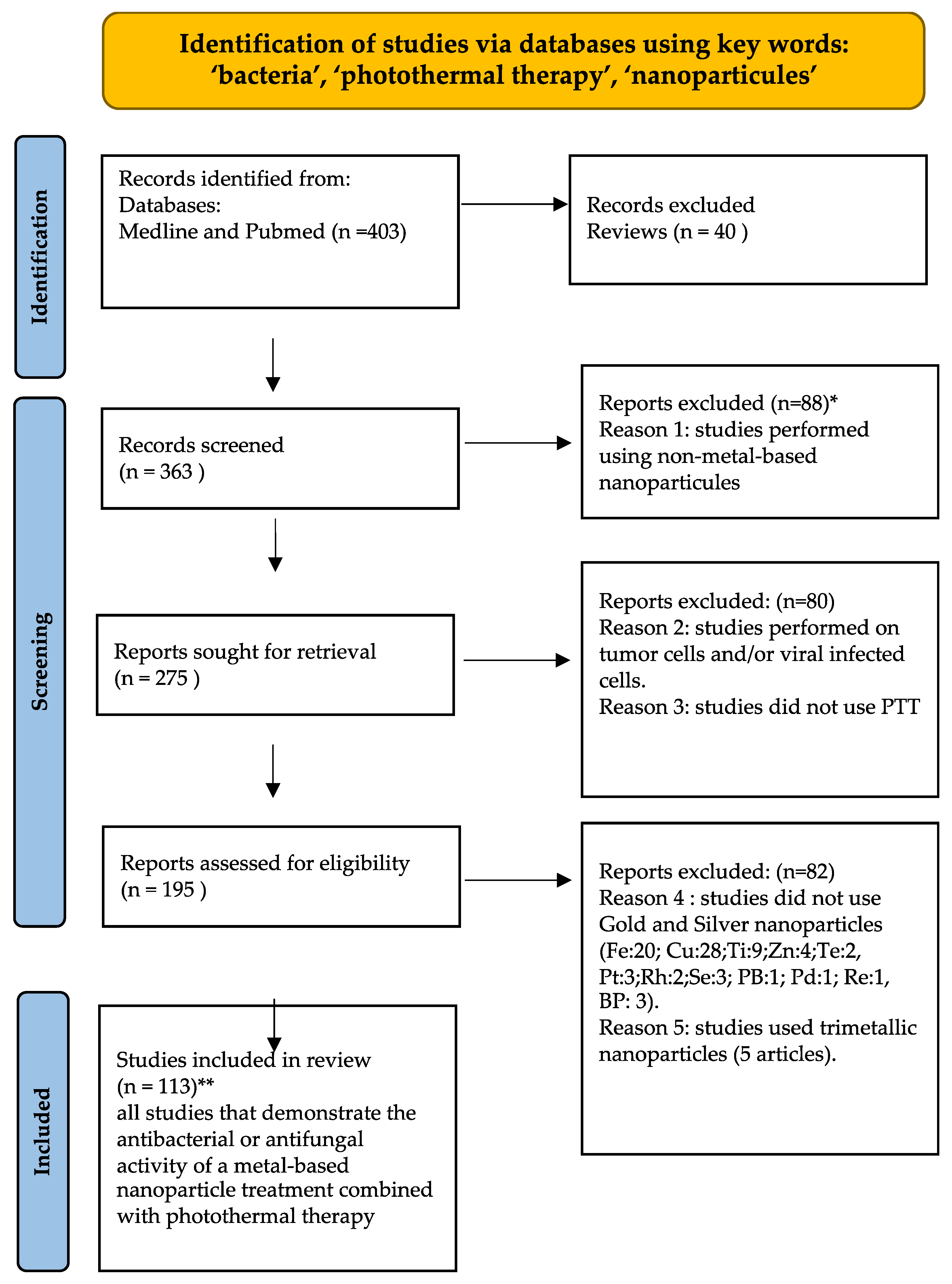
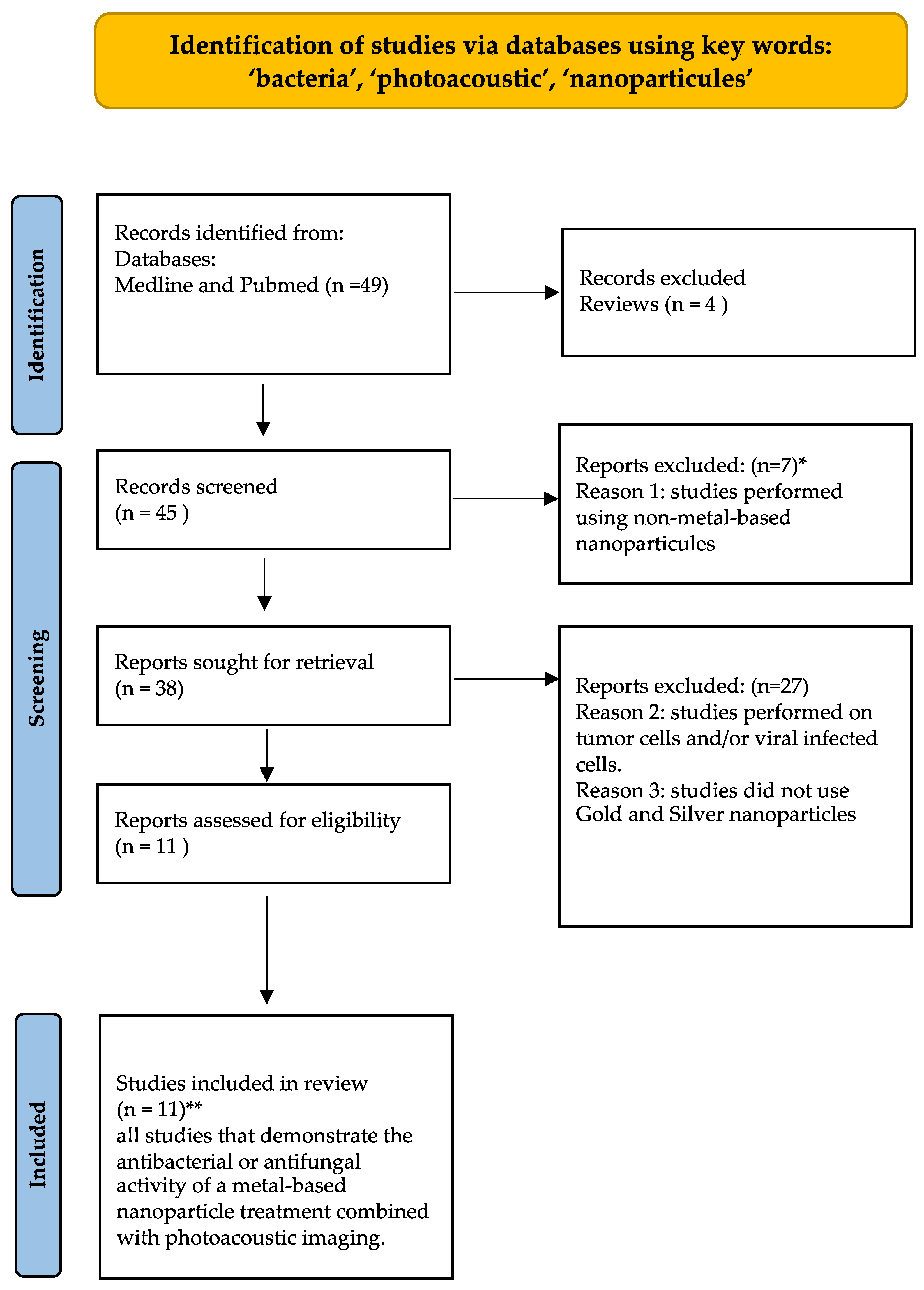
2. Methodology
3. PTT Combined with Metal or Metal Oxide Nanoparticles as Antibacterial Treatment Agents
3.1. Silver Nanoparticles (AgNPs)
3.2. Gold Nanoparticles (AuNPs)
3.3. Bimetallic Nanocomponents Based on Au or Ag
4. Ag- and/or Au-Based Compounds Used in Photoacoustic Therapy (PTAT)
5. Conclusions
Author Contributions
Funding
Data Availability Statement
Acknowledgments
Conflicts of Interest
References
- Dabbousi, A.A.; Dabboussi, F.; Hamze, M.; Osman, M.; Kassem, I.I. The Emergence and Dissemination of Multidrug Resistant Pseudomonas Aeruginosa in Lebanon: Current Status and Challenges during the Economic Crisis. Antibiotics 2022, 11, 687. [Google Scholar] [CrossRef]
- Thorley, K.; Charles, H.; Greig, D.R.; Prochazka, M.; Mason, L.C.E.; Baker, K.S.; Godbole, G.; Sinka, K.; Jenkins, C. Emergence of Extensively Drug-Resistant and Multidrug-Resistant Shigella Flexneri Serotype 2a Associated with Sexual Transmission among Gay, Bisexual, and Other Men Who Have Sex with Men, in England: A Descriptive Epidemiological Study. Lancet Infect. Dis. 2023, 23, 732–739. [Google Scholar] [CrossRef]
- Klemm, E.J.; Wong, V.K.; Dougan, G. Emergence of Dominant Multidrug-Resistant Bacterial Clades: Lessons from History and Whole-Genome Sequencing. Proc. Natl. Acad. Sci. USA 2018, 115, 12872–12877. [Google Scholar] [CrossRef] [Green Version]
- Aslam, B.; Wang, W.; Arshad, M.I.; Khurshid, M.; Muzammil, S.; Rasool, M.H.; Nisar, M.A.; Alvi, R.F.; Aslam, M.A.; Qamar, M.U.; et al. Antibiotic Resistance: A Rundown of a Global Crisis. Infect. Drug Resist. 2018, 11, 1645–1658. [Google Scholar] [CrossRef] [Green Version]
- European Centre for Disease Prevention and Control; World Health Organization. Antimicrobial Resistance Surveillance in Europe: 2022: 2020 Data; Publications Office: Luxembourg, 2022.
- Yeh, Y.-C.; Huang, T.-H.; Yang, S.-C.; Chen, C.-C.; Fang, J.-Y. Nano-Based Drug Delivery or Targeting to Eradicate Bacteria for Infection Mitigation: A Review of Recent Advances. Front. Chem. 2020, 8, 286. [Google Scholar] [CrossRef] [Green Version]
- Mammari, N.; Lamouroux, E.; Boudier, A.; Duval, R.E. Current Knowledge on the Oxidative-Stress-Mediated Antimicrobial Properties of Metal-Based Nanoparticles. Microorganisms 2022, 10, 437. [Google Scholar] [CrossRef]
- Franco, D.; Calabrese, G.; Guglielmino, S.P.P.; Conoci, S. Metal-Based Nanoparticles: Antibacterial Mechanisms and Biomedical Application. Microorganisms 2022, 10, 1778. [Google Scholar] [CrossRef] [PubMed]
- Levy, L.; Pottier, A.; Poul, L.; Maggiorella, L. Metallic Nanoparticles, Preparation and Uses Thereof. EP3178494A1, 14 October 2020. [Google Scholar]
- Zinn, A.A.; Lu, P.P. Nanoparticle Composition and Methods of Making the Same. US9378861B2, 28 June 2016. [Google Scholar]
- Hetta, H.F.; Ramadan, Y.N.; Al-Harbi, A.I.; Ahmed, E.A.; Battah, B.; Abd Ellah, N.H.; Zanetti, S.; Donadu, M.G. Nanotechnology as a Promising Approach to Combat Multidrug Resistant Bacteria: A Comprehensive Review and Future Perspectives. Biomedicines 2023, 11, 413. [Google Scholar] [CrossRef] [PubMed]
- Huang, Y.; Zou, L.; Wang, J.; Jin, Q.; Ji, J. Stimuli-Responsive Nanoplatforms for Antibacterial Applications. Wiley Interdiscip. Rev. Nanomed. Nanobiotechnol. 2022, 14, e1775. [Google Scholar] [CrossRef] [PubMed]
- Qi, X.; Shen, N.; Al Othman, A.; Mezentsev, A.; Permyakova, A.; Yu, Z.; Lepoitevin, M.; Serre, C.; Durymanov, M. Metal-Organic Framework-Based Nanomedicines for the Treatment of Intracellular Bacterial Infections. Pharmaceutics 2023, 15, 1521. [Google Scholar] [CrossRef]
- Sultana, A.; Zare, M.; Thomas, V.; Kumar, T.S.S.; Ramakrishna, S. Nano-Based Drug Delivery Systems: Conventional Drug Delivery Routes, Recent Developments and Future Prospects. Med. Drug Discov. 2022, 15, 100134. [Google Scholar] [CrossRef]
- Anderson, D.; Anderson, T.; Fahmi, F. Advances in Applications of Metal Oxide Nanomaterials as Imaging Contrast Agents. Phys. Status Solidi A 2019, 216, 1801008. [Google Scholar] [CrossRef]
- Wang, X.; Zhu, L.; Gu, Z.; Dai, L. Carbon Nanomaterials for Phototherapy. Nanophotonics 2022, 11, 4955–4976. [Google Scholar] [CrossRef]
- Fedatto Abelha, T.; Rodrigues Lima Caires, A. Light-Activated Conjugated Polymers for Antibacterial Photodynamic and Photothermal Therapy. Adv. NanoBiomed Res. 2021, 1, 2100012. [Google Scholar] [CrossRef]
- Chen, J.; Ning, C.; Zhou, Z.; Yu, P.; Zhu, Y.; Tan, G.; Mao, C. Nanomaterials as Photothermal Therapeutic Agents. Prog. Mater. Sci. 2019, 99, 1–26. [Google Scholar] [CrossRef]
- Roelandts, R. The History of Phototherapy: Something New under the Sun? J. Am. Acad. Dermatol. 2002, 46, 926–930. [Google Scholar] [CrossRef]
- Pucelik, B.; Dąbrowski, J.M. Chapter Three—Photodynamic Inactivation (PDI) as a Promising Alternative to Current Pharmaceuticals for the Treatment of Resistant Microorganisms. In Advances in Inorganic Chemistry; Recent Highlights II; van Eldik, R., Hubbard, C.D., Eds.; Academic Press: Cambridge, MA, USA, 2022; Volume 79, pp. 65–108. [Google Scholar]
- Khan, S.; Rayis, M.; Rizvi, A.; Alam, M.M.; Rizvi, M.; Naseem, I. ROS Mediated Antibacterial Activity of Photoilluminated Riboflavin: A Photodynamic Mechanism against Nosocomial Infections. Toxicol. Rep. 2019, 6, 136–142. [Google Scholar] [CrossRef]
- Chen, Y.; Gao, Y.; Chen, Y.; Liu, L.; Mo, A.; Peng, Q. Nanomaterials-Based Photothermal Therapy and Its Potentials in Antibacterial Treatment. J. Control Release 2020, 328, 251–262. [Google Scholar] [CrossRef]
- Qi, X.; Huang, Y.; You, S.; Xiang, Y.; Cai, E.; Mao, R.; Pan, W.; Tong, X.; Dong, W.; Ye, F.; et al. Engineering Robust Ag-Decorated Polydopamine Nano-Photothermal Platforms to Combat Bacterial Infection and Prompt Wound Healing. Adv. Sci. 2022, 9, e2106015. [Google Scholar] [CrossRef]
- Ye, Y.; He, J.; Wang, H.; Li, W.; Wang, Q.; Luo, C.; Tang, X.; Chen, X.; Jin, X.; Yao, K.; et al. Cell Wall Destruction and Internal Cascade Synergistic Antifungal Strategy for Fungal Keratitis. ACS Nano 2022, 16, 18729–18745. [Google Scholar] [CrossRef]
- Palau, M.; Muñoz, E.; Larrosa, N.; Gomis, X.; Márquez, E.; Len, O.; Almirante, B.; Gavaldà, J. Hyperthermia Prevents In Vitro and In Vivo Biofilm Formation on Endotracheal Tubes. Microbiol. Spectr. 2023, 11, e0280722. [Google Scholar] [CrossRef] [PubMed]
- Ma, R.; Hu, X.; Zhang, X.; Wang, W.; Sun, J.; Su, Z.; Zhu, C. Strategies to Prevent, Curb and Eliminate Biofilm Formation Based on the Characteristics of Various Periods in One Biofilm Life Cycle. Front. Cell. Infect. Microbiol. 2022, 12, 1003033. [Google Scholar] [CrossRef]
- Dai, X.; Zhao, Y.; Yu, Y.; Chen, X.; Wei, X.; Zhang, X.; Li, C. All-in-One NIR-Activated Nanoplatforms for Enhanced Bacterial Biofilm Eradication. Nanoscale 2018, 10, 18520–18530. [Google Scholar] [CrossRef]
- Huo, J.; Jia, Q.; Huang, H.; Zhang, J.; Li, P.; Dong, X.; Huang, W. Emerging Photothermal-Derived Multimodal Synergistic Therapy in Combating Bacterial Infections. Chem. Soc. Rev. 2021, 50, 8762–8789. [Google Scholar] [CrossRef] [PubMed]
- Yang, N.; Guo, H.; Cao, C.; Wang, X.; Song, X.; Wang, W.; Yang, D.; Xi, L.; Mou, X.; Dong, X. Infection Microenvironment-Activated Nanoparticles for NIR-II Photoacoustic Imaging-Guided Photothermal/Chemodynamic Synergistic Anti-Infective Therapy. Biomaterials 2021, 275, 120918. [Google Scholar] [CrossRef]
- Gellini, C.; Feis, A. Optothermal Properties of Plasmonic Inorganic Nanoparticles for Photoacoustic Applications. Photoacoustics 2021, 23, 100281. [Google Scholar] [CrossRef]
- Chen, Q.; Qi, M.; Shi, F.; Liu, C.; Shi, Y.; Sun, Y.; Bai, X.; Wang, L.; Sun, X.; Dong, B.; et al. Novel Twin-Crystal Nanosheets with MnO2 Modification to Combat Bacterial Biofilm against Periodontal Infections via Multipattern Strategies. Adv. Healthc. Mater. 2023, e2300313. [Google Scholar] [CrossRef]
- Yang, Y.; Zhou, X.; Chan, Y.K.; Wang, Z.; Li, L.; Li, J.; Liang, K.; Deng, Y. Photo-Activated Nanofibrous Membrane with Self-Rechargeable Antibacterial Function for Stubborn Infected Cutaneous Regeneration. Small 2022, 18, e2105988. [Google Scholar] [CrossRef]
- Yan, L.; Mu, J.; Ma, P.; Li, Q.; Yin, P.; Liu, X.; Cai, Y.; Yu, H.; Liu, J.; Wang, G.; et al. Gold Nanoplates with Superb Photothermal Efficiency and Peroxidase-like Activity for Rapid and Synergistic Antibacterial Therapy. Chem. Commun. 2021, 57, 1133–1136. [Google Scholar] [CrossRef]
- Zhang, Z.; Wen, J.; Zhang, J.; Guo, D.; Zhang, Q. Vacancy-Modulated of CuS for Highly Antibacterial Efficiency via Photothermal/Photodynamic Synergetic Therapy. Adv. Healthc. Mater. 2023, 12, e2201746. [Google Scholar] [CrossRef]
- Yu, Y.; Mei, L.; Shi, Y.; Zhang, X.; Cheng, K.; Cao, F.; Zhang, L.; Xu, J.; Li, X.; Xu, Z. Ag-Conjugated Graphene Quantum Dots with Blue Light-Enhanced Singlet Oxygen Generation for Ternary-Mode Highly-Efficient Antimicrobial Therapy. J. Mater. Chem. B 2020, 8, 1371–1382. [Google Scholar] [CrossRef]
- Gong, P.; Wang, F.; Guo, F.; Liu, J.; Wang, B.; Ge, X.; Li, S.; You, J.; Liu, Z. Fluorescence Turn-off Ag/Fluorinated Graphene Composites with High NIR Absorption for Effective Killing of Cancer Cells and Bacteria. J. Mater. Chem. B 2018, 6, 7926–7935. [Google Scholar] [CrossRef]
- Xia, J.; Yao, J.; Wang, L.V. Photoacoustic Tomography: Principles and Advances. Electromagn. Waves 2014, 147, 1–22. [Google Scholar] [CrossRef]
- Ouyang, J.; Liu, R.-Y.; Chen, W.; Liu, Z.; Xu, Q.; Zeng, K.; Deng, L.; Shen, L.; Liu, Y.-N. A Black Phosphorus Based Synergistic Antibacterial Platform against Drug Resistant Bacteria. J. Mater. Chem. B 2018, 6, 6302–6310. [Google Scholar] [CrossRef]
- Qiao, Y.; Ma, F.; Liu, C.; Zhou, B.; Wei, Q.; Li, W.; Zhong, D.; Li, Y.; Zhou, M. Near-Infrared Laser-Excited Nanoparticles To Eradicate Multidrug-Resistant Bacteria and Promote Wound Healing. ACS Appl. Mater. Interfaces 2018, 10, 193–206. [Google Scholar] [CrossRef]
- Zhang, H.; Yu, S.; Wu, S.; Xu, M.; Gao, T.; Wu, Q.; Xu, H.; Liu, Y. Rational Design of Silver NPs-Incorporated Quaternized Chitin Nanomicelle with Combinational Antibacterial Capability for Infected Wound Healing. Int. J. Biol. Macromol. 2023, 224, 1206–1216. [Google Scholar] [CrossRef]
- Zhou, K.; Zhang, Z.; Xue, J.; Shang, J.; Ding, D.; Zhang, W.; Liu, Z.; Yan, F.; Cheng, N. Hybrid Ag Nanoparticles/Polyoxometalate-Polydopamine Nano-Flowers Loaded Chitosan/Gelatin Hydrogel Scaffolds with Synergistic Photothermal/Chemodynamic/Ag+ Anti-Bacterial Action for Accelerated Wound Healing. Int. J. Biol. Macromol. 2022, 221, 135–148. [Google Scholar] [CrossRef]
- Li, Y.; Fu, R.; Duan, Z.; Zhu, C.; Fan, D. Mussel-Inspired Adhesive Bilayer Hydrogels for Bacteria-Infected Wound Healing via NIR-Enhanced Nanozyme Therapy. Colloids Surf. B Biointerfaces 2022, 210, 112230. [Google Scholar] [CrossRef]
- Huang, H.; Su, Y.; Wang, C.; Lei, B.; Song, X.; Wang, W.; Wu, P.; Liu, X.; Dong, X.; Zhong, L. Injectable Tissue-Adhesive Hydrogel for Photothermal/Chemodynamic Synergistic Antibacterial and Wound Healing Promotion. ACS Appl. Mater. Interfaces 2023, 15, 2714–2724. [Google Scholar] [CrossRef]
- Zhu, H.; Cheng, X.; Zhang, J.; Wu, Q.; Liu, C.; Shi, J. Constructing a Self-Healing Injectable SABA/Borax/PDA@AgNPs Hydrogel for Synergistic Low-Temperature Photothermal Antibacterial Therapy. J. Mater. Chem. B 2023, 11, 618–630. [Google Scholar] [CrossRef]
- Chang, R.; Zhao, D.; Zhang, C.; Liu, K.; He, Y.; Guan, F.; Yao, M. Nanocomposite Multifunctional Hyaluronic Acid Hydrogel with Photothermal Antibacterial and Antioxidant Properties for Infected Wound Healing. Int. J. Biol. Macromol. 2023, 226, 870–884. [Google Scholar] [CrossRef] [PubMed]
- Liu, Z.; Li, S.; Yin, Z.; Zhu, Z.; Chen, L.; Tan, W.; Chen, Z. Stabilizing Enzymes in Plasmonic Silk Film for Synergistic Therapy of In Situ SERS Identified Bacteria. Adv. Sci. 2022, 9, e2104576. [Google Scholar] [CrossRef]
- Bi, X.; Bai, Q.; Liang, M.; Yang, D.; Li, S.; Wang, L.; Liu, J.; Yu, W.W.; Sui, N.; Zhu, Z. Silver Peroxide Nanoparticles for Combined Antibacterial Sonodynamic and Photothermal Therapy. Small 2022, 18, e2104160. [Google Scholar] [CrossRef]
- Wen, S.; Wu, T.; Long, H.; Ke, L.; Deng, S.; Huang, L.; Zhang, J.; Tan, S. Mechanism Insight into Rapid Photodriven Sterilization Based on Silver Bismuth Sulfide Quantum Dots. ACS Appl. Mater. Interfaces 2021, 13, 21979–21993. [Google Scholar] [CrossRef]
- Li, M.; Huang, L.; Wang, X.; Song, Z.; Zhao, W.; Wang, Y.; Liu, J. Direct Generation of Ag Nanoclusters on Reduced Graphene Oxide Nanosheets for Efficient Catalysis, Antibacteria and Photothermal Anticancer Applications. J. Colloid Interface Sci. 2018, 529, 444–451. [Google Scholar] [CrossRef] [PubMed]
- Ran, X.; Du, Y.; Wang, Z.; Wang, H.; Pu, F.; Ren, J.; Qu, X. Hyaluronic Acid-Templated Ag Nanoparticles/Graphene Oxide Composites for Synergistic Therapy of Bacteria Infection. ACS Appl. Mater. Interfaces 2017, 9, 19717–19724. [Google Scholar] [CrossRef]
- Wang, D.; Peng, J.; Huang, Y.; Sun, L.; Liu, M.; Li, H.; Chao, M.; Gong, P.; Liu, Z.; You, J. Rational Construction of Fluorescence Turn-Off Fluorinated Carbon Fiber/Ag Composites and Their Anticancer and Antibacterial Activities. ACS Appl. Bio Mater. 2021, 4, 1749–1759. [Google Scholar] [CrossRef]
- Yan, Y.; Liu, Y.; Li, J.; Li, Y.; Wu, H.; Li, H.; Ma, X.; Tang, Y.; Tong, Y.; Yi, K.; et al. A Molecular Switch-Integrated Nanoplatform Enables Photo-Unlocked Antibacterial Drug Delivery for Synergistic Abscess Therapy. Adv. Healthc. Mater. 2023, e2301157. [Google Scholar] [CrossRef] [PubMed]
- Zhou, T.; Huang, J.; Zhao, W.; Guo, R.; Cui, S.; Li, Y.; Zhang, X.; Liu, Y.; Zhang, Q. Multifunctional Plasmon-Tunable Au Nanostars and Their Applications in Highly Efficient Photothermal Inactivation and Ultra-Sensitive SERS Detection. Nanomaterials 2022, 12, 4232. [Google Scholar] [CrossRef]
- He, X.; Dai, L.; Ye, L.; Sun, X.; Enoch, O.; Hu, R.; Zan, X.; Lin, F.; Shen, J. A Vehicle-Free Antimicrobial Polymer Hybrid Gold Nanoparticle as Synergistically Therapeutic Platforms for Staphylococcus aureus Infected Wound Healing. Adv. Sci. 2022, 9, e2105223. [Google Scholar] [CrossRef]
- Mocan, L.; Matea, C.; Tabaran, F.A.; Mosteanu, O.; Pop, T.; Puia, C.; Agoston-Coldea, L.; Gonciar, D.; Kalman, E.; Zaharie, G.; et al. Selective in Vitro Photothermal Nano-Therapy of MRSA Infections Mediated by IgG Conjugated Gold Nanoparticles. Sci. Rep. 2016, 6, 39466. [Google Scholar] [CrossRef] [Green Version]
- Zharov, V.P.; Mercer, K.E.; Galitovskaya, E.N.; Smeltzer, M.S. Photothermal Nanotherapeutics and Nanodiagnostics for Selective Killing of Bacteria Targeted with Gold Nanoparticles. Biophys. J. 2006, 90, 619–627. [Google Scholar] [CrossRef] [Green Version]
- Li, C.; Xian, J.; Hong, J.; Cao, X.; Zhang, C.; Deng, Q.; Qin, Z.; Chen, M.; Zheng, X.; Li, M.; et al. Dual Photothermal Nanocomposites for Drug-Resistant Infectious Wound Management. Nanoscale 2022, 14, 11284–11297. [Google Scholar] [CrossRef]
- Liang, Z.; Liu, W.; Wang, Z.; Zheng, P.; Liu, W.; Zhao, J.; Zhong, Y.; Zhang, Y.; Lin, J.; Xue, W.; et al. Near-Infrared Laser-Controlled Nitric Oxide-Releasing Gold Nanostar/Hollow Polydopamine Janus Nanoparticles for Synergistic Elimination of Methicillin-Resistant Staphylococcus aureus and Wound Healing. Acta Biomater. 2022, 143, 428–444. [Google Scholar] [CrossRef]
- Xu, X.; Liu, X.; Tan, L.; Cui, Z.; Yang, X.; Zhu, S.; Li, Z.; Yuan, X.; Zheng, Y.; Yeung, K.W.K.; et al. Controlled-Temperature Photothermal and Oxidative Bacteria Killing and Acceleration of Wound Healing by Polydopamine-Assisted Au-Hydroxyapatite Nanorods. Acta Biomater. 2018, 77, 352–364. [Google Scholar] [CrossRef]
- Ocsoy, I.; Yusufbeyoglu, S.; Yılmaz, V.; McLamore, E.S.; Ildız, N.; Ülgen, A. DNA Aptamer Functionalized Gold Nanostructures for Molecular Recognition and Photothermal Inactivation of Methicillin-Resistant Staphylococcus aureus. Colloids Surf. B Biointerfaces 2017, 159, 16–22. [Google Scholar] [CrossRef]
- Wu, Q.; Peng, R.; Luo, Y.; Cui, Q.; Zhu, S.; Li, L. Antibacterial Activity of Porous Gold Nanocomposites via NIR Light-Triggered Photothermal and Photodynamic Effects. ACS Appl. Bio Mater. 2021, 4, 5071–5079. [Google Scholar] [CrossRef] [PubMed]
- Fan, H.; Fan, Y.; Du, W.; Cai, R.; Gao, X.; Liu, X.; Wang, H.; Wang, L.; Wu, X. Enhanced Type I Photoreaction of Indocyanine Green via Electrostatic-Force-Driven Aggregation. Nanoscale 2020, 12, 9517–9523. [Google Scholar] [CrossRef] [PubMed]
- Annesi, F.; Pane, A.; Losso, M.A.; Guglielmelli, A.; Lucente, F.; Petronella, F.; Placido, T.; Comparelli, R.; Guzzo, M.G.; Curri, M.L.; et al. Thermo-Plasmonic Killing of Escherichia coli TG1 Bacteria. Materials 2019, 12, 1530. [Google Scholar] [CrossRef] [Green Version]
- Yin, M.; Yang, M.; Yan, D.; Yang, L.; Wan, X.; Xiao, J.; Yao, Y.; Luo, J. Surface-Charge-Switchable and Size-Transformable Thermosensitive Nanocomposites for Chemo-Photothermal Eradication of Bacterial Biofilms in Vitro and in Vivo. ACS Appl. Mater. Interfaces 2022, 14, 8847–8864. [Google Scholar] [CrossRef]
- Tang, Y.; Wang, T.; Feng, J.; Rong, F.; Wang, K.; Li, P.; Huang, W. Photoactivatable Nitric Oxide-Releasing Gold Nanocages for Enhanced Hyperthermia Treatment of Biofilm-Associated Infections. ACS Appl. Mater. Interfaces 2021, 13, 50668–50681. [Google Scholar] [CrossRef]
- Ma, H.; Tang, Y.; Rong, F.; Wang, K.; Wang, T.; Li, P. Surface Charge Adaptive Nitric Oxide Nanogenerator for Enhanced Photothermal Eradication of Drug-Resistant Biofilm Infections. Bioact. Mater. 2023, 27, 154–167. [Google Scholar] [CrossRef]
- Meeker, D.G.; Wang, T.; Harrington, W.N.; Zharov, V.P.; Johnson, S.A.; Jenkins, S.V.; Oyibo, S.E.; Walker, C.M.; Mills, W.B.; Shirtliff, M.E.; et al. Versatility of Targeted Antibiotic-Loaded Gold Nanoconstructs for the Treatment of Biofilm-Associated Bacterial Infections. Int. J. Hyperth. 2018, 34, 209–219. [Google Scholar] [CrossRef] [PubMed] [Green Version]
- Wu, Y.; Deng, G.; Jiang, K.; Wang, H.; Song, Z.; Han, H. Photothermally Triggered Nitric Oxide Nanogenerator Targeting Type IV Pili for Precise Therapy of Bacterial Infections. Biomaterials 2021, 268, 120588. [Google Scholar] [CrossRef] [PubMed]
- Cui, T.; Wu, S.; Sun, Y.; Ren, J.; Qu, X. Self-Propelled Active Photothermal Nanoswimmer for Deep-Layered Elimination of Biofilm In Vivo. Nano Lett. 2020, 20, 7350–7358. [Google Scholar] [CrossRef] [PubMed]
- Wang, S.-G.; Chen, Y.-C.; Chen, Y.-C. Antibacterial Gold Nanoparticle-Based Photothermal Killing of Vancomycin-Resistant Bacteria. Nanomedicine 2018, 13, 1405–1416. [Google Scholar] [CrossRef]
- Kataria, M.; Yadav, K.; Nain, A.; Lin, H.-I.; Hu, H.-W.; Paul Inbaraj, C.R.; Chang, T.-J.; Liao, Y.-M.; Cheng, H.-Y.; Lin, K.-H.; et al. Self-Sufficient and Highly Efficient Gold Sandwich Upconversion Nanocomposite Lasers for Stretchable and Bio-Applications. ACS Appl. Mater. Interfaces 2020, 12, 19840–19854. [Google Scholar] [CrossRef]
- Gao, X.; Wu, H.; Hao, Z.; Ji, X.; Lin, X.; Wang, S.; Liu, Y. A Multifunctional Plasmonic Chip for Bacteria Capture, Imaging, Detection, and in Situ Elimination for Wound Therapy. Nanoscale 2020, 12, 6489–6497. [Google Scholar] [CrossRef]
- Wang, J.; Zhang, J.; Liu, K.; He, J.; Zhang, Y.; Chen, S.; Ma, G.; Cui, Y.; Wang, L.; Gao, D. Synthesis of Gold Nanoflowers Stabilized with Amphiphilic Daptomycin for Enhanced Photothermal Antitumor and Antibacterial Effects. Int. J. Pharm. 2020, 580, 119231. [Google Scholar] [CrossRef]
- Wang, H.; Song, Z.; Li, S.; Wu, Y.; Han, H. One Stone with Two Birds: Functional Gold Nanostar for Targeted Combination Therapy of Drug-Resistant Staphylococcus aureus Infection. ACS Appl. Mater. Interfaces 2019, 11, 32659–32669. [Google Scholar] [CrossRef]
- Wang, H.; Ouyang, W.; Zhang, X.; Xue, J.; Lou, X.; Fan, R.; Zhao, X.; Shan, L.; Jiang, T. Bacteria-Induced Aggregation of Bioorthogonal Gold Nanoparticles for SERS Imaging and Enhanced Photothermal Ablation of Gram-Positive Bacteria. J. Mater. Chem. B 2019, 7, 4630–4637. [Google Scholar] [CrossRef] [PubMed]
- Xie, Y.; Zheng, W.; Jiang, X. Near-Infrared Light-Activated Phototherapy by Gold Nanoclusters for Dispersing Biofilms. ACS Appl. Mater. Interfaces 2020, 12, 9041–9049. [Google Scholar] [CrossRef]
- Manivasagan, P.; Khan, F.; Hoang, G.; Mondal, S.; Kim, H.; Hoang Minh Doan, V.; Kim, Y.-M.; Oh, J. Thiol Chitosan-Wrapped Gold Nanoshells for near-Infrared Laser-Induced Photothermal Destruction of Antibiotic-Resistant Bacteria. Carbohydr. Polym. 2019, 225, 115228. [Google Scholar] [CrossRef]
- Zhang, Y.; Li, P.; Su, R.; Wen, F.; Jia, Z.; Lv, Y.; Cai, J.; Su, W. Curcumin-Loaded Multifunctional Chitosan Gold Nanoparticles: An Enhanced PDT/PTT Dual-Modal Phototherapeutic and PH-Responsive Antimicrobial Agent. Photodiagn. Photodyn. Ther. 2022, 39, 103011. [Google Scholar] [CrossRef] [PubMed]
- El Zorkany, H.E.; Youssef, T.; Mohamed, M.B.; Amin, R.M. Photothermal versus Photodynamic Treatment for the Inactivation of the Bacteria Escherichia coli and Bacillus cereus: An in Vitro Study. Photodiagn. Photodyn. Ther. 2019, 27, 317–326. [Google Scholar] [CrossRef]
- Kirui, D.K.; Weber, G.; Talackine, J.; Millenbaugh, N.J. Targeted Laser Therapy Synergistically Enhances Efficacy of Antibiotics against Multi-Drug Resistant Staphylococcus aureus and Pseudomonas aeruginosa Biofilms. Nanomed. Nanotechnol. Biol. Med. 2019, 20, 102018. [Google Scholar] [CrossRef] [PubMed]
- Ngo-Duc, T.-T.; Alibay, Z.; Plank, J.M.; Cheeney, J.E.; Haberer, E.D. Gold-Decorated M13 I-Forms and S-Forms for Targeted Photothermal Lysis of Bacteria. ACS Appl. Mater. Interfaces 2020, 12, 126–134. [Google Scholar] [CrossRef]
- Alhmoud, H.; Cifuentes-Rius, A.; Delalat, B.; Lancaster, D.G.; Voelcker, N.H. Gold-Decorated Porous Silicon Nanopillars for Targeted Hyperthermal Treatment of Bacterial Infections. ACS Appl. Mater. Interfaces 2017, 9, 33707–33716. [Google Scholar] [CrossRef]
- Hu, D.; Li, H.; Wang, B.; Ye, Z.; Lei, W.; Jia, F.; Jin, Q.; Ren, K.-F.; Ji, J. Surface-Adaptive Gold Nanoparticles with Effective Adherence and Enhanced Photothermal Ablation of Methicillin-Resistant Staphylococcus aureus Biofilm. ACS Nano 2017, 11, 9330–9339. [Google Scholar] [CrossRef]
- Candreva, A.; De Rose, R.; Perrotta, I.D.; Guglielmelli, A.; La Deda, M. Light-Induced Clusterization of Gold Nanoparticles: A New Photo-Triggered Antibacterial against E. coli Proliferation. Nanomaterials 2023, 13, 746. [Google Scholar] [CrossRef]
- Yougbaré, S.; Chou, H.-L.; Yang, C.-H.; Krisnawati, D.I.; Jazidie, A.; Nuh, M.; Kuo, T.-R. Facet-Dependent Gold Nanocrystals for Effective Photothermal Killing of Bacteria. J. Hazard. Mater. 2021, 407, 124617. [Google Scholar] [CrossRef]
- Peng, H.; Borg, R.E.; Dow, L.P.; Pruitt, B.L.; Chen, I.A. Controlled Phage Therapy by Photothermal Ablation of Specific Bacterial Species Using Gold Nanorods Targeted by Chimeric Phages. Proc. Natl. Acad. Sci. USA 2020, 117, 1951–1961. [Google Scholar] [CrossRef] [PubMed] [Green Version]
- Xu, X.; Ding, Y.; Hadianamrei, R.; Lv, S.; You, R.; Pan, F.; Zhang, P.; Wang, N.; Zhao, X. Antimicrobial Peptide Functionalized Gold Nanorods Combining Near-Infrared Photothermal Therapy for Effective Wound Healing. Colloids Surf. B Biointerfaces 2022, 220, 112887. [Google Scholar] [CrossRef] [PubMed]
- Tian, H.; Hong, J.; Li, C.; Qiu, Y.; Li, M.; Qin, Z.; Ghiladi, R.A.; Yin, X. Electrospinning Membranes with Au@carbon Dots: Low Toxicity and Efficient Antibacterial Photothermal Therapy. Biomater. Adv. 2022, 142, 213155. [Google Scholar] [CrossRef] [PubMed]
- Lin, Y.; Hamme Ii, A.T. Targeted Highly Sensitive Detection/Eradication of Multi-Drug Resistant Salmonella DT104 through Gold Nanoparticle-SWCNT Bioconjugated Nanohybrids. Analyst 2014, 139, 3702–3705. [Google Scholar] [CrossRef] [Green Version]
- Kim, T.; Braun, G.B.; She, Z.-G.; Hussain, S.; Ruoslahti, E.; Sailor, M.J. Composite Porous Silicon-Silver Nanoparticles as Theranostic Antibacterial Agents. ACS Appl. Mater. Interfaces 2016, 8, 30449–30457. [Google Scholar] [CrossRef] [PubMed]
- Feng, Q.L.; Wu, J.; Chen, G.Q.; Cui, F.Z.; Kim, T.N.; Kim, J.O. A Mechanistic Study of the Antibacterial Effect of Silver Ions on Escherichia coli and Staphylococcus aureus. J. Biomed. Mater. Res. 2000, 52, 662–668. [Google Scholar] [CrossRef]
- Hu, B.; Zhang, L.-P.; Chen, X.-W.; Wang, J.-H. Gold Nanorod-Covered Kanamycin-Loaded Hollow SiO2 (HSKAu(Rod)) Nanocapsules for Drug Delivery and Photothermal Therapy on Bacteria. Nanoscale 2013, 5, 246–252. [Google Scholar] [CrossRef] [PubMed]
- Wang, H.; Wang, D.; Huangfu, H.; Chen, S.; Qin, Q.; Ren, S.; Zhang, Y.; Fu, L.; Zhou, Y. Highly Efficient Photothermal Branched Au-Ag Nanoparticles Containing Procyanidins for Synergistic Antibacterial and Anti-Inflammatory Immunotherapy. Biomater. Sci. 2023, 11, 1335–1349. [Google Scholar] [CrossRef]
- Nudelman, R.; Gavriely, S.; Bychenko, D.; Barzilay, M.; Gulakhmedova, T.; Gazit, E.; Richter, S. Bio-Assisted Synthesis of Bimetallic Nanoparticles Featuring Antibacterial and Photothermal Properties for the Removal of Biofilms. J. Nanobiotechnol. 2021, 19, 452. [Google Scholar] [CrossRef]
- Jiang, X.; Fan, X.; Xu, W.; Zhang, R.; Wu, G. Biosynthesis of Bimetallic Au-Ag Nanoparticles Using Escherichia coli and Its Biomedical Applications. ACS Biomater. Sci. Eng. 2020, 6, 680–689. [Google Scholar] [CrossRef]
- Yang, J.; Zhao, Y.; Cao, J.; Gong, C.; Zuo, J.; Zhang, N.; Zhao, Y. Hyaluronic Acid and Antimicrobial Peptide-Modified Gold/Silver Hybrid Nanocages to Combat Bacterial Multidrug Resistance. Int. J. Pharm. 2020, 586, 119505. [Google Scholar] [CrossRef] [PubMed]
- Wang, C.; Wang, Y.; Zhang, L.; Miron, R.J.; Liang, J.; Shi, M.; Mo, W.; Zheng, S.; Zhao, Y.; Zhang, Y. Pretreated Macrophage-Membrane-Coated Gold Nanocages for Precise Drug Delivery for Treatment of Bacterial Infections. Adv. Mater. 2018, 30, e1804023. [Google Scholar] [CrossRef] [PubMed]
- Du, T.; Cao, J.; Xiao, Z.; Liu, J.; Wei, L.; Li, C.; Jiao, J.; Song, Z.; Liu, J.; Du, X.; et al. Van-Mediated Self-Aggregating Photothermal Agents Combined with Multifunctional Magnetic Nickel Oxide Nanoparticles for Precise Elimination of Bacterial Infections. J. Nanobiotechnol. 2022, 20, 325. [Google Scholar] [CrossRef]
- Ma, W.; Dong, W.; Zhao, S.; Du, T.; Wang, Y.; Yao, J.; Liu, Z.; Sun, D.; Zhang, M. An Injectable Adhesive Antibacterial Hydrogel Wound Dressing for Infected Skin Wounds. Biomater. Adv. 2022, 134, 112584. [Google Scholar] [CrossRef]
- Zhao, X.; Chang, L.; Hu, Y.; Xu, S.; Liang, Z.; Ren, X.; Mei, X.; Chen, Z. Preparation of Photocatalytic and Antibacterial MOF Nanozyme Used for Infected Diabetic Wound Healing. ACS Appl. Mater. Interfaces 2022, 14, 18194–18208. [Google Scholar] [CrossRef]
- Lin, S.; Chen, H.; Wang, R.; Jiang, T.; Wang, R.; Yu, F. Hollow Silver-Gold Alloy Nanoparticles for Enhanced Photothermal/Photodynamic Synergetic Therapy against Bacterial Infection and Acceleration of Wound Healing. Biomater. Sci. 2023, 11, 4874–4889. [Google Scholar] [CrossRef]
- Xiong, Q.; Fang, Q.; Xu, K.; Liu, G.; Sang, M.; Xu, Y.; Hao, L.; Xuan, S. Near-Infrared Light-Responsive Photothermal α-Fe2O3@Au/PDA Core/Shell Nanostructure with on-off Controllable Anti-Bacterial Effects. Dalton Trans. 2021, 50, 14235–14243. [Google Scholar] [CrossRef]
- Huang, H.; Wang, X.; Wang, W.; Qu, X.; Song, X.; Zhang, Y.; Zhong, L.; Yang, D.P.; Dong, X.; Zhao, Y. Injectable Hydrogel for Postoperative Synergistic Photothermal-Chemodynamic Tumor and Anti-Infection Therapy. Biomaterials 2022, 280, 121289. [Google Scholar] [CrossRef]
- Naskar, A.; Lee, S.; Kim, K.-S. Au-ZnO Conjugated Black Phosphorus as a Near-Infrared Light-Triggering and Recurrence-Suppressing Nanoantibiotic Platform against Staphylococcus aureus. Pharmaceutics 2021, 13, 52. [Google Scholar] [CrossRef] [PubMed]
- Cao, C.; Ge, W.; Yin, J.; Yang, D.; Wang, W.; Song, X.; Hu, Y.; Yin, J.; Dong, X. Mesoporous Silica Supported Silver-Bismuth Nanoparticles as Photothermal Agents for Skin Infection Synergistic Antibacterial Therapy. Small 2020, 16, e2000436. [Google Scholar] [CrossRef] [PubMed]
- He, J.; Qiao, Y.; Zhang, H.; Zhao, J.; Li, W.; Xie, T.; Zhong, D.; Wei, Q.; Hua, S.; Yu, Y.; et al. Gold-Silver Nanoshells Promote Wound Healing from Drug-Resistant Bacteria Infection and Enable Monitoring via Surface-Enhanced Raman Scattering Imaging. Biomaterials 2020, 234, 119763. [Google Scholar] [CrossRef] [PubMed]
- Hsueh, Y.-H.; Hsieh, C.-T.; Chiu, S.-T.; Tsai, P.-H.; Liu, C.-Y.; Ke, W.-J. Antibacterial Property of Composites of Reduced Graphene Oxide with Nano-Silver and Zinc Oxide Nanoparticles Synthesized Using a Microwave-Assisted Approach. Int. J. Mol. Sci. 2019, 20, 5394. [Google Scholar] [CrossRef] [Green Version]
- Zhang, S.; Lu, Q.; Wang, F.; Xiao, Z.; He, L.; He, D.; Deng, L. Gold-Platinum Nanodots with High-Peroxidase-like Activity and Photothermal Conversion Efficiency for Antibacterial Therapy. ACS Appl. Mater. Interfaces 2021, 13, 37535–37544. [Google Scholar] [CrossRef]
- Yang, T.; Wang, D.; Liu, X. Assembled Gold Nanorods for the Photothermal Killing of Bacteria. Colloids Surf. B Biointerfaces 2019, 173, 833–841. [Google Scholar] [CrossRef] [PubMed]
- Yang, Y.; Wu, X.; He, C.; Huang, J.; Yin, S.; Zhou, M.; Ma, L.; Zhao, W.; Qiu, L.; Cheng, C.; et al. Metal-Organic Framework/Ag-Based Hybrid Nanoagents for Rapid and Synergistic Bacterial Eradication. ACS Appl. Mater. Interfaces 2020, 12, 13698–13708. [Google Scholar] [CrossRef] [PubMed]
- Hui, S.; Liu, Q.; Han, Y.; Zhang, L.; Yang, J.; Jiang, S.; Qian, H.; Yang, W. ICG@ZIF-8/PDA/Ag Composites as Chemo-Photothermal Antibacterial Agents for Efficient Sterilization and Enhanced Wound Disinfection. J. Mater. Chem. B 2021, 9, 9961–9970. [Google Scholar] [CrossRef]
- Huang, Y.; Geng, H.; Wu, Z.; Sun, L.; Ji, C.; Grimes, C.A.; Feng, X.; Cai, Q. An Ag2S@ZIF-Van Nanosystem for NIR-II Imaging of Bacterial-Induced Inflammation and Treatment of Wound Bacterial Infection. Biomater. Sci. 2022, 10, 3972–3980. [Google Scholar] [CrossRef]
- Liu, M.; He, D.; Yang, T.; Liu, W.; Mao, L.; Zhu, Y.; Wu, J.; Luo, G.; Deng, J. An Efficient Antimicrobial Depot for Infectious Site-Targeted Chemo-Photothermal Therapy. J. Nanobiotechnol. 2018, 16, 23. [Google Scholar] [CrossRef] [Green Version]
- Ye, Y.; He, J.; Qiao, Y.; Qi, Y.; Zhang, H.; Santos, H.A.; Zhong, D.; Li, W.; Hua, S.; Wang, W.; et al. Mild Temperature Photothermal Assisted Anti-Bacterial and Anti-Inflammatory Nanosystem for Synergistic Treatment of Post-Cataract Surgery Endophthalmitis. Theranostics 2020, 10, 8541–8557. [Google Scholar] [CrossRef]
- Qiao, Y.; He, J.; Chen, W.; Yu, Y.; Li, W.; Du, Z.; Xie, T.; Ye, Y.; Hua, S.Y.; Zhong, D.; et al. Light-Activatable Synergistic Therapy of Drug-Resistant Bacteria-Infected Cutaneous Chronic Wounds and Nonhealing Keratitis by Cupriferous Hollow Nanoshells. ACS Nano 2020, 14, 3299–3315. [Google Scholar] [CrossRef]
- Yang, Y.; Chu, B.; Cheng, J.; Tang, J.; Song, B.; Wang, H.; He, Y. Bacteria Eat Nanoprobes for Aggregation-Enhanced Imaging and Killing Diverse Microorganisms. Nat. Commun. 2022, 13, 1255. [Google Scholar] [CrossRef]
- Afkhami, F.; Ahmadi, P.; Chiniforush, N.; Sooratgar, A. Effect of Different Activations of Silver Nanoparticle Irrigants on the Elimination of Enterococcus faecalis. Clin. Oral Investig. 2021, 25, 6893–6899. [Google Scholar] [CrossRef] [PubMed]
- Li, Z.; Fu, Q.; Ye, J.; Ge, X.; Wang, J.; Song, J.; Yang, H. Ag+-Coupled Black Phosphorus Vesicles with Emerging NIR-II Photoacoustic Imaging Performance for Cancer Immune-Dynamic Therapy and Fast Wound Healing. Angew. Chem. Int. Ed. 2020, 59, 22202–22209. [Google Scholar] [CrossRef] [PubMed]
- Lu, S.-Z.; Guo, X.-Y.; Zou, M.-S.; Zheng, Z.-Q.; Li, Y.-C.; Li, X.-D.; Li, L.-L.; Wang, H. Bacteria-Instructed In Situ Aggregation of AuNPs with Enhanced Photoacoustic Signal for Bacterial Infection Bioimaging. Adv. Healthc. Mater. 2020, 9, e1901229. [Google Scholar] [CrossRef] [PubMed]
- Zhi, X.; Liu, Y.; Lin, L.; Yang, M.; Zhang, L.; Zhang, L.; Liu, Y.; Alfranca, G.; Ma, L.; Zhang, Q.; et al. Oral PH Sensitive GNS@ab Nanoprobes for Targeted Therapy of Helicobacter Pylori without Disturbance Gut Microbiome. Nanomed. Nanotechnol. Biol. Med. 2019, 20, 102019. [Google Scholar] [CrossRef] [PubMed]
- Kim, T.; Zhang, Q.; Li, J.; Zhang, L.; Jokerst, J.V. A Gold/Silver Hybrid Nanoparticle for Treatment and Photoacoustic Imaging of Bacterial Infection. ACS Nano 2018, 12, 5615–5625. [Google Scholar] [CrossRef]
- Millenbaugh, N.J.; Baskin, J.B.; DeSilva, M.N.; Elliott, W.R.; Glickman, R.D. Photothermal Killing of Staphylococcus Aureus Using Antibody-Targeted Gold Nanoparticles. Int. J. Nanomed. 2015, 10, 1953–1960. [Google Scholar] [CrossRef] [Green Version]
- Nedosekin, D.A.; Juratli, M.A.; Sarimollaoglu, M.; Moore, C.L.; Rusch, N.J.; Smeltzer, M.S.; Zharov, V.P.; Galanzha, E.I. Photoacoustic and Photothermal Detection of Circulating Tumor Cells, Bacteria and Nanoparticles in Cerebrospinal Fluid in Vivo and Ex Vivo. J. Biophotonics 2013, 6, 523–533. [Google Scholar] [CrossRef] [Green Version]
- Galanzha, E.I.; Shashkov, E.; Sarimollaoglu, M.; Beenken, K.E.; Basnakian, A.G.; Shirtliff, M.E.; Kim, J.-W.; Smeltzer, M.S.; Zharov, V.P. In Vivo Magnetic Enrichment, Photoacoustic Diagnosis, and Photothermal Purging of Infected Blood Using Multifunctional Gold and Magnetic Nanoparticles. PLoS ONE 2012, 7, e45557. [Google Scholar] [CrossRef]
- Zharov, V.P.; Galanzha, E.I.; Shashkov, E.V.; Kim, J.-W.; Khlebtsov, N.G.; Tuchin, V.V. Photoacoustic Flow Cytometry: Principle and Application for Real-Time Detection of Circulating Single Nanoparticles, Pathogens, and Contrast Dyes in Vivo. J. Biomed. Opt. 2007, 12, 051503. [Google Scholar] [CrossRef] [PubMed] [Green Version]
- Liu, L.; Li, S.; Yang, K.; Chen, Z.; Li, Q.; Zheng, L.; Wu, Z.; Zhang, X.; Su, L.; Wu, Y.; et al. Drug-Free Antimicrobial Nanomotor for Precise Treatment of Multidrug-Resistant Bacterial Infections. Nano Lett. 2023, 23, 3929–3938. [Google Scholar] [CrossRef] [PubMed]
- Hajfathalian, M.; de Vries, C.R.; Hsu, J.C.; Amirshaghaghi, A.; Dong, Y.C.; Ren, Z.; Liu, Y.; Huang, Y.; Li, Y.; Knight, S.; et al. Theranostic Gold in a Gold Cage Nanoparticle for Photothermal Ablation and Photoacoustic Imaging of Skin and Oral Infections. BioRxiv Prepr. Serv. Biol. 2023. [Google Scholar] [CrossRef]


| Nanostructure | PT | Light Source | PT Parameters | Bacteria or Type of Infection | References |
|---|---|---|---|---|---|
| CG/PDA@Ag | PTT | Laser |
808 nm (1 W/cm2)
3 min (37 to 49.1 °C) |
E. coli S. aureus | [23] |
| MX@AgP nanoparticle (NPs) | PTT | Laser |
808 nm (1.5 W/cm2)
10 min | S. aureus (ATCC 25923) E. coli (ATCC 25922) | [32] |
|
AuNPTs
(Gold nanoplates) | PTT | Laser |
808 nm (1 W/cm2)
3 min | MRSA | [33] |
| GQD–AgNP (Ag nanoparticle-conjugated graphene quantum dots) | PTD, PTT | Laser | 450 nm (14.2 mW/cm2) 10 min (40 °C) | E. coli S. aureus | [35] |
|
FGO–Ag
(Graphene oxide–silver) | PTT | Laser | 808 nm (2.0 W/cm2) 5 min | E. coli S. aureus | [36] |
| Ag@BP nanohybrids | PTT | Laser | 808 nm (0.8 W/cm2) 5 min | MRSA | [38] |
|
Tri–Ag
Silver triangular nanoparticles | PTT | Laser | 808 nm (1.3 W/cm2) 10 min | E. coli E. coli (ESBL) S. aureus MRSA | [39] |
| Ag NPs-incorporated quaternized chitin (DQCA) nanomicelle | PTT | Laser | 660 nm (1.0 W/cm2) 10 min | S. aureus E. coli | [40] |
| AgNPs/POM-PDA (a three-in-one bactericidal flower-like nanocomposite–Ag nanoparticles/phosphotungstic acid–polydopamine nano-flowers) | PTT | Laser | 808 nm (0.75 W/cm2) 6 min | E. coli S. aureus | [41] |
| Polydopamine (PDA) coating-reduced Ag nanoparticles (AgNPs) | PTT | Laser |
808 nm
10 min | E. coli S. aureus | [42] |
| Ag-doped Mo2C-derived polyoxometalate (AgPOM) nanoparticles urea, gelatin, and tea polyphenols (TPs) | PTD, PTT | Laser | 1060 nm (1.0 W/cm2) 10 min | S. aureus | [43] |
| SABA/Borax/PDA@AgNPs hydrogel | PTT | Laser | 808 nm, 5 min (≤45 °C) | S. aureus E. coli | [44] |
| AgNPs@TA (Hyaluronic acid–tyramine (HT) hydrogel loaded with antioxidant and photothermal silver nanoparticles (AgNPs)) | PTT | Laser | 808 nm (0.92 W/cm2) 10 min | S. aureus E. coli | [45] |
|
Soie-GOx-Ag @ G, SGA
(Silk-GOx-Ag@G, SGA (glucose oxidase (GOx) is embedded in Ag graphitic nanocapsule (Ag@G)) | PTT | Laser | 808 nm (5 W/cm2) ( 60 °C) |
S. aureus MRSA | [46] |
| Ag2O2 NPs (Silver peroxide nanoparticles) | PTT | Laser |
808 nm (0.7 W/cm2)
10 min | MRSA S. aureus E. coli P. aeruginosa | [47] |
| AgBiS2 QDs (Silver bismuth sulfide quantum dots) | PTD, PTT | Laser | 808 nm (1.6 W/cm2) 10 min |
E. coli S. aureus | [48] |
|
AgNC/GSH-rGO
(Ag nanoclusters, graphene oxide (rGO), glutathione (GSH)) | PTT | Laser |
808 nm (2 W/cm2)
5 min | E. coli S. aureus | [49] |
|
GO–HA–AgNPs
(a hyaluronidase, silver nanoparticles (AgNPs) and graphene oxide (GO)) | PTT | Laser | 808 nm (1.0 W/cm2) 2 min | S. aureus | [50] |
| Carbon fiber oxide (FCO)/Ag composite | PTT | Laser | 808 nm (2 W/cm2) 5 min | E. coli S. aureus | [51] |
| Mesoporous silica nanospheres (HMSN)/silver nanoparticles (Ag NPs)/vancomycin (Van)/hemin (HAVH) | PTT | Laser | 808 nm (1 W/cm2) 10 min | MRSA | [52] |
| AuNSs (Gold nanoclusters) | PTT | Laser | 808 nm (1 W/cm2) 300 s (53.1 °C) | S. aureus | [53] |
| PHMB@Au NPs (Polymer polyhexamethylene biguanide (PHMB, with bactericidal and anti-biofilm functions) hybrid gold nanoparticle (Au NPs)) | PTT | Laser |
808 nm (2.0 W/cm2)
10 min ( 45.0 °C) (4.5 µg/mL), 58.4 °C (9.0 µg/mL), 65.2 °C (18.0 µg/mL) | S. aureus | [54] |
|
IgG-AuNPs
(Gold nanoparticles) | PTT | Laser |
808 nm (2 W/cm2)
10 min | MRSA | [55] |
| AuNPs Gold nanoparticles | PTT | Laser pulses | 420–570 nm, 12 ns, 0.1–5 J/cm2, 100 pulses | S. aureus | [56] |
| PDA-AuNPs (Polydopamine–gold nanoparticles) | PTT | Laser | 808 nm (1 W/cm2) 15 min (55 °C) | S. aureus (ATCC 6538) MRSA E. coli (ATCC 25922) | [57] |
|
GNS/HPDA JNPs
(Gold nanostar/hollow polydopamine Janus nanostructure) | PTT | Laser |
808 nm (1.5 W/cm2)
5 min (125 µg/mL) | E. coli (ATCC 25922) S. aureus (ATCC 29213) MRSA (ATCC 43300) | [58] |
| PDA@Au-HAp NPs (a polydopamine (PDA) coating on hydroxyapatite (HAp) incorporated with gold nanoparticles (Au-Hap)) | PTT | Laser |
808 nm (1.0 W/cm2)
10 min | E. coli S. aureus | [59] |
|
Apt@AuNPs
(DNA aptamer-functionalized gold nanoparticles) | PTT | Laser |
808 nm (1.1 W/cm2)
2 min | MRSA (ATCC 43300) | [60] |
|
AuNPs-ICG
(Porous gold nanoparticles–indocyanine green) | PTD, PTT | Laser | 808 nm | S. aureus | [61] |
| AuNPs_ICG | PTT | Laser | 808 nm (1 W/cm2) 1 min | E. coli (ATCC 8393) S. aureus (ATCC 6538P) | [62] |
|
AuNRs
(Gold nanorods) | PTT | Laser |
810 nm (6.3 W/cm2)
10 min | E. coli E. coli/AuNRs | [63] |
| AuNR@P(NIPAM-AA-MAA) (N-isopropyl acrylamide (NIPAM), acrylic acid (AA), and N-allylmethylamine (MAA)–gold nanorods (AuNRs)) | PTT | Laser | 808 nm (1.0 W/cm2) 10 min | E. coli S. aureus | [64] |
| AuNC@NO | PTT | Laser | 808 nm (0.5 W/cm2) 5 min | MRSA | [65] |
|
PDG@Au-NO/PBAM
(dopamine-co-glucosamine,Gold, Nitric oxide, phenylboronic acid, and acryloylmorpholine) | PTT | Laser | 808 nm (1.0 W/cm2) 10 min |
MRSA (ATCC BAA-40)
TREC (ATCC ER2738) | [66] |
|
PDA-AuNCs
(Antibody-conjugated, polydopamine (PDA)-coated gold nanocages (AuNCs)) | PTT | Laser | 808 nm (0.8 W/cm2) 10 min | MRSA P. aeruginosa (ATCC 27317) | [67] |
| SNP@MOF@Au-Mal nanogenerator | PTT | Laser |
808 nm (1.5 W/cm2)
10 min | P. aeruginosa | [68] |
| Gold asymmetrically functionalized mesoporous silica half-shell nanoswimmer (HSMV) | PTT | Laser | 650 nm (1.5 W/cm2) 10 min | S. aureus | [69] |
|
Au@Van NPs
(Vancomycin-immobilized gold nanoparticles) | PTT | Laser |
808 nm
5 min | Vancomycin-resistant Enterococci (VRE) | [70] |
|
(gold (Au1)–UCNP–gold (Au2))
(Gold sandwich UCNP nanocomposites) | PTT | Laser | 980 nm (0.2 kW/cm2) 20 min | E. coli (BCRC 12438) S. aureus (BCRC 10 780) | [71] |
| MPBA/pAu chip (4-mercaptophenylboronic acid (4-MPBA)/Au) | PTT | Laser | 808 nm 10 min | S. aureus (ATCC 29213) E. coli (ATCC 8739) | [72] |
|
Da-AunNFs
(Daptomycin–gold nanoflowers) | PTT | Laser | 808 nm (1.75 W/cm2) 10 min | E. coli S. aureus | [73] |
| AuNSs@Van (Vancomycin (Van)-modified gold nanostars (AuNSs)) | PTT | Laser | 808 nm (2.5 W/cm2) 10 min | S. aureus (1213P46B) S. aureus (AB91093) MRSA (011P6B5A) ampicillin-resistant E. coli (PCN033) E. coli (AB 93154) | [74] |
|
Van-TCO-NHS-AuNPs
(Vancomycine-E-cyclooct-4-enyl-2,5-dioxo-1-pyrrolidinyl carbonate–gold nanoparticle) | PTT | Laser | 808 nm (2 W/cm2) 5 min | Bacillus subtilis (ATCC 6633) S. aureus (ATCC 700698) Enterococcus faecalis (ATCC 29212) E. coli (ATCC 53868) | [75] |
|
DNase-AuNCs
(Eoxyribonuclease (DNase)-functionalized gold nanoclusters (AuNCs)) | PTT | Laser | 808 nm (2 W/cm2) 10 min | E. coli S. aureus | [76] |
|
TC-AuNSs
(Thiol chitosan-wrapped gold nanoshells) | PTT | Laser |
808 nm (0.95 W/cm2)
5 min | S. aureus E. coli P. aeruginosa | [77] |
| AuNPs/CS-Cur | PTD, PTT | Laser | 405 + 808 nm 5 min | E. coli P. aeruginosa Bacillus subtilis S. aureus | [78] |
|
TBO-AuNPs
(Toluidine blue O (TBO) and gold nanoparticles (AuNPs)) | PDT, PTT |
helium-neon laser light
light-emitting diode (LED) | PDT: 633 nm, 530 nm (85 mW) |
E. coli Bacillus cereus | [79] |
| Gold nanoparticle (AuNP)-targeted pulsed laser therapy + ATB | PTT | LED | 530 nm (85 mW) 5 min | MRSA (SA5120) MDR P. aeruginosa (PA 60–65) | [80] |
|
Au/i-form/Au/s-form
(a gold-binding peptide motif displayed on the pVIII major coat protein templated Au nanoparticles) | PTT | Laser | 532 nm (0, 100, 200, and 300 mW/cm2) 20 min | E. coli (K12 ER2738) | [81] |
| Gold-nanoparticle-decorated porous silicon nanopillars | PTT | Laser | 808 nm (1.25 W/cm2) 10 min | E. coli (ATCC 25922) S. aureus (ATCC 29213) | [82] |
| surface-adaptive gold nanoparticles (AuNPs) zwitterionic self-assembled monolayers 11-mercaptoundecanoic acid (HS-C10-COOH) and (10-mercaptodecyl) trimethylammonium bromide (HS-C10-N4) | PTT | Laser | 808 nm (0.91 W/cm2) 10 min | MRSA (ATCC 43300) | [83] |
| AuNS@PEG-SH (gold nanoparticles (AuNP) coated with polyethylene glycol) | PTT | CW laser |
532 nm (60 mW)
5 min | E. coli | [84] |
| Gold nanorods (AuNRs) with (200) plane and gold nanobipyramids (AuNBPs) | PTT | Laser |
808 nm (1.0 W/cm2)
10 min | E. coli | [85] |
|
Phanorods
(phages to gold nanorods) | PTT | Laser |
808 nm (3.0 W/cm2)
10 min |
E. coli P. aeruginosa Vibrio cholerae | [86] |
| AuNR@C-At5 gold nanorods (AuNRs)/peptide | PTT | Laser | 808 nm (2.5 W/cm2) 10 min | E. coli (ATCC 25922) S. aureus (ATCC 25923) | [87] |
| Au@CDs composite nanoparticles comprised of gold nanoparticles (AuNPs) and carbon dots (N,S-CDs) | PTT | Laser | 808 nm (3.0 W/cm2) 10 min (50 °C) | S. aureus (ATCC 25923) E. coli (ATCC 25922) | [88] |
|
SWCNT-AuNPs
(Monoclonal antibody-conjugated sphere-shaped gold nanoparticles were combined with single-walled carbon | PTT | LED |
670 nm (2 W/cm2)
15 min | MDR Salmonella typhimurium DT104 | [89] |
| Bimetallic Nanostructure | PT | Light Source | PT Parameters | Bacteria or Type of Infection | References |
|---|---|---|---|---|---|
|
HSKAu(rod)
(Hybrid bactericidal material, gold nanorod-covered kanamycin-loaded hollow SiO2) | PTT | Emitting diode laser |
785 nm (120 mW)
20 min | E. coli BL21 | [92] |
| AuAg-PC procyanidins | PTT | Laser | 808 nm (2.5 W cm2) 10 min | Porphyromonas gingivalis (ATCC33277) | [93] |
| Au-AgNPs | PTT | Laser |
808 nm (1 W/cm2)
5 min | E. coli
(K-12 strain, WT)
S. epidermidis (ATCC 12228) P. aeruginosa (ATCC 27853) | [94] |
| Au-AgNPs | PTT | Laser | 808 nm (2 W/cm2) 5 min | S. aureus | [95] |
|
Au/AgNCs
(Gold–silver hybrid nanocage) | PTT | Laser |
808 nm (1 W/cm2)
10 min | Multidrug-Resistant Acinetobacter baumannii (MDR-AB) | [96] |
| Sa-M-AuAgNC (Gold–silver nanocage (AuAgNC)) | PTT | Laser | 808 nm (1.0 W/cm2) 5 min | E. coli (ATCC 43888) S. aureus (ATCC BAA-1721) | [97] |
| NiO NPs@AuNPs@Van (NAV) | PTT | Laser |
808 nm (1.8 W/cm2)
10 min | MRSA | [98] |
| HydrogelGFA/PDA@Ag NPs: PDA@Ag NPs_ADA_gel Cat | PTT | Laser | 808 nm (1.3 W/cm2) 10 min | S. aureus E. coli | [99] |
|
Au NCs@PCN
(Gold nanoclusters modified with zirconium-based porphyrin metal–organic frameworks) | PTT | Laser |
808 nm (1 W/cm2)
10 min (56.2 °C) | S. aureus (ATCC 25923) MRSA (ATCC 43300) E. coli (ATCC 25922) Ampr E. coli (ATCC 35218) | [100] |
|
Ag@Au-Ce6 NPs
(silver–gold alloy nanoparticles immobilized with the photosensitizer molecule Ce6) | PTT | Laser | 808 nm (800 mW/cm2 for 5 min) and a 660 nm laser (200 mW/cm2 for 5 min) | S. aureus (ATCC 25923) E. coli (ATCC 25922) | [101] |
| α-Fe2O3@Au/PDA core/shell nanoparticles | PTT | Laser | 808 nm (2 W/cm2) 5 min |
E. coli S. aureus | [102] |
| BGN-Fe-Ag2S (Ag2S nanodots conjugated Fe-doped bioactive glass nanoparticles) | PTT | Laser |
808 nm laser (1 W/cm2)
10 min | S. aureus (ATCC 43300) | [103] |
| Au-ZnO-BP nanocomposite (phosphorus (BP)-based non-damaging near-infrared light-responsive platform conjugated with ZnO and Au nanoparticles) | PTT | Laser |
808 nm (2.5 W/cm2)
5 min | S. aureus (ATCC 25923) MRSA clinical isolates | [104] |
| Ag-Bi@SiO2 NPs (mesoporous silica supported silver–bismuth nanoparticles) | PTT | Laser |
808 nm (1 W/cm2)
15 min | MRSA | [105] |
| DTTC AuAgNSs (3,3′-diethylthiatricarbocyanine iodide (DTTC)-conjugated gold–silver nanoshells) | PTT | Laser |
808-nm (1.0 W/cm2)
10 min | E. coli (ATCC 25922) E. coli (ESBL) S. aureus (ATCC 6538) MRSA | [106] |
|
Ag/ZnO/rGO
(Silver, Graphene oxide, zinc oxide) | PTT | Xenon lamps | rapid microwave irradiation | S. aureus (SA113) E. coli (K12) | [107] |
|
AuPtNDs
(Gold–platinum nanodots) | PTT | Laser | 808 nm (1 W/cm2) 15 min |
E. coli S. aureus | [108] |
|
AuNRs/Ti
(Gold nanorods–Titanium) | PTT | Laser |
808 nm (0.5 W/cm2)
20 min | E. coli (ATCC 25922) P. aeruginosa (ATCC 27853) S. aureus (ATCC 25923) S. epidermidis (ATCC 12228) | [109] |
|
MOF/Ag-derived nanocomposite
(MOF-derived nanocarbon consisting of metallic zinc and a graphitic-like carbon framework is first synthesized, and then Ag nanoparticles (AgNPs)) | PTT | Laser | 808 nm (3 W/cm2) 10 min | E. coli S. aureus | [110] |
| ICG@ZIF-8/PDA/Ag | PTT | Laser |
808 nm (1.5 W/cm2)
20 min | E. coli S. aureus | [111] |
| Ag2S@ZIF-Van NS | PTT | 808 nm (1 W/cm2) | S. aureus | [112] | |
| Ag+-GCS-PDA@AuNRs (Silver Polydopamine (PDA)-coated gold nanorods (AuNRs)) | PTT | Laser | 808 nm (0.5 W/cm2) 7 min | MRSA E. coli | [113] |
|
AuAgCu2O-BS NPs
(AuAgCu2O-bromfenac sodium nanoparticles) | PTT | Laser |
808 nm (0.75 W/cm2)
10 min | MRSA | [114] |
| AuAgCu2O NS | PTT | Laser | 808 nm (2.55 W/cm2) 5 min | E. coli (ESBL ATCC 35218) MRSA (ATCC 43300) | [115] |
| Nanostructures | PT/PTAT | Light Source | PT/PTAT Parameters | Bacteria or Type of Infection | References |
|---|---|---|---|---|---|
| AgCuE NPs (Ethylenediaminetetraacetic acid (EDTA)-modified AgCu2O nanoparticles) | PDT/PTT/PTAT | Laser | 808 nm (0.25 W/cm2) 5 min | C. albicans | [24] |
| GP-dAuNPs@Ce6 (Glucose polymer (GP)-modified gold nanoparticles through ATP-binding cassette (ABC)) | PTT/PTAT | Lasers | 405 nm (1 W/cm2) 25 min 660 nm (12 mW/cm2) 5 min 808nm (1 W/cm2) 5 min | E. coli (ATCC 11303) S. aureus Micrococcus luteus (BNCC 102589) P. aeruginosa (BNCC 125486) | [116] |
| Silver nanoparticles | - | - | - | [117] | |
| BP Ve-Ag+ QD (Silver-ion-coupled black phosphorus (BP) vesicle quantum dot (QD)) | PTT/PTAT | Laser | 660 nm (150 mW/cm2) | E. coli (BNCC 133264) S. aureus (ATCC 6538) | [118] |
| AuNPs@P1 (Peptide modified gold nanoparticles) | PTAT | Laser | 710 nm | S. aureus (ATCC 6538) E. coli (ATCC 25922) | [119] |
| AuNS@Ab (Gold nanostars@H. pylori-antibodies nanoprobes) | PTT/PTAT | Laser |
PTAT: 790 nm (3 W/cm2)
PTT: 790 nm (1 W/cm2) 8 min | Helicobacter pylori | [120] |
|
Au/AgNRs
(Au/Ag nanoparticles by coating AuNRs with silver (Ag)) | PTAT | Pulsed laser | 800 nm at 30 MHz |
MRSA
E. coli | [121] |
| AuNPs (Gold nanoparticle) | PTAT | Pulsed laser | 532 nm, an 8 ns pulse duration pulse repetition rate of 1 Hz |
MRSA (ATCC 33591)
MSSA (ATCC 29213) | [122] |
| AuNRs (gold nanorods) | PTAT | Laser-induced photoacoustic waves | 710 nm and 1 J/cm2 | S. aureus (ATCC 49230) | [123] |
|
Gold nanorods (AuNRs)
golden carbon nanotubes (AuNTs) silica-coated magnetic MNPs (siMNPs) | PTT/PTAT | Laser-induced photoacoustic waves Pulsed laser |
PTAT: (50 mJ/cm2) at 671 nm
PTT: 1-h laser exposure, laser fluence of 0.8 mJ/cm2 at 850 nm and a pulse rate of 10 Hz | S. aureus | [124] |
|
Gold nanoparticles (AuNRs)
nanoshells (AuNSs) | PTAT | Laser | 830 nm, 100 mJ/cm2 | S. aureus (ATCC 49230) E. coli K12 | [125] |
| AuNR-SiO2-Cu7S4 | PTT/PTAT | Laser | 1064 nm (0.75 W/cm2), 5 min | MRSA (ATCC43300) | [126] |
| PTNP gold in a gold cage photothermal nanoparticles | PTT/PTAT | Laser | 808 nm (2 W/cm2), 10 min | Streptococcus mutans UA159 (ATCC 700610) MRSA (ATCC43300) | [127] |
Disclaimer/Publisher’s Note: The statements, opinions and data contained in all publications are solely those of the individual author(s) and contributor(s) and not of MDPI and/or the editor(s). MDPI and/or the editor(s) disclaim responsibility for any injury to people or property resulting from any ideas, methods, instructions or products referred to in the content. |
© 2023 by the authors. Licensee MDPI, Basel, Switzerland. This article is an open access article distributed under the terms and conditions of the Creative Commons Attribution (CC BY) license (https://creativecommons.org/licenses/by/4.0/).
Share and Cite
Mammari, N.; Duval, R.E. Photothermal/Photoacoustic Therapy Combined with Metal-Based Nanomaterials for the Treatment of Microbial Infections. Microorganisms 2023, 11, 2084. https://doi.org/10.3390/microorganisms11082084
Mammari N, Duval RE. Photothermal/Photoacoustic Therapy Combined with Metal-Based Nanomaterials for the Treatment of Microbial Infections. Microorganisms. 2023; 11(8):2084. https://doi.org/10.3390/microorganisms11082084
Chicago/Turabian StyleMammari, Nour, and Raphaël E. Duval. 2023. "Photothermal/Photoacoustic Therapy Combined with Metal-Based Nanomaterials for the Treatment of Microbial Infections" Microorganisms 11, no. 8: 2084. https://doi.org/10.3390/microorganisms11082084