Article

Stimulation of the Defense Mechanisms of Potatoes to a Late Blight Causative Agent When Treated with Bacillus subtilis Bacteria and Chitosan Composites with Hydroxycinnamic Acids

by
Liubov Yarullina
1,2,*,
Ekaterina A. Cherepanova
1,
Guzel F. Burkhanova
1,
Antonina V. Sorokan
1,
Evgenia A. Zaikina
1,
Vyacheslav O. Tsvetkov
2,
Ildar S. Mardanshin
3,
Ildus Y. Fatkullin
1,
Joanna N. Kalatskaja
4,
Ninel A. Yalouskaya
4 and
Victoria V. Nikalaichuk
5
1
Institute of Biochemistry and Genetics, Ufa Federal Research Centre, Russian Academy of Sciences, 450054 Ufa, Russia
2
Department of Biology, Ufa University of Science and Technology, 450076 Ufa, Russia
3
Bashkir Research Institute of Agriculture, Ufa Federal Research Center, Russian Academy of Sciences, 450054 Ufa, Russia
4
Institute of Experimental Botany Named after V. F. Kuprevich of the National Academy of Sciences of Belarus, 220072 Minsk, Belarus
5
Institute of New Materials Chemistry, National Academy of Sciences of Belarus, 220141 Minsk, Belarus
*
Author to whom correspondence should be addressed.
Microorganisms 2023, 11(8), 1993; https://doi.org/10.3390/microorganisms11081993
Submission received: 29 June 2023 / Revised: 18 July 2023 / Accepted: 28 July 2023 / Published: 2 August 2023
(This article belongs to the Special Issue Advances in Microbial and Plant Biotechnology)

Abstract

:
Phytophthora infestans is, worldwide, one of the main causal agents of epiphytotics in potato plantings. Prevention strategies demand integrated pest management, including modeling of beneficial microbiomes of agroecosystems combining microorganisms and natural products. Chitooligosaccharides and their derivatives have great potential to be used by agrotechnology due to their ability to elicit plant immune reactions. The effect of combining Bacillus subtilis 26D and 11VM and conjugates of chitin with hydroxycinnamates on late blight pathogenesis was evaluated. Mechanisms for increasing the resistance of potato plants to Phytophthora infestans were associated with the activation of the antioxidant system of plants and an increase in the level of gene transcripts that encode PR proteins: basic protective protein (PR-1), thaumatin-like protein (PR-5), protease inhibitor (PR-6), and peroxidase (PR-9). The revealed activation of the expression of marker genes of systemic acquired resistance and induced systemic resistance under the influence of the combined treatment of plants with B. subtilis and conjugates of chitin with hydroxycinnamates indicates that, in this case, the development of protective reactions in potato plants to late blight proceeds synergistically, where B. subtilis primes protective genes, and chitosan composites act as a trigger for their expression.

1. Introduction

Recently, numerous studies have demonstrated that the use of endophytic bacteria of the genus Bacillus can lead to advantageous effects on plant fitness through a broad spectrum of mechanisms, including phytohormone synthesizing and augmenting the availability of minerals to plants [1]. Some of them bring certain benefits, acting as plant-growth-promoting microorganisms (PGPM) [1,2,3]. Endophytic strains of PGPMs which live inside plant tissues without causing damage form the basis of the plant microbiome community. This group is of great interest in terms of the practical application of plant–microbe relationships. Many endophytes are facultative plant symbionts from the genus Bacillus [3,4].
B. subtilis and B. thuringiensis account for more than 75% of the total biopesticides market [5]. The disadvantages of biopesticides include a relatively low rate of eradication of pathogens and high sensitivity to adverse environmental factors [6]. In our opinion, it is very important to increase the stability and diversity of the spectrum of activities of microbiological preparations for plant protection from different biotic and abiotic environmental factors by supplementing the bacterial strain with biologically active substances [2,3,4].
Chitosan and its derivatives are elicitors of plant immunity, which are often recommended to increase the biological activity of biological control agents [1]. It has been demonstrated to inhibit pathogenic microorganisms in vitro by damaging the cell membrane of pathogens and inhibiting synthesis of nucleic acids, leading to cell death [7,8]. These molecules possess the ability to induce plant resistance against a spectrum of invaders and to enhance biodiversity in the rhizosphere [9].
The biological activity of chitosan depends on its molecular weight (varies from 102 kDa for oligomers to up to several hundred kDa for high molecular weight forms), degree of polymerization and deacetylation (combination of N-acetylglucosamine and glucosamine residues), concentration, plant species, soil chemistry, and environmental conditions [10]. The highest biological activity is characteristic of chitosan with a degree of deacetylation in the range of 70–90% [11]. Chitooligosaccharides are degradation products of chitosan consisting of depolymerized derivatives of chitosan with a degree of polymerization ranging from 2 to 10 [12]. Chitosan has been shown to be degraded to chitooligosaccharides by chemical methods or enzymatic processes. Many enzymes have been reported to hydrolyze chitosan, such as cellulase, papain, pepsin, pectinase, lipase, and chitosanase (EC. 3.2.1.132), which hydrolyzes chitosan specifically. In recent years, the production of chitosanase by Bacillus, Staphylococcus, Microbacterium, Streptomyces, and other bacterial genera has been reported, among which Streptomyces and Bacillus have provided the greatest numbers of these enzymes [13]. It is suggested that Bacillus sp. could be used to efficiently produce active forms of chitosan that are biocontrol agents applicable for safe and sustainable agricultural production.
Thus, B. velezensis RC218, in combination with chitosan, demonstrated the ability to reduce Fusarium head blight disease severity and Fusarium-derived toxin deoxynivalenol accumulation in T. aestivum plants in greenhouse and field trials [14]. In addition, the combination of 0.5% chitin oligomers with B. subtilis HS93 or B. licheniformis LS674 significantly (by 62% and 70%, respectively) reduced the development of Phytophthora and Rhizoctonia pepper root rot compared to control plants [15].
Some plant-associated bacteria possess chitinase activity and can be used in the control of pathogens. The expression of chitinase genes in bacteria demands the availability of chitin substrates [16]. Thus, the positive influence of the set of chitinolytic bacterial strains, Azospirillum lipoferum and Pseudomonas fluorescens, with chitosan on the germinating of Zea mays seeds was evaluated. In addition, P. putida effectively increased the weight of roots and germinated seeds, but chitosan in combination with P. putida increased the weight of the shoots [17].
The chemical modification of chitosan makes it possible to obtain derivatives with increased solubility, and antimicrobial, growth-stimulating, and antioxidant activity [18]. The modern vector of chemical modification of chitosan is the inclusion of phenolic compounds in their composition, such as hydroxycinnamic acids, which are precursors of most phenolic compounds and can regulate plant defense responses [19], including those associated with the development of an oxidative burst in response to a pathogenic attack.
Defense reactions against pathogens involve an increase in the production of reactive oxygen species (ROS). H2O2 can be considered as the most important molecule involved in the transmission of intracellular signals that regulate gene expression and the activity of defense systems [20]. It has been shown that H2O2 is involved in the activation of the gene expression of stress proteins, including PR1, PR6, PR9, and PR10. However, ROS can cause a variety of damage, such as the inhibition of enzymes, denaturation and inactivation of proteins, and membrane and cytoskeleton injuries [21,22].
Changes in the concentration of H2O2 in plant tissues during pathogenesis can occur as a result of many metabolic processes, but this primarily occurs as a result of changes in the activity of the antioxidant system, in particular, catalase (EC 1.11.1.6), superoxide dismutase (EC 1.15.1.1.), peroxidase (EC 1.11.1.7), enzymes of the ascorbate–glutathione cycle, proline, etc. [23].
Catalase is a common antioxidant enzyme with the highest turnover rate. It is present in living tissues and is a key clinical enzyme involved in the breakdown of hydrogen peroxide to water and molecular oxygen [24]. Catalase activity can be significantly changed by signaling molecules. The effect of H2O2 on catalase activity in plants is ambiguous. Thus, in wheat seedlings, H2O2, depending on the concentration, inhibited [25] or stimulated [26] catalase activity. Stress factors can lead to an increase in ROS generation and, as a result, to oxidative injury to plant organisms via inhibition of SOD activity [27]. Peroxidases take part in the strengthening of cell walls due to lignification of cell walls, setting a physical barrier to invaders [28]. It was shown that activation of peroxidase chitinase and phenylalanine ammonia-lyase in tomato plants inoculated with B. tequilensis PKDN31 and B. licheniformis PKDL10 led to the activation of induced systemic resistance (ISR) against Fusarium oxysporum. F. sp. Lycopersici [29]. Activation of the transcription of PR-genes, such as encoding peroxidase (PR-9), endo-1,3(4)-beta-glucanase (PR-2), PR-4, and PR-5 (thaumatin-like protein), was an important mechanism of the biocontrol ability of B.subtilis MBI600 [30].
The amino acid proline takes part in the stabilization of plant membranes and the structure of proteins and plays a role in ROS detoxifying [31]. A significant accumulation of proline in plants which are inoculated with endophytic bacteria B. subtilis was shown, and its level positively correlated with plant disease resistance [32].
In this regard, the aim of our work was to investigate the effect of chitosan conjugates with caffeic and ferulic acids and their combined preparations with endophytic B. subtilis 26D and B. subtilis 11VM [33] bacteria on the status of the pro/antioxidant system and changes in the gene expression of pathogen-induced proteins in potato plants infected with the late blight pathogen.

2. Materials and Methods

2.1. Plant, Microbe, and Insect Material

Plants: Mini-tubers of Solanum tuberosum L. cv. Udacha were planted in pots with soil (TerraVita, Nord Pulp) to a depth of 3–4 cm. Plants were propagated in the KBW E6 climatic chamber (Binder GmbH, Tuttlingen, Germany) (16 h light period, 20–22 °C).
PGPM bacteria: Gram-positive aerobic strains B. subtilis 26D and B. subtilis 11VM were held in the collection of the Laboratory of Biochemistry of Plant Immunity of IBG UFRC RAS [34]. Strains under investigation were grown on liquid LB (lysogeny broth) medium. Bacteria were cultivated at 37 °C using an ES-20 laboratory shaker with an oscillation speed of 120 rpm (Biosan, Tarnėnai, Lithuania). Culture growth was measured spectrophotometrically (Mini-SpecBio, BioRad, Hercules, CA, USA) at 590 nm and expressed as optical density units (OD590).
“Plant+endophyte” System: 15-day-old plants were inoculated according to the previously described method [35] with 5 mL of (1) water (control plants); (2) solutions of conjugates of chitosan with caffeic (ChCA, 0.30 mg/mL) or ferulic (ChFA, 0.25 mg/mL) acids; (3) suspensions of bacteria B. subtilis strains 26D or 11 VM (108 cells/mL); or (4) a mixture of ChCA or ChFA solutions with bacterial suspensions of the corresponding strains (compositions for treatment were 0.30 mg/mL ChCA+108 cells/mL and 0.25 mg/mL ChFA+108 cells/mL of bacterial strains).
Pathogen and infection of plants: Zoospores of the oomycete P. infestans 1840 were used to infect potato plants. It was re-inoculated from infected potato mini-tubers to restore aggressiveness and grown on potato glucose agar for 7 days. The surfaces of colonies were washed with distilled water at 4 °C for 30 min. The concentration of sporangia was evaluated in a Fuchs-Rosenthal chamber. Plants were infected with 5 mL per plant of a 1 × 10⁵ spores/mL suspension on the 3rd day after treatment with chitosan conjugates and inoculation with the Bacillus suspension. On the 10th day after infection of plants with the late blight pathogen, the disease severity was measured by the percentage of the lesioned area to the total area of the leaves. Images were examined with the ImageJ software ver. 1.54a (NIH, Bethesda, MD, USA). Plants treated with water and not infected with phytophthora were used as a control.

2.2. Obtaining Conjugates of Chitosan with Hydroxycinnamic Acids

The conjugates of chitosan with CA and FA were obtained by the carbodiimide method according to the procedure described in [36]. For the synthesis of conjugates, we used 30 kDa oligomers of chitosan (degree of deacetylation 98.3%, degree of polymerization ~186) manufactured by Glentham Life Sciences (Corsham, UK), CA (M = 180.16 g/mol, Sigma-Aldrich, Burlington, MA, USA), FA (M = 194,18 g/mol, Sigma-Aldrich, USA), and 1-ethyl-3-(3-dimethylaminopropyl)carbodiimide hydrochloride (EDC, Sigma-Aldrich). The conjugate was synthesized at a mass ratio of ChCA/ChFA = 5:1; EDC was taken in a threefold molar excess with respect to CA and FA. The content of CA and FA in the synthesized conjugates was determined spectrophotometrically (Specord-50, Jena, Germany). The absorption spectrum of the conjugate was recorded in the range of 200–400 nm, and the content of CA and FA was calculated using a preliminarily built calibration graph. The degree of attachment of the caffeic and ferulic acids to the chitosan was 5.0 ± 0.6% or 53.8 ± 7.2 μg/mg of chitosan.

2.3. Hydrogen Peroxide Content Measurement

Leaves (200 mg fresh weight) were homogenized using a mortar and pestle in 600 μL of 25 mM sodium phosphate buffer (PB) with a pH of 6.2. The homogenates were centrifuged for 10 min at 13,000× g in a 5415R centrifuge (Eppendorf, Ulm, Germany). The H2O2 production in plants was measured using xylenol orange test [37] with the following reagents: reagent 1: 0.074% Mohr salt (Fe2(NH4)2SO4, purity 99.997%) diluted with 5.81% H2SO4; reagent 2: 0.009% xylenol orange diluted by 1.82% sorbite. Reagent 1 was mixed with reagent 2 at a ratio of 1:100; a total of 25 μL of homogenized and centrifuged sample was added to 250 μL of reaction mixture. The ferric product–xylenol orange complex optical density (OD) was detected spectrophotometrically at 560 nm (EnSpire plate reader; Perkin Elmer, Waltham, MA, USA). The concentration of H2O2 was defined using the calibration curve.

2.4. SOD Activity

Activity of the enzyme was evaluated using the previously described method [38]. After homogenization in 50 mM of TrisHCl buffer (pH 7.4), plant extracts were centrifuged for 10 min at 10,000× g and 4 °C in a 5415R centrifuge (Eppendorf, Germany), and 100 µL of supernatants were added to the mixture of reagents as described in [38]. Optical density was immediately measured (OD0) in a LS 55 luminescence spectrometric cell (Perkin Elmer, USA) at 540 nm. Then, after 10 min, a second measurement was taken (OD10). The absorbance change was used to calculate the percentage of the nitro blue tetrazolium reduction: U = (OD10 − OD0)/D0 × 100% was taken as an activity unit. SOD activity was expressed in units/mg protein min.

2.5. Peroxidase (PO) and Catalase (CAT) Activity

Plant leaves were homogenized with mortar and pestle in 50 mM of Na-phosphate buffer with a pH of 6.2 (1:5). After incubation (30 min at 4 °C), extracts were centrifuged at 13,000× g for 15 min (5415 K; Eppendorf, Hamburg, Germany). A number of 96-well plates (Corning-Costar, Glendale, AZ, USA) were used for the measures. Optical density was measured on the EnSpire plate reader (Perkin Elmer, USA) spectrophotometer. PO activity was accessed by the OD of oxidased (o) phenylenediamine in the presence of H2O2 at 490 nm [39]. The enzyme activity was expressed in OD/mg of protein minute. CAT activity was assessed based on the ability of H2O2 to form a stable-colored complex with molybdate salts (OD 405 nm) [40]. Catalase activity was calculated using the formula U = (Ac − Ae)/(KVT) (Ac and Ae are the absorption of the control (containing water instead of the sample) and experimental samples, respectively; V is the sample volume, 0.1 mL; T is the incubation time, 600 s; and K is the coefficient of H2O2 molar absorption equal to 22.2 × 10³ mol−1 cm−1). CAT activity was expressed as units/mg of protein min. Protein content was determined by the Bradford method [41].

2.6. Native Electrophoresis of PO

To investigate the isoenzyme spectrum of PO, 30 µL of plant extracts (60 µg of protein in 0.05 M PB, pH 6.2) were applied to a 10% polyacrylamide gel (PAAG) plate (1.5 mm thick) and prepared in a buffer containing 50 mM TRIS-HCl, pH = 6.8; 0.3 M sucrose; 0.02% sodium ascorbate; 2 mM MgCl2; and 1 mM PMSF.
Electrophoresis in PAAG was carried out for 4 h in two modes: at 10 mA, 40 V before entering the separating gel and at 20 mA, 140 V in the separating gel on the SE-250 mini-electrophoresis device (Amersham Bioscienses, Buckinghamshire, UK).
The isoenzyme spectrum of the plant PO was determined after isoelectrofocusing (IEF) of protein extracts (60 µg of protein in 0.05 M PB, pH 6.2) on a Hiiu-Kallur electroforetic cell (Estonia) using 7% polyacrylamide gel (PAAG) and 2.5% ampholytes (BioRad, USA) with a pH range of 3.0–10.0. The gel, after electrophoresis, was immersed in a 0.05% solution of diaminobenzidine 0.1 M PB with 0.47 μM H2O2 until the appearance of brown coloration on the bands of PO isoenzymes. After the appearance of bands, the PAAG was scanned using a HPScanjet G405 device (HP, Shaanxi, China). Identification of the isoelectric point (pI) of potato PO was performed using the IEF Standards kit with a pI range of 4.45–9.6 (BioRad, USA). Marker proteins were stained with Coomassie Blue. Images were analyzed using the ImageJ program (NIH, USA).

2.7. Proline Content

Potato leaves (250 mg) were immersed in 2.5 mL of distilled water and processed according to [35]. The optical density of the reaction products was measured on EnSpire equipment (Perkin Elmer, USA) at 522 nm.

2.8. RNA Extraction and Real-Time PCR (qPCR)

Total RNA was isolated from plants with Lira® reagent (Biolabmix, Novosibirsk, 630090 Russia) according to the manufacturer’s protocol on the 3rd day post infection. Synthesizing of first strand cDNA and the real-time procedure were performed as described previously [35]. The expression of genes was shown as a fold change normalized to the transcription of the reference gene StAct encoding potato actin. iCycler iQ5 Real-Time Detection System 181 equipment and software ver. 2.1 (BioRad, USA) were used. The primers used for qPCR are shown in Table 1. The efficiency of primers was accessed using a 10-fold cDNA dilution series.

2.9. Statistical Analysis

There was established five biological and three technical repetitions of experiments. Data presented are mean values with standard errors (±SE). Asterisks on figures’ labels indicate significant differences between treatments and the control (treated with distilled water, non-inoculated and non-infected plants) according to the Kruskal–Wallis test. In order to assess the statistical significance of the differences among some biological groups, Duncan’s test was performed (Supplementary Material). The software Statistica 12.0 (Stat Soft, Moscow, Russia) was applied.

3. Results

3.1. Susceptibility of Leaves to P. infestans

The degree of susceptibility was measured by the percentage of leaf area damaged (Figure 1). ChCA had no effect on this parameter. Treatment of plants with ChFA, B. subtilis strains, and combinations of B. subtilis + ChCA or ChFA resulted in increased resistance to P. infestans, with lower percentages of leaf area damaged than the non-treated plants. Individual treatment of plants with ChFA and B. subtilis 11VM resulted in 30% and 40% decreases in the lesioned area, respectively. B. subtilis 26D treatment reduced lesion margins on leaves to a greater degree, and the addition of chitin hydroxycinnamates, in particular ChFA, significantly increased B. subtilis 26D effectiveness. Treatment with ChCA and ChFA combined with inoculation by B. subtilis 11VM reduced susceptibility to P. infestans in comparison to the control and individual ChFA treatment, but treated plants showed high leaf area damage (24.3% and 35.2%, respectively) compared to plants inoculated with B. subtilis 26D (Figure 1).

3.2. Content of Proline and H2O2

Figure 2 shows that ChFA promoted high levels of H2O2 individually and in both combinations. Infection of ChFA-treated (including ChFA + B. subtils 26D or ChFA + B. subtilis 11VM) plants did not stimulate these levels. Under the influence of ChCA, a significant decrease in H2O2 levels was observed in B. subtilis non-infected and non-inoculated plants, but in infected plants, this parameter was at least a quarter higher than in the control plants. Treatment of plants with B. subtilis 26D individually did not lead to increase in H2O2 content, but B. subtilis 11VM promoted it (to the same degree as ChCA) in non-infected plants.
P. infestans damage did not change the level of proline in potato plants within 3 days after inoculation (Figure 2). The level of proline was significantly increased in infected plants treated with B. subtilis 26D and B. subtilis 11VM + ChCA, but the highest degree of increase was seen in plants treated with the combination of B. subtilis 26D with ChCA.

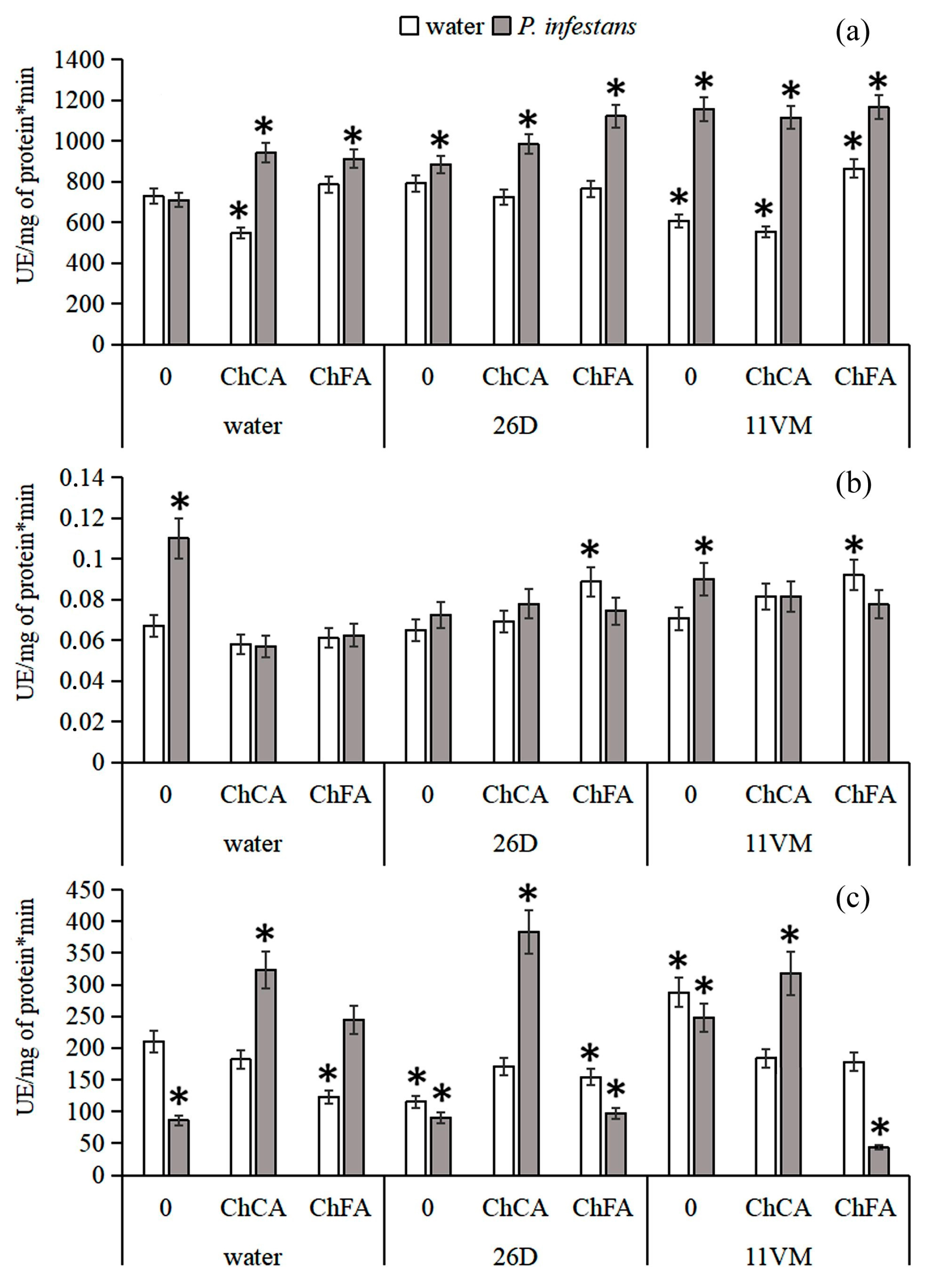
3.3. Activity of Antioxidant Enzymes

Figure 3a shows that treatment with ChFA and ChCA + B. subtilis 11VM led to decreased levels of PO activity in approximately 20–25% of all non-infected plants as compared with control ones. Infection of plants treated with ChFA, ChCA, and B. subtilis 26D increased these levels by 25%; all other combinations under investigation produced more significant effects and led to an increase in this parameter by 50% as compared with the control. The maximal level of SOD activity was observed in non-treated plants infected with P. infestans (60% higher than in the control) (Figure 3b). Combined treatment of plants with both B. subtilis strains and ChFA led to the increase in this parameter in 35% of non-infected plants, but only under the individual influence of B. subtilis 11VM was a 30% increase in SOD activity demonstrated in the infected ones. In contrast, the activity of catalase in non-treated plants consisted of less than one-half of the parameter in non-infected and non-treated plants (Figure 3c). It was found that in non-infected plants only B. subtilis 11VM promoted catalase activity. Under the influence of ChFA and B. subtilis 26D individually and in combination, activity of the enzyme was lower than in the control ones by 30–40%, and catalase activity decreasing was more significant under P. infestans inoculation.

3.4. Activity of PO Isoenzymes

It is shown (Figure 4) that in ChCA-treated uninfected potato plants, the activity of cationic isoperoxidases, especially pI~9.0 and one of the anionic isoforms (pI~4.1) of peroxidase, that is constitutively present in untreated uninfected plants, significantly decreased. Treatment of plants with ChFA or B. subtilis 26D led to significant increase in the activity of anionic isoperoxidases with pI~3.7–4.3 and cationic isoperoxidases with pI~9.0–9.3, as well as isoforms in the neutral region (with pI~7.1–7.4), compared to untreated controls. Treatment with B. subtilis 11VM stimulated the activity of isoperoxidases to a lesser extent compared with B. subtilis 26D. Combined treatment of B. subtilis 11VM + ChCA did not influence the pool of potato isoperoxidases. In contrast, B. subtilis 11VM + ChFA markedly increased the activity of most isoperoxidases under investigation, especially the activity of isoforms with pI~4.1 and pI~9.3.
In infected plants, the activity of anionic isoperoxidases with pI~3.7–4.3 and cationic isoperoxidases with pI~9.0–9.3, as well as isoforms in the neutral region (pI~7.1–7.4), increased significantly. The intensity of this pattern was the most pronounced in plants treated with B. subtilis 26D + ChCA. Thus, isoperoxidases, which activate upon infection, increased their activity in uninfected plants treated with endophytic bacteria in combination with ChFA composites and in infected plants inoculated with both bacterial strains individually and in combination with chitin hydroxycinnamates.

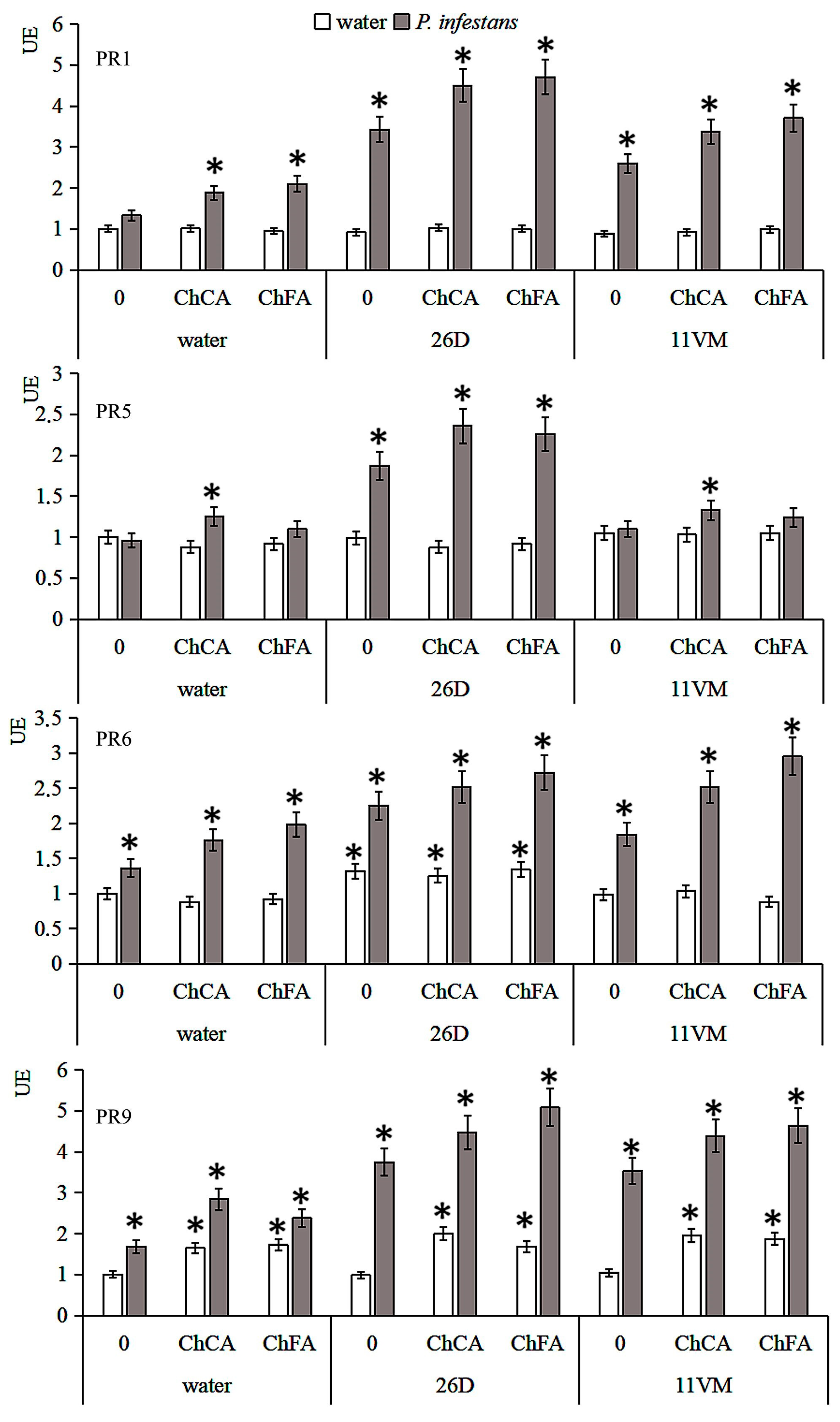
3.5. Transcriptional Activity of PR Genes

The influence of Bacillus and conjugates of chitin with hydroxycinnamates on the level of gene transcripts of StPR1, StPR5, StPR6, and StPR9, encoding the protein PR-1 (marker of the salicylate-dependent pathway), PR-5 (thaumatin-like), PR-6 (proteinase inhibitor, marker of the jasmonate-dependent pathway) and PR-9 (peroxidase), respectively, (Table 1) in intact and P. infestans infected plants was investigated. Treatment of potato seeds with cells of Bacillus spp. Strains did not influence the transcript levels of the genes under study in non-infected plants (Figure 5). The transcript level of the StPR9 gene, which encodes anionic peroxidase, unlike StPR1 and StPR6, was slightly upregulated by P. infestans inoculation. Treatments of potato plants with conjugates of chitin with hydroxycinnamates increased the transcript levels of the StPR1, StPR6, and StPR9 genes and did not alter StPR5 gene transcription in plant leaves infected with P. infestans.
Bacillus sp., especially B. subtilis 26D, stimulated transcription of StPR1 and StPR6 two-fold and StPR9 four-fold in infected plants. The StPR5 gene increased in infected Bs26D-treated plants, but did not in Bs11VM-treated plants. Treatments of plants with B. subtilis 26D strain cells and conjugates of chitin with hydroxycinnamates significantly increased the transcript levels of StPR1, StPR5, StPR6, and StPR9. Conversely, infecting plants treated with B. subtilis 11VM strain and conjugates of chitin with hydroxycinnamates with cells of P. infestans led to the increase in transcript levels of StPR1, StPR6, and StPR9, but did not influence StPR5 transcription.

4. Discussion

It was shown that the area of lesions on plant leaves caused by infection with the oomycete P. infestans decreased when plants were treated with composites of chitosan with hydroxycinnamic acids, as well as their mixtures with B. subtilis bacteria, especially in plants treated with B. subtilis 26D + ChFA (Figure 1).
The generation of ROS (superoxide O2, hydroxyl OH·, and H2O2) and cascade of subsequent defense reactions is the earliest response of the plant organism to pathogen attacks. At the same time, ROS accumulated in affected plants causes injury to the organism.
In our studies, in healthy plants treated with the ChCA composite, both separately and in a mixture with B. subtilis 26D and 11VM strains, the content of hydrogen peroxide exceeded the level of control plants (Figure 2). An increased level of H2O2 was also detected in infected plants treated with a complex of bacteria with a ChCA composite. This could be due to the protective effect of bacterial metabolites. It is known that under the action of B. subtilis, the activity of antioxidant enzymes and the level of proline in plants can increase [42]. H2O2 is involved in triggering the hyper-sensitive reaction and lignification processes and may also exhibit antimicrobial activity [43]. It is demonstrated that potato resistance to the late blight pathogen strictly demands the mechanisms related to H2O2 action [44].
Chitin, chitosan, and their oligomers are active elicitors of plant immunity, including through the regulation of the redox status of plant cells [45,46]. It is known that hydroxycinnamic acids have pronounced antioxidant properties, reducing the formation of reactive oxygen species by neutralizing free radicals [47] and activating antioxidant enzymes [48]. Under the influence of stress factors of different natures, hydroxycinnamic acids can be included in the phenylpropanoid pathway, changing the direction of the synthesis of their own derivatives [49] and generally enhancing the formation of phenolic compounds involved in the mechanisms of increasing plant resistance [50].
H2O2 can be considered as the most important molecule involved in the transmission of intracellular signals that regulate gene expression and the activity of defense systems [44], including increases in the concentration of calcium ions in the cytosol, which play an important role in the transmission of signaling information to the plant genome. It has been shown that H2O2 is involved in the activation of the expression of stress protein genes [51]. The combination of B. subtilis bacteria and chitosan composites with hydroxycinnamic acids has a modulating effect on the generation of ROS and facilitates the rapid transmission of signals that trigger other defense mechanisms to prevent the development of the pathogenic invasion.
Our investigations have revealed an antistress effect of the treatment with B. subtilis combined with ChCA on potato plants infected with P. infestans (Figure 2). Proline is considered as an important antistress metabolite, which can regulate intracellular processes. In addition, proline takes part in systemic plant defense reactions as a signaling molecule during the attacking of plants by different pathogens [31]. In our work, inoculation of plants with both endophytic strains of B. subtilis led to the accumulation of proline [32].
Proline synthesis can be induced under the influence of exogenic signaling mediators salicylic acid [52], brassinosteroids [53], and chitosan [54]. The increase in the content of proline in the leaves of infected potato plants, revealed in this study, is probably associated with both the elicitor nature of chitosan and the action of hydroxycinnamic acids [55]. It is known that increased proline synthesis and activation of its transport between cell compartments and plant tissues is one of the primary and universal plant responses to stress [56]. Changes in the concentration of H2O2 in plant tissues during pathogenesis can occur as a result of many metabolic processes, but to a greater extent, this occurs as a result of changes in the activity of antioxidant enzymes [57].
In infected plants, we observed an increase in SOD activity (Figure 3); however, treatment with both Bacillus strains + ChCA/ChFA decreased SOD activity in the affected ones. It is known that the intensity and the time of exposure to stress factors can modify SOD activity in various manners [58]. Thus, oxidative damage to plant cells and tissues can be a result of a decrease in SOD activity under severe stress influence [27]. Consequently, the utilization of ROS in infected plants is determined by a broad spectrum of other enzymes (catalases, peroxidases, and enzymes of the ascorbate–glutathione cycle) [59].
In our work, in potato plants infected with P. infestans, a decrease in catalase activity was observed compared to the control (Figure 3). However, in plants treated with composites of ChCA and ChFA, as well as in their combination with bacteria, catalase activity increased several times. Catalase activity can be significantly modified with the participation of signaling molecules. H2O2 is not only a signaling molecule, but also a catalase substrate. At the same time, its effect on catalase activity in plants is ambiguous. Thus, in wheat seedlings, H2O2 can inhibit [25] or stimulate [26] catalase activity depending on its concentration.
PO (PR-9 family) activity involves both the generation and utilization of H2O2 [60]. It is involved in the strengthening of cell walls due to the polymerization of phenolic compounds into lignin in cell walls with the participation of H2O2. It was shown that when plants were infected with the late blight pathogen, a significant activation of PO was observed in all variants of treatment, most significantly when both bacterial strains were combined with ChFA (Figure 3). The regulation of H2O2 content in potato plants under the influence of both strains of B. subtilis with ChCA and ChFA under P. infestans infection can likely occur in various ways: through a decrease in SOD activity, an increase in catalase and PO activity, the regulation of the balance of synthesis/inactivation of ROS, and the stabilization of plant metabolism. These parameters can be crucial factors for maintaining plant resistance to environmental fluctuations [61].
Plants produce a wide range of PO isoforms sensitive to various impacts [62]. For example, anionic peroxidases are involved in the formation of resistance to hypo- and hyperthermia, changes in the pH of the environment, and pathogen attacks [63,64].
Analysis of the isoenzyme spectrum of the soluble fraction of peroxidase showed that infection increased the activity of anionic POs with pI ~3.7 and 4.3 and cationic isoPOs with pI ~9.0–9.3 (Figure 5). It is interesting that the same isoPOs were activated during the joint treatment with B. subtilis 26D bacteria with the ChFA composite as during infection with P. infestans. It has been shown that cationic isoperoxidases, which have an affinity for the cell walls of the oomycete P. infestans, occupy an important place in potato responses to infection with late blight pathogen [40]. The decrease in the lesion area under P. infestans on potato leaves treated with B. subtilis 26D + ChFA may be directly related to the primed status of the plant. This set makes it possible to implement stronger and faster defense reactions against subsequent pathogen invasion, which manifests itself as a common feature of systemic resistance induced by beneficial microorganisms [65].
Plant treatment with the strains under investigation promoted the development of jasmonate-mediated ISR, as could be identified by the high rate of expression of the PR-6 gene, which is considered as marker of ISR in plants [66]. It is worth noting that the development of SAR occurs when a plant recognizes a biotrophic pathogen or its elicitors, which leads to the synthesis of salicylate-dependent PR proteins [67], including the expression of the PR-1 gene (marker of SAR) [68].
It should be noted that in infected plants pretreated with B. subtilis 26D and 11VM together with ChCA and CaFA composites, a high level of transcriptional activity of the main antimicrobial protein PR-1 and the protease inhibitor PR-6 genes was observed. It is possible that in these plants, salicylate- and jasmonate-dependent pathways occurred simultaneously. In infected plants treated with B. subtilis 26D in combination with ChFA, the transcriptional activity of the thaumatin-like protein (PR-5) and PO (PR-9) genes significantly increased. It is believed that the activity of proteins of the PR-5 family is associated with an increase in membrane permeability [69]. Infected plants treated with both B. subtilis strains under investigation in combination with ChFA significantly increased their expression of the PR-9 gene, which correlates with their resistance. Thus, the induction of plant resistance mediated by the Bacillus strains under consideration can be characterized by a multifaceted priming process, including the expression of PR genes and fine-tuning regulation of the redox status of plants [70].

5. Conclusions

Thus, joint treatment of plants with bacterial strains B. subtilis 26D and 11VM with conjugates of chitosan with caffeic and ferulic acids led to an increase in potato plants’ resistance to the late blight pathogen. This status involved the activation of CAT, PO, and SOD, accumulation of H2O2, and promotion of the expression of genes encoding the PR-1, PR-5, PR-6, and PR-9 proteins. The revealed activation of the expression of the PR-1 (systemic acquired resistance marker) and PR-6 (induced systemic resistance marker) genes in plants treated with endophytic B. subtilis 26D and 11VM and chitosan composites with hydroxycinnamates indicates that, in these cases, the different branches of protective reactions in potato plants against the pathogen proceed synergistically. It is possible that endophytes prime plant defense genes, and chitosan composites initiate their expression. The mechanism of the influence of ChCA and ChFA on the formation of resistance to the pathogen is determined by the composition (polymer–antioxidant) and the structure of the conjugate itself. Due to the polymer matrix, a prolonged effect of hydroxycinnamic acids on the functional state of the plant cell is probably provided.

Supplementary Materials

The following supporting information can be downloaded at: https://www.mdpi.com/article/10.3390/microorganisms11081993/s1, Table S1: The experimental groups, demonstrating significant differences by Duncan’s test.

Author Contributions

Conceptualization, L.Y. and J.N.K.; methodology, A.V.S. and E.A.C.; investigation, A.V.S., E.A.C., G.F.B., E.A.Z. and V.V.N.; resources, I.S.M., V.V.N. and N.A.Y.; writing—original draft preparation, A.V.S.; writing—review and editing, V.O.T., I.Y.F. and L.Y.; visualization, I.Y.F.; project administration, L.Y. and J.N.K.; funding acquisition, L.Y. All authors have read and agreed to the published version of the manuscript.

Funding

This research was funded by the Russian Scientific Foundation, grant number 23-16-00139.

Data Availability Statement

Not applicable.

Acknowledgments

The authors are grateful to the staffs of the “Biomika” Shared Access Centre (Branch of Biochemical Methods and Nanobiotechnology, “Agidel” Resource Centre for Collective Use) and the “KODINK” Complex of Equipment for the Study of Nucleic Acids for access to the equipment. The authors are grateful to the head of the Laboratory of Plant–Microbial Interactions of the Bashkir Research Institute of Agriculture UFRC RAS, Ludmila Pusenkova, for the kindly provided pathogenic cultures.

Conflicts of Interest

The authors declare no conflict of interest.

References

  1. Rkhaila, A.; Chtouki, T.; Erguig, H.; El Haloui, N.; Ounine, K. Chemical Proprieties of Biopolymers (Chitin/Chitosan) and Their Synergic Effects with Endophytic Bacillus Species: Unlimited Applications in Agriculture. Molecules 2021, 26, 1117. [Google Scholar] [CrossRef]
  2. Kumawat, K.C.; Razdan, N.; Saharan, K. Rhizospheric microbiome: Bio-based emerging strategies for sustainable agriculture development and future perspectives. Microbiol. Res. 2022, 254, 126901. [Google Scholar] [CrossRef]
  3. Pinski, A.; Betekhtin, A.; Hupert-Kocurek, K.; Mur, L.A.J.; Hasterok, R. Defining the Genetic Basis of Plant-Endophytic Bacteria Interactions. Int. J. Mol. Sci. 2019, 20, 1947. [Google Scholar] [CrossRef] [Green Version]
  4. Sharma, M.; Mallubhotla, S. Diversity, Antimicrobial Activity, and Antibiotic Susceptibility Pattern of Endophytic Bacteria Sourced from Cordia dichotoma L. Front. Microbiol. 2022, 13, 879386. [Google Scholar] [CrossRef] [PubMed]
  5. Chakraborty, N.; Mitra, R.; Pal, S.; Ganguly, R.; Acharya, K.; Minkina, T.; Sarkar, A.; Keswani, C. Biopesticide Consumption in India: Insights into the Current Trends. Agriculture 2023, 13, 557. [Google Scholar] [CrossRef]
  6. Chandler, D.; Bailey, A.S.; Tatchell, G.M.; Davidson, G.; Greaves, J.; Grant, W.P. The development, regulation and use of biopesticides for integrated pest management. Philos. Trans. R. Soc. Lond. B Biol. Sci. 2011, 366, 1987–1998. [Google Scholar] [CrossRef]
  7. Kocięcka, J.; Liberacki, D. The Potential of Using Chitosan on Cereal Crops in the Face of Climate Change. Plants 2021, 10, 1160. [Google Scholar] [CrossRef] [PubMed]
  8. Maurya, A.; Prasad, J.; Das, S.; Dwivedy, A.K. Essential Oils and Their Application in Food Safety. Front. Sustain. Food Syst. 2021, 5, 653420. [Google Scholar] [CrossRef]
  9. Fitza, K.N.E.; Payn, K.G.; Steenkamp, E.T.; Myburg, A.A.; Naidoo, S. Chitosan Application Improves Resistance to Fusarium circinatum in Pinus patula. S. Afr. J. Bot. 2013, 85, 70–78. [Google Scholar] [CrossRef] [Green Version]
  10. Gonçalves, C.; Ferreira, N.; Lourenço, L. Production of Low Molecular Weight Chitosan and Chitooligosaccharides (COS): A Review. Polymers 2021, 13, 2466. [Google Scholar] [CrossRef]
  11. Aranaz, I.; Alcántara, A.R.; Civera, M.C.; Arias, C.; Elorza, B.; Heras Caballero, A.; Acosta, N. Chitosan: An Overview of Its Properties and Applications. Polymers 2021, 13, 3256. [Google Scholar] [CrossRef]
  12. Benchamas, G.; Huang, G.L.; Huang, S.Y.; Huang, H.L. Preparation and biological activities of chitosan oligosaccharides. Trends Food Sci. Tech. 2021, 107, 38–44. [Google Scholar] [CrossRef]
  13. Yang, G.S.; Sun, H.H.; Cao, R.; Liu, Q.; Mao, X.Z. Characterization of a novel glycoside hydrolase family 46 chitosanase, Csn-BAC, from Bacillus sp. MD-5. Int. J. Biol. Macromol. 2020, 146, 518–523. [Google Scholar] [CrossRef]
  14. Palazzini, J.; Reynoso, A.; Yerkovich, N.; Zachetti, V.; Ramírez, M.; Chulze, S. Combination of Bacillus velezensis RC218 and Chitosan to Control Fusarium Head Blight on Bread and Durum Wheat under Greenhouse and Field Conditions. Toxins 2022, 14, 499. [Google Scholar] [CrossRef]
  15. Ahmed, A.S.; Ezziyyani, M.; Sánchez, C.P.; Candela, M.E. Effect of chitin on biological control activity of Bacillus spp. and Trichoderma harzianum against root rot disease in pepper (Capsicum annuum) plants. Eur. J. Plant Pathol. 2003, 109, 633–637. [Google Scholar] [CrossRef]
  16. Brzezinska, M.S.; Kalwasińska, A.; Świątczak, J.; Żero, K.; Jankiewicz, U. Exploring the properties of chitinolytic Bacillus isolates for the pathogens biological control. Microb. Pathog. 2020, 148, 104462. [Google Scholar] [CrossRef]
  17. Agbodjato, N.A.; Noumavo, P.A.; Adjanohoun, A.; Agbessi, L.; Baba-Moussa, L. Synergistic effects of plant growth promoting rhizobacteria and chitosan on in vitro seeds germination, greenhouse growth, and nutrient uptake of maize (Zea mays L.). Biotechnol. Res. Int. 2016, 2016, 7830182. [Google Scholar] [CrossRef]
  18. Chen, Q.; Qi, Y.; Jiang, Y.; Quan, W.; Luo, H.; Wu, K.; Li, S.; Ouyang, Q. Progress in Research of Chitosan Chemical Modification Technologies and Their Applications. Mar. Drugs 2022, 20, 536. [Google Scholar] [CrossRef]
  19. Di Santo, M.C.; D’Antoni, C.L.; Domínguez Rubio, A.P.; Alaimo, A.; Pérez, O.E. Chitosan-tripolyphosphate nanoparticles designed to encapsulate polyphenolic compounds for biomedical and pharmaceutical applications–A review. Biomed. Pharmacother. 2021, 142, 111970. [Google Scholar] [CrossRef]
  20. Qi, J.; Song, C.P.; Wang, B.; Zhou, J.; Kangasjärvi, J.; Zhu, J.K.; Gong, Z. Reactive oxygen species signaling and stomatal movement in plant responses to drought stress and pathogen attack. J. Integr. Plant Biol. 2018, 60, 805–826. [Google Scholar] [CrossRef] [Green Version]
  21. Fortunato, S.; Lasorella, C.; Dipierro, N.; Vita, F.; de Pinto, M.C. Redox Signaling in Plant Heat Stress Response. Antioxidants 2023, 12, 605. [Google Scholar] [CrossRef] [PubMed]
  22. Haider, S.; Iqbal, J.; Naseer, S.; Yaseen, T.; Shaukat, M.; Bibi, H.; Ahmad, Y.; Daud, H.; Abbasi, N.L.; Mahmood, T. Molecular Mechanisms of Plant Tolerance to Heat Stress: Current Landscape and Future Perspectives. Plant Cell Rep. 2021, 40, 2247–2271. [Google Scholar] [CrossRef] [PubMed]
  23. Rajput, V.D.; Harish; Singh, R.K.; Verma, K.K.; Sharma, L.; Quiroz-Figueroa, F.R.; Meena, M.; Gour, V.S.; Minkina, T.; Sushkova, S.; et al. Recent Developments in Enzymatic Antioxidant Defence Mechanism in Plants with Special Reference to Abiotic Stress. Biology 2021, 10, 267. [Google Scholar] [CrossRef] [PubMed]
  24. Mahomoodally, M.F.; Daphne Désiré, A.-L.; Elodie Rosette, M.A.-L. Chapter 2.2–Catalase. In Antioxidants Effects in Health; Seyed, M.N., Silva, A.S., Eds.; Elsevier: Amsterdam, The Netherlands, 2022; pp. 81–90. [Google Scholar] [CrossRef]
  25. Bakalova, S.; Nikolova, A.; Wedera, D. Isoenzyme profiles of peroxidase, catalase and superoxide dismutase as affected by dehydration stress and ABA during germination of wheat seeds. J. Plant Physiol. 2004, 30, 64–77. [Google Scholar] [CrossRef]
  26. Luna, C.M.; Pastori, G.M.; Driscoll, S.; Groten, K.; Bernard, S.; Foyer, C.H. Drought controls on H2O2 accumulation, catalase (CAT) activity and CAT gene expression in wheat. J. Exp. Bot. 2005, 56, 417–423. [Google Scholar] [CrossRef] [Green Version]
  27. Jiang, Y.; Huang, B. Drought and Heat Stress Injury to Two Cool-Season Turfgrasses in Relation to Antioxidant Metabolism and Lipid Peroxidation. Crop. Sci. 2001, 41, 436–442. [Google Scholar] [CrossRef]
  28. Fornera, S.; Walde, P. Spectrophotometric quantification of horseradish peroxidase with o-phenylenediamine. Anal. Biochem. 2010, 407, 293–295. [Google Scholar] [CrossRef]
  29. Karthika, S.; Remya, M.; Varghese, S.; Dhanraj, N.D.; Sali, S.; Rebello, S.; Jose, S.M.; Jisha, M.S. Bacillus tequilensis PKDN31 and Bacillus licheniformis PKDL10-As double headed swords to combat Fusarium oxysporum f. sp. lycopersici induced tomato wilt. Microb. Pathog. 2022, 172, 105784. [Google Scholar] [CrossRef]
  30. Samaras, A.; Kamou, N.; Tzelepis, G.; Karamanoli, K.; Menkissoglu-Spiroudi, U.; Karaoglanidis, G.S. Root Transcriptional and Metabolic Dynamics Induced by the Plant Growth Promoting Rhizobacterium (PGPR) Bacillus subtilis Mbi600 on Cucumber Plants. Plants 2022, 11, 1218. [Google Scholar] [CrossRef]
  31. Fabro, G.; Kovács, I.; Pavel, V.; Szabados, L.; Alvarez, M.E. Proline accumulation and AtP5CS2 gene activation are induced by plant-pathogen incompatible interactions in Arabidopsis. Mol. Plant Microb. Intrract. 2004, 17, 343–350. [Google Scholar] [CrossRef] [Green Version]
  32. Lastochkina, O.; Seifikalhor, M.; Aliniaeifard, S.; Baymiev, A.; Pusenkova, L.; Garipova, S.; Kulabuhova, D.; Maksimov, I. Bacillus Spp.: Efficient Biotic Strategy to Control Postharvest Diseases of Fruits and Vegetables. Plants 2019, 8, 97. [Google Scholar] [CrossRef] [Green Version]
  33. Sorokan, A.; Cherepanova, E.; Burkhanova, G.; Veselova, S.; Rumyantsev, S.; Alekseev, V.; Mardanshin, I.; Sarvarova, E.; Khairullin, R.; Benkovskaya, G.; et al. Endophytic Bacillus spp. as a Prospective Biological Tool for Control of Viral Diseases and Non-vector Leptinotarsa decemlineata Say. in Solanum tuberosum L. Front. Microbiol. 2020, 11, 569457. [Google Scholar] [CrossRef]
  34. Catalog of Strains and Isolates of the Collection of Endophytic Microorganisms IBG UFRC RAS. Available online: http://ibg.anrb.ru/wp-content/uploads/2019/04/Katalog-endofit.doc (accessed on 25 June 2023). (In Russian).
  35. Yarullina, L.G.; Burkhanova, G.F.; Cherepanova, E.A.; Sorokan, A.V.; Zaikina, E.A.; Tsvetkov, V.O.; Mardanshin, I.S.; Kalatskaya, J.N.; Balyuk, N.V. Effect of Bacillus subtilis and signaling molecules on the state of the pro/antioxidant system and the expression of protective protein genes in potato plants upon phytophthorosis and a moisture deficit. Appl. Biochem. Microbiol. 2021, 57, 760–769. [Google Scholar] [CrossRef]
  36. Kraskouski, A.; Nikalaichuk, V.; Kulikouskaya, V.; Hileuskaya, K.; Kalatskaja, J.; Nedved, H.; Laman, N.; Agabekov, V. Synthesis and properties of hydrogel particles based on chitosan-ferulic acid conjugates. Soft Mater. 2021, 19, 495–502. [Google Scholar] [CrossRef]
  37. Bindschedler, L.V.; Minibayeva, F.; Gardner, S.L.; Gerrish, C.; Davies, D.R.; Bolwell, G.P. Early signalling events in the apoplastic oxidative burst in suspension cultured French bean cells involve cAMP and Ca2+. New Phytol. 2001, 151, 185–194. [Google Scholar] [CrossRef]
  38. Bonnet, M.; Camares, O.; Veisseire, P. Effect of Zinc and influence of Acromonium lolli on growth parameters, chlorophyll fluorescence and antioxidant enzyme activities of rye grass (Lolium perenne L. cv. Apollo). J. Exp. Bot. 2000, 51, 945–953. [Google Scholar]
  39. Minibayeva, F.; Kolesnikov, O.; Chasov, A.; Beckett, R.P.; Lüthje, S.; Vylegzhanina, N.; Buck, F.; Böttger, M. Wound-induced apoplastic peroxidase activities: Their roles in the production and detoxification of reactive oxygen species. Plant Cell Environ. 2009, 32, 497–508. [Google Scholar] [CrossRef]
  40. Maksimov, I.V.; Sorokan, A.V.; Cherepanova, E.A.; Surina, O.B.; Troshina, N.B.; Yarullina, L.G. Effects of salicylic and jasmonic acids on the components of pro/antioxidant system in potato plants infected with late blight. Russ. J. Plant Physiol. 2011, 58, 299–306. [Google Scholar] [CrossRef]
  41. Kruger, N.J. The Bradford Method for Protein Quantitation. In The Protein Protocols Handbook; Springer Protocols Handbooks; Walker, J.M., Ed.; Humana Press: Totowa, NJ, USA, 2009; pp. 17–24. [Google Scholar]
  42. Lastochkina, O.V.; Aliniaeifard, S.; Seifikalhor, M.; Yuldashev, R.; Pusenkova, L.; Garipova, S. Plant growth promoting bacteria–Biotic strategy to cope with abiotic stresses in wheat. In Wheat Production in Changing Environments. Responses, Adaptation and Tolerance; Hasanuzzaman, M., Nahar, K., Hossain, A., Eds.; Springer: Singapore, 2019; pp. 579–614. [Google Scholar]
  43. Bolwell, G.P.; Bindschedler, L.V.; Blee, K.A.; Butt, V.S.; Davies, D.R.; Gardner, S.L.; Gerrish, C.; Minibayeva, F. The apoplastic oxidative burst in response to biotic stress in plants: A tree component system. J. Exp. Bot. 2002, 53, 1367–1376. [Google Scholar]
  44. Galvez-Valdivieso, G.; Fryer, M.J.; Lawson, T.; Slattery, K.; Truman, W.; Smirnoff, N.; Asami, T.; Davies, W.J.; Jones, A.M.; Baker, N.R. The High Light Response in Arabidopsis Involves ABA Signaling between Vascular and Bundle Sheath Cells. Plant Cell 2009, 21, 2143–2162. [Google Scholar] [CrossRef] [Green Version]
  45. Jia, X.; Rajib, M.; Yin, H. Recognition Pattern, Functional Mechanism and Application of Chitin and Chitosan Oligosaccharides in Sustainable Agriculture. Curr. Pharm. Des. 2020, 26, 3508–3521. [Google Scholar] [CrossRef] [PubMed]
  46. Chakraborty, M.; Hasanuzzaman, M.; Rahman, M.; Khan, M.D.; Bhowmik, P.; Mahmud, N.U.; Tanveer, M.; Islam, T. Mechanism of Plant Growth Promotion and Disease Suppression by Chitosan Biopolymer. Agriculture 2020, 10, 624. [Google Scholar] [CrossRef]
  47. Siquet, C.; Paiva-Martins, F.; Lima, J.L.; Reis, S.; Borges, F. Antioxidant profile of dihydroxy- and trihydroxyphenolic acids–A structure–activity relationship study. Free Radic. Res. 2006, 40, 433–442. [Google Scholar] [CrossRef] [PubMed]
  48. Rivero, R.M.; Ruiz, J.M.; Garcia, P.C.; Lopez-Lefebre, L.R.; Sanchez, E.; Romero, L. Resistance to Cold and Heat Stress: Accumulation of Phenolic Compounds in Tomato and Watermelon Plants. Plant Sci. 2001, 160, 315–321. [Google Scholar] [CrossRef] [PubMed]
  49. Krol, A.; Amarowicz, R.; Weidner, S. Changes in the composition of phenolic compounds and antioxidant properties of grapevine roots and leaves (Vitis vinifera L.) under continuous of long-term drought stress. Acta Physiol. Plant. 2014, 36, 1491–1499. [Google Scholar] [CrossRef] [Green Version]
  50. Budna, G.A.; Lima, R.B.; Zanardo, D.Y.; dos Santos, W.D.; Ferrarese, M.; Ferrarese-Filho, O. Exogenous caffeic acid inhibits the growth and enhances the lignification of the roots of soybean (Glycine max). J. Plant Physiol. 2011, 168, 1627–1633. [Google Scholar] [CrossRef]
  51. Pfannschmidt, T.; Bräutigam, K.; Wagner, R.; Dietzel, L.; Schröter, Y. Potential regulation of gene expression in photosynthetic cells by redox and energy state: Approaches towards better understanding. Ann. Bot. 2009, 103, 599–607. [Google Scholar] [CrossRef] [Green Version]
  52. Mishra, S.; Dubey, R.S. Inhibition of ribonuclease and protease activities in arsenic exposed rice seedlings: Role of proline as enzyme protectant. J. Plant Physiol. 2006, 163, 927–936. [Google Scholar] [CrossRef]
  53. Muley, A.B.; Shingote, P.R.; Patil, A.P.; Dalvi, S.G.; Suprasanna, P. Gamma radiation degradation of chitosan for application in growth promotion and induction of stress tolerance in potato (Solanum tuberosum L.). Carbohydr. Polym. 2019, 210, 289–301. [Google Scholar] [CrossRef]
  54. Rahman, M.; Mukta, J.A.; Sabir, A.A.; Gupta, D.R.; Mohi-Ud-Din, M.; Hasanuzzaman, M.; Miah, M.G.; Rahman, M.; Islam, M.T. Chitosan biopolymer promotes yield and stimulates accumulation of antioxidants in strawberry fruit. PLoS ONE 2018, 13, e0203769. [Google Scholar] [CrossRef]
  55. Puzina, T.I.; Makeeva, I.Y. Participation of caffeic acid in regulation of the production process in Solanum tuberosum L. Agrochemistry 2015, 6, 53–58. (In Russian) [Google Scholar]
  56. Kolupaev, Y.E.; Karpets, Y.V.; Kabashnikova, L.F. Antioxidative system of plants: Cellular compartmentalization, protective and signaling functions, mechanisms of regulation (review). Appl. Biochem. Microbiol. 2019, 55, 441–459. [Google Scholar] [CrossRef]
  57. Smirnoff, N.; Arnaud, D. Hydrogen peroxide metabolism and functions in plants. New Phytol. 2019, 221, 1197–1214. [Google Scholar] [CrossRef] [Green Version]
  58. Rizhsky, L.; Liang, H.; Mittler, R. The water-water cycle is essential for chloroplast protection in the absence of stress. J. Biol. Chem. 2003, 278, 38921–38925. [Google Scholar] [CrossRef] [Green Version]
  59. Pradedova, E.V.; Isheeva, O.D.; Salyaev, R.K. Antioxidant defense enzymes in cell vacuoles of red beet roots. Russ. J. Plant Physiol. 2011, 58, 36–44. [Google Scholar] [CrossRef]
  60. Gazaryan, I.G.; Khushpulyan, D.M.; Tishkov, V.I. Structure and mechanism of action of plant peroxidases. Adv. Biol. Chem. 2006, 46, 303–322. (In Russian) [Google Scholar]
  61. Sathyamurthy, B.; Dama, G.; Balasubramanian, A.; Vvs, S. Evaluation on antioxidant activity of curry leaf extracts in cisplatin induced wistar rats. World J. Pharm. Res. 2015, 4, 927–939. [Google Scholar]
  62. Cosio, C.; Dunand, C. Specific functions of individual class III peroxidase genes. J. Exp. Bot. 2009, 60, 391–408. [Google Scholar] [CrossRef]
  63. Spoel, S.H.; Johnson, J.S.; Dong, X. Regulation of tradeoffs between plant defenses against pathogens with different lifestyles. Proc. Nat. Acad. Sci. USA 2007, 104, 18842–18847. [Google Scholar] [CrossRef]
  64. Marjamaa, K.; Kukkola, E.M.; Fagerstedt, K.V. The role of xylem class III peroxidases in lignification. J. Exp. Bot. 2009, 60, 367–376. [Google Scholar] [CrossRef]
  65. Conrath, U.; Beckers, G.J.M.; Flors, V.; García-Agustín, P.; Jakab, G.; Mauch, F.; Newman, M.-A.; Pieterse, C.M.J.; Poinssot, B.; Pozo, M.J. Priming: Getting ready for battle. Mol. Plant Microbe Interact. 2006, 19, 1062–1071. [Google Scholar] [CrossRef] [PubMed] [Green Version]
  66. Veselova, S.V.; Nuzhnaya, T.V.; Maksimov, I.V. Role of Jasmonic Acid in Interaction of Plants with Plant Growth Promoting Rhizobacteria during Fungal Pathogenesis. In Jasmonic Acid: Biosynthesis, Functions and Role in Plant Development, Series Plant Science Research and Practices; Morrison, L., Ed.; Nova Sci. Publishers: Hauppauge, NY, USA, 2015; pp. 33–66. [Google Scholar]
  67. Glazebrook, J. Contrasting Mechanisms of Defense Against Biotrophic and Necrotrophic Pathogens. Annu. Rev. Phytopathol. 2005, 43, 205. [Google Scholar] [CrossRef]
  68. Gimenez-Ibanez, S.; Solano, R. Nuclear jasmonate and salicylate signaling and crosstalk in defense against pathogens. Front. Plant Sci. 2013, 4, 72. [Google Scholar] [CrossRef] [PubMed] [Green Version]
  69. Vasyukova, N.I.; Ozeretskovskaya, O.L. Jasmonate-dependent defense signaling in plant tissues. Russ. J. Plant Physiol. 2009, 56, 581–590. [Google Scholar] [CrossRef]
  70. Martinez-Medina, A.; Flors, V.; Heil, M.; Mauch-Mani, B.; Corné, M.J.; Pieterse, C.M.J. Recognizing Plant Defense Priming. Trends Plant Sci. 2016, 21, 818–822. [Google Scholar] [CrossRef] [Green Version]
Figure 1. The influence of conjugates of chitin oligomers with caffeic (ChCA) and ferulic (ChFA) acids in compositions with B. subtilis 26D and B. subtilis 11VM on late blight disease symptoms on potato leaves on the 10th day after P. infestans inoculation. Values marked by an asterisk are significantly different from the control as measured by the Kruskal–Wallis test.
Microorganisms 11 01993 g001
Figure 2. The influence of conjugates of chitin oligomers with caffeic (ChCA) and ferulic (ChFA) acids in compositions with B. subtilis 26D and B. subtilis 11VM on the content of H2O2 (a) and proline (b) in potato plants on the 3rd day after P. infestans inoculation. Values marked by an asterisk are significantly different from the control as measured by the Kruskal–Wallis test.
Microorganisms 11 01993 g002
Figure 3. The influence of conjugates of chitin oligomers with caffeic (ChCA) and ferulic (ChFA) acids in compositions with B. subtilis 26D and B. subtilis 11VM on the activity of peroxidase (a), SOD (b), and catalase (c) in potato plants on the 3rd day after P. infestans inoculation. Values marked by an asterisk are significantly different from the control as measured by the Kruskal–Wallis test.
Microorganisms 11 01993 g003
Figure 4. The influence of conjugates of chitin oligomers with caffeic (ChCA) and ferulic (ChFA) acids in compositions with water (a), B. subtilis 26D (b), and B. subtilis 11VM (c) on the activity of different isoforms of peroxidase on the 3rd day after P. infestans inoculation. Arrows indicate important peaks on densitograms.
Microorganisms 11 01993 g004
Figure 5. The influence of conjugates of chitin oligomers with caffeic (ChCA) and ferulic (ChFA) acids in compositions with B. subtilis 26D and B. subtilis 11VM on the content of potato gene transcripts encoding PR1, PR5, PR6, and PR9 proteins on the 3rd day after P. infestans inoculation. Expression values were normalized to the housekeeping gene as an internal reference and expressed relative to the normalized expression levels in control plants. Values marked by an asterisk are significantly different from the control as measured by the Kruskal–Wallis test.
Microorganisms 11 01993 g005
Table 1. Primers used in PCR for investigation of the activity of genes under study.
Gene ProductsGenesGenBank Accession NumberSequence (5′-3′)
Forward PrimersReverse Primer
ActinStActX55749gat-ggt-gtc-agc-cac-acatt-cca-gca-gct-tcc-att-cc
PR1StPR1AY050221tgg-gtg-gtg-gtt-cat-ttc-ttg-tcat-tta-att-cct-tac-aca-tca-taa-g
Thaumatin-like, PR5StPR5AY737317ccc-gtt-tga-cat-tga-cct-ttgcga-ata-cgg-tgg-aac-atg-ga
Proteinase inhibitor, PR6StPR6JX683427ggg-aaa-gaa-tat-gct-caa-gtt-ataat-tct-cca-tca-tct-tcc-act-g
Peroxidase, PR9StPR9M21334gta-atc-ctg-ccg-cac-aac-tgca-gca-aaa-tct-cca-agg-aa
Disclaimer/Publisher’s Note: The statements, opinions and data contained in all publications are solely those of the individual author(s) and contributor(s) and not of MDPI and/or the editor(s). MDPI and/or the editor(s) disclaim responsibility for any injury to people or property resulting from any ideas, methods, instructions or products referred to in the content.

