Article

Association of Virulence, Biofilm, and Antimicrobial Resistance Genes with Specific Clonal Complex Types of Listeria monocytogenes

1
Department of Biological Sciences, Munster Technological University, Bishopstown, T12 P928 Cork, Ireland
2
Teagasc Food Research Centre, Moorepark, Fermoy, Co., P61 C996 Cork, Ireland
*
Author to whom correspondence should be addressed.
Microorganisms 2023, 11(6), 1603; https://doi.org/10.3390/microorganisms11061603
Submission received: 28 April 2023 / Revised: 14 June 2023 / Accepted: 15 June 2023 / Published: 17 June 2023
(This article belongs to the Special Issue An Update on Listeria monocytogenes 2.0)

Abstract

:
Precise classification of foodborne pathogen Listeria monocytogenes is a necessity in efficient foodborne disease surveillance, outbreak detection, and source tracking throughout the food chain. In this study, a total of 150 L. monocytogenes isolates from various food products, food processing environments, and clinical sources were investigated for variations in virulence, biofilm formation, and the presence of antimicrobial resistance genes based on their Whole-Genome Sequences. Clonal complex (CC) determination based on Multi-Locus Sequence Typing (MLST) revealed twenty-eight CC-types including eight isolates representing novel CC-types. The eight isolates comprising the novel CC-types share the majority of the known (cold and acid) stress tolerance genes and are all genetic lineage II, serogroup 1/2a-3a. Pan-genome-wide association analysis by Scoary using Fisher’s exact test identified eleven genes specifically associated with clinical isolates. Screening for the presence of antimicrobial and virulence genes using the ABRicate tool uncovered variations in the presence of Listeria Pathogenicity Islands (LIPIs) and other known virulence genes. Specifically, the distributions of actA, ecbA, inlF, inlJ, lapB, LIPI-3, and vip genes across isolates were found to be significantly CC-dependent while the presence of ami, inlF, inlJ, and LIPI-3 was associated with clinical isolates specifically. In addition, Roary-derived phylogenetic grouping based on Antimicrobial-Resistant Genes (AMRs) revealed that the thiol transferase (FosX) gene was present in all lineage I isolates, and the presence of the lincomycin resistance ABC-F-type ribosomal protection protein (lmo0919_fam) was also genetic-lineage-dependent. More importantly, the genes found to be specific to CC-type were consistent when a validation analysis was performed with fully assembled, high-quality complete L. monocytogenes genome sequences (n = 247) extracted from the National Centre for Biotechnology Information (NCBI) microbial genomes database. This work highlights the usefulness of MLST-based CC typing using the Whole-Genome Sequence as a tool in classifying isolates.

1. Introduction

L. monocytogenes is an opportunistic, Gram-positive, food-borne pathogen that causes listeriosis, which is a major concern from a food safety and public health perspective. Elderly and immunocompromised people may have greater hospitalisation with sepsis, meningitis, and meningoencephalitis as well as higher fatality rates (13% in 2020) [1] due to the disease. The pathogen can cause symptoms such as fever, muscle aches, and gastrointestinal symptoms such as nausea and diarrhoea. While severe disease is rare, in pregnant women, it can cause miscarriage, stillbirth, preterm labour, sepsis, or meningitis in new-borns. L. monocytogenes infections are caused mainly through the consumption of ready-to-eat foods, such as dairy products, processed meat, fish, and fresh produce. This problem is compounded by the microbe’s propensity to survive/grow in stressful conditions (acidic environments, high salt concentrations, and low temperatures), which are commonly used in the food industry to control microbial safety.
The capability of L. monocytogenes to proliferate at lower temperatures [2,3] and lower pH [4,5], the resistance to commonly used disinfectants [6] in the food industry, and the ability to form biofilms [7] is primarily responsible for their stress survival and persistence. One of the key problems in microbial risk profiling is strain variation. To distinguish between strains, multiple typing approaches such as serotyping, Multi-Locus Sequence Typing (MLST), and various fragment-based typing methods have been employed. Subtyping allows researchers and public health officials to differentiate between different strains of L. monocytogenes, which can help with identifying the source of an outbreak and implementing appropriate control measures. In addition, studies of the genetic diversity of L. monocytogenes have helped shed light on key aspects of virulence, persistence, and environmental stress adaptation.
Traditional molecular typing methods such as serotyping and phage typing are labour-intensive, time-consuming, and can produce inconsistent results between methods [5]. They also have a lower resolution compared to genomic analysis with next-generation sequencing (NGS) technology [8,9,10]. MLST based on whole-genome sequences (WGSs) is robust, convenient, and efficient [11] and has been utilised in epidemiological research [12], pathogen surveillance, and food-related stress tolerance investigations [5,13]. In addition, high-throughput Real-Time PCR methods have recently been developed that allow for the rapid determination of L. monocytogenes isolate CC-type [14]. Increasingly accessible pan-genome annotation software such as Prokka v1.13.4 [15] and Prokaryotic Genome Annotation Pipeline (PGAP) [16] makes it possible to efficiently and consistently characterise the specific genotype. Furthermore, pan-genome comparative genomic studies offer more insights on phylogeny, virulence, and other differentiating characteristics to help with the better management of this foodborne illness [17,18,19].
Undissociated acid [5], salt, and pH tolerance [20] have already been described for the isolates in this study. However, the differences in genetic makeup related to the critical characteristics of virulence, biofilm formation, and antimicrobial resistance had not been reported with this set of isolates. The aim of this work was to perform comparative genomic analysis of the 150 L. monocytogenes isolates from food, food processing environments, and clinical sources to determine specific genetic markers associated with virulence markers, their ability to form biofilms, and antimicrobial resistance based on WGS data. More importantly, associations between CC-type and genetic markers were validated using additional genomes extracted from NCBI.

2. Materials and Methods

2.1. Strains

A total of 150 L. monocytogenes isolates from various food production and clinical sources (Figure 1) were sourced from the Listeria collection at Teagasc Food Research Centre, Moorepark, Co Cork. All isolates used in this study were previously published by Myintzaw et al. in 2022 [5]. In addition, reference strains of L. monocytogenes, EGDe, 10403S [21], 6179 [22], and F2365 [23] were included.

2.2. Whole-Genome Sequences

For each isolate (n = 150), raw reads in fastq format obtained from paired-end libraries and Illumina MiSeq sequencing were available from a previous study [5]. The raw reads were quality-checked and the adaptors were removed using the Trimmomatic tool [24] available on the online platform Galaxy (https://usegalaxy.org/, (accessed on 21 March 2021)) [25]. Genome assemblies were performed using SPAdes [26], also available on the Galaxy online platform (https://usegalaxy.org/, (accessed on 29 March 2021)).

2.3. Pan-Genome Analysis

Prokka was employed to annotate all of the genomes in the UseGalaxy.org online platform using default parameters, and the resultant gff3 files were used for pan-genome analysis with Roary 3.13.0 version [27], available at (http://sanger-pathogens.github.io/Roary/, (accessed on 24 October 2021)). The presence or absence of the pan-genome gene file generated by Roary was compared with the source of isolates in Scoary (Gene-wise counting and Fisher’s exact tests for trait) to establish gene clusters associated with each trait [28], available at (https://github.com/AdmiralenOla/Scoary, (accessed on 21 January 2022)).

2.4. Genotypic Characterisation

Tools available on the Centre for Genomic Epidemiology’s (CGE) online platform (https://cge.cbs.dtu.dk/services/, accessed on 12 December 2022) were used to perform the genotypic characterisation of the isolates utilising core genome cgMLST (cgMLSTFinder) typing based on single-nucleotide polymorphisms at 1748 loci. MLST typing was also performed, which is based on the nucleotide variation in seven conserved genes, the presence of plasmids (PlasmidFinder), and the presence of virulence genes (VirulenceFinder) on WGS data of each of the isolates [29]. The resultant cgMLST data files were organised in a matrix used as an input for visualisation of the loci variation and phylogeny construction based on the method outlined on the Norwegian Veterinary Institute (NVI) github page (https://norwegianveterinaryinstitute.github.io/BioinfTraining/R_trees.html, (accessed on 3 May 2021)) with slight modifications. Briefly, 1748 loci-based similarities were calculated from cgMLST files and a sequential comparison was performed to create a symmetrical dissimilarity matrix. Finally, hierarchical clustering based on the Unweighted Pair Group Method with Arithmetic (UPGMA) method was performed on all cgMLST data for all of the isolates. The isolate typing obtained by the cgMLST phylogeny was linked to clonal complexes assigned by cgMLST types and genetic lineages assigned by the Pasteur Institute classification scheme, available at: (https://bigsdb.pasteur.fr/cgi-bin/bigsdb/bigsdb.pl?db=pubmlst_Listeria_isolates&page=query, (accessed on 7 March 2023)). L. monocytogenes gene sequences previously shown to be linked to stress tolerance and virulence genes were collected from Listilist (http://genolist.pasteur.fr/, (accessed on 21 March 2023)). The presence or absence of virulence and biofilm formation genes [30] (see Supplementary Table S1) for each genome was established by performing a standalone local NCBI BLAST+ 2.10.0+ with a cut-off of >95% of nucleotide identity and an e-value of <10−6 (https://ftp.ncbi.nlm.nih.gov/blast/executables/blast+/LATEST/). Serogroups were assigned in silico based on the four marker genes, namely lmo0737, lmo1118, ORF2819, and ORF2110 [31,32]. In addition, virulence, antibiotic-resistant, and Antimicrobial-Resistant (AMR) genes were identified by using a custom script to automatically loop the analyses of all genomes in the ABRicate program (v1.0.1) (https://github.com/tseemann/abricate, (accessed on 8 January 2023)) against the Virulence Factor Database (VFDB), Comprehensive Antibiotic Resistance Database (CARD), and antimicrobial-resistant (AMR) databases of NCBI [33,34].
To validate the presence of specific genes identified by the ABRicate tool that are unique to the CC-type in our isolates, fully assembled, high-quality complete L. monocytogenes genome sequences (n = 247) available at https://www.ncbi.nlm.nih.gov/data-hub/genome/?taxon=1639, (accessed on 16 January 2023) were acquired from the National Center for Biotechnology Information (NCBI) for similar analysis. Moreover, all available clinical isolates were specifically examined to link the presence of the genes (virulence, biofilm, stress tolerance, and antibiotic resistance) and evidence of human infections. The dataset of all L. monocytogenes (n = 98,297) from the Institut Pasteur MLST database accessible at http://bigsdb.pasteur.fr/Listeria (accessed on 9 February 2023) was exported for this purpose, and the clinical frequency was determined by dividing the clinically sourced isolates by the total of the clinical and food source isolates.

2.5. Statistical Analysis

Data visualisation and analysis were performed in R v.4.1.1. Fisher’s exact tests or Pearson’s chi-square were used for the association testing, and over-representation was identified by a Pearson residual value of more than 2. The alpha p-value threshold for significance was set at <0.05.

3. Results

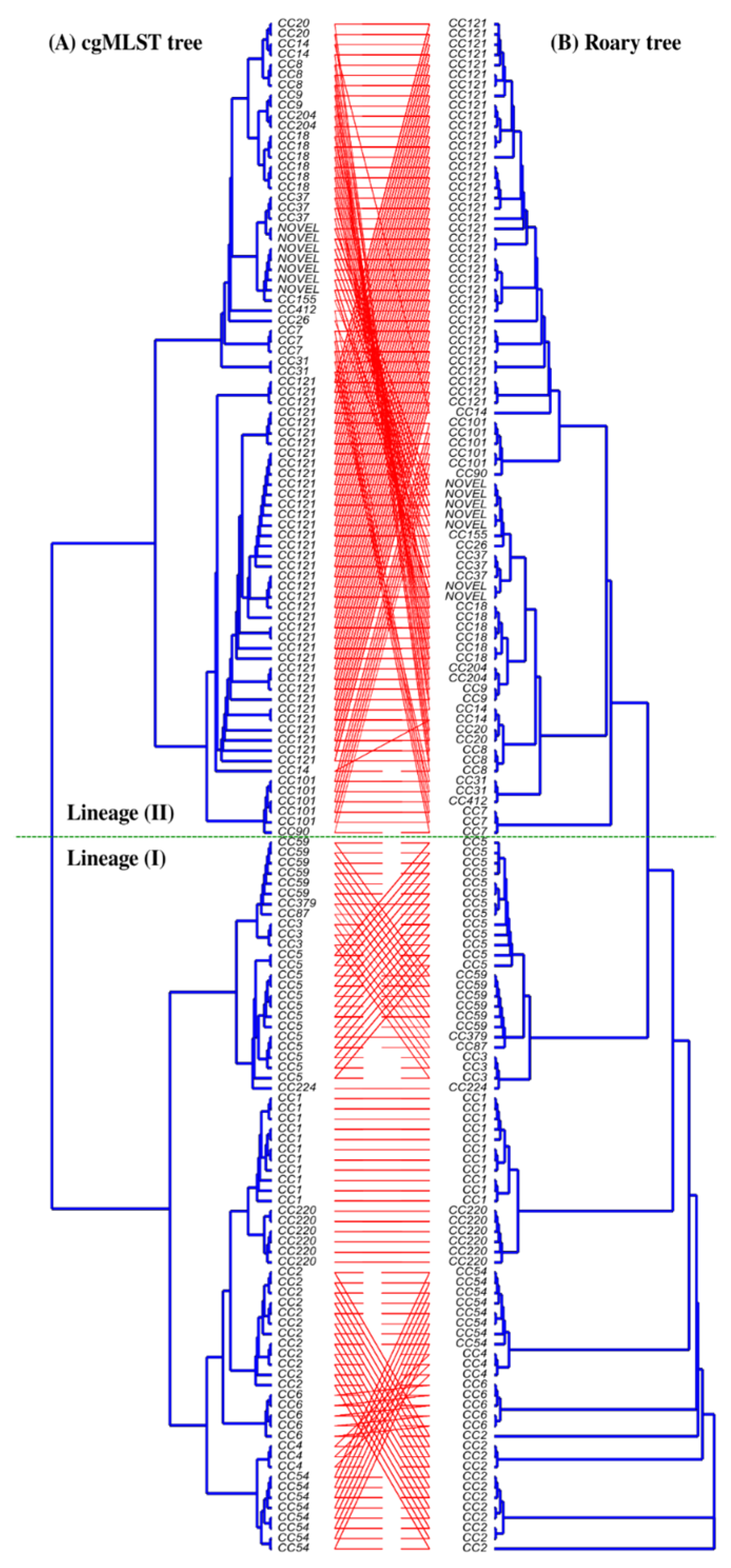
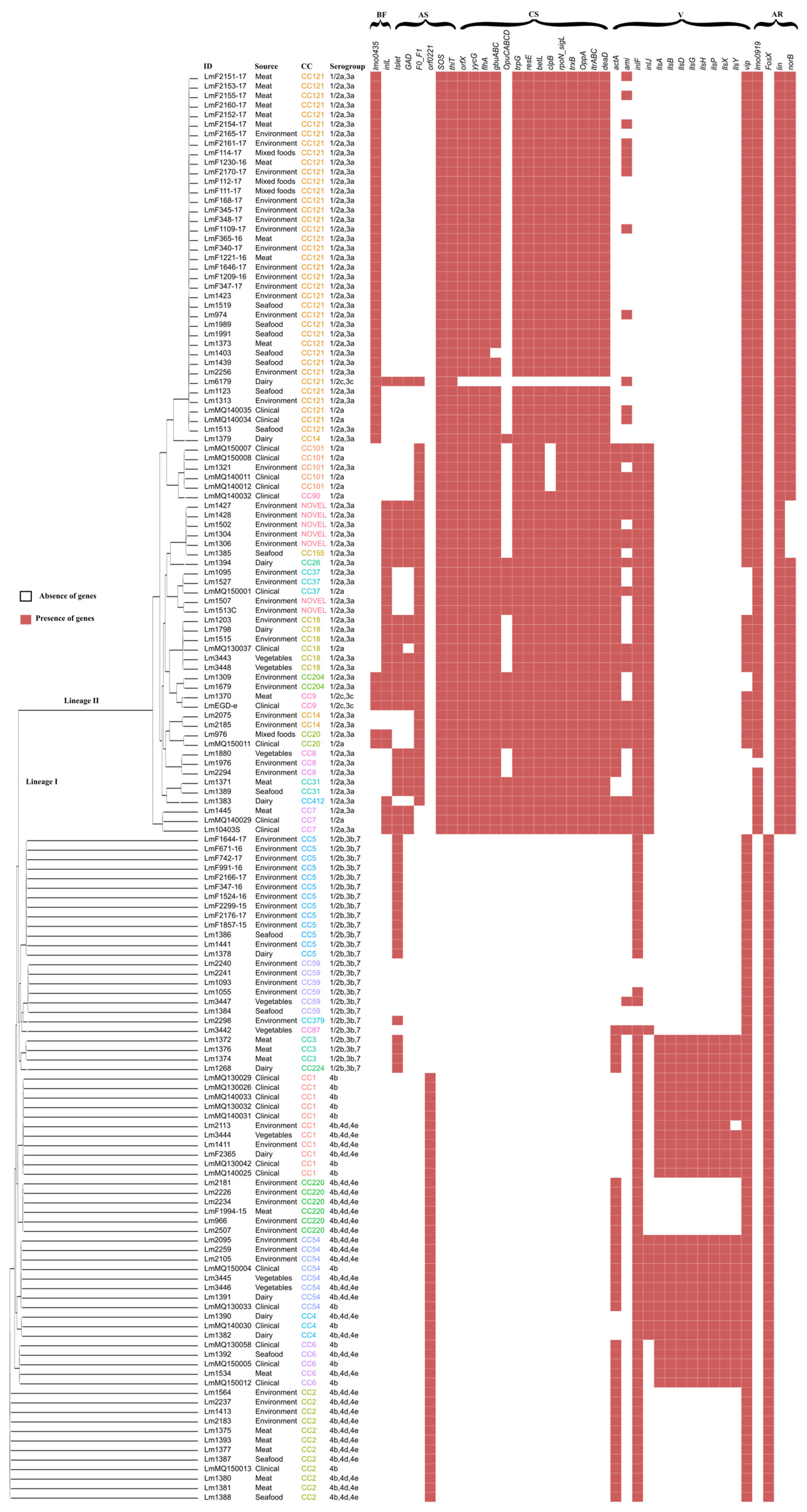
3.1. Clonal Distribution and Phylogenetic Clustering

A 1748-gene-based cgMLST phylogenetic analysis on organic acid variability was reported previously with the same set of isolates used in this study [5]. This cgMLST phylogeny was replotted to visualise the Sequence Type (ST), clonal structure, and cgMLST alignment. Closely related CC-types, which were assigned by seven-gene in silico MLST and genetic lineages, were clustered and aligned consistently (Figure 1). To understand the genetic relatedness of CC-type to the presence of particular genes of interest, a pan-genome based phylogenetic tree was constructed using Roary analysis. As shown in Figure 2, the cgMLST tree generally showed a highly similar alignment to the Roary pan-genome phylogenetic tree. In particular, a comparison of CC-type distribution between the pan-genome analysis based on orthologous genes and a previously reported cgMLST-based phylogeny showed that CC-types from lineage I and lineage II are arranged in a similar way in both trees, whereas strains from lineage II showed more variability. In many instances, discrepancies were related simply to tree presentation and rotating clades could eliminate discrepancies across trees without changing their main structure (Figure 2).

3.2. Pan-Genome Analysis

Scoary (v1.6.16) results from a pan-genome-wide association study revealed 11 genes (Table 1) that were specifically associated (p < 0.05) with clinical isolates. Some of the Scoary-identified clinical-isolate-specific genes are similar to known virulence genes required for successful infection of human hosts but others of unknown function were also identified by the analysis.

3.3. Presence of Virulence Genes and Their Genotype Association

In order to investigate potential associations of virulence genes with genetic grouping (CC, ST, Serogroup, Lineage), virulence genes were initially predicted in silico using VirulenceFinder. Eighty-nine virulence genes were found in the CC20, CC18, CC26, CC9, CC7, and CC155 isolates, as well as in the EGDe (CC9) reference genome. The fewest number of virulence genes were predicted in CC6 (seventy-seven). The number of virulence genes present in genomes of ST20 and ST18 isolates and reference genome EGD-e were the same as reported previously [38]. An independent t-test revealed that there are no significance differences in the presence of the total number of virulence genes in clinical versus non-clinical isolates. To fully understand the observed virulence gene differences, further analysis was performed with the ABRicate tool, which relies on the more limited, highly curated VFDB database. Only forty-one different virulence genes were predicted to be present in our isolates, including Listeria Pathogenicity Island LIPI-1, LIPI-2, and LIPI-3. Twenty-five of the forty-one virulence genes identified by ABRicate were found in all of the isolates. Interestingly, apart from the presence of twenty-five virulence genes in all the isolates, the remaining presence or absence was found to be CC-type-specific with very few exceptions (Figure 3 and Figure 4). In contrast, the presence and absence of the autolysin amidase gene (ami), collagen binding MSCRAMM (microbial surface components recognising adhesive matrix molecules) (ecbA), internalin family of surface proteins (inlF), one of the LIPI-3 genes llsY, and genes encoding the LPXTG surface protein (vip) were found to be ambiguous or not CC-type-specific. However, Fisher’s exact test revealed a significant association between the presence of ami with CC101, CC20, and CC7; ecbA with CC2, inlJ with CC101, CC18, CC54, and Novel CC-type; as well as LIPI-3 with CC1, CC3, CC4, and CC54. To corroborate such associations of genes specific to CC-types, a total of 247 assembled, high-quality, complete L. monocytogenes genomes were downloaded from the NCBI dataset and the same analysis was performed using the ABRicate tool. MLST typing using tseemann/mlst (available at: https://github.com/tseemann/mlst (accessed on 25 January 2023)) and the CC-type assignment based on the Pasteur Institute classification scheme revealed seventy different MLST types and forty-two different CC-types with five unassignable ST-types in the fully assembled NCBI genomes. The NCBI collection of strains shared the same number of virulence genes (n = 41), and the presence/absence of CC-type-specific genes was consistent across the strains used in this study (Supplementary Table S2).

3.4. Antimicrobial Resistance, Antibiotic Resistance, and Biofilm Formation Genes

AMRFinderPlus and CARD databases in the ABRicate tool revealed the lincomycin resistance ABC-F-type ribosomal protection gene (lin) [39], fosfomycin resistance thiol transferase (FosX) [40], an integral membrane gene (mprF) [41], fluoroquinolones, and other structurally unrelated tetracycline resistant genes (norB) [39] in our isolates (Figure 4). A recent study reported that L. monocytogenes EGDe lmo0919 is associated with resistance to lincomycin [42]. A standalone BLAST search in our isolates database revealed that lmo0919 was exclusively present in the Lineage II isolates. It is worth noting that these resistance genes were found to be specific to cgMLST lineages (i.e., lmo0919, lin, and norB are present solely in Lineage II as well as FoxS solely in Lineage I). Specificity to lineage was also consistent among the NCBI L. monocytogenes genomes studied. BLAST searches (95% identity) for biofilm formation genes (Supplementary Table S3) revealed that all of the isolates carried a biofilm-formation-associated gene (lmo0673), the product of which catalyses the hydrolysis of S-ribosylhomocysteine to homocysteine (luxS), the cell-wall-binding gene (lmo2504), and the gene encoding a DNA repair protein (recO). The presence/absence of peptidoglycan binding genes lmo0435 (bapL) and class 1 internalin (inlL) were found to be specific to CC-type; the presence of bapL was specific to CC121, CC14, CC204, CC9, and CC20. Likewise, inlL was exclusively present in the NOVELCC-type, CC155, CC26, CC37, CC18, CC204, CC20, CC412, and CC7 with the exception of reference strain 6179. The trend of genes specific to CC-type (bapL and inlL) was also found to be consistent when the same analysis was performed using NCBI strains (Supplementary Table S3).

3.5. Stress Tolerance Genes

A genetic basis for acid tolerance and cold tolerance among the 150 L. monocytogenes isolates was investigated in our previous work [5]. The stress tolerance genes used in those studies are included in Supplementary Table S1. Apart from the subset of stress tolerance genes that are present in all the isolates, the stress tolerance genes were clearly specific to CC-type with the exception of an ATP-binding cassette (ABC) transporter and gbuABC operon [43] (Supplementary Table S4). In particular, thiT, yycG, flhA, trpG, resE, betL, clpB, rpoN_sigL, trxB, OppA, itrABC, and deaD were found to be specific to CC121, CC18, CC101, CC8, CC7, CC37, CC14, CC9, CC31, CC204, and CC20. It was noted that these CC types are phylogenetically close on both cgMLST- and Roary-gene-based phylogenies and are all Lineage II.

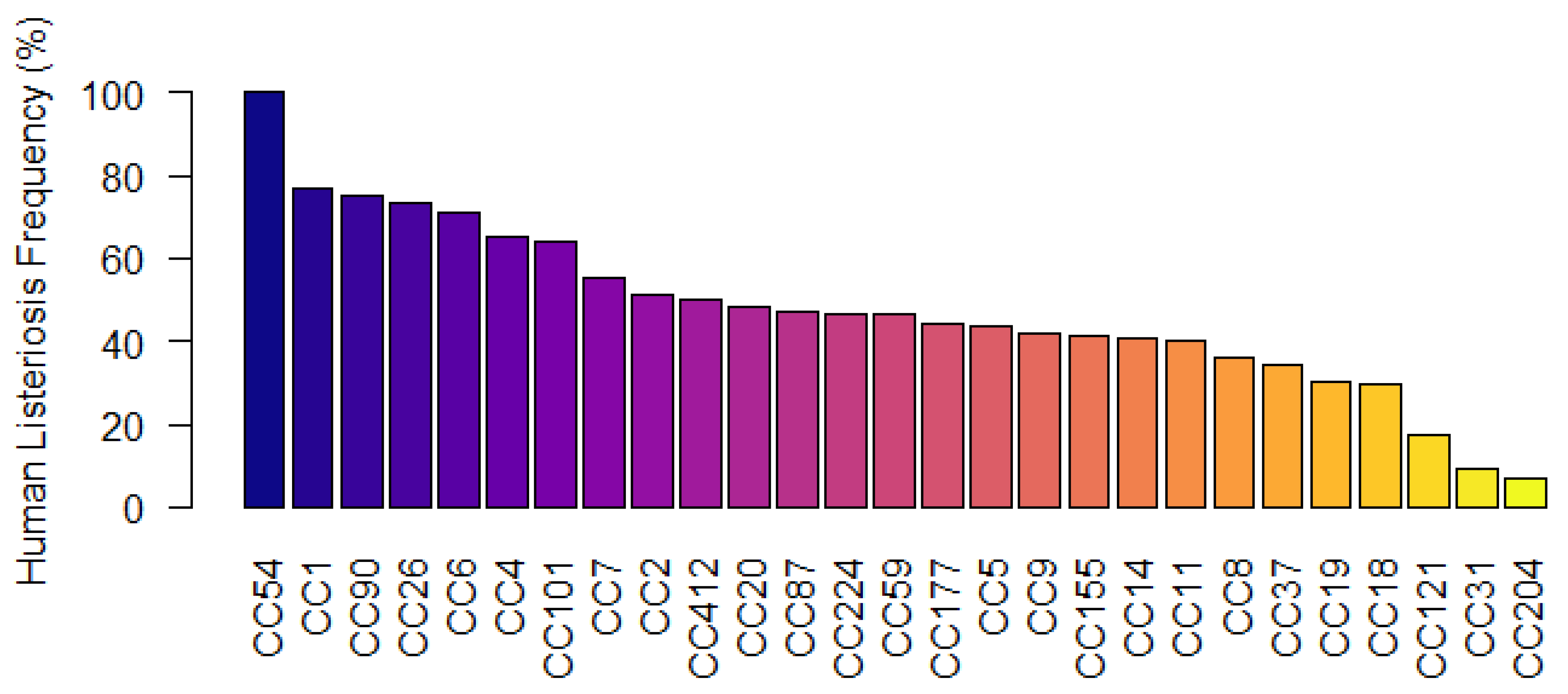
3.6. CC-Types Associated with Human Listeriosis

Adaptation to environmental stress enables L. monocytogenes to survive and even proliferate along the food chain. In particular, the capability for biofilm formation contributes to the persistence in food production facilities. Moreover, the presence of several virulence genes is important for the L. monocytogenes to survive within a host [44]. Since many of the analysed genes (virulence, biofilm, antimicrobial resistance, and stress tolerance) were found to be specific to clonal structure, we analysed all the L. monocytogenes data from the Institute Pasteur MLST database to calculate the frequency of human clinical isolation for each CC-type by dividing isolates from clinical sources by the total number of isolates. Analysis of 98,297 isolates showed that CC54 had the highest (100%) number of clinical isolates, followed by CC1 (76.94%), while the most prevalent clone, namely CC121 17.28%, had the lowest frequency (Figure 5).

4. Discussion

This study exploits WGS-based comparative genomic analysis of L. monocytogenes strains mainly isolated in the Republic of Ireland from food, food processing environments, and clinical sources and included CC-types known to associate with listeriosis outbreaks, namely CC1, CC2, CC4, and CC6 (Lineage I) and CC9 and CC121 (Lineage II), which are the most frequently isolated from immunocompromised patients [18,45]. It also included lineage II isolates that are prevalent in food processing environments belonging to CC9 [46,47,48], CC155 [49], and CC121 [18]. The analysis linked the presence or absence of key genes with specific CC-types and validated the linkage using genomes from the NCBI databases.

4.1. Heterogeneity in Virulence Genes

L. monocytogenes’ capability to colonise the gastrointestinal tract [50] and spread throughout the host dictates pathogen virulence. In the past couple of decades, multiple genes encoding virulence determinants have been described [19]. Most of the virulence genes are highly conserved but not all strains carry the full set. The important virulence factor LIPI contains several genes that encode virulence factors, including internalin proteins that allow the bacteria to invade host cells. For example, Listeriolysin O (LLO) specifically helps the bacteria escape from phagosomes and into the host cell cytoplasm, while other proteins enable the bacteria to replicate and spread within host tissues [51]. According to the ABRicate and CGE-based virulence-gene-finding analysis in this study, all of the virulence genes from LIPI-1 and LIPI-2 were present in all the isolates except actA from LIPI-1 and inlJ from LIPI-2. Both actA and inlJ encode essential virulence factors of L. monocytogenes [52,53] but were missing in all our CC1 and CC4 isolates, the most prevalent CC-type associated with human listeriosis globally [45,54], and this was consistent among the 247 NCBI genomes (Supplementary Table S2). However, standalone local BLAST with actA and inlJ genes from reference genome EGDe determined that these two genes were present in all genomes but with nucleotide identities as low as 95.18 and 86.18, respectively (Supplementary Table S5), which may be linked to the fact it has been reported that CC1, CC4, CC5, and CC59 strains possess disrupted versions of actA [55]. Another virulence gene found to associate with CC-type by ABRicate analysis was inlF, which was present in all CC-types except CC121, one CC3 isolate, and some CC59 isolates. Notably, among the isolates used in this work, CC121 and CC59, which are isolated from humans less frequently (2 out of 45), did not carry the inlF genes identified by ABRicate, which was in agreement with local BLAST searches (Supplementary Table S5). According to a recent study, InlF promotes entry into endothelial cells to breach the blood–brain barrier [56,57]. The inlF gene is part of a larger gene cluster, which includes the inlA and inlB genes. These three genes facilitate the entry of L. monocytogenes into host cells through interaction with distinct host surface receptors [53]. Since all of the isolates in this study carry inlA and inlB (Supplementary Table S2), the absence of inlF in the CC121 and CC59 group may limit successful infection.
Overall, VirulenceFinder detected more genes than ABRicate, although none of the biosynthetic cluster involved in the production of Listeriolysin S (LLS) (llsAGHXBYDP) belonging to LIPI-3 [57] were predicted, because the VirulenceFinder analysis was performed (December 2022) utilising eighty-nine virulence genes based on the EGD-e reference genome, which does not encode LIPI-3. ABRicate predicted a total of forty-one virulence genes among the isolates of this study using the Virulence Factor Database (VFDB) including LIPI-3 genes exclusively in the Lineage I isolates CC3, CC54, CC4, CC6, and CC1. In fact, LIPI-3 is specifically present in most of the clinical isolates, where it is proposed to increase virulence [58]. However, it has also been found in food isolates [58,59] and CC-types with full LIPI-3, which may present the most significant risk. This study also found that isolates belonging to CC3, CC54, CC4, CC6, and CC1 were more likely to be isolated from cases of human listeriosis based on analysis of the Institute Pasteur MLST database (Figure 5). Notably, inlF and the full set of LIPI-3 genes are harboured by CC54, CC1, CC6, CC4, CC3, and CC224 (Figure 4), which showed a higher clinical isolation frequency of >46.66% (Figure 5) and are closely related by Roary-based phylogeny. However, other lineage II isolates such as CC90 and CC110 were also present at a higher clinical frequency in the Institute Pasteur MLST database even though these CC-types do not carry LIPI-3. It is possible that the genotype of lineage II CC-types harbouring more stress tolerance genes but fewer virulence genes than lineage I isolates (Figure 4) allows these organisms to persist in the food and food processing environments and they are still able to infect highly immunocompromised individuals [18].

4.2. Prevalence of Antimicrobial Resistance Genes

Antimicrobial resistance amongst L. monocytogenes strains appear to differ considerably [60] and multiple antibiotic resistance genes may contribute to the development of novel antibiotic-resistant strains [61]. Recent studies have reported the high prevalence of antibiotic resistance genes in L. monocytogenes [62,63]. The screening of AMR genes in the WGS by the ABRicate tool using CARD revealed that five different AMR genes were present in all the isolates. These AMR resistance genes may have evolved or transferred horizontally, resulting in an increase in antibiotic resistance [64,65]. The presence of other AMR genes in the WGS were generally found to be specific to genetic lineages. For instance, lmo0919 [42] and norB [39] were harboured by Lineage II while FosX [63] was harboured by Lineage I. The link between WGS-based subtyping techniques and the presence of AMR genes may be important information for determining an appropriate treatment strategy for patients.

4.3. Stress Response and Biofilm Formation Genes

When exposed to stress, L. monocytogenes develops strategies for altering cellular functions so that the pathogen can endure and proliferate [19]. It is able to adapt to stressful conditions, such as those encountered in food processing and storage environments, through the activation of specific stress response genes. In this study, all the Lineage II isolates, including CC20, CC18, CC26, CC9, CC7, and CC155, were predicted to contain more stress tolerance genes than Lineage I isolates (Figure 4). Unsurprisingly, these CC-type isolates have been observed to grow faster in cold temperatures [3] and demonstrate increased tolerance to acetic and propionic acid [5]. This may explain why these CC-types were more prevalent in food and food processing environments with the presence of combinations of more stress tolerance genes. In addition, L. monocytogenes is also capable of producing biofilm—extracellular polymeric substances (EPSs) [66] that encase the cells and shield them from environmental stresses such desiccation, pH fluctuations, and antimicrobial agents. Although the genes involved in L. monocytogenes biofilm formation are not yet fully characterised, a number of studies have identified an array of genes that could be involved (see Supplementary Table S1). Apart from the biofilm formation genes predicted as present in all the isolates, CC20, CC18, CC26, CC9, CC7, and CC155 types specifically possess class I internalin inlL biofilm formation genes potentially involved in mucin binding, sessile growth, and adhesion [64]. A similar linking of the presence of inlL to these CC-types was reported among L. monocytogenes in Norwegian food chains [65]. All of these CC-types belong to the most frequently reported serogroup 1/2a,3a in the European Union [66] and are second highest in the United States [67]. The combination of a higher proportion of stress tolerance genes and the presence of inlL in the genome may enable these CC-types to persist in the food processing environment and eventually infect immunocompromised individuals.

5. Conclusions

Comparative genomic analysis of 150 L. monocytogenes isolates from food, the food-processing environment, and clinical sources was performed in this study. The relationship between CC-type and the prevalence of virulence, stress tolerance, biofilm formation, and antimicrobial-resistant genes was investigated. It was observed that several of these genes were clonal-structure-specific, and this specificity was verified using 247 completely assembled, high-quality genomes extracted from NCBI. Particularly, we highlight that CC20, CC18, CC26, CC9, CC7, and CC155 were predicted to harbour a higher proportion of biofilm, stress-survival, and antimicrobial genes than other CC-types. In addition, the CC-types, which carry both inlF and a full set of LIPI-3 genes, were found to have a higher human clinical isolation frequency than others. This WGS-based study provides a deeper understanding of this pathogen’s persistence within food chains and the ability to infect the host, but further in vitro adherence and invasion studies using host cell lines and in vivo studies using animal infection models are needed to support the in silico bioinformatics analysis.

Supplementary Materials

The following supporting information can be downloaded at: https://www.mdpi.com/article/10.3390/microorganisms11061603/s1, Table S1: The presence/absence of virulence, biofilm formation, acid stress tolerance, and cold stress tolerance genes included in this study. Table S2: The presence/absence of CC-type-specific virulence genes among isolates in this study. Table S3: The presence/absence of CC-type-specific virulence genes among NCBI strains. Table S4: Variation in presence of stress tolerance genes among CC-types in 150 L. monocytogenes isolates apart from genes, which are present in all isolates. Table S5: Local BLAST search result of actA and inlJ genes among the isolates of this study. References [68,69,70,71,72,73,74,75,76,77,78,79,80,81,82,83,84,85,86,87,88,89,90,91,92] are cited in the Supplementary Materials.

Author Contributions

Conceptualization, V.P., M.B. and M.C.; Methodology, P.M. and V.P.; Investigation, P.M.; Data curation, P.M.; Writing—original draft, P.M.; Writing—review & editing, M.B. and M.C.; Supervision, O.M., M.B. and M.C.; Project administration, O.M.; Funding acquisition, O.M. and M.C. All authors have read and agreed to the published version of the manuscript.

Funding

PM is in receipt of a Munster Technological University RÍSAM PhD Scholarship and is supported by the Department of Agriculture, Food and the Marine under the Food Institutional Research Measure, grant numbers 15F604 and 2019R495.

Data Availability Statement

Genome sequence data was previously published [5].

Conflicts of Interest

The authors declare no conflict of interest.

References

  1. European Food Safety Authority (EFSA). EU One Health Zoonoses Report 2020. EFSA J. 2021, 19, e06971. [Google Scholar] [CrossRef]
  2. Arguedas-Villa, C.; Stephan, R.; Tasara, T. Evaluation of Cold Growth and Related Gene Transcription Responses Associated with Listeria monocytogenes Strains of Different Origins. Food Microbiol. 2010, 27, 653–660. [Google Scholar] [CrossRef]
  3. Myintzaw, P.; Pennone, V.; McAuliffe, O.; Begley, M.; Callanan, M. Variability in Cold Tolerance of Food and Clinical Listeria monocytogenes Isolates. Microorganisms 2023, 11, 65. [Google Scholar] [CrossRef]
  4. Cotter, P.D.; Ryan, S.; Gahan, C.G.M.; Hill, C. Presence of GadD1 Glutamate Decarboxylase in Selected Listeria monocytogenes Strains Is Associated with an Ability to Grow at Low PH. Appl. Environ. Microbiol. 2005, 71, 2832–2839. [Google Scholar] [CrossRef] [PubMed] [Green Version]
  5. Myintzaw, P.; Pennone, V.; McAuliffe, O.; Begley, M.; Callanan, M. Correlation of Organic Acid Tolerance and Genotypic Characteristics of Listeria monocytogenes Food and Clinical Isolates. Food Microbiol. 2022, 104, 104004. [Google Scholar] [CrossRef]
  6. Centorotola, G.; Guidi, F.; D’aurizio, G.; Salini, R.; Di Domenico, M.; Ottaviani, D.; Petruzzelli, A.; Fisichella, S.; Duranti, A.; Tonucci, F.; et al. Intensive Environmental Surveillance Plan for Listeria monocytogenes in Food Producing Plants and Retail Stores of Central Italy: Prevalence and Genetic Diversity. Foods 2021, 10, 1944. [Google Scholar] [CrossRef] [PubMed]
  7. Lourenço, A.; de Las Heras, A.; Scortti, M.; Vazquez-Boland, J.; Frank, J.F.; Brito, L. Comparison of Listeria monocytogenes Exoproteomes from Biofilm and Planktonic State: Lmo2504, a Protein Associated with Biofilms. Appl. Environ. Microbiol. 2013, 79, 6075–6082. [Google Scholar] [CrossRef] [Green Version]
  8. Moran-Gilad, J.; Sintchenko, V.; Pedersen, S.K.; Wolfgang, W.J.; Pettengill, J.; Strain, E.; Hendriksen, R.S. Proficiency Testing for Bacterial Whole Genome Sequencing: An End-User Survey of Current Capabilities, Requirements and Priorities. BMC Infect. Dis. 2015, 15, 174. [Google Scholar] [CrossRef] [PubMed] [Green Version]
  9. Sintchenko, V.; Holmes, E.C. The Role of Pathogen Genomics in Assessing Disease Transmission. BMJ 2015, 350, h1314. [Google Scholar] [CrossRef]
  10. Schürch, A.C.; van Schaik, W. Challenges and Opportunities for Whole-Genome Sequencing–Based Surveillance of Antibiotic Resistance. Ann. N. Y. Acad. Sci. 2017, 1388, 108–120. [Google Scholar] [CrossRef] [PubMed]
  11. Belén, A.; Pavón, I.; Maiden, M.C.J. Multilocus Sequence Typing. Methods Mol. Biol. 2009, 551, 129–140. [Google Scholar] [CrossRef] [Green Version]
  12. Caruso, M.; Fraccalvieri, R.; Pasquali, F.; Santagada, G.; Latorre, L.M.; Difato, L.M.; Miccolupo, A.; Normanno, G.; Parisi, A. Antimicrobial Susceptibility and Multilocus Sequence Typing of Listeria monocytogenes Isolated over 11 Years from Food, Humans, and the Environment in Italy. Foodborne Pathog. Dis. 2020, 17, 284–294. [Google Scholar] [CrossRef] [Green Version]
  13. Hilliard, A.; Leong, D.; O’Callaghan, A.; Culligan, E.P.; Morgan, C.A.; Delappe, N.; Hill, C.; Jordan, K.; Cormican, M.; Gahan, C.G.M. Genomic Characterization of Listeria monocytogenes Isolates Associated with Clinical Listeriosis and the Food Production Environment in Ireland. Genes 2018, 9, 171. [Google Scholar] [CrossRef] [Green Version]
  14. Félix, B.; Capitaine, K.; Te, S.; Felten, A.; Gillot, G.; Feurer, C.; Bosch, T. Van Den. Identification by High-Throughput Real-Time PCR of 30 Major Circulating Listeria monocytogenes Clonal Complexes in Europe. Am. Soc. Microbiol. 2023, 11, e03954-22. [Google Scholar] [CrossRef]
  15. Seemann, T. Prokka: Rapid Prokaryotic Genome Annotation. Bioinformatics 2014, 30, 2068–2069. [Google Scholar] [CrossRef] [Green Version]
  16. Tatusova, T.; Dicuccio, M.; Badretdin, A.; Chetvernin, V.; Nawrocki, E.P.; Zaslavsky, L.; Lomsadze, A.; Pruitt, K.D.; Borodovsky, M.; Ostell, J. NCBI Prokaryotic Genome Annotation Pipeline. Nucleic Acids Res. 2016, 44, 6614–6624. [Google Scholar] [CrossRef] [PubMed]
  17. Kuenne, C.; Billion, A.; Mraheil, M.A.; Strittmatter, A.; Daniel, R.; Goesmann, A.; Barbuddhe, S.; Hain, T.; Chakraborty, T. Reassessment of the Listeria monocytogenes Pan-Genome Reveals Dynamic Integration Hotspots and Mobile Genetic Elements as Major Components of the Accessory Genome. BMC Genom. 2013, 14, 47. [Google Scholar] [CrossRef] [PubMed] [Green Version]
  18. Maury, M.M.; Tsai, Y.-H.; Charlier, C.; Touchon, M.; Chenal-Francisque, V.; Leclercq, A.; Criscuolo, A.; Gaultier, C.; Roussel, S.; Brisabois, A.; et al. Uncovering Listeria monocytogenes Hypervirulence by Harnessing Its Biodiversity. Nat. Genet. 2016, 48, 308–313. [Google Scholar] [CrossRef] [Green Version]
  19. Sibanda, T.; Buys, E.M. Listeria monocytogenes Pathogenesis: The Role of Stress Adaptation. Microorganisms 2022, 10, 1522. [Google Scholar] [CrossRef] [PubMed]
  20. Wu, J.; Nicaogáin, K.; McAuliffe, O.; Jordan, K.; O’Byrne, C. Phylogenetic and Phenotypic Analyses of a Collection of Food and Clinical Listeria monocytogenes Isolates Reveal Loss of Function of Sigma B from Several Clonal Complexes. Appl. Environ. Microbiol. 2022, 88, 1–21. [Google Scholar] [CrossRef] [PubMed]
  21. Lauer, P.; Chow, M.Y.N.; Loessner, M.J.; Portnoy, D.A.; Calendar, R. Construction, Characterization, and Use of Two Listeria monocytogenes Site-Specific Phage Integration Vectors. J. Bacteriol. 2002, 184, 4177. [Google Scholar] [CrossRef] [Green Version]
  22. Schmitz-Esser, S.; Müller, A.; Stessl, B.; Wagner, M. Genomes of Sequence Type 121 Listeria monocytogenes Strains Harbor Highly Conserved Plasmids and Prophages. Front. Microbiol. 2015, 6, 380. [Google Scholar] [CrossRef] [Green Version]
  23. Nelson, K.E.; Fouts, D.E.; Mongodin, E.F.; Ravel, J.; DeBoy, R.T.; Kolonay, J.F.; Rasko, D.A.; Angiuoli, S.V.; Gill, S.R.; Paulsen, I.T.; et al. Whole Genome Comparisons of Serotype 4b and 1/2a Strains of the Food-Borne Pathogen Listeria monocytogenes Reveal New Insights into the Core Genome Components of This Species. Nucleic Acids Res. 2004, 32, 2386–2395. [Google Scholar] [CrossRef] [Green Version]
  24. Bolger, A.M.; Lohse, M.; Usadel, B. Trimmomatic: A Flexible Trimmer for Illumina Sequence Data. Bioinformatics 2014, 30, 2114–2120. [Google Scholar] [CrossRef] [Green Version]
  25. Afgan, E.; Baker, D.; Bér´, B.; Batut, B.; Van Den Beek, M.; Bouvier, D.; Cech, M.; Chilton, J.; Clements, D.; Coraor, N.; et al. The Galaxy Platform for Accessible, Reproducible and Collaborative Biomedical Analyses: 2018 Update. Nucleic Acids Res. 2018, 46, 537–544. [Google Scholar] [CrossRef] [PubMed] [Green Version]
  26. Bankevich, A.; Nurk, S.; Antipov, D.; Gurevich, A.A.; Dvorkin, M.; Kulikov, A.S.; Lesin, V.M.; Nikolenko, S.I.; Pham, S.; Prjibelski, A.D.; et al. SPAdes: A New Genome Assembly Algorithm and Its Applications to Single-Cell Sequencing. J. Comput. Biol. 2012, 19, 455. [Google Scholar] [CrossRef] [PubMed] [Green Version]
  27. Page, A.J.; Cummins, C.A.; Hunt, M.; Wong, V.K.; Reuter, S.; Holden, M.T.G.; Fookes, M.; Falush, D.; Keane, J.A.; Parkhill, J. Roary: Rapid Large-Scale Prokaryote Pan Genome Analysis. Bioinformatics 2015, 31, 3691–3693. [Google Scholar] [CrossRef] [PubMed] [Green Version]
  28. Brynildsrud, O.; Bohlin, J.; Scheffer, L.; Eldholm, V. Rapid Scoring of Genes in Microbial Pan-Genome-Wide Association Studies with Scoary. Genome Biol. 2016, 17, 238. [Google Scholar] [CrossRef] [PubMed] [Green Version]
  29. Quainoo, S.; Coolen, J.P.M.; van Hijum, S.A.F.T.; Huynen, M.A.; Melchers, W.J.G.; van Schaik, W.; Wertheim, H.F.L. Whole-Genome Sequencing of Bacterial Pathogens: The Future of Nosocomial Outbreak Analysis. Clin. Microbiol. Rev. 2017, 30, 1015–1063. [Google Scholar] [CrossRef] [PubMed] [Green Version]
  30. Mafuna, T.; Matle, I.; Magwedere, K.; Pierneef, R.E.; Reva, O.N. Whole Genome-Based Characterization of Listeria monocytogenes Isolates Recovered from the Food Chain in South Africa. Front. Microbiol. 2021, 12, 669287. [Google Scholar] [CrossRef]
  31. Doumith, M.; Buchrieser, C.; Glaser, P.; Jacquet, C.; Martin, P. Differentiation of the Major Listeria monocytogenes Serovars by Multiplex PCR. J. Clin. Microbiol. 2004, 42, 3819–3822. [Google Scholar] [CrossRef] [PubMed] [Green Version]
  32. Hingston, P.; Chen, J.; Dhillon, B.K.; Laing, C.; Bertelli, C.; Gannon, V.; Tasara, T.; Allen, K.; Brinkman, F.S.L.; Truelstrup Hansen, L.; et al. Genotypes Associated with Listeria monocytogenes Isolates Displaying Impaired or Enhanced Tolerances to Cold, Salt, Acid, or Desiccation Stress. Front. Microbiol. 2017, 8, 369. [Google Scholar] [CrossRef] [PubMed] [Green Version]
  33. Liu, B.; Zheng, D.; Jin, Q.; Chen, L.; Yang, J. VFDB 2019: A Comparative Pathogenomic Platform with an Interactive Web Interface. Nucleic Acids Res. 2019, 47, D687–D692. [Google Scholar] [CrossRef] [PubMed]
  34. Chen, L.; Zheng, D.; Liu, B.; Yang, J.; Jin, Q. VFDB 2016: Hierarchical and Refined Dataset for Big Data Analysis—10 Years On. Nucleic Acids Res. 2016, 44, D694–D697. [Google Scholar] [CrossRef]
  35. Reis, O.; Sousa, S.; Camejo, A.; Villiers, V.; Gouin, E.; Cossart, P.; Cabanes, D. LapB, a Novel Listeria monocytogenes LPXTG Surface Adhesin, Required for Entry into Eukaryotic Cells and Virulence. J. Infect. Dis. 2010, 202, 551–562. [Google Scholar] [CrossRef] [Green Version]
  36. Wagner, E.; Fagerlund, A.; Thalguter, S.; Jensen, M.R.; Heir, E.; Møretrø, T.; Moen, B.; Langsrud, S.; Rychli, K. Deciphering the Virulence Potential of Listeria monocytogenes in the Norwegian Meat and Salmon Processing Industry by Combining Whole Genome Sequencing and in Vitro Data. Int. J. Food Microbiol. 2022, 383, 109962. [Google Scholar] [CrossRef]
  37. Caliandro, R.; de Diego, I.; Gomis-Rüth, F.X. Crystal Structure Report of the ImmR Transcriptional Regulator DNA-Binding Domain of the Bacillus subtilis ICEBs1 Transposon. Sci. Rep. 2022, 12, 5258. [Google Scholar] [CrossRef]
  38. Løvdal, T.; Brandal, L.T.; Sundaram, A.Y.M.; Naseer, U.; Roth, B.; Lunestad, B.T. Small-Scale Comparative Genomic Analysis of Listeria monocytogenes Isolated from Environments of Salmon Processing Plants and Human Cases in Norway. Hygiene 2021, 1, 43–55. [Google Scholar] [CrossRef]
  39. Mota, M.I.; Vázquez, S.; Cornejo, C.; D’Alessandro, B.; Braga, V.; Caetano, A.; Betancor, L.; Varela, G. Does Shiga Toxin-Producing Escherichia coli and Listeria monocytogenes Contribute Significantly to the Burden of Antimicrobial Resistance in Uruguay? Front. Vet. Sci. 2020, 7, 583930. [Google Scholar] [CrossRef]
  40. Lüth, S.; Halbedel, S.; Rosner, B.; Wilking, H.; Holzer, A.; Roedel, A.; Dieckmann, R.; Vincze, S.; Prager, R.; Flieger, A.; et al. Backtracking and Forward Checking of Human Listeriosis Clusters Identified a Multiclonal Outbreak Linked to Listeria monocytogenes in Meat Products of a Single Producer. Emerg. Microbes Infect. 2020, 9, 1600–1608. [Google Scholar] [CrossRef]
  41. Thedieck, K.; Hain, T.; Mohamed, W.; Tindall, B.J.; Nimtz, M.; Chakraborty, T.; Wehland, J.; Jänsch, L. The MprF Protein Is Required for Lysinylation of Phospholipids in Listerial Membranes and Confers Resistance to Cationic Antimicrobial Peptides (CAMPs) on Listeria monocytogenes. Mol. Microbiol. 2006, 62, 1325–1339. [Google Scholar] [CrossRef] [Green Version]
  42. Duval, M.; Dar, D.; Carvalho, F.; Rocha, E.P.C.; Sorek, R.; Cossart, P. HflXr, a Homolog of a Ribosome-Splitting Factor, Mediates Antibiotic Resistance. Proc. Natl. Acad. Sci. USA 2018, 115, 13359–13364. [Google Scholar] [CrossRef] [Green Version]
  43. Dreux, N.; Albagnac, C.; Sleator, R.D.; Hill, C.; Carlin, F.; Morris, C.E.; Nguyen-The, C. Glycine Betaine Improves Listeria monocytogenes Tolerance to Desiccation on Parsley Leaves Independent of the Osmolyte Transporters BetL, Gbu and OpuC. J. Appl. Microbiol. 2008, 104, 1221–1227. [Google Scholar] [CrossRef]
  44. Roberts, A.J.; Wiedmann, M. Pathogen, Host and Environmental Factors Contributing to the Pathogenesis of Listeriosis. Cell. Mol. Life Sci. 2003, 60, 904–918. [Google Scholar] [CrossRef] [PubMed]
  45. Bergis, H.; Bonanno, L.; Asséré, A.; Lombard, B. EURL Lm Technical Guidance Document on Challenge Tests and Durability Studies for Assessing Shelf-Life of Ready-to-Eat Foods Related to Listeria monocytogenes; EURL Lm: Maisons-Alfort, France, 2021; Volume 4, p. 60. [Google Scholar]
  46. Li, H.; Wang, P.; Lan, R.; Luo, L.; Cao, X.; Wang, Y.; Wang, Y.; Li, H.; Zhang, L.; Ji, S.; et al. Risk Factors and Level of Listeria Monocytogenes Contamination of Raw Pork in Retail Markets in China. Front. Microbiol. 2018, 9, 1090. [Google Scholar] [CrossRef] [PubMed]
  47. Alvarez-Molina, A.; Cobo-Díaz, J.F.; López, M.; Prieto, M.; de Toro, M.; Alvarez-Ordóñez, A. Unraveling the Emergence and Population Diversity of Listeria monocytogenes in a Newly Built Meat Facility through Whole Genome Sequencing. Int. J. Food Microbiol. 2021, 340, 109043. [Google Scholar] [CrossRef] [PubMed]
  48. Fagerlund, A.; Langsrud, S.; Moen, B.; Heir, E. Complete Genome Sequences of Six Listeria monocytogenes Sequence Type 9 Isolates from Meat Processing Plants in Norway. Am. Soc. Microbiol. 2018, 6, 9–10. [Google Scholar] [CrossRef] [PubMed] [Green Version]
  49. Muchaamba, F.; Stephan, R.; Tasara, T. Β-Phenylethylamine as a Natural Food Additive Shows Antimicrobial Activity Against Listeria monocytogenes on Ready-to-Eat Foods. Foods 2020, 9, 1363. [Google Scholar] [CrossRef]
  50. Davis, M.L.; Ricke, S.C.; Donaldson, J.R. Establishment of Listeria monocytogenes in the Gastrointestinal Tract. Microorganisms 2019, 7, 75. [Google Scholar] [CrossRef] [Green Version]
  51. Guidi, F.; Chiaverini, A.; Repetto, A.; Lorenzetti, C.; Centorotola, G.; Bazzucchi, V.; Palombo, B.; Gattuso, A.; Pomilio, F.; Blasi, G. Hyper-Virulent Listeria monocytogenes Strains Associated with Respiratory Infections in Central Italy. Front. Cell. Infect. Microbiol. 2021, 11, 765540. [Google Scholar] [CrossRef]
  52. Pillich, H.; Puri, M.; Chakraborty, T. The Actin Cytoskeleton and Bacterial Infection. In Current Topics in Microbiology and Immunology; Springer: Berlin/Heidelberg, Germany, 2016; Volume 399, pp. 113–132. [Google Scholar]
  53. Smith, A.; Moorhouse, E.; Monaghan, J.; Taylor, C.; Singleton, I. Sources and Survival of Listeria monocytogenes on Fresh, Leafy Produce. J. Appl. Microbiol. 2018, 125, 930–942. [Google Scholar] [CrossRef] [Green Version]
  54. Bergholz, T.M.; Shah, M.K.; Burall, L.S.; Rakic-Martinez, M.; Datta, A.R. Genomic and Phenotypic Diversity of Listeria monocytogenes Clonal Complexes Associated with Human Listeriosis. Appl. Microbiol. Biotechnol. 2018, 102, 3475–3485. [Google Scholar] [CrossRef] [PubMed]
  55. Lake, F.B.; van Overbeek, L.S.; Baars, J.J.P.; Koomen, J.; Abee, T.; den Besten, H.M.W. Genomic Characteristics of Listeria monocytogenes Isolated during Mushroom (Agaricus bisporus) Production and Processing. Int. J. Food Microbiol. 2021, 360, 109438. [Google Scholar] [CrossRef] [PubMed]
  56. Ireton, K.; Mortuza, R.; Gyanwali, G.C.; Gianfelice, A.; Hussain, M. Role of Internalin Proteins in the Pathogenesis of Listeria monocytogenes. Mol. Microbiol. 2021, 116, 1407–1419. [Google Scholar] [CrossRef] [PubMed]
  57. Ghosh, P.; Halvorsen, E.M.; Ammendolia, D.A.; Mor-vaknin, N.; Riordan, M.X.D.O.; Brumell, J.H.; Markovitz, D.M.; Higgins, D.E. Invasion of the Brain by Listeria monocytogenes Is Mediated by InlF and Host Cell Vimentin. MBio 2018, 9, e00160-18. [Google Scholar] [CrossRef] [Green Version]
  58. Vilchis-Rangel, R.E.; Espinoza-Mellado, M.D.R.; Salinas-Jaramillo, I.J.; Martinez-Peña, M.D.; Rodas-Suárez, O.R. Association of Listeria monocytogenes LIPI-1 and LIPI-3 Marker LlsX with Invasiveness. Curr. Microbiol. 2019, 76, 637–643. [Google Scholar] [CrossRef]
  59. Muchaamba, F.; Eshwar, A.K.; von Ah, U.; Stevens, M.J.A.; Tasara, T. Evolution of Listeria monocytogenes During a Persistent Human Prosthetic Hip Joint Infection. Front. Microbiol. 2020, 11, 1726. [Google Scholar] [CrossRef]
  60. Wilson, A.; Gray, J.; Scott Chandry, P.; Fox, E.M. Phenotypic and Genotypic Analysis of Antimicrobial Resistance among Listeria monocytogenes Isolated from Australian Food Production Chains. Genes 2018, 9, 80. [Google Scholar] [CrossRef] [Green Version]
  61. Baquero, F.; Lanza, V.F.; Duval, M.; Coque, T.M. Ecogenetics of Antibiotic Resistance in Listeria monocytogenes. Mol. Microbiol. 2020, 113, 570–579. [Google Scholar] [CrossRef] [Green Version]
  62. Hughes, D.; Andersson, D.I. Environmental and Genetic Modulation of the Phenotypic Expression of Antibiotic Resistance. FEMS Microbiol. Rev. 2017, 41, 374–391. [Google Scholar] [CrossRef]
  63. Yu, W.; Huang, Y.; Ying, C.; Zhou, Y.; Zhang, L.; Zhang, J.; Chen, Y.; Qiu, Y. Analysis of Genetic Diversity and Antibiotic Options for Clinical Listeria monocytogenes Infections in China. Open Forum Infect. Dis. 2021, 8, ofab177. [Google Scholar] [CrossRef]
  64. Popowska, M.; Krawczyk-balska, A.; Ostrowski, R. InlL from Listeria monocytogenes Is Involved in Biofilm Formation and Adhesion to Mucin. Front. Microbiol. 2017, 8, 660. [Google Scholar] [CrossRef] [Green Version]
  65. Fagerlund, A.; Wagner, E.; Møretrø, T.; Heir, E.; Moen, B.; Rychli, K.; Langsrud, S. Pervasive Listeria monocytogenes Is Common in the Norwegian. Appl. Environ. Microbiol. 2022, 88, e00861-22. [Google Scholar] [PubMed]
  66. European Centre for Disease Prevention and Control (ECDC). Surveillance Atlas of Infectious Diseases. Available online: https://atlas.ecdc.europa.eu/public/index.aspx (accessed on 15 August 2022).
  67. Centres for Disease Control and Prevention (CDC). Pathogen Surveillance—Listeria. Available online: https://www.cdc.gov/listeria/pdf/listeriainitiativeoverview_508.pdf (accessed on 27 April 2023).
  68. Cabanes, D.; Dussurget, O.; Dehoux, P.; Cossart, P. Auto, a Surface Associated Autolysin of Listeria monocytogenes Required for Entry into Eukaryotic Cells and Virulence. Mol. Microbiol. 2004, 51, 1601–1614. [Google Scholar] [CrossRef] [PubMed]
  69. Wang, W.; Xu, J.; Chen, Y.; Zhu, Z.; He, F. An Insight into the Genome of a Listeria monocytogenes Strain Isolated from a Bloodstream Infection and Phylogenetic Analysis. J. Clin. Lab. Anal. 2021, 35, e23824. [Google Scholar] [CrossRef]
  70. Hein, I.; Klinger, S.; Dooms, M.; Flekna, G.; Stessl, B.; Leclercq, A.; Hill, C.; Allerberger, F.; Wagner, M. Stress Survival Islet 1 (SSI-1) Survey in Listeria monocytogenes Reveals an Insert Common to Listeria innocua in Sequence Type 121 L. Monocytogenes Strains. Appl. Environ. Microbiol. 2011, 77, 2169–2173. [Google Scholar] [CrossRef] [Green Version]
  71. Cotter, P.D.; Gahan, C.G.M.; Hill, C. Analysis of the Role of the Listeria monocytogenes F0F1-ATPase Operon in the Acid Tolerance Response. Int. J. Food Microbiol. 2000, 60, 137–146. [Google Scholar] [CrossRef] [PubMed]
  72. Cotter, P.D.; Emerson, N.; Gahan, C.G.M.; Hill, C. Identification and Disruption of LisRK, a Genetic Locus Encoding a Two-Component Signal Transduction System Involved in Stress Tolerance and Virulence in Listeria monocytogenes. J. Bacteriol. 1999, 181, 6840. [Google Scholar] [CrossRef] [Green Version]
  73. Pöntinen, A.; Lindström, M.; Skurnik, M.; Korkeala, H. Screening of the Two-Component-System Histidine Kinases of Listeria monocytogenes EGD-e. LiaS Is Needed for Growth under Heat, Acid, Alkali, Osmotic, Ethanol and Oxidative Stresses. Food Microbiol. 2017, 65, 36–43. [Google Scholar] [CrossRef] [PubMed] [Green Version]
  74. Wiedmann, M.; Arvik, T.J.; Hurley, R.J.; Boor, K.J. General Stress Transcription Factor ΣB and Its Role in Acid Tolerance and Virulence of Listeria monocytogenes. J. Bacteriol. 1998, 180, 3650. [Google Scholar] [CrossRef] [Green Version]
  75. Chen, J.; Cheng, C.; Xia, Y.; Zhao, H.; Fang, C.; Shan, Y.; Wu, B.; Fang, W. Lmo0036, an Ornithine and Putrescine Carbamoyltransferase in Listeria monocytogenes, Participates in Arginine Deiminase and Agmatine Deiminase Pathways and Mediates Acid Tolerance. Microbiology 2011, 157, 3150–3161. [Google Scholar] [CrossRef] [PubMed] [Green Version]
  76. Chen, J.; Jiang, L.; Chen, Q.; Zhao, H.; Luo, X.; Chen, X.; Fang, W. Lmo0038 Is Involved in Acid and Heat Stress Responses and Specific for Listeria monocytogenes Lineages i and II, and Listeria Ivanovii. Foodborne Pathog. Dis. 2009, 6, 365–376. [Google Scholar] [CrossRef] [PubMed] [Green Version]
  77. Ryan, S.; Begley, M.; Gahan, C.G.M.; Hill, C. Molecular Characterization of the Arginine Deiminase System in Listeria monocytogenes: Regulation and Role in Acid Tolerance. Environ. Microbiol. 2009, 11, 432–445. [Google Scholar] [CrossRef] [PubMed]
  78. van der Veen, S.; van Schalkwijk, S.; Molenaar, D.; de Vos, W.M.; Abee, T.; Wells-Bennik, M.H.J. The SOS Response of Listeria monocytogenes Is Involved in Stress Resistance and Mutagenesis. Microbiology 2010, 156, 374–384. [Google Scholar] [CrossRef] [Green Version]
  79. Van Der Veen, S.; Moezelaar, R.; Abee, T.; Wells-Bennik, M.H.J. The Growth Limits of a Large Number of Listeria monocytogenes Strains at Combinations of Stresses Show Serotype- and Niche-Specific Traits. J. Appl. Microbiol. 2008, 105, 1246–1258. [Google Scholar] [CrossRef]
  80. Madeo, M.; O’Riordan, N.; Fuchs, T.M.; Utratna, M.; Karatzas, K.A.G.; O’Byrne, C.P. Thiamine Plays a Critical Role in the Acid Tolerance of Listeria monocytogenes. FEMS Microbiol. Lett. 2012, 326, 137–143. [Google Scholar] [CrossRef] [Green Version]
  81. Borezee, E.; Pellegrini, E.; Berche, P. OppA of Listeria monocytogenes, an Oligopeptide-Binding Protein Required for Bacterial Growth at Low Temperature and Involved in Intracellular Survival. Infect. Immun. 2000, 68, 7069–7077. [Google Scholar] [CrossRef] [Green Version]
  82. Angelidis, A.S.; Smith, G.M. Role of the Glycine Betaine and Carnitine Transporters in Adaptation of Listeria monocytogenes to Chill Stress in Defined Medium. Appl. Environ. Microbiol. 2003, 69, 7492–7498. [Google Scholar] [CrossRef] [Green Version]
  83. Liu, S.; Graham, J.E.; Bigelow, L.; Morse, P.D.; Wilkinson, B.J. Identification of Listeria monocytogenes Genes Expressed in Response to Growth at Low Temperature. Appl. Environ. Microbiol. 2002, 68, 1697–1705. [Google Scholar] [CrossRef] [Green Version]
  84. Brøndsted, L.; Kallipolitis, B.H.; Ingmer, H.; Knöchel, S. KdpE and a Putative RsbQ Homologue Contribute to Growth of Listeria monocytogenes at High Osmolarity and Low Temperature. FEMS Microbiol. Lett. 2003, 219, 233–239. [Google Scholar] [CrossRef] [Green Version]
  85. Becker, L.A.; Evans, S.N.; Hutkins, R.W.; Benson, A.K. Role of ΣB in Adaptation of Listeria monocytogenes to Growth at Low Temperature. J. Bacteriol. 2000, 182, 7083–7087. [Google Scholar] [CrossRef] [PubMed] [Green Version]
  86. Christiansen, J.K.; Larsen, M.H.; Ingmer, H.; Søgaard-Andersen, L.; Kallipolitis, B.H. The RNA-Binding Protein Hfq of Listeria monocytogenes: Role in Stress Tolerance and Virulence. J. Bacteriol. 2004, 186, 3355–3362. [Google Scholar] [CrossRef] [PubMed] [Green Version]
  87. Knudsen, G.M.; Olsen, J.E.; Dons, L. Characterization of DegU, a Response Regulator in Listeria monocytogenes, Involved in Regulation of Motility and Contributes to Virulence. FEMS Microbiol. Lett. 2004, 240, 171–179. [Google Scholar] [CrossRef] [PubMed] [Green Version]
  88. Schmid, B.; Klumpp, J.; Raimann, E.; Loessner, M.J.; Stephan, R.; Tasara, T. Role of Cold Shock Proteins in Growth of Listeria monocytogenes under Cold and Osmotic Stress Conditions. Appl. Environ. Microbiol. 2009, 75, 1621–1627. [Google Scholar] [CrossRef] [PubMed] [Green Version]
  89. Zheng, W.; Kathariou, S. Differentiation of Epidemic-Associated Strains of Listeria monocytogenes by Restriction Fragment Length Polymorphism in a Gene Region Essential for Growth at Low Temperatures (4 °C). Appl. Environ. Microbiol. 1995, 61, 4310–4314. [Google Scholar] [CrossRef] [PubMed] [Green Version]
  90. Markkula, A.; Mattila, M.; Lindström, M.; Korkeala, H. Genes Encoding Putative DEAD-Box RNA Helicases in Listeria monocytogenes EGD-e Are Needed for Growth and Motility at 3 °C. Environ. Microbiol. 2012, 14, 2223–2232. [Google Scholar] [CrossRef] [PubMed]
  91. Pöntinen, A.; Markkula, A.; Lindström, M.; Korkeala, H. Two-Component-System Histidine Kinases Involved in Growth of Listeria monocytogenes EGD-e at Low Temperatures. Appl. Environ. Microbiol. 2015, 81, 3994–4004. [Google Scholar] [CrossRef] [Green Version]
  92. Mattila, M.; Lindström, M.; Somervuo, P.; Markkula, A.; Korkeala, H. Role of FlhA and MotA in Growth of Listeria monocytogenes at Low Temperatures. Int. J. Food Microbiol. 2011, 148, 177–183. [Google Scholar] [CrossRef]
Figure 1. Unweighted Pair Group Method with Arithmetic Mean (UPGMA) tree based on the allelic profiles of the cgMLST target genes (n = 1748) of 150 L. monocytogenes isolates and their sequence type based on nucleotide differences in genomic sequences of seven different loci of housekeeping genes (MLST) along with assigned CC-type and genetic lineages. Reference strain names are highlighted in purple.
Microorganisms 11 01603 g001
Figure 2. Comparison of phylogenetic trees constructed using the core genome Multi-Locus Sequence Typing (cgMLST) and Roary pan-genome-gene-based phylogeny for the 150 L. monocytogenes isolates. (A) Phylogenetic tree based on the UPGMA hierarchically clustered cgMLST of 1748 loci distances. (B) A pan-genome tree based on the presence or absence of genes in the Roary pan-genome pipeline.
Microorganisms 11 01603 g002
Figure 3. Variation in the presence (green) and absence (red) of virulence genes among CC-type in 150 L. monocytogenes isolates. The presence and absence of genes were determined by the ABRicate tool and using the Virulence Factor Database (VFDB) (http://www.mgc.ac.cn/VFs/, (accessed on 8 January 2023)).
Microorganisms 11 01603 g003
Figure 4. Phylogenetic relatedness and presence of respective genes among 150 L. monocytogenes isolates. Isolates ID, sources of isolation, MLST-based CC-type, and in silico-based serogrouping [31] are presented along with gene-based phylogeny constructed by Roary pangenome analysis. The presence of known biofilm (BF) formation, acid stress tolerance (AS), and cold stress tolerance (CS) genes identified by standalone BLAST and virulence (V) and antibiotic resistance (AR) genes identified by ABRicate (VFDB, CARD database) is indicated by red bars.
Microorganisms 11 01603 g004
Figure 5. Human listeriosis frequency distribution analysis of L. monocytogenes (n = 97,397) based on CC-types (from PasteurMLST database available at https://bigsdb.pasteur.fr/cgi-bin/bigsdb/bigsdb.pl, (accessed on 9 February 2023)). Human listeriosis frequency for CC-types was calculated by dividing the number of clinical isolates by total number of isolates (food and human isolates).
Microorganisms 11 01603 g005
Table 1. Pan-genome-wide association analysis identified genes specific to clinical isolates and their predicted function.
IDQuery (bp)AccessionIdentity (%)Predicted Function
group_3609WP_242211654 100LPXTG * cell-wall-anchored domain-containing protein
group_8773276WP_003726175.1100carbohydrate-binding protein
group_71306EEO3657903.1100transposase [Listeria monocytogenes] protein
group_12541515WP_012951969.1100putative DNA-binding-domain-containing protein
group_102754WP_014601151.199.89autolysin Ami *
immR_1423WP_003724014.1100helix-turn-helix transcriptional regulator protein
group_902210WP_003733688.1100helix-turn-helix domain-containing protein
group_12516EAC8754143.1100GW-domain-containing glycosaminoglycan-binding protein
immA426EDD2318653.1100ImmA */IrrE family metallo-endopeptidase protein
group_3548204WP_222949761.1 100hypothetical protein
ispDF2592EEW21888.1100LOW-QUALITY PROTEIN: peptidoglycan-bound protein
* Virulence contributor genes, LPXTG [35]; Ami [36]; immA [37].
Disclaimer/Publisher’s Note: The statements, opinions and data contained in all publications are solely those of the individual author(s) and contributor(s) and not of MDPI and/or the editor(s). MDPI and/or the editor(s) disclaim responsibility for any injury to people or property resulting from any ideas, methods, instructions or products referred to in the content.

