Article

Molecular Investigation Confirms Myotis Genus Bats as Common Hosts of Polychromophilus in Brazil

by
Bruno da Silva Mathias
1,
Guilherme Augusto Minozzo
2,
Alexander Welker Biondo
3,4,
Jaciara de Oliveira Jorge Costa
5,
Herbert Sousa Soares
6,
Arlei Marcili
5,6,
Lilian de Oliveira Guimarães
7,
Carolina Clares dos Anjos
1,
Andrea Pires Dos Santos
4,
Irina Nastassja Riediger
2,
Alan Fecchio
8,
Marina Galvão Bueno
9,
João Batista Pinho
10 and
Karin Kirchgatter
1,7,*
1
Programa de Pós-Graduação em Medicina Tropical, Faculdade de Medicina, Universidade de São Paulo, São Paulo 05403-000, SP, Brazil
2
Laboratório Central de Saúde Pública do Paraná, São José dos Pinhais 83060-500, PR, Brazil
3
Departamento de Medicina Veterinária, Universidade Federal do Paraná, Curitiba 80035-050, PR, Brazil
4
Department of Comparative Pathobiology, Purdue University, West Lafayette, IN 47907, USA
5
Departamento de Medicina Veterinária Preventiva e Saúde Animal, Universidade de São Paulo, São Paulo 05508-270, SP, Brazil
6
Programa de Medicina e Bem-Estar Animal e Saúde Única, Universidade Santo Amaro, São Paulo 04829-300, SP, Brazil
7
Laboratório de Bioquímica e Biologia Molecular, Instituto Pasteur, São Paulo 01027-000, SP, Brazil
8
Centro de Investigación Esquel de Montaña y Estepa Patagónica (CIEMEP), CONICET, Universidad Nacional de la Patagonia San Juan Bosco, Esquel 9200, Chubut, Argentina
9
Laboratório de Virologia Comparada e Ambiental (LVCA), Instituto Oswaldo Cruz, Fundação Oswaldo Cruz, Fiocruz Rio de Janeiro, Rio de Janeiro 21041-250, RJ, Brazil
10
Programa de Pós-Graduação em Ecologia e Conservação da Biodiversidade, Universidade Federal de Mato Grosso, Cuiabá 78060-900, MT, Brazil
*
Author to whom correspondence should be addressed.
Microorganisms 2023, 11(6), 1531; https://doi.org/10.3390/microorganisms11061531
Submission received: 16 May 2023 / Revised: 30 May 2023 / Accepted: 30 May 2023 / Published: 8 June 2023
(This article belongs to the Special Issue Vector-Borne Infections in Wildlife)

Abstract

:
Plasmodium spp. and some other blood parasites belonging to the order Haemosporida are the focus of many epidemiological studies worldwide. However, haemosporidian parasites from wild animals are largely neglected in scientific research. For example, Polychromophilus parasites, which are exclusive to bats, are described in Europe, Asia, Africa, and Oceania, but little is known about their presence and genetic diversity in the New World. In this study, 224 samples of bats from remaining fragments of the Atlantic Forest and Pantanal biomes, as well as urbanized areas in southern and southeastern Brazil, were analyzed for the presence of haemosporidian parasites by PCR of the mitochondrial gene that encodes cytochrome b (cytb). The PCR fragments of the positive samples were sequenced and analyzed by the Bayesian inference method to reconstruct the phylogenetic relationships between Polychromophilus parasites from bats in Brazil and other countries. Sequences from Brazilian lineages of Polychromophilus were recovered in a clade with sequences from Polychromophilus murinus and close to the one Polychromophilus sequence obtained in Panama, the only available sequence for the American continent. This clade was restricted to bats of the family Vespertilionidae and distinct from Polychromophilus melanipherus, a parasite species mainly found in bats of the family Miniopteridae. The detection of Polychromophilus and the genetic proximity to P. murinus were further confirmed with the amplification of two other genes (clpc and asl). We also found a Haemosporida parasite sequence in a sample of Noctilio albiventris collected in the Pantanal biome, which presents phylogenetic proximity with avian Haemoproteus sequences. Morphological and molecular studies are still needed to conclude and describe the Polychromophilus species in Brazilian Myotis bats in more detail and to confirm Haemoproteus parasites in bats. Nevertheless, these molecular results in Brazilian bats confirm the importance of studying these neglected genera.

1. Introduction

Bats harbor a large diversity of pathogens, causing emerging infectious diseases, including bacteria and viruses [1]. They also are hosts for protozoa, such as a variety of haemosporidian parasites (Apicomplexa: Haemosporida) [2]. The parasites belonging to the Order Haemosporida (Danilewsky, 1885) are characterized by being obligatorily heteroxenous, i.e., they need more than one host to complete their evolutionary cycle. The merogony stage of a haemosporidian occurs in vertebrate hosts (reptiles, birds, and mammals), the intermediate hosts, while the sporogony stage occurs in many species of hematophagous dipterans, the definitive hosts. Haemosporidian parasites are cosmopolitan organisms widely distributed on all continents, but their diversity and phylogenetic relationships are not well established [3].
Currently, more than 500 species of haemosporidians parasitizing different groups of vertebrate hosts have been described, and new species continue to be described [4,5]. In mammals, haemosporidian infections are mainly known from primates, rodents, ungulates, and bats [6]. Bats stand out for the diversity of haemosporidian genera that can parasitize them, demonstrating a well-established parasite-host-vector relationship [3,7,8,9].
Nine genera of the order Haemosporida have been described in bats in the Old World. Plasmodium Marchiafava and Celli, 1885, Polychromophilus Dionisi 1898, Hepatocystis Miller 1908, Nycteria Garnham and Heisch, 1953, Bioccala [10], Biguetiella [11], Dionisia [10], all from the Plasmodiidae family Mesnil, 1903; and Johnsprentia [12] and Sprattiella [13] from the Haemoproteidae family Doflein, 1916. The two new genera, Johnsprentia and Sprattiella, were only recently described, and only morphological data is available.
Polychromophilus is the most widely distributed parasite genus, mainly being found in insectivorous bats of the Miniopteridae and Vespertilionidae families in temperate areas of Europe and tropical areas in Africa, Southeast Asia, and Oceania [6]. In the New World, parasitism in bats by Haemosporida species is rare, with only one species described in South America, Polychromophilus deanei, found in Myotis nigricans in Pará State, Brazil [14]. In 2014, a Polychromophilus sequence was recovered from Myotis nigricans from Panama [15], and recently, Polychromophilus was reported for the first time by molecular detection in Brazilian bats [16]. Here, we aimed to screen for haemosporidian parasites in bats sampled in three Brazilian biomes, extending the survey for these parasites in the Neotropics. Moreover, we amplified the caseinolytic protease C (clpc) gene, present in the genome of the parasite’s apicoplast, and adenylosuccinate lyase (asl) nuclear gene, in addition to cytochrome b (cytb) to confirm the close phylogenetic relationship of Polychromophilus parasites in Brazil to P. murinus.

2. Materials and Methods
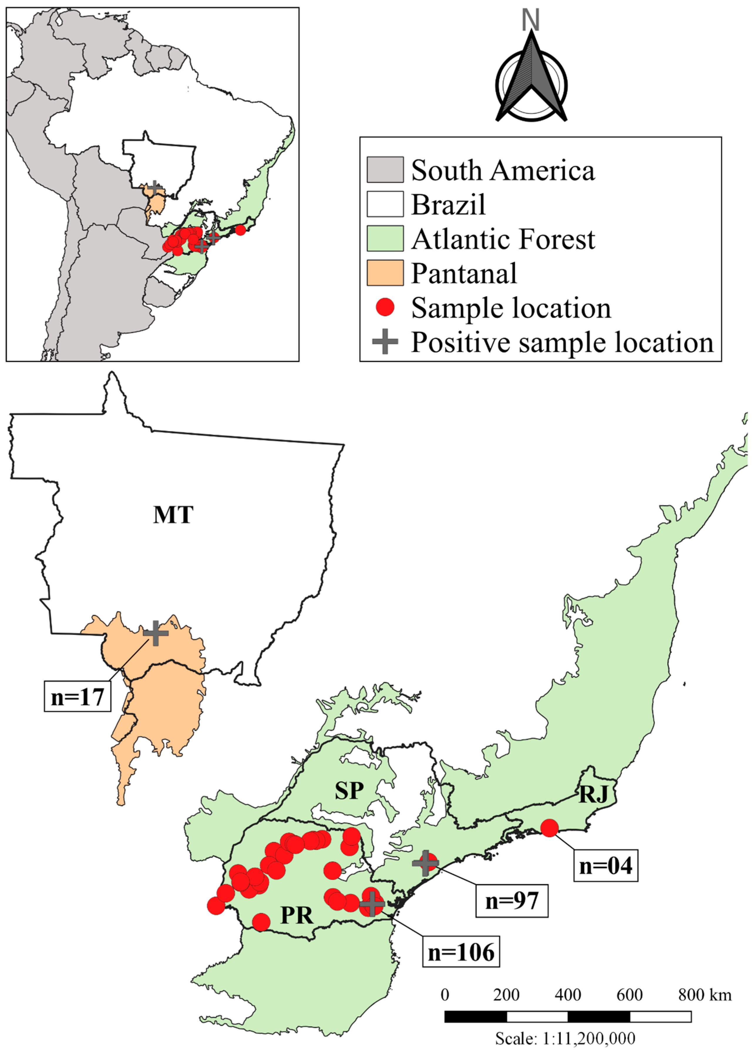
2.1. Sampling

Samples were collected from different species of bats belonging to the Atlantic Forest and Pantanal biomes from four states of different regions, including southern (Paraná/PR), central-western (Mato Grosso/MT), and southeastern (São Paulo/SP and Rio de Janeiro/RJ) Brazil (Figure 1 and Table 1). Specific data for each state are presented in the following items.

2.1.1. Paraná/PR

Brains from bats not identified to species (n = 96) from the Paraná State Reference Laboratory (LACEN) were acquired as part of the rabies virus circulation monitoring program. The collections were carried out between September 2019 and August 2020 in 29 different municipalities in the Paraná State, inserted in remnant fragments of the Atlantic Forest biome and urbanized areas. Samples were stored at −80 °C and thawed immediately prior to DNA/RNA extraction.
Eight blood samples from bats (six Desmodus rotundus and two Diphylla ecaudata, both family Phyllostomidae) were collected in EDTA-anticoagulated tubes by intracardiac puncture from sedated bats, between October and November 2015, in two caves in the municipality of Rio Branco do Sul, PR, which is located in a fragment of Atlantic Forest. Since they are hematophagous species, those bats were collected as part of the rabies monitoring program in herbivores (ruminants and horses) carried out by municipal and state public agencies. In addition, two non-hematophagous bats of the genus Molossus, family Molossidae, were collected for rabies surveillance in Curitiba city, as they were found in public areas nearby the Municipal Zoo of Curitiba, PR [17]. Blood samples were stored at −20 °C until processing.

2.1.2. Mato Grosso/MT and Rio de Janeiro/RJ

Bat blood samples stored on FTA® Whatman® cards (Whatman, Sigma-Aldrich, Darmstadt, Germany) were collected in Mato Grosso (17 samples) and Rio de Janeiro (four samples). Blood smears were also prepared for microscopical examination from Rio de Janeiro.
In Mato Grosso, samples were collected in October 2019 in the municipality of Poconé, in rural areas within the Pantanal biome. Species and families sampled: Glossophaga soricina, family Phyllostomidae (five samples); Molossus molossus, family Molossidae (five samples); Myotis cf. nigricans, family Vespertilionidae (four samples); Rhynchonycteris naso, family Emballonuridae (two samples); Noctilio albiventris, family Noctilionidae (one sample).
Samples from Rio de Janeiro were collected in October 2020 in the city of Rio de Janeiro, in a remnant of Atlantic Forest (Pedra Branca State Park), near Fiocruz Atlantic Forest Biological Station, a highly urbanized region in the central portion of the city, under severe anthropogenic pressure. Only bats of the species Artibeus lituratus, family Phyllostomidae were sampled.

2.1.3. São Paulo/SP

Blood samples from bats (n = 97) were collected in EDTA-anticoagulated tubes by intracardiac puncture from sedated bats. Certain specimens were euthanized using xylazine and ketamine, followed by inhalation of isoflurane. Subsequently, they were preserved in a 10% formaldehyde solution for future identification and storage at the Museum of Zoology of the University of São Paulo (MZUSP). From these specimens, liver (n = 30) and spleen (n = 12) were also collected.
Samples were collected from 2018 to 2021 in Legado das Águas reserve, the largest private reserve of Atlantic Forest in the country. It is inserted in the municipalities of Miracatu and Tapiraí, in the Ribeira Valley, south of the São Paulo State, located 122 km from the state capital, in the southern portion of the Serra do Mar ecological corridor. Legado das Águas covers 31,000 hectares contiguously connected to several other Conservation Units, contributing to an important ecological corridor between the coastal and inland areas of the southern region of São Paulo State. It is the largest continuous area of remaining Atlantic Forest that has suffered minimal human intervention, a factor attributed to the low demographic density and little economic development in the region.
Families and species sampled: family Phyllostomidae, Anoura caudifer (seven samples), Artibeus cinereus (two samples), Artibeus fimbriatus (three samples), Artibeus gnomus (one sample), Artibeus lituratus (nine samples), Artibeus obscurus (nine samples), Artibeus planirostris (three samples), Artibeus sp. (one sample), Carollia perspicillata (eighteen samples), Chrotopterus auritus (one sample), Desmodus rotundus (one sample), Ectophylla sp. (one sample), Lonchorhina aurita (one sample), Platyrrhinus lineatus (six samples), Platyrrhinus sp. (one sample), Rhinophylla pumilio (one sample), Sturnira lilium (seven samples), Thrachops cirrhosus (one sample), Uroderma bilobatum (one sample); family Emballonuridae, Pteropteryx sp. (three samples); family Molossidae, Molossus ater (one sample), Nyctinomops sp. (four samples); family Vespertilionidae, Eptesicus sp. (one sample), Myotis nigricans (eight samples), Myotis riparius (one sample), Myotis ruber (one sample), Myotis sp. (four samples). All these samples are also described in Table 1.

2.2. Ethics Statement

All animals and their tissue samples were collected and handled under appropriate authorizations by the Brazilian government. The project was authorized by SISBIO (Sistema de Autorização e Informação em Biodiversidade), ICMBio/MMA (Instituto Chico Mendes de Conservação da Biodiversidade/Ministério do Meio Ambiente), numbers 72790, 51714-1, and 19037-1. The study was approved by the Ethics in Use of Animals Committee, CEUA/SESA, of the Centro de Produção e Pesquisa de Imunobiológicos—CPPI/PR (approval number 01/2019 and date of approval 3 March 2020), CEUA/FIOCRUZ (approval number LM-6/18/2021 and date of approval 14 May 2018) and CEUA/SUCEN (approval number 09/2021 and date of approval 30 September 2021). Rio de Janeiro sampling was carried out under SisGen authorization A46B0E1.

2.3. Optical Microscopy Diagnosis

The four blood smears available for analysis acquired from bats in Rio de Janeiro were fixed with 100% methanol within 24 h of collection and stained with 10% Giemsa solution for one hour, up to 30 days after collection [3]. The smears were examined for approximately 15–20 min, viewing 100 fields at low magnification (400×) and 100 fields at high magnification (1000×) [3], using a Leica® DM3000LED light microscope. The search for parasitic blood stages was carried out following previous morphological studies on haemosporidians in wild animals [3,8].

2.4. DNA Extraction

Brain tissue samples from Paraná State were extracted using the BioGene DNA/RNA Viral Kit (K204-4, Bioclin, Belo Horizonte, MG, Brazil), following the manufacturer’s instructions. For the blood (200 μL), the DNA was prepared according to the Illustra Kit Mini Genomic Blood Preparation Spin (GE Healthcare, Chalfont, St. Giles, UK), according to the manufacturer’s instructions.
Samples from Rio de Janeiro and Mato Grosso, stored on FTA® Whatman® cards (Whatman, Sigma-Aldrich, Darmstadt, Germany), were extracted using the commercial Wizard SV 96 Genomic DNA Purification System kit (PROMEGA®, Madison, WI, USA), according to the manufacturer’s instructions.
For samples collected in the São Paulo State, genomic DNA was extracted from blood, liver and spleen tissues using PureLink™ Genomic DNA Mini Kit (Thermo Fisher Scientific, Waltham, MA, USA), according to the manufacturer’s instructions.

2.5. Molecular Detection of Haemosporidian Parasites

A fragment of ~1.1 kb (approximately 92% of the gene) from the mitochondrial cytb gene was amplified using a nested polymerase chain reaction (PCR), taking standard precautions to prevent cross-contamination of samples. The PCR reactions were conducted as described in [18] using primers DW2 and DW4 and 5 µL (50 ng) of genomic DNA in the first reaction. An aliquot of 1 µL of the PCR product was used as a template for a nested reaction with primers DW1 and DW6.
For clpc, a fragment of approximately 500 bp was amplified using a nested PCR, as described in [8], using primers clpcF and clpcR and 5 µL (50 ng) of genomic DNA in the first reaction. An aliquot of 1 µL of the PCR product was used as a template for a nested reaction with primers clpcF2 and clpcR2.
For asl, a fragment of approximately 240 bp was amplified using a nested PCR, as described in [8], using the primers aslF and aslR and 5 µL (50 ng) of genomic DNA in the first reaction. An aliquot of 1 µL of the PCR product was used as a template for a nested reaction with primers aslF2 and aslR2.
All PCR amplifications included two controls: one positive control (DNA sample with known Polychromophilus sp. infection) and one negative control (ultrapure water without DNA); the last one was included to check for possible contamination and false positives. All PCR products were evaluated by running 10 μL on 1% agarose gel.
PCR products were sequenced using BigDye® Terminator v3.1 Cycle Sequencing Kit in ABI PRISM® 3500 Genetic Analyzer (Applied Biosystems, Carlsbad, CA, USA) using nested PCR primers. For cytb, the oligonucleotides DW8 and DW3 were also used for sequencing [18]. The cytb, clpc or asl sequences were obtained and aligned with the sequences available in the GenBank® database.

2.6. Phylogenetic Analysis

The phylogenetic relationships among the haemosporidian parasites were inferred using partial sequences of cytb (1116 bp) or the concatenated analysis of three genes, the mitochondrial cytochrome b gene (cytb, 725 bp), the nuclear adenylosuccinate lyase gene (asl, 206 bp), and the apicoplast caseinolytic protease C gene (clpc, 531 bp). Sequences acquired from the GenBank® database used are shown in the phylogenetic trees. The alignment was obtained using the ClustalW algorithm [19] implemented in MEGAX software [20]. The phylogenetic reconstructions were performed using the Bayesian inference method implemented in MrBayes v3.2.0 [21]. The best evolution model was selected using MEGAX software [20]. The model GTR + G + I was considered to describe the substitution pattern the best (with the lowest Bayesian Information Criterion scores). Bayesian inferences were executed with two Markov Chain Monte Carlo searches of 8 million generations, with each sampling 1 of 300 trees. After a burn-in of 25%, the remaining 15,002 trees were used to calculate the 50% majority-rule consensus tree. The standard deviation of split frequencies was <0.01. The phylogenies were visualized using FigTree version 1.4.0 [22].

2.7. Host Species Identification
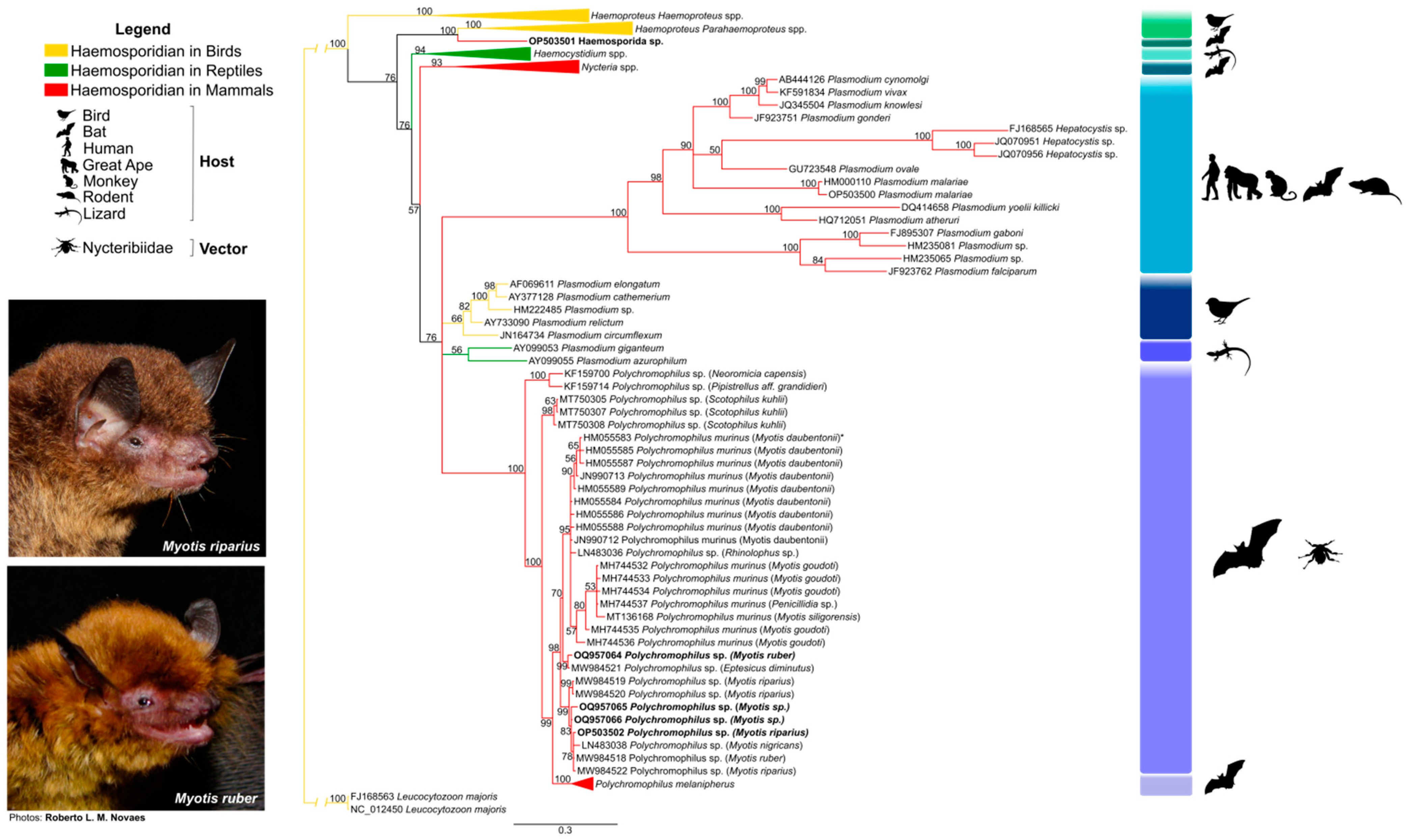
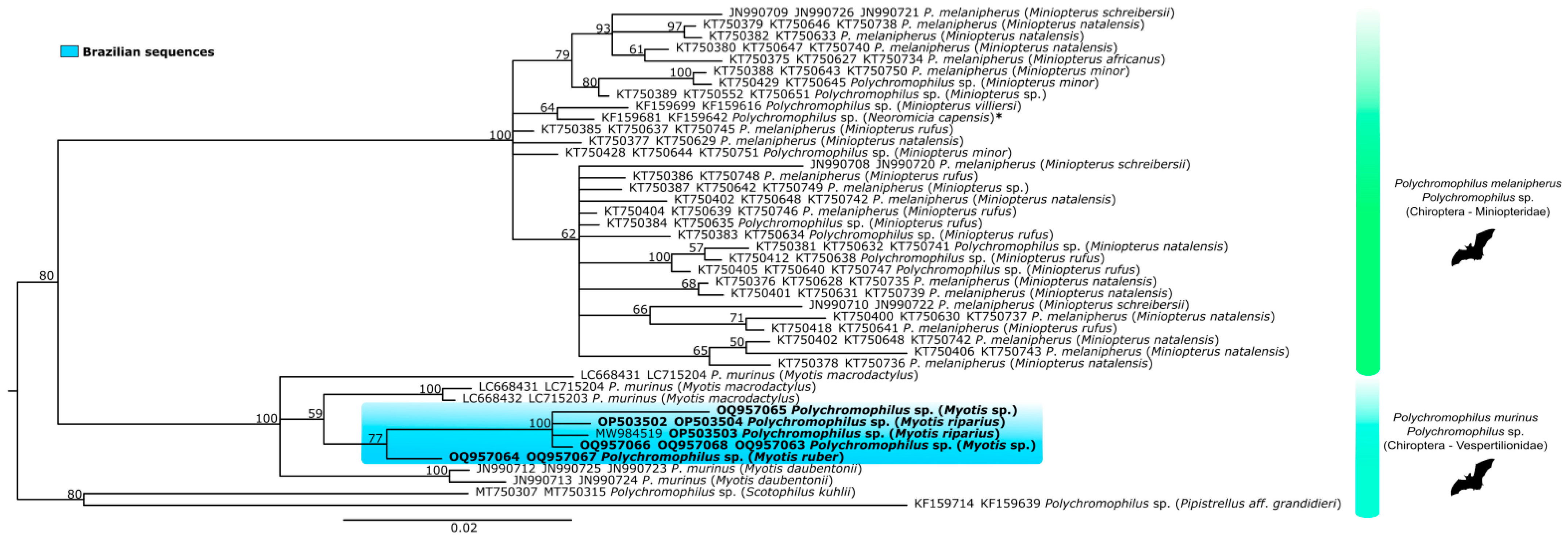
A fragment with ~650 bp from the mitochondrial cytochrome c oxidase (coi) gene was amplified using the universal primers LCO1490 and HCO2198 [23] and PCR protocol based on [24]. Amplified fragments were sequenced directly using the corresponding flanking primers. Sequences (at least 550 bp) were entered in the BOLD platform (Barcode of Life Data system) by the option “Species Level Barcode Records”. Sequences with >99% similarity were used for species identification. For sequences showing <98% identity, the neighbor-joining (NJ) tree of K2P distances showing intra and interspecific variation generated in BOLD was analyzed. Samples were considered identified if assigned to a monophyletic group of sequences corresponding to a single species.

3. Results

3.1. Host Species Identification

We used DNA barcodes and BOLD to identify bat species in the samples obtained from LACEN. Five of the 95 tested samples did not amplify (in two independent experiments). A total of 80 (84.2%) could be identified to the species level using DNA barcoding and BOLD, and 10 (10.5%) generated ambiguous identification and were identified just to genera (one Artibeus sp., one Eumops sp., four Molossus sp., one Sturnira sp., two Eptesicus sp. and one Myotis sp.). From the samples identified to species level, 50 samples showed >99% while 30 had <99% of similarity in the BOLD database. In the last case, the BOLD NJ tree was analyzed to consider the sample identified. Ten different species were obtained: 15 specimens were Vespertilionidae (two Myotis nigricans, one Myotis riparius, and 12 Eptesicus furinalis); 65 bats were Molossidae (23 Molossus rufus, 29 Molossus molossus, five Tadarida brasiliensis, three Promops nasutus, three Eumops glaucinus, one Molossops temminckii, and one Molossops neglectus).

3.2. Haemosporidian Parasites and Phylogeny

The percentage of positive samples by PCR was 2.67%, with six out of 224 bats infected (Table 1). No positive samples were obtained by microscopy. Positive samples were: two brain tissue samples of Myotis riparius (sample IDs 198 and 607), bats belonging to the Vespertilionidae family, collected in two municipalities in the Paraná State (Curitiba and São José dos Pinhais, respectively); one spleen tissue sample of Myotis ruber collected in Tapiraí-SP (sample ID 125); two samples, spleen and liver tissues respectively, of Myotis sp. from Miracatu-SP (sample IDs 138 and 141); and a blood sample of Noctilio albiventris (sample ID Bat17) that belongs to the Noctilionidae family. The latter species was collected near a lake in the Pantanal of Mato Grosso, municipality of Poconé (Figure 1). Interestingly, paired samples from bats ID 125, 138 and 141 obtained variable results, with liver samples being the worst for Polychromophilus detection (Table 2).
Table 1. Bats examined in this study and the prevalence of haemosporidian infection.
Host SpeciesLocalityYear of CollectionBats ExaminedBats Infected with Haemosporidians (%)
Vespertilionidae Gray, 1821
Myotis nigricansRio Bom, Toledo-PR2019–202020
Myotis ripariusSão José dos Pinhais, Curitiba-PR2019–202022 (100%)
Eptesicus furinalisAssis Chateaubriand, Curitiba, Foz do Iguaçu, Maringá, Ramilândia-PR2019–2020120
Eptesicus sp.Curitiba-PR2019–202020
Myotis sp.Curitiba-PR2019–202010
Myotis cf. nigricansPoconé-MT201940
Eptesicus sp.Miracatu-SP201810
Myotis nigricansTapiraí, Miracatu-SP2018–201980
Myotis ripariusMiracatu-SP201910
Myotis ruberTapiraí-SP201911 (100%)
Myotis sp.Miracatu-SP201942 (50%)
Molossidae Gervais, 1856
Molossus sp.Curitiba, Araruna-PR2019–202040
Molossus sp.Curitiba-PR201520
Eumops sp.Foz do Iguaçu-PR2019–202010
Molossus rufusBraganey, Cascavel, Céu Azul, Francisco Beltrão, Jacarezinho, Londrina, Mamborê, Maringá, Maripá, Sarandi, Telêmaco Borba, Vera Cruz do Oeste-PR2019–2020230
Molossus molossusAraucária, Assis Chateaubriand, Cascavel, Curitiba, Foz do Iguaçu, Guapirama, Guaratuba, Mandaguaçu, Maringá, Paulo Frontin, Ramilândia, Sarandi-PR2019–2020290
Molossus molossusPoconé-MT201950
Tadarida brasiliensisCuritiba, Imbituva, Mamborê-PR2019–202050
Promops nasutusCascavel, União da Vitória-PR2019–202030
Eumops glaucinusAssis Chateaubriand, Foz do Iguaçu, Maringá-PR2019–202030
Molossops temminckiiFoz do Iguaçu-PR2019–202010
Molossops neglectusSalto do Lontra-PR2019–202010
Molossus aterTapiraí-SP201810
Nyctinomops sp.Tapiraí-SP2019–202040
Phyllostomidae Gray, 1825
Sturnira sp.Curitiba-PR2019–202010
Artibeus sp.Foz do Iguaçu-PR2019–202010
Artibeus lituratusRio de Janeiro-RJ202040
Desmodus rotundusRio Branco do Sul-PR201560
Diphylla ecaudataRio Branco do Sul-PR201520
Glossophaga soricinaPoconé-MT201950
Anoura caudiferTapiraí-SP201870
Artibeus cinereusTapiraí-SP201820
Artibeus fimbriatusTapiraí, Miracatu-SP201830
Artibeus gnomusTapiraí-SP201810
Artibeus lituratusTapiraí-SP201890
Artibeus obscurusTapiraí-SP201890
Artibeus planirostrisTapiraí-SP201830
Artibeus sp.Tapiraí-SP201810
Carollia perspicillataTapiraí, Miracatu-SP2018180
Chrotopterus auritusTapiraí-SP201810
Desmodus rotundusTapiraí-SP201810
Ectophylla sp.Tapiraí-SP201810
Lonchorhina auritaMiracatu-SP201810
Platyrrhinus lineatusTapiraí-SP202060
Platyrrhinus sp.Tapiraí-SP202010
Rhinophylla pumilioTapiraí-SP202010
Sturnira liliumTapiraí-SP202170
Thrachops cirrhosusTapiraí-SP202110
Uroderma bilobatumMiracatu-SP202110
Emballonuridae Gervais, 1856
Rhynchonycteris nasoPoconé-MT201920
Pteropteryx sp.Tapiraí-SP202030
Noctilionidae Gray, 1821
Noctilio albiventrisPoconé-MT201911 (100%)
unknownCuritiba, Paulo Frontin, Rolândia, Salto do Lontra-PR2019–202050
TOTAL 2246 (2.67%)
PR = Paraná State; MT = Mato Grosso State; RJ = Rio de Janeiro State; SP = São Paulo State.
Sequencing of the PCR amplicon revealed that sample ID 607 (M. riparius) was infected with Polychromophilus sp. It was not possible to identify the haemosporidian parasite in sample Bat17 (Noctilio albiventris) since an unprecedented sequence was obtained. Its closest sequence available on GenBank® (KY653763) was obtained from Haemoproteus minutus, infecting Turdus merula, a passerine collected in Lithuania [25] with a 94% identity.
The cytb gene phylogenetic tree generated using reference sequences available in the GenBank® database covering different haemosporidian genera from different hosts, as well as the sequences found herein are shown in Appendix A. Cytb-based phylogenetic analysis produced no conflict in any of the major nodes. All major genera and subgenera were recovered and represented in the phylogenetic tree by separate monophyletic clades (Figure 2). The results show eight clades within the order Haemosporida analyzed here. All Polychromophilus sequences from bats from different regions of the world were grouped into a monophyletic clade (posterior probability of 100) and consisted of six subclades (with posterior probabilities > 95), with all Polychromophilus found in Brazilian Myotis bats segregated in two of them (Figure 2).
The first Polychromophilus distinct subclade comprised two samples of Pipistrellus aff. grandidieri and Neoromicia capensis, both vespertilionid species from Guinea (KF159700 and KF159714). The second subclade contained the Polychromophilus sequences from Scotophilus kuhlii, a vespertilionid species from Thailand (MT750305, MT750307, and MT750308) (Figure 2). The third Polychromophilus subclade (posterior probability of 95) was composed of sequences of P. murinus from bats in Europe (Switzerland, Bulgaria), Madagascar, and Thailand. This subclade included a sequence of Polychromophilus sp. obtained in Rhinolophus sp. (Rhinolophidae) that stands out for not being part of the vespertilionids (Figure 2). The fourth subclade comprises sequences obtained in Eptesicus diminutus (MW984521) and Myotis ruber (OQ957064) from Brazil. The fifth subclade comprises the sequence of Polychromophilus obtained from M. nigricans from Panama and all other Brazilian sequences isolated from the genus Myotis. This subclade exclusively included Polychromophilus sequences from vespertilionids (including Brazilian ones). All P. melanipherus sequences from hosts of bats of the genus Miniopterus were distinctly separated into a subclade, confirming a clear separation of parasites from miniopterid and vespertilionid hosts (Figure 2).
The sequence of Bat17 (N. albiventris) clustered close to the subclade of Haemoproteus (Parahaemoproteus) spp., a specific genus of bird parasites, being positioned as a sister clade. Thus, although in the phylogenetic tree, the sequence obtained in bat was grouped with others from Haemoproteidae, it was not supported in a monophyletic clade.
The finding of Polychromophilus in Brazilian bats was confirmed with the amplification of the clpc gene, from the apicoplast of the parasite, in three samples (IDs 141, 198 and 607), presenting fragments of approximately 500 bp and also of the asl gene from the nuclear genome, in two samples (IDs 125 and 141), with 244 bp. Compared to sequences of the same target gene on GenBank® for the genus Polychromophilus, the clpc sequences showed 97% similarities with the closest available sequences (LC715203 and LC715204), sequences from P. murinus described in Myotis macrodactylus, a bat collected in Japan [26].
A phylogenetic tree was generated with concatenated sequences from three genes: cytb, asl, and clpc (Figure 3), including the Polychromophilus from this study and all available sequences of this genus in the GenBank® database (Table A2, Appendix A). The tree topology of concatenated genes confirmed the separation of parasites from miniopterid and vespertilionid hosts, except for Neoromicia capensis, a vespertilionid species grouped with miniopterid hosts. The vespertilionid Polychromophilus subclade was divided into three branches: one with P. murinus sequences from Swiss bats (Myotis daubentonii), one with just a sequence from Myotis macrodactylus from Japan, and the third with all the Polychromophilus found in Brazilian bats and another two from Myotis macrodactylus from Japan.

4. Discussion

The study of haemosporidian parasites in bats can significantly contribute to understanding the evolution of these parasites in mammals since seven out of nine genera of this family occurring in bats are considered specific to these hosts [6]. Haemosporidians have been found mainly in Old World bats, except for Polychromophilus from vespertilionid bats: Myotis nigricans from Brazil [14], Myotis nigricans from Panama [15] and, more recently, in Myotis riparius, Myotis ruber and Eptesicus diminutus from Brazil [16].
This study extended the search for haemosporidian parasites in bats to two additional Brazilian areas, including the Pantanal biome. We found a low haemosporidian positivity rate prevalence (2.67%), consistent with our previous study (1.2%) [16]. It is important to note that 52% of the analyzed samples were obtained from tissues (brain, spleen or liver), sample sources that are not common in haemoparasite studies but confirmed its usefulness in the screening of Polychromophilus parasites since we obtained the same amount of positives found in the group of blood samples.
We hypothesize that the low positivity found in our studies is related to the number of samples collected from bats of the Myotis genus (10% in this study), which we believe to be the main host of Polychromophilus in Brazil. In fact, considering only the Myotis bats tested, we found 21% of positives. Of all the nine samples already found positive for Polychromophilus by molecular methods in Brazil (this study and [16]), only one was not within the Myotis species.
The four new Polychromophilus cytb sequences obtained in this study conserved the two nucleotides T (thymine) at positions 247 and 512 of the gene, which is also observed in other Brazilian isolates, but not in the sequence from Panama [16]. Future studies analyzing the cytb sequence of more isolates are needed to verify whether these SNPs are molecular markers of Brazilian Polychromophilus isolates.
The order Chiroptera corresponds to approximately one-quarter of the mammal species in the world [27]. In Brazil, there are nine families with 182 species [28]. The Brazilian families with their respective numbers of species are Emballonuridae (17), Phyllostomidae (94), Mormoopidae (4), Noctilionidae (2), Furipteridae (1), Thyropteridae (5), Natalidae (1), Molossidae (32) and Vespetilionidae (26) [28,29]. They inhabit the entire national territory and are distributed in the most diverse biomes and urban areas, occurring in the Amazon, Cerrado, Caatinga, Atlantic Forest, Pantanal, and Pampas [28,29,30,31,32,33]. To know the diversity of bat species tested in the present study, we used DNA barcoding to identify the bat species in samples with unknown species. The results showed that most of our samples come from the Phyllostomidae family (41.5%), followed by Molossidae (36.6%), Vespertilionidae (16.9%), and Noctilionidae plus Emballonuridae (2.6%), with 2.2% unidentified. Polychromophilus infection in Brazilian bats continues to be limited to just one family (Vespertilionidae). However, a Haemosporida sp. sequence was obtained from a Noctilionidae bat (Noctilio albiventris), a family with just one sample analyzed. It is important to note that there is one record of P. melanipherus in Emballonuridae (Taphozous melanopogon from Thailand) but no previous record of haemosporidian parasites in Molossidae, Phyllostomidae, and Noctilionidae families. Therefore, it is very likely that the prevalence of haemosporidian parasites was low in our study because the vast majority of samples analyzed were from species that are uncommon hosts for these parasites. Since molecular studies showed that 89% of Polychromophilus-positive samples in Brazil were from Myotis species, further studies are needed to confirm their host specificity and to determine if Myotis spp. are the primary hosts for Polychromophilus in the Neotropics.
The bat Noctilio albiventris has a wide geographic distribution, occurring practically throughout Latin America and almost the entire Brazilian territory. It has an insectivorous diet and is always related to humid forest habitats and environments close to rivers, lakes, or coastal marine habitats [33], making this species more susceptible to parasitic diseases transmitted by vectors available in the environment. Moreover, its involvement with dipteran ectoparasites has not been shown [33], reinforcing the possibility of transmission of Haemoproteidae by ceratopogonid dipterans of the genus Culicoides, known vectors of Haemoproteus (Parahaemoproteus) spp. in birds, as well as Hepatocystis in bats [3,7].
The generalist feeding preferences of vector species could provide opportunities for cross-species transmission of Haemoproteus between avian and bat hosts. In this case, the Haemosporida sp. parasite detected in Bat17 likely represents an abortive spill-over infection [3]. In fact, detecting DNA in the blood without the demonstration of parasites in blood smears does not necessarily indicate successful infection, being plausible that its development cannot be completed in bats.
The Haemosporida sp. sequence described here, with the closest sequence identity of 94% with Haemoproteus (Parahaemoproteus) minutus, is insufficient to identify this parasite as any of those previously described in bats or other animals. However, if this finding is not a spill-over, the parasite sequence position in the phylogenetic tree points to a parasite of the Haemoproteidae family. In fact, the Haemoproteidae family harbors genera of haemosporidian parasites that are exclusive to bats, such as Johnsprentia and Sprattiella, which have not been analyzed molecularly yet, and sequences are lacking for comparison.
A combination of morphological evaluation and molecular studies are needed to conclude and further describe the Polychromophilus parasite lineage, as well as the Haemosporida sp. found in Brazilian bats. Nevertheless, these results confirm the importance of studying these neglected haemosporidian parasites in bats in Brazil.

Author Contributions

Conceptualization, A.W.B. and K.K.; formal analysis, B.d.S.M. and C.C.d.A.; investigation, B.d.S.M., L.d.O.G. and C.C.d.A.; resources, G.A.M., A.W.B., J.d.O.J.C., H.S.S., A.M., I.N.R., A.F., J.B.P., M.G.B. and K.K.; data curation, B.d.S.M. and C.C.d.A.; writing—original draft preparation, B.d.S.M., A.P.D.S. and K.K.; writing—review and editing, B.d.S.M., G.A.M., A.W.B., J.d.O.J.C., H.S.S., A.M., I.N.R., L.d.O.G., C.C.d.A., A.P.D.S., A.F., M.G.B. and K.K.; visualization, B.d.S.M. and C.C.d.A.; supervision, K.K.; project administration, K.K.; funding acquisition, K.K. All authors have read and agreed to the published version of the manuscript.

Funding

A.F. was funded by a PNPD scholarship from Coordenação de Aperfeiçoamento de Pessoal de Nível Superior—CAPES (Process number 88887.342366/2019-00). K.K. is a CNPq research fellow (Process number 309396/2021-2). This research benefited from the State Research Institutes Modernization Program, funded by Fundação de Amparo à Pesquisa do Estado de São Paulo (FAPESP 2017/50345-5).

Data Availability Statement

The data presented in this study are available in Appendix A and also in the GenBank® database. https://www.ncbi.nlm.nih.gov/genbank/ (accessed on 19 March 2022) (accession numbers OP503500-OP503504).

Acknowledgments

We are grateful to Roberto Leonan M. Novaes from Fiocruz Mata Atlântica, Fundação Oswaldo Cruz-Fiocruz, Rio de Janeiro, Brazil, for sharing the bats’ photographs and reviewing the manuscript. We would also thank Fiocruz Mata Atlântica-Fiocruz team for the fieldwork support and Rogério V. Rossi from the University of Mato Grosso, who kindly identified the bats collected in Pantanal. Finally, we are extremely indebted to Juliane Schaer (Humboldt University, Berlin, Germany) for her valuable comments and insightful suggestions on the manuscript.

Conflicts of Interest

The authors declare no conflict of interest.

Appendix A

Table A1. Mitochondrial cytochrome b (cytb) gene sequences used in phylogenetic analyzes and their respective GenBank® accession numbers. Sequences from this study are highlighted in bold.
GenBank®
Accession Number
Parasite
Species
Host
Species
Country of Source
MN316537, MN316538Haemocystidium cf. chelodinaeMyuchelys georgesiAustralia
MK976708-MK976710Haemocystidium pacayaePodocnemis vogliColombia
MH177855Haemocystidium ptyodactyliiSquamate *unknown
KT364883Haemocystidium sp.Hemidactylus luqueorumOman
KT364884Haemocystidium sp.Ptyodactylus hasselquistiiOman
KX148083-KX148085Haemocystidium sp.Kinixys erosaGabon
KX148088-KX148090Haemocystidium sp.Kinixys erosaGabon
KX148086, KX148087Haemocystidium sp.Pelusios castaneusGabon
MT684458Haemocystidium sp.Podocnemis vogliColombia
MT684459Haemocystidium sp.Trachylepis spilogasterAngola
MT684460Haemocystidium sp.Rhacodactylus auriculatusNew Caledonia
DQ630007Haemoproteus balmoraliLuscinia luscinaLithuania
DQ630014Haemoproteus balmoraliMuscicapa striataLithuania
DQ630006Haemoproteus belopolskyiHippolais icterinaSweden
MK843310Haemoproteus belopolskyiHippolais icterinaLithuania
FJ168562Haemoproteus columbaeColumba liviaUSA
MK843311Haemoproteus hirundinisDelichon urbicumLithuania
KY653778Haemoproteus iwaFregata magnificensEcuador
KY653760Haemoproteus jenniaeCreagrus furcatusEcuador
DQ630010Haemoproteus laniiLanius collurioRussia
MK843313Haemoproteus laniiLanius collurioLithuania
AY099045Haemoproteus majorisParus caeruleusSweden
JN164727, JN164728Haemoproteus majorisSylvia atricapillaSpain
KU160476Haemoproteus minchiniCorythaeola cristataSingapore
DQ630013Haemoproteus minutusTurdus merulaLithuania
KY653756Haemoproteus multipigmentatusZenaida galapagoensisEcuador
MK843312Haemoproteus nucleocondensusAcrocephalus arundinaceusLithuania
JN164720Haemoproteus pallidulusSylvia atricapillaSpain
DQ630004, DQ630005Haemoproteus pallidusFicedula hypoleucaSweden, Russia
JN164718, JN164719, JN164722Haemoproteus parabelopolskyiSylvia atricapillaSpain
DQ630009Haemoproteus payevskyAcrocephalus scipaceusLithuania
AY099040Haemoproteus sylvaeAcrocephalus arundinaceusSweden
OP503501Haemosporida sp.Noctilio albiventris (ID Bat17)Brazil
FJ168565Hepatocystis sp.Pteropus hypomelanusUSA
JQ070951, JQ070956Hepatocystis sp.Cercopithecus nictitansCameroon
FJ168563Leucocytozoon majorisZonotrichia leucophrys orianthaUSA
NC_012450Leucocytozoon majorisZonotrichia leucophrys orianthaUSA
KF159690Nycteria sp.Rhinolophus landeriGuinea
KF159720Nycteria sp.Rhinolophus alcyoneCôte d’Ivoire
MK098843-MK098847Nycteria sp.Rhinolophus sp., R. landeriGabon
FJ168561Parahaemoproteus vireonisVireo gilvusUSA
NC_012447Parahaemoproteus vireonisVireo gilvusUSA
HQ712051Plasmodium atheruriAtherurus africanusMadagascar
AY099055Plasmodium azurophilumAnolis oculatusDominica
AY377128Plasmodium cathemeriumSerinus canariaGermany
JN164734Plasmodium circumflexumSylvia atricapillaSpain
AB444126Plasmodium cynomolgiMonkey *Japan
AF069611Plasmodium elongatumPasser domesticusNorth America
JF923762Plasmodium falciparumCercopithecus nictitansGabon
FJ895307Plasmodium gaboniPan sp.Gabon
AY099053Plasmodium giganteumAgama agamaGhana
JF923751Plasmodium gonderiMandrillus sphinxGabon
JQ345504Plasmodium knowlesiHomo sapiensMalaysia
HM000110Plasmodium malariaePan troglodytes elliotiCameroon
OP503500Plasmodium malariaeHomo sapiens (ID I11)Brazil
GU723548Plasmodium ovaleHomo sapiensEngland
AY733090Plasmodium relictumHemignathus virensUSA
HM222485Plasmodium sp.Icteria virensUSA
HM235065Plasmodium sp.Gorilla sp.Cameroon
HM235081Plasmodium sp.Gorilla sp.Cameroon
KF591834Plasmodium vivaxHomo sapiensCongo
DQ414658Plasmodium yoelii killickiThamnomys rutilansCongo
JN990708-JN990711Polychromophilus melanipherusMiniopterus schreibersiiSwitzerland
KJ131270-KJ131275Polychromophilus melanipherusMiniopterus schreibersiiEuropa
KU182361-KU182367Polychromophilus melanipherusNycteribia schmidlii scottiGabon
KU182368Polychromophilus melanipherusPenicillidia fulvidaGabon
MH744504, MH744505Polychromophilus melanipherusMiniopterus mahafaliensisMadagascar
MH744506, MH744519Polychromophilus melanipherusMiniopterus griffithsiMadagascar
MH744508Polychromophilus melanipherusMiniopterus griveaudiMadagascar
MH744522-MH744525Polychromophilus melanipherusMiniopterus griveaudiMadagascar
MH744509-MH744511 Polychromophilus melanipherusMiniopterus gleniMadagascar
MH744518, MH744521Polychromophilus melanipherusMiniopterus gleniMadagascar
MH744512, MH744526Polychromophilus melanipherusMiniopterus manaviMadagascar
MH744514-MH744516Polychromophilus melanipherusMiniopterus griveaudiMadagascar
MH744520Polychromophilus melanipherusParatriaenops furculusMadagascar
MH744527Polychromophilus melanipherusNycteribia stylidiopsisMadagascar
MH744528-MH744531Polychromophilus melanipherusPenicillidia leptothrinaxMadagascar
MK088162-MK088164, MK088168Polychromophilus melanipherusMiniopterus orianaeAustralia
MT136167Polychromophilus melanipherusTaphozous melanopogonThailand
MW007671-MW007674 Polychromophilus melanipherusNycteribia schmidlii scottiSouth Africa
MW007676Polychromophilus melanipherusNycteribia schmidlii scottiSouth Africa
MW007677Polychromophilus melanipherusMiniopterus natalensisSouth Africa
MW007680-MW007682 Polychromophilus melanipherusNycteribia schmidliiHungary
MW007685Polychromophilus melanipherusNycteribia schmidliiSpain
MW007689Polychromophilus melanipherusMiniopterus schreibersiiSpain
HM055583Polychromophilus murinusMyotis daubentoniiSwitzerland
HM055583Polychromophilus murinusEptesicus serotinusSwitzerland
HM055583Polychromophilus murinusNyctalus noctulaSwitzerland
HM055583Polychromophilus murinusMyotis myotisSwitzerland
HM055584-HM055589Polychromophilus murinusMyotis daubentoniiSwitzerland
JN990712, JN990713Polychromophilus murinusMyotis daubentoniiSwitzerland
MH744532-MH744536Polychromophilus murinusMyotis goudotiMadagascar
MH744537Polychromophilus murinusPenicillidia sp.Madagascar
MT136168Polychromophilus murinusMyotis siligorensisThailand
KF159675, KF159681Polychromophilus sp.Miniopterus villiersiGuinea
KF159699Polychromophilus sp.Miniopterus villiersiGuinea
KF159700Polychromophilus sp.Neoromicia capensisGuinea
LN483036Polychromophilus sp.Rhinolophus sp.Bulgaria
LN483038Polychromophilus sp.Myotis nigricansPanama
MK098848, MK098849Polychromophilus sp.Miniopterus minorGabon
OP503502Polychromophilus sp.Myotis riparius (ID 607)Brazil
JQ995284-JQ995288Polychromophilus sp.Miniopterus inflatusGabon
KF159714Polychromophilus sp.Pipistrellus aff. grandidieriGuinea
MT750305, MT750307, MT750308Polychromophilus sp.Scotophilus kuhliiThailand
MW984518Polychromophilus sp.Myotis ruberBrazil
MW984519, MW984520 Polychromophilus sp.Myotis ripariusBrazil
MW984522Polychromophilus sp.Myotis ripariusBrazil
MW984521Polychromophilus sp.Eptesicus diminutusBrazil
OQ957064Polychromophilus sp.Myotis ruber (ID 125)Brazil
OQ957065Polychromophilus sp.Myotis sp. (ID 138)Brazil
OQ957066Polychromophilus sp.Myotis sp. (ID 141)Brazil
* unreported species.
Table A2. Mitochondrial gene cytochrome b (cytb), nuclear gene adenylosuccinate lyase (asl) and apicoplast gene caseinolytic protease C (clpc) sequences from Polychromophilus species used in phylogenetic analyzes and their respective GenBank® accession numbers. Sequences from this study are highlighted in bold.
Host SpeciesParasite SpeciescytbaslclpcCountry of Source
Miniopterus schreibersiiPolychromophilus melanipherusJN990708-JN990720Switzerland
Miniopterus schreibersiiPolychromophilus melanipherusJN990709JN990726JN990721Switzerland
Miniopterus schreibersiiPolychromophilus melanipherusJN990710-JN990722Switzerland
Myotis daubentoniiPolychromophilus murinusJN990712JN990725JN990723Switzerland
Myotis daubentoniiPolychromophilus murinusJN990713-JN990724Switzerland
Miniopterus villiersiPolychromophilus sp.KF159699-KF159616Guinea
Neoromicia capensisPolychromophilus sp.KF159681-KF159642Guinea
Pipistrellus aff. grandidieriPolychromophilus sp.KF159714-KF159639Guinea
Miniopterus natalensisPolychromophilus melanipherusKT750379KT750646KT750738Kenya
Miniopterus natalensisPolychromophilus melanipherusKT750382KT750633-Kenya
Miniopterus natalensisPolychromophilus melanipherusKT750380KT750647KT750740Kenya
Miniopterus rufusPolychromophilus melanipherusKT750385KT750637KT750745Kenya
Miniopterus rufusPolychromophilus melanipherusKT750386-KT750748Kenya
Miniopterus sp.Polychromophilus melanipherusKT750387KT750642KT750749Kenya
Miniopterus natalensisPolychromophilus melanipherusKT750377KT750629-Kenya
Miniopterus africanusPolychromophilus melanipherusKT750375KT750627KT750734Kenya
Miniopterus natalensisPolychromophilus melanipherusKT750400KT750630KT750737Kenya
Miniopterus rufusPolychromophilus melanipherusKT750403KT750636KT750744Kenya
Miniopterus rufusPolychromophilus melanipherusKT750404KT750639KT750746Kenya
Miniopterus rufusPolychromophilus melanipherusKT750418KT750641-Kenya
Miniopterus natalensisPolychromophilus melanipherusKT750376KT750628KT750735Kenya
Miniopterus natalensisPolychromophilus melanipherusKT750401KT750631KT750739Kenya
Miniopterus natalensisPolychromophilus melanipherusKT750402KT750648KT750742Kenya
Miniopterus natalensisPolychromophilus melanipherusKT750406-KT750743Kenya
Miniopterus natalensisPolychromophilus melanipherusKT750378-KT750736Kenya
Miniopterus minorPolychromophilus sp.KT750388KT750643KT750750Tanzania
Miniopterus minorPolychromophilus sp.KT750428KT750644KT750751Tanzania
Miniopterus minorPolychromophilus sp.KT750429KT750645-Tanzania
Miniopterus sp.Polychromophilus sp.KT750389KT750552KT750651Mozambique
Miniopterus rufusPolychromophilus sp.KT750384KT750635-Kenya
Miniopterus rufusPolychromophilus sp.KT750383KT750634-Kenya
Miniopterus natalensisPolychromophilus sp.KT750381KT750632KT750741Kenya
Miniopterus rufusPolychromophilus sp.KT750412KT750638-Kenya
Miniopterus rufusPolychromophilus sp.KT750405KT750640KT750747Kenya
Scotophilus kuhliiPolychromophilus sp.MT750307-MT750315Thailand
Myotis macrodactylusPolychromophilus murinus LC668431-LC715204Japan
Myotis macrodactylusPolychromophilus murinusLC668432-LC715203Japan
Myotis macrodactylusPolychromophilus murinusLC668433-LC715205Japan
Myotis riparius (ID 198)Polychromophilus sp.MW984519-OP503503Brazil
Myotis riparius (ID 607)Polychromophilus sp.OP503502-OP503504Brazil
Myotis ruber (ID 125)Polychromophilus sp.OQ957064OQ957067-Brazil
Myotis sp. (ID 138)Polychromophilus sp.OQ957065--Brazil
Myotis sp. (ID 141)Polychromophilus sp.OQ957066OQ957068OQ957063Brazil

References

  1. Beltz, L.A. Bats and Human Health: Ebola, SARS, Rabies and Beyond; John Wiley & Sons: Hoboken, NJ, USA, 2018. [Google Scholar]
  2. Pacheco, M.A.; Escalante, A.A. Origin and diversity of malaria parasites and other Haemosporida. Trends Parasitol. 2023, 16, S1471-4922(23)00096-X. [Google Scholar] [CrossRef] [PubMed]
  3. Valkiūnas, G. Avian Malaria Parasites and Other Haemosporidia; CRC Press: Boca Raton, FL, USA, 2005; ISBN 978-0-415-30097-1. [Google Scholar]
  4. Perkins, S.L.; Austin, C.C. Four New Species of Plasmodium from New Guinea Lizards: Integrating Morphology and Molecules. J. Parasitol. 2009, 95, 424–433. [Google Scholar] [CrossRef]
  5. Votýpka, J.; Modrý, D.; Oborník, M.; Šlapeta, J.; Lukeš, J. Apicomplexa. In Handbook of the Protists; Archibald, J.M., Simpson, A.G.B., Slamovits, C.H., Eds.; Springer International Publishing: Cham, Switzerland, 2017; pp. 567–624. ISBN 978-3-319-28147-6. [Google Scholar]
  6. Perkins, S.L.; Schaer, J. A Modern Menagerie of Mammalian Malaria. Trends Parasitol. 2016, 32, 772–782. [Google Scholar] [CrossRef] [PubMed]
  7. Carreno, R.A.; Kissinger, J.C.; McCutchan, T.F.; Barta, J.R. Phylogenetic Analysis of Haemosporinid Parasites (Apicomplexa: Haemosporina) and Their Coevolution with Vectors and Intermediate Hosts. Arch. Für Protistenkd. 1997, 148, 245–252. [Google Scholar] [CrossRef]
  8. Martinsen, E.S.; Perkins, S.L.; Schall, J.J. A Three-Genome Phylogeny of Malaria Parasites (Plasmodium and Closely Related Genera): Evolution of Life-History Traits and Host Switches. Mol. Phylogenet. Evol. 2008, 47, 261–273. [Google Scholar] [CrossRef]
  9. Schaer, J.; Perkins, S.L.; Decher, J.; Leendertz, F.H.; Fahr, J.; Weber, N.; Matuschewski, K. High Diversity of West African Bat Malaria Parasites and a Tight Link with Rodent Plasmodium Taxa. Proc. Natl. Acad. Sci. USA 2013, 110, 17415–17419. [Google Scholar] [CrossRef] [Green Version]
  10. Landau, I.; Rosin, G.; Miltgen, F.; Hugot, J.-P.; Leger, N.; Beveridge, I.; Baccam, D. Sur Le Genre Polycbromophilus: (Haemoproteidae, Parasite de Microchiroptères)1. Ann. Parasitol. Hum. Comp. 1980, 55, 13–32. [Google Scholar] [CrossRef] [Green Version]
  11. Landau, I.; Baccam, D.; Ratanaworabhan, N.; Yenbutra, S.; Boulard, Y.; Chabaud, A.G. [New Haemoproteidae parasites of Chiroptera in Thailand]. Ann. Parasitol. Hum. Comp. 1984, 59, 437–447. [Google Scholar] [CrossRef] [Green Version]
  12. Landau, I.; Chavatte, J.M.; Beveridge, I. Johnsprentia Copemani Gen. Nov., Sp. Nov. (Haemoproteidae), a Parasite of the Flying-Fox, Pteropus Alecto (Pteropidae). Mem. Qld. Mus. 2012, 56, 61–66, ISSN 0079-8835. [Google Scholar]
  13. Landau, I.; Chavatte, J.M.; Karadjian, G.; Chabaud, A.; Beveridge, I. The Haemosporidian Parasites of Bats with Description of Sprattiella Alecto Gen. Nov., Sp. Nov. Parasite 2012, 19, 137–146. [Google Scholar] [CrossRef] [Green Version]
  14. Garnham, P.C.C.; Lainson, R.; Shaw, J.J. A Contribution to the Study of the Haematozoon Parasites of Bats. A New Mammalian Haemoproteid, Polychromophilus Deanei n. Sp. Mem. Inst. Oswaldo Cruz 1971, 69, 119–125. [Google Scholar] [CrossRef]
  15. Borner, J.; Pick, C.; Thiede, J.; Kolawole, O.M.; Kingsley, M.T.; Schulze, J.; Cottontail, V.M.; Wellinghausen, N.; Schmidt-Chanasit, J.; Bruchhaus, I.; et al. Phylogeny of Haemosporidian Blood Parasites Revealed by a Multi-Gene Approach. Mol. Phylogenet. Evol. 2016, 94, 221–231. [Google Scholar] [CrossRef]
  16. Minozzo, G.A.; da Silva Mathias, B.; Riediger, I.N.; de Oliveira Guimarães, L.; dos Anjos, C.C.; Monteiro, E.F.; dos Santos, A.P.; Biondo, A.W.; Kirchgatter, K. First Molecular Detection of Polychromophilus Parasites in Brazilian Bat Species. Microorganisms 2021, 9, 1240. [Google Scholar] [CrossRef]
  17. Correia dos Santos, L.; Vidotto, O.; dos Santos, N.J.R.; Ribeiro, J.; Pellizzaro, M.; dos Santos, A.P.; Haisi, A.; Wischral Jayme Vieira, T.S.; de Barros Filho, I.R.; Cubilla, M.P.; et al. Hemotropic Mycoplasmas (Hemoplasmas) in Free-Ranging Bats from Southern Brazil. Comp. Immunol. Microbiol. Infect. Dis. 2020, 69, 101416. [Google Scholar] [CrossRef]
  18. Perkins, S.L.; Schall, J.J. A molecular phylogeny of malarial parasites recovered from cytochrome b gene sequences. J. Parasitol. 2002, 88, 972–978. [Google Scholar] [CrossRef]
  19. Thompson, J.D.; Higgins, D.G.; Gibson, T.J. CLUSTAL W: Improving the sensitivity of progressive multiple sequence alignment through sequence weighting, position-specific gap penalties and weight matrix choice. Nucleic Acids Res. 1994, 22, 4673–4680. [Google Scholar] [CrossRef] [Green Version]
  20. Kumar, S.; Stecher, G.; Li, M.; Knyaz, C.; Tamura, K. MEGA X: Molecular Evolutionary Genetics Analysis across Computing Platforms. Mol. Biol. Evol. 2018, 35, 1547–1549. [Google Scholar] [CrossRef]
  21. Huelsenbeck, J.P.; Ronquist, F. MRBAYES: Bayesian Inference of Phylogenetic Trees. Bioinformatics 2001, 17, 754–755. [Google Scholar] [CrossRef] [Green Version]
  22. Rambaut FigTree: Tree Figure Drawing Tool; Version 1.4.0; Institute of Evolutionary Biology, University of Edinburgh: Edinburgh, UK, 2010.
  23. Folmer, O.; Black, M.; Hoeh, W.; Lutz, R.; Vrijenhoek, R. DNA Primers for Amplification of Mitochondrial Cytochrome c Oxidase Subunit I from Diverse Metazoan Invertebrates. Mol. Mar. Biol. Biotechnol. 1994, 3, 294–299. [Google Scholar]
  24. Ruiz, F.; Linton, Y.-M.; Ponsonby, D.J.; Conn, J.E.; Herrera, M.; Quiñones, M.L.; Vélez, I.D.; Wilkerson, R.C. Molecular Comparison of Topotypic Specimens Confirms Anopheles (Nyssorhynchus) Dunhami Causey (Diptera: Culicidae) in the Colombian Amazon. Mem. Inst. Oswaldo Cruz 2010, 105, 899–903. [Google Scholar] [CrossRef]
  25. Pacheco, M.A.; Matta, N.E.; Valkiūnas, G.; Parker, P.G.; Mello, B.; Stanley, C.E.; Lentino, M.; Garcia-Amado, M.A.; Cranfield, M.; Kosakovsky Pond, S.L.; et al. Mode and Rate of Evolution of Haemosporidian Mitochondrial Genomes: Timing the Radiation of Avian Parasites. Mol. Biol. Evol. 2018, 35, 383–403. [Google Scholar] [CrossRef] [Green Version]
  26. Rosyadi, I.; Shimoda, H.; Takano, A.; Yanagida, T.; Sato, H. Isolation and molecular characterization of Polychromophilus spp. (Haemosporida: Plasmodiidae) from the Asian long-fingered bat (Miniopterus fuliginosus) and Japanese large-footed bat (Myotis macrodactylus) in Japan. Parasitol. Res. 2022, 121, 2547–2559. [Google Scholar] [CrossRef]
  27. Simmons, N.B. Order Chiroptera. In Mammal Species of the World: A Taxonomic and Geographic Reference; Wilson, D.E., Reeder, D.M., Eds.; Johns Hopkins University Press: Baltimore, MD, USA, 2005; pp. 312–529. ISBN 978-0-8018-8221-0. [Google Scholar]
  28. Abreu, E.F.; Casali, D.; Costa-Araújo, R.; Garbino, G.S.T.; Libardi, G.S.; Loretto, D.; Loss, A.C.; Marmontel, M.; Moras, L.M.; Nascimento, M.C.; et al. Lista de Mamíferos do Brasil (Version 2022-1) [Data Set]. Available online: https://zenodo.org/record/7469767 (accessed on 19 March 2022).
  29. Nogueira, M.R.; de Lima, I.P.; Moratelli, R.; da Cunha Tavares, V.; Gregorin, R.; Peracchi, A.L. Checklist of Brazilian Bats, with Comments on Original Records. Check List 2014, 10, 808–821. [Google Scholar] [CrossRef] [Green Version]
  30. Paglia, A.P.; Fonseca, G.A.B.; Rylands, A.B.; Herrmann, G.; Aguiar, L.M.S.; Chiarello, A.G.; Leite, Y.L.R.; Costa, L.P.; Siciliano, S.; Kierulff, A.M.; et al. Lista Anotada Dos Mamíferos Do Brasil. In Occasional Papers in Conservation Biology; Conservação Internacional: Belo Horizonte, MG, Brazil, 2012. [Google Scholar]
  31. Dias, D.; Esbérard, C.E.L.; Moratelli, R. A New Species of Lonchophylla (Chiroptera, Phyllostomidae) from the Atlantic Forest of Southeastern Brazil, with Comments on L. Bokermanni. Zootaxa 2013, 3722, 347. [Google Scholar] [CrossRef] [Green Version]
  32. Reis, N.R.; Fregonezi, M.N.; Peracchi, A.L.; Shibatta, O.A. Morcegos Do Brasil: Guia de Campo, 1st ed.; Série Manuais & guias TB; Technical Books Editora: Rio de Janeiro, Brazil, 2013; ISBN 978-85-61368-31-9. [Google Scholar]
  33. Reis, N.R.; Peracchi, A.L.; Batista, C.B.; de Lima, I.P.; Pereira, A.D. História Natural Dos Morcegos Brasileiros: Chave de Identificação de Espécies, 1st ed.; Technical Books Editora: Rio de Janeiro, Brazil, 2017; ISBN 978-85-61368-55-5. [Google Scholar]
Figure 1. Map showing the different biomes and sample origin sites.
Microorganisms 11 01531 g001
Figure 2. Bayesian phylogeny based on the mitochondrial cytochrome b gene (cytb) from haemosporidian parasites from this study and reference sequences, totaling 180 sequences (Table A1, Appendix A) in 1116 bp alignment. Leucocytozoon spp. was used as the external group. The support values of the nodes (in percentage) indicate posterior probabilities. The red branches highlight the haemosporidian sequences found in mammals. The yellow branches highlight the haemosporidian sequences found in birds. The green branches highlight the haemosporidian sequences found in reptiles. Sequences from this study are highlighted in bold. * Sequence HM055583 was also reported in P. murinus from Eptesicus serotinus, Nyctalus noctula, and Myotis myotis.
Microorganisms 11 01531 g002
Figure 3. Bayesian phylogeny based on the concatenated analysis of three genes, the mitochondrial cytochrome b gene (cytb, 725 bp), the nuclear adenylosuccinate lyase gene (asl, 206 bp), and the apicoplast caseinolytic protease C gene (clpc, 531 bp) from Polychromophilus spp. of the sequences identified in the present study (highlighted in bold) and reference sequences listed in Table A2 (Appendix A), totalizing 43 sequences. The support values of the nodes (in percentage) indicate posterior probabilities. Brazilian sequences are highlighted in blue. * Neoromicia capensis is a vespertilionid species.
Microorganisms 11 01531 g003
Table 2. Bats examined in this study with positive PCR results in paired samples.
Sample IDBlood SampleSpleen SampleLiver Sample
125NAPositiveNegative
138PositivePositiveNegative
141PositiveNAPositive
NA = not available.
Disclaimer/Publisher’s Note: The statements, opinions and data contained in all publications are solely those of the individual author(s) and contributor(s) and not of MDPI and/or the editor(s). MDPI and/or the editor(s) disclaim responsibility for any injury to people or property resulting from any ideas, methods, instructions or products referred to in the content.

