Review

Association between Intestinal Microecological Changes and Atherothrombosis

1
Department of Clinical Medicine, Affiliated Hospital of Hangzhou Normal University, Hangzhou Normal University, Wenzhou Road, Gongshu District, Hangzhou 310000, China
2
Hangzhou Institute of Cardiovascular Disease, Hangzhou 310000, China
*
Authors to whom correspondence should be addressed.
These authors contributed equally to this work.
Microorganisms 2023, 11(5), 1223; https://doi.org/10.3390/microorganisms11051223
Submission received: 11 April 2023 / Revised: 28 April 2023 / Accepted: 2 May 2023 / Published: 6 May 2023
(This article belongs to the Special Issue Gut Microbiota in Disease)

Abstract

:
Atherosclerosis (AS) is a chronic inflammatory disease of large- and medium-sized arteries that causes ischemic heart disease, strokes, and peripheral vascular disease, collectively called cardiovascular disease (CVD), and is the leading cause of CVD resulting in a high rate of mortality in the population. AS is pathological by plaque development, which is caused by lipid infiltration in the vessel wall, endothelial dysfunction, and chronic low-grade inflammation. Recently, more and more scholars have paid attention to the importance of intestinal microecological disorders in the occurrence and development of AS. Intestinal G-bacterial cell wall lipopolysaccharide (LPS) and bacterial metabolites, such as oxidized trimethylamine (TMAO) and short-chain fatty acids (SCFAs), are involved in the development of AS by affecting the inflammatory response, lipid metabolism, and blood pressure regulation of the body. Additionally, intestinal microecology promotes the progression of AS by interfering with the normal bile acid metabolism of the body. In this review, we summarize the research on the correlation between maintaining a dynamic balance of intestinal microecology and AS, which may be potentially helpful for the treatment of AS.

1. Introduction

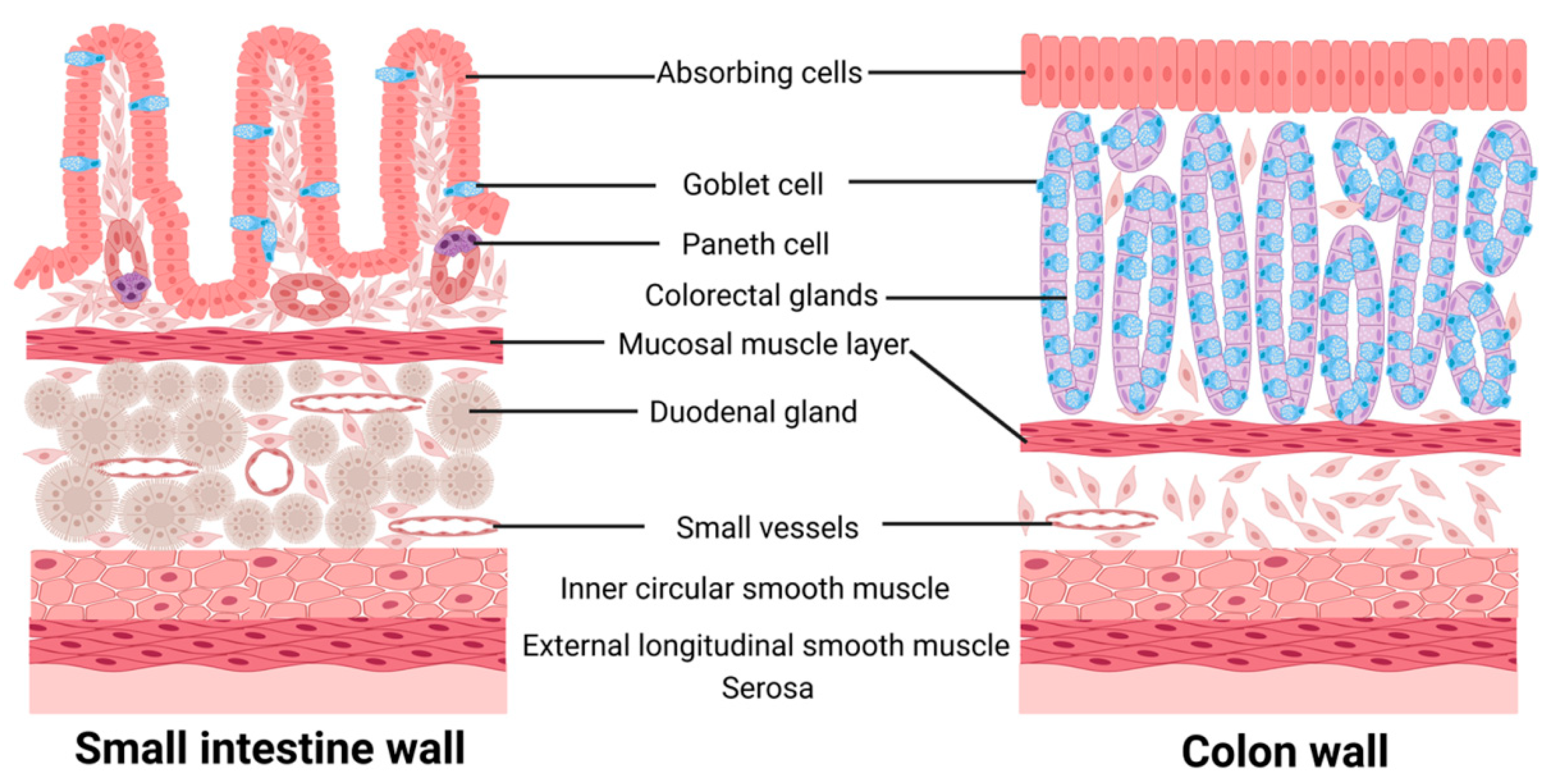
1.1. Intestinal Microecology

Intestinal microecology is a dynamic and balanced system consisting of four parts: the intestinal anatomy, mucosal secretions, food intake, and microorganisms colonizing the intestine [1]. The human intestine, a part of the digestive tract, is divided into the small intestine and large intestine according to its morphology, function, and structure. The wall of the small intestine consists of a mucosal layer, sub-mucosal layer, muscular layer, and outer membrane, and the mucosal epithelium is composed of absorptive cells, cup cells, and Paneth cells, which make the small intestine the main part of nutrient absorption in the human body. The wall of the large intestine is similar to the wall of the small intestine, but its epithelial lamina propria contains dense colonic glands, which are the main sites for absorbing water from food residues and forming feces [2,3,4,5] (Figure 1). The intestinal microbiota consists of over 100 trillion microorganisms, with bacteria accounting for 90% of the total intestinal microorganisms, primarily from the Bacteroides and Firmicutes phyla [6]. Other intestinal microflora, such as Fusobacteria, Bacteroidetes, and Verrucomicrobiaphylum, are present in much smaller quantities [7].
The structure and function of intestinal flora vary across populations and are primarily influenced by the hosts’ diet, age, and living environment. The proportion of intestinal microflora varies among different age groups, with the bifidobacteria-dominated microbiota in infants shifting to a microbiota dominated by Bacteroidetes and Firmicutes in adults, while the relative proportion of the phylum Bacteroides dominates in the elderly [8]. Diet structure can also change the colonization environment of normal intestinal flora, affecting their growth, development, and reproduction, such as a high-fat diet increases the proportion of thick-walled bacteria phylum and decreases the proportion of bacteriophage phylum [9]. Antibiotic use leads to a downregulation of the density of the normal flora which is sensitive to antibiotics, while the non-sensitive flora proliferates and becomes dominant, thus showing a dysbiosis of microecology. With the development of molecular tools and technologies, such as macroeconomics, metabolomics, lipidomic, and Meta transcriptomics, it has been found that alterations in the intestinal flora are associated with obesity, type 2 diabetes, hepatic steatosis, intestinal diseases (IBDs), and various cancers. This discovery may provide a new way of thinking for the treatment of associated diseases [10] (Table 1). Furthermore, it has been demonstrated that gut microecology is indispensable in human immune regulation, metabolism, and food catabolism and metabolism [10].

1.2. Atherosclerosis

Atherosclerosis (AS) is a pathological process characterized by chronic inflammatory changes, lipid accumulation, fibrous tissue proliferation, and calcium deposition in the arterial vessel wall. This condition is accompanied by intraplaque hemorrhage, plaque rupture, and local thrombosis, which are the main causes of ischemic heart disease (IHD). According to the Global Burden of Disease study, the world prevalence of coronary heart disease (CHD) has increased from 100 million cases in 1990 to 180 million in 2019, with AS being the leading cause of CHD [11]. The concept of AS was first introduced in 1904 by the German pathologist Marchand, and subsequently, Russian pathologist Nikolai Anitschkow successfully induced rabbit AS models by feeding them cholesterol-rich sunflower oil [11]. AS is a multifactorial process that develops due to imbalances in lipid metabolism, inflammation, atherothrombosis, and changes in blood pressure. Inflammatory response, which is a major influencing factor, interacts with unbalanced lipid metabolism and other traditional risk factors that jointly contribute to the development of AS [12]. Low-density lipoprotein (LDL) is a particle encapsulated by Apo B that transports cholesterol to peripheral tissue cells, and AS occurs when the concentration of LDL is high [13,14,15]. These lipid particles can readily cross the intima and bind to chondroitin sulfate proteoglycans in the arterial wall matrix, leading to the deposition of cholesterol and cholesterol esters in the intima, which causes hyperplasia of arterial tissue and thickening and hardening of the arterial well [16].
The loss of elastic fibers in arteries and the thickening of small arterial walls are thought to be closely related to AS [17]. As atherosclerosis develops, the compliance of the arterial wall gradually decreases, and the shear forces generated by blood flow are highly susceptible to damage in the vascular endothelial cells, which leads to the adhesion of monocytes in the blood to the endothelium, and subsequent migration of monocytes to the subendothelial, where they differentiate into macrophages and then transform into foam cells after phagocytosis of lipid particles [17,18,19]. Clinical complications of atherosclerotic plaques include thrombosis caused by fibrous cap fracture or surface erosion of the plaque, which prevents perfusion, causes an acute ischemic syndrome, and promotes plaque healing and progressive intimal thickening. The combination of both will readily lead to further aggravating arterial stenosis, which leads to the limitation of perfusion, eventually resulting in ischemic tissue necrosis [20]. The intestinal flora analysis in patients with hyperlipidemia and hypercholesterolemia combined with colon cancer showed alterations in intestinal microbial composition, as evidenced by increased Escherichia/Shigella ratios and abundance of Streptococcus, as well as a decreased mass of Clostridium XIVa and Clostridium tumefaciens [21]. Jie et al. have conducted a metagenome-wide association study on the stools of 218 atherosclerotic patients and 187 healthy controls and found that increased E. coli and Streptococcus in the gut microbiome of atherosclerotic patients affected the transport or metabolism of molecules critical to cardiovascular health [22]. In recent years, the association between intestinal microecological alterations and AS development has gained increased attention [18,23,24,25,26]. Microbial metabolites represented by trimethylamine n-oxide (TMAO) have become important mediators in the development of atherosclerosis, which serve as substrates required for biochemical reactions in the organism and are involved in regulating intestinal homeostasis and influencing the development of atherosclerosis, thus yielding a wide range of potential therapeutic targets. In this review, we detail how changes in gut microecology affect the development of AS in terms of gut microbial composition, metabolites of gut microbes such as TMAO and SCFAs, cellular components of gut flora, and interference of gut microbes with bile acid metabolism in the body, and provide new ideas for the treatment and prevention of AS.

1.3. Intestinal Microecology and AS

1.3.1. Intestinal G Bacterial Cell Wall Lipopolysaccharide (LPS) and AS

Endotoxin, also known as LPS, is present in the outer wall of the cell wall of Gram-negative bacteria and consists of O antigen, core polysaccharide, and lipid A. Typically, it is released during bacterial cell wall disruption or bacterial division, triggering a robust immune response in the host that results in acute infection symptoms, such as fever, sepsis, infectious shock, and respiratory distress syndrome [27,28]. Additionally, LPS is closely associated with other chronic diseases in the host that involve immune responses, and lipid-A plays a major immunostimulatory role among them [29].
Several studies have demonstrated that individuals at high risk of AS exhibits increased levels of LPS and its binding protein [25,30,31]. LPS, which is a component of the outer membrane of Gram-negative bacteria, triggers Toll-like receptor 4 (TLR4) activation, thereby promoting the development of AS through the activation of the body’s inflammatory response [25]. LPS can bind with lipopolysaccharide-binding protein (LBP) and either high-density lipoprotein-2 (HDL2) or chylomicrons, allowing it to reach the liver via the portal vein or the body’s circulation via the lymphatic system [32]. Targeting intestinal-derived oxidized phospholipids may reduce the uptake of LPS at the intestinal level, thus limiting the inflammatory response that promotes AS [33]. LPS stimulates inflammatory responses in a TLR4-dependent manner and plays an important role in pro-leukocyte activation and release of inflammatory mediators, dendritic cell maturation and migration, macrophage autophagy, increase in reactive oxygen species (ROS) and reactive nitrogen species (RNS) formation, and exertion of a wide range of effects on vascular endothelial cells [34,35,36]. LPS also promotes oxidized-LDL/IgM complex binding and foam cell formation by upregulating the expression of Fcα/μ receptors on the surface of macrophages [37]. Additionally, subclinical doses of LPS can reduce interleukin-1 receptor-associated kinase M (IRAK-M) in mice and induce increased expression of miR-24 in monocytes, thereby disrupting the low-grade inflammatory phenotype of monocytes and promoting AS development [33,38]. It has been demonstrated that LPS accelerates atherosclerotic plaque formation through TLR4 by promoting lipid accumulation and the production of chemokines such as MCP-1 in human extravascular fibroblasts via the TLR4-dependent pathway [39]. In contrast, inhibition of Sema3A, a specific membrane-associated secretory protein, weakens LPS-stimulated inflammatory factor release by suppressing the NF-κB and MAPKs signaling pathways, alleviating LPS-induced oxidative stress, and reducing IL-1β, IL-6, NLRP3, SOD-1 antioxidant protein, and reactive oxygen species. This suggests that inhibition of Sema3A could be a potential therapeutic strategy for treating AS [40]. In summary, the activation of TLR4 by LPS and its downstream inflammatory responses play a critical role in the development of atherosclerosis, and targeting this pathway may be a promising therapeutic strategy for treating this disease (Figure 2).

1.3.2. Trimethylamine Oxide (TMAO) with AS

Production and Metabolism of TMAO

Trimethylamine oxide (TMAO) is a metabolite produced by intestinal bacteria that has been linked to the progression of atherosclerotic lesions and atherothrombosis [41,42,43]. TMAO is derived from trimethylamine (TMA), which is found in foods containing choline, phosphatidylcholine, and L-carnitine, such as red meat, dairy products, eggs, some fruits, vegetables, and cereals [44]. Gut microorganisms promote the release of TMA precursors from these foods and facilitate the formation of TMA through various microbial enzymes such as Choline TMA lyase, Carnitine monooxygenase, TMAO reductase, Betaine reductase enzyme, which are then converted to TMAO by flavin-containing monooxygenase-3 (FMO3) [26,45,46]. TMA and TMAO are primarily excreted in the urine via renal filtration [47]. Many types of bacteria can produce TMA, including common human commensal bacteria, such as Aspergillus, Clostridium, Shigella, and Aeromonas, as well as non-commensal groups, such as Burkholderia, Shigella, Vibrio, Campylobacter, Aeromonas and Salmonella [48,49]. The concentration of TMAO in blood is influenced by the balance between dietary precursors, TMA-producing bacteria, and TMA-metabolizing bacteria, as well as the activity and number of FMO3 [50]. As more is learned about the various functions and mechanisms of TMAO, its connection to atherosclerosis and thrombosis becomes increasingly clear. Consequently, TMAO has emerged as a promising target for treating AS (Figure 3).

TMAO and Lipid Metabolism

Hypertension is a major risk factor for the development of AS, which is influenced by several factors including inflammation, the sympathetic nervous system, water and sodium retention, and glycolipid metabolism [51]. The gut microbial metabolite TMAO has been shown to contribute to the progression of hypertension and AS by inhibiting cholesterol metabolism [52]. TMA, FMO3, and TMAO play important roles in regulating lipid metabolism in the body [53]. Studies have shown that mice with knockout FMO3 fed with a high-cholesterol diet exhibited a decline in intestinal lipid absorption and hepatic cholesterol production, and an increase in cholesterol reverse transportation, thus facilitating the restoration of cholesterol homeostasis [54]. Knocking out coiled-coil 1 in mice resulted in elevated plasma TMAO levels, leading to lipid deposition in atherosclerotic plaques, and increased plasma lipid levels, as well as impaired hepatic cholesterol transportation [55]. Feeding male apoE−/− mice with 0.3% trimethylamine oxide for eight weeks resulted in a significant increase in the total aortic plaque area and in the elevation of serum triglyceride, total cholesterol, and LDL-c levels in mice [56]. TMAO also affects glucose metabolism in vivo, which directly affects the progression of atherosclerosis [57]. A selective hepatic insulin resistance rat model showed a significant upregulation of plasma TMAO levels and hepatic FMO3 levels. FMO3 knockout mice had a lower rate of obesity and atherosclerosis than wild-type mice after high-fat diet induction [49,57,58]. These findings suggest that TMAO may be an emerging target for the treatment of atherosclerosis and related metabolic disorders (Figure 3).

TMAO and the Inflammatory Response of the Arterial Wall

Cholesterol-rich macrophages that infiltrate the arterial wall contribute to the development of AS through oxidative stress and inflammation [59]. Through investigating miRNAs and their target sites related to lipid metabolism and cardiovascular expression in TMAO metabolism-related macrophage and stem cell models, it was discovered that TMAO promotes inflammation and AS by upregulating miR-30c-5p and miR-21-5p and downregulating the target gene of miRNAs, Period2 (PER2), which controls lipid metabolism and the inflammatory response [60]. Additionally, several studies have shown that TMAO activates scavenger receptors and CD36 on the surface of macrophages, promoting recognition and phagocytosis of oxidized LDL via the CD36/MAPK/JNK pathway, leading to foam cells formation and accelerating the progression of AS [61,62]. Elevated plasma TMAO levels increase the expression of pro-inflammatory cytokines, such as IL-1β, IL-18, and TNF-α, while decreasing the expression of anti-inflammatory cytokines, such as IL-10 [63,64,65]. This process is regulated by TMAO activation of human umbilical vein endothelial cells (HUVECs) oxidation and the ROS/TXNIP/NLRP3 inflammatory vesicle pathway [66]. It was found that TMAO-treated hepatocytes (TMAO-Exos) significantly reduced cell migration and angiogenesis in vitro and impaired endothelial cell function by downregulating CXR4 expression, which affects the recovery of post-ischemic perfusion and angiogenesis [67] (Figure 3).

TMAO and Platelet Reactivity

Platelet activation and aggregation are important factors of atherosclerotic thrombosis as enhanced platelet reactivity is closely linked to thrombogenesis, which in turn can result in hypoxic damage and a poor prognosis for the affected tissue [68]. However, there is a debate over whether the intestinal microbial metabolite TMAO actually enhances platelet reactivity and promotes thrombosis. While it has been shown that prothrombotic and pro-atherothrombotic effects are important mechanisms of TMAO’s detrimental impact, there is evidence to suggest that the correlation between TMAO and platelet reactivity is not particularly strong [69,70]. Furthermore, studies have demonstrated that choline and TMAO activate the NOX/ROS/Nrf2/CES1 pathway, which decreases the formation of clopidogrel active metabolite and impairs platelet response to clopidogrel [71]. Despite this, elevated TMAO levels have been significantly associated with coronary CVD in patients who received antiplatelet therapy for CVD [72].

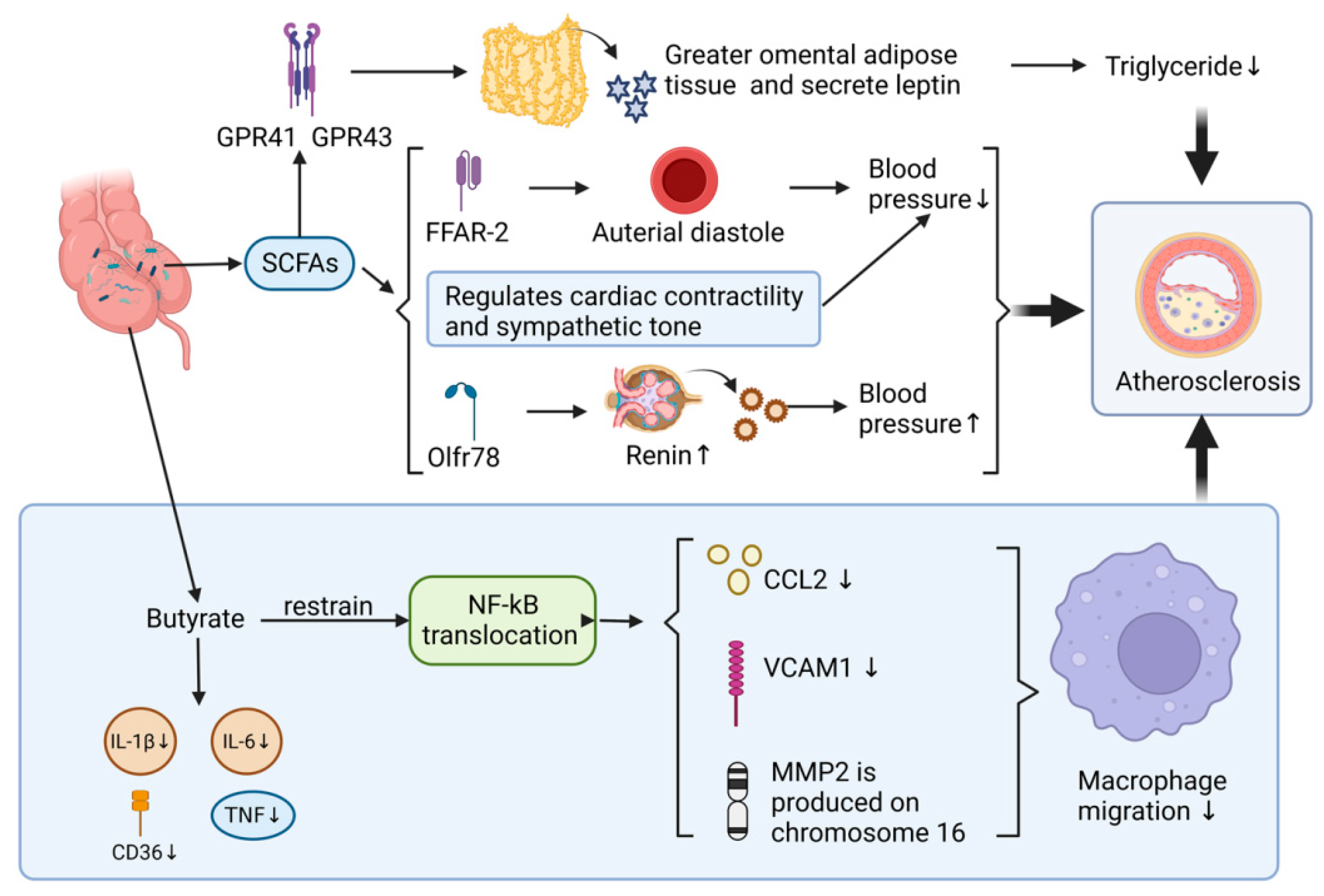
1.3.3. Effect of Gut Microbial Metabolites SCFAs on AS

Production and Metabolism of SCFAs

Short-chain fatty acids (SCFAs) are produced in humans by the microbial glycolysis of carbohydrates, such as dietary fiber and resistant starch that are not digested and absorbed, which concludes formate, acetate, propionate, and butyrate [73]. In addition, small amounts of branched-chain fatty acids are produced from the fermentative of protein-derived branched-chain amino acids [74]. The acetate production pathway is widely distributed in multiple floras, while the propionate and butyrate production pathways are highly conserved and substrate-specific [75]. Numerous studies have demonstrated the anti-inflammatory, antihypertensive, and metabolic regulatory effects of SCFAs, which can have a protective effect on the development of AS [76,77,78,79].

SCFAs and the Inflammatory Response of the Vessel Wall

SCFAs have been shown to inhibit the development of AS by reducing the migration and activation of macrophages. This effect is achieved by inhibiting the production of cytokines and chemokines [76]. For instance, when butyrate was added to the diet of apoE/ gene, NF-kappa B translocation was inhibited and the production of chemokine ligand-2 (CCL2), vascular cell adhesion molecule-1 (VCAM-1) and MMP2 gene on chromosome 16 in lesion area was reduced. As a result, the migration of macrophages was decreased, deposition of collagen was increased and plaque stability was improved at the lesion site. In addition, the uptake of oxLDL and production of CD36, TNF, IL-1β, and IL-6 were also reduced, leading to a 50% decrease in the development of AS in the aorta [77,78]. Furthermore, propionic acid and butyric acid have been shown to antagonize the production of LPS-induced cytokines IL-6 and IL-12p40 in humans [79]. This suggests that SCFAs have the potential as therapeutic agents for the treatment of AS and other inflammatory diseases (Figure 4).

SCFAs and Blood Pressure Regulation

It is well established that hypertension is one of the major risk factors for the development of AS, and SCFAs have been found to play a protective role against its progression by the regulation of hypertension [77]. Studies have shown that higher concentrations of SCFAs in human feces are associated with higher blood pressure and that hypertensive individuals have a lower number of SCFA-producing microorganisms in their gut microbiota compared to healthy controls [80]. SCFAs regulate blood pressure by targeting different receptors, including G protein-coupled receptors, olfactory receptor 78, and short-chain fatty acid receptors such as FFAR-2 and FFAR-3. For instance, SCFAs acting on FFAR-2 in the renal artery cause arterial diastole and lower blood pressure, whereas SCFAs operating on Olfr78 promote the release of renin and increase blood pressure [81,82,83,84]. Additionally, SCFAs modulate cardiac contractility and sympathetic tone, with mice injected with acetic acid showing a simultaneous decrease in arterial pressure and heart rate, as well as a load-independent decrease in myocardial contractility. These effects were antagonized by beta-1-adrenergic receptor antagonists such as atenolol and tyramine [85] (Figure 4).

SCFAs and Adipose Tissue

SCFAs have also been found to directly or indirectly participate in adipogenesis, catabolism, and inflammatory response, leading to inhibition of the progression of AS [86]. For example, propionic acid promotes leptin secretion from the greater omental adipose tissue and subcutaneous adipose tissue by activating GPR41 and GPR43, resulting in significantly increased leptin levels. Moreover, SCFAs have been found to reduce human blood triglyceride concentrations by regulating adipogenesis and impairing lipolysis [87,88,89]. Finally, SCFAs have been shown to inhibit the inflammatory response in adipocytes and adipose tissue, which further prevents the development of cardiovascular diseases, including AS [90] (Figure 4).

1.3.4. Association of Intestinal Microbial Interference with Bile Acid Metabolism and AS

Hydrophobic cholesterol is hydroxylated by cholesterol 7 alpha-hydroxylase (CYP7A1), which is catalyzed in hepatocytes and transforms into hydrophilic primary bile acids [91]. After primary bile acids are excreted through the bile duct, most of which are reabsorbed at the end of the ileum via bile acid transport proteins, some primary bile acids enter the colon and are changed into hydrophobic secondary bile acids through the removal of nuclear hydroxyl groups, oxidation, or epimerization [92], which are reabsorbed by the colon into the circulation and complete the enterohepatic circulation of bile acids. Only about 5% of secondary bile acids are excreted in feces [93]. Bile acids influence the composition of the intestinal flora, and the intestinal flora determines the distribution of intestinal bile acids [84] (Figure 5).
Secondary bile acids have been shown to act as potent signaling molecules that exert different effects by activating the corresponding receptors such as farnesoid X receptor (FXR) and Takeda G protein-coupled receptor 5 (TGR5; GPBAR1). Other receptors that are activated directly or indirectly include sphingosine-1-phosphate receptor 2 (S1PR2), pregnane X receptor (PXR), constitutive androstane receptor (CAR), vitamin D receptor (VDR), and retinoic acid-related receptor γt (R O Rγt) [94]. The activation of these receptors has been linked directly with the development of inflammatory bowel disease, which has been associated with an increased risk of atherosclerosis, cerebrovascular accidents, and atrial fibrillation [95]. Studies have also shown that TGR5 agonist INT-777 has immunosuppressive effects that include reducing macrophage production of pro-inflammatory cytokines, and delaying atherosclerotic plaque formation in LD−/− mice [96]. Researches have shown that polyphenol-rich substances reduce plasma TMAO by regulating intestinal flora, thereby affecting the development of AS [97]. The earlier confirmed extract was resveratrol (RSV), which has been shown to increase levels of Lactobacillus spp. and Bifidobacterium spp. in the intestine through intestinal flora remodeling. It also enhanced bile salt hydrolase activity, decreased ileal bile acid content, inhibited enterohepatic foresaid X receptor-fibroblast growth factor 15 (FGF15) axis, increased cholesterol 7a-hydroxylase (CYP7A1) expression, and promoted hepatic bile acid synthesis. All of these diminished the atherosclerosis-promoting effects of TMAO [98]. A recent study has reported that geraniin may also inhibit the progression of atherosclerosis by reducing plasma TMAO concentrations in mice [61] (Figure 5 and Figure 6).

1.3.5. Altered Intestinal Microecology Induced by Increased Intestinal Permeability and AS

The intestinal barrier system, which includes physical, biochemical, and immune components, interacts with intestinal microorganisms and increases intestinal permeability leading to the translocation of intestinal bacterial DNA, as well as promotes the absorption of intestinal flora metabolites and endotoxins into the circulation, thereby accelerating the progression of AS.
The intestinal epithelial cells (IECs) are interconnected, and the symbiotic bacteria in the intestinal lumen secrete antimicrobial substances to inhibit the growth of pathogenic bacteria [99]. The intestinal microenvironment is composed of the glycocalyx, mucus, and water layers, all of which together form the intestinal physical barrier [99,100]. There are three types of intercellular junctions between intestinal epithelial cells: zonula occludens (ZO), zonula adhesion, and bridging granules, which together constitute the apical linkage complex [101]. The tight junction protein (TJ) is involved in regulating epithelial barrier function and intercellular transport, and its regulatory effect leads to the formation of two different paracellular epithelial permeability pathways in intestinal epithelial cells, which are known as the “leakage” pathway and the “pore” pathway, and they are critical factors in controlling intestinal permeability [100,102]. The mucus layer in the intestinal lumen consists of a firmly adherent inner layer and a loosely adherent outer layer; the inner layer contains fewer bacteria and more antimicrobial substances, such as defensins and lysozyme; and the outer layer includes more bacteria and bacterial products, both of which together serve to prevent antigens, toxins, microbial metabolites, and bacteria directly contacting the intestinal epithelial cells. Two layers are essential parts of the physical defense of the intestinal mucosa [103]. The primary participating cells include cup cells and Pan cells [103]. Other cells, which include endocrine cells that secrete GLP-2 and participate in the regeneration and repair of epithelial cells and induce tight protein synthesis, cluster cells that produce IL-25 and IL-13, and M cells that participate in the mucosal immune response, all play essential roles as components of the intestinal mucosal barrier [100]. Intestinal barrier disruption and increased mucosal permeability promote the transport of LPS through the intestinal barrier, which facilitates the involvement of LPS participation in the development of inflammatory responses and AS in vivo. Moreover, LPS affects the tight junction permeability between intestinal epithelial cells through a TLR4-dependent mechanism, which increases the absorption of LPS, bacteria, and metabolites of the intestinal flora (e.g., TMAO, SCFAs, etc.) [34]. It has been shown that one of the ways that daily dietary structure affects the progression of AS is by altering intestinal mucosal permeability, for example, excessive sugar intake and hyperglycemia disruption in the intestinal barrier, which increases intestinal permeability and leads to deregulation of the intestinal microenvironment, thereby leading to an increased incidence of cardiovascular disease [104]. However, the mechanisms of their interaction with intestinal microecology need to be further investigated.

2. Intestinal Microecological Disorders Regulation and Atherosclerosis Prevention

Assessment of the composition of gut microbial is an essential foundation for understanding the relationship between gut microecology and AS and is also a prerequisite for developing relevant control strategies. Since most intestinal flora belong to prokaryotes, their 16S rRNA genes, which encode rRNA that is highly conserved and specific, are widely used for researching the composition and distribution of microbial communities through 16S rRNA gene sequencing [105]. In addition, combining 16S rRNA gene sequencing with enzyme-linked immunosorbent assay and lipid metabolism assessment further evaluates the association between the inflammatory responses caused by intestinal microecological disorders, abnormal lipid metabolism, and AS [106]. Based on the results of these tests, the balance of the intestinal can be adjusted to inhibit the development of AS [105,106]. Currently, several strategies have been applied, including intestinal flora transplantation, probiotics, prebiotics, symbiotics, and short-term antibiotic applications, all of which can also help to adjust the microecology of intestinal disorders.

3. Discussion

Intestinal flora transplantation is performed by extracting and pretreating microbial communities from healthy feces stool and then transplanting them into the recipient’s colon in order to create a new dynamic balance of intestinal microecology in the recipient’s intestinal microecology [107]. It has been demonstrated that fecal microbiota transplantation (FMT) is effective in the treatment of intestinal bacterial infections, depression, and metabolic diseases such as type 2 diabetes and obesity [108,109,110]. In the practical application of this method, there are still some factors that lead to unstable efficacy, such as the safety and stability of the microbial composition of the donor’s gut, the degree of preparation in the recipient’s gut, the method and standardization of the transplantation operation, etc [107,111]. Therefore, the use of animal models for relevant efficacy assessment is essential for the application of this method. Moreover, prebiotics, probiotics, and postbiotics, such as short-chain fatty acids, can modulate gut microbiota composition and reduce LPS levels, thereby exerting a protective effect against AS [112]. Probiotics are originally referred to as the beneficial flora in yogurt; they affect health by stimulating intestinal flora, interfering with the host immune response, reducing cholesterol absorption, etc [113]. Prebiotics are defined as a particular fermentation component that promotes the growth of probiotics in the human intestinal tract and the function of the body’s immune system; thus, prebiotics are often used as a health food, and lactulose is widely used as a common clinical preparation [114]. Synbiotics are a combination of probiotics and prebiotics, which have characteristics of both and have a more significant and lasting effect on the adjustment of intestinal microecology [115].
The use of inhibitors that reduce TMAO production has been shown to attenuate the promotion of lipid metabolism and inflammation formation by TMAO in the intestinal flora [116,117]. RSV and broad-spectrum antibiotics such as ciprofloxacin can remodel the flora, inhibiting TMAO production by reducing TMA [98]. RSV also attenuates TMAO-induced AS by reducing bile acid (BA) de novo synthesis in the liver [49,98]. The choline analog 3, 3-dimethyl-1-butanol has also been used to reduce TMAO production in the body, which not only weakens the promotion of TMAO in the development of AS but also plays an essential role in the treatment of hypertension and heart failure [118,119]. In addition, adjusting the daily diet structure by reducing the intake of choline-rich foods, such as red meat and seafood, and increasing the intake of plant-based foods, is important in reducing the production and absorption of TMAO in the intestine [120].
Gut microecological disorders affect the development of AS through multiple pathways, and elucidating the corresponding pathways helps to develop a wide range of potential therapeutic targets, which provide new therapeutic modalities and support for clinical mitigation of the development of AS.

4. Conclusions and Future Directions

Changes in intestinal microecology mainly refer to alterations in the composition of intestinal microorganisms and the production or absorption capacity of related metabolites, which affect the development of AS by exerting corresponding effects on human immunity, metabolism, and food breakdown. This review details the protective or promotive effects of microbial constituents LPS, microbial metabolites TMAO and SCFAs, microbial interference with bile acid metabolism, and organismal intestinal barrier disruption on AS. This information provides a new idea and direction for the development of clinical prevention and treatment strategies for AS. Numerous studies have demonstrated the relevance of intestinal microecology to the development of AS through multiple pathways. However, research is mostly limited to animal studies, and their feasibility and generalizability in the population still need further exploration. In addition, the current therapeutic measures, such as intestinal flora transplantation, TMAO inhibitors, probiotics, and prebiotics, still have disadvantages including high cost and unstable efficacy, which need further improvement.

Author Contributions

X.Z. (Xinyu Zhuo), R.L. and W.G. drafted the manuscript. X.Z. (Xinyu Zhuo), J.B.; X.L.; J.G.; and H.L. (Hao Luo) helped to prepare the manuscript. H.L. (Hui Luo), X.Z. (Xingwei Zhang), W.G.; and Q.J. were involved in revising it critically. All authors have read and agreed to the published version of the manuscript.

Funding

This work was supported by research grants from the National Natural Science Foundation of China (No. 81900745), the National Innovation and Entrepreneurship Training Program for college students (202210346019), and the Zhejiang Provincial Traditional Chinese Medicine Science and Technology Plan (2023ZF121).

Data Availability Statement

All supporting data are available within the article.

Conflicts of Interest

The authors declare no conflict of interest.

Abbreviations

AS: Atherosclerosis; BA: bile acid; CAR: constitutive androstane receptor; CCL2: chemokine ligand-2; CHD: coronary heart disease; CYP7A1: cholesterol 7 a-hydroxylase; FMT: fecal microbiota transplantation; FXR: foresaid X receptor; FGF15: fibroblast growth factor 15; FMO3: flavin-containing monooxygenase-3; HDL: high-density lipoprotein; HUVECs: human umbilical vein endothelial cells; IBDs: intestinal diseases; IECs: intestinal epithelial cells; IHD: ischemic heart disease; LBP: lipopolysaccharide-binding protein; LDL: low-density lipoprotein; LPS: lipopolysaccharide; PXR: pregnane X receptor; ROS: reactive oxygen species; RNS: reactive nitrogen species; RSV: resveratrol; S1PR2: sphingosine-1-phosphate receptor 2; SCFAs: short-chain fatty acids; TGR5: Takeda G protein-coupled receptor 5; TJ: tight junction; TLR4: Toll-like receptor 4; TMAO: trimethylamine; VDR: vitamin D receptor; ZO: zonula occludens.

References

  1. Jia, Q.; Li, H.; Zhou, H.; Zhang, X.; Zhang, A.; Xie, Y.; Li, Y.; Lv, S.; Zhang, J. Role and Effective Therapeutic Target of Gut Microbiota in Heart Failure. Cardiovasc. Ther. 2019, 2019, 5164298. [Google Scholar] [CrossRef]
  2. Fish, E.M.; Burns, B. Physiology, Small Bowel. In StatPearls; StatPearls Publishing: Treasure Island, FL, USA, 2022. [Google Scholar]
  3. Patricia, J.J.; Dhamoon, A.S. Physiology, Digestion. In StatPearls; StatPearls Publishing: Treasure Island, FL, USA, 2022. [Google Scholar]
  4. Malone, J.C.; Shah, A.B. Embryology, Midgut. In StatPearls; StatPearls Publishing: Treasure Island, FL, USA, 2022. [Google Scholar]
  5. Chikina, A.S.; Vignjevic, M.D.; Lennon-Dumenil, A.M. Roles of the macrophages in colon homeostasis. Comptes Rendus Biol. 2021, 344, 337–356. [Google Scholar] [CrossRef] [PubMed]
  6. Ley, R.E.; Peterson, D.A.; Gordon, J.I. Ecological and evolutionary forces shaping microbial diversity in the human intestine. Cell 2006, 124, 837–848. [Google Scholar] [CrossRef]
  7. Zhou, B.; Yuan, Y.; Zhang, S.; Guo, C.; Li, X.; Li, G.; Xiong, W.; Zeng, Z. Intestinal Flora and Disease Mutually Shape the Regional Immune System in the Intestinal Tract. Front. Immunol. 2020, 11, 575. [Google Scholar] [CrossRef] [PubMed]
  8. Cresci, G.A.; Bawden, E. Gut Microbiome: What We Do and Don’t Know. Nutr. Clin. Pract. 2015, 30, 734–746. [Google Scholar] [CrossRef] [PubMed]
  9. Wu, J.; Zhao, Y.; Wang, X.; Kong, L.; Johnston, L.J.; Lu, L.; Ma, X. Dietary nutrients shape gut microbes and intestinal mucosa via epigenetic modifications. Crit. Rev. Food Sci. Nutr. 2022, 62, 783–797. [Google Scholar] [CrossRef]
  10. de Vos, W.M.; Tilg, H.; Van Hul, M.; Cani, P.D. Gut microbiome and health: Mechanistic insights. Gut 2022, 71, 1020–1032. [Google Scholar] [CrossRef]
  11. Buja, L.M.; Nikolai, N. Anitschkow and the lipid hypothesis of atherosclerosis. Cardiovasc. Pathol. 2014, 23, 183–184. [Google Scholar] [CrossRef]
  12. Libby, P.; Hansson, G.K. From Focal Lipid Storage to Systemic Inflammation JACC Review Topic of the Week. J. Am. Coll. Cardiol. 2019, 74, 1594–1607. [Google Scholar] [CrossRef]
  13. Domanski, M.J.; Tian, X.; Wu, C.O.; Reis, J.P.; Dey, A.K.; Gu, Y.; Zhao, L.; Bae, S.; Liu, K.; Hasan, A.A.; et al. Time Course of LDL Cholesterol Exposure and Cardiovascular Disease Event Risk. J. Am. Coll. Cardiol. 2020, 76, 1507–1516. [Google Scholar] [CrossRef]
  14. Qiao, Y.N.; Zou, Y.L.; Guo, S.D. Low-density lipoprotein particles in atherosclerosis. Front. Physiol. 2022, 13, 931931. [Google Scholar] [CrossRef] [PubMed]
  15. Borén, J.; Chapman, M.J.; Krauss, R.M.; Packard, C.J.; Bentzon, J.F.; Binder, C.J.; Daemen, M.J.; Demer, L.L.; Hegele, R.A.; Nicholls, S.J.; et al. Low-density lipoproteins cause atherosclerotic cardiovascular disease: Pathophysiological, genetic, and therapeutic insights: A consensus statement from the European Atherosclerosis Society Consensus Panel. Eur. Heart J. 2020, 41, 2313–2330. [Google Scholar] [CrossRef]
  16. Jebari-Benslaiman, S.; Galicia-García, U.; Larrea-Sebal, A.; Olaetxea, J.R.; Alloza, I.; Vandenbroeck, K.; Benito-Vicente, A.; Martín, C. Pathophysiology of Atherosclerosis. Int. J. Mol. Sci. 2022, 23, 3346. [Google Scholar] [CrossRef] [PubMed]
  17. Pasta, A.; Cremonini, A.L.; Pisciotta, L.; Buscaglia, A.; Porto, I.; Barra, F.; Ferrero, S.; Brunelli, C.; Rosa, G.M. PCSK9 inhibitors for treating hypercholesterolemia. Expert Opin. Pharmacother. 2020, 21, 353–363. [Google Scholar] [CrossRef]
  18. Fan, J.; Watanabe, T. Atherosclerosis: Known and unknown. Pathol. Int. 2022, 72, 151–160. [Google Scholar] [CrossRef] [PubMed]
  19. Poznyak, A.; Grechko, A.V.; Poggio, P.; Myasoedova, V.A.; Alfieri, V.; Orekhov, A.N. The Diabetes Mellitus-Atherosclerosis Connection: The Role of Lipid and Glucose Metabolism and Chronic Inflammation. Int. J. Mol. Sci. 2020, 21, 1835. [Google Scholar] [CrossRef]
  20. Libby, P. Inflammation during the life cycle of the atherosclerotic plaque. Cardiovasc. Res. 2021, 117, 2525–2536. [Google Scholar] [CrossRef]
  21. Han, S.; Pan, Y.; Yang, X.; Da, M.; Wei, Q.; Gao, Y.; Qi, Q.; Ru, L. Intestinal microorganisms involved in colorectal cancer complicated with dyslipidosis. Cancer Biol. Ther. 2019, 20, 81–89. [Google Scholar] [CrossRef]
  22. Jie, Z.; Xia, H.; Zhong, S.L.; Feng, Q.; Li, S.; Liang, S.; Zhong, H.; Liu, Z.; Gao, Y.; Zhao, H.; et al. The gut microbiome in atherosclerotic cardiovascular disease. Nat. Commun. 2017, 8, 845. [Google Scholar] [CrossRef]
  23. Choroszy, M.; Litwinowicz, K.; Bednarz, R.; Roleder, T.; Lerman, A.; Toya, T.; Kamiński, K.; Sawicka-Śmiarowska, E.; Niemira, M.; Sobieszczańska, B. Human Gut Microbiota in Coronary Artery Disease: A Systematic Review and Meta-Analysis. Metabolites 2022, 12, 1165. [Google Scholar] [CrossRef]
  24. Anto, L.; Blesso, C.N. Interplay between diet, the gut microbiome, and atherosclerosis: Role of dysbiosis and microbial metabolites on inflammation and disordered lipid metabolism. J. Nutr. Biochem. 2022, 105, 108991. [Google Scholar] [CrossRef]
  25. Wang, H.; Reddy, S.T.; Fogelman, A.M. The role of gut-derived oxidized lipids and bacterial lipopolysaccharide in systemic inflammation and atherosclerosis. Curr. Opin. Lipidol. 2022, 33, 277–282. [Google Scholar] [CrossRef]
  26. Wen, Y.; Sun, Z.; Xie, S.; Hu, Z.; Lan, Q.; Sun, Y.; Yuan, L.; Zhai, C. Intestinal Flora Derived Metabolites Affect the Occurrence and Development of Cardiovascular Disease. J. Multidiscip. Healthc. 2022, 15, 2591–2603. [Google Scholar] [CrossRef]
  27. Kerr, A.W.; Efron, P.A.; Larson, S.D.; Rincon, J.C. T-Cell Activation and LPS: A Dangerous Duo for Organ Dysfunction. J. Leukoc. Biol. 2022, 112, 219–220. [Google Scholar] [CrossRef]
  28. Heinrich, M.A.; Mangia, M.; Prakash, J. Impact of endotoxins on bioengineered tissues and models. Trends Biotechnol. 2022, 40, 532–534. [Google Scholar] [CrossRef]
  29. Franco-Gonzalez, J.F.; Matamoros-Recio, A.; Torres-Mozas, A.; Rodrigo-Lacave, B.; Martin-Santamaria, S. Lipid-A-dependent and cholesterol-dependent dynamics properties of liposomes from gram-negative bacteria in ESKAPE. Sci. Rep. 2022, 12, 19474. [Google Scholar] [CrossRef]
  30. Hakoupian, M.; Ferino, E.; Jickling, G.C.; Amini, H.; Stamova, B.; Ander, B.P.; Alomar, N.; Sharp, F.R.; Zhan, X. Bacterial lipopolysaccharide is associated with stroke. Sci. Rep. 2021, 11, 6570. [Google Scholar] [CrossRef] [PubMed]
  31. Asada, M.; Oishi, E.; Sakata, S.; Hata, J.; Yoshida, D.; Honda, T.; Furuta, Y.; Shibata, M.; Suzuki, K.; Watanabe, H.; et al. Serum Lipopolysaccharide-Binding Protein Levels and the Incidence of Cardiovascular Disease in a General Japanese Population: The Hisayama Study. J. Am. Heart Assoc. 2019, 8, e013628. [Google Scholar] [CrossRef] [PubMed]
  32. Han, Y.H.; Onufer, E.J.; Huang, L.H.; Sprung, R.W.; Davidson, W.S.; Czepielewski, R.S.; Wohltmann, M.; Sorci-Thomas, M.G.; Warner, B.W.; Randolph, G.J. Enterically derived high-density lipoprotein restrains liver injury through the portal vein. Science 2021, 373, eabe6729. [Google Scholar] [CrossRef] [PubMed]
  33. Ghosh, S.S.; Wang, J.; Yannie, P.J.; Ghosh, S. Intestinal Barrier Dysfunction, LPS Translocation, and Disease Development. J. Endocr. Soc. 2020, 4, bvz039. [Google Scholar] [CrossRef]
  34. Page, M.J.; Kell, D.B.; Pretorius, E. The Role of Lipopolysaccharide-Induced Cell Signalling in Chronic Inflammation. Chronic Stress 2022, 6, 24705470221076390. [Google Scholar] [CrossRef] [PubMed]
  35. Zhang, Y.; Liang, X.; Bao, X.; Xiao, W.; Chen, G. Toll-like receptor 4 (TLR4) inhibitors: Current research and prospective. Eur. J. Med. Chem. 2022, 235, 114291. [Google Scholar] [CrossRef]
  36. Siddiqui, M.A.; Yamashita, M. Toll-Like Receptor (TLR) Signaling Enables Cyclic GMP-AMP Synthase (cGAS) Sensing of HIV-1 Infection in Macrophages. mBio 2021, 12, e0281721. [Google Scholar] [CrossRef]
  37. Feng, X.; Zhang, Y.; Xu, R.; Xie, X.; Tao, L.; Gao, H.; Gao, Y.; He, Z.; Wang, H. Lipopolysaccharide up-regulates the expression of Fcalpha/mu receptor and promotes the binding of oxidized low-density lipoprotein and its IgM antibody complex to activated human macrophages. Atherosclerosis 2010, 208, 396–405. [Google Scholar] [CrossRef]
  38. Geng, S.; Chen, K.; Yuan, R.; Peng, L.; Maitra, U.; Diao, N.; Chen, C.; Zhang, Y.; Hu, Y.; Qi, C.F.; et al. The persistence of low-grade inflammatory monocytes contributes to aggravated atherosclerosis. Nat. Commun. 2016, 7, 13436. [Google Scholar] [CrossRef] [PubMed]
  39. Wang, J.; Si, Y.; Wu, C.; Sun, L.; Ma, Y.; Ge, A.; Li, B. Lipopolysaccharide promotes lipid accumulation in human adventitial fibroblasts via TLR4-NF-κB pathway. Lipids Health Dis. 2012, 11, 139. [Google Scholar] [CrossRef]
  40. Fu, Y.; Liu, J.W.; Wu, J.; Wu, Z.X.; Li, J.; Ji, H.F.; Liang, N.P.; Zhang, H.J.; Lai, Z.Q.; Dong, Y.F. Inhibition of semaphorin-3a alleviates lipopolysaccharide-induced vascular injury. Microvasc. Res. 2022, 142, 104346. [Google Scholar] [CrossRef]
  41. Qi, J.; You, T.; Li, J.; Pan, T.; Xiang, L.; Han, Y.; Zhu, L. Circulating trimethylamine N-oxide and the risk of cardiovascular diseases: A systematic review and meta-analysis of 11 prospective cohort studies. J. Cell Mol. Med. 2018, 22, 185–194. [Google Scholar] [CrossRef]
  42. Fu, B.C.; Hullar, M.A.J.; Randolph, T.W.; Franke, A.A.; Monroe, K.R.; Cheng, I.; Wilkens, L.R.; Shepherd, J.A.; Madeleine, M.M.; Le Marchand, L.; et al. Associations of plasma trimethylamine N-oxide, choline, carnitine, and betaine with inflammatory and cardiometabolic risk biomarkers and the fecal microbiome in the Multiethnic Cohort Adiposity Phenotype Study. Am. J. Clin. Nutr. 2020, 111, 1226–1234. [Google Scholar] [CrossRef] [PubMed]
  43. Yao, M.E.; Liao, P.D.; Zhao, X.J.; Wang, L. Trimethylamine-N-oxide has prognostic value in coronary heart disease: A meta-analysis and dose-response analysis. BMC Cardiovasc. Disord. 2020, 20, 7. [Google Scholar] [CrossRef]
  44. Razavi, A.C.; Potts, K.S.; Kelly, T.N.; Bazzano, L.A. Sex, gut microbiome, and cardiovascular disease risk. Biol. Sex Differ. 2019, 10, 29. [Google Scholar] [CrossRef] [PubMed]
  45. Day-Walsh, P.; Shehata, E.; Saha, S.; Savva, G.M.; Nemeckova, B.; Speranza, J.; Kellingray, L.; Narbad, A.; Kroon, P.A. The use of an in-vitro batch fermentation (human colon) model for investigating mechanisms of TMA production from choline, L-carnitine and related precursors by the human gut microbiota. Eur. J. Nutr. 2021, 60, 3987–3999. [Google Scholar] [CrossRef]
  46. Li, Y.; Cui, M.; Sun, J.; Wei, Q.; Liu, M.; Zhang, J.; Qi, H.; Zhao, L.; Fang, H.; Chen, Z.; et al. Gut microbiota and its metabolite trimethylamine-N-oxide (TMAO): A novel regulator in coronary artery disease. Sheng Wu Gong Cheng Xue Bao 2021, 37, 3745–3756. [Google Scholar] [PubMed]
  47. Kyaw, T.S.; Sukmak, M.; Nahok, K.; Sharma, A.; Silsirivanit, A.; Lert-Itthiporn, W.; Sansurin, N.; Senthong, V.; Anutrakulchai, S.; Sangkhamanon, S.; et al. Monosodium glutamate consumption reduces the renal excretion of trimethylamine N-oxide and the abundance of Akkermansia muciniphila in the gut. Biochem. Biophys. Res. Commun. 2022, 630, 158–166. [Google Scholar] [CrossRef] [PubMed]
  48. Subramaniam, S.; Fletcher, C. Trimethylamine N-oxide: Breathe new life. Br. J. Pharmacol. 2018, 175, 1344–1353. [Google Scholar] [CrossRef] [PubMed]
  49. Zhang, W.Q.; Wang, Y.J.; Zhang, A.; Ding, Y.J.; Zhang, X.N.; Jia, Q.J.; Zhu, Y.P.; Li, Y.Y.; Lv, S.C.; Zhang, J.P. TMA/TMAO in Hypertension: Novel Horizons and Potential Therapies. J. Cardiovasc. Transl. Res. 2021, 14, 1117–1124. [Google Scholar] [CrossRef]
  50. Dai, Y.; Zhang, J.; Wang, Z.; Xu, S.; Zhang, Q.; Duan, Z.; Tan, R.; Qi, X.; Guo, J.; Chang, X.; et al. Associations of Diet with Urinary Trimethylamine-N-Oxide (TMAO) and Its Precursors among Free-Living 10-Year-Old Children: Data from SMBCS. Nutrients 2022, 14, 3419. [Google Scholar] [CrossRef]
  51. Poznyak, A.V.; Sadykhov, N.K.; Kartuesov, A.G.; Borisov, E.E.; Melnichenko, A.A.; Grechko, A.V.; Orekhov, A.N. Hypertension as a risk factor for atherosclerosis: Cardiovascular risk assessment. Front. Cardiovasc. Med. 2022, 9, 959285. [Google Scholar] [CrossRef]
  52. Zhen, J.; Zhou, Z.; He, M.; Han, H.X.; Lv, E.H.; Wen, P.B.; Liu, X.; Wang, Y.T.; Cai, X.C.; Tian, J.Q.; et al. The gut microbial metabolite trimethylamine N-oxide and cardiovascular diseases. Front. Endocrinol. 2023, 14, 1085041. [Google Scholar] [CrossRef]
  53. Xiong, X.; Zhou, J.; Fu, Q.; Xu, X.; Wei, S.; Yang, S.; Chen, B. The associations between TMAO-related metabolites and blood lipids and the potential impact of rosuvastatin therapy. Lipids Health Dis. 2022, 21, 60. [Google Scholar] [CrossRef]
  54. Warrier, M.; Shih, D.M.; Burrows, A.C.; Ferguson, D.; Gromovsky, A.D.; Brown, A.L.; Marshall, S.; McDaniel, A.; Schugar, R.C.; Wang, Z.; et al. The TMAO-Generating Enzyme Flavin Monooxygenase 3 Is a Central Regulator of Cholesterol Balance. Cell Rep. 2015, 10, 326–338. [Google Scholar] [CrossRef] [PubMed]
  55. Luo, T.; Guo, Z.; Liu, D.; Guo, Z.; Wu, Q.; Li, Q.; Lin, R.; Chen, P.; Ou, C.; Chen, M. Deficiency of PSRC1 accelerates atherosclerosis by increasing TMAO production via manipulating gut microbiota and flavin monooxygenase 3. Gut Microbes 2022, 14, 2077602. [Google Scholar] [CrossRef]
  56. Ding, L.; Chang, M.; Guo, Y.; Zhang, L.; Xue, C.; Yanagita, T.; Zhang, T.; Wang, Y. Trimethylamine-N-oxide (TMAO)-induced atherosclerosis is associated with bile acid metabolism. Lipids Health Dis. 2018, 17, 286. [Google Scholar] [CrossRef]
  57. Miao, J.; Ling, A.V.; Manthena, P.V.; Gearing, M.E.; Graham, M.J.; Crooke, R.M.; Croce, K.J.; Esquejo, R.M.; Clish, C.B.; Morbid Obesity Study Group; et al. Flavin-containing monooxygenase 3 as a potential player in diabetes-associated atherosclerosis. Nat. Commun. 2015, 6, 6498. [Google Scholar] [CrossRef] [PubMed]
  58. Canyelles, M.; Pérez, A.; Junza, A.; Miñambres, I.; Yanes, O.; Sardà, H.; Rotllan, N.; Julve, J.; Sánchez-Quesada, J.L.; Tondo, M.; et al. Divergent Effects of Glycemic Control and Bariatric Surgery on Circulating Concentrations of TMAO in Newly Diagnosed T2D Patients and Morbidly Obese. Diagnostics 2022, 12, 2783. [Google Scholar] [CrossRef]
  59. Eshghjoo, S.; Kim, D.M.; Jayaraman, A.; Sun, Y.; Alaniz, R.C. Macrophage Polarization in Atherosclerosis. Genes 2022, 13, 756. [Google Scholar] [CrossRef]
  60. Díez-Ricote, L.; Ruiz-Valderrey, P.; Micó, V.; Blanco-Rojo, R.; Tomé-Carneiro, J.; Dávalos, A.; Ordovás, J.M.; Daimiel, L. Trimethylamine n-Oxide (TMAO) Modulates the Expression of Cardiovascular Disease-Related microRNAs and Their Targets. Int. J. Mol. Sci. 2021, 22, 11145. [Google Scholar] [CrossRef] [PubMed]
  61. Lin, K.; Wang, X.; Li, J.; Zhao, P.; Xi, X.; Feng, Y.; Yin, L.; Tian, J.; Li, H.; Liu, X.; et al. Anti-atherosclerotic effects of geraniin through the gut microbiota-dependent trimethylamine N-oxide (TMAO) pathway in mice. Phytomedicine 2022, 101, 154104. [Google Scholar] [CrossRef]
  62. Geng, J.; Yang, C.; Wang, B.; Zhang, X.; Hu, T.; Gu, Y.; Li, J. Trimethylamine N-oxide promotes atherosclerosis via CD36-dependent MAPK/JNK pathway. Biomed. Pharmacother. 2018, 97, 941–947. [Google Scholar] [CrossRef] [PubMed]
  63. Chen, K.; Zheng, X.; Feng, M.; Li, D.; Zhang, H. Gut Microbiota-Dependent Metabolite Trimethylamine N-Oxide Contributes to Cardiac Dysfunction in Western Diet-Induced Obese Mice. Front. Physiol. 2017, 8, 139. [Google Scholar] [CrossRef]
  64. Garcia, E.; Osté, M.C.J.; Bennett, D.W.; Jeyarajah, E.J.; Shalaurova, I.; Gruppen, E.G.; Hazen, S.L.; Otvos, J.D.; Bakker, S.J.L.; Dullaart, R.P.F.; et al. High Betaine, a Trimethylamine N-Oxide Related Metabolite, Is Prospectively Associated with Low Future Risk of Type 2 Diabetes Mellitus in the PREVEND Study. J. Clin. Med. 2019, 8, 1813. [Google Scholar] [CrossRef]
  65. Zhang, L.; Xie, F.; Tang, H.; Zhang, X.; Hu, J.; Zhong, X.; Gong, N.; Lai, Y.; Zhou, M.; Tian, J.; et al. Gut microbial metabolite TMAO increases peritoneal inflammation and peritonitis risk in peritoneal dialysis patients. Transl. Res. 2022, 240, 50–63. [Google Scholar] [CrossRef]
  66. Sun, X.; Jiao, X.; Ma, Y.; Liu, Y.; Zhang, L.; He, Y.; Chen, Y. Trimethylamine N-oxide induces inflammation and endothelial dysfunction in human umbilical vein endothelial cells via activating ROS-TXNIP-NLRP3 inflammasome. Biochem. Biophys. Res. Commun. 2016, 481, 63–70. [Google Scholar] [CrossRef] [PubMed]
  67. Liu, X.; Shao, Y.; Tu, J.; Sun, J.; Dong, B.; Wang, Z.; Zhou, J.; Chen, L.; Tao, J.; Chen, J. TMAO-Activated Hepatocyte-Derived Exosomes Impair Angiogenesis via Repressing CXCR4. Front. Cell Dev. Biol. 2021, 9, 804049. [Google Scholar] [CrossRef] [PubMed]
  68. Wei, K.; Huang, H.; Liu, M.; Shi, D.; Ma, X. Platelet-Derived Exosomes and Atherothrombosis. Front. Cardiovasc. Med. 2022, 9, 886132. [Google Scholar] [CrossRef]
  69. Wang, Z.; Klipfell, E.; Bennett, B.J.; Koeth, R.; Levison, B.S.; Dugar, B.; Feldstein, A.E.; Britt, E.B.; Fu, X.; Chung, Y.M.; et al. Gut flora metabolism of phosphatidylcholine promotes cardiovascular disease. Nature 2011, 472, 57–63. [Google Scholar] [CrossRef] [PubMed]
  70. Zhu, W.; Wang, Z.; Tang, W.H.W.; Hazen, S.L. Gut Microbe-Generated Trimethylamine N-Oxide from Dietary Choline Is Prothrombotic in Subjects. Circulation 2017, 135, 1671–1673. [Google Scholar] [CrossRef]
  71. Ge, P.X.; Tai, T.; Jiang, L.P.; Ji, J.Z.; Mi, Q.Y.; Zhu, T.; Li, Y.F.; Xie, H.G. Choline and trimethylamine N-oxide impair metabolic activation of and platelet response to clopidogrel through activation of the NOX/ROS/Nrf2/CES1 pathway. J. Thromb. Haemost. 2023, 21, 117–132. [Google Scholar] [CrossRef]
  72. Witkowski, M.; Witkowski, M.; Friebel, J.; Buffa, J.A.; Li, X.S.; Wang, Z.; Sangwan, N.; Li, L.; DiDonato, J.A.; Tizian, C.; et al. Vascular endothelial tissue factor contributes to trimethylamine N-oxide-enhanced arterial thrombosis. Cardiovasc. Res. 2022, 118, 2367–2384. [Google Scholar] [CrossRef]
  73. Rauf, A.; Khalil, A.A.; Rahman, U.U.; Khalid, A.; Naz, S.; Shariati, M.A.; Rebezov, M.; Urtecho, E.Z.; de Albuquerque, R.D.D.G.; Anwar, S.; et al. Recent advances in the therapeutic application of short-chain fatty acids (SCFAs): An updated review. Crit. Rev. Food Sci. Nutr. 2022, 62, 6034–6054. [Google Scholar] [CrossRef]
  74. Zeng, X.; Xing, X.; Gupta, M.; Keber, F.C.; Lopez, J.G.; Lee, Y.J.; Roichman, A.; Wang, L.; Neinast, M.D.; Donia, M.S.; et al. Gut bacterial nutrient preferences quantified in vivo. Cell 2022, 185, 3441–3456.e19. [Google Scholar] [CrossRef]
  75. Ohira, H.; Tsutsui, W.; Fujioka, Y. Are Short Chain Fatty Acids in Gut Microbiota Defensive Players for Inflammation and Atherosclerosis? J. Atheroscler. Thromb. 2017, 24, 660–672. [Google Scholar] [CrossRef]
  76. Lymperopoulos, A.; Suster, M.S.; Borges, J.I. Short-Chain Fatty Acid Receptors and Cardiovascular Function. Int. J. Mol. Sci. 2022, 23, 3303. [Google Scholar] [CrossRef] [PubMed]
  77. Hu, T.; Wu, Q.; Yao, Q.; Jiang, K.; Yu, J.; Tang, Q. Short-chain fatty acid metabolism and multiple effects on cardiovascular diseases. Ageing Res. Rev. 2022, 81, 101706. [Google Scholar] [CrossRef]
  78. Aguilar, E.C.; Leonel, A.J.; Teixeira, L.G.; Silva, A.R.; Silva, J.F.; Pelaez, J.M.; Capettini, L.S.; Lemos, V.S.; Santos, R.A.; Alvarez-Leite, J.I. Butyrate impairs atherogenesis by reducing plaque inflammation and vulnerability and decreasing NFκB activation. Nutr. Metab. Cardiovasc. Dis. 2014, 24, 606–613. [Google Scholar] [CrossRef]
  79. Nastasi, C.; Candela, M.; Bonefeld, C.M.; Geisler, C.; Hansen, M.; Krejsgaard, T.; Biagi, E.; Andersen, M.H.; Brigidi, P.; Ødum, N.; et al. The effect of short-chain fatty acids on human monocyte-derived dendritic cells. Sci. Rep. 2015, 5, 16148. [Google Scholar] [CrossRef] [PubMed]
  80. Huart, J.; Leenders, J.; Taminiau, B.; Descy, J.; Saint-Remy, A.; Daube, G.; Krzesinski, J.M.; Melin, P.; de Tullio, P.; Jouret, F. Gut Microbiota and Fecal Levels of Short-Chain Fatty Acids Differ Upon 24-Hour Blood Pressure Levels in Men. Hypertension 2019, 74, 1005–1013. [Google Scholar] [CrossRef] [PubMed]
  81. Pluznick, J.L. Microbial Short-Chain Fatty Acids and Blood Pressure Regulation. Curr. Hypertens. Rep. 2017, 19, 25. [Google Scholar] [CrossRef] [PubMed]
  82. Wang, L.; Zhu, Q.; Lu, A.; Liu, X.; Zhang, L.; Xu, C.; Liu, X.; Li, H.; Yang, T. Sodium butyrate suppresses angiotensin II-induced hypertension by inhibition of renal (pro)renin receptor and intrarenal renin-angiotensin system. J. Hypertens. 2017, 35, 1899–1908. [Google Scholar] [CrossRef]
  83. Vallianou, N.G.; Geladari, E.; Kounatidis, D. Microbiome and hypertension: Where are we now? J. Cardiovasc. Med. 2020, 21, 83–88. [Google Scholar] [CrossRef]
  84. Verhaar, B.J.H.; Prodan, A.; Nieuwdorp, M.; Muller, M. Gut Microbiota in Hypertension and Atherosclerosis: A Review. Nutrients 2020, 12, 2982. [Google Scholar] [CrossRef] [PubMed]
  85. Poll, B.G.; Xu, J.; Jun, S.; Sanchez, J.; Zaidman, N.A.; He, X.; Lester, L.; Berkowitz, D.E.; Paolocci, N.; Gao, W.D.; et al. Acetate, a Short-Chain Fatty Acid, Acutely Lowers Heart Rate and Cardiac Contractility Along with Blood Pressure. J. Pharmacol. Exp. Ther. 2021, 377, 39–50. [Google Scholar] [CrossRef] [PubMed]
  86. Li, D.; Xu, Z.; Li, Y.; Gan, L.; Wu, P.; Wu, R.; Jin, J.; Zheng, X.; Zhang, K.; Ma, H.; et al. Polysaccharides from Callerya speciosa alleviate metabolic disorders and gut microbiota dysbiosis in diet-induced obese C57BL/6 mice. Food Funct. 2022, 13, 8662–8675. [Google Scholar] [CrossRef]
  87. Al-Lahham, S.; Roelofsen, H.; Rezaee, F.; Weening, D.; Hoek, A.; Vonk, R.; Venema, K. Propionic acid affects immune status and metabolism in adipose tissue from overweight subjects. Eur. J. Clin. Investig. 2012, 42, 357–364. [Google Scholar] [CrossRef]
  88. Zaibi, M.S.; Stocker, C.J.; O’Dowd, J.; Davies, A.; Bellahcene, M.; Cawthorne, M.A.; Brown, A.J.; Smith, D.M.; Arch, J.R. Roles of GPR41 and GPR43 in leptin secretory responses of murine adipocytes to short chain fatty acids. FEBS Lett. 2010, 584, 2381–2386. [Google Scholar] [CrossRef] [PubMed]
  89. Chen, X.F.; Chen, X.; Tang, X. Short-chain fatty acid, acylation and cardiovascular diseases. Clin. Sci. 2020, 134, 657–676. [Google Scholar] [CrossRef]
  90. Kawai, T.; Autieri, M.V.; Scalia, R. Adipose tissue inflammation and metabolic dysfunction in obesity. Am. J. Physiol. Cell Physiol. 2021, 320, C375–C391. [Google Scholar] [CrossRef]
  91. Chiang, J.Y.L.; Ferrell, J.M. Up to date on cholesterol 7 alpha-hydroxylase (CYP7A1) in bile acid synthesis. Liver Res. 2020, 4, 47–63. [Google Scholar] [CrossRef]
  92. Cai, J.; Rimal, B.; Jiang, C.; Chiang, J.Y.L.; Patterson, A.D. Bile acid metabolism and signaling, the microbiota, and metabolic disease. Pharmacol. Ther. 2022, 237, 108238. [Google Scholar] [CrossRef]
  93. Simbrunner, B.; Trauner, M.; Reiberger, T. Review article: Therapeutic aspects of bile acid signalling in the gut-liver axis. Aliment Pharmacol. Ther. 2021, 54, 1243–1262. [Google Scholar] [CrossRef]
  94. Thibaut, M.M.; Bindels, L.B. Crosstalk between bile acid-activated receptors and microbiome in entero-hepatic inflammation. Trends Mol. Med. 2022, 28, 223–236. [Google Scholar] [CrossRef]
  95. Chen, B.; Collen, L.V.; Mowat, C.; Isaacs, K.L.; Singh, S.; Kane, S.V.; Farraye, F.A.; Snapper, S.; Jneid, H.; Lavie, C.J.; et al. Inflammatory Bowel Disease and Cardiovascular Diseases. Am. J. Med. 2022, 135, 1453–1460. [Google Scholar] [CrossRef] [PubMed]
  96. Pols, T.W.; Nomura, M.; Harach, T.; Lo Sasso, G.; Oosterveer, M.H.; Thomas, C.; Rizzo, G.; Gioiello, A.; Adorini, L.; Pellicciari, R.; et al. TGR5 activation inhibits atherosclerosis by reducing macrophage inflammation and lipid loading. Cell Metab. 2011, 14, 747–757. [Google Scholar] [CrossRef] [PubMed]
  97. Rehman, A.; Tyree, S.M.; Fehlbaum, S.; DunnGalvin, G.; Panagos, C.G.; Guy, B.; Patel, S.; Dinan, T.G.; Duttaroy, A.K.; Duss, R.; et al. A water-soluble tomato extract rich in secondary plant metabolites lowers trimethylamine-n-oxide and modulates gut microbiota: A randomized, double-blind, placebo-controlled cross-over study in overweight and obese adults. J. Nutr. 2023, 153, 96–105. [Google Scholar] [CrossRef] [PubMed]
  98. Chen, M.L.; Yi, L.; Zhang, Y.; Zhou, X.; Ran, L.; Yang, J.; Zhu, J.D.; Zhang, Q.Y.; Mi, M.T. Resveratrol Attenuates Trimethylamine-N-Oxide (TMAO)-Induced Atherosclerosis by Regulating TMAO Synthesis and Bile Acid Metabolism via Remodeling of the Gut Microbiota. mBio 2016, 7, e02210-15. [Google Scholar] [CrossRef] [PubMed]
  99. Camilleri, M. Leaky gut: Mechanisms, measurement and clinical implications in humans. Gut 2019, 68, 1516–1526. [Google Scholar] [CrossRef]
  100. Camilleri, M.; Vella, A. What to do about the leaky gut. Gut 2022, 71, 424–435. [Google Scholar] [CrossRef]
  101. Wang, Q.; Margolis, B. Apical junctional complexes and cell polarity. Kidney Int. 2007, 72, 1448–1458. [Google Scholar] [CrossRef] [PubMed]
  102. Odenwald, M.A.; Turner, J.R. The intestinal epithelial barrier: A therapeutic target? Nat. Rev. Gastroenterol. Hepatol. 2017, 14, 9–21. [Google Scholar] [CrossRef] [PubMed]
  103. Paone, P.; Cani, P.D. Mucus barrier, mucins and gut microbiota: The expected slimy partners? Gut 2020, 69, 2232–2243. [Google Scholar] [CrossRef] [PubMed]
  104. Arnone, D.; Chabot, C.; Heba, A.C.; Kökten, T.; Caron, B.; Hansmannel, F.; Dreumont, N.; Ananthakrishnan, A.N.; Quilliot, D.; Peyrin-Biroulet, L. Sugars and Gastrointestinal Health. Clin. Gastroenterol. Hepatol. 2022, 20, 1912–1924.e7. [Google Scholar] [CrossRef]
  105. Han, Y.; Shao, D.; Han, C.; Huang, Q.; Zhao, W. Response of human gut microbiota under simulated microgravity. Appl. Microbiol. Biotechnol. 2022, 106, 5221–5231. [Google Scholar] [CrossRef]
  106. Huang, X.; Cao, Q.; Chen, C.; Xie, Z.; Fan, J. Beneficial effects of fermented barley extracts on inflammatory status and gut microbiota in high-fat diet-induced obese rats. J. Appl. Microbiol. 2022, 133, 3708–3718. [Google Scholar] [CrossRef]
  107. Tian, Y.; Zhou, Y.; Huang, S.; Li, J.; Zhao, K.; Li, X.; Wen, X.; Li, X.A. Fecal microbiota transplantation for ulcerative colitis: A prospective clinical study. BMC Gastroenterol. 2019, 19, 116. [Google Scholar] [CrossRef] [PubMed]
  108. Mayneris-Perxachs, J.; Castells-Nobau, A.; Arnoriaga-Rodríguez, M.; Martin, M.; de la Vega-Correa, L.; Zapata, C.; Burokas, A.; Blasco, G.; Coll, C.; Escrichs, A.; et al. Microbiota alterations in proline metabolism impact depression. Cell Metab. 2022, 34, 681–701.e10. [Google Scholar] [CrossRef] [PubMed]
  109. Iatcu, C.O.; Steen, A.; Covasa, M. Gut Microbiota and Complications of Type-2 Diabetes. Nutrients 2021, 14, 166. [Google Scholar] [CrossRef]
  110. Aron-Wisnewsky, J.; Clément, K.; Nieuwdorp, M. Fecal Microbiota Transplantation: A Future Therapeutic Option for Obesity/Diabetes? Curr. Diab. Rep. 2019, 19, 51. [Google Scholar] [CrossRef]
  111. Fang, H.; Fu, L.; Li, X.; Lu, C.; Su, Y.; Xiong, K.; Zhang, L. Long-term efficacy and safety of monotherapy with a single fresh fecal microbiota transplant for recurrent active ulcerative colitis: A prospective randomized pilot study. Microb. Cell Fact. 2021, 20, 18. [Google Scholar] [CrossRef]
  112. Manzoor, S.; Wani, S.M.; Ahmad Mir, S.; Rizwan, D. Role of probiotics and prebiotics in mitigation of different diseases. Nutrition 2022, 96, 111602. [Google Scholar] [CrossRef]
  113. Sorbara, M.T.; Pamer, E.G. Microbiome-based therapeutics. Nat. Rev. Microbiol. 2022, 20, 365–380. [Google Scholar] [CrossRef] [PubMed]
  114. El Hage, R.; Al-Arawe, N.; Hinterseher, I. The Role of the Gut Microbiome and Trimethylamine Oxide in Atherosclerosis and Age-Related Disease. Int. J. Mol. Sci. 2023, 24, 2399. [Google Scholar] [CrossRef] [PubMed]
  115. Nguyen, T.T.; Nguyen, P.T.; Pham, M.N.; Razafindralambo, H.; Hoang, Q.K.; Nguyen, H.T. Synbiotics: A New Route of Self-production and Applications to Human and Animal Health. Probiotics Antimicrob. Proteins 2022, 14, 980–993. [Google Scholar] [CrossRef]
  116. Roberts, A.B.; Gu, X.; Buffa, J.A.; Hurd, A.G.; Wang, Z.; Zhu, W.; Gupta, N.; Skye, S.M.; Cody, D.B.; Levison, B.S.; et al. Development of a gut microbe-targeted nonlethal therapeutic to inhibit thrombosis potential. Nat. Med. 2018, 24, 1407–1417. [Google Scholar] [CrossRef]
  117. Gupta, N.; Buffa, J.A.; Roberts, A.B.; Sangwan, N.; Skye, S.M.; Li, L.; Ho, K.J.; Varga, J.; DiDonato, J.A.; Tang, W.H.W.; et al. Targeted Inhibition of Gut Microbial Trimethylamine N-Oxide Production Reduces Renal Tubulointerstitial Fibrosis and Functional Impairment in a Murine Model of Chronic Kidney Disease. Arterioscler. Thromb. Vasc. Biol. 2020, 40, 1239–1255. [Google Scholar] [CrossRef] [PubMed]
  118. Hsu, C.N.; Hou, C.Y.; Lee, C.T.; Chang-Chien, G.P.; Lin, S.; Tain, Y.L. Maternal 3,3-Dimethyl-1-Butanol Therapy Protects Adult Male Rat Offspring against Hypertension Programmed by Perinatal TCDD Exposure. Nutrients 2021, 13, 3041. [Google Scholar] [CrossRef] [PubMed]
  119. Wang, G.; Kong, B.; Shuai, W.; Fu, H.; Jiang, X.; Huang, H. 3,3-Dimethyl-1-butanol attenuates cardiac remodeling in pressure-overload-induced heart failure mice. J. Nutr. Biochem. 2020, 78, 108341. [Google Scholar] [CrossRef]
  120. Riccardi, G.; Giosuè, A.; Calabrese, I.; Vaccaro, O. Dietary recommendations for prevention of atherosclerosis. Cardiovasc. Res. 2022, 118, 1188–1204. [Google Scholar] [CrossRef]
Figure 1. Structure of the intestine.
Microorganisms 11 01223 g001
Figure 2. LPS promotes atherosclerotic pathways.
Microorganisms 11 01223 g002
Figure 3. TMAO promotes atherosclerotic pathways. ↑: increase; ↓: decrease.
Microorganisms 11 01223 g003
Figure 4. Effect of gut microbial metabolites SCFAs on AS. ↑: increase; ↓: decrease.
Microorganisms 11 01223 g004
Figure 5. Effect of intestinal microecology on bile acid metabolism.
Microorganisms 11 01223 g005
Figure 6. Effect of bile acid metabolism on atherosclerosis.
Microorganisms 11 01223 g006
Table 1. Common omics techniques and their application.
Common Omics Application
16S rRNA sequencing analysis1. Diversity analyses of microbial communities.
2. Abundance and density of species in microbial communities.
3. Interrelationships between microorganisms and hosts.
Macrogenomics1. Genome assembly analysis of microbial communities.
2. Identification of microorganisms down to species level or even strain level.
Proteomics1. Protein structure and activity of microbial communities.
2. Complementary or corrected genomic data.
3. Less influenced by the extraction process and more stable than RNA sequencing.
Metabonomics1. Dynamic changes of metabolites in gut microbiota.
2. Screening for differential metabolites of intestinal flora.
Lipidomics1. Effect of altered microbiota on lipid metabolism.
2. Interrelationships between microorganisms and hosts.
Transcriptomics1. Expression levels of mRNAs in microbial communities
2. Metabolic potential of microbial communities
3. Metabolically active members of microbial communities
Disclaimer/Publisher’s Note: The statements, opinions and data contained in all publications are solely those of the individual author(s) and contributor(s) and not of MDPI and/or the editor(s). MDPI and/or the editor(s) disclaim responsibility for any injury to people or property resulting from any ideas, methods, instructions or products referred to in the content.

