
Effect of pH, Norepinephrine and Glucose on Metabolic and Biofilm Activity of Uropathogenic Microorganisms

 , and
, and 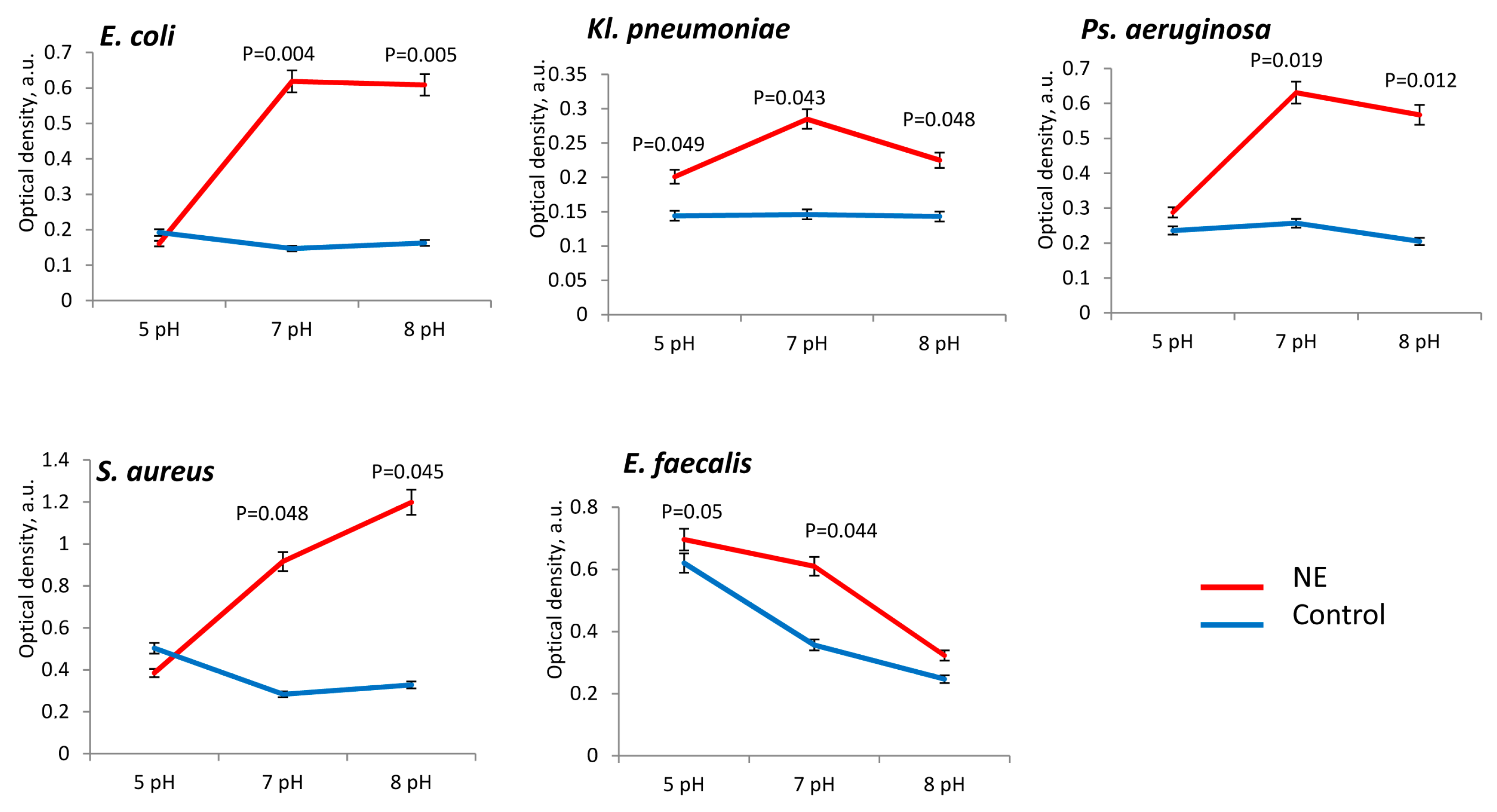
Abstract
:1. Introduction
2. Materials and Methods
2.1. Bacterial Strains and Cultivation
2.2. pH and Soluble Components of Urine
2.3. Effects on the Growth of Bacterial Biomass
2.4. Matrix Production Assay
2.5. Metabolic Activity Assay
2.6. Statistical Analysis
3. Results
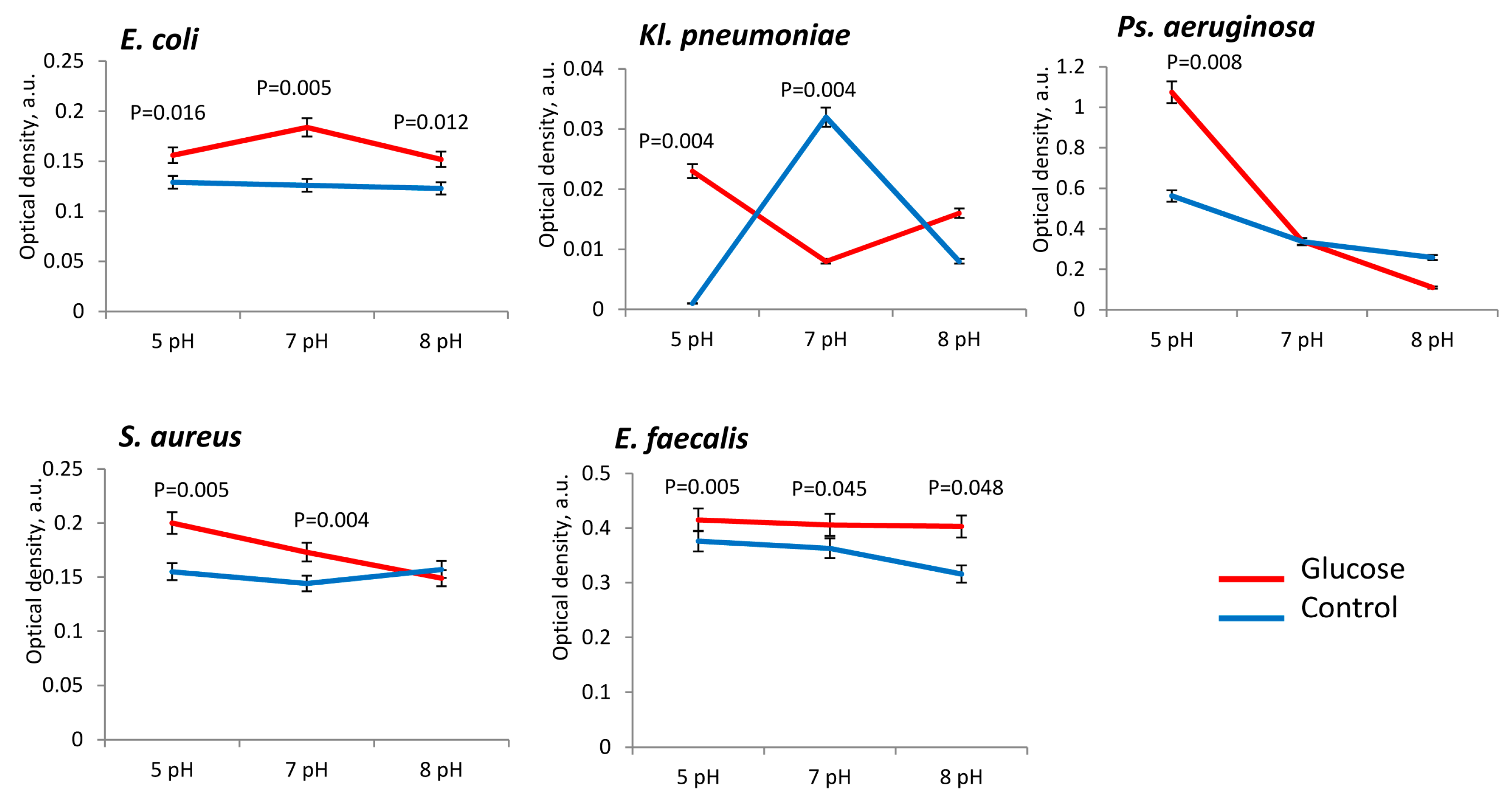
3.1. Biomass Production of Uropathogenic Microorganisms
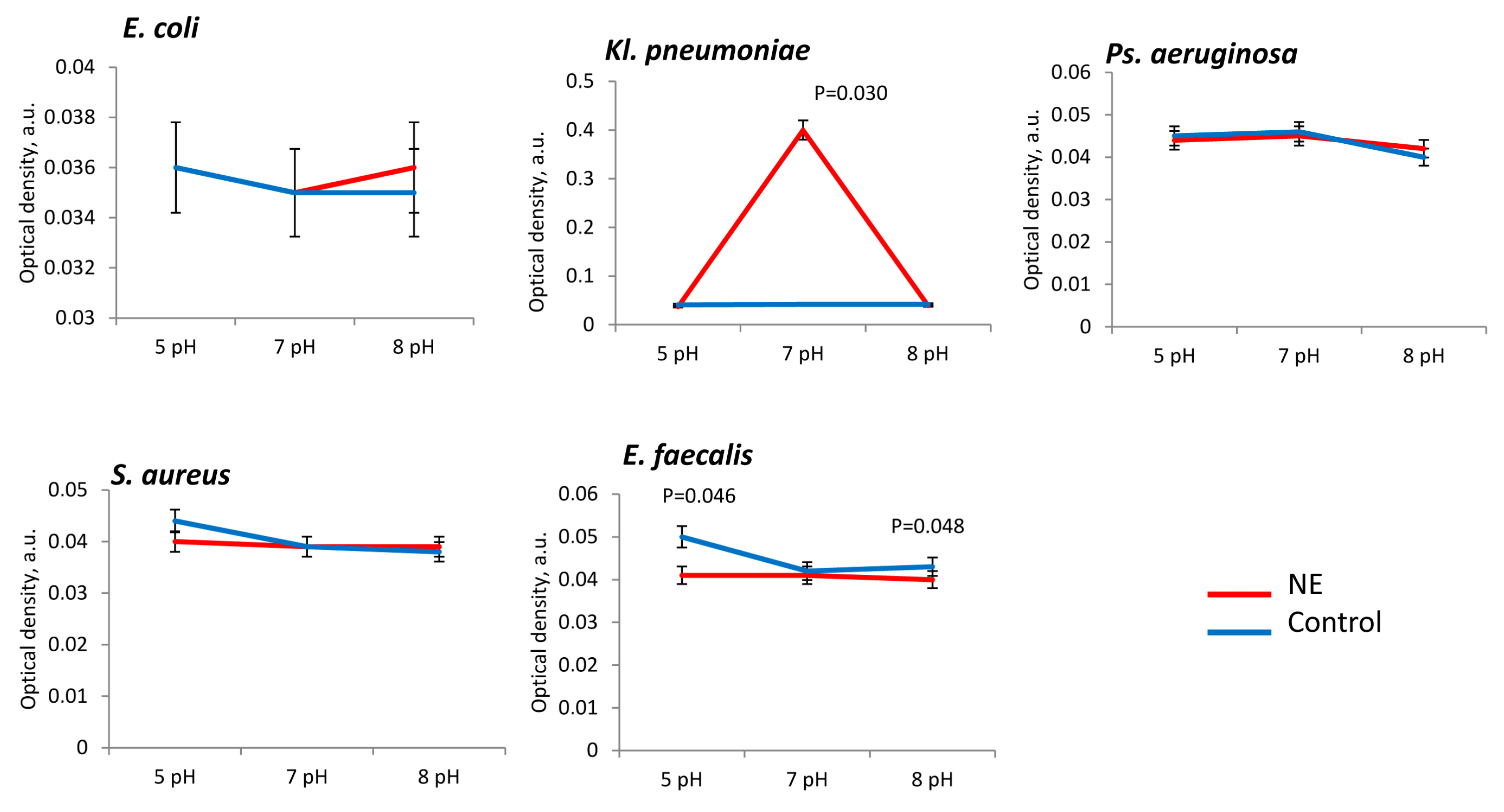
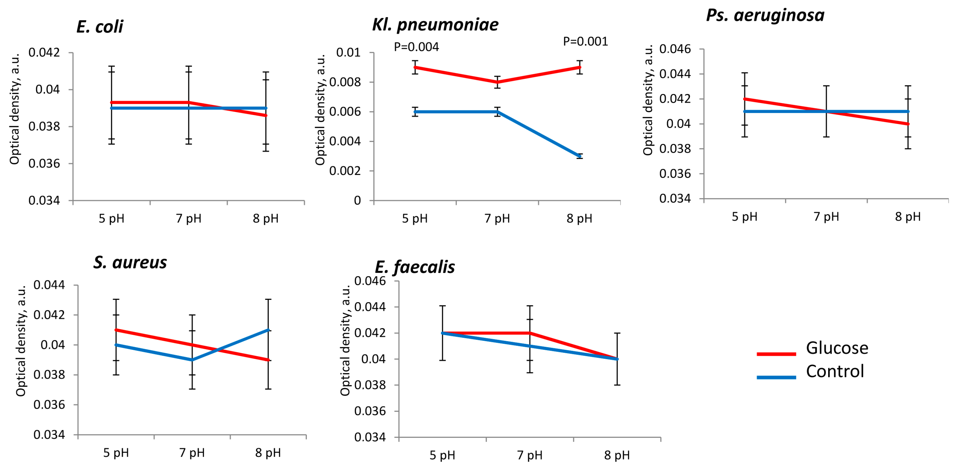
3.2. Matrix Production of Uropathogenic Microorganisms
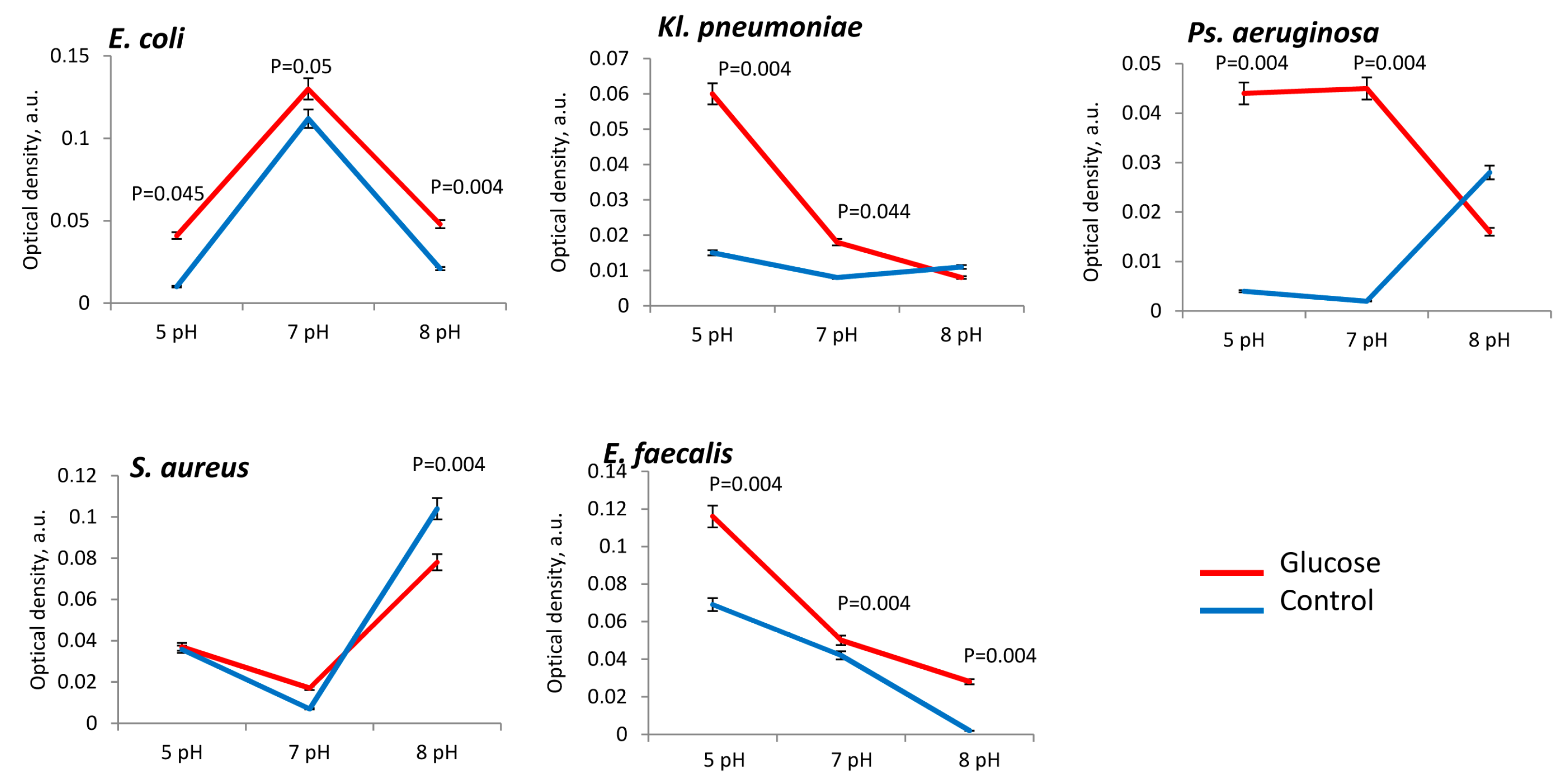
3.3. Metabolic Activity of Uropathogenic Microorganisms
4. Discussion
5. Conclusions
Author Contributions
Funding
Acknowledgments
Conflicts of Interest
References
- Wu, J.; Gao, Y. Physiological conditions can be reflected in human urine proteome and metabolome. Expert Rev. Proteom. 2015, 12, 623–636. [Google Scholar] [CrossRef] [PubMed]
- Dobrek, Ł. Drug-related urinary tract infections. Wiad. Lek. 2021, 74, 1728–1736. [Google Scholar] [CrossRef] [PubMed]
- Ferraro, P.M.; Bargagli, M.; Trinchieri, A.; Gambaro, G. Risk of Kidney Stones: Influence of Dietary Factors, Dietary Patterns, and Vegetarian-Vegan Diets. Nutrients 2020, 12, 779. [Google Scholar] [CrossRef] [PubMed] [Green Version]
- Siener, R.; Bitterlich, N.; Birwé, H.; Hesse, A. The Impact of Diet on Urinary Risk Factors for Cystine Stone Formation. Nutrients 2021, 13, 528. [Google Scholar] [CrossRef]
- Nouvenne, A.; Ticinesi, A.; Morelli, I.; Guida, L.; Borghi, L.; Meschi, T. Fad diets and their effect on urinary stone formation. Transl. Androl. Urol. 2014, 3, 303–312. [Google Scholar] [CrossRef]
- Hopkins, E.; Sanvictores, T.; Sharma, S. Physiology, Acid Base Balance. In StatPearls [Internet]; StatPearls: Treasure Island, FL, USA, 2022. [Google Scholar]
- Ogg, C. Chronic renal failure. Br. Med. J. 1970, 4, 223–225. [Google Scholar] [CrossRef] [Green Version]
- Ferrari, C.; Santo, G.; Mammucci, P.; Pisani, A.R.; Sardaro, A.; Rubini, G. Diagnostic Value of Choline PET in the Preoperative Localization of Hyperfunctioning Parathyroid Gland(s): A Comprehensive Overview. Biomedicines 2021, 9, 231. [Google Scholar] [CrossRef]
- Go, T. Effect of antiepileptic drug polytherapy on urinary pH in children and young adults. Child’s Nerv. Syst. 2009, 25, 237–240. [Google Scholar] [CrossRef]
- Cook, J.D.; Strauss, K.A.; Caplan, Y.H.; Lodico, C.P.; Bush, D.M. Urine pH: The effects of time and temperature after collection. J. Anal. Toxicol. 2007, 31, 486–496. [Google Scholar] [CrossRef] [Green Version]
- Lee, J.; Chang, H.K.; Lee, S. Association of low urine pH as a metabolic feature with abdominal obesity. J. Int. Med. Res. 2020, 48, 0300060519898615. [Google Scholar] [CrossRef] [Green Version]
- Hetey, S.K.; Kleinberg, M.L.; Parker, W.D.; Johnson, E.W. Effect of ascorbic acid on urine pH in patients with injured spinal cords. Am. J. Hosp. Pharm. 1980, 37, 235–237. [Google Scholar] [CrossRef]
- Grases, F.; Costa-Bauza, A.; Prieto, R.M. Renal lithiasis and nutrition. Nutr. J. 2006, 5, 23. [Google Scholar] [CrossRef] [PubMed] [Green Version]
- Wang, R.C. Managing Urolithiasis. Ann. Emerg. Med. 2016, 67, 449–454. [Google Scholar] [CrossRef] [PubMed] [Green Version]
- von Euler, U.S.; Luft, R. Noradrenaline output in urine after infusion in man. Br. J. Pharmacol. Chemother. 1951, 6, 286–288. [Google Scholar] [CrossRef] [Green Version]
- Ignatova, N.; Abidullina, A.; Streltsova, O.; Elagin, V.; Kamensky, V. Norepinephrine Effects on Uropathogenic Strains Virulence. Microorganisms 2022, 10, 2248. [Google Scholar] [CrossRef]
- Fujitani, T.; Fujii, Y.; Lyu, Z.; Harada Sassa, M.; Harada, K.H. Urinary equol levels are positively associated with urinary estradiol excretion in women. Sci Rep. 2021, 11, 19532. [Google Scholar] [CrossRef]
- Theorell, T. A long-term perspective on cardiovascular job stress research. J. Occup. Health 2019, 61, 3–9. [Google Scholar] [CrossRef] [PubMed] [Green Version]
- Kolouri, S.; Daneshfard, B.; Jaladat, A.M.; Tafazoli, V. Green Urine in Traditional Persian Medicine: Differential Diagnosis and Clinical Relevance. J. Evid. -Based Complement. Altern. Med. 2017, 22, 232–236. [Google Scholar] [CrossRef] [Green Version]
- Liman, M.N.P.; Jialal, I. Physiology, Glycosuria. In StatPearls [Internet]; StatPearls Publishing: Treasure Island, FL, USA, 2022. [Google Scholar]
- Aubron, C.; Huet, O.; Ricome, S.; Borderie, D.; Pussard, E.; Leblanc, P.E.; Bouvet, O.; Vicaut, E.; Denamur, E.; Duranteau, J. Changes in urine composition after trauma facilitate bacterial growth. BMC Infect. Dis. 2012, 12, 330. [Google Scholar] [CrossRef] [PubMed] [Green Version]
- Ignatova, N.I.; Alexandrova, N.A.; Zaslavskaya, M.I.; Abramycheva, D.V. Evaluation of the influence of culturing on the intensity of biofilm formation by Klebsiella pneumoniae strains. Klin. Lab. Diagn. (Russ. Clin. Lab. Diagn.) 2020, 65, 512–515. [Google Scholar] [CrossRef]
- Manoharan, A.; Ognenovska, S.; Paino, D.; Whiteley, G.; Glasbey, T.; Kriel, F.H.; Farrell, J.; Moore, K.H.; Manos, J.; Das, T. N-Acetylcysteine Protects Bladder Epithelial Cells from Bacterial Invasion and Displays Antibiofilm Activity against Urinary Tract Bacterial Pathogens. Antibiotics 2021, 10, 900. [Google Scholar] [CrossRef]
- Mart’yanov, S.V.; Botchkova, E.A.; Plakunov, V.K.; Gannesen, A.V. The Impact of Norepinephrine on Mono-Species and Dual-Species Staphylococcal Biofilms. Microorganisms 2021, 9, 820. [Google Scholar] [CrossRef] [PubMed]
- Gond, D.P.; Singh, S.; Agrawal, N.K. Norepinephrine augmented in vitro growth of uropathogenic E. coli in Type 2 diabetes mellitus and its suppression by silodosin (alpha blocker). Diagn. Microbiol. Infect. Dis. 2018, 92, 85–89. [Google Scholar] [CrossRef] [PubMed]
- Mohanty, S.; Kamolvit, W.; Scheffschick, A.; Björklund, A.; Tovi, J.; Espinosa, A.; Brismar, K.; Nyström, T.; Schröder, J.M.; Östenson, C.G.; et al. Diabetes downregulates the antimicrobial peptide psoriasin and increases E. coli burden in the urinary bladder. Nat. Commun. 2022, 13, 4983. [Google Scholar] [CrossRef] [PubMed]
- Altabas, V.; Altabas, K.; Berković-Cigrovski, M.; Malosevac, S.; Vrkljan, M.; Nikolić Heitzler, V. Glucose metabolism disorders in patients with acute coronary syndromes. Acta Clin. Croat 2012, 51, 71–77. [Google Scholar]
- Vachvanichsanong, P.; McNeil, E.B.; Dissaneewate, P. Extended-spectrum beta-lactamase Escherichia coli and Klebsiella pneumoniae urinary tract infections. Epidemiol. Infect. 2020, 149, e12. [Google Scholar] [CrossRef]
- Geerlings, S.E.; Brouwer, E.C.; Gaastra, W.; Verhoef, J.; Hoepelman, A.I.M. Effect of glucose and pH on uropathogenic and non-uropathogenic Escherichia coli: Studies with urine from diabetic and non-diabetic individuals. J. Med. Microbiol. 1999, 48, 535–539. [Google Scholar] [CrossRef] [Green Version]
- Islam, M.J.; Bagale, K.; John, P.P.; Kurtz, Z.; Kulkarni, R. Glycosuria alters uropathogenic Escherichia coli global gene expression and virulence. mSphere 2022, 7, e0000422. [Google Scholar] [CrossRef]
- Elagin, V.; Budruev, I.; Antonyan, A.; Bureev, P.; Ignatova, N.; Streltsova, O.; Kamensky, V. Enhancement of the Efficacy of Photodynamic Therapy against Uropathogenic Gram-Negative Bacteria Species. Photonics 2023, 10, 310. [Google Scholar] [CrossRef]


Disclaimer/Publisher’s Note: The statements, opinions and data contained in all publications are solely those of the individual author(s) and contributor(s) and not of MDPI and/or the editor(s). MDPI and/or the editor(s) disclaim responsibility for any injury to people or property resulting from any ideas, methods, instructions or products referred to in the content. |
© 2023 by the authors. Licensee MDPI, Basel, Switzerland. This article is an open access article distributed under the terms and conditions of the Creative Commons Attribution (CC BY) license (https://creativecommons.org/licenses/by/4.0/).
Share and Cite
Ignatova, N.; Abidullina, A.; Streltsova, O.; Elagin, V.; Kamensky, V. Effect of pH, Norepinephrine and Glucose on Metabolic and Biofilm Activity of Uropathogenic Microorganisms. Microorganisms 2023, 11, 862. https://doi.org/10.3390/microorganisms11040862
Ignatova N, Abidullina A, Streltsova O, Elagin V, Kamensky V. Effect of pH, Norepinephrine and Glucose on Metabolic and Biofilm Activity of Uropathogenic Microorganisms. Microorganisms. 2023; 11(4):862. https://doi.org/10.3390/microorganisms11040862
Chicago/Turabian StyleIgnatova, Nadezhda, Alina Abidullina, Olga Streltsova, Vadim Elagin, and Vladislav Kamensky. 2023. "Effect of pH, Norepinephrine and Glucose on Metabolic and Biofilm Activity of Uropathogenic Microorganisms" Microorganisms 11, no. 4: 862. https://doi.org/10.3390/microorganisms11040862