Article

Selected Gut Bacteria from Water Monitor Lizard Exhibit Effects against Pathogenic Acanthamoeba castellanii Belonging to the T4 Genotype

1
Department of Clinical Sciences, College of Medicine, University of Sharjah, University City, Sharjah 27272, United Arab Emirates
2
Sharjah Institute for Medical Research, University of Sharjah, Sharjah 27272, United Arab Emirates
3
Department of Medical Biology, Faculty of Medicine, Istinye University, Istanbul 34010, Turkey
4
Department of Medicinal Chemistry, College of Pharmacy, University of Sharjah, Sharjah 27272, United Arab Emirates
5
Department of Clinical Laboratory Sciences, College of Applied Medical Sciences, Taif University, Taif 21944, Saudi Arabia
6
Department of Medical Microbiology, Faculty of Medicine, Al-Baha University, P.O. Box. 1988, Al-Baha 65799, Saudi Arabia
7
College of Arts and Sciences, American University of Sharjah, University City, Sharjah 26666, United Arab Emirates
*
Author to whom correspondence should be addressed.
Microorganisms 2023, 11(4), 1072; https://doi.org/10.3390/microorganisms11041072
Submission received: 29 March 2023 / Revised: 10 April 2023 / Accepted: 17 April 2023 / Published: 20 April 2023
(This article belongs to the Special Issue Microbiome and One Health)

Abstract

:
Water monitor lizards (WMLs) reside in unhygienic and challenging ecological surroundings and are routinely exposed to various pathogenic microorganisms. It is possible that their gut microbiota produces substances to counter microbial infections. Here we determine whether selected gut bacteria of water monitor lizards (WMLs) possess anti-amoebic properties using Acanthamoeba castellanii of the T4 genotype. Conditioned media (CM) were prepared from bacteria isolated from WML. The CM were tested using amoebicidal, adhesion, encystation, excystation, cell cytotoxicity and amoeba-mediated host cell cytotoxicity assays in vitro. Amoebicidal assays revealed that CM exhibited anti-amoebic effects. CM inhibited both excystation and encystation in A. castellanii. CM inhibited amoebae binding to and cytotoxicity of host cells. In contrast, CM alone showed limited toxic effects against human cells in vitro. Mass spectrometry revealed several antimicrobials, anticancer, neurotransmitters, anti-depressant and other metabolites with biological functions. Overall, these findings imply that bacteria from unusual places, such as WML gut, produce molecules with anti-acanthamoebic capabilities.

1. Introduction

Acanthamoeba is pervasive in a variety of environments [1,2,3]. Its life cycle includes a dormant cyst stage and an active trophozoite stage [1,4]. Pathogenic Acanthamoeba can cause granulomatous amoebic encephalitis (GAE) and blinding keratitis [1]. Despite advances in antimicrobial chemotherapy, treatment remains problematic, suggesting the need to find therapeutic agents [3,5,6].
Secondary metabolites are produced by a variety of organisms, including different animals, microorganisms (bacteria, fungi and parasites, etc.), marine organisms, terrestrial plants, vertebrate and invertebrate species [7,8,9,10,11,12]. Although plant-derived products have been used frequently in folk medicine, the discovery of penicillin signaled a significant shift in the source of natural molecules from plants to microorganisms [13]. Since then, metabolites produced by bacteria have been widely used in the food industries, agriculture, pharmaceuticals and scientific research [14]. For example, Bacillus, cyanobacteria, myxobacteria and Pseudomonas produce secondary metabolites with therapeutic values [15]. Streptomyces venezuelae produced pikromycin, the first known polyketide antibiotic [16]. Streptomyces noursei produced a potent polyene antifungal agent, i.e., Nystatin [17]. Amphotericin B is another antifungal drug produced by Streptomyces nodosus [18]. Pseudomonas species isolated from the Antarctic region produced broad-spectrum antiparasitic and antiproliferative effects [19]. Streptomyces hachijoensis derived Trichomycin exhibited antifungal and antitrichomonal activity [20].
Water monitor lizards (WMLs) breathe in unhygienic and challenging ecological surroundings, commonly feeding on rotten food contaminated with various pathogenic microorganisms. Therefore, the purpose of this investigation was to extract and characterize the selected gut bacterial species of WML. CM were tested against pathogenic A. castellanii of the genotype T4 in different assays to determine their anti-amoebic potential.

2. Materials and Methods

2.1. Dissection of Water Monitor Lizard and Isolation of Gut Bacteria

In brief, the Department of Wildlife and National Parks (PERHILITAN), Malaysia, approved this study, as well as Sunway University, Malaysia (SUNREC 2019/023). Prior to the experiments, all operating and dissecting tools were properly sterilized in an autoclave. Additionally, during the dissection procedure, tools were surface sterilized with 70% ethanol. WML was dissected, gut was removed using the aseptic technique and culturable bacteria were isolated with sterile swabs. The swabs were cultured on blood agar plates, and the plates were incubated at 37 °C for 24 h. Bacterial colonies were differentiated from each other, and pure cultures were grown in nutrient broth overnight at 37 °C.

2.2. DNA Extraction, Amplification and Identification of Isolated Bacteria

DNA were extracted from pure bacterial cultures using the AllPrep Bacterial DNA extraction kit (Qiagen, Germantown, MD, USA). The extracted DNA were amplified using 16S rRNA universal primers (27F 5′ AGAGTTTGATCMTGGCTCAG 3′ and 1492R 5′ TACGGYTACCTTGTTACGACTT 3′) [21]. The amplified products were sequenced commercially (1st Base; Axil Scientific Pt. Ltd., Singapore). In order to find matches with pre-existing reference sequences, the output sequences were exported into the alignment program Basic Local Alignment Search Tool (BLAST). Using the Maximum Likelihood (ML) approach in MEGA 7 and the GTR+G model with concatenated 16S rRNA sequences, the phylogenetic tree was reconstructed.

2.3. Preparation of Bacterial Conditioned Media

Pure bacterial colonies were cultured in Roswell Park Memorial Institute (RPMI) at 37 °C with constant shaking at 90 rpm for 48 h [11,12,22]. Next, bacterial cultures were centrifuged at 10,000× g for 60 min at 4 °C. Cell free culture supernatants were collected and filtered using 0.22 μm pore size filters and kept at −80 °C until tested.

2.4. Acanthamoeba castellanii Cultures

A. castellanii genotype T4 (ATCC 50492) was grown in a proteose-peptone-yeast-glucose (PYG) growth medium containing 0.75% yeast extract, 1.5% d-glucose and 0.75% proteose-peptone [6]. Cultures were kept at 30 °C until confluent. For assays, adhering trophozoites were detached by placing the flask on ice for 10 min and then gentle tapping. The culture containing amoebae was then centrifuged at 2500× g for 5 min. Next, supernatants were discarded, and the pellet was re-suspended in 1 mL of RPMI medium and amoebae were enumerated using a hemocytometer.

2.5. Amoebicidal Assays

Amoebicidal assays were performed as described previously [6,22]. Briefly, 5 × 10⁵ amoebae were incubated with different CM for 2 and 24 h at 30 °C. Controls include amoebae alone in RPMI (negative control) and with 25 µM chlorhexidine (positive control). Finally, 0.1% Trypan blue dye was added to each well, and amoebae were enumerated using a hemocytometer. The number of viable unstained amoebae was determined as before [6].

2.6. HeLa Cell Cultivation

HeLa cells were obtained from the American Type Culture Collection (ATCC CCL-2) and maintained in RPMI with L-glutamine complemented with 1% minimum essential amino acids, 10% fetal bovine serum (FBS) and 1% Penicillin-Streptomycin (Pen-Strep) at 37 °C in an incubator with 5% CO2 [6].

2.7. Adhesion Assays

CM were tested in adhesion assays to see their effects on amoebae binding to human cells [23]. Briefly, 100 µL of each CM was incubated with 5 × 10⁵ amoebae at 30 °C for 120 min. Next, amoebae were centrifuged at 2500× g for 5 min, and pelleted amoebae (pre-treated amoebae) were obtained. The pellet was resuspended in RPMI (200 µL) and subsequently transferred to HeLa cells. Plates were incubated at 37 °C for 60 min. Finally, unbound amoebae were enumerated using a hemocytometer, and the percentage amoebae was estimated using the given equation: % amoebae (bound) = 100 − amoebae (unbound).

2.8. Encystation Assays

Using 16% glucose as an encystation medium, approximately 1 × 10⁶ amoeba trophozoites were incubated with different CM for 48 h at 30 °C as before [24]. Next, amoebae were treated with sodium dodecyl sulphate (SDS) (0.1%) for approximately 20 min. Using a hemocytometer, the residual cysts were counted, and the results were recorded.

2.9. Excystation Assays

To prepare cysts, A. castellanii trophozoites were placed on non-nutrient agar plates for two weeks at 30 °C, as described earlier [5,6]. Cysts were collected by scrapping off the agar plates using a cell scrapper using PBS. Cysts were pelleted by centrifugation at 3000× g for 10 min. Next, half a million cysts were incubated with 100 µL of different CM in PYG medium with a final volume of 500 µL for up to 72 h [6]. Finally, a hemocytometer was used to count viable amoebae trophozoites.

2.10. Host Cell Cytotoxicity Assays

To determine the cytotoxic effects of CM, 100 µL of the CM were incubated with HeLa cell monolayers in 96-well plate as described earlier [6,11]. The plate was maintained for an overnight period in the cell culture incubator supplied with 5% CO2 at 37 °C. Finally, cell supernatants were collected, and LDH release was determined using Cytotoxicity Detection kit (Roche Diagnostics, Indianapolis, IN, USA). Percent cell damage was determined by the following equation,
%   C y t o t o x i c i t y = s a m p l e   m e a n   v a l u e     n e g a t i v e   c o n t r o l   m e a n   v a l u e p o s i t i v e   c o n t r o l   m e a n   v a l u e     n e g a t i v e   c o n t r o l   m e a n   v a l u e × 100

2.11. Cytopathogenicity Assays

Cytopathogenicity assays were performed to determine whether CM inhibits amoebae-mediated host cell death. In brief, amoebae were treated with CM as described in adhesion assays, and then, pre-treated amoebae were incubated with HeLa cell monolayers overnight at 37 °C. The amoebae-mediated cell damage was evaluated by measuring the amount of LDH enzyme released into cell media by damaged cells as before ([25]).

2.12. Ultra-Performance Liquid Chromatography Tandem Mass Spectrometry (HPLC-MS/MS)

For metabolomics, the sample metabolite extraction was carried out as before [26]. Next, each sample was given two injections of 1 µL for metabolomics, which were followed by 30 min of elution using the gradient described below: Over the course of 15 min, ACN was raised from 1% to 99%, held there for 3 min, and then reduced to 1% ACN for another 10 min. Elution and re-equilibration flow rates were 250 and 350 µL/min, respectively. The analysis was carried out using an Apollo II electrospray ionization (ESI) source and a TimsTOF (Bruker, Darmstadt, Germany). The drying temperature, gas flow and nebulizer pressure levels were adjusted to 220 °C, 10 µL/min and 2.2 bar, respectively. The capillary voltage and end plate offset were 4500 V and 500 V, respectively. For metabolites processing and statistical assessment, MetaboScape® 4.0 was utilized. The T-ReX 2D/3D method for molecular characterization and “bucketing” was modified using the following criteria: Peak identification requires a minimum peak length of 7 spectra and a minimum intensity threshold of 1000 counts with characteristic verification based on peak area. The file masses were recalibrated using an external calibrant that was applied between 0 and 0.3 min. Only data having retention times somewhere between 0.3 and 25 min and m/z ranges between 50 and 1000 were properly considered, whereas the MS/MS import strategy was set to average. All the experimentations were performed in three biological replicates while each sample (extracts) was analyzed in duplicate using LC-TIMS-QTOF MS.
MetaboScape compared features to the MetaboPharmHam database (HMDB) using retention time and accurate mass or MS2 spectral identification. Then, among the numerous other entries within the same molecule, the data of the same compounds that perfectly matched the maximum number of variables, such as m/z values, MS/MS, analyte list, spectrum library and retention time were chosen as the metabolite entries to be further filtered. T-test (two sample, two-tailed distribution) statistical analyses were performed to highlight the impact of CM on amoebae. The data are shown as the average standard deviation of multiple replicated experiments. The software Graph Pad Prism version 8.0 was used for all of the analyses and visualizations. p ≤ 0.05 was chosen as the statistical significance threshold.

3. Results

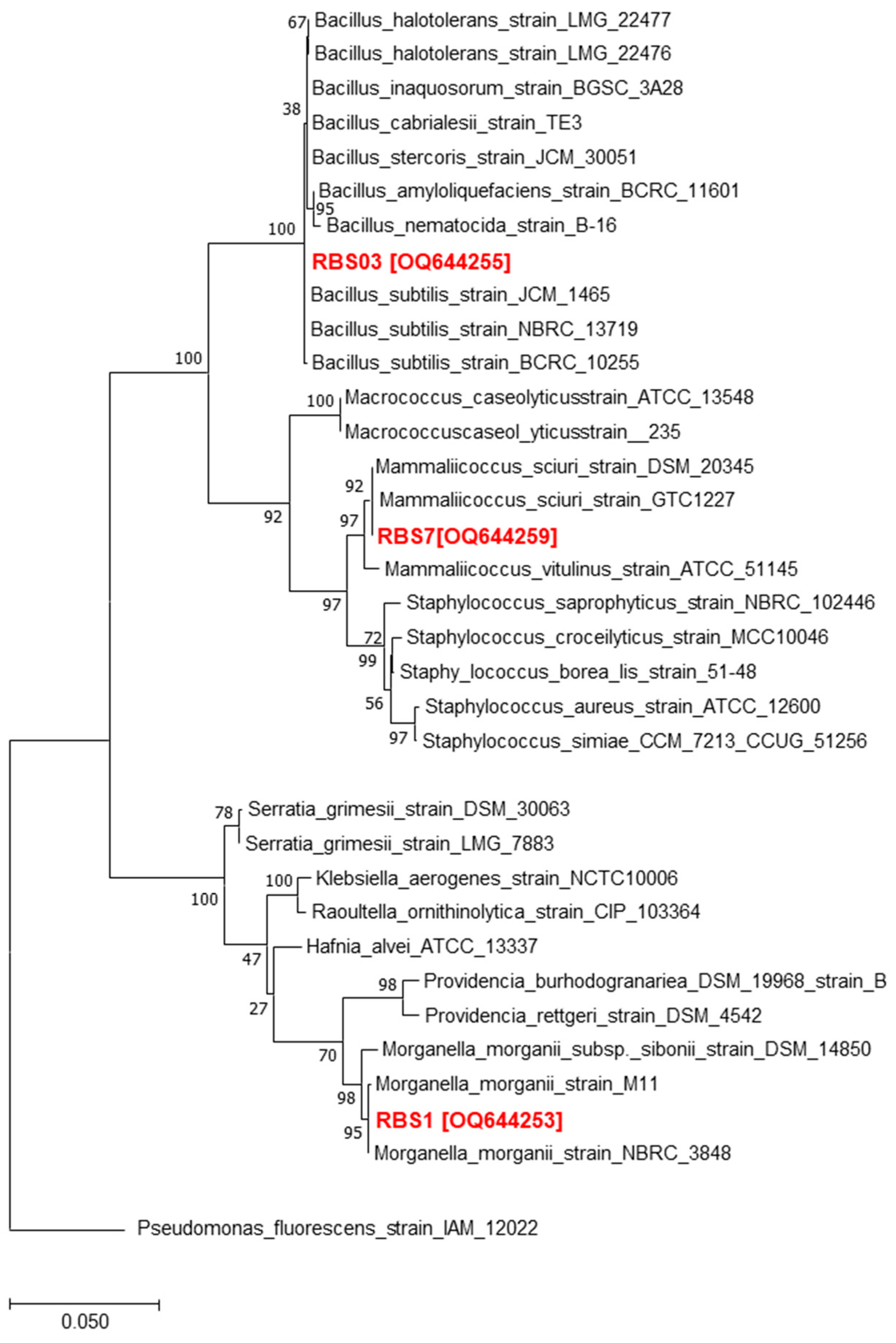
3.1. Isolation and Identifcation of Bacterial Isolates Derived from the Gut of Water Monitor Lizard

Several bacterial species were isolated from water monitor lizard gut. Sequencing results revealed numerous bacterial species (Table 1). Several bacteria, including Morganella morganii, Bacillus subtilis and Mammaliicoccus sciuri, were identified (Figure 1 and Table 1).

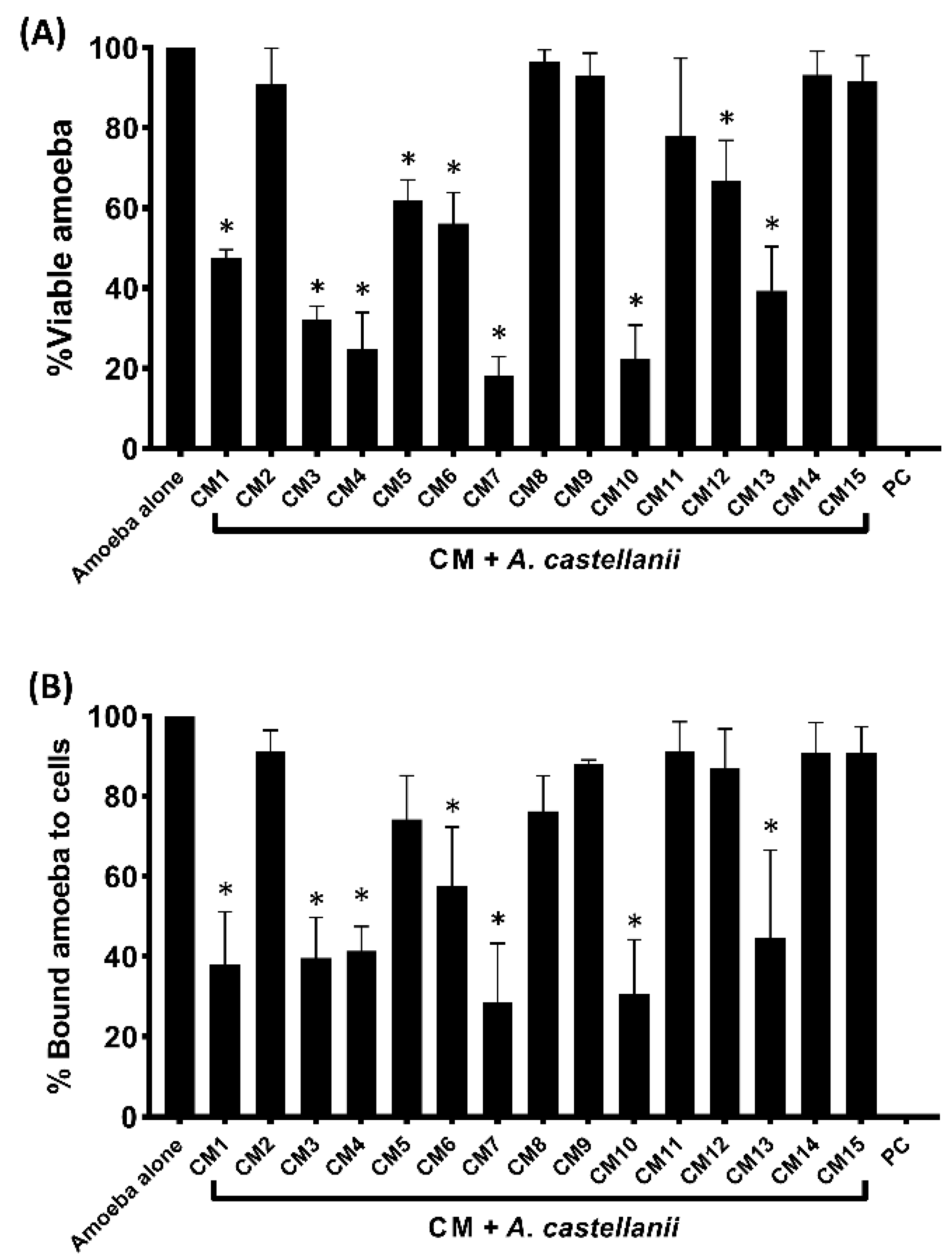
3.2. Conditioned Media Isolated from Selected Gut Bacteria of Water Monitor Lizard Showed Amoebicidal Activity

The findings showed that against A. castellanii, CM1, CM3-CM7, CM10, CM12 and CM13 showed anti-amoebic effects (Figure 2A). CM7 reduced amoeba viability by 82%, followed by CM10 (78%), CM4 (76%), CM3 (68%) and CM1 (53%). Contrarily, CM5, CM6, and CM12 demonstrated 38%, 44%, and 33% amoebicidal effects (Figure 2A).

3.3. Bacterial Conditioned Media Inhibited the Binding of Amoebae to Human Cells

The findings showed that with the exception of CM5 and CM12, all CM with amoebicidal activity decreased amoebae adhesion to HeLa cells (Figure 2B). CM7 and CM10 reduced amoeba binding by 72% and 69%, respectively, while CM1, CM3, CM4, CM13 and CM6 inhibited amoebae adhesion to human cells by 62%, 61%, 59%, 56% and 42%, respectively (Figure 2B). Amoeba attachment to HeLa cells was not prevented by the remaining CM (Figure 2B).

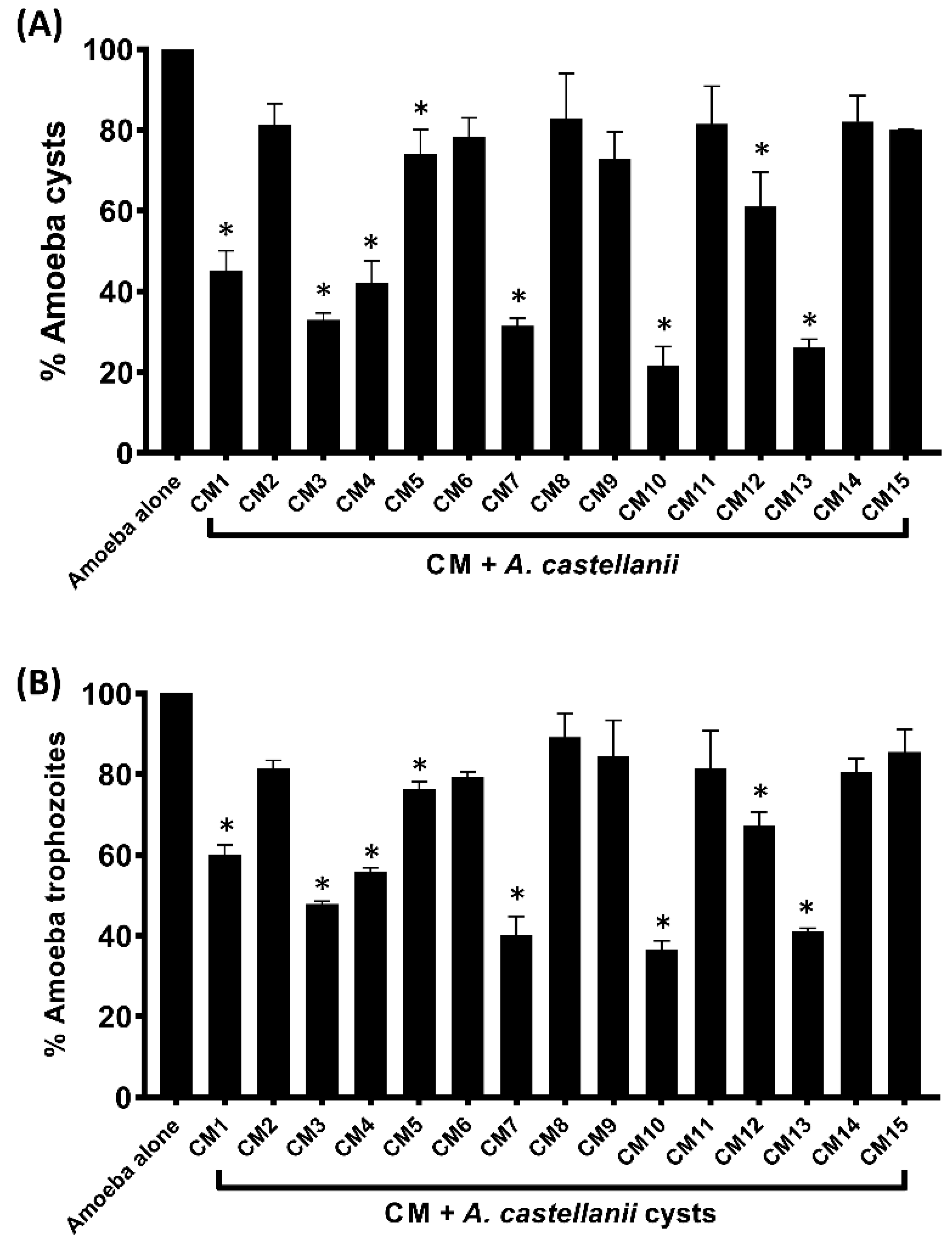
3.4. Encystation and Excystation of Amoebae were Inhibited

A. castellanii trophozoites treated with 16% glucose (encystation media) were used as the negative control in encystation experiments, and A. castellanii cysts obtained were considered to be 100%. The results revealed that several CM inhibited A. castellanii encystation when compared to the negative control (Figure 3A). In the presence of CM10 and CM13, cysts formation declined to 21% and 26%, respectively. Similarly, CM1, CM3, CM4, CM7 and CM12 arrested 55%, 67%, 58%, 69% and 39% amoebae (Figure 3B).

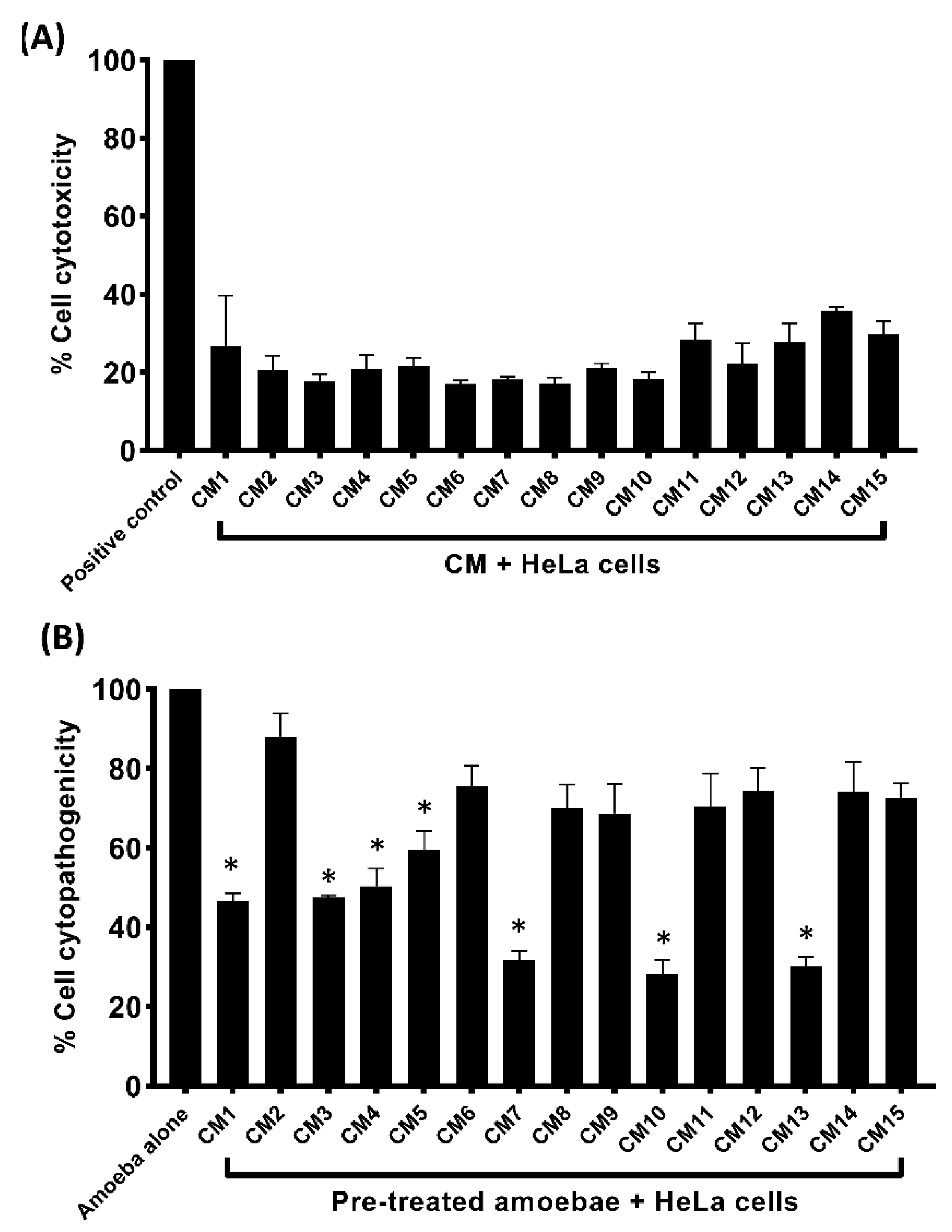
3.5. Conditioned Media Exhibited Limited Cytotoxicity and Inhibited Amoebae–Mediated Host Cell Damage

The CM produced minimal toxicity against HeLa cell lines, except CM1, CM11 and CM13-15, which exhibited low to moderate cell cytotoxicity (Figure 4A). Similarly, CM decreased host cell cytotoxicity caused by amoebae (Figure 4B). Less than 30% of the cells in CM7, CM10 and CM13 were found to have damage compared to CM1 and CM3, which limited the damage to 46% (Figure 4B).

3.6. Mass Spectrometry Revealed Numerous Metabolites

Mass spectrometry revealed a total of 189 tentatively identified metabolites (Table 2 and Supplementary Table S1). Among the identified metabolites, the most abundant and important compounds were Carvone, 1,11-Undecanedicarboxylic acid, 4-Ethylphenol, Benzoic acid, Hydrocinnamic acid, Indole-3-carbinol, Indolelactic acid, Quinaldic acid, Succinic acid and several other compounds, some of which with known antimicrobial effects (Table 2 and Supplementary Table S1). Principal Components Analysis (PCA) shows the relative (dis)similarity between samples according to the first two principal components. Points represent samples coloured according to the location of the point of origin. Circles represent Gram-negative and triangles Gram-positive bacterial species (Supplementary Figure S1a). Among these metabolites, there are 12 metabolites with significant altered abundance across sampling locations. Androsterone is more abundant in samples taken from the mouth and oesophagus while nearly absent in samples from other locations. Estrone is nearly absent in samples drawn from the anus/rectum and large intestine but abundant in samples from all other locations (Supplementary Figure S1b). Furthermore, hierarchical clustering shows that bacteria collected from the mouth generally clustered together, as did samples from the anus/rectum and large intestine, with samples from origins between those extremes mostly forming a cluster between those two (Supplementary Figure S2).

4. Discussion

Acanthamoeba, being a free-living amoeba, can be found in a wide range of habitats, including water, soil, swimming pools and hospitals [47]. It can endure extreme conditions and cause eye and brain infections [4]. Many available drugs are used to fight amoebic infections, but their non-specificity and potent cytotoxicity are challenges to researchers [5]. As a result, new anti-amoebic drugs with high bioactivity are required. In the present study, conditioned media from WML gut bacteria were prepared and tested against the pathogenic A. castellanii genotype T4. The results have shown that CM from water monitor gut bacteria showed remarkable amoebicidal effects against A. castellanii. CM also restricted the binding capability of the parasite to human cell lines and amoeba-mediated host cell death. Furthermore, the CM inhibited both the encystation and excystation processes in amoebae and showed limited cytotoxicity towards HeLa cells. These findings are consistent with earlier research [11,22,48]. CM isolated from bacteria identified from WML intestine indicated considerable anticancer effects [49]. Streptomyces avermitilis produces avermectins and their derivatives that possess strong anti-parasitic activity against Onchocerca volvulus (parasite) [50]. Saccharopolyspora spinose produces spinosad that exhibited notable insecticidal activity against ecto-parasite of the livestock [14].
In this study, CM produced by the gut microbiota of WML showed significant amoebicidal action against pathogenic A. castellanii. In our previous study, CM from cockroach gut bacteria possessed important amoebicidal activity against A. castellanii [22]. Iqbal et al. reported that the CM from E. coli K1 and Enterobacter species exhibited noteworthy amoebicidal and amoebistatic effects against A. castellanii [51]. Therefore, our findings are consistent with earlier findings emphasizing the significance of WML gut bacteria as an anti-amoebic agent(s).
Also, we investigated the effects of the CM isolated from WML gut bacteria on the adhesion capabilities of A. castellanii to human cells. Following therapy, CM precluded amoebae from attaching to human cells, reducing cell damage. Matin et al. reported the interaction of human brain microvascular endothelial cells (HBMECs) and Balamuthia mandrillaris, where galactose impeded the binding capability of B. mandrillaris to HBMECs [52].
Conditioned medium minimized amoeba-mediated host cell damage and inhibited the excystation and encystation of A. castellanii while exhibiting low cytotoxicity. Quercetin and kolavenic acid, which were extracted from Polyalthia longifolia, have virtually no harmful effects on human cell lines [24]. Similarly, CM of bacteria isolated from animals gut produced minimal cytotoxic properties against human cells [11,12,21,48].
It is interesting to note that the isolated bacteria from WML gut produced a variety of bioactive metabolites with antimicrobial effects. These metabolites include benzoic acid [33,34,35,36,37,39], carvone [27], 4-Ethylphenol [32], succinic acid [46] and 1,11-Undecanedicarboxylic acid etc. [29,30]. These bacteria also produced anticancer molecules, such as Carvone and Indole-3-carbinol [27,41,42], enzyme inhibitors [38] and various pharmacologically and commercially important metabolites. The compound 4-Ethylphenol produced by Lactobacillus plantarum is a strong odorant and is used as a strong anti-fungal agent [31,32]. Likewise, Candida species produced Indolelactic acid, which exhibited antibacterial effects against Gram-positive and Gram-negative bacteria [44]. Quinaldic acid extracted from Ephedra pachyclada showed notable antibacterial activity against intestinal bacteria [45]. Likewise, various antimicrobial (antibacterial and antifungal) lipopeptides (surfactin and iturin A) were generated by Bacillus subtills found in Rattus rattus (rat) intestine [21].
The identified molecules revealed different modes of action. For example, succinic acid identified from dried and pickled mustard damaged the cell membrane in Staphylococcus aureus and Pseudomonas fluorescens [52]. Similarly, Indole-3-carbinol showed anti-cancer activity by arresting the cells at G1 phase and inhibiting the expression of cyclin-dependent kinase-6 [53]. Benzoic acid showed antimicrobial effects against Zygosaccharomyces bailii by depleting ATP generation [54]. In this study, these metabolites have been identified from WML gut bacteria. The anti-amoebic effects may be due to the presence of such bioactive compounds that could lead to membrane damage, depletion of energy and inhibition of growth related enzymes or other targets. Several metabolites with antimicrobial, anticancer and other well-known biological activities were identified. Our findings propose that microbial species from unexplored sources, such as the gut of a water monitor lizard, produce anti-amoebic compounds that have the potential to become potent anti-parasitic medicines.

Supplementary Materials

The following supporting information can be downloaded at: https://www.mdpi.com/article/10.3390/microorganisms11041072/s1.

Author Contributions

Conceptualization, N.A.K. and R.S.; Methodology, N.A., N.A.K., A.D.G. and N.C.S.; Formal analysis, N.A., A.D.G. and N.C.S.; Investigation, N.A., N.A.K., N.C.S., A.M.A., H.A. and R.S.; Resources, A.M.A., H.A. and R.S.; Writing—original draft, N.A. and R.S.; Writing—review & editing, N.A.K., A.D.G., N.C.S., A.M.A. and H.A.; Supervision, N.A.K. and R.S.; Project administration, N.A.K.; Funding acquisition, N.A.K. and R.S. All authors have read and agreed to the published version of the manuscript.

Funding

This work was funded, in part, by the Air Force Office of Scientific Research (AFOSR), grant number: FA 8655-20-1-7004.

Data Availability Statement

The data described in this study are accessible from the corresponding author upon request.

Acknowledgments

Authors acknowledge the American University of Sharjah and the University of Sharjah for their support.

Conflicts of Interest

The authors declare no conflict of interest.

References

  1. Visvesvara, G.S.; Moura, H.; Schuster, F.L. Pathogenic and opportunistic free-living amoebae: Acanthamoeba spp., Balamuthia mandrillaris, Naegleria fowleri, and Sappinia diploidea. FEMS Immunol. Med. Microbiol. 2007, 50, 1–26. [Google Scholar] [CrossRef] [PubMed]
  2. Niederkorn, J.Y.; Alizadeh, H.; Leher, H.; McCulley, J.P. The pathogenesis of Acanthamoeba keratitis. Microbes Infect. 1999, 1, 437–443. [Google Scholar] [CrossRef]
  3. Marciano-Cabral, F.; Cabral, G. Acanthamoeba spp. as agents of disease in humans. Clin. Microbiol. Rev. 2003, 16, 273–307. [Google Scholar] [CrossRef]
  4. Khan, N.A. Acanthamoeba: Biology and increasing importance in human health. FEMS Microbiol. Rev. 2006, 30, 564–595. [Google Scholar] [CrossRef]
  5. Anwar, A.; Mungroo, M.R.; Anwar, A.; Sullivan, W.J., Jr.; Khan, N.A.; Siddiqui, R. Repositioning of guanabenz in conjugation with gold and silver nanoparticles against pathogenic amoebae Acanthamoeba castellanii and Naegleria fowleri. ACS Infect. Dis. 2019, 5, 2039–2046. [Google Scholar] [CrossRef]
  6. Akbar, N.; Kawish, M.; Jabri, T.; Khan, N.A.; Shah, M.R.; Siddiqui, R. Cinnamic acid and lactobionic acid based nanoformulations as a potential antiamoebic therapeutics. Exp. Parasitol. 2023, 246, 108474. [Google Scholar] [CrossRef] [PubMed]
  7. Chin, Y.W.; Balunas, M.J.; Chai, H.B.; Kinghorn, A.D. Drug discovery from natural sources. AAPS J. 2006, 8, E239–E253. [Google Scholar] [CrossRef]
  8. Simmons, T.L.; Coates, R.C.; Clark, B.R.; Engene, N.; Gonzalez, D.; Esquenazi, E.; Dorrestein, P.C.; Gerwick, W.H. Biosynthetic origin of natural products isolated from marine microorganism–invertebrate assemblages. Proc. Natl. Acad. Sci. USA 2008, 105, 4587–4594. [Google Scholar] [CrossRef]
  9. Show, P.L.; Oladele, K.O.; Siew, Q.Y.; Aziz Zakry, F.A.; Lan, J.C.W.; Ling, T.C. Overview of citric acid production from Aspergillus niger. Front. Life Sci. 2015, 8, 271–283. [Google Scholar] [CrossRef]
  10. Zida, A.; Bamba, S.; Yacouba, A.; Ouedraogo-Traore, R.; Guiguemdé, R. Anti-Candida albicans natural products, sources of new antifungal drugs: A review. J. Mycol. Med. 2017, 27, 1–19. [Google Scholar] [CrossRef]
  11. Akbar, N.; Khan, N.A.; Sagathevan, K.; Iqbal, M.; Tawab, A.; Siddiqui, R. Gut bacteria of Cuora amboinensis (turtle) produce broad-spectrum antibacterial molecules. Sci. Rep. 2019, 9, 1–19. [Google Scholar] [CrossRef] [PubMed]
  12. Akbar, N.; Siddiqui, R.; Iqbal, M.; Khan, N.A. Antibacterial activities of selected pure compounds isolated from gut bacteria of animals living in polluted environments. Antibiotics 2020, 9, 190. [Google Scholar] [CrossRef]
  13. Tan, S.Y.; Tatsumura, Y. Alexander Fleming (1881–1955): Discoverer of penicillin. Singap. Med. J. 2015, 56, 366. [Google Scholar] [CrossRef] [PubMed]
  14. Sanchez, S.; Guzmán-Trampe, S.; Ávalos, M.; Ruiz, B.; Rodríguez-Sanoja, R.; Jiménez-Estrada, M. “Microbial Natural Products,” in Natural Products in Chemical Biology; Civjan, N., Ed.; Wiley: Hoboken, NJ, USA, 2012; pp. 65–108. [Google Scholar]
  15. Berdy, J. Bioactive microbial metabolites. J. Antibiot. 2005, 58, 1–26. [Google Scholar] [CrossRef] [PubMed]
  16. Jung, W.S.; Lee, S.K.; Hong, J.S.J.; Park, S.R.; Jeong, S.J.; Han, A.R.; Sohng, J.K.; Kim, B.G.; Choi, C.Y.; Sherman, D.H.; et al. Heterologous expression of tylosin polyketide synthase and production of a hybrid bioactive macrolide in Streptomyces venezuelae. Appl. Microbiol. Biotechnol. 2006, 72, 763–769. [Google Scholar] [CrossRef]
  17. Stanley, V.C.; English, M.P. Some effects of nystatin on the growth of four Aspergillus species. Microbiology 1965, 40, 107–118.x. [Google Scholar] [CrossRef]
  18. Tevyashova, A.N.; Olsufyeva, E.N.; Solovieva, S.E.; Printsevskaya, S.S.; Reznikova, M.I.; Trenin, A.S.; Galatenko, O.A.; Treshalin, I.D.; Pereverzeva, E.R.; Mirchink, E.P.; et al. Structure-antifungal activity relationships of polyene antibiotics of the amphotericin B group. Antimicrob. Agents Chemother. 2013, 57, 3815–3822. [Google Scholar] [CrossRef]
  19. Silva, T.R.; Duarte, A.W.; Passarini, M.R.; Ruiz, A.L.T.; Franco, C.H.; Moraes, C.B.; de Melo, I.S.; Rodrigues, R.A.; Fantinatti-Garboggini, F.; Oliveira, V.M. Bacteria from Antarctic environments: Diversity and detection of antimicrobial, antiproliferative, and antiparasitic activities. Polar Biol. 2018, 41, 1505–1519. [Google Scholar] [CrossRef]
  20. Shiomi, K. Antiparasitic antibiotics from Japan. Parasitol. Int. 2021, 82, 102298. [Google Scholar] [CrossRef]
  21. Akbar, N.; Siddiqui, R.; Iqbal, M.; Sagathevan, K.; Kim, K.S.; Habib, F.; Khan, N.A. Gut Bacteria of Rattus rattus (Rat) Produce Broad-Spectrum Antibacterial Lipopeptides. ACS Omega 2021, 6, 12261–12273. [Google Scholar] [CrossRef]
  22. Akbar, N.; Siddiqui, R.; Iqbal, M.; Sagathevan, K.; Khan, N.A. Gut bacteria of cockroaches are a potential source of antibacterial compound (s). Lett. Appl. Microbiol. 2018, 66, 416–426. [Google Scholar] [CrossRef]
  23. Akbar, N.; Siddiqui, R.; Khamis, M.; Ibrahim, T.; Khan, N.A. Cationic surfactant-natural clay complex as a novel agent against Acanthamoeba castellanii belonging to the T4 genotype. Eye Contact Lens. 2021, 47, 592–597. [Google Scholar] [CrossRef]
  24. Anwar, A.; Ting, E.L.S.; Anwar, A.; ul Ain, N.; Faizi, S.; Shah, M.R.; Khan, N.A.; Siddiqui, R. Antiamoebic activity of plant-based natural products and their conjugated silver nanoparticles against Acanthamoeba castellanii (ATCC 50492). AMB Express 2020, 10, 24. [Google Scholar] [CrossRef]
  25. Ali, S.M.; Khan, N.A.; Sagathevan, K.; Anwar, A.; Siddiqui, R. Biologically active metabolite (s) from haemolymph of redheaded centipede Scolopendra subspinipes possess broad-spectrum antibacterial activity. AMB Express 2019, 9, 95. [Google Scholar] [CrossRef]
  26. Faris, M.; Madkour, M.; Al Bustanji, Y.; Semreen, M.; Giddey, A.D.; Soares, N.C.; Zeb, F. Ramadan diurnal intermittent fasting is associated with significant plasma metabolomics changes in subjects with overweight and obese: A prospective cohort study. Front. Nutr. 2022, 1–17. [Google Scholar]
  27. Bouyahya, A.; Mechchate, H.; Benali, T.; Ghchime, R.; Charfi, S.; Balahbib, A.; Burkov, P.; Shariati, M.A.; Lorenzo, J.M.; Omari, N.E. Health benefits and pharmacological properties of carvone. Biomolecules 2021, 11, 1803. [Google Scholar] [CrossRef]
  28. De Sousa, D.P.; De Farias Nóbrega, F.F.; De Almeida, R.N. Influence of the chirality of (R)-(−)-and (S)-(+)-carvone in the central nervous system: A comparative study. Chirality Pharmacol. Biol. Chem. Conseq. Mol. Asymmetry 2007, 19, 264–268. [Google Scholar] [CrossRef]
  29. Gibka, J.; Kunicka-Styczyńska, A.; Gliński, M. Experimental immunology antimicrobial activity of undecan-3-one, undecan-3-ol and undec-3-yl acetate. Cent. Eur. J. Immunol. 2009, 34, 154–157. [Google Scholar]
  30. Jarboe, L.R.; Royce, L.A.; Liu, P. Understanding biocatalyst inhibition by carboxylic acids. Front. Microbiol. 2013, 4, 272. [Google Scholar] [CrossRef] [PubMed]
  31. Santamaría, L.; Reverón, I.; de Felipe, F.L.; de Las Rivas, B.; Muñoz, R. Ethylphenol formation by Lactobacillus plantarum: Identification of the enzyme involved in the reduction of vinylphenols. Appl. Environ. Microbiol. 2018, 84, e01064-18. [Google Scholar] [CrossRef] [PubMed]
  32. Ge, T.; Gao, W.; Liang, C.; Han, C.; Wang, Y.; Xu, Q.; Wang, Q. 4-Ethylphenol, A Volatile Organic Compound Produced by Disease-Resistant Soybean, Is a Potential Botanical Agrochemical Against Oomycetes. Front. Plant Sci. 2021, 12, 717258. [Google Scholar] [CrossRef]
  33. Cho, J.Y.; Moon, J.H.; Seong, K.Y.; Park, K.H. Antimicrobial activity of 4-hydroxybenzoic acid and trans 4-hydroxycinnamic acid isolated and identified from rice hull. Biosci. Biotechnol. Biochem. 1998, 62, 2273–2276. [Google Scholar] [CrossRef]
  34. Melliou, E.; Chinou, I. Chemical analysis and antimicrobial activity of Greek propolis. Planta Med. 2004, 70, 515–519. [Google Scholar] [CrossRef]
  35. Kong, W.; Zhao, Y.; Shan, L.; Xiao, X.; Guo, W. Thermochemical studies on the quantity–antibacterial effect relationship of four organic acids from Radix Isatidis on Escherichia coli growth. Biol. Pharm. Bull. 2008, 31, 1301–1305. [Google Scholar] [CrossRef]
  36. Alvesalo, J.; Vuorela, H.; Tammela, P.; Leinonen, M.; Saikku, P.; Vuorela, P. Inhibitory effect of dietary phenolic compounds on Chlamydia pneumoniae in cell cultures. Biochem. Pharmacol. 2006, 71, 735–741. [Google Scholar] [CrossRef]
  37. Kubo, I.; Xiao, P.; Nihei, K.I.; Fujita, K.I.; Yamagiwa, Y.; Kamikawa, T. Molecular design of antifungal agents. J. Agric. Food Chem. 2002, 50, 3992–3998. [Google Scholar] [CrossRef]
  38. Innocenti, A.; Hall, R.A.; Schlicker, C.; Mühlschlegel, F.A.; Supuran, C.T. Carbonic anhydrase inhibitors. Inhibition of the β-class enzymes from the fungal pathogens Candida albicans and Cryptococcus neoformans with aliphatic and aromatic carboxylates. Bioorg. Med. Chem. 2009, 17, 2654–2657. [Google Scholar] [CrossRef]
  39. Ngurah, B.I.G.M.; Nyoman, Y.N.; Dafroyati, Y.; Gunadi, I.G.A.; Taneo, M. Antibacterial Evaluation of 2, 4-Dihidroxy Benzoic Acid on Escherichia coli and Vibrio Alginolyticus. J. Phys. Conf. Ser. 2020, 1503, 012027. [Google Scholar] [CrossRef]
  40. Korneev, S.M. Hydrocinnamic acids: Application and strategy of synthesis. Synthesis 2013, 45, 1000–1015. [Google Scholar] [CrossRef]
  41. He, Y.H.; Friesen, M.D.; Ruch, R.J.; Schut, H.A.J. Indole-3-carbinol as a chemopreventive agent in 2-amino-1-methyl-6-phenylimidazo [4, 5-b] pyridine (PhIP) carcinogenesis: Inhibition of PhIP–DNA adduct formation, acceleration of PhIP metabolism, and induction of cytochrome P450 in female F344 rats. Food Chem. Toxicol. 2000, 38, 15–23. [Google Scholar] [CrossRef]
  42. Nachshon-Kedmi, M.; Yannai, S.; Haj, A.; Fares, F.A. Indole-3-carbinol and 3, 3′-diindolylmethane induce apoptosis in human prostate cancer cells. Food Chem. Toxicol. 2003, 41, 745–752. [Google Scholar] [CrossRef]
  43. Sarkar, F.H.; Li, Y. Indole-3-carbinol and prostate cancer. J. Nutr. 2004, 134, 3493S–3498S. [Google Scholar] [CrossRef] [PubMed]
  44. Narayanan, T.K.; Rao, G.R. Beta-indoleethanol and beta-indolelactic acid production by Candida species: Their antibacterial and autoantibiotic action. Antimicrob. Agents Chemother. 1976, 9, 375–380. [Google Scholar] [CrossRef]
  45. Lee, C.H.; Lee, H.S. Growth inhibiting activity of quinaldic acid isolated from Ephedra pachyclada against intestinal bacteria. J. Korean Soc. Appl. Biol. 2009, 52, 331–335. [Google Scholar] [CrossRef]
  46. Kumar, R.; Chandar, B.; Parani, M. Use of succinic & oxalic acid in reducing the dosage of colistin against New Delhi metallo-β-lactamase-1 bacteria. Indian J. Med. Res. 2018, 147, 97. [Google Scholar]
  47. De Jonckheere, J.F. Ecology of acanthamoeba. Rev. Infect. Dis. 1991, 13 (Suppl. S5), S385–S387. [Google Scholar] [CrossRef]
  48. Akbar, N.; Siddiqui, R.; Sagathevan, K.; Iqbal, M.; Khan, N.A. Gut bacteria of water monitor lizard (Varanus salvator) are a potential source of antibacterial compound (s). Antibiotics 2019, 8, 164. [Google Scholar] [CrossRef] [PubMed]
  49. Soopramanien, M.; Khan, N.A.; Sagathevan, K.; Siddiqui, R. Gut bacteria of Varanus salvator possess potential antitumour molecules. Int. Microbiol. 2021, 24, 47–56. [Google Scholar] [CrossRef] [PubMed]
  50. Shen, B. A new golden age of natural products drug discovery. Cell 2015, 163, 1297–1300. [Google Scholar]
  51. Iqbal, J.; Siddiqui, R.; Khan, N.A. Acanthamoeba and bacteria produce antimicrobials to target their counterpart. Parasit. Vectors 2014, 7, 56. [Google Scholar] [CrossRef]
  52. Huang, S.; Chen, X.; Yan, R.; Huang, M.; Chen, D. Isolation, identification and antibacterial mechanism of the main antibacterial component from pickled and dried mustard (Brassica juncea Coss. Var. foliosa Bailey). Molecules 2022, 27, 2418. [Google Scholar] [CrossRef] [PubMed]
  53. Katz, E.; Nisani, S.; Chamovitz, D.A. Indole-3-carbinol: A plant hormone combatting cancer. F1000Research 2018, 7, 689. [Google Scholar] [CrossRef]
  54. Warth, A.D. Mechanism of action of benzoic acid on Zygosaccharomyces bailii: Effects on glycolytic metabolite levels, energy production, and intracellular pH. Appl. Environ. Microbiol. 1991, 57, 3410–3414. [Google Scholar] [CrossRef]
Figure 1. Based on the phylogenetic analysis of their 16 S rRNA genes, the phylogenetic tree shows the isolated bacteria from the water monitor lizard stomach that were employed in this work (red) and Pseudomonas fluorescens as an outgroup. The GTR+G model with concatenated 16 S rRNA sequences was used as the foundation for the dendrogram reconstruction using the Maximum Likelihood (ML) approach (MEGA 7). Branching nodes show percentage bootstrap values higher than 50% of 1000 repetitions.
Microorganisms 11 01072 g001
Figure 2. (A) Conditioned media were prepared by incubating each bacterium in RPMI at 37 °C for 48 h. Next, bacterial cultures were centrifuged for 60 min at 10,000× g at 4 °C, and supernatants, i.e., CM, were filtered using 0.22 μm pore size filters. Equal doses (100 µL) of various bacterial CM were used to determine amoebic effects. Briefly, A. castellanii, i.e., 5 × 10⁵, were incubated with 100 µL of CM for 24 h at 30 °C. After this, viable amoebae were counted using a hemocytometer microscopically. Amoebae grown in RPMI alone were taken as the negative control, while amoebae incubated with 25 µM chlorhexidine were used as the positive control. The data are expressed as the mean ± standard error. p values were determined using two-sample t-test, two-tailed distribution, and (*) is p ≤ 0.05. p ≤ 0.05 was chosen as the statistical significance threshold. (B) Conditioned media from water monitor lizard gut bacteria repressed the A. castellanii binding to human cells. Adhesion assays were performed to determine whether A. castellanii interacts with human cells. Note that CM repressed A. castellanii significantly. p values were determined using two-sample t-test, two-tailed distribution, (*) is <0.05. The data are expressed as the mean ± standard error.
Microorganisms 11 01072 g002
Figure 3. (A) Effects of CM on the encystation of A. castellanii trophozoites. The results showed that CM inhibited the encystment of trophozoite form compared to the negative control. The data are presented as the mean ± standard error. p values were calculated using a two-sample t-test with a two-tailed distribution, and (*) denotes that p ≤ 0.05. (B) Effects of CM on A. castellanii excystation. When compared to the negative control, the results demonstrated that CM reduced the excystment of trophozoites. Overall, the CM prevented the excystation process in amoebae. The data are presented as the mean ± standard error. p values were calculated using a two-sample t-test with a two-tailed distribution, and (*) denotes that p ≤ 0.05.
Microorganisms 11 01072 g003
Figure 4. (A) The effects of CM on human cell lines. The compounds alone and in complex form exhibited limited cytotoxicity. (B) The cytopathogenicity of human cells was decreased by pre-treating A. castellanii with CM. The results revealed substantial inhibition of amoeba-mediated host cytotoxicity when compared to amoeba (untreated). Data are presented as mean  ±  standard error. (*) denotes where p ≤ 0.05.
Microorganisms 11 01072 g004
Table 1. Bacterial species isolated from the gut of water monitor lizard.
S. No.Conditioned Media CodeBacteria SpeciesAccession Number
1.CM1Morganella morganiiOQ644253
2.CM2Empedobacter brevisOQ644254
3.CM3Bacillus subtilisOQ644255
4.CM4Pseudomonas otitidisOQ644256
5.CM5Pseudoclavibacter albaOQ644257
6.CM6Pseudomonas aeruginosaOQ644258
7.CM7Mammaliicoccus sciuriOQ644259
8.CM8Proteus terraeOQ644260
9.CM9Chryseobacterium taklimakanenseOQ644261
10.CM10Bacillus aeriusOQ644262
11.CM11Morganella morganiiOQ644263
12.CM12Corynebacterium atrinaeOQ644264
13.CM13Mammaliicoccus sciuriOQ644265
14.CM14Chryseobacterium taklimakanenseOQ644266
15.CM15Corynebacterium freneyiOQ644267
Table 2. Mass spectrometry revealed several compounds with biological activities.
Compounds NamesRT [min]m/z Meas.Chemical FormulaStructureActivity
(+)-(S)-Carvone11.9151.113C10H14OMicroorganisms 11 01072 i001Antimicrobial activity (Antibacterial, antifungal, antiparasitic) by damaging plasma membrane [27], neurological activity [28], anti-neuraminidase, antioxidant, anti-inflammatory and anticancer activities [27].
1,11-Undecanedicarboxylic acid10.47245.175C13H24O4Microorganisms 11 01072 i002The analogues exhibited antimicrobial activity [29,30].
4-Ethylphenol9.88123.0804C8H10OMicroorganisms 11 01072 i003This is a strong odorant compound produced by Lactobacillus plantarum [31]. This is an agrochemical agent produced by the disease-resistant soybean eliminating fungal infections caused by P. sojae and Phytophthora nicotianae. The volatile compound has potent anti-fungal effects against soil-borne fungi, i.e., Rhizoctonia solani, Gaeumannomyces graminis var tritici and Fusarium graminearum [32].
Benzoic acid3.41123.0484C7H6O2Microorganisms 11 01072 i004Benzoic acid alone is considered to be a general-purpose antimicrobial agent with a broad spectrum of actions towards human pathogenic bacteria and fungi with varying minimum inhibitory concentrations (MIC) values [33,34,35,36,37]. Additionally, it was being tested as a potential inhibitor of -carbonic anhydrase, a novel molecular target found in Cryptococcus neoformans and C. albicans [38]. 2,4-dihidroxy benzoic acid showed significant antibacterial activity against Vibrio alginolyticus and E. coli [39].
Hydrocinnamic acid5.95151.0728C9H10O2Microorganisms 11 01072 i005It belongs to the phenylpropanoids class, having an important role in pharmaceuticals, cosmetics and as a food additive [40].
Indole-3-carbinol9.52148.076C9H9NOMicroorganisms 11 01072 i006It is used to inhibit several types of cancers in human and prevent carcinogenesis in animal models [41,42]. The dimeric counterpart of indole-3-carbinol, i.e., 3,3′-diindolylmethane, is a very crucial anti-cancer agent [43].
Indolelactic acid7.31206.084C11H11NO3Microorganisms 11 01072 i007Candida species produce Indolelactic acid that inhibit several Gram-negative and Gram-positive bacteria [44].
Quinaldic acid5.98174.0551C10H7NO2Microorganisms 11 01072 i008The Quinaldic acid inhibit the growth of several intestinal bacteria [45].
Succinic acid8.36119.0351C4H6O4Microorganisms 11 01072 i009Succinic acid has a strong antibacterial effect against E. coli [46].
Disclaimer/Publisher’s Note: The statements, opinions and data contained in all publications are solely those of the individual author(s) and contributor(s) and not of MDPI and/or the editor(s). MDPI and/or the editor(s) disclaim responsibility for any injury to people or property resulting from any ideas, methods, instructions or products referred to in the content.

