Article

Prevalence of Small Intestinal Bacterial Overgrowth Syndrome in Patients with Non-Alcoholic Fatty Liver Disease/Non-Alcoholic Steatohepatitis: A Cross-Sectional Study

by
Paraskevas Gkolfakis
1,*,
Georgios Tziatzios
1,
Gabriela Leite
2,
Ioannis S. Papanikolaou
1,
Elias Xirouchakis
3,
Ioannis G. Panayiotides
4,
Athanasios Karageorgos
5,
Maria Jesus Millan
2,
Ruchi Mathur
2,
Stacy Weitsman
2,
George D. Dimitriadis
1,
Evangelos J. Giamarellos-Bourboulis
5,
Mark Pimentel
2 and
Konstantinos Triantafyllou
1
1
Hepatogastroenterology Unit, Second Department of Internal Medicine-Propaedeutic, Medical School, National and Kapodistrian University of Athens, 12462 Athens, Greece
2
Medically Associated Science and Technology (MAST) Program, Cedars-Sinai, Los Angeles, CA 90048, USA
3
Department of Gastroenterology and Hepatology, Athens Medical Palaio Faliron General Hospital, 17562 Palaio Faliron, Greece
4
2nd Department of Pathology, Medical School, National and Kapodistrian University of Athens, 124622 Athens, Greece
5
4th Department of Internal Medicine, Medical School, National and Kapodistrian University of Athens, 12462 Athens, Greece
*
Author to whom correspondence should be addressed.
Microorganisms 2023, 11(3), 723; https://doi.org/10.3390/microorganisms11030723
Submission received: 6 February 2023 / Revised: 4 March 2023 / Accepted: 8 March 2023 / Published: 10 March 2023
(This article belongs to the Special Issue Gut Microbiota and Host Diseases)

Abstract

:
Introduction: Non-alcoholic fatty liver disease (NAFLD) is a multifactorial, wide-spectrum liver disorder. Small intestinal bacterial overgrowth (SIBO) is characterized by an increase in the number and/or type of colonic bacteria in the upper gastrointestinal tract. SIBO, through energy salvage and induction of inflammation, may be a pathophysiological factor for NAFLD development and progression. Aim/Methods: Consecutive patients with histological, biochemical, or radiological diagnosis of any stage of NAFLD (non-alcoholic fatty liver [NAFL], non-alcoholic steatohepatitis [NASH], cirrhosis) underwent upper gastrointestinal endoscopy. Duodenal fluid (2cc) was aspirated from the 3rd–4th part of duodenum into sterile containers. SIBO was defined as ≥103 aerobic colony-forming units (CFU)/mL of duodenal aspirate and/or the presence of colonic-type bacteria. Patients without any liver disease undergoing gastroscopy due to gastroesophageal reflux disease (GERD) comprised the healthy control (HC) group. Concentrations (pg/mL) of tumor necrosis factor alpha (TNFα), interleukin (IL)-1β, and IL-6 were also measured in the duodenal fluid. The primary endpoint was to evaluate the prevalence of SIBO in NAFLD patients, while the comparison of SIBO prevalence among NAFLD patients and healthy controls was a secondary endpoint. Results: We enrolled 125 patients (51 NAFL, 27 NASH, 17 cirrhosis, and 30 HC) aged 54 ± 11.9 years and with a weight of 88.3 ± 19.6 kg (NAFLD vs. HC 90.7 ± 19.1 vs. 80.8 ± 19.6 kg, p = 0.02). Overall, SIBO was diagnosed in 23/125 (18.4%) patients, with Gram-negative bacteria being the predominant species (19/23; 82.6%). SIBO prevalence was higher in the NAFLD cohort compared to HC (22/95; 23.2% vs. 1/30; 3.3%, p = 0.014). Patients with NASH had higher SIBO prevalence (6/27; 22.2%) compared to NAFL individuals (8/51; 15.7%), but this difference did not reach statistical significance (p = 0.11). Patients with NASH-associated cirrhosis had a higher SIBO prevalence compared to patients with NAFL (8/17; 47.1% vs. 8/51; 15.7%, p = 0.02), while SIBO prevalence between patients with NASH-associated cirrhosis and NASH was not statistically different (8/17; 47.1% vs. 6/27; 22.2%, p = 0.11). Mean concentration of TNF-α, IL-1β, and IL-6 did not differ among the different groups. Conclusion: The prevalence of SIBO is significantly higher in a cohort of patients with NAFLD compared to healthy controls. Moreover, SIBO is more prevalent in patients with NASH-associated cirrhosis compared to patients with NAFL.

1. Introduction

Non-alcoholic fatty liver disease (NAFLD) remains the main cause of abnormal liver function tests in Western countries [1,2]. The definition of NAFLD requires the exclusion of ongoing or recent excessive alcohol consumption and other causes of liver steatosis [3]. Histologic evidence of liver fatty infiltration or, in the absence of histology, gamma-glutamyl transferase (γ-GT) and/or aminotransferase elevation, as well as compatible sonographic findings, are sufficient for diagnosis. Moreover, modern imaging modalities, such as controlled attenuation parameter (CAP) elastography and magnetic resonance proton density fat fraction, have been developed to quantify fatty infiltration and provide reliable evidence of NAFLD for diagnosis and clinical follow up [4]. The spectrum of the disease encompasses non-alcoholic fatty liver (NAFL), non-alcoholic steatohepatitis (NASH), NASH-related cirrhosis, and hepatocellular carcinoma [5]. Despite the fact that obesity, diabetes mellitus, and hypertriglyceridemia are the main risk factors for the development and progression of NAFLD [6], the pathogenetic factors that lead to NAFLD development have not been fully clarified. An emerging approach engaging liver steatosis with metabolic syndrome and their cumulative impact on cardiovascular diseases, has generated the new term of metabolic syndrome-associated fatty liver disease (MAFLD). However, this new nomenclature has not been universally accepted to replace NAFLD, due to the identified limitations on its applicability, especially regarding liver steatosis cases without metabolic syndrome [7]. More specifically, the historical “two hits” hypothesis regarding NAFLD pathogenesis [8] has been replaced by the “multiple parallel” hits hypothesis [9,10], where inflammation may either precede or follow simple steatosis with multiple factors, namely lipotoxicity, increased oxidative stress, mitochondrial dysfunction, and iron overload, acting in parallel to promote NASH [10]. This is also supported by studies where NAFLD is diagnosed in patients with chronic inflammatory conditions, such as inflammatory bowel diseases, even when their metabolic profile is normal, as indicated by the dominant term of sarcopenic NAFLD [11]. Moreover, a genetic predisposition to the disease development and progression cannot be excluded [12].
Interesting experimental data from the field of obesity have suggested a potential correlation between gut microbiota and NAFLD/NASH development [13,14,15,16]. This correlation could be mediated by a variety of mechanisms including direct action of the bacteria or their by-products (e.g., LPS-endotoxin) on hepatic parenchyma, bacterial translocation due to the increased intestinal permeability, and redundant salvage energy by altered gut microbiota [17,18]. Studies using fecal bacterial counts have demonstrated a clear correlation between the presence of NASH and a decreased percentage of Bacteroidetes, which represent one of the main bacterial phyla in the human gastrointestinal lumen [19,20,21,22]. In this regard, small intestinal bacterial overgrowth (SIBO), characterized by the abnormal presence of large numbers of colonic-type bacterial populations in the small intestine and normal microbiota imbalance, could be postulated to have a role in the pathogenesis of the disease, as the small intestine remains an important site for the functions of digestion and nutrient absorption. In this context, the present study aimed to evaluate the prevalence of SIBO in a cohort of patients with NAFLD, using a quantitative microbiological assessment of duodenal aspirate, a diagnostic modality that despite being considered the “gold standard” for SIBO diagnosis is not commonly used.

2. Patients and Methods

2.1. Study Design

This was a prospective, cross-sectional study conducted from April 2015 until March 2018. We designed this study according to the Strengthening the Reporting of Observational studies in Epidemiology (STROBE) guidelines (Supplementary Table S1) [23].

2.2. Study Population

2.2.1. Non-Alcoholic Fatty Liver Disease Group

The NAFLD group consisted of patients visiting the outpatient hepatology clinic of the tertiary institutions ATTIKON University General Hospital and Athens Medical Palaio Faliron General Hospital with radiological and/or biochemical and/or histological evidence of fatty liver [3]. Patients with an age less than 18 years, use of antibiotics or probiotics within the last 3 months, ongoing alcohol consumption of any volume, presence of any other liver-related disease (e.g., viral hepatitis, metabolic liver disease, hepatocellular carcinoma), inability to provide informed consent, comorbidities (including stroke, chronic obstructive pulmonary disease), systemic diseases (e.g., systemic sclerosis), Crohn’s disease, chronic constipation (to exclude small intestinal methanogenic flora overgrowth [24]), previous gastrointestinal surgeries with postsurgical structural changes (ileocecal valve resection, gastric bypass, and Roux-en-Y), known jejunal diverticulum or stricture, chronic pancreatitis, or recent upper gastrointestinal bleeding or malignancies were considered exclusion criteria. Patient demographics, medical history, physical examination, and recent and ongoing medications were recorded at enrollment. Based on the baseline radiological and/or biochemical and/or histological assessment, each patient was classified either as non-alcoholic fatty liver (NAFL), non-alcoholic steatohepatitis (NASH), or as NASH-associated cirrhosis.

2.2.2. Control Group

Individuals with negative medical history for comorbidities, no drug intake, no current or recent (within the last 3 months) use of proton pump inhibitors (PPI), or with H2 receptor antagonists undergoing upper gastrointestinal (GI) endoscopy for gastroesophageal reflux symptom evaluation were considered eligible for inclusion. Those finally having neither pathological findings after an endoscopy nor histological evidence of Helicobacter pylori (H. pylori) infection [25]—identified as non-erosive reflux disease patients—were enrolled in the control group.

2.3. Interventions

Enrolled study participants underwent upper GI endoscopy under conscious sedation with incremental doses of midazolam to exclude significant upper GI pathology and to test for H. pylori infection, after overnight fasting. No other specific recommendations regarding diet, smoking, or alcohol modification or cessation prior to endoscopy were given. In the case of significant pathological endoscopy findings, such as peptic ulcer disease, reflux esophagitis, Barrett’s esophagus, or malignancy, patients were excluded from the study. Prior to the procedure, an assistant using sterile gloves assembled the catheter and all other collecting devices and placed a sterile cap on the syringe in order to maintain aseptic conditions for all components of specimen collection and handling. For the next step, suction was disconnected to prevent cross-contamination by oral and upper gut flora. A sterilized upper endoscope, previously stored in a sterile place, was used by an endoscopist who also wore sterile gloves, to intubate the second/third portion of the duodenum with no air insufflation or water flushing. Upon entrance of the endoscope into the third section of the duodenum, the endoscopist passed the endoscopic retrograde cholangiopancreatography (ERCP) catheter through the biopsy channel of the endoscope and advanced it into the third and fourth portion of the duodenum for collection of the small bowel aspirate. At that time, gentle repeated suction with a sterile syringe connected to a 3-way stopcock was applied, allowing approximately 2–4 mL of duodenal fluid to be aspirated into a sterile container, as previously described in a standardized method [26]. Within approximately 2–5 min, the required amount of duodenal liquid was successfully aspirated, and the sterile containers were immediately transported to the laboratory.

2.4. Laboratory Investigation

Small bowel aspirates were quantitatively cultured under aerobic conditions, as in previous studies [26,27,28]. Briefly, one 0.1-mL aliquot with aspirate fluid was placed onto MacConkey agar (Becton Dickinson, Cockeysville, MD, USA), while another 0.1-mL aliquot of fluid was diluted 1:10 for four consecutive times into Mueller–Hinton broth and another 0.1 mL of each dilution was plated onto MacConkey agar. Plates were cultured for 24 h at 37 °C under aerobic conditions. The number of bacteria is expressed as colony-forming units (CFU)/mL by their log10 value, with a lower limit of detection of 10 CFU/mL. The study was focused on SIBO by aerobes since it has been suggested that third-part duodenum aspirates do not experience anaerobic conditions [26]. A colony count of ≥103 CFU/mL in the duodenal culture was considered to be suggestive of the presence of SIBO [24]. In addition to the aforementioned diagnostic threshold for SIBO in the duodenal aspirate, results are also presented for diagnostic cutoffs of ≥104 and ≥105 CFU/mL. Commercial enzyme immunosorbent assays were used for the measurement of tumor necrosis factor (TNF) alpha, interleukin (IL)-1β (IL-1b), and IL-6 (Affymetrix Inc., Santa Clara, CA, USA), as previously described [29]. The lower limit of detection was 7.8 pg/mL for TNFα, 3.12 pg/mL for IL-6, and 2.34 for IL-1b. All measurements were performed by a technician blind to patient identity and clinical information.

2.5. Study Endpoints

The primary end point of the study was SIBO prevalence in a cohort of NAFLD patients based on the small bowel aspirate culture results. Secondary end points included the following: (i) comparison of SIBO prevalence between NAFLD and healthy controls; (ii) investigation of SIBO prevalence in the different stages of NAFLD (NAFL, NASH, and NASH-associated cirrhosis); (iii) comparison of SIBO prevalence between the different stages of NAFLD and healthy controls; and (iv) comparison of cytokine levels in duodenal fluid between NAFLD patients and healthy controls.

2.6. Statistical Analysis

Categorical data are presented as number and the prevalence as a percentage (%). Distribution of quantitative data was assessed for normality after Kolmogorov–Smirnov’s statistic and expressed as a mean (±SD or ±SEM) according to their distribution. Student’s t test was used for comparisons of variables with normal distribution, while we used non-parametrical tests to analyze categorical and non-continuous quantitative variables (chi-square statistic, Mann–Whitney U Test). Correlation of SIBO presence with other variables was assessed using logistic regression analysis. Any p value < 0.05 was considered significant for all statistical assessments.

2.7. Sample Size Estimation

Taking into account that SIBO prevalence is in NAFLD patients and that the general population is 25% and 5%, respectively, and in order to detect a difference at a significance level of 5% with a power of 80%, a G-power analysis determined that 30 patients should be recruited into the NAFLD group and 30 into the control group.

2.8. Ethical Approval

The protocol of this study was approved by the Attikon University General Hospital Ethics Committee (EBΔ 639/11-2-15) and is available at the Deutsches Register Klinischer Studien (DRKS)—German Clinical Trials Register, under registration number DRKS00009875. The study was conducted in accordance with the ethical principles of the Declaration of Helsinki and in compliance with good clinical practice guidelines. All participants were prospectively enrolled after ensuring them for anonymity and providing written informed consent.

3. Results

Overall, we enrolled 125 patients: 95 in the NAFLD cohort and 30 healthy controls. Of them, 61 (48.8%) were female, and the mean age was 54.01 ± 11.95 years. Twenty-five patients (20%) suffered from diabetes mellitus type 2 and were on antidiabetic treatment and 19 (15%) used drugs that could affect the gastric pH (PPIs and/or H2-receptor antagonists). Regarding the NAFLD cohort, and based on the predefined clinical, laboratory, and radiological work-up, 51 (53.7%), 27 (28.4%), and 17 (17.9%) patients were classified as having NAFL, NASH, and NASH-associated cirrhosis, respectively. The baseline characteristics of the included patients are shown in Table 1.

3.1. Primary Endpoint

Based on the culture results, SIBO (prevalence [95% confidence interval (CI)]) was diagnosed in 22/95 (23.2% [14.7–31.6]) patients in the NAFLD cohort [male/female 16/6 (72.7%/27.3%); mean age (±SD) 58.2 (±11.3) years]. When applying a diagnostic threshold of >104 CFU/mL of colonic-type aerobic bacteria in the duodenal aspirate for SIBO, the syndrome was diagnosed in 18/95 (18.9% [11.1–26.8]) patients, while with a diagnostic threshold of >105 CFU/mL, SIBO was diagnosed in 11/95 (11.6% [5.1–18]) patients. Escherichia coli was the most common species identified in the culture of small bowel aspirate (10/22; 45.5%), followed by Staphylococcus aureus (4/22; 18.2%; Table 2).

3.2. Secondary Endpoints

(i)
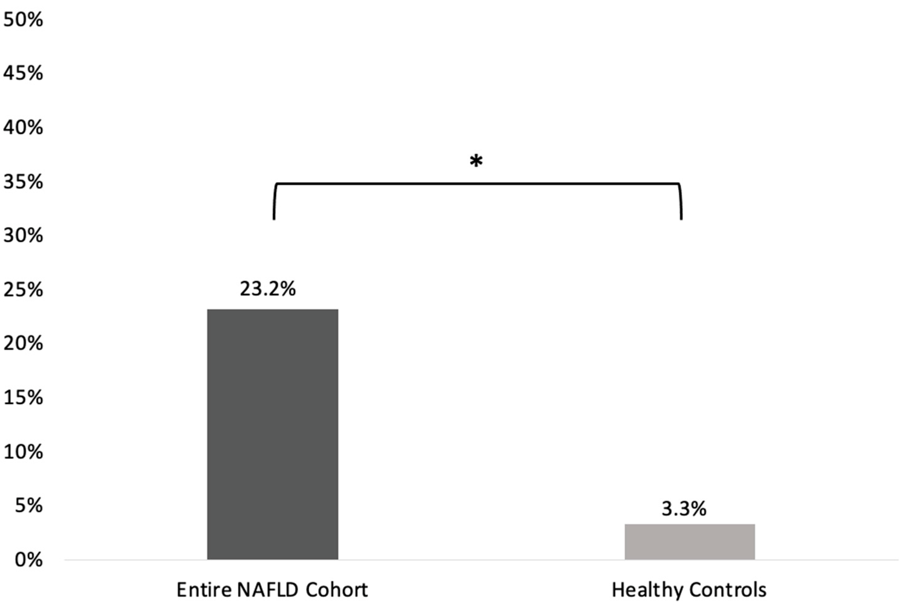
SIBO prevalence was statistically significantly higher in the entire NAFLD cohort compared to the healthy controls group [22/95 (23.2%) vs. 1/30 (3.3%); p = 0.014; odds ratio (OR) (95%CI): 8.77 (1.12–66.7); Figure 1].
(ii)
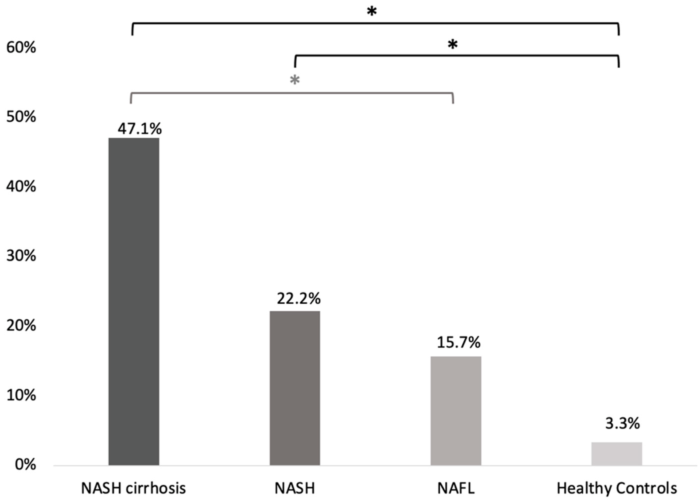
Regarding the different stages of the disease, SIBO prevalence was higher among patients with NASH-associated cirrhosis (8/17; 47.1% [23.3–70.8]) compared to patients with NAFL (8/51; 15.7% [5.7–25.7]; p = 0.02, OR (95%CI): 4.8 [1.4–16.1]) and patients with NASH (6/27; 22.2% [6.5–37.9]). However, the difference in SIBO preavalence between NASH-associated cirrhosis and NASH patients did not reach statistical significance (p = 0.11; OR (95%CI): 3.1 [0.8–11.6]). No statistically significant difference regarding SIBO prevalence was noted between patients with NASH and NAFL (p = 0.54; OR (95%CI): 1.5 [0.5–4.9]; Figure 2).
(iii)
SIBO prevalence was significantly higher in NASH and NASH-associated cirrhosis patients compared to healthy controls (p = 0.04; OR (95%CI): 8.3 [1.01–74.1] and p < 0.001; OR (95%CI): 25.8 [2.8–234.8], respectively). Although numerically higher in NAFL, the difference in SIBO prevalence between NAFL patients and healthy controls did not reach significance (p = 0.14; OR (95%CI): 5.4 [0.6–45.4]; Figure 2).
(iv)
Cytokine concentrations in the duodenal aspirates (mean pg/mL ± SEM) in NAFLD patients and healthy controls are presented in Table 3. TNFα concentrations in the duodenal aspirates did not differ between NAFLD subjects and the healthy control group (8.79 ± 0.86 vs. 7.80 ± 0.10, p = 0.533). NAFLD patients also had similar levels of IL-1β and IL-6 compared to healthy controls (7.63 ± 1.74 vs. 6.26 ± 2.76, p = 0.701 and 3.17 ± 0.04 vs. 3.14 ± 0.10, p = 0.648, respectively; Table 3). There were no differences in duodenal aspirate cytokine levels among the different stages of NAFLD (p ≤ 0.524).
Figure 1. SIBO prevalence in the entire NAFLD cohort and the healthy controls group. SIBO: small intestinal bacterial overgrowth; NAFLD: non-alcoholic fatty liver disease. * denotes statistical significance.
Microorganisms 11 00723 g001
Figure 2. SIBO prevalence among patients with different stages of NAFLD and healthy controls. SIBO: small intestinal bacterial overgrowth; NAFL: non-alcoholic fatty liver; NASH: non-alcoholic steatohepatitis. * denotes statistical significance.
Microorganisms 11 00723 g002
Similar levels of TNFα, IL-1β, and IL-6 were also observed in those NAFLD patients who tested positive for SIBO compared to healthy controls (8.39 ± 0.56 vs. 7.8 ± 0.01, p = 0.240; 11.66 ± 5.55 vs. 6.26 ± 2.76, p = 0.362; and 3.12 ± 0.05 vs. 3.14 ± 0.10, p = 0.372, respectively). Finally, cytokine levels were also similar between NAFLD patients who tested negative for SIBO and those found to be positive for SIBO (8.39 ± 0.56 vs. 8.92 ± 1.12, p = 0.799; 11.66 ± 5.55 vs. 6.36 ± 1.48, p = 0.195; and 3.12 ± 0.02 vs. 3.18 ± 0.40, p = 0.439 for TNFα, IL-1β, and IL-6, respectively).

4. Discussion

Recently published data have suggested that SIBO could be implicated in the pathogenesis of NAFLD [17]. In the current study of a well-defined cohort of NAFLD patients, SIBO was diagnosed in almost one-fourth of them using quantitative culture of the proximal small intestine, which is considered the gold standard for SIBO diagnosis. Moreover, NAFLD patients had a significantly higher prevalence of SIBO compared to healthy controls, while SIBO was more frequent in patients with NASH cirrhosis compared to NAFL.
Although the definition of a clear pathophysiological relationship between SIBO and NAFLD remains an open question, a recent systematic review and meta-analysis demonstrated that patients with NAFLD are prone to suffering from SIBO at the same time [OR (95%CI): 3.82 (1.93–7.59)] [30]. However, the exact prevalence of SIBO among NAFLD patients varies in the literature, potentially due to significant variability in the criteria and methods used to define and diagnose both NAFLD and SIBO [31]. In this context, our results are in line with the results of the study by Guimaraes et al., who reported that the prevalence of SIBO among histologically confirmed NAFLD patients using the lactulose breath test as a diagnostic modality was 26.2% [32]. Different reports in the literature, both in NAFLD [33,34,35] and in NASH [36] patients that used either cultures of jejunal aspirates [33,34,36] or a lactulose breath test [35] to diagnose SIBO, reported an even higher prevalence of the syndrome, highlighting its frequent occurrence in this population. Specifically, SIBO prevalence was reported to be 40% among NASH individuals [36], while it approached or exceeded 35% in those with NAFL [33,34,35]. Moreover, among individuals with biopsy-proven NASH, the prevalence of SIBO was >43% both in those with concomitant metabolic syndrome as well as in those without [37]. Interestingly, in two of the studies that used the 105 cfu/mL threshold after jejunal aspirate culture to diagnose SIBO, the prevalence of SIBO was reported to be 37.5% and 47.2% [33,34]. Finally, it is worth mentioning that the prevalence of SIBO in the control group was similar to those reported in the literature in studies performed either in the field of NAFLD [38] or other diseases [39].
Our study showed that the prevalence of SIBO among patients with NASH-associated cirrhosis was significantly higher compared with patients with NAFL. The relationship between SIBO prevalence and disease stage has been the focus of many studies in this field [33,34,35,38,40,41,42], with inconclusive results. One of the first studies, that included 136 patients with available liver biopsy who underwent a glucose hydrogen breath test for SIBO detection, demonstrated that SIBO was more frequent among patients with non-severe hepatic steatosis (26.3% vs. 10.3%, p = 0.127), whereas it did not differ in patients with NASH [41]. Moreover, in their study of 35 liver biopsy-proven NAFLD patients, Miele et al. concluded that the prevalence of SIBO increased significantly with the severity of steatosis (p = 0.001), without a significant correlation between SIBO and the presence of NASH (p = 0.84) [40]. Similar to our results, in a study by Scarpellini et al., a higher prevalence of SIBO, diagnosed using the lactulose breath test, was observed in patients with cirrhosis compared with non-cirrhotic patients (41.2% vs. 13.1%; p < 0.05), but no association was observed between SIBO prevalence and the grade of liver steatosis, as diagnosed using abdominal ultrasound [38]. On the other hand, a study by Shi et al. reported that as the severity of steatosis, expressed as the controlled attenuation parameter, increased, the prevalence of SIBO also increased, ranging from 32.6% to 88% for mild and severe steatosis, respectively [35]. Additionally, of interest, a recent study by Mikolasevic et al. that used jejunal aspirate culture to diagnose SIBO, reported that the prevalence of SIBO was higher among patients with biopsy-proven NASH compared with patients with NAFLD, but provided no evidence of NASH in the liver biopsy (68.1% vs. 29.1%; p < 0.001) [33]. However, in two other studies using either jejunal aspirate [34] culture or the glucose hydrogen breath test [42], SIBO prevalence did not differ by the histological stage of the disease [34] or according to the presence of severe steatosis [42]. More precisely, Fitriakusumah et al. did not find any significant association between SIBO and NAFLD development, while SIBO was also not associated with significant hepatic steatosis [42].
Despite these conflicting results, two points should be highlighted regarding the higher prevalence of SIBO with advanced stages of NAFLD, and especially with NASH-associated cirrhosis. First, patients with cirrhosis often have impaired intestinal motility that could lead to an increased orocecal transit time that could promote bacterial overgrowth, including in the small intestine, due to decreased intestinal clearance [35,43]. Second, proton pump inhibitor (PPI) intake can lead to a moderately increased risk of SIBO development as demonstrated in a meta-analysis of 7055 patients (OR [95%CI]: 1.71[1.20–2.43]) [44]. At the same time, proton pump inhibitor consumption is a frequent phenomenon among cirrhotic patients [45]. Thus, one cannot preclude an association between increased PPI intake and higher SIBO prevalence among NASH-related cirrhotic patients. However, in our cohort, PPI consumption did not differ among patients with different stages of the disease.
Accumulating evidence from published studies suggests a multifactorial contribution of SIBO in the development and progression of NAFLD [17,18,30,31,32,33,34,35,36,37,38]. The main proposed mechanisms include a disturbance of energy homeostasis leading to energy salvage, increased oxidative stress, enhancement of insulin resistance, increased ethanol production, and alterations in the metabolism of choline and bile acids [18,31,46]. Nucleotide-binding oligomerisation domain-like receptor protein NLRP3 and NLRP6, which belong to the family of inflammasomes, balance the immune response with the expression of interleukin IL-1b and IL-18, serving as regulators of normal microbiota and ameliorating NAFLD, whereas dysbiosis has been associated with a decrease in those inflammasomes and NAFLD occurrence in animal models [47,48,49]. Portal circulation and the liver–gut axis can also comprise the pathways of bacterial translocation to the liver, accompanied by pathogen-associated molecular patterns (PAMPs), danger-associated molecular patterns (DAMPs), which are recognized triggering factors of chronic inflammatory responses [50]. Moreover, increased permeability due to tight cell junctions disruption found in SIBO patients can lead to the translocation of bacteria and their products, such as endotoxins, while endotoxemia further induces CD14 mRNA and nuclear factor kappa B (NF-kB) expression that consequently activates Toll-like receptor 4 (TLR-4) and stimulates the production of proinflammatory cytokines such as tumor necrosis factor α (TNF-α), IL-1β, IL-6, and IL-8 [31,34,35,46]. Overproduction of these cytokines, but also the direct effect of bacterial products, such as lipopolysaccharides, contribute to the development of inflammation and insulin resistance and may be essential in the pathogenesis of NASH, liver fibrosis, and hepatocellular carcinoma [34]. In order to move a step beyond systematic responses with the aim of elaborating upon the potential role of mucosal immunity of the GI compartment on disease development and progression, we assessed cytokine levels (TNFα, IL-1β, and IL-6) in the duodenal aspirate of patients with NAFLD as well as in healthy controls. However, our analysis did not reveal any differences in any of the cytokine levels in duodenal samples among NAFLD subjects and healthy controls or between individuals with different stages of the disease. In any case, further confirmation of these findings in future trials is warranted.
Regarding the composition of the intestinal microbiota of NAFLD subjects, this has been the focus of different studies that have used culture-independent techniques, such as quantitative real-time polymerase chain reaction (qRT-PCR) and sequencing of the bacterial 16S ribosomal RNA (16S rRNA) gene, to assess, mainly, stool samples. One of the main outcomes of these studies was to evaluate the abundance of different phyla, particularly the Firmicutes/Bacteroidetes ratio, an indirect index of dysbiosis. While a lower abundance of Firmicutes was demonstrated in the feces of both children and adults with NASH compared to their healthy comparators [19,21], other studies did not demonstrate any alteration in the Firmicutes/Bacteroidetes ratio among NAFLD patients and healthy controls [20,22]. However, while the use of stool samples may underestimate or even cloak the contribution of small intestinal flora and small intestinal bacterial overgrowth in NAFLD pathophysiology [51], data on the duodenal microbiome composition of both NAFLD and SIBO patients are lacking. In a recently published study, Leite et al. used 16-s RNA sequencing to characterize the duodenal microbiome of patients diagnosed with SIBO by using duodenal aspirates [52]. The authors showed that duodenal aspirates from SIBO subjects had 4 × 10³-fold higher bacterial counts on MacConkey agar than duodenal aspirates from non-SIBO subjects (p < 0.0001) [52]. Moreover, SIBO subjects presented with decreased α-diversity, with a significant strong inverse correlation between the Proteobacteria and Firmicutes phyla [52]. In our study, we did not use culture-independent techniques to describe the duodenal microbiome, but it should be noted that the vast majority of the isolated bacteria in NAFLD patients with SIBO (72.6%) belonged to the Proteobacteria phylum, followed by Firmicutes (27.4%). In any case, direct application of advanced, culture-independent techniques on duodenal aspirates could reveal a likely site-dependent interaction between NAFLD and gut microbiota.
Duodenal fluid aspiration, although a broadly applied and acceptable technique, reflects the microbiota composition of a minor part of the small bowel. Bacterial diversity and phyla that could determine SIBO development and its impact on diseases, such as NAFLD, may be not isolated in all duodenal aspirate cultures, thus requiring jejunal or ileal fluid. However, the interventional nature of enteroscopic techniques, with the associated risks, does not allow spiral or balloon enteroscopy for the indication of fluid aspiration and culture. Emerging studies have assessed the role of dedicated devices to collect samples from the small bowel for microbiota analysis. More specifically, a simple low-cost 3D capsule and a robot capsule have been designed to obtain intestinal fluid or tissue, respectively, with the aim to provide a representative mapping of bowel microbiota and diagnose SIBO [53,54]. These promising technologies, needing clinical evaluation, could provide detailed information about the role and the identity of bacterial species in SIBO, with regard to NAFLD incidence and severity.
We should acknowledge a number of strengths related to our study. First, we used cultures of small bowel aspirates to diagnose SIBO, with a cut-off of 103 cfu/mL, which is the suggested threshold for SIBO diagnosis in the literature [24]. This technique may be time-consuming, more expensive, and invasive and difficult for the patient requiring sedation, but it is now considered as the gold standard to make a SIBO diagnosis [24,55]. Second, the study design allowed us to enroll a large number of individuals, overcoming the initially estimated overall sample size of 60 subjects. Finally, all participants underwent a detailed laboratory and clinical work-up, allowing us to exclude any other liver-related pathologies and get a well-defined cohort of NAFLD patients. On the other hand, our study also has limitations that merit consideration. First, the cross-sectional design of the study precluded the establishment of a causal relationship between SIBO and NAFLD. A second limitation is that nonerosive reflux disease patients were enrolled in the control group. We acknowledge the fact that these patients are not typically healthy subjects, but it would be unethical to enroll patients to undergo a non-indicated endoscopy given the existing, albeit low, risk of complications. Third, we did not perform cultures for anaerobic organisms and deep sequencing analysis; however, this should be taken into consideration by future studies. Finally, liver biopsy, as an invasive modality with considerable complication rates, was not considered mandatory to enroll patients for ethical reasons and, as expected in a real life study, it was performed, as recommended [56], if there was uncertainty regarding the cause of liver injury or the stage of liver fibrosis.

5. Conclusions

To conclude, this prospective cross-sectional study in a well-defined cohort of NAFLD patients demonstrated that the prevalence of SIBO, using duodenal aspirate culture, is significantly higher among patients with NAFLD compared to healthy controls. In addition, SIBO is more prevalent among patients with NASH-associated cirrhosis compared to NAFL or NASH individuals. Further data are warranted in order to better explain a potential causal relationship.

Supplementary Materials

The following supporting information can be downloaded at: https://www.mdpi.com/article/10.3390/microorganisms11030723/s1, Table S1: STROBE Statement—checklist of items that should be included in reports of observational studies.

Author Contributions

Conceptualization, K.T.; Methodology, P.G., G.T. and A.K.; Formal analysis, P.G.; Investigation, P.G. and G.T.; Resources, P.G., G.T., M.J.M., R.M. and S.W.; Data curation, P.G., G.T. and E.X.; Writing—original draft, P.G. and I.S.P.; Writing—review & editing, P.G., G.T., G.L., I.G.P., A.K., M.J.M., R.M., S.W., G.D.D., E.J.G.-B., M.P. and K.T.; Supervision, I.G.P., G.D.D. and K.T.; Project administration, P.G., G.D.D. and K.T. All authors have read and agreed to the published version of the manuscript.

Funding

This research received no external funding.

Data Availability Statement

The data presented in this study are available on request from the corresponding author. The data are not publicly available due to.

Acknowledgments

We would like to acknowledge the contribution of medical writer, Sandy Field, for English language editing and formatting of this manuscript.

Conflicts of Interest

The authors have no conflicts of interest to declare.

References

  1. Bugianesi, E.; McCullough, A.J.; Marchesini, G. Insulin resistance: A metabolic pathway to chronic liver disease. Hepatology 2005, 42, 987–1000. [Google Scholar] [CrossRef] [PubMed]
  2. Bhala, N.; Younes, R.; Bugianesi, E. Epidemiology and natural history of patients with NAFLD. Curr. Pharm. Des. 2013, 19, 5169–5176. [Google Scholar] [CrossRef] [PubMed]
  3. Chalasani, N.; Younossi, Z.; LaVine, J.E.; Diehl, A.M.; Brunt, E.M.; Cusi, K.; Charlton, M.; Sanyal, A.J. The diagnosis and management of non-alcoholic fatty liver disease: Practice Guideline by the American Association for the Study of Liver Diseases, American College of Gastroenterology, and the American Gastroenterological Association. Hepatology 2012, 55, 2005–2023. [Google Scholar] [CrossRef] [PubMed]
  4. Han, M.A.T.; Saouaf, R.; Ayoub, W.; Todo, T.; Mena, E.; Noureddin, M. Magnetic resonance imaging and transient elastography in the management of Nonalcoholic Fatty Liver Disease (NAFLD). Expert Rev. Clin. Pharmacol. 2017, 10, 379–390. [Google Scholar] [CrossRef]
  5. Duseja, A.; Chawla, Y.K. Obesity and NAFLD: The role of bacteria and microbiota. Clin. Liver Dis. 2014, 18, 59–71. [Google Scholar] [CrossRef]
  6. Bedogni, G.; Miglioli, L.; Masutti, F.; Tiribelli, C.; Marchesini, G.; Bellentani, S. Prevalence of and risk factors for nonalcoholic fatty liver disease: The Dionysos nutrition and liver study. Hepatology 2005, 42, 44–52. [Google Scholar] [CrossRef]
  7. Méndez-Sánchez, N.; Bugianesi, E.; Gish, R.G.; Lammert, F.; Tilg, H.; Nguyen, M.H.; Sarin, S.K.; Fabrellas, N.; Zelber-Sagi, S.; Fan, J.-G.; et al. Global multi-stakeholder endorsement of the MAFLD definition. Lancet Gastroenterol. Hepatol. 2022, 7, 388–390. [Google Scholar] [CrossRef]
  8. Day, C.P.; James, O.F. Steatohepatitis: A tale of two “hits”? Gastroenterology 1998, 114, 842–845. [Google Scholar] [CrossRef]
  9. Dowman, J.K.; Tomlinson, J.W.; Newsome, P.N. Pathogenesis of non-alcoholic fatty liver disease. Qjm: Int. J. Med. 2010, 103, 71–83. [Google Scholar] [CrossRef] [Green Version]
  10. Tilg, H.; Moschen, A.R. Evolution of inflammation in nonalcoholic fatty liver disease: The multiple parallel hits hypothesis. Hepatology 2010, 52, 1836–1846. [Google Scholar] [CrossRef]
  11. Papaefthymiou, A.; Potamianos, S.; Goulas, A.; Doulberis, M.; Kountouras, J.; Polyzos, S.A. Inflammatory Bowel Disease-associated Fatty Liver Disease: The Potential Effect of Biologic Agents. J. Crohn’s Colitis 2022, 16, 852–862. [Google Scholar] [CrossRef]
  12. Speliotes, E.K.; Butler, J.L.; Palmer, C.D.; Voight, B.F. PNPLA3 variants specifically confer increased risk for histologic nonalcoholic fatty liver disease but not metabolic disease. Hepatology 2010, 52, 904–912. [Google Scholar] [CrossRef] [Green Version]
  13. Turnbaugh, P.J.; Ley, R.E.; Hamady, M.; Fraser-Liggett, C.M.; Knight, R.; Gordon, J.I. The human microbiome project. Nature 2007, 449, 804–810. [Google Scholar] [CrossRef] [Green Version]
  14. Schwiertz, A.; Taras, D.; Schäfer, K.; Beijer, S.; Bos, N.A.; Donus, C.; Hardt, P.D. Microbiota and SCFA in lean and overweight healthy subjects. Obesity 2010, 18, 190–195. [Google Scholar] [CrossRef]
  15. Ley, R.E.; Bäckhed, F.; Turnbaugh, P.; Lozupone, C.A.; Knight, R.D.; Gordon, J.I. Obesity alters gut microbial ecology. Proc. Natl. Acad. Sci. USA 2005, 102, 11070–11075. [Google Scholar] [CrossRef] [Green Version]
  16. Novljan, U.; Pintar, T. Small Intestinal Bacterial Overgrowth in Patients with Roux-en-Y Gastric Bypass and One-Anastomosis Gastric Bypass. Obes. Surg. 2022, 32, 4102–4109. [Google Scholar] [CrossRef]
  17. Ghoshal, U.C.; Goel, A.; Quigley, E.M.M. Gut microbiota abnormalities, small intestinal bacterial overgrowth, and non-alcoholic fatty liver disease: An emerging paradigm. Indian J. Gastroenterol. 2020, 39, 9–21. [Google Scholar] [CrossRef]
  18. Gkolfakis, P.; Dimitriadis, G.; Triantafyllou, K. Gut microbiota and non-alcoholic fatty liver disease. Hepatobiliary Pancreat. Dis. Int. 2015, 14, 572–581. [Google Scholar] [CrossRef]
  19. Zhu, L.; Baker, S.S.; Gill, C.; Liu, W.; Alkhouri, R.; Baker, R.D.; Gill, S.R. Characterization of gut microbiomes in nonalcoholic steatohepatitis (NASH) patients: A connection between endogenous alcohol and NASH. Hepatology 2013, 57, 601–609. [Google Scholar] [CrossRef]
  20. Mouzaki, M.; Comelli, E.M.; Arendt, B.M.; Bonengel, J.; Fung, S.K.; Fischer, S.E.; McGilvray, I.D.; Allard, J.P. Intestinal microbiota in patients with nonalcoholic fatty liver disease. Hepatology 2013, 58, 120–127. [Google Scholar] [CrossRef] [Green Version]
  21. Wong, V.W.-S.; Tse, C.-H.; Lam, T.-T.Y.; Wong, G.L.-H.; Chim, A.M.-L.; Chu, W.C.-W.; Yeung, D.K.-W.; Law, P.T.-W.; Kwan, H.S.; Yu, J.; et al. Molecular characterization of the fecal microbiota in patients with nonalcoholic steatohepatitis—A longitudinal study. PLoS ONE 2013, 8, e62885. [Google Scholar] [CrossRef] [PubMed] [Green Version]
  22. Raman, M.; Ahmed, I.; Gillevet, P.M.; Probert, C.S.; Ratcliffe, N.M.; Smith, S.; Greenwood, R.; Sikaroodi, M.; Lam, V.; Crotty, P.; et al. Fecal microbiome and volatile organic compound metabolome in obese humans with nonalcoholic fatty liver disease. Clin. Gastroenterol. Hepatol. 2013, 11, 868–875.e3. [Google Scholar] [CrossRef]
  23. Von Elm, E.; Altman, D.G.; Egger, M.; Pocock, S.J.; Gøtzsche, P.C.; Vandenbroucke, J.P. The Strengthening the Reporting of Observational Studies in Epidemiology (STROBE) statement: Guidelines for reporting observational studies. J. Clin. Epidemiol. 2008, 61, 344–349. [Google Scholar] [CrossRef] [PubMed] [Green Version]
  24. Pimentel, M.; Saad, R.J.; Long, M.D.; Rao, S.S.C. ACG Clinical Guideline: Small Intestinal Bacterial Overgrowth. Am. J. Gastroenterol. 2020, 115, 165–178. [Google Scholar] [CrossRef] [PubMed]
  25. Sugano, K.; Tack, J.; Kuipers, E.J.; Graham, D.Y.; El-Omar, E.M.; Miura, S.; Haruma, K.; Asaka, M.; Uemura, N.; Malfertheiner, P. Kyoto global consensus report on Helicobacter pylori gastritis. Gut 2015, 64, 1353–1367. [Google Scholar] [CrossRef] [Green Version]
  26. Pyleris, E.; Giamarellos-Bourboulis, E.J.; Tzivras, D.; Koussoulas, V.; Barbatzas, C.; Pimentel, M. The prevalence of overgrowth by aerobic bacteria in the small intestine by small bowel culture: Relationship with irritable bowel syndrome. Dig. Dis. Sci. 2012, 57, 1321–1329. [Google Scholar] [CrossRef]
  27. Giamarellos-Bourboulis, E.J.; Pyleris, E.; Barbatzas, C.; Pistiki, A.; Pimentel, M. Small intestinal bacterial overgrowth is associated with irritable bowel syndrome and is independent of proton pump inhibitor usage. BMC Gastroenterol. 2016, 16, 67. [Google Scholar] [CrossRef] [Green Version]
  28. Giamarellos-Bourboulis, E.; Tang, J.; Pyleris, E.; Pistiki, A.; Barbatzas, C.; Brown, J.; Lee, C.C.; Harkins, T.T.; Kim, G.; Weitsman, S.; et al. Molecular assessment of differences in the duodenal microbiome in subjects with irritable bowel syndrome. Scand. J. Gastroenterol. 2015, 50, 1076–1087. [Google Scholar] [CrossRef]
  29. Leventogiannis, K.; Gkolfakis, P.; Spithakis, G.; Tsatali, A.; Pistiki, A.; Sioulas, A.; Giamarellos-Bourboulis, E.J.; Triantafyllou, K. Effect of a Preparation of Four Probiotics on Symptoms of Patients with Irritable Bowel Syndrome: Association with Intestinal Bacterial Overgrowth. Probiotics Antimicrob. Proteins 2019, 11, 627–634. [Google Scholar] [CrossRef] [Green Version]
  30. Wijarnpreecha, K.; Lou, S.; Watthanasuntorn, K.; Kroner, P.T.; Cheungpasitporn, W.; Lukens, F.J.; Pungpapong, S.; Keaveny, A.P.; Ungprasert, P. Small intestinal bacterial overgrowth and nonalcoholic fatty liver disease: A systematic review and meta-analysis. Eur. J. Gastroenterol. Hepatol. 2020, 32, 601–608. [Google Scholar] [CrossRef]
  31. Augustyn, M.; Grys, I.; Kukla, M. Small intestinal bacterial overgrowth and nonalcoholic fatty liver disease. Clin. Exp. Hepatol. 2019, 5, 1–10. [Google Scholar] [CrossRef]
  32. GuimarÃes, V.M.; Santos, V.N.; Borges PS de, A.; DEFarias, J.L.R.; Grillo, P.; Parise, E.R. Peripheral blood endotoxin levels are not associated with small intestinal bacterial overgrowth in nonalcoholic fatty liver disease without cirrhosis. Arq. Gastroenterol. 2020, 57, 471–476. [Google Scholar] [CrossRef]
  33. Mikolasevic, I.; Delija, B.; Mijic, A.; Stevanovic, T.; Skenderevic, N.; Sosa, I.; Krznaric-Zrnic, I.; Abram, M.; Krznaric, Z.; Domislovic, V.; et al. Small intestinal bacterial overgrowth and non-alcoholic fatty liver disease diagnosed by transient elastography and liver biopsy. Int. J. Clin. Pract. 2021, 75, e13947. [Google Scholar] [CrossRef]
  34. Kapil, S.; Duseja, A.; Sharma, B.K.; Singla, B.; Chakraborti, A.; Das, A.; Ray, P.; Dhiman, R.K.; Chawla, Y. Small intestinal bacterial overgrowth and toll-like receptor signaling in patients with non-alcoholic fatty liver disease. J. Gastroenterol. Hepatol. 2016, 31, 213–221. [Google Scholar] [CrossRef]
  35. Shi, H.; Mao, L.; Wang, L.; Quan, X.; Xu, X.; Cheng, Y.; Zhu, S.; Dai, F. Small intestinal bacterial overgrowth and orocecal transit time in patients of nonalcoholic fatty liver disease. Eur. J. Gastroenterol. Hepatol. 2021, 33 (Suppl. S1), e535–e539. [Google Scholar] [CrossRef]
  36. Ghoshal, U.C.; Baba, C.S.; Ghoshal, U.; Alexander, G.; Misra, A.; Saraswat, V.A.; Choudhuri, G. Low-grade small intestinal bacterial overgrowth is common in patients with non-alcoholic steatohepatitis on quantitative jejunal aspirate culture. Indian J. Gastroenterol. 2017, 36, 390–399. [Google Scholar] [CrossRef]
  37. De Oliveira, J.M.; Pace, F.L.; Ghetti, F.D.F.; Barbosa, K.V.B.D.; Cesar, D.E.; Chebli, J.M.F.; Ferreira, L.E.V.V.D.C. Non-alcoholic Steatohepatitis: Comparison of Intestinal Microbiota between Different Metabolic Profiles. A Pilot Study. J. Gastrointest. Liver Dis. 2020, 29, 369–376. [Google Scholar] [CrossRef]
  38. Scarpellini, E.; Abenavoli, L.; Cassano, V.; Rinninella, E.; Sorge, M.; Capretti, F.; Rasetti, C.; Baroni, G.S.; Luzza, F.; Santori, P.; et al. The Apparent Asymmetrical Relationship Between Small Bowel Bacterial Overgrowth, Endotoxemia, and Liver Steatosis and Fibrosis in Cirrhotic and Non-Cirrhotic Patients: A Single-Center Pilot Study. Front. Med. 2022, 9, 872428. [Google Scholar] [CrossRef]
  39. Tziatzios, G.; Gkolfakis, P.; Papanikolaou, I.S.; Mathur, R.; Pimentel, M.; Damoraki, G.; Giamarellos-Bourboulis, E.J.; Dimitriadis, G.; Triantafyllou, K. High Prevalence of Small Intestinal Bacterial Overgrowth among Functional Dyspepsia Patients. Dig. Dis. 2021, 39, 382–390. [Google Scholar] [CrossRef]
  40. Miele, L.; Valenza, V.; La Torre, G.; Montalto, M.; Cammarota, G.; Ricci, R.; Mascianà, R.; Forgione, A.; Gabrieli, M.L.; Perotti, G.; et al. Increased intestinal permeability and tight junction alterations in nonalcoholic fatty liver disease. Hepatology 2009, 49, 1877–1887. [Google Scholar] [CrossRef]
  41. Sabaté, J.-M.; Jouët, P.; Harnois, F.; Mechler, C.; Msika, S.; Grossin, M.; Coffin, B. High prevalence of small intestinal bacterial overgrowth in patients with morbid obesity: A contributor to severe hepatic steatosis. Obes. Surg. 2008, 18, 371–377. [Google Scholar] [CrossRef] [PubMed]
  42. Fitriakusumah, Y.; Lesmana, C.R.A.; Bastian, W.P.; Jasirwan, C.O.M.; Hasan, I.; Simadibrata, M.; Kurniawan, J.; Sulaiman, A.S.; Gani, R.A. The role of Small Intestinal Bacterial Overgrowth (SIBO) in Non-alcoholic Fatty Liver Disease (NAFLD) patients evaluated using Controlled Attenuation Parameter (CAP) Transient Elastography (TE): A tertiary referral center experience. BMC Gastroenterol. 2019, 19, 43. [Google Scholar] [CrossRef] [PubMed] [Green Version]
  43. Juanola, O.; Ferrusquía-Acosta, J.; García-Villalba, R.; Zapater, P.; Magaz, M.; Marín, A.; Olivas, P.; Baiges, A.; Bellot, P.; Turon, F.; et al. Circulating levels of butyrate are inversely related to portal hypertension, endotoxemia, and systemic inflammation in patients with cirrhosis. FASEB J. 2019, 33, 11595–11605. [Google Scholar] [CrossRef] [PubMed]
  44. Su, T.; Lai, S.; Lee, A.; He, X.; Chen, S. Meta-analysis: Proton pump inhibitors moderately increase the risk of small intestinal bacterial overgrowth. J. Gastroenterol. 2018, 53, 27–36. [Google Scholar] [CrossRef]
  45. Cole, H.L.; Pennycook, S.; Hayes, P.C. The impact of proton pump inhibitor therapy on patients with liver disease. Aliment. Pharmacol. Ther. 2016, 44, 1213–1223. [Google Scholar] [CrossRef]
  46. Bakhshimoghaddam, F.; Alizadeh, M. Contribution of gut microbiota to nonalcoholic fatty liver disease: Pathways of mechanisms. Clin. Nutr. ESPEN 2021, 44, 61–68. [Google Scholar] [CrossRef]
  47. Pierantonelli, I.; Rychlicki, C.; Agostinelli, L.; Giordano, D.M.; Gaggini, M.; Fraumene, C.; Saponaro, C.; Manghina, V.; Sartini, L.; Mingarelli, E.; et al. Author Correction: Lack of NLRP3-inflammasome leads to gut-liver axis derangement, gut dysbiosis and a worsened phenotype in a mouse model of NAFLD. Sci. Rep. 2017, 7, 17568. [Google Scholar] [CrossRef] [Green Version]
  48. Wood, N.J. Dysbiosis driven by inflammasome deficiency exacerbates hepatic steatosis and governs rate of NAFLD progression. Nat. Rev. Gastroenterol. Hepatol. 2012, 9, 123. [Google Scholar] [CrossRef]
  49. Henao-Mejia, J.; Elinav, E.; Jin, C.; Hao, L.; Mehal, W.Z.; Strowig, T.; Thaiss, C.A.; Kau, A.L.; Eisenbarth, S.C.; Jurczak, M.J.; et al. Inflammasome-mediated dysbiosis regulates progression of NAFLD and obesity. Nature 2012, 482, 179–185. [Google Scholar] [CrossRef] [Green Version]
  50. Abu-Shanab, A.; Quigley, E.M.M. The role of the gut microbiota in nonalcoholic fatty liver disease. Nat. Rev. Gastroenterol. Hepatol. 2010, 7, 691–701. [Google Scholar] [CrossRef]
  51. Lin, E.A.; Barlow, G.M.; Mathur, R. The microbiome in non-alcoholic fatty liver disease: Associations and implications. Ann. Gastroenterol. 2014, 27, 181–183. [Google Scholar]
  52. Leite, G.; Morales, W.; Weitsman, S.; Celly, S.; Parodi, G.; Mathur, R.; Barlow, G.M.; Sedighi, R.; Millan, M.J.V.; Rezaie, A.; et al. The duodenal microbiome is altered in small intestinal bacterial overgrowth. PLoS ONE 2020, 15, e0234906. [Google Scholar] [CrossRef]
  53. Rehan, M.; Al-Bahadly, I.; Thomas, D.G.; Avci, E. Capsule robot for gut microbiota sampling using shape memory alloy spring. Int. J. Med. Robot. 2020, 16, 1–14. [Google Scholar] [CrossRef]
  54. Waimin, J.F.; Nejati, S.; Jiang, H.; Qiu, J.; Wang, J.; Verma, M.S.; Rahimi, R. Smart capsule for non-invasive sampling and studying of the gastrointestinal microbiome. RSC Adv. 2020, 10, 16313–16322. [Google Scholar] [CrossRef] [Green Version]
  55. Rezaie, A.; Buresi, M.; Lembo, A.; Lin, H.; McCallum, R.; Rao, S.; Schmulson, M.; Valdovinos, M.; Zakko, S.; Pimentel, M. Hydrogen and Methane-Based Breath Testing in Gastrointestinal Disorders: The North American Consensus. Am. J. Gastroenterol. 2017, 112, 775–784. [Google Scholar] [CrossRef] [Green Version]
  56. Long, M.T.; Noureddin, M.; Lim, J.K. AGA Clinical Practice Update: Diagnosis and Management of Nonalcoholic Fatty Liver Disease in Lean Individuals: Expert Review. Gastroenterology 2022, 163, 764–774.e1. [Google Scholar] [CrossRef]
Table 1. Baseline patient characteristics.
Entire NAFLD Cohort
(n = 95)
NAFL
(n = 51)
NASH
(n = 27)
NASH Cirrhosis
(n = 17)
Healthy Controls
(n = 30)
* p-Value** p-Value
Age (years), mean (±SD)55 (10.9)54.7 (9.2)49.8 (11.8)63.9 (8.5)50.9 (14.6)0.103<0.001
Gender, n (%) 0.2090.110
Female43 (45.3)27 (52.9)12 (44.4)4 (23.5)18 (60)
Male52 (54.7)24 (47.1)15 (55.6)13 (76.5)12 (40)
Weight (kg), mean (±SD)90.7 (19.1)89.2 (16.6)91.1 (21.1)94.4 (23.2)80.8 (19.5)0.0160.630
Height (cm), mean (±SD)176 (6.3)180 (8.6)171 (7.4)171 (11.5)171 (6.4)0.6440.786
Diabetes mellitus type 2, (%) 0.2950.391
Yes21 (22.1)14 (27.5)4 (14.8)3 (17.6)4 (13.3)
No74 (77.9)37 (72.5)23 (85.2)14 (82.4)26 (86.7)
PPI and/or H2RA intake, n (%) 0.0070.398
Yes19 (20)12 (23.5)3 (11.1)4 (23.5)0 (0)
No76 (80)39 (76.5)24 (88.9)13 (76.5)30 (100)
NAFLD: non-alcoholic fatty liver disease; NAFL: non-alcoholic fatty liver; NASH: non-alcoholic steatohepatitis; SD: standard deviation; PPI: proton pump inhibitor; H2RA: H2 receptor antagonist. * refers to differences between the entire NAFLD cohort and the healthy controls group. ** refers to differences between the different stages of the NAFLD cohort (NAFL, NASH, and NASH-associated cirrhosis).
Table 2. Bacterial species from duodenal aspirates of study participants with small intestinal bacterial overgrowth (SIBO).
Bacterial Species, n (%)Entire NAFLD Cohort, n (%)Healthy Controls, n (%)
Escherichia coli10 (45.5)-
Staphylococcus aureus4 (18.2)-
Pseudomonas aeruginosa3 (13.6)-
Enterococus faecalis2 (9.1)-
Klebsiella pneumoniae1 (4.5)-
Providencia alcalifaciens1 (4.5)-
Acinetobacter spp. 1 (4.5)-
Enterobacter cloacae-1 (100)
Total, n/N (%)22/95 (23.2%)1/30 (3.3%)
NAFLD: non-alcoholic fatty liver disease.
Table 3. TNFα, IL-1b, and IL-6 concentrations in duodenal fluid from the NAFLD cohort and healthy controls.
Concentration in Duodenal Fluid *Entire NAFLD Cohort
(n = 95)
NAFL
(n = 51)
NASH
(n = 27)
NASH-Cirrhosis
(n = 17)
Healthy Controls
(n = 30)
p-Value **p-Value ***
TNFα8.79 ± 0.867.80 ± 0.1010.69 ± 2.898.57 ± 0.727.80 ± 0.100.5330.345
IL-1β7.63 ± 1.748.51 ± 2.928.69 ± 2.753.45 ± 1.116.26 ± 2.760.7010.524
IL-63.17 ± 0.043.22 ± 0.073.12 ± 0.043.12 ± 0.043.14 ± 0.100.6480.417
* Measured and presented as mean (pg/mL) ± standard error of mean (SEM); ** difference between the entire NAFLD cohort and the healthy controls; *** difference among the different stages of the NAFLD cohort. NAFLD, non-alcoholic fatty liver disease; NAFL, non-alcoholic fatty liver; NASH, non-alcoholic steatohepatitis; IL, interleukin; TNF, tumor necrosis factor.
Disclaimer/Publisher’s Note: The statements, opinions and data contained in all publications are solely those of the individual author(s) and contributor(s) and not of MDPI and/or the editor(s). MDPI and/or the editor(s) disclaim responsibility for any injury to people or property resulting from any ideas, methods, instructions or products referred to in the content.

