Article

Inherited Chromosomally Integrated Human Herpesvirus 6: Laboratory and Clinical Features

by
Liliana Gabrielli
1,*,
Alice Balboni
2,
Eva Caterina Borgatti
2,
Giulio Virgili
3,
Evangelia Petrisli
1,
Alessia Cantiani
2,
Matteo Pavoni
2,
Federico Baiesi Pillastrini
1,
Simona Venturoli
1,
Giulia Piccirilli
1 and
Tiziana Lazzarotto
1,2
1
Microbiology Unit, IRCCS Azienda Ospedaliero-Universitaria di Bologna, 40138 Bologna, Italy
2
Department of Medical and Surgical Sciences, University of Bologna, 40138 Bologna, Italy
3
Infective Disease Unit, IRCCS Azienda Ospedaliero-Universitaria di Bologna, 40138 Bologna, Italy
*
Author to whom correspondence should be addressed.
Microorganisms 2023, 11(3), 548; https://doi.org/10.3390/microorganisms11030548
Submission received: 8 February 2023 / Accepted: 15 February 2023 / Published: 21 February 2023

Abstract

:
Inherited chromosomally integrated human herpesvirus 6 (iciHHV-6) is a condition in which the complete HHV-6 genome is integrated into the chromosomes of the host germ cell and is vertically transmitted. The aims of this study were to identify iciHHV-6 prevalence in hospitalized patients and clinical features in individuals carrying this integration. HHV-6 PCR on hair follicles was used to confirm iciHHV-6 status when the blood viral load was more than 5 Log10 copies/mL. From January 2012 to June 2022, HHV-6 DNAemia was investigated in 2019 patients. In particular, 49 had a viral load higher than 6 Log10 copies/mL and HHV-6 DNA in hair follicles was positive. A viral load between 5.0 and 5.9 Log10 copies/mL was observed in 10 patients: 6 infants with acute HHV-6 infection and 4 patients with leukopenia and HHV-6 integration. Therefore, the iciHHV-6 prevalence in our population was 2.6% (53/2019). Adult patients with integration presented hematological (24%), autoimmune (11%), autoimmune neurological (19%), not-autoimmune neurological (22%), and other diseases (19%), whereas 5% had no clinically relevant disease. Although in our study population a high percentage of iciHHV-6 adult hospitalized patients presented a specific pathology, it is still unknown whether the integration is responsible for, or contributes to, the disease development.

1. Introduction

Human Herpesvirus 6 (HHV-6) is a Roseolovirus that comprises two distinct viruses, HHV-6A and HHV-6B [1,2].
HHV-6B infects nearly 100% of human beings [3], typically before the age of three and is the causative agent in exanthema subitum (also known as roseola infantum) [4,5,6], a childhood disease characterized by high fever, diarrhea, and a mild skin rash [7]. This initial infection can also cause febrile seizures and febrile status epilepticus [7,8].
Little is known about the prevalence of HHV-6A or how it is acquired [5,9]. It is generally believed that primary HHV-6A infection is acquired later than HHV-6B, through asymptomatic infection [6]. Recent findings suggest that high HHV-6A antibody levels are associated with increased multiple sclerosis risk and act synergistically with common environmental/lifestyle risk factors [10].
Like other herpesviruses, HHV-6 establishes latency [6,11], and reactivation can cause disease in severely immunocompromised hosts [9,12]. Multiple studies have described an association between reactivation of latent HHV-6 and encephalitis, graft rejection, acute Graft-versus-Host-Disease (GvHD), and bone marrow suppression in recipients of hematopoietic stem cell transplantation (HSCT) [13,14,15,16,17,18].
Unlike other herpesviruses, HHV-6 species establish latency through integration of the HHV-6 genome into the chromosomal telomeres [19,20,21,22], similarly to Marek’s disease virus, whose genomic integration is tightly associated with chicken lymphoma development [23]. HHV-6 integration occurs in a small proportion of somatic cells [7], including monocytes, macrophages, T-cells, and bone marrow progenitor cells [7,24,25]. The integration process is facilitated by telomeric repeats present at the ends of the viral DNA [26]. This allows maintaining the viral genome in latently infected cells, while no circular episome was observed [7].
Apart from integration into somatic cells, HHV-6A and B integration into the chromosomes of germ cells is possible in a selected number of patients and leads to offspring that carry a copy of the viral genome in every nucleated cell of the body [11,21,27,28,29]. This condition is referred as inherited chromosomally integrated HHV-6 (iciHHV-6) and occurs in approximately 1% of the human population [21,28,30,31,32,33], which corresponds to nearly 70 million people [19]. It remains unknown if integration in the germ cells occurs during primary infection or reactivation [7].
Heritable integration might occur in sperm progenitor cells or in an ovum [7]. The inherited viral genome is passed on to subsequent generations according to Mendelian laws [27]: half of the gametes harbor the virus resulting in about 50% of descendants carrying the virus in their germline [21]. iciHHV-6 individuals harbor one copy of the viral genome in every nucleated cell which accounts for the persistent high viral load in whole blood [5,34]. HHV-6 levels in whole blood that exceed 5.5 Log10 copies/mL are strongly suggestive of iciHHV-6 [5]. Rarely, in the absence of iciHHV-6, patients with GvHD or drug induced hypersensitivity syndrome can have transient viral load above 5.5 Log10 copies/mL [5,35].
In addition to genetic transmission, populations of cells harboring iciHHV-6 may be horizontally transmitted via allogenic HSCT [5] and via solid organ transplantation [36,37].
The prevalence of iciHHV-6 was found to be higher in hospitalized individuals (2.9 and 3.3%) than in blood donors (0.8 and 1.5%), possibly suggesting an impact on health, although not associated with any particular disease [20,38]. In fact, the consequences of having iciHHV-6A/B are not well defined [5,39], although there are evidences of association to a higher risk of developing angina pectoris [9]. Furthermore, iciHHV-6 subjects have shorter telomeres, which may explain, at least in part, how iciHHV-6 may contribute to the development of angina [9].
The aims of this study are to evaluate the iciHHV-6 frequency in hospitalized patients in Bologna, Italy, and the clinical features in iciHHV-6 patients. We used a cut-off of 5 Log10 HHV-6 copies/mL in whole blood and testing of hair follicles to confirm or exclude iciHHV-6 status. The occurrence of hematological, autoimmune, autoimmune and not-autoimmune neurological and other diseases in iciHHV-6 patients was investigated.

2. Materials and Methods

2.1. Patients Studied

Patients admitted to IRCCS St. Orsola Polyclinic, Maggiore Hospital, and Bellaria Hospital in Bologna investigated for HHV-6 DNA in whole blood from January 2012 to June 2022 were studied. When the HHV-6 DNA load in blood was greater than or equal to 5 Log10 copies/mL, the HHV-6 PCR test on hair follicles was used to confirm or exclude iciHHV-6 status [38,40], according to our protocol.
Clinical data and laboratory findings regarding iciHHV-6 patients were discussed in multidisciplinary team meetings. iciHHV-6 patients were divided in two age groups (<18 years and ≥18 years) and then classified according to their past and present medical history.

2.2. DNA Extraction and HHV-6 Detection Using Real-Time PCR

HHV-6 PCR was performed on EDTA-anticoagulated whole blood samples by using the HHV-6 ELITe MGB® kit (ELITech Group, Turin, Italy), a commercial quantitative real-time PCR assay with the limit of detection (LOD) equal to 10 copies of target DNA per amplification reaction. In each well, two amplification reactions were performed: one for the ORF13R region, belonging to the U67 gene of HHV6, and one human beta globin gene (internal control). The test is not able to discriminate between HHV-6 A and B. The assay was used in association with different extractions and amplification platforms, as follows.
Until May 2018, DNA extraction was performed with the QIAsymphony SP instrument (Qiagen, Hilden, Germany) and the quantification of HHV-6 was performed by using HHV-6 ELITe MGB® kit on the ABI Prism 7500 real-time PCR System (PE Applied Biosystem, Foster City, CA, USA), according to the assay manufacturer’s instructions. In brief: 0.5 mL of samples were extracted; the DNA was eluted in 100 µL, and 20 µL of DNA were used for amplification. The LOD and the lower limit of quantification (LLQ) of such method were 120 copies/mL, whereas the upper limit of quantification (ULQ) of the assay was 12,000,000 copies/mL.
From June 2018, HHV-6 ELITe MGB® Kit was used in association to the ELITeInGenius® instrument, an integrated platform for fully automated nucleic acids extraction and real-time PCR amplification. The extraction was performed with ELITe InGenius SP 200 (ELITechGroup S.p.A., Torino, Italy), a set of dedicated reagents for nucleic acid extraction from different matrices. The samples were processed using a pre-set protocol: 0.2 mL of samples were extracted, the DNA was eluted in 100 µL, and 20 µL of DNA were used for amplification. The LOD and LLQ of this method were 131 copies/mL, and ULQ was 17,857,143 copies/mL.
Considering hair follicles testing, at least 4 hair follicles were collected for each patient. The samples were added to 180 µL of tissue lysis buffer (ATL buffer, Qiagen) and 20 µL of protease (Proteinase K solution, Qiagen, Germany), incubated overnight at 56 °C and then for one hour at 90 °C. Extraction of nucleic acids and real-time PCR amplification were performed using the same protocol of whole blood and qualitative results were expressed as positive or negative. A sample was considered truly negative when ORF13R was not amplified (Ct undetermined for HHV-6) and beta globin gene was detected with Ct ≤ 35. The human beta globin region was used as an endogenous internal control for sufficient specimen cellularity and for extraction and amplification steps.

2.3. Statistical Analysis
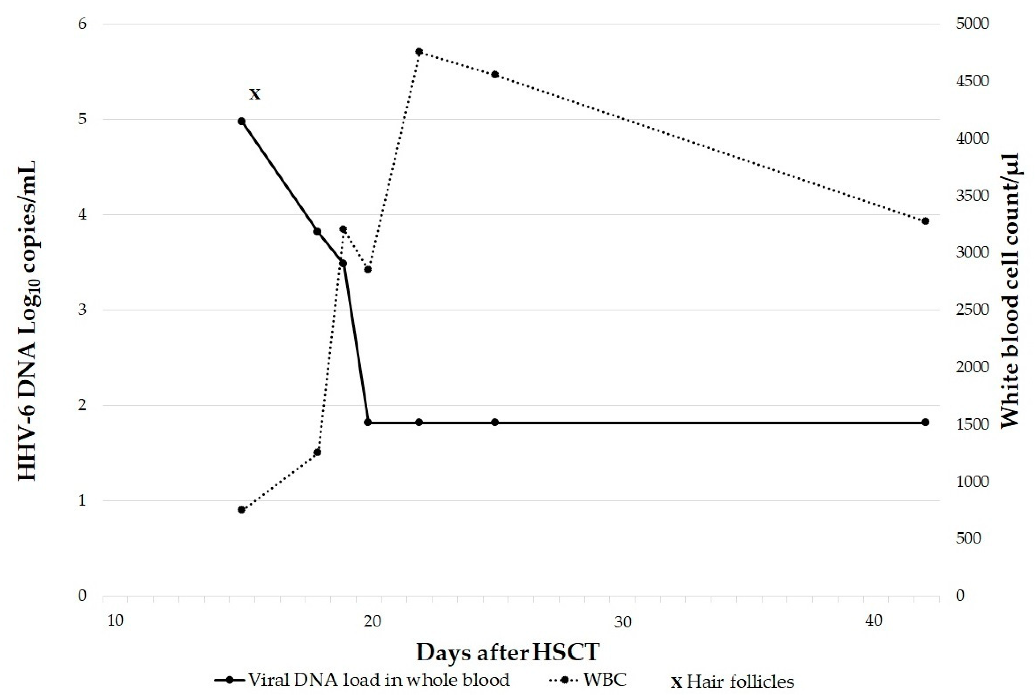
The Fisher’s exact test was used to compare the distribution of iciHHV-6 between male and female subjects and of patients without underlying medical conditions between the two age groups. p < 0.05 was considered statistically significant.
95% confidence intervals (CI) analysis were computed. Analyses were performed using Statsoft Version 7.1 (StatSoft Europe GmbH, 22301 Hamburg, Germany) for Windows 10.

3. Results

From January 2012 to June 2022, HHV-6 DNA in whole blood was investigated in 2019 patients (age range: 0–92, mean age: 37) admitted to IRCCS St. Orsola Polyclinic, Maggiore Hospital, and Bellaria Hospital in Bologna, under clinicians’ requests.
Of the 2019 hospitalized patients, 1464 (72.5%) had HHV-6 DNA negative result in whole blood, whereas 555 (27.5%) patients had a positive result. HHV-6 positive patients were classified according to the viral load: 365 (66%) patients had a viral load <2 Log10 copies/mL (LLQ); 131 (23%) had a viral load between 2.0–5.0 Log10 copies/mL (range 2.5–4.5 Log10 copies/mL; IQR 3.4) and 59 (11%) had a viral load ≥5.0 Log10 copies/mL (range 5–7.2 Log10 copies/mL; IQR 6.4).
The 59 patients with a viral load ≥5.0 Log10 copies/mL were then tested by HHV-6 PCR on hair follicles. The results obtained are summarized in Table 1.
In particular, 49/59 (83%) patients had a viral load ≥6 Log10 copies/mL (range: 6.0–7.2) and all were positive for HHV-6 DNA in hair follicles, thus confirming the assumption of iciHHV-6.
The remaining 10 patients had a viral load between 5 and 5.9 Log10 copies/mL. Six patients were infants, 2 affected by hematologic malignancy, and 2 were transplant recipients.
The 6 infants (age: 10 days–2 years) had fever and rash clinically compatible with roseola infantum and the viral load was between 5.1 and 5.5 Log10 copies/mL. Their clinical condition was so critical as to require admission to hospital and further investigations. In particular, one infant was 2 years old and had had recent infection with severe acute respiratory syndrome coronavirus-2 and acute hepatitis. The others were between 10 to 19 days of life, two with neutropenia, two with febrile seizures and one with concomitant urinary tract infection. The acute HHV-6 infection was confirmed by the negative result of hair follicles and the progressive reduction of blood viral load without antiviral treatment.
Of the 2 patients with hematologic malignancy, one had acute myeloid leukemia, and presented with fever, rash and leukopenia (leukocytes: 730/µL) at the onset of the disease. HHV-6 viral load in the blood was 5 Log10 copies/mL. The suspected iciHHV-6 status was confirmed by the hair follicles’ positive PCR. The other hematologic malignancy patient had an acute lymphoblastic leukemia, and HHV-6 was investigated during screening exams prior to the chemotherapy administration. The blood viral load was 5.2 Log10 copies/mL associated with leukopenia (leukocytes: 330/µL), and the iciHHV-6 suspect was confirmed by the hair follicles’ positive PCR. After 14 days from the first investigation the HHV-6 load increased up to 6.0 Log10 copies/mL in correspondence with the increase in the number of leukocytes (leukocytes: 2760/µL).
Two patients were transplant recipients with transient leukopenia at the moment of blood collection. One was a multivisceral transplant recipient (stomach, duodenum, pancreas, intestine and liver) with HHV-6 DNAemia of 5.9 Log10 copies/mL and leukopenia (leukocytes: 710/µL) during routine blood tests. The integration was confirmed by the positive result of the hair follicles test and the subsequent increase of the DNAemia (>6 Log10 copies/mL) 1 month after, in correspondence with the leukocyte recovery (leukocytes: 7590/µL). The other patient was an allogeneic hematopoietic stem cell recipient affected by acute myeloid leukemia. He had a facial rash related to GvHD 2 weeks after the transplant and a HHV-6 load in blood at 5 Log10 copies/mL (donor-derived leukocytes: 750/µL; chimerism test: 100% donor) with positive hair follicles. The patient was successfully treated with corticosteroids and the viral load quickly decreased without antiviral treatment, confirming the absence of both HHV-6 reactivation in the recipient and iciHHV-6 status in the donor. A persistent HHV-6 low level was detectable in blood after the resolution of GvHD (Figure 1).
Overall, of the 2019 subjects analyzed, 1226 males (61%) and 793 females (39%), 53 were identified as iciHHV-6, indicating a prevalence of 2.6% reaching 9.5% (53/555) considering only the patients with a HHV-6 positive result in blood.
Among the 53 iciHHV-6 patients, 33 were males (62%) and 20 (38%) were females. The sex distribution of iciHHV-6 negative and iciHHV-6 positive patients was similar (p = 0.88).
iciHHV-6 cases were divided in 2 age groups: <18 years (16 patients) and ≥18 years (37 patients).
Clinical data from the 53 iciHHV-6 patients are summarized in Table 2 and Table 3. For each patient, we describe: clinical features that required HHV-6 investigation; viral load in blood and, when requested, in cerebrospinal fluid (CSF); patient’s diseases including those already known in medical history and those diagnosed during the current hospitalization. In particular, diseases were classified in 5 groups: hematological, autoimmune, autoimmune neurological, not-autoimmune neurological and other disease (a disease not included in the previous disease categories). Patients without already known pathologies in anamnesis at the hospital admission and without new clinically relevant diseases diagnosed during the hospitalization, were defined as patients without underlying medical conditions (nUMC).
In 23 patients (43%), HHV-6 DNAemia was performed after a HHV-6 positive result obtained in CSF collected to investigate an infectious or neurological or autoimmune disease. The clinical feature leading to HHV-6 DNA investigation was mainly the occurrence of fever, especially in pediatric patients (12/16, 75%).
No iciHHV-6 patients reported angina at the moment of HHV-6 investigation or in the medical history.

4. Discussion

Like other herpesviruses, HHV-6 establishes latency following primary infection, allowing the virus to persist in the host for life. In contrast to other human herpesviruses, HHV-6 can integrate its genome into the telomere region of host chromosomes of latently infected cells [22,41].
The HHV-6 integration process is facilitated by telomeric repeats present at the ends of the viral DNA [26] and occurs in a small proportion of somatic cells [7], including monocytes, macrophages, T-cell, or bone marrow progenitor cells [24,25]. However, if integration occurs in the chromosomes of germ cells, vertical transmission of integrated HHV-6 results in offspring with an integrated copy of the virus in each nucleated cell, a phenomenon referred as iciHHV-6 [11,21]. These individuals will transmit the condition to half of their offspring via Mendelian mechanism and exhibit a persistent high viral load in whole blood. Pellet et al. [5] reported that iciHHV-6 individuals have at least one copy of viral genome per white blood cell, which corresponds to >5.5 Log10 copies/mL of whole blood. Thus, HHV-6 levels > 5.5 Log10 copies/mL in whole blood are suggestive of iciHHV-6 and confirmation can be made by DNA PCR testing of blood-free samples such as hair follicles or fingernails [34,42].
Other authors [3,7] reported that iciHHV-6 individuals can be identified by their constant viral load of 6–7 Log10 copies/mL in whole blood. However, when the white blood cell count is significantly decreased, as observed in transplant recipients, the results for absolute viral loads in blood may be ambiguous [6,39].
In the last ten years, we investigated HHV-6 DNAemia in 2019 patients obtaining a positive result in 555 (27.5%). In order to investigate iciHHV-6 status, we used a cut-off of 5 Log10 copies/mL in whole blood, lower than the one usually reported (5.5), considering that transplant recipients with HHV-6 reactivation and children with primary HHV-6 infection have transient virus DNA loads typically not more than 5.0 Log10 copies/mL in whole blood [5].
In all 59 (2.9%) patients with HHV-6 load greater than or equal to 5 Log10 copies/mL in blood, the HHV-6 PCR test on hair follicles was used to confirm or exclude iciHHV-6 status.
We observed that in the 49 patients with HHV-6 DNAemia exceeding 6 Log10 copies/mL (range: 6.0–7.2), viral DNA was also detected in hair follicles, confirming iciHHV-6 status.
In the 10 patients with a viral load between 5 and 5.9 Log10 copies/mL, hair follicles assay was negative in 6 of them, all infants. They were under 2 years of age with clinical features that required admission to hospital and had a transient viral load between 5.1 and 5.5 Log10 copies/mL, higher than the value described by other authors in case of primary infection [5,40]. These results indicate that viral loads corresponding to active infections are located within a wide range, reaching several hundreds of thousands of copies per mL of blood in infants with primary infection. These values may be confused with those resulting from iciHHV-6 [5].
In the remaining 4 patients with a viral load between 5 and 5.9 Log10 copies/mL, all with leukopenia at the moment of HHV-6 blood investigation, the suspected iciHHV-6 status was confirmed by the hair follicles’ positive PCR. It is known that in individuals with iciHHV-6, the HHV-6 DNA load in blood varies according to the number of cells included in the specimen, especially when the patient has leukopenia [5,43]. At the moment of blood collection, 2 patients had an acute hematologic malignancy, one was a multivisceral transplant recipient and one was an HSCT.
In HSCTs there are two different scenarios [44,45]: (i) HSCT recipients receiving a hematopoietic cell graft from iciHHV-6 positive donors will have HHV-6 DNA in all donor hematopoietic cells following engraftment [46,47,48]; (ii) HSCT recipients who independently have iciHHV-6 will retain iciHHV-6 in their non-hematopoietic cells after transplant [49]. We observed the second scenario, in particular our iciHHV-6 HSCT patient (Figure 1) developed GvHD 2 weeks after the transplant alongside with a viral load of 5 Log10 copies/mL. He was treated with corticosteroids and blood viral load quickly decreased, excluding HHV-6 reactivation. It is known that HSCT patients, particularly those receiving allogeneic transplants, are at high risk of developing a reactivation within the first 4 weeks after cell transfer [6]. The clinical symptoms associated with HHV-6 reactivations are included in a wide spectrum of syndromes: some symptoms may be considered nonspecific, such as fever, and rash, while subacute limbic encephalitis and delayed engraftment are recognized as typical opportunistic diseases due to HHV-6 reactivation in HSCT recipients [6,12,50]. In our case we also excluded iciHHV-6 in the donor because of the extremely low HHV-6 DNA loads after the transplant. Clark et al. [46] have reported the transmission of iciHHV-6 via hematopoietic stem cells from an iciHHV-6 donor to a recipient originally without viral integration, with a consequent enormous increase in the amount of HHV-6 DNA in recipient’s blood. Here, we reported the opposite donor–recipient situation: a recipient with iciHHV-6 who received a graft from a donor without iciHHV-6. Therefore, because the virus remains in the non-hematopoietic cells in the iciHHV-6 recipient’s body, the HHV-6 DNA in blood after transplantation was most likely due to the release of chromosomal DNA from non-hematopoietic cells [49]. During inflammation and GvHD this phenomenon is more evident and the viral load increases [49]. It was also observed in our iciHHV-6 patient affected by acute myeloid leukemia, who developed hepatic and intestinal GvHD after HSCT associated with HHV-6 load of 3.4 Log10 copies/mL. Hill et al. [51] reported that acute GvHD was more frequent when recipients or donors had iciHHV-6 and suggested that screening iciHHV-6 could guide donor selection, risk stratification and treatment strategies post-HCT, but further studies are needed.
The results of this study indicate that the prevalence of iciHHV-6 in our population of hospitalized patients is 2.6% (95%CI 2.0–3.4), falling within the 0.2–2.9% range previously reported, depending on the population and region investigated [28,30,31,32,33]. In the literature, in healthy blood donors it was found that 0.8% (4/500) [31] and 1.5% (10/653) [52] had high HHV-6 loads most likely attributed to integration in the germline. In contrast, prevalence of iciHHV-6 in hospitalized patients was higher, 2.9% (13/449) [31] and 3.3% (6/184) [52], respectively. Moreover, we observed that the prevalence of iciHHV-6 did not differ in males and females, similarly to the sex distribution of iciHHV-6 negative and iciHHV-6 positive patients reported in a large scale study [9].
Considering the higher incidence in hospitalized individuals, some authors suggested that iciHHV-6 represents a risk factor for the development of certain medical conditions [5,9].
Recently, the first large scale analysis of disease association with iciHHV-6 has shown that iciHHV-6 individuals have three times greater risk of developing angina pectoris. Authors analyzed DNA from 19,597 participants aged 40–69 years for the identification of iciHHV-6 and determined the telomere length in randomly selected samples. They observed that iciHHV-6 individuals have shorter telomeres than aged-matched control subjects, a result that may partially explain how iciHHV-6 may contribute to the development of angina. Their results support literature data linking short telomere lengths and cardiovascular disease development [9]. In our study, the lack of angina at the moment of HHV-6 investigation or in the medical history could be correlated with the small number of iciHHV-6 positive patients.
HHV-6 integration is not site-specific and can potentially occur in the telomere region of any host chromosome, either the long or short arms [39,53]. To date, iciHHV-6 has been identified in a number of chromosome including 1q, 6q, 7q, 9q, 10q, 11p, 17p, 18p, 18q, 19q, 22q, and Xp [11,21,27,28,29,41,46,54,55,56,57,58,59,60,61].
Integration of HHV-6 within that telomeric region is unlikely to be without clinical consequences [9,19]. Since integration can occur in different chromosomes, the potential pathologies associated with this condition are likely to vary greatly [9,62]. Huang et al. [63] reported that human telomeres carrying integrated HHV-6 are often shorter and unstable. Indeed, it is known that telomeres are the main protectors of chromosomes, and their integrity could be compromised by a viral insertion at this site: the long-term consequence of a shortened telomere could cause premature senescence or cell apoptosis when numerous cell divisions are required [9,62,64]. Several diseases are linked with telomere dysfunctions and/or telomerase mutations such as cardiovascular diseases, hematopoietic dysfunction, pulmonary fibrosis, liver disease, degenerative diseases, and cancer [65,66,67,68,69,70,71,72,73,74,75]. Alterations within telomeric regions are therefore a likely cause for cellular dysfunctions linked to diseases [9,21].
More recently, Wight et al. [76] developed a genome imaging approach, which revealed, contrary to what was previously thought, the telomere lengths of chromosomes harboring the integrated virus genome were comparable to other chromosomes. Therefore, this feature needs further investigations.
In addition, several researchers reported an increase in anti-HHV-6 antibody titers as well as HHV-6 DNA copies while monitoring clinical relapse of multiple sclerosis in patients compared to control subjects [20]. However, pertaining to iciHHV-6, the clinical link between neurological disease and viral replication is still being explored.
In the present study we investigated the occurrence of hematological, autoimmune, autoimmune and not-autoimmune neurological and other diseases in 53 iciHHV-6 patients. All of them had HHV-6 load > 5 Log10 copies/mL in whole blood and we had performed this evaluation mainly for the occurrence of fever, especially in pediatric patients (12/16, 75%), or of symptoms requiring lumbar puncture to investigate an infectious or neurological or autoimmune disease.
Patients without a diagnosis of a specific pathology in anamnesis and at hospital discharge were defined nUMC patients and, as expected, the frequency was significantly higher in pediatric patients (9/16) versus adults (2/37) (p = 0.002). Focusing our attention on the 37 adult iciHHV-6 patients, we observed in 41% of cases a neurological disease and in 24% and 11% a hematological and autoimmune disease, respectively. All the 9 adult hematological patients had a blood tumor and 3 of them also had a solid tumor. Overall, 13/37 (35%) adults were affected by neoplasia. Nevertheless, the small number of iciHHV-6 positive patients and the condition of hospitalization do not permit us to achieve a correlation between HHV-6 integration and specific pathologies. In addition, given the retrospective nature of our study, we do not have information regarding the medical condition in patients without iciHHV6. Further prospective studies that evaluate the prevalence of iciHHV-6 positive and negative status in specific medical conditions are needed.
Although there is no evidence linking iciHHV-6 to any cancer type, some authors suggested that iciHHV-6 might predispose the formation of marker chromosomes [77]. In the case of a patient with diffuse large B-cell lymphoma (DLBCL) and iciHHV-6 integration in chromosome 17p the authors pointed out that structurally abnormal chromosomes are commonly found in DLBCL, and that the high expression of HHV-6 U94, a protein with DNA-binding, exonuclease, and helicase-ATP activities, may have been involved in tumorigenesis [77,78].
Our study presents the following limitations: first, the high frequency of neurological and autoimmune diseases could be correlated with the high percentage of patients (43%) in whom HHV-6 DNAemia was performed after a positive result in CSF collected to evaluate an infectious or neurological or autoimmune disease; and second, the clinical data and laboratory findings regarding iciHHV-6 negative patients were not available.
We observed that HHV-6 DNAemia values more than 6.0 Log10 copies/mL were always correlated with iciHHV-6. In case of viral loads between 5.0 to 5.9 Log10 copies/mL, we identified the following patients’ categories: iciHHV-6 positive leukopenic patients; iciHHV-6 positive patients receiving HSCT from donors without integration; iciHHV-6 negative pediatric patients with primary HHV-6 infection. Therefore, attention should be paid to avoid erroneous diagnosis of active infection and unnecessary treatment.
The prevalence of iciHHV-6 in our hospitalized patients was 2.6%, higher than what reported in literature in blood donors (0.8–1.5%), suggesting that iciHHV-6 represents a risk factor for the development of certain medical conditions.
In our study population, 95% of iciHHV-6 adult hospitalized patients presented a wide range of pathologies. The current understanding of clinical consequences of iciHHV-6 is only in its initial stage of development. By altering the telomeric regions, HHV-6 may affect the ability of cells to divide, leading to organ dysfunctions or may provoke chromosomal instability. Additional large-scale clinical-epidemiological studies are required to explore association between the specific HHV-6 integration sites and the occurrence of specific diseases.

Author Contributions

L.G., A.B. and T.L. conceptualized the study and with E.C.B., A.C., M.P., F.B.P., S.V. and G.P. were responsible for methodology. Data and statistical analysis were done by L.G., A.B. and G.V. Validation of the study protocol and project supervision were done by L.G., A.B. and T.L. The original draft was prepared by L.G. and A.B., while revision was undertaken by L.G., A.B., E.P. and T.L. All authors have read and agreed to the published version of the manuscript.

Funding

This research received no external funding.

Informed Consent Statement

Not applicable. The Local Ethical Committee (CE-AVEC area vasta Emilia centro) stated that the study did not require any kind of approval or informed consent, as it was a retrospective study with anonymous data collection.

Data Availability Statement

All the data used in this study are available from the corresponding author upon request.

Acknowledgments

The authors would like to thank Lucy Scioscia and Maria Paola Bonasoni for editing the English language text.

Conflicts of Interest

The authors declare no conflict of interest.

References

  1. Adams, M.J.; Carstens, E.B. Ratification Vote on Taxonomic Proposals to the International Committee on Taxonomy of Viruses. Arch. Virol. 2012, 157, 1411–1422. [Google Scholar] [CrossRef] [PubMed] [Green Version]
  2. Ablashi, D.; Agut, H.; Alvarez-Lafuente, R.; Clark, D.A.; Dewhurst, S.; DiLuca, D.; Flamand, L.; Frenkel, N.; Gallo, R.; Gompels, U.A.; et al. Classification of HHV-6A and HHV-6B as Distinct Viruses. Arch. Virol. 2014, 159, 863–870. [Google Scholar] [CrossRef] [PubMed]
  3. Pantry, S.N.; Medveczky, P.G. Latency, Integration, and Reactivation of Human Herpesvirus-6. Viruses 2017, 9, 194. [Google Scholar] [CrossRef] [PubMed] [Green Version]
  4. Yamanishi, K.; Okuno, T.; Shiraki, K.; Takahashi, M.; Kondo, T.; Asano, Y.; Kurata, T. Identification of Human Herpesvirus-6 as a Causal Agent for Exanthem Subitum. Lancet Lond. Engl. 1988, 1, 1065–1067. [Google Scholar] [CrossRef] [PubMed]
  5. Pellett, P.E.; Ablashi, D.V.; Ambros, P.F.; Agut, H.; Caserta, M.T.; Descamps, V.; Flamand, L.; Gautheret-Dejean, A.; Hall, C.B.; Kamble, R.T.; et al. Chromosomally Integrated Human Herpesvirus 6: Questions and Answers. Rev. Med. Virol. 2012, 22, 144–155. [Google Scholar] [CrossRef]
  6. Agut, H.; Bonnafous, P.; Gautheret-Dejean, A. Laboratory and Clinical Aspects of Human Herpesvirus 6 Infections. Clin. Microbiol. Rev. 2015, 28, 313–335. [Google Scholar] [CrossRef] [Green Version]
  7. Aimola, G.; Beythien, G.; Aswad, A.; Kaufer, B.B. Current Understanding of Human Herpesvirus 6 (HHV-6) Chromosomal Integration. Antivir. Res. 2020, 176, 104720. [Google Scholar] [CrossRef]
  8. Mohammadpour Touserkani, F.; Gaínza-Lein, M.; Jafarpour, S.; Brinegar, K.; Kapur, K.; Loddenkemper, T. HHV-6 and Seizure: A Systematic Review and Meta-Analysis. J. Med. Virol. 2017, 89, 161–169. [Google Scholar] [CrossRef]
  9. Gravel, A.; Dubuc, I.; Morissette, G.; Sedlak, R.H.; Jerome, K.R.; Flamand, L. Inherited Chromosomally Integrated Human Herpesvirus 6 as a Predisposing Risk Factor for the Development of Angina Pectoris. Proc. Natl. Acad. Sci. USA 2015, 112, 8058–8063. [Google Scholar] [CrossRef] [Green Version]
  10. Wu, J.; Engdahl, E.; Gustafsson, R.; Fogdell-Hahn, A.; Waterboer, T.; Hillert, J.; Olsson, T.; Alfredsson, L.; Hedström, A.K. High Antibody Levels against Human Herpesvirus-6A Interact with Lifestyle Factors in Multiple Sclerosis Development. Mult. Scler. Houndmills Basingstoke Engl. 2022, 28, 383–392. [Google Scholar] [CrossRef]
  11. Daibata, M.; Taguchi, T.; Nemoto, Y.; Taguchi, H.; Miyoshi, I. Inheritance of Chromosomally Integrated Human Herpesvirus 6 DNA. Blood 1999, 94, 1545–1549. [Google Scholar] [CrossRef] [PubMed]
  12. Zerr, D.M.; Corey, L.; Kim, H.W.; Huang, M.-L.; Nguy, L.; Boeckh, M. Clinical Outcomes of Human Herpesvirus 6 Reactivation after Hematopoietic Stem Cell Transplantation. Clin. Infect. Dis. Off. Publ. Infect. Dis. Soc. Am. 2005, 40, 932–940. [Google Scholar] [CrossRef] [PubMed] [Green Version]
  13. Ogata, M.; Satou, T.; Kawano, R.; Takakura, S.; Goto, K.; Ikewaki, J.; Kohno, K.; Ikebe, T.; Ando, T.; Miyazaki, Y.; et al. Correlations of HHV-6 Viral Load and Plasma IL-6 Concentration with HHV-6 Encephalitis in Allogeneic Stem Cell Transplant Recipients. Bone Marrow Transplant. 2010, 45, 129–136. [Google Scholar] [CrossRef] [PubMed] [Green Version]
  14. Mori, Y.; Miyamoto, T.; Nagafuji, K.; Kamezaki, K.; Yamamoto, A.; Saito, N.; Kato, K.; Takenaka, K.; Iwasaki, H.; Harada, N.; et al. High Incidence of Human Herpes Virus 6-Associated Encephalitis/Myelitis Following a Second Unrelated Cord Blood Transplantation. Biol. Blood Marrow Transplant. J. Am. Soc. Blood Marrow Transplant. 2010, 16, 1596–1602. [Google Scholar] [CrossRef] [Green Version]
  15. Shimazu, Y.; Kondo, T.; Ishikawa, T.; Yamashita, K.; Takaori-Kondo, A. Human Herpesvirus-6 Encephalitis during Hematopoietic Stem Cell Transplantation Leads to Poor Prognosis. Transpl. Infect. Dis. Off. J. Transplant. Soc. 2013, 15, 195–201. [Google Scholar] [CrossRef]
  16. De Bolle, L.; Naesens, L.; De Clercq, E. Update on Human Herpesvirus 6 Biology, Clinical Features, and Therapy. Clin. Microbiol. Rev. 2005, 18, 217–245. [Google Scholar] [CrossRef] [Green Version]
  17. Ablashi, D.V.; Devin, C.L.; Yoshikawa, T.; Lautenschlager, I.; Luppi, M.; Kühl, U.; Komaroff, A.L. Review Part 3: Human Herpesvirus-6 in Multiple Non-Neurological Diseases. J. Med. Virol. 2010, 82, 1903–1910. [Google Scholar] [CrossRef]
  18. Phan, T.L.; Carlin, K.; Ljungman, P.; Politikos, I.; Boussiotis, V.; Boeckh, M.; Shaffer, M.L.; Zerr, D.M. Human Herpesvirus-6B Reactivation Is a Risk Factor for Grades II to IV Acute Graft-versus-Host Disease after Hematopoietic Stem Cell Transplantation: A Systematic Review and Meta-Analysis. Biol. Blood Marrow Transplant. J. Am. Soc. Blood Marrow Transplant. 2018, 24, 2324–2336. [Google Scholar] [CrossRef] [Green Version]
  19. Morissette, G.; Flamand, L. Herpesviruses and Chromosomal Integration. J. Virol. 2010, 84, 12100–12109. [Google Scholar] [CrossRef] [Green Version]
  20. Arbuckle, J.H.; Medveczky, P.G. The Molecular Biology of Human Herpesvirus-6 Latency and Telomere Integration. Microbes Infect. 2011, 13, 731–741. [Google Scholar] [CrossRef] [Green Version]
  21. Kaufer, B.B.; Flamand, L. Chromosomally Integrated HHV-6: Impact on Virus, Cell and Organismal Biology. Curr. Opin. Virol. 2014, 9, 111–118. [Google Scholar] [CrossRef]
  22. Caselli, E.; D’Accolti, M.; Caccuri, F.; Soffritti, I.; Gentili, V.; Bortolotti, D.; Rotola, A.; Cassai, E.; Fiorentini, S.; Zani, A.; et al. The U94 Gene of Human Herpesvirus 6: A Narrative Review of Its Role and Potential Functions. Cells 2020, 9, 2608. [Google Scholar] [CrossRef]
  23. Osterrieder, N.; Kamil, J.P.; Schumacher, D.; Tischer, B.K.; Trapp, S. Marek’s Disease Virus: From Miasma to Model. Nat. Rev. Microbiol. 2006, 4, 283–294. [Google Scholar] [CrossRef] [PubMed]
  24. Luppi, M.; Barozzi, P.; Morris, C.; Maiorana, A.; Garber, R.; Bonacorsi, G.; Donelli, A.; Marasca, R.; Tabilio, A.; Torelli, G. Human Herpesvirus 6 Latently Infects Early Bone Marrow Progenitors in Vivo. J. Virol. 1999, 73, 754–759. [Google Scholar] [CrossRef] [PubMed] [Green Version]
  25. Andre-Garnier, E.; Milpied, N.; Boutolleau, D.; Saiagh, S.; Billaudel, S.; Imbert-Marcille, B.-M. Reactivation of Human Herpesvirus 6 during Ex Vivo Expansion of Circulating CD34+ Haematopoietic Stem Cells. J. Gen. Virol. 2004, 85, 3333–3336. [Google Scholar] [CrossRef]
  26. Komaroff, A.L.; Zerr, D.M.; Flamand, L. Summary of the 11th International Conference on Human Herpesviruses-6A, -6B, and -7. J. Med. Virol. 2020, 92, 4–10. [Google Scholar] [CrossRef]
  27. Daibata, M.; Taguchi, T.; Sawada, T.; Taguchi, H.; Miyoshi, I. Chromosomal Transmission of Human Herpesvirus 6 DNA in Acute Lymphoblastic Leukaemia. Lancet Lond. Engl. 1998, 352, 543–544. [Google Scholar] [CrossRef] [PubMed]
  28. Tanaka-Taya, K.; Sashihara, J.; Kurahashi, H.; Amo, K.; Miyagawa, H.; Kondo, K.; Okada, S.; Yamanishi, K. Human Herpesvirus 6 (HHV-6) Is Transmitted from Parent to Child in an Integrated Form and Characterization of Cases with Chromosomally Integrated HHV-6 DNA. J. Med. Virol. 2004, 73, 465–473. [Google Scholar] [CrossRef]
  29. Torelli, G.; Barozzi, P.; Marasca, R.; Cocconcelli, P.; Merelli, E.; Ceccherini-Nelli, L.; Ferrari, S.; Luppi, M. Targeted Integration of Human Herpesvirus 6 in the p Arm of Chromosome 17 of Human Peripheral Blood Mononuclear Cells in Vivo. J. Med. Virol. 1995, 46, 178–188. [Google Scholar] [CrossRef] [PubMed]
  30. Hubacek, P.; Muzikova, K.; Hrdlickova, A.; Cinek, O.; Hyncicova, K.; Hrstkova, H.; Sedlacek, P.; Stary, J. Prevalence of HHV-6 Integrated Chromosomally among Children Treated for Acute Lymphoblastic or Myeloid Leukemia in the Czech Republic. J. Med. Virol. 2009, 81, 258–263. [Google Scholar] [CrossRef] [PubMed]
  31. Leong, H.N.; Tuke, P.W.; Tedder, R.S.; Khanom, A.B.; Eglin, R.P.; Atkinson, C.E.; Ward, K.N.; Griffiths, P.D.; Clark, D.A. The Prevalence of Chromosomally Integrated Human Herpesvirus 6 Genomes in the Blood of UK Blood Donors. J. Med. Virol. 2007, 79, 45–51. [Google Scholar] [CrossRef] [PubMed]
  32. Potenza, L.; Barozzi, P.; Masetti, M.; Pecorari, M.; Bresciani, P.; Gautheret-Dejean, A.; Riva, G.; Vallerini, D.; Tagliazucchi, S.; Codeluppi, M.; et al. Prevalence of Human Herpesvirus-6 Chromosomal Integration (CIHHV-6) in Italian Solid Organ and Allogeneic Stem Cell Transplant Patients. Am. J. Transplant. Off. J. Am. Soc. Transplant. Am. Soc. Transpl. Surg. 2009, 9, 1690–1697. [Google Scholar] [CrossRef] [PubMed]
  33. Ward, K.N.; Leong, H.N.; Thiruchelvam, A.D.; Atkinson, C.E.; Clark, D.A. Human Herpesvirus 6 DNA Levels in Cerebrospinal Fluid Due to Primary Infection Differ from Those Due to Chromosomal Viral Integration and Have Implications for Diagnosis of Encephalitis. J. Clin. Microbiol. 2007, 45, 1298–1304. [Google Scholar] [CrossRef] [PubMed] [Green Version]
  34. Ward, K.N.; Leong, H.N.; Nacheva, E.P.; Howard, J.; Atkinson, C.E.; Davies, N.W.S.; Griffiths, P.D.; Clark, D.A. Human Herpesvirus 6 Chromosomal Integration in Immunocompetent Patients Results in High Levels of Viral DNA in Blood, Sera, and Hair Follicles. J. Clin. Microbiol. 2006, 44, 1571–1574. [Google Scholar] [CrossRef] [PubMed] [Green Version]
  35. Brands-Nijenhuis, A.V.M.; van Loo, I.H.M.; Schouten, H.C.; van Gelder, M. Temporal Relationship between HHV 6 and Graft vs Host Disease in a Patient after Haplo-Identical SCT and Severe T-Cell Depletion. Bone Marrow Transplant. 2011, 46, 1151–1152. [Google Scholar] [CrossRef] [PubMed]
  36. Bonnafous, P.; Marlet, J.; Bouvet, D.; Salamé, E.; Tellier, A.-C.; Guyetant, S.; Goudeau, A.; Agut, H.; Gautheret-Dejean, A.; Gaudy-Graffin, C. Fatal Outcome after Reactivation of Inherited Chromosomally Integrated HHV-6A (IciHHV-6A) Transmitted through Liver Transplantation. Am. J. Transplant. Off. J. Am. Soc. Transplant. Am. Soc. Transpl. Surg. 2018, 18, 1548–1551. [Google Scholar] [CrossRef] [PubMed] [Green Version]
  37. Bonnafous, P.; Phan, T.L.; Himes, R.; Eldin, K.; Gautheret-Dejean, A.; Prusty, B.K.; Agut, H.; Munoz, F.M. Evaluation of Liver Failure in a Pediatric Transplant Recipient of a Liver Allograft with Inherited Chromosomally Integrated HHV-6B. J. Med. Virol. 2020, 92, 241–250. [Google Scholar] [CrossRef] [PubMed]
  38. Wood, M.L.; Royle, N.J. Chromosomally Integrated Human Herpesvirus 6: Models of Viral Genome Release from the Telomere and Impacts on Human Health. Viruses 2017, 9, 184. [Google Scholar] [CrossRef] [Green Version]
  39. Clark, D.A. Clinical and Laboratory Features of Human Herpesvirus 6 Chromosomal Integration. Clin. Microbiol. Infect. Off. Publ. Eur. Soc. Clin. Microbiol. Infect. Dis. 2016, 22, 333–339. [Google Scholar] [CrossRef] [Green Version]
  40. Caserta, M.T.; Hall, C.B.; Schnabel, K.; Lofthus, G.; Marino, A.; Shelley, L.; Yoo, C.; Carnahan, J.; Anderson, L.; Wang, H. Diagnostic Assays for Active Infection with Human Herpesvirus 6 (HHV-6). J. Clin. Virol. Off. Publ. Pan Am. Soc. Clin. Virol. 2010, 48, 55–57. [Google Scholar] [CrossRef] [Green Version]
  41. Arbuckle, J.H.; Medveczky, M.M.; Luka, J.; Hadley, S.H.; Luegmayr, A.; Ablashi, D.; Lund, T.C.; Tolar, J.; De Meirleir, K.; Montoya, J.G.; et al. The Latent Human Herpesvirus-6A Genome Specifically Integrates in Telomeres of Human Chromosomes in Vivo and in Vitro. Proc. Natl. Acad. Sci. USA 2010, 107, 5563–5568. [Google Scholar] [CrossRef] [PubMed] [Green Version]
  42. Wang, X.; Patel, S.A.; Haddadin, M.; Cerny, J. Post-Allogeneic Hematopoietic Stem Cell Transplantation Viral Reactivations and Viremias: A Focused Review on Human Herpesvirus-6, BK Virus and Adenovirus. Ther. Adv. Infect. Dis. 2021, 8, 20499361211018028. [Google Scholar] [CrossRef]
  43. Gautheret-Dejean, A.; Henquell, C.; Mousnier, F.; Boutolleau, D.; Bonnafous, P.; Dhédin, N.; Settegrana, C.; Agut, H. Different Expression of Human Herpesvirus-6 (HHV-6) Load in Whole Blood May Have a Significant Impact on the Diagnosis of Active Infection. J. Clin. Virol. Off. Publ. Pan Am. Soc. Clin. Virol. 2009, 46, 33–36. [Google Scholar] [CrossRef] [PubMed]
  44. Heldman, M.R.; Job, C.; Maalouf, J.; Morris, J.; Xie, H.; Davis, C.; Stevens-Ayers, T.; Huang, M.-L.; Jerome, K.R.; Fann, J.R.; et al. Association of Inherited Chromosomally Integrated Human Herpesvirus 6 with Neurologic Symptoms and Management after Allogeneic Hematopoietic Cell Transplantation. Transplant. Cell. Ther. 2021, 27, 795.e1–795.e8. [Google Scholar] [CrossRef] [PubMed]
  45. Sedlak, R.H.; Hill, J.A.; Nguyen, T.; Cho, M.; Levin, G.; Cook, L.; Huang, M.-L.; Flamand, L.; Zerr, D.M.; Boeckh, M.; et al. Detection of Human Herpesvirus 6B (HHV-6B) Reactivation in Hematopoietic Cell Transplant Recipients with Inherited Chromosomally Integrated HHV-6A by Droplet Digital PCR. J. Clin. Microbiol. 2016, 54, 1223–1227. [Google Scholar] [CrossRef] [PubMed] [Green Version]
  46. Clark, D.A.; Nacheva, E.P.; Leong, H.N.; Brazma, D.; Li, Y.T.; Tsao, E.H.F.; Buyck, H.C.E.; Atkinson, C.E.; Lawson, H.M.; Potter, M.N.; et al. Transmission of Integrated Human Herpesvirus 6 through Stem Cell Transplantation: Implications for Laboratory Diagnosis. J. Infect. Dis. 2006, 193, 912–916. [Google Scholar] [CrossRef]
  47. Luppi, M.; Barozzi, P.; Bosco, R.; Vallerini, D.; Potenza, L.; Forghieri, F.; Torelli, G. Human Herpesvirus 6 Latency Characterized by High Viral Load: Chromosomal Integration in Many, but Not All, Cells. J. Infect. Dis. 2006, 194, 1020–1021; author reply 1021–1023. [Google Scholar] [CrossRef] [PubMed]
  48. Kamble, R.T.; Clark, D.A.; Leong, H.N.; Heslop, H.E.; Brenner, M.K.; Carrum, G. Transmission of Integrated Human Herpesvirus-6 in Allogeneic Hematopoietic Stem Cell Transplantation. Bone Marrow Transplant. 2007, 40, 563–566. [Google Scholar] [CrossRef] [Green Version]
  49. Hubacek, P.; Hyncicova, K.; Muzikova, K.; Cinek, O.; Zajac, M.; Sedlacek, P. Disappearance of Pre-Existing High HHV-6 DNA Load in Blood after Allogeneic SCT. Bone Marrow Transplant. 2007, 40, 805–806. [Google Scholar] [CrossRef] [Green Version]
  50. Yoshikawa, T. Human Herpesvirus 6 Infection in Hematopoietic Stem Cell Transplant Patients. Br. J. Haematol. 2004, 124, 421–432. [Google Scholar] [CrossRef]
  51. Hill, J.A.; Magaret, A.S.; Hall-Sedlak, R.; Mikhaylova, A.; Huang, M.-L.; Sandmaier, B.M.; Hansen, J.A.; Jerome, K.R.; Zerr, D.M.; Boeckh, M. Outcomes of Hematopoietic Cell Transplantation Using Donors or Recipients with Inherited Chromosomally Integrated HHV-6. Blood 2017, 130, 1062–1069. [Google Scholar] [CrossRef]
  52. Ward, K.N.; Thiruchelvam, A.D.; Couto-Parada, X. Unexpected Occasional Persistence of High Levels of HHV-6 DNA in Sera: Detection of Variants A and B. J. Med. Virol. 2005, 76, 563–570. [Google Scholar] [CrossRef] [PubMed]
  53. Zhang, E.; Bell, A.J.; Wilkie, G.S.; Suárez, N.M.; Batini, C.; Veal, C.D.; Armendáriz-Castillo, I.; Neumann, R.; Cotton, V.E.; Huang, Y.; et al. Inherited Chromosomally Integrated Human Herpesvirus 6 Genomes Are Ancient, Intact, and Potentially Able To Reactivate from Telomeres. J. Virol. 2017, 91, e01137-17. [Google Scholar] [CrossRef] [PubMed] [Green Version]
  54. Luppi, M.; Marasca, R.; Barozzi, P.; Ferrari, S.; Ceccherini-Nelli, L.; Batoni, G.; Merelli, E.; Torelli, G. Three Cases of Human Herpesvirus-6 Latent Infection: Integration of Viral Genome in Peripheral Blood Mononuclear Cell DNA. J. Med. Virol. 1993, 40, 44–52. [Google Scholar] [CrossRef] [PubMed]
  55. Morris, C.; Luppi, M.; McDonald, M.; Barozzi, P.; Torelli, G. Fine Mapping of an Apparently Targeted Latent Human Herpesvirus Type 6 Integration Site in Chromosome Band 17p13.3. J. Med. Virol. 1999, 58, 69–75. [Google Scholar] [CrossRef]
  56. Nacheva, E.P.; Ward, K.N.; Brazma, D.; Virgili, A.; Howard, J.; Leong, H.N.; Clark, D.A. Human Herpesvirus 6 Integrates within Telomeric Regions as Evidenced by Five Different Chromosomal Sites. J. Med. Virol. 2008, 80, 1952–1958. [Google Scholar] [CrossRef]
  57. Hubacek, P.; Virgili, A.; Ward, K.N.; Pohlreich, D.; Keslova, P.; Goldova, B.; Markova, M.; Zajac, M.; Cinek, O.; Nacheva, E.P.; et al. HHV-6 DNA throughout the Tissues of Two Stem Cell Transplant Patients with Chromosomally Integrated HHV-6 and Fatal CMV Pneumonitis. Br. J. Haematol. 2009, 145, 394–398. [Google Scholar] [CrossRef]
  58. Strenger, V.; Aberle, S.W.; Nacheva, E.P.; Urban, C. Chromosomal Integration of the HHV-6 Genome in a Patient with Nodular Sclerosis Hodgkin Lymphoma. Br. J. Haematol. 2013, 161, 594–595. [Google Scholar] [CrossRef]
  59. Endo, A.; Watanabe, K.; Ohye, T.; Suzuki, K.; Matsubara, T.; Shimizu, N.; Kurahashi, H.; Yoshikawa, T.; Katano, H.; Inoue, N.; et al. Molecular and Virological Evidence of Viral Activation from Chromosomally Integrated Human Herpesvirus 6A in a Patient with X-Linked Severe Combined Immunodeficiency. Clin. Infect. Dis. Off. Publ. Infect. Dis. Soc. Am. 2014, 59, 545–548. [Google Scholar] [CrossRef] [Green Version]
  60. Ohye, T.; Inagaki, H.; Ihira, M.; Higashimoto, Y.; Kato, K.; Oikawa, J.; Yagasaki, H.; Niizuma, T.; Takahashi, Y.; Kojima, S.; et al. Dual Roles for the Telomeric Repeats in Chromosomally Integrated Human Herpesvirus-6. Sci. Rep. 2014, 4, 4559. [Google Scholar] [CrossRef] [Green Version]
  61. Ohye, T.; Kawamura, Y.; Inagaki, H.; Yoshikawa, A.; Ihira, M.; Yoshikawa, T.; Kurahashi, H. A Simple Cytogenetic Method to Detect Chromosomally Integrated Human Herpesvirus-6. J. Virol. Methods 2016, 228, 74–78. [Google Scholar] [CrossRef] [PubMed]
  62. Collin, V.; Flamand, L. HHV-6A/B Integration and the Pathogenesis Associated with the Reactivation of Chromosomally Integrated HHV-6A/B. Viruses 2017, 9, 160. [Google Scholar] [CrossRef] [PubMed] [Green Version]
  63. Huang, Y.; Hidalgo-Bravo, A.; Zhang, E.; Cotton, V.E.; Mendez-Bermudez, A.; Wig, G.; Medina-Calzada, Z.; Neumann, R.; Jeffreys, A.J.; Winney, B.; et al. Human Telomeres That Carry an Integrated Copy of Human Herpesvirus 6 Are Often Short and Unstable, Facilitating Release of the Viral Genome from the Chromosome. Nucleic Acids Res. 2014, 42, 315–327. [Google Scholar] [CrossRef] [PubMed] [Green Version]
  64. Hemann, M.T.; Strong, M.A.; Hao, L.Y.; Greider, C.W. The Shortest Telomere, Not Average Telomere Length, Is Critical for Cell Viability and Chromosome Stability. Cell 2001, 107, 67–77. [Google Scholar] [CrossRef] [Green Version]
  65. Sahin, E.; Colla, S.; Liesa, M.; Moslehi, J.; Müller, F.L.; Guo, M.; Cooper, M.; Kotton, D.; Fabian, A.J.; Walkey, C.; et al. Telomere Dysfunction Induces Metabolic and Mitochondrial Compromise. Nature 2011, 470, 359–365. [Google Scholar] [CrossRef] [Green Version]
  66. Artandi, S.E.; Chang, S.; Lee, S.L.; Alson, S.; Gottlieb, G.J.; Chin, L.; DePinho, R.A. Telomere Dysfunction Promotes Non-Reciprocal Translocations and Epithelial Cancers in Mice. Nature 2000, 406, 641–645. [Google Scholar] [CrossRef] [PubMed]
  67. Vulliamy, T.; Marrone, A.; Dokal, I.; Mason, P.J. Association between Aplastic Anaemia and Mutations in Telomerase RNA. Lancet Lond. Engl. 2002, 359, 2168–2170. [Google Scholar] [CrossRef]
  68. Yamaguchi, H.; Calado, R.T.; Ly, H.; Kajigaya, S.; Baerlocher, G.M.; Chanock, S.J.; Lansdorp, P.M.; Young, N.S. Mutations in TERT, the Gene for Telomerase Reverse Transcriptase, in Aplastic Anemia. N. Engl. J. Med. 2005, 352, 1413–1424. [Google Scholar] [CrossRef] [PubMed] [Green Version]
  69. Wilkie, A.O.; Lamb, J.; Harris, P.C.; Finney, R.D.; Higgs, D.R. A Truncated Human Chromosome 16 Associated with Alpha Thalassaemia Is Stabilized by Addition of Telomeric Repeat (TTAGGG)n. Nature 1990, 346, 868–871. [Google Scholar] [CrossRef]
  70. Mitchell, J.R.; Wood, E.; Collins, K. A Telomerase Component Is Defective in the Human Disease Dyskeratosis Congenita. Nature 1999, 402, 551–555. [Google Scholar] [CrossRef]
  71. Qazilbash, M.H.; Liu, J.M.; Vlachos, A.; Fruchtman, S.; Messner, H.; Zipursky, A.; Alter, B.P.; Young, N.S. A New Syndrome of Familial Aplastic Anemia and Chronic Liver Disease. Acta Haematol. 1997, 97, 164–167. [Google Scholar] [CrossRef] [PubMed]
  72. Armanios, M.Y.; Chen, J.J.-L.; Cogan, J.D.; Alder, J.K.; Ingersoll, R.G.; Markin, C.; Lawson, W.E.; Xie, M.; Vulto, I.; Phillips, J.A.; et al. Telomerase Mutations in Families with Idiopathic Pulmonary Fibrosis. N. Engl. J. Med. 2007, 356, 1317–1326. [Google Scholar] [CrossRef] [PubMed] [Green Version]
  73. O’Sullivan, J.N.; Bronner, M.P.; Brentnall, T.A.; Finley, J.C.; Shen, W.-T.; Emerson, S.; Emond, M.J.; Gollahon, K.A.; Moskovitz, A.H.; Crispin, D.A.; et al. Chromosomal Instability in Ulcerative Colitis Is Related to Telomere Shortening. Nat. Genet. 2002, 32, 280–284. [Google Scholar] [CrossRef] [PubMed]
  74. Calado, R.T.; Regal, J.A.; Hills, M.; Yewdell, W.T.; Dalmazzo, L.F.; Zago, M.A.; Lansdorp, P.M.; Hogge, D.; Chanock, S.J.; Estey, E.H.; et al. Constitutional Hypomorphic Telomerase Mutations in Patients with Acute Myeloid Leukemia. Proc. Natl. Acad. Sci. USA 2009, 106, 1187–1192. [Google Scholar] [CrossRef] [PubMed] [Green Version]
  75. Ohyashiki, J.H.; Ohyashiki, K.; Fujimura, T.; Kawakubo, K.; Shimamoto, T.; Iwabuchi, A.; Toyama, K. Telomere Shortening Associated with Disease Evolution Patterns in Myelodysplastic Syndromes. Cancer Res. 1994, 54, 3557–3560. [Google Scholar] [PubMed]
  76. Wight, D.J.; Aimola, G.; Aswad, A.; Jill Lai, C.-Y.; Bahamon, C.; Hong, K.; Hill, J.A.; Kaufer, B.B. Unbiased Optical Mapping of Telomere-Integrated Endogenous Human Herpesvirus 6. Proc. Natl. Acad. Sci. USA 2020, 117, 31410–31416. [Google Scholar] [CrossRef]
  77. Campioni, D.; Gentili, V.; Cavazzini, F.; Bortolotti, D.; Nacheva, E.P.; Cuneo, A.; Di Luca, D.; Rizzo, R. Detection of inherited chromosomally integrated HHV-6 (ciHHV-6) in a marker chromosome. Eur. J. Haematol. 2017, 98, 635–637. [Google Scholar] [CrossRef]
  78. Eliassen, E.; Lum, E.; Pritchett, J.; Ongradi, J.; Krueger, G.; Crawford, J.R.; Phan, T.L.; Ablashi, D.; Hudnall, S.D. Human Herpesvirus 6 and Malignancy: A Review. Front. Oncol. 2018, 8, 512. [Google Scholar] [CrossRef] [Green Version]
Figure 1. Monitoring of blood HHV-6 DNA post-hematopoietic stem cell transplantation (HSCT) in a patient with iciHHV-6 who received a graft from a donor without iciHHV-6. HHV-6 DNA was related to white blood cell (WBC) count in the early period after transplantation.
Microorganisms 11 00548 g001
Table 1. Identification of iciHHV-6 status among the 59 patients with a viral load in blood ≥ 5.0 Log10 copies/mL.
HHV-6 DNA
Log10 Copies/mL Whole Blood
No. of PatientsHHV-6 DNA Hair Follicles
≥6.049positive
5.0–5.9(5.1–5.5) 6 a negative
(5.0–5.2)2 bpositive
(5.0–5.9)2 cpositive
a: infants with acute HHV-6 infection; b: patients with hematologic malignancy; c: transplant recipients.
Table 2. Overview of the 37 iciHHV-6 adult patients: clinical features leading to HHV-6 investigation, HHV-6 DNA load in CSF and WB and patients’ disease.
CaseSexAge (y)Clinical FeaturesCSF (Log10
Copies/mL)
WB (Log10
Copies/mL)
CategoryDisease
1M74Fever/6.0HChronic lymphocytic leukemia and diffuse large B-cell non-Hodgkin lymphoma
Other: prostate cancer
2M70Screening /6.4HB-cell non-Hodgkin lymphoma
Other: hypertensive heart disease
3M67Fever, erythema/5.0HAcute myeloid leukemia
Other: allergy to cephalosporins; bladder neoplasia; metasteroid diabetes mellitus; gastrointestinal and hepatic GvHD; hypertension
4F48Facial rash (GvHD)/5.0HAcute myeloid leukemia; allogeneic transplant
Other: papillary thyroid carcinoma
5M46Screening /6.1HSezary syndrome
6M46Fever, hematemesis,
melena
/6.2HCastelman’s disease
Other: HIV infection
7F42Fever, headache 5.36.3HHodgkin’s lymphoma
Other: infertility; pericarditis; enterovirus meningitis
8M33Fever, rash/6.4HAcute myeloid leukemia; allogeneic transplant
Other: Gilbert’s syndrome
9M18Screening in
chemotherapy
/5.2HAcute lymphoblastic leukemia
10F41Worsening subacute
medullary syndrome
3.16.7AGraves’ disease
Other: inflammatory cervical spinal cord injury
11M37Fever,
confusional state
4.46.4AAutoimmune thrombocytopenia
Other: HIV infection; allergy to acetylcysteine
12F36Acute renal failure,
severe anemia
/7.2AHashimoto’s thyroiditis; Goodpasture’s syndrome
13M31Limbs weakness,
dysesthesia
3.46.5ARheumatoid arthritis
14F56Diplopia, ataxia,
asthenia, tetraparesis
/6.6N(A)Axonal and demyelinating polyradiculoneuropathy
15F49Dysesthesia and
limbs weakness
3.66.1N(A)Multiple sclerosis
Other: Hay–Wells syndrome
16F46Weakness,
paraesthesia in limbs
4.86.5N(A)Acute disseminated rncephalomyelitis
Other: allergy to penicillin, cephalosporins, amoxicillin
17M33Diplopia, ataxia3.06.4N(A)Inflammatory demyelinating disease of the CNS
18F30Dysesthesia in
lower limbs
/6.5N(A)Multiple sclerosis
19M28Dysarthria,
nuchal rigidity
3.26.7N(A)CNS demyelinating disease
20M20Left hemiplegia3.36.7N(A)CNS demyelinating disease
21F84Fever, sleepy state3.86.7N(nA)Parkinsonism
Other: chronic ischemic heart disease; hypertension
22F81Weakness of lower
limbs in suspected
polyradiculoneuritis
3.36.4N(nA)Encephalopathy and peripheral neuropathy
23M59Confusional state3.36.7N(nA)Cerebral ischemia and microbleeds
Other: insulin dependent diabetes mellitus;
hypertension
24M54Dengue infection/6.0N(nA)Bell’s palsy
Other: metabolic syndrome; prostatic hyperplasia
25M51Sensitive limbs
disorders
2.96.3N(nA)History of neurally-mediated sincopes
26F40Fever, seizures/6.5N(nA)Super refractory cryptogenic status epilepticus
Other: primary immune regulatory disorder; sclerosing cholangitis; ovarian cancer; skin psoriasis; allergy to phenobarbital, phenytoin, augmentin
27M38Worsening of
respiratory dynamics
/6.6N(nA)Amyotrophic lateral sclerosis
Other: history of febrile seizures
28F20Decreased vision,
headache
3.17.2N(nA)Idiopathic intracranial hypertension
29M61Pneumonia
(HHV-6 positive
bronchoalveolar lavage)
/6.5OLiver transplant for cirrhosis
30M58Fever, headache/6.6OLiver transplant for hepatocellular carcinoma
31M44Heart failure/6.5ODilated cardiomiopathy
32F40Screening
in transplant
/5.9OMultivisceral transplant for small bowel syndrome;
resistance to ganciclovir
33F39Jaundice/6.3OLiver transplant for type IV Klatskin tumor
34M29Screening/6.1OKidney transplant for renal hypoplasia
35M24Syncope, chest pain/6.0OEwing’s sarcoma with pulmonary metastasis
36F84Confusional state,
auditory and visual
hallucinations
3.06.7nUMC
37M37Fever, headache5.86.8nUMC
GvHD—graft-versus-host-disease; CSF—cerebrospinal fluid; /—not performed; WB—whole blood; H—hematological; A—autoimmune; N(A)—autoimmune neurological; N(nA)—not-autoimmune neurological; O—other; nUMC—no underlying medical disease; CNS—central nervous system.
Table 3. Overview of the 16 iciHHV-6 pediatric patients: clinical features leading to HHV-6 investigation, HHV-6 DNA load in CSF and WB and patients’ disease.
CaseSexAge (y)Clinical FeaturesCSF (Log10
Copies/mL)
WB (Log10
Copies/mL)
CategoryDisease
1M8Screening for bone
marrow transplant
/6.4HMyd88 deficiency
2F16Fever, headache,
syncopal episodes
4.06.6AHenoch–Schönlein purpura
Other: ostium secundum atrial septal defect
3F0Fever, vomiting3.06.5AKawasaki disease with coronary aneurysms
4M11Fever, soporous state3.36.6N(nA)Syndrome of transient headache and neurologic deficits with CSF lymphocytosis
5M7Fever, hemolytic
uremic syndrome
/6.8N(nA)Posterior reversible encephalopathy syndrome
6M1Fever, seizures2.76.9ODeath from unknown causes
Meningitis
7M0Fever, tremors/6.9OVentricular septal defect
8M15Fever, asthenia/6.4nUMC
9F15Fever3.16.3nUMC
10M8Bone marrow donor
to his twin brother
/6.4nUMC
11M4Rash/6.6nUMC
12M1Fever, seizures,
acute Epstein-Barr
virus infection
/6.5nUMC
13F0Fever, jaundice/6.2nUMC
14M0Fever, rash/6.7nUMC
15F0Fever3.06.6nUMC
16M0Dyspnea5.66.1nUMC
CSF—cerebrospinal fluid; /—not performed; WB—whole blood; H—hematological; A—autoimmune; N(nA)—not-autoimmune neurological; O—other; nUMC—no underlying medical disease.
Disclaimer/Publisher’s Note: The statements, opinions and data contained in all publications are solely those of the individual author(s) and contributor(s) and not of MDPI and/or the editor(s). MDPI and/or the editor(s) disclaim responsibility for any injury to people or property resulting from any ideas, methods, instructions or products referred to in the content.

