Article

Spatio-Temporal Patterns of Ticks and Molecular Survey of Anaplasma marginale, with Notes on Their Phylogeny

1
Department of Zoology, Abdul Wali Khan University Mardan, Mardan 23200, Pakistan
2
King Abdulaziz City for Science and Technology, Riyadh 12354, Saudi Arabia
3
Department of Pharmacology and Toxicology, College of Pharmacy, King Saud University, Riyadh 11451, Saudi Arabia
4
Department of Chemistry, Abdul Wali Khan University Mardan, Mardan 23200, Pakistan
5
Faculty of Medicine, Bioscience and Nursing, MAHSA University, Jenjarom 42610, Selangor, Malaysia
6
Laboratory of Infectious Diseases, Joint Faculty of Veterinary Medicine, Kagoshima University, 1-21-24 Korimoto, Kagoshima 890-0065, Japan
*
Authors to whom correspondence should be addressed.
Microorganisms 2022, 10(8), 1663; https://doi.org/10.3390/microorganisms10081663
Submission received: 25 July 2022 / Revised: 11 August 2022 / Accepted: 16 August 2022 / Published: 17 August 2022
(This article belongs to the Special Issue Molecular Detection and Genotypic Analysis of Tick-Borne Pathogens)

Abstract

:
Hard ticks (Ixodida: Ixodidae) are medically important ectoparasites that feed on all classes of terrestrial vertebrates. Recently, we molecularly characterized hard ticks and associated Anaplasma spp. in the northern and central regions of Khyber Pakhtunkhwa (KP), Pakistan; however, this knowledge was missing in the southern regions. This study aimed to investigate tick prevalence, host range, genetic diversity, and molecular survey of Anaplasma spp. in a wide range of tick species in two distinct physiographic regions of southern KP. A total of 1873 hard ticks were randomly collected from 443/837 hosts (cattle, Asian water buffaloes, horses, goats, sheep, dogs, and camels) in Lakki Marwat, Bannu, and Orakzai districts of KP. Overall, 12 tick species were morphologically identified, among which Hyalomma dromedarii was the most prevalent species (390/1873, 20.9%), followed by Hy. anatolicum (294, 15.7%), Rhipicephalus microplus (262, 14%), Hy. scupense (207, 11.1%), R. sanguineus (136, 7.3%), R. turanicus (121, 6.5%), Haemaphysalis cornupunctata (107, 5.7%), R. haemaphysaloides (110, 5.9%), Ha. montgomeryi (87, 4.6%), Hy. isaaci (58, 3.1%), Ha. bispinosa (54, 2.9%), and Ha. sulcata (47, 2.5%). The extracted DNA from a subset of each tick species was subjected to PCR to amplify cox1 or 16S rRNA sequences of ticks and 16S rRNA sequences of Anaplasma spp. The tick cox1 sequences showed 99–100% identities with the sequences of the same species, whereas 16S rRNA sequences of R. turanicus, Ha. montgomeryi and Ha. sulcata showed 97–100% identities with the corresponding species. The 16S rRNA sequence of Ha. cornupunctata showed 92% identity with the species from the same subgenus, such as Ha. punctata. The 16S rRNA sequence of Anaplasma spp. showed 100% identity with Anaplasma marginale. Moreover, 54 ticks were found positive for A. marginale with a total infection rate of 17.2%. The highest infection rate was recorded in Hy. dromedarii (31.1%) and the lowest in each R. haemaphysaloides and R. sanguineus (20%). All the cox1 or 16S rRNA sequences in phylogenetic trees clustered with the same species, except Ha. cornupunctata, which clustered with the Ha. (Aboimisalis) punctata. In this study, Ha. cornupunctata was reported for the first time at the molecular level. The genetic characterization of ixodid ticks and molecular detection of associated A. marginale will assist in the epidemiological surveillance of these parasites in the region.

1. Introduction

Ticks are obligatory blood feeders that infest terrestrial and semi-aquatic vertebrates in tropical and subtropical regions [1,2,3]. Ticks damage their hosts through several mechanisms, including the transmission of different disease-causing agents such as bacteria (Anaplasma, Borrelia, Ehrlichia, and Rickettsia), viruses (Bunyaviridae, Iridoviridae, and Reoviridae), and protozoans (Babesia and Theileria) [4].
According to the Global Climate Risk Index 2021, Pakistan is the eighth country prone to climate change [5]. The distribution patterns of ticks will be highly affected within such a region [6] and may favor the transmission of tick-borne pathogens (TBPs) [2,7]. However, there are few reports about tick distribution in different zones of Pakistan [2,3,7,8,9,10,11,12]. These studies have described the abundance of Rhipicephalus, Hyalomma, and Haemaphysalis ticks infesting animals and humans in Pakistan. Additionally, Amblyomma (Am. gervaisi, Am. javanense), Ixodes (I. hyatti and I. redikorzevi), Ornithodoros (Pavlovskyella) spp. (an undetermined species), and Nosomma (N. monstrosum) are rarely reported tick genera in the country [10,12,13,14,15,16]. The commonly reported species of the genus Rhipicephalus are: R. haemaphysaloides, R. microplus, R. annulatus, R. turanicus, and R. sanguineus. In contrast, the Hyalomma genus is comprised of Hy. anatolicum, Hy. isaaci, Hy. scupense, and Hy. dromedarii and species of genus Haemaphysalis are Ha. cornupunctata, Ha. montgomeryi, Ha. kashmirensis, Ha. bispinosa, and Ha. sulcata, with varying prevalence in different ecological regions of the country [2,3]. The most important TBPs causing animal health issues in Pakistan include species of Anaplasma, Babesia, and Theileria [2,17,18].
The pathogenic agents of anaplasmosis are highly prevalent worldwide, particularly in tropical and subtropical regions [19]. These pathogens have a wide genetic range and adversely affect the livestock industry [20,21]. Knowing that this field of research has attracted attention, there are still very few available studies restricted to a few areas in Pakistan about the molecular data of ticks and Anaplasma spp. [2,8,13,18,22,23,24]. Our previous study has demonstrated the molecular assessment of hard ticks and associated A. marginale collected from livestock hosts in the northern and central regions of Khyber Pakhtunkhwa (KP), Pakistan [24]. Still, similar studies are missing from the southern regions. This study aimed to investigate tick prevalence, genetic diversity, and molecular survey of associated Anaplasma spp. in a wide range of tick species in two distinct physiographic regions of southern KP.

2. Materials and Methods

2.1. Ethical Approval

Before this study, ethical approval was taken from the Advanced Studies and Research Board (Dir/A&R/AWKUM/2020/4871) of the Faculty of Chemical and Life Sciences, Abdul Wali Khan University Mardan, KP, Pakistan. Furthermore, written and/or oral consents were obtained from the animals’ owners for tick collection.

2.2. Study Area

The current study investigated three districts of southern KP, including Lakki Marwat (32.6135° N, 70.9012° E), Bannu (32.9910° N, 70.6455° E), and Orakzai (33.6671° N, 70.9547° E). These districts belong to two distinct physiographic regions, one with a “hot semi-arid climate” (Bannu and Lakki Marwat) and the other with a “humid subtropical climate” (Orakzai). Based on the ecological zones, the former is mainly a “desert plain,” and the latter is mainly a semi-arid piedmont. The geographic coordinates of each collection site were obtained using Global Positioning System (GPS) and loaded into a Microsoft Excel sheet to design a map using ArcGIS 10.3.1.3 (ESRI, Redlands, CA, USA) (Figure 1).

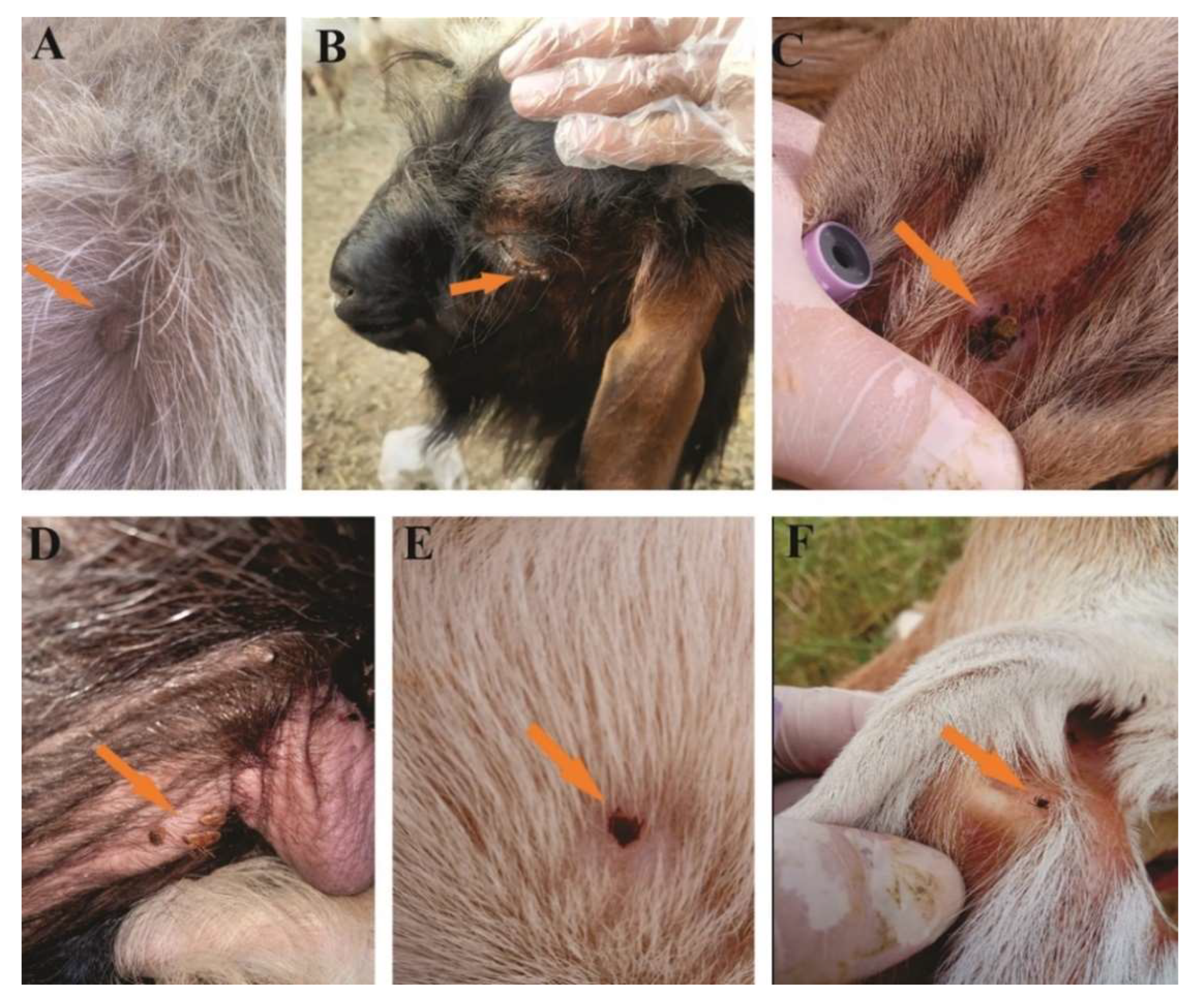
2.3. Tick Collection and Preservation

Tick collection was carried out from March 2019 to February 2020 with a regular visit to the study area once a month. Ticks were randomly collected using forceps from different vertebrate hosts, including cattle, Asian water buffaloes, horses, goats, sheep, dogs, and camels (Figure 2). Tick specimens were rinsed with distilled water followed by 70% ethanol and were stored in 100% ethanol in properly labeled tubes for onward molecular experiments. During tick collection, the relevant information regarding collection date, host type, and place of collection of the ticks were noted.

2.4. Morphological Identification of Ticks

Ticks were morphologically identified using stereo zoom microscope (SZ61, Olympus Corporation, Tokyo, Japan) and standard morphological keys [6,25,26,27,28,29,30,31,32,33].

2.5. DNA Extraction and PCR

Before the genomic DNA extraction, ticks were washed with distilled water and dried on sterile filter paper. The ticks were crushed with sterilized pestles in 1.5 mL sterile Eppendorf tubes. Genomic DNA was extracted individually from each tick using the phenol–chloroform method according to the standard protocol. The DNA pellet was hydrated by adding 30 µL of nuclease-free water [34]. The quality and quantity of genomic DNA were determined through Nano-Q (Optizen, Daejeon, Korea).
By using reference primers and PCR conditions (Table 1), the extracted DNA was subjected to amplifying partial fragments of ticks cox1 and 16S rRNA genes and screened for 16S rRNA of Anaplasma spp. in Table 2 through a PCR. Each PCR reaction was prepared in a 20 μL reaction mixture and contained: 12 μL of DreamTaq MasterMix (Thermo Fisher Scientific, Inc., Waltham, MA, USA), 1 μL of each forward and reverse primers (10 μM), 2 μL (50 ng/μL) genomic DNA template and 4 μL PCR water (nuclease-free). The DNA of R. microplus and Rickettsia massiliae were used as positive controls for ticks and Anaplasma spp., respectively, while PCR water (nuclease-free) was used as a negative control. The amplified DNA was run on a 1.5% agarose gel, dyed with 2 µL ethidium bromide, and observed by a Gel documentation system (BioDoc-It™ Imaging Systems UVP, LLC, Upland, CA, USA).

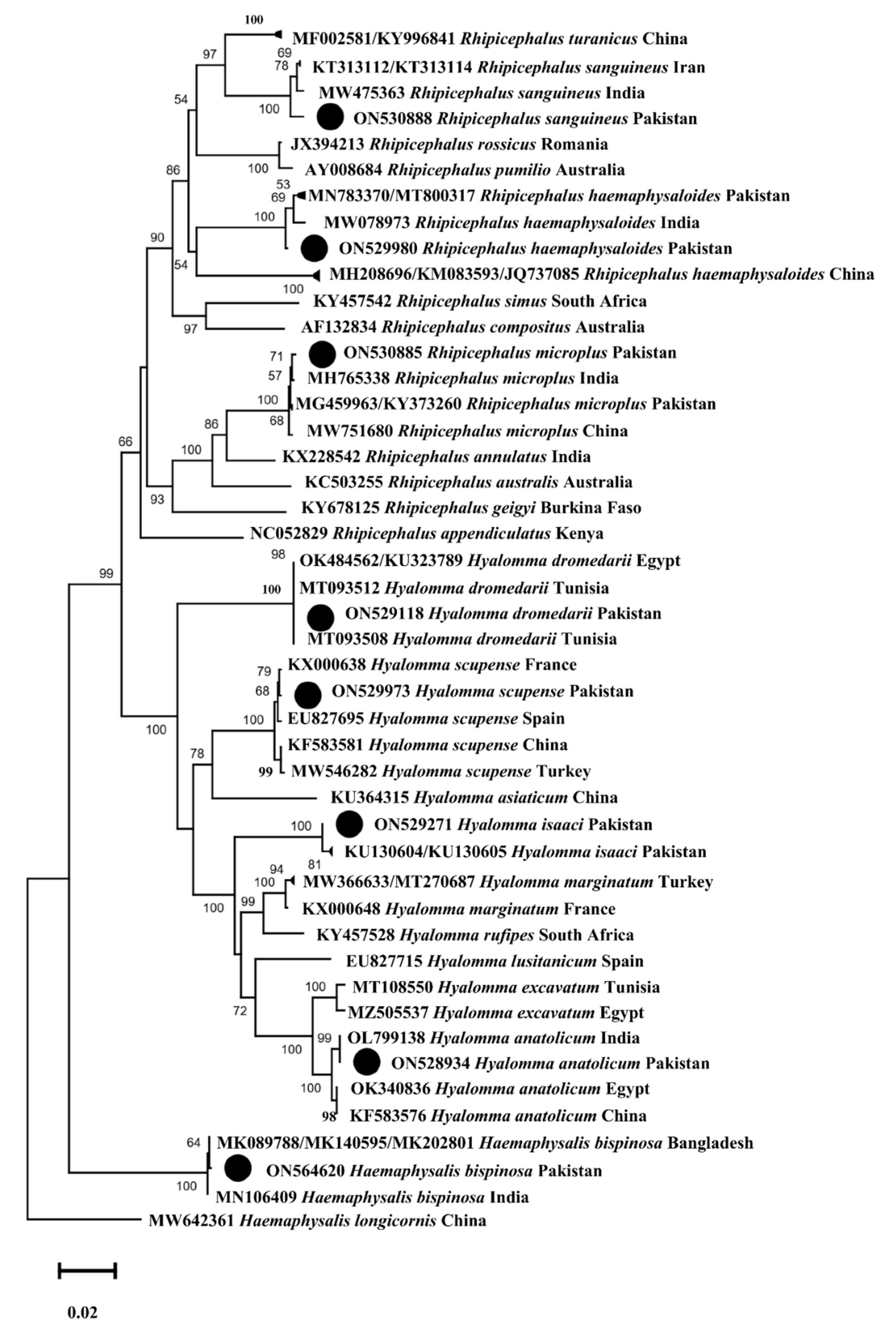
2.6. DNA Sequencing and Phylogenetic Analysis

Purification of PCR products was performed using GeneClean II Kit (Qbiogene, Illkirch, France) following the manufacturer’s protocol. A total of 64 (cox1 40 and 16S rRNA 24) amplified PCR products for ticks and 18 (3 from each Anaplasma positive tick species amplicons) for 16S rRNA Anaplasma spp. were submitted for bidirectional DNA sequencing (Macrogen, Inc., Seoul, South Korea). The sequences were cropped to remove the primers and poor reading regions through SeqMan V. 5 (DNASTAR). The obtained purified sequences were subjected to the Basic Local Alignment Search Tool (BLAST) [38] at National Center for Biotechnology Information (NCBI), and the homologous sequences were downloaded. These sequences were aligned with obtained sequences along with an outgroup in BioEdit Sequence Alignment Editor V. 7.0.5 (Raleigh, NC, USA) [39]. The phylogenetic trees were constructed by using the Maximum-Likelihood model (1000 bootstrap replicons) in Molecular Evolutionary Genetics Analysis (MEGA-X) [40].

3. Results

3.1. Morphologically Identified Ticks

The morphological identification confirmed 12 tick species belonging to the three genera of hard ticks. The genus Hyalomma included Hy. dromedarii, Hy. anatolicum, Hy. scupense and Hy. isaaci, the genus Rhipicephalus contained R. microplus, R. sanguineus, R. haemaphysaloides, and R. turanicus, while the genus Haemaphysalis included Ha. montgomeryi, Ha. bispinosa, Ha. sulcata and Ha. cornupunctata.

3.2. Prevalence of Ticks

A total of 1873 ticks were randomly collected from 443/837 infested hosts (cattle, Asian water buffaloes, horses, goats, sheep, dogs, and camels) comprising Hyalomma (949/1873, 50.6%), Rhipicephalus (629/1873, 33.6%), and Haemaphysalis (295/1873, 15.8%). Overall, Hy. dromedarii was the most prevalent species (390/1873, 20.8%), followed by Hy. anatolicum (294, 15.7%), R. microplus (262, 14%), Hy. scupense (207, 11%), R. sanguineus (136, 7.3%), R. turanicus (121, 6.5%), Ha. cornupunctata (107, 5.7%), R. haemaphysaloides (110, 5.9%), Ha. montgomeryi (87, 4.6%), Hy. isaaci (58, 3.1%), Ha. bispinosa (54, 2.9%), and Ha. sulcata (47, 2.5%). Detailed data about each tick species’ number and percentage of life stages are shown (Table 2).

3.3. Spatial Pattern of Ticks

The highest number of ticks were recorded from Lakki Marwat (679/1873, 36.3%), followed by Orakzai (641/1873, 34.2%), and Bannu (553/1873, 29.5%). Herein, eight tick species were reported representing two genera from Lakki Marwat in which Hy. dromedarii (208/679, 30.6%) was the most abundant, followed by Hy. anatolicum (131/679, 19.3%), and Hy. scupense (96/679, 14.1%). Eight tick species comprising two tick genera were recorded from the Bannu district in which Hy. dromedarii (118/553, 21.3%) was the most abundant species, followed by Hy. anatolicum (107/553, 19.3%), and R. microplus (87/553, 15.7%). In contrast, Ha. cornupunctata (107/641, 16.6%) was the most dominant species in the Orakzai district, followed by R. microplus (103/641, 16%) and Ha. montgomeryi (87/641, 13.5%). Haemaphysalis species were only found in the Orakzai district, while we could not collect these species in the other two districts. The details of each tick species reported from the study area are provided (Figure 3).

3.4. Seasonal Abundance of Ticks

Tick abundance was highly fluctuated by seasonal variations. The highest number of ticks were reported in summer (June–August) (1009/1873, 53.9%), followed by spring (March–May) (522/1873, 27.9%), autumn (September–November) (230/1873, 12.3%), and winter (Dec–Feb) (112/1873, 5.9%) (Figure 4). Details about the seasonal abundance of each tick species in all four seasons are presented in the graph (Figure 4).

3.5. Detection of Anaplasma spp. in Ticks

Anaplasma spp. was detected in 54 out of 314 selected ticks with a total infection rate of 17.2% (54/314). Out of 12 examined tick species, Anaplasma spp. were detected in six species, such as Hy. dromedarii, Hy. anatolicum, Hy. scupense, R. sanguineus, R. microplus and R. haemaphysaloides. The highest infection rate was recorded in Hy. dromedarii 31.1% (14/45), followed by R. microplus 30% (12/40), Hy. scupense 27.3% (9/33), Hy. anatolicum 23.8% (10/42), and in each R. haemaphysaloides and R. sanguineus 20% (6/30), with no amplification of Anaplasma DNA in the selected Haemaphysalis species. The detailed information regarding the infection rate of the selected species is shown in Table 2.

3.6. Sequencing Analysis

From the extracted tick DNA of 12 tick species, the partial fragments of cox1 were amplified for eight tick species, whereas 16S rRNA was amplified for four tick species. Clean cox1 sequences were obtained from eight tick species: Hy. dromedarii (743 bp), Hy. anatolicum (791 bp), Hy. scupense (775 bp), Hy. isaaci (771 bp), Ha. bispinosa (728 bp), R. microplus (800 bp), R. sanguineus (612 bp), and R. haemaphysaloides (797 bp), while 16S rRNA sequences were obtained from four tick species: R. turanicus (398 bp), Ha. cornupunctata (394 bp), Ha. montgomeryi (265 bp) and Ha. sulcata (396 bp). The identical sequences were considered as a single consensus sequence. The BLAST results of the obtained cox1 sequences of Hy. dromedarii, Hy. anatolicum, Hy. scupense, Hy. isaaci, Ha. bispinosa, R. microplus, R. sanguineus, and R. haemaphysaloides showed maximum identities of 99–100%, with the same species reported from Egypt, India, France, Pakistan, Bangladesh, and Iran. In the case of 16S rRNA, the BLAST results of R. turanicus, Ha. montgomeryi and Ha. sulcata showed the highest identities of 99.75%, 96.99%, and 98.75%, respectively, with the same species reported from Afghanistan, China, and Pakistan, while the 16S rRNA sequence of Ha. cornupunctata showed the maximum identity of 92% with the Ha. punctata reported from China, Turkey, Italy, Spain, and Portugal. The 16S rRNA sequences (931 bp) of Anaplasma spp. were subjected to BLAST and showed 100% identity with the A. marginale.

3.7. Phylogenetic Analysis

The phylogenetic tree for the cox1 sequences of Hy. dromedarii, Hy. anatolicum, Hy. scupense, Hy. isaaci, Ha. bispinosa, R. microplus, R. sanguineus, and R. haemaphysaloides were constructed combinedly with 49 sequences downloaded from NCBI based on the maximum identity. In the phylogenetic tree, the obtained cox1 sequences were clustered to the corresponding species reported from different countries, such as Hy. dromedarii from Egypt and Tunisia, Hy. anatolicum from India, Egypt, and China, Hy. scupense from France, Spain, China, and Turkey, Hy. isaaci from Pakistan, Ha. bispinosa from India and Bangladesh, R. microplus from Pakistan, India, and China, R. sanguineus from Iran and India, and R. haemaphysaloides from Pakistan, India, and China. In the case of 16S rRNA, the phylogenetic tree of R. turanicus, Ha. cornupunctata, Ha. montgomeryi and Ha. sulcata was constructed with 27 sequences downloaded from NCBI based on the maximum identity. In the phylogenetic tree, 16S rRNA sequences of R. turanicus, Ha. montgomeryi and Ha. sulcata clustered with the same species reported from Afghanistan, Pakistan, and China, while Ha. cornupunctata clustered with the species of the same subgenus Ha. (Aboimisalis) punctata reported from China, Turkey, Italy, Spain, and Portugal.
All the obtained cox1 sequences were uploaded to the GenBank under accession numbers: ON529118 (Hy. dromedarii), ON528934 (Hy. anatolicum), ON529973 (Hy. scupense), ON529271 (Hy. isaaci), ON564620 (Ha. bispinosa), ON530885 (R. microplus), ON530888 (R. sanguineus), and ON529980 (R. haemaphysaloides). The obtained 16S rRNA sequences were uploaded under accession numbers: ON911440 (R. turanicus), ON911369 (Ha. cornupunctata), ON911371 (Ha. montgomeryi), and ON911372 (Ha. sulcata). The phylogenetic trees of the obtained cox1 and 16S rRNA sequences are shown in Figure 5 and Figure 6, respectively.
A total of 29 sequences of 16S rRNA for A. marginale were downloaded from GenBank in FASTA format based on maximum identity with query sequences. In the phylogenetic tree, the obtained partial 16S rRNA sequence of A. marginale clustered with the same sequences reported from Kenya, Thailand, Australia, Pakistan, and China (Figure 7). The obtained partial 16S rRNA sequence of A. marginale was uploaded to the GenBank (ON528757).

4. Discussion

Pakistan has an agrarian economy where agriculture contributes approximately 21% to gross domestic product (GDP) and 45% to the labor force [41]. Ticks pose severe threats to the livestock and economy of the country. Knowledge regarding molecular surveillance of ticks and A. marginale and their host range in different physiographic is essential for implementing adequate measures against these parasites in Pakistan. The present study was executed in two distinct physiographic regions in southern KP, Pakistan. The targeted areas were selected because ticks and tick-borne diseases are common in these regions but mainly remained unexplored, and to compare tick diversity in two regions that are geographically close but physiographically and climatically different. Herein, 12 tick species were morphologically and molecularly identified. Four tick species, including Ha. bispinosa, Ha. cornupunctata, Hy. dromedarii and Hy. isaaci were genetically characterized for the first time from Pakistan. Furthermore, the molecular survey was conducted to screen a subset of the collected 12 species for A. marginale, in which this pathogen was detected in six species. Among these species, A. marginale was detected for the first time in Hy. dromedarii, Hy. scupense, R. sanguineus and R. haemaphysaloides from Pakistan.
Environmental and climatic factors influence the distribution and prevalence of ticks within a specific region [42]. Previous studies considered Hyalomma spp. as successful ticks in harsh desert regions [43,44]. Similarly, as a larger proportion of the current study area was a desert plain, the genus Hyalomma was the most prevalent, followed by genus Rhipicephalus and Haemaphysalis. Herein, unlike [2,3,8], Hy. dromedarii was the most prevalent in the region owing to the screening of a larger number of camels compared to other hosts. According to the studies performed in the region [2,3,8], R. microplus and Ha. bispinosa were the most prevalent tick species in the genus Rhipicephalus and genus Haemaphysalis, respectively.
The highest prevalence of A. marginale occurs in those regions where R. microplus is endemic [45]. This implies that R. microplus is one of the most competent vectors for A. marginale. Comparatively, A. marginale was highly detected in Hy. dromedarii, followed by R. microplus in the present study. This pathogen was also detected in four other tick species, including Hy. anatolicum, Hy. dromedarii, R. sanguineus and Hy. scupense. To the best of our knowledge, the detection of A. marginale in R. sanguineus is exceptionally rare [46], and this pathogen has not been detected in Hy. scupense. However, experimentally it has been demonstrated that this pathogen can be successfully transmitted by R. sanguineus [47,48]. Therefore, such unexpected outcomes need to be further evaluated because the presence of a pathogen DNA in a tick species does not ensure it as a biological vector. Moreover, a global increase in moments of infected/carrier livestock and/or tick-infested livestock across international borders can further worsen the situation regarding this pathogen [49].
For the host range of ticks, the resemblance among hosts’ ecology might be more significant than evolutionary similarity [50]. A wide host range was recorded for Hy. anatolicum that could be attributed to its two or three host life cycle with the infestation on different ungulates [51]. A comparatively wide host range was also noted for one host tick species such as R. microplus. This might be due to common practices in the study area, such as placing different hosts in the same shelter, overcrowded herds, and combined grazing.
Research has been focused on understanding the evolutionary history and taxonomy of ticks and TBPs using standard genetic markers [36,52,53]. The mitochondrial gene cox1 has been considered an appropriate genetic marker for understanding tick phylogenetic relationships, especially at the species level [52]. The 16S rRNA gene has also been considered a reliable marker for tick identification [36,52] and is of prime importance in evaluating bacterial phylogeny and taxonomy [54,55]. When taking these into account, the cox1 sequences were obtained for eight tick species (Hy. dromedarii, Hy. anatolicum, Hy. scupense, Hy. isaaci, Ha. bispinosa, R. microplus, R. sanguineus, and R. haemaphysaloides). For the remaining four tick species (R. turanicus, Ha. cornupunctata, Ha. montgomeryi, and Ha. sulcata), we were able to obtain only 16S rRNA sequences. The A. marginale associated with these ticks was molecularly assessed by targeting the partial 16S rRNA gene. Except for Ha. cornupunctata, all other Haemaphysalis species were clustered with related species reported from the Oriental and neighboring Palearctic zoogeographical regions (Figure 5 and Figure 6). In the cox1-based phylogenetic tree, the monophyletic clade containing Ha. bispinosa was basal to the remaining ixodid tick species. In tick 16S rRNA-based tree, the monophyletic clade having Ha. sulcata was at a basal position to all other hard tick species. The clade that had Ha. montgomeryi appeared as sister to the clade possessing both Ha. obesa and Ha. parva. Before the genetic data, based on morphological resemblance, these species were placed in the same subgenus Segalia of Haemaphysalis [56]. Due to the lack of previous genetic data, Ha. cornupunctata was displayed individually as sister taxa to the clade, which constitutes Ha. punctata. The closeness between these species has already been well established from morphological similarities; accordingly, they were assigned the same subgenus Aboimisalis of Haemaphysalis [6,56]. Hyalomma species were clustered with related species from Oriental, neighboring Palearctic, and Afrotropic regions. In the phylogenetic tree inferred from tick cox1, the clade formed by Hy. anatolicum clustered as sisters to the clade of Hy. excavatum. This concurs with the morphological resemblance among Hy. anatolicum and Hy. excavatum. The clade containing Hy. dromedarii was sister to the clade formed by Hy. scupense (jointly with seven other Hyalomma species). In the same phylogenetic tree, the clades of Hy. scupense and Hy. asiaticum were revealed as sister taxa. These studies were concordant with previous demonstrations [57]. Hyalomma isaaci clade appeared as a distinct species that did not support this species as a sub-species of Hy. marginatum [58,59,60] but supported this species as a valid species [31]. Rhipicephalus species clustered with the same species from Oriental and neighboring Palearctic regions. In the tick cox1-based phylogenetic tree, the R. sanguineus clade appeared to be sister to the R. turanicus clade, and both were jointly sister to the R. rossicus and R. pumilio clade. In the phylogenetic tree inferred from tick 16S rRNA, R. turanicus, along with R. sanguineus and R. linnaei, appeared to be sister to R. camicasi. These mentioned Rhipicephalus species are included in the R. sanguineus species complex, and R. sanguineus from the temperate lineage was found closest to R. turanicus [11,61]. Following a previous study [62], different genetic groups were depicted within R. haemaphysaloides in the present phylogenetic analysis. Rhipicephalus microplus of clade-c was found close to R. annulatus [3,11,63]. In the phylogenetic tree based on bacterial 16S rRNA, the clade of A. marginale and A. ovis clustered as a sister clade to the clade of A. centrale. This relatedness is also represented by their ecological and epidemiological aspects because these three species commonly infect ruminants [24].

5. Conclusions

In this study, 12 hard tick species were morphologically and molecularly identified; among them, four species (Ha. bispinosa, Ha. cornupunctata, Hy. dromedarii, and Hy. isaaci) were molecularly characterized for the first time from Pakistan. Notably, this is the first report providing Ha. cornupunctata genetic data and preliminary phylogenetic analysis. Furthermore, A. marginale was molecularly assessed in six tick species; among them, this pathogen was molecularly detected for the first time in four tick species (Hy. dromedarii, Hy. scupense, R. sanguineus, and R. haemaphysaloides) from Pakistan. Further studies should assess the genetic diversity of ticks and associated Anaplasma spp. in the country. This study might help in recognizing knowledge gaps and provide future direction to veterinary and health authorities in controlling ticks and A. marginale.

Author Contributions

A.A. (Abid Ali), S.A., A.A. (A. Alouffi), M.M.A. and T.T. designed the study and performed the experimental designing of this study. S.A., M.K., S.U., M.N., N.I., Z.K. and O.A. collected the ticks. A.A. (Abid Ali), S.A., M.K., A.A. (A. Alouffi), M.M.A., S.U., M.N., O.A., S.Z.S. and T.T. carried out experiments. A.A. (Abid Ali), M.K., S.U. and M.N. conducted the phylogenetic and statistical analysis. All authors have read and agreed to the published version of the manuscript.

Funding

This work was supported by JSPS KAKENHI, Grant Number JP22H02522 and Heiwa Nakajima Foundation.

Institutional Review Board Statement

This study was approved by Advanced Studies and Research Board (Dir/A&R/AWKUM/2020/4871) committee members of Abdul Wali Khan University Mardan, KP, Pakistan.

Informed Consent Statement

Not applicable.

Data Availability Statement

All the relevant data are within the manuscript.

Acknowledgments

This study was carried out under the financial support given by the Higher Education Commission and Pakistan Science Foundation of Pakistan and the researchers supporting project number (RSP2022R494), King Saud University, Riyadh, Saudi Arabia.

Conflicts of Interest

The authors declare no conflict of interest.

References

  1. Sonenshine, D.E.; Roe, R.M. Biology of Ticks; Oxford University Press: Oxford, UK, 2013; Volume 2. [Google Scholar]
  2. Karim, S.; Budachetri, K.; Mukherjee, N.; Williams, J.; Kausar, A.; Hassan, M.J.; Adamson, S.; Dowd, S.E.; Apanskevich, D.; Arijo, A. A study of ticks and tick-borne livestock pathogens in Pakistan. PLoS Negl. Trop. Dis. 2017, 11, e0005681. [Google Scholar] [CrossRef] [PubMed]
  3. Ali, A.; Khan, M.A.; Zahid, H.; Yaseen, P.M.; Qayash Khan, M.; Nawab, J.; Ur Rehman, Z.; Ateeq, M.; Khan, S.; Ibrahim, M. Seasonal dynamics, record of ticks infesting humans, wild and domestic animals, and molecular phylogeny of Rhipicephalus microplus in Khyber Pakhtunkhwa Pakistan. Front. Physiol. 2019, 10, 793. [Google Scholar] [CrossRef] [PubMed]
  4. Boulanger, N.; Boyer, P.; Talagrand-Reboul, E.; Hansmann, Y. Ticks and tick-borne diseases. Med. Mal. Infect. 2019, 49, 87–97. [Google Scholar] [CrossRef]
  5. Eckstein, D.; Künzel, V.; Schäfer, L. Global Climate Risk Index 2021. Who Suffers Most from Extreme Weather Events; Germanwatch: Bonn, Germany, 2021; pp. 2000–2019. [Google Scholar]
  6. Hoogstraal, H.; Varma, M.G.R. Haemaphysalis cornupunctata sp. n. and H. kashmirensis sp. n. from Kashmir, with Notes on H. sundrai Sharif and H. sewelli Sharif of India and Pakistan (Ixodoidea, Ixodidae). J. Parasitol. 1962, 48, 185–194. [Google Scholar] [CrossRef]
  7. Ali, A.; Mulenga, A.; Vaz, I.S., Jr. Tick and tick-borne pathogens: Molecular and immune targets for control strategies. Front. Physiol. 2020, 11, 744. [Google Scholar] [CrossRef]
  8. Rehman, A.; Nijhof, A.M.; Sauter-Louis, C.; Schauer, B.; Staubach, C.; Conraths, F.J. Distribution of ticks infesting ruminants and risk factors associated with high tick prevalence in livestock farms in the semi-arid and arid agro-ecological zones of Pakistan. Parasites Vectors 2017, 10, 190. [Google Scholar] [CrossRef] [PubMed]
  9. Ali, A.; Zahid, H.; Zeb, I.; Tufail, M.; Khan, S.; Haroon, M.; Bilal, M.; Hussain, M.; Alouffi, A.S.; Muñoz-Leal, S.; et al. Risk factors associated with tick infestations on equids in Khyber Pakhtunkhwa, Pakistan, with notes on Rickettsia massiliae detection. Parasites Vectors 2021, 14, 363. [Google Scholar] [CrossRef] [PubMed]
  10. Aiman, O.; Ullah, S.; Chitimia-Dobler, L.; Nijhof, A.M.; Ali, A. First report of Nosomma monstrosum ticks infesting Asian water buffaloes (Bubalus bubalis) in Pakistan. Ticks Tick-Borne Dis. 2022, 13, 101899. [Google Scholar] [CrossRef]
  11. Ali, A.; Shehla, S.; Zahid, H.; Ullah, F.; Zeb, I.; Ahmed, H.; da Silva Vaz, I.; Tanaka, T. Molecular survey and spatial distribution of Rickettsia spp. in ticks infesting free-ranging wild animals in Pakistan (2017–2021). Pathogens 2022, 11, 162. [Google Scholar] [CrossRef]
  12. Ali, A.; Numan, M.; Khan, M.; Aiman, O.; Muñoz-Leal, S.; Chitimia-Dobler, L.; Labruna, M.B.; Nijhof, A.M. Ornithodoros (Pavlovskyella) ticks associated with a Rickettsia sp. in Pakistan. Parasites Vectors 2022, 15, 138. [Google Scholar] [CrossRef]
  13. Begum, F.; Wisseman, C.L., Jr.; Casals, J. Tick-borne viruses of West Pakistan: II. Hazara virus, a new agent isolated from Ixodes redikorzevi ticks from the Kaghan valley, W. Pakistan. Am. J. Epidemiol. 1970, 92, 192–194. [Google Scholar] [CrossRef] [PubMed]
  14. Clifford, C.M.; Hoogstraal, H.; Kohls, G.M. Ixodes hyatti, n. sp.; and I. shahi, n. sp. (Acarina: Ixodidae), parasites of Pikas (Lagomorpha: Ochotonidae) in the Himalayas of Nepal and West Pakistan. J. Med. Entomol. 1971, 8, 430–438. [Google Scholar] [CrossRef] [PubMed]
  15. Auffenberg, W.; Auffenberg, T. The reptile tick Aponomma gervaisi: (Acarina Ixodidae) as a parasite of monitor lizards in Pakistan and India. Bull. Fla. Mus. Nat. Hist. Biol. Sci. 1990, 35, 1–34. [Google Scholar]
  16. McCarthy, V.C. Ixodid Ticks (Acarina, Ixodidae) of West Pakistan. Doctoral dissertation, University of Maryland, College Park, MD, USA, 1967; p. 533. [Google Scholar]
  17. Jabbar, A.; Abbas, T.; Sandhu, Z.U.D.; Saddiqi, H.A.; Qamar, M.F.; Gasser, R.B. Tick-borne diseases of bovines in Pakistan: Major scope for future research and improved control. Parasites Vectors 2015, 8, 283. [Google Scholar] [CrossRef]
  18. Rehman, A.; Conraths, F.J.; Sauter-Louis, C.; Krücken, J.; Nijhof, A.M. Epidemiology of tick-borne pathogens in the semi-arid and the arid agro-ecological zones of Punjab province, Pakistan. Transbound. Emerg. Dis. 2019, 66, 526–536. [Google Scholar] [CrossRef] [PubMed]
  19. Aktas, M.; Altay, K.; Dumanli, N. Molecular detection and identification of Anaplasma and Ehrlichia species in cattle from Turkey. Ticks Tick-Borne Dis. 2011, 2, 62–65. [Google Scholar] [CrossRef] [PubMed]
  20. Raoult, D.; Parola, P. (Eds.) Rickettsial Diseases; Informa Healthcare: New York, NY, USA, 2007; p. 379. [Google Scholar]
  21. Ybañez, A.P.; Ybanez, R.H.D.; Claveria, F.G.; Cruz-Flores, M.J.; Xuenan, X.; Yokoyama, N.; Inokuma, H. High genetic diversity of Anaplasma marginale detected from Philippine cattle. J. Vet. Med. Sci. 2014, 76, 1009–1014. [Google Scholar] [CrossRef]
  22. Begum, F.; Wisseman, C.L., Jr.; Traub, R. Tick-borne viruses of West Pakistan: I. Isolation and general characteristics. Am. J. Epidemiol. 1970, 92, 180–191. [Google Scholar] [CrossRef]
  23. Ghafar, A.; Gasser, R.B.; Rashid, I.; Ghafoor, A.; Jabbar, A. Exploring the prevalence and diversity of bovine ticks in five agro-ecological zones of Pakistan using phenetic and genetic tools. Ticks Tick-Borne Dis. 2020, 11, 101472. [Google Scholar] [CrossRef]
  24. Khan, Z.; Shehla, S.; Alouffi, A.; Kashif Obaid, M.; Zeb Khan, A.; Almutairi, M.M.; Numan, M.; Aiman, O.; Alam, S.; Ullah, S.; et al. Molecular survey and genetic characterization of Anaplasma marginale in ticks collected from livestock hosts in Pakistan. Animals 2022, 12, 1708. [Google Scholar] [CrossRef]
  25. Hoogstraal, H.; Kaiser, M.N. Observations on Egyptian Hyalomma ticks (Ixodoidea, Ixodidae). Biological notes and differences in identity of H. anatolicum and its subspecies anatolicum Koch and excavatum Koch among Russian and other workers. Identity of H. lusitanicum Koch. Ann. Entomol. Soc. Am. 1959, 52, 243–261. [Google Scholar] [CrossRef]
  26. Hoogstraal, H.; Trapido, H.; Kohls, G.M. Studies on southeast Asian Haemaphysalis ticks (Ixodoidea, Ixodidae). Speciation in the H. (Kaiseriana) obesa group: H. semermis Neumann, H. obesa Larrousse, H. roubaudi Toumanoff, H. montgomeryi Nuttall, and H. hirsuta sp. n. J. Parasitol. 1966, 52, 169–191. [Google Scholar] [CrossRef] [PubMed]
  27. Hoogstraal, H.; Trapido, H. Redescription of the type materials of Haemaphysalis (Kaiseriana) bispinosa Neumann (India), H. (K.) neumanni Dönitz (Japan), H. (K.) lagrangei Larrousse (Vietnam), and H. (K.) yeni Toumanoff (Vietnam) (Ixodoidea, Ixodidae). J. Parasitol. 1966, 52, 1188–1198. [Google Scholar] [CrossRef]
  28. Apanaskevich, D.A. Differentiation of closely related species Hyalomma anatolicum and H. excavatum (Acari: Ixodidae) based on a study of all life cycle stages, throughout entire geographical range. Parazitologiia 2003, 37, 259–280. [Google Scholar] [PubMed]
  29. Walker, A.R. Ticks of Domestic Animals in Africa: A Guide to Identification of Species; Bioscience Reports: Edinburgh, UK, 2003; pp. 3–210. [Google Scholar]
  30. Walker, J.B.; Keirans, J.E.; Horak, I.G. The Genus Rhipicephalus (Acari, Ixodidae): A Guide to the Brown Ticks of the World; Cambridge University Press: Cambridge, UK, 2005. [Google Scholar]
  31. Apanaskevich, D.A.; Horak, I.G. The genus Hyalomma Koch, 1844: V. Re-evaluation of the taxonomic rank of taxa comprising the H. (Euhyalomma) marginatum Koch complex of species (Acari: Ixodidae) with redescription of all parasitic stages and notes on biology. Int. J. Acarol. 2008, 34, 13–42. [Google Scholar] [CrossRef]
  32. Apanaskevich, D.A.; Schuster, A.L.; Horak, I.G. The genus Hyalomma: VII. Redescription of all parasitic stages of H. (Euhyalomma) dromedarii and H. (E.) schulzei (Acari: Ixodidae). J. Med. Entomol. 2008, 45, 817–831. [Google Scholar] [CrossRef]
  33. Apanaskevich, D.A.; Filippova, N.A.; Horak, I.G. The genus Hyalomma Koch, 1844. X. Redescription of all parasitic stages of H. (Euhyalomma) scupense Schulze, 1919 (= H. detritum Schulze) (Acari: Ixodidae) and notes on its biology. Folia Parasitol. 2010, 57, 69–78. [Google Scholar] [CrossRef]
  34. Sambrook, J.; Fritsch, E.F.; Maniatis, T. Molecular Cloning: A Laboratory Manual, 2nd ed.; Cold Spring Harbor Laboratory Press: New York, NY, USA, 1989. [Google Scholar]
  35. Chitimia, L.; Lin, R.Q.; Cosoroaba, I.; Wu, X.Y.; Song, H.Q.; Yuan, Z.G.; Zhu, X.Q. Genetic characterization of ticks from southwestern Romania by sequences of mitochondrial cox1 and nad5 genes. Exp. App. Acarol. 2010, 52, 305–311. [Google Scholar] [CrossRef]
  36. Mangold, A.J.; Bargues, M.D.; Mas-Coma, S. Mitochondrial 16S rDNA sequences and phylogenetic relationships of species of Rhipicephalus and other tick genera among Metastriata (Acari: Ixodidae). Parasitol. Res. 1998, 84, 478–484. [Google Scholar] [CrossRef]
  37. Hailemariam, Z.; Ahmed, J.S.; Clausen, P.H.; Nijhof, A.M. A comparison of DNA extraction protocols from blood spotted on FTA cards for the detection of tick-borne pathogens by Reverse Line Blot hybridization. Ticks Tick-Borne Dis. 2017, 8, 185–189. [Google Scholar] [CrossRef]
  38. Altschul, S.F.; Gish, W.; Miller, W.; Myers, E.W.; Lipman, D.J. Basic Local Alignment Search Tool. J. Mol. Biol. 1990, 215, 403–410. [Google Scholar] [CrossRef]
  39. Hall, T.; Biosciences, I.; Carlsbad, C. BioEdit: An important software for molecular biology. GERF Bull. Biosci. 2011, 2, 60–61. [Google Scholar]
  40. Kumar, S.; Stecher, G.; Li, M.; Knyaz, C.; Tamura, K. MEGA-X: Molecular Evolutionary Genetics Analysis across computing platforms. Mol. Biol. Evol. 2018, 35, 1547–1549. [Google Scholar] [CrossRef] [PubMed]
  41. Pakistan Economic Survey. Government of Pakistan. Ministry of Finance, Islamabad. Available online: https://www.finance.gov.pk/survey (accessed on 2 October 2021).
  42. MacDonald, A.J. Abiotic and habitat drivers of tick vector abundance, diversity, phenology and human encounter risk in southern California. PLoS ONE 2018, 13, e0201665. [Google Scholar] [CrossRef] [PubMed]
  43. Uspensky, I. Preliminary observations on specific adaptations of exophilic ixodid ticks to forests or open country habitats. Exp. App. Acarol. 2002, 28, 147–154. [Google Scholar] [CrossRef] [PubMed]
  44. Kamran, K.; Ali, A.; Villagra, C.A.; Bazai, Z.A.; Iqbal, A.; Sajid, M.S. Hyalomma anatolicum resistance against ivermectin and fipronil is associated with indiscriminate use of acaricides in southwestern Balochistan, Pakistan. Parasitol. Res. 2021, 120, 15–25. [Google Scholar] [CrossRef]
  45. Palmer, G.H.; Rurangirwa, F.R.; McElwain, T.F. Strain composition of the Ehrlichia Anaplasma marginale within persistently infected cattle, a mammalian reservoir for tick transmission. J. Clin. Microbiol. 2001, 39, 631–635. [Google Scholar] [CrossRef]
  46. Hegab, A.A.; Omar, H.M.; Abuowarda, M.; Ghattas, S.G.; Mahmoud, N.E.; Fahmy, M.M. Screening and phylogenetic characterization of tick-borne pathogens in a population of dogs and associated ticks in Egypt. Parasites Vectors 2022, 15, 1–15. [Google Scholar] [CrossRef]
  47. Parker, R.J. The Australian brown dog tick Rhipicephalus sanguineus as an experimental parasite of cattle and vector of Anaplasma marginale. Aust. Vet. J. 1982, 58, 47–50. [Google Scholar] [CrossRef]
  48. Shkap, V.; Kocan, K.; Molad, T.; Mazuz, M.; Leibovich, B.; Krigel, Y.; Michoytchenko, A.; Blouin, E.; De la Fuente, J.; Samish, M.; et al. Experimental transmission of field Anaplasma marginale and the A. centrale vaccine strain by Hyalomma excavatum, Rhipicephalus sanguineus and Rhipicephalus (Boophilus) annulatus ticks. Vet. Microbiol. 2009, 134, 254–260. [Google Scholar] [CrossRef]
  49. Quiroz-Castañeda, R.E.; Amaro-Estrada, I.; Rodríguez-Camarillo, S.D. Anaplasma marginale: Diversity, virulence, and vaccine landscape through a genomics approach. BioMed Res. Int. 2016, 2016, 9032085. [Google Scholar] [CrossRef]
  50. Krasnov, B.R.; Mouillot, D.; Shenbrot, G.I.; Khokhlova, I.S.; Vinarski, M.V.; Korallo-Vinarskaya, N.P.; Poulin, R. Similarity in ectoparasite faunas of Palearctic rodents as a function of host phylogenetic, geographic, or environmental distances: Which matters the most? Int. J. Parasitol. 2010, 40, 807–817. [Google Scholar] [CrossRef] [PubMed]
  51. Vatansever, Z. Hyalomma anatolicum Koch, 1844 (Figs. 158–160). In Ticks of Europe and North Africa; Springer: Cham, Switzerland, 2017; pp. 391–395. [Google Scholar]
  52. Lv, J.; Wu, S.; Zhang, Y.; Chen, Y.; Feng, C.; Yuan, X.; Jia, G.; Deng, J.; Wang, C.; Wang, Q.; et al. Assessment of four DNA fragments (COI, 16S rDNA, ITS2, 12S rDNA) for species identification of the Ixodida (Acari: Ixodida). Parasites Vectors 2014, 7, 1–11. [Google Scholar] [CrossRef] [PubMed]
  53. Beati, L.; Klompen, H. Phylogeography of ticks (Acari: Ixodida). Ann. Rev. Entomol. 2019, 64, 379–397. [Google Scholar] [CrossRef] [PubMed]
  54. Patel, J.B. 16S rRNA gene sequencing for bacterial pathogen identification in the clinical laboratory. Mol Diagn. 2001, 6, 313–321. [Google Scholar] [CrossRef]
  55. Janda, J.M.; Abbott, S.L. 16S rRNA gene sequencing for bacterial identification in the diagnostic laboratory: Pluses, perils, and pitfalls. J. Clin. Microbiol. 2007, 45, 2761–2764. [Google Scholar] [CrossRef]
  56. Geevarghese, G.; Mishra, A.C. Haemaphysalis Ticks of India; Elsevier: Amsterdam, The Netherlands, 2011. [Google Scholar]
  57. Sands, A.F.; Apanaskevich, D.A.; Matthee, S.; Horak, I.G.; Harrison, A.; Karim, S.; Mohammad, M.K.; Mumcuoglu, K.Y.; Rajakaruna, R.S.; Santos-Silva, M.M.; et al. Effects of tectonics and large-scale climatic changes on the evolutionary history of Hyalomma ticks. Mol. Phylogenet. Evol. 2017, 114, 153–165. [Google Scholar] [CrossRef]
  58. Pomerantzev, B.I. Ticks (fam. Ixodidae) of the USSR and neighboring countries. Opred. Faune SSSR Zool. Inst. Akad. Nauk SSSR 1946, 26, 26–28. (in Russian). [Google Scholar]
  59. Hoogstraal, H.; Kaiser, M.N. Observations on ticks (Ixodoidea) of Libya. Ann. Entomol. Soc. Am. 1960, 53, 445–457. [Google Scholar] [CrossRef]
  60. Kaiser, M.N.; Hoogstraal, H. The Hyalomma ticks (Ixodoidea, Ixodidae) of Afghanistan. J. Parasitol. 1963, 49, 130–139. [Google Scholar] [CrossRef]
  61. Bakkes, D.K.; Ropiquet, A.; Chitimia-Dobler, L.; Matloa, D.E.; Apanaskevich, D.A.; Horak, I.G.; Mans, B.J.; Matthee, C.A. Adaptive radiation and speciation in Rhipicephalus ticks: A medley of novel hosts, nested predator-prey food webs, off-host periods, and dispersal along temperature variation gradients. Mol. Phylogenet. Evol. 2021, 162, 107178. [Google Scholar] [CrossRef] [PubMed]
  62. Li, H.; Zheng, Y.C.; Ma, L.; Jia, N.; Jiang, B.G.; Jiang, R.R.; Huo, Q.B.; Wang, Y.W.; Liu, H.B.; Chu, Y.L.; et al. Human infection with a novel tick-borne Anaplasma species in China: A surveillance study. Lancet Infect. Dis. 2015, 15, 663–670. [Google Scholar] [CrossRef]
  63. Low, V.L.; Tay, S.T.; Kho, K.L.; Koh, F.X.; Tan, T.K.; Lim, Y.A.L.; Ong, B.L.; Panchadcharam, C.; Norma-Rashid, Y.; Sofian-Azirun, M. Molecular characterization of the tick Rhipicephalus microplus in Malaysia: New insights into the cryptic diversity and distinct genetic assemblages throughout the world. Parasites Vectors 2015, 8, 341. [Google Scholar] [CrossRef] [PubMed]
Figure 1. Map showing tick collection sites in southern Khyber Pakhtunkhwa, Pakistan.
Microorganisms 10 01663 g001
Figure 2. Tick infestation on different hosts: Hy. dromedarii from camels (A). Ha. bispinosa from goats (B). R. turanicus from sheep (C). Hy. anatolicum from male Asian water buffaloes (D). R. sanguineus from dogs (E). R. turanicus from goats (F).
Microorganisms 10 01663 g002
Figure 3. Spatial patterns of the collected ixodid ticks in the study regions.
Microorganisms 10 01663 g003
Figure 4. Seasonal abundance of the collected ixodid ticks in the study regions.
Microorganisms 10 01663 g004
Figure 5. Maximum likelihood phylogenetic tree based on cox1 sequences of Hy. dromedarii, Hy. anatolicum, Hy. scupense, Hy. isaaci, Ha. bispinosa, R. microplus, R. sanguineus and R. haemaphysaloides. Haemaphysalis longicornis was used as an outgroup, using supporting values (1000 replicons) at each node. The scale bar indicates the number of substitutions per site. The obtained sequences were represented with black circles.
Microorganisms 10 01663 g005
Figure 6. Maximum likelihood phylogenetic tree based on 16S rRNA sequences of R. turanicus, Ha. cornupunctata, Ha. montgomeryi and Ha. sulcata. Haemaphysalis longicornis was used as an outgroup, using supporting values (1000 replicons) at each node. The scale bar indicates the number of substitutions per site. The obtained sequences were represented with black circles.
Microorganisms 10 01663 g006
Figure 7. Maximum likelihood phylogenetic tree based on the partial 16S rRNA sequence of A. marginale. The Anaplasma boleense was used as an outgroup, using supporting values (1000 replicons) at each node. The scale bar indicates the number of substitutions per site. The obtained sequence was represented with a black circle.
Microorganisms 10 01663 g007
Table 1. Primers used for the detection of ticks and associated Anaplasma spp.
Organism/GeneSequence (5′-3′)Amplicon SizePCR ConditionRef.
Ticks/cox1cox1 F, GGAACAATATATTTAATTTTTGG
cox1 R, ATCTATCCCTACTGTAAATATATG
801 bp95 °C 5 min, 40× (95 °C 30 s, 55 °C 60 s, 72 °C 1 min), 72 °C 5 min[35]
Ticks/16S rRNA16S+1, CCGGTCTGAACTCAGATCAAGT
16S−1, GCTCAATGATTTTTTAAATTGCTG
460 bp95 °C 3 min, 40× (95 °C 30 s, 55 °C 60 s, 72 °C 1 min), 72 °C 7 min[36]
Anaplasma spp./16S rRNAEhr-F2, AGAGTTTGATCCTGGCTCAG
Ehr-R, AGTTTGCCGGGACTTYTTCT
1100 bp95 °C 3 min, 35× (95 °C 30 s, 50 °C 30 s, 72 °C 1 min), 72 °C 7 min[37]
Table 2. Prevalence of ticks and the detection rate of Anaplasma marginale.
Tick SpeciesTick Life StagesTotal Ticks (%)Ticks Subjected to PCRInfested HostsAnaplasma marginale
Female • (%)Male (%)Nymph (%)Positive TicksInfection Rate %
Hy. dromedarii187 (47.9)170 (43.6)33 (8.5)390 (20.8)45Camels, Sheep, Cattle1431.1
Hy. anatolicum140 (47.6)128 (43.5)26 (8.9)294 (15.7)42Cattle, Sheep, Goats, Dogs, Asian water buffaloes, Horses, Camels1023.8
Hy. scupense103 (49.7)86 (41.6)18 (8.7)207 (11.0)33Cattle, Asian water buffaloes, Horses927.3
Hy. isaaci33 (56.9)19 (32.8)6 (10.3)58 (3.1)15Sheep, Cattle, Goats00
Ha. cornupunctata51 (47.7)42 (39.3)14 (13)107 (5.7)8Sheep, Goats00
Ha. montgomeryi42 (48.3)36 (41.4)9 (10.3)87 (4.6)6Goats, Sheep00
Ha. bispinosa26 (48.2)18 (33.3)10 (18.5)54 (2.9)8Goats, Sheep00
Ha. sulcata21 (44.7)17 (36.2)9 (19.1)47 (2.5)7Sheep, Goats00
R. microplus126 (48.1)78 (29.8)58 (22.1)262 (14)40Cattle, Asian water buffaloes, Sheep, Goats, Dogs1230
R. turanicus61 (50.4)51 (42.2)9 (7.4)121 (6.5)6Sheep, Goats, Dogs, Horses00
R. sanguineus71 (52.2)52 (38.2)13 (9.6)136 (7.3)30Dogs, Sheep, Goats620
R. haemaphysaloides53 (48.2)45 (40.9)12 (10.9)110 (5.9)30Dogs, Sheep, Goats620
Total914 (48.8)742 (39.6)217 (11.6)1873314 5417.2
Count for fully, partially and unengorged.
Publisher’s Note: MDPI stays neutral with regard to jurisdictional claims in published maps and institutional affiliations.

