Review

Homeostasis and Dysbiosis of the Intestinal Microbiota: Comparing Hallmarks of a Healthy State with Changes in Inflammatory Bowel Disease

1
Faculty of Dental Medicine and Health, Josip Juraj Strossmayer University of Osijek, Crkvena 21, 31000 Osijek, Croatia
2
University Centre Varaždin, University North, 42000 Varaždin, Croatia
3
Institute for Health Metrics and Evaluation and the Department of Health Metrics Sciences, University of Washington, Seattle, WA 98195, USA
4
Faculty of Natural Sciences, Juraj Dobrila University of Pula, 52100 Pula, Croatia
5
General Hospital Vukovar, Županijska 35, 32000 Vukovar, Croatia
*
Authors to whom correspondence should be addressed.
Microorganisms 2022, 10(12), 2405; https://doi.org/10.3390/microorganisms10122405
Submission received: 7 November 2022 / Revised: 28 November 2022 / Accepted: 1 December 2022 / Published: 5 December 2022
(This article belongs to the Special Issue Gut Microbiota: Health, Clinical & Beyonds)

Abstract

:
The gut microbiota, which represent a community of different microorganisms in the human intestinal tract, are crucial to preserving human health by participating in various physiological functions and acting as a metabolic organ. In physiological conditions, microbiota–host partnership exerts homeostatic stability; however, changes in intestinal microbiota composition (dysbiosis) are an important factor in the pathogenesis of inflammatory bowel disease and its two main disease entities: ulcerative colitis and Crohn’s disease. The incidence and prevalence of these inflammatory conditions have increased rapidly in the last decade, becoming a significant problem for the healthcare system and a true challenge in finding novel therapeutic solutions. The issue is that, despite numerous studies, the etiopathogenesis of inflammatory bowel disease is not completely clear. Based on current knowledge, chronic intestinal inflammation occurs due to altered intestinal microbiota and environmental factors, as well as a complex interplay between the genetic predisposition of the host and an inappropriate innate and acquired immune response. It is important to note that the development of biological and immunomodulatory therapy has led to significant progress in treating inflammatory bowel disease. Certain lifestyle changes and novel approaches—including fecal microbiota transplantation and nutritional supplementation with probiotics, prebiotics, and synbiotics—have offered solutions for dysbiosis management and paved the way towards restoring a healthy microbiome, with only minimal long-term unfavorable effects.

1. Introduction

The human gastrointestinal tract (GIT) is a home to an abundant and protean congregation of more than 100 trillion microorganisms that live in peaceful coexistence with their hosts [1]. This complex but well-organized community is known as intestinal microbiota, microflora, or normal gut flora [2]. During the last decade, novel and emerging technologies have enabled phylogenetical identification and quantification of the gut microbiota constituents, primarily by relying on the analysis of nucleic acids extracted from stools [3,4]. The term for the collective genetic content of this living microbiota is known as a microbiome [5]. Although these terms are often used interchangeably, this difference should be acknowledged for much better understanding of studies in the field.
The availability of whole-genome sequencing, in conjunction with metagenomic/metabolomic techniques, have opened the door to appraising the composition of gut microflora in different contexts. As a result, it has become possible to pinpoint differences in healthy and diseased states [6,7]. However, it quickly became evident that due to various external and internal factors (most notably genetics, diet, and the environment), it is impossible to generalize a “healthy” gut microbiome on a population level [8,9,10,11]. Nevertheless, specific combinations of microbial species have been repeatedly linked to certain conditions, diseases, or geographical regions. Furthermore, dysbiosis, or disturbed balance of microbiota, is a hallmark of many different diseases—most notably inflammatory bowel disease [12,13].
In this review, we aimed to define the role and composition of normal gut microbiota and its importance for maintaining intestinal homeostasis, but also to highlight the differences of microbial communities observed between and within individuals. We also emphasized intestinal dysbiosis and its links to enteric and other diseases, with an emphasis on inflammatory bowel disease. Manifold ways of manipulating gut microbiota have entered quotidian clinical practice and enabled the reinstitution of a lost balance of gut bacteria, which is a very pertinent discussion as well. The ultimate goal of a large body of research on human microbiota is translational in nature and aimed at answering the most important question: how to optimize disease management and restore a healthy state.

2. The Diversity of Gut Microbiota

The intestinal tract is one of the most colonized known habitats for microorganisms, and this is particularly true for the colon, which harbors between 1011 and 1012 bacterial cells per milliliter [14,15]. Since the totality of gut microbiota and its symbiotic relationship with the host organism are characterized not only by substantial diversity but also profound resilience and stability, such host–microbiota interrelation can be viewed in terms of a “superorganism” performing various metabolic and immune functions [16,17].

2.1. The Role and Composition of Gut Microbiota

Intestinal bacteria play a significant role as principal regulators of digestion; specifically, microbial residents of the gastrointestinal tract have a role in extracting, synthesizing, and absorbing many metabolites and nutrients (such as amino acids, lipids, bile acids, vitamins, and short-chain fatty acids) [17]. Furthermore, they are responsible for antagonistic microbial interactions, which means that their mere presence (alongside the production of biocins and the utilization of available nutrients) prevents potentially pathogenic bacteria from colonizing this niche, but also preserves the integrity of the gut epithelium [17,18,19]. Other competition mechanisms include the secretion of antimicrobial peptides [20], pH modifications [21], the control of innate and adaptive immune cells [22], and influencing cell signaling pathways [23].
The native gut microbiota can be recognized very early in life (i.e., between 4 and 36 months of age), but its relative stability can be seen upon reaching the age of two [17]. The constituents of gut microbiota are several types of microorganisms, which include bacteria, fungi, viruses, protists, and even archaea; however, most studies have mainly concentrated on bacteria. Additionally, although more than 160 bacterial species have been described to date, they belong to only a handful of bacterial phyla [17]. Of those, the dominant ones are phyla Bacteroidetes, Firmicutes (or Bacillota), Proteobacteria, Actinobacteria, Verrucomicrobia, and Fusobacteria, with 90% of intestinal microbiota represented by Bacteroidetes and Firmicutes [24,25]. The most dominant genera within the latter phylum (Firmicutes) are Clostridium, Lactobacillus, Bacillus, Enterococcus, and Ruminicoccus [17,25]. Conversely, the Bifidobacterium genus is well known but belongs to the less abundant Actinobacteria phylum [24]. Moreover, fungal genera which are found in the gut include yeasts such as Candida and Saccharomyces, as well as molds such as Aspergillus, Rhodotorula, and Penicillium, among others [26,27]. In addition, the human gut virome harbors eukaryotic viruses (infecting human cells in the gut) and prokaryotic viruses (primarily infecting bacteria), of which the latter predominate (i.e., more than 90%) [28]. Further studies are needed to fully elucidate archaeal and parasitic constituents of human gut microbiota.
Different studies have tried to define a core microbiota in the human intestine with the use of longitudinal analysis and comparisons of fecal 16S ribosomal RNA (rRNA) [29]. A stable bacterial core was represented by the genera Bacteroides, Faecalibacterium, Eubacterium, Ruminococcus, Alistipes, Roseburia, Clostridium, and Blautia; moreover, Faecalibacterium prausnitzii, Ruminococcus obeum, and Oscillospira guillermondii were the top three taxa shared by all adults in many studies [21,29]. This is important to understand, as this can provide additional insight into bacterial metabolites that can impact host processes and metabolic capabilities. Likewise, the characterization of intestinal microbes that are able to produce vitamins belonging to the B complex via coordinated bacterial cross-feeding is increasingly important in our understanding of a sundry of their metabolic functions [29].

2.2. The Variations of Gut Microbiota

Although the functions of the intestinal microbiota are highly conserved between different individuals, a specific combination of microbial genera and species is found within each person’s gut as a result of inter- and intra-individual variability during a lifespan [17,30]. Hence, a specific hallmark of every individual is a particular cluster of bacteria, which can be subsequently grouped into different enterotypes [24,25]. More specifically, there are three enterotypes distinguished by dominant clusters of one of three bacterial taxa: enterotype I, characterized by Bacteroides; enterotype II, characterized by Prevotella; and enterotype III, characterized by Ruminococcus [17,30]. It has to be noted that they do not merely represent a systematic tally of bacterial species but also convey a true functional and well-balanced association. Additionally, even though enterotypes cannot be considered permanent for any individual, they are indeed stable, highly characteristic, defined by nutritional habits, and may be restored in instances when they are modified [17,25]. By encompassing specific clusters of bacteria with their functional traits, each enterotype has a specific pathway for utilizing fermentable substrates in the colon to generate energy. For example, energy in the enterotype I cluster is derived from carbohydrates using pentose phosphate and glycolysis pathways. In contrast, enterotypes II and III are known for degrading mucin glycoproteins found in the mucosal layer [17].
Gut microbiota can also show variations in accordance with the anatomical regions of the intestines due to differences in substrate availability, physiological processes, pH levels, oxygen tension, flow rates of digested food, and secretion content from the host [31]. For example, the small intestine is a rather unfavorable milieu for microbial colonizers due to high bile concentrations and rapid transit time. Conversely, neutral/gently acidic pH and protracted flow rates shape a suitable environment to harbor the largest community of microorganisms [31]. Many studies imply how the gut microbiome can be affected by a myriad of host characteristics, including geographical region, ethnicity, and socioeconomic status [30,32,33,34]. There is also increasing evidence based on the seasonality of gut microbiota that has to be considered, as such shifts in microbiota composition can affect the seasonal pattern of infectious and non-infectious disease incidence and recurrence [35]. Likewise, shifts in the intestinal microbiota composition can also be influenced by physical activity and exercise [36]; for example, studies have found that the genus Veillonella is enriched within the intestinal flora of athletes [37].

3. Maintaining Homeostasis and Gastrointestinal Health

Mammals and their commensal microorganisms have co-evolved towards mutualism and homeostasis. Consequently, the microbiota–host partnership is crucial for maintaining a healthy state, but also for influencing susceptibility to disease development [38]. In short, homeostasis requires a healthy gut microbiota. In the homeostatic stage, the microbiota is requisite for the host’s metabolic functions, the development of the immune system, and resistance to exogenous pathogens. It strongly influences short- and long-term homeostasis [39,40,41]; however, as already mentioned, the GIT microbiota varies over time and differs among healthy individuals [17,30,42].

3.1. What Influences Homeostasis?

Homeostatic stability is greatly influenced by environmental factors, as well as host genetics [41]. Thus, salient environmental factors such as diet can undoubtedly influence the homeostatic configuration of the GIT microbial ecosystem. The most common ingredients of the human diet are carbohydrates, proteins, and fats; consequently, a change in their ratio can lead to changes in microbial diversity in the GIT. Other factors, such as antibiotic usage, lifestyle, immunodeficiency, age, and gender, certainly have an impact as well [40,42,43,44,45,46,47,48,49,50,51].
Regarding microbial ecology, factors affecting microbial growth, metabolic functions, colonization efficiency, and communication between microbial species significantly influence the state of homeostasis [52]. Although environmental factors are the main determinants of GIT microbiota diversity and may have an impact on homeostasis, host genetic variation should not be discounted either. Finally, genetic predisposition to diseases can depend on the microbiome [53,54]. More specifically, as stated previously, a healthy microbiome depends on an assemblage of microbial species that can carry out specific sets of biomolecular functions [38].

3.2. What Is Affected by Homeostasis?

Microbiome research has revealed that the gut microbiome actively influences multiple host functions, including circadian rhythmicity, nutritional responses, metabolism, and immunity [55,56,57].
In fact, host–microbiota interactions on the intestinal mucosa are the best-studied interactions, as the intestinal mucosa represents the largest surface area in contact with the antigens from the external environment. The immune system plays a vital role in gut homeostasis as the gut microbiota maintains a symbiotic relationship with the gut mucosa [39,58]. A special ability of the intestinal immune system is immune tolerance against a large and changing number of harmless microorganisms. In contrast, immune responses against pathogenic infections and commensal intrusions into sterile parts of the body are preserved [59]. During homeostasis, the host’s immune response to the intestinal microbiota is strictly compartmentalized to the mucosal surface. A dense mucus layer separates the intestinal epithelium cells from the microbiome [60]. Intestinal IgA and microbiota have a regulated mutualistic relationship, where a diverse and selected IgA repertoire maintains a well-balanced and diverse microbiome [61].
Emerging evidence has underscored specific crosstalk between the GIT microbiota and extra-intestinal organ immunity; more specifically, microbiome-associated metabolites translocate from the intestinal lumen to various organs such as the brain, lung, and liver through the circulatory system, and thus induce tissue-specific local immune responses [62,63,64,65].
In order to affirm the importance of homeostasis, it is necessary to emphasize the functions of the normal gut microbiota, such as the metabolism of nutrients (digestion of carbohydrates, proteins, and lipids), the synthesis of vitamin K, the breakdown of various polyphenols, the metabolism of xenobiotics and drugs, antimicrobial protection, immunomodulation, and the preservation of the integrity of the GIT barrier and structure [39,66,67,68].

3.3. Resistance and Resilience

From a microbial ecology perspective, a healthy microbiome is resistant to stress and perturbation and has the propensity to recover to a healthy functional profile. Therefore, it has a certain degree of resilience to external (change in diet, entry of pathogens, medication) or internal changes (age, immunodeficiency) [38,40,69,70]. Thus, either perturbation can be counteracted, or the microbiome can shift from a healthy state. After that, the resilient microbiome can return to a healthy state, which may or may not be the original state, or it can transition to an unhealthy state. From an ecological point of view, factors that assist resilience and resist perturbation could promote human health [41,71]. Although certain members of the bacterial community may play an important functional role in the field of resistance to infection, the ability to occupy host niches and generate effective resistance to pathogens may depend on the unique functional activity of a specific microorganism [69,71].
Considering all of the above, it is challenging to define microbial health since it does not encompass a static state, but rather a dynamic balance [40,69].

4. Intestinal Dysbiosis and Enteric Diseases

Bacterial species found in the digestive tract, through direct contact with host cells or indirect communication through bacterial metabolites, can affect the maintenance of homeostasis and trigger inflammatory mechanisms [72]. An intact intestinal epithelium represents the first line of defense against pathogenic and commensal bacteria invasion with complex mucosal and solid intercellular junctions [73]. In situations where the intestinal barrier is damaged, there is a possibility for bacteria and their toxins to translocate by using a paracellular route through damaged tight intercellular junctions, or a transcellular route directly through cells into the portal circulation [74].
The integrity of the intestinal barrier can be disrupted by dysbiosis, which worsens a whole series of intestinal defense functions, and creates a predisposition to the onset of various diseases. For this reason, the microbiota and the integrity of the intestinal barrier are necessary to preserve the health of the digestive system [75]. Dysbiosis occurs when the balance of intestinal microbiota is disturbed and when potentially pathogenic microorganisms predominate at the expense of commensal bacteria, based on which a metabolic or immune response of the host may occur [76] (Figure 1). It has been observed that, in people with immune-mediated diseases, there are differences in the microbial communities compared to healthy individuals, which suggests that the pathogenesis stems from the disruption of the structure of the commensal bacterial community, subsequently leading to the development of immune-mediated diseases [77]. Three types of dysbiosis are known (Table 1), and in most instances, they occur simultaneously.
Various environmental factors often trigger dysbiosis, so broad-spectrum antibiotics used to treat infections significantly impact the microbiota [79]. In addition, some antibiotics have long-lasting effects on the microbiota, which lead to the permanent loss of certain microorganisms; specifically, the overgrowth and persistence of pathogenic microorganisms [80]. Environmental factors include nutrition, stress, and various infections [81], which highlights their pivotal role in the complex pathogenesis of inflammatory bowel disease, but also other important conditions (Table 2) [78].
The pathogenic involvement of the host’s microbiota in inflammatory bowel diseases represents a two-way relationship between altered immune function (mucosal barrier, immune regulation) and modified bacterial community (its features, functions, and metabolites) [82]. Pathogenesis is triggered by metabolic signaling from the intestinal microbiota, which carries a frank potential to influence the host, i.e., its health status [81]. Microbial signaling occurs through the structural components of bacteria or metabolites, and is transmitted through the intestinal epithelium, which is responsible for communicating with distant organs [83]. Upon conveying these signals, their influence on the organs is enabled through subsequent signaling with the help of hormones or nerves [84]. The association between inflammatory bowel diseases and gut microbiota has been revealed by 16S rRNA sequencing analysis [85].
In dysbiosis, the increased number of organisms known as pathobionts can modulate the expression and activation of Toll-Like Receptors (TLRs), which can then lead to a pro-inflammatory response in the gut and locations outside the intestines [86]. In contrast, NOD-Like Receptors (NLRs) have beneficial or detrimental effects that rely on antimicrobial factors and the pro-inflammatory cytokine profile upon activation of the intestinal microbiota [87].
In patients with inflammatory bowel disease (IBD), there is a decrease in the number of bacteria from the phylum Firmicutes, which includes Gram-positive bacteria with rigid or semi-rigid cell walls that are predominantly from the genera Bacillus, Clostridium, Enterococcus, and Lactobacillus Ruminicoccus, and an increase in Bacteroidetes, which includes approximately 7000 different types of Gram-negative bacteria that are predominantly from the genera Alistipes, Bacteroides, Parabacteroides and Prevotella [88] (Table 3). In addition, there is a significant increase in the number of bacteria from the family Enterobacteriaceae, which includes Escherichia coli, Klebsiella spp., and Shigella spp. [89]. In the digestive system of patients with IBD, there are an increased number of bacteria belonging to the genera Streptococcus, Lactobacillus, and Enterococcus, which can produce significant amounts of hydrogen peroxide; this, in turn, leads to a decrease in the population of anaerobic bacteria and can stimulate the cells of the immune system to release pro-inflammatory cytokines, but also stimulate apoptosis of intestinal epithelial cells deprived of the protective mucus layer [90]. Furthermore, the Enterococcus genus contributes to the disruption of the intestinal barrier and inflammation of the intestine using a metalloproteinase that cleaves epithelial cadherins [91]. Likewise, a positive correlation between alkaline phosphatase levels was observed, indicating biliary pathology in connection with Enterococcus [92]. Additionally, patients with IBD showed increased biofilm production of Enterococcus strains compared to strains from the control group [93]. Based on recent research results, Fusobacterium is associated with the severity of intestinal inflammation [94]. In addition to bacterial dysbiosis, there is a significant role of fungal dysbiosis in IBD, resulting in a higher representation of the genus Exophiala and a reduced proportion of Saccharomyces cerevisiae species [95].

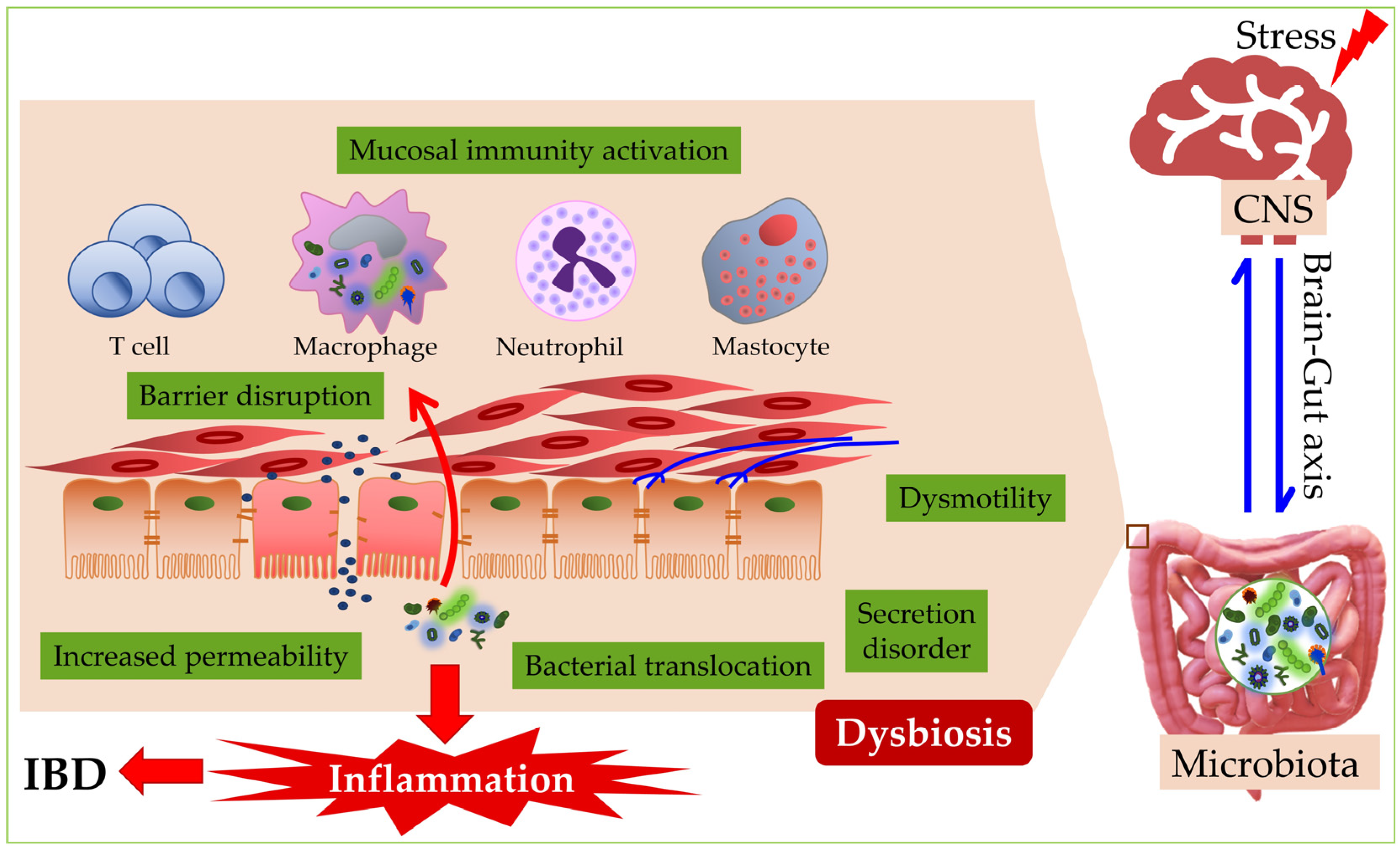
5. The Connection of Dysbiosis to Inflammatory Bowel Disease

The main conditions within the umbrella of IBD, Crohn’s disease (CD) and ulcerative colitis (UC), are chronic conditions of the gastrointestinal system characterized by alternating episodes of relapse and remission, which affect about 3 million people in the USA and Europe [96]. Interactions between environmental factors, dysregulated immune response, host genetics, and changes in intestinal microbiological composition are involved in the disease pathogenesis [97]. CD can affect any part of the digestive system, but most often, it is observed in the last part of the small intestine or the large intestine, while UC disease is exclusively related to the large intestine [98]. In addition, the incidence and prevalence of IBD are closely related to urban lifestyle and northern latitudes, while these conditions are relatively rare in Africa, Asia, and Latin America [99].
IBD can appear throughout the lifetime, from the first year of life to old age, with pronounced peaks of incidence between 15 and 30 and between 60 and 80 years [100]. In the last decade, a significant increase in incidence has been recorded worldwide [101,102]. Although the etiology and pathogenesis of IBD are thus far unclear, it is evident that dysbiosis within the intestinal microbiome represents a crucial factor in the development of IBD and an essential piece in the development of mucosal lesions [103]. The microbiota of patients with IBD significantly differs from those in healthy individuals and is characterized by a smaller abundance and diversity of microorganisms [104]. Both qualitative and quantitative changes are present in the composition and function of the microbiota associated with IBD [105].
The microbial population changes in the digestive system of patients with CD and UC. Bacteroidetes and Firmicutes, as well as Clostridium leptum and Clostridium coccoides groups, were shown to be reduced [106]. In several studies on the same group of patients, a reduced representation of F. prausnitzii (a member of the C. leptum group), characterized by anti-inflammatory properties, may increase the risk of postoperative recurrent ileal disease and lead to an increased abundance of E. coli [107].
The reduced prevalence of F. prausnitzii (Firmicute), which belongs to the butyrate-producing Clostridia cluster IV species, illuminates the reduced amount of short-chain fatty acids in the fecal samples of patients with IBD [108]. Butyrate, in addition to strengthening the mucosal barrier by inducing the production of antimicrobial peptides and mucins [109], represents the primary source of energy for the epithelial cells of the large intestine [110] and also serves as an inhibitor of the expression of pro-inflammatory cytokines in intestinal mucosa through a mechanism involving hyper-acetylation of histones and suppression of NF-κB signaling [111].
Ulcerative colitis and Crohn’s disease belong to the group of chronic inflammatory bowel diseases [112]. In people with active CD, as a sign of dysbiosis, there is a decrease in the number of Firmicutes bacteria belonging to the Ruminococcaceae and Lachnospiraceae families [113]. This is important because these are genera of bacteria that normally belong to the families of human intestine bacteria that produce butyrate [114]. In CD, an increase in the number of sulfate-reducing bacteria has been noted, during which hydrogen sulfate is formed, which damages the intestinal barrier and promotes inflammation. An example of such a bacterium is Desulfovibrio [115]. Additionally, the abundance of Escherichia and Shigella was significantly increased compared to healthy people [116]. The adherent-invasive E. coli (AIEC) group is involved considerably in CD development. The data show that it was isolated from the intestines of 6% of healthy people, and this percentage is significantly higher in CD and amounts to about 38% [117]. In addition, the number of mucolytic bacteria such as Bifidobacterium bifidum, Bacteroides fragilis, Runinococcus gnavas, and Ruminococcus torques is rising, which leads to the breakdown of protective mucus and increased bacterial invasion of the mucous membrane [118]. Mycobacterium avium paratuberculosis (MAP), which usually causes chronic granulomatous ileitis, is associated with CD; namely, a more frequent presence of antibodies and reactive T cells against MAP and a higher level of MAP-DNA in the mucosa of patients with CD compared to the control group [119].
The research results determined that the composition of bacterial species is substantially modified in people suffering from UC; more specifically, bacteria belonging to the phylum Firmicutes are of lower abundance, while the number of bacteria from the phylum Proteobacteria is high [106]. In addition, it is important to note that the number of bacteria that have a protective role in GIS is significantly decreased, including the genera Bacteroides, Eubacterium, and Lactobacillus [106]. The number of bacteria that produce short-chain fatty acids, such as F. prausnitzii and Clostridium butyricum, is decreased in patients presenting with UC, which can affect regulatory T cells, primarily their differentiation and expansion [120]. Individuals experiencing a relapse of UC have a higher proportion of Bacteroidetes, but a much lower proportion of Clostridiales [121]. Moreover, a correlation between butyrate-producing bacteria (such as the aforementioned F. prausnitzii and Roseburia hominis) and UC disease activity has repeatedly shown an inverse trend [122]. Several other studies demonstrated a link between active UC and lower abundance of Akkermansia municiphila and Roseburia spp. [123,124]. Finally, it has to be emphasized that the alterations in intestinal microbiota composition in patients with UC are also related to changes in microbial metabolism; for example, increases in sphingosine-1-phosphate and trimethylamine N-oxide are often observed [122].
In patients with CD, the fecal fungal community is also disturbed, accompanied by an increased diversity and prevalence of the fungi Candida albicans, C. neoformans, and Aspergillus clavatus [125]. In addition, norovirus infection is mentioned as a possible trigger for the development and relapse of CD disease [126]. In patients with UC, some changes have been observed in regard to bacteriophage populations; more specifically, a large number of DNA viruses have been observed in gut mucosa (such as Caudovirales bacteriophages), but there also a decrease in Caudovirales richness and diversity [122,127].
Significant differences were found in a study that studied the microbiota of pediatric patients with CD and compared it with a healthy control group. Specifically, CD patients had an increased abundance of Enterobacteriaceae, Pasteurellaceae, Veillonellaceae, and Fusobacteriaceae and a decreased amount of Erysipelotrichales, Bacteroidales, and Clostridiales compared to healthy controls [128]. Additionally, although dysbiosis is more pronounced in children with CD when compared to children with UC, there are also some characteristic hallmarks in pediatric patients with UC, such as a reduction in abundance of Akkermansia spp. and Eubacterium rectale, and the expansion of E. coli [129]. In addition, an absent response to steroids given to children with acute and severe forms of UC has been linked to a substantial reduction in bacterial diversity in comparison to steroid responders and healthy controls [130].
Hence, as a result of dysbiosis, various harmful products are produced that break down and thin the protective mucus layer and damage the epithelial layer [104]. Due to the impaired integrity of the epithelial barrier, microorganisms can penetrate the lamina propria, which results in an excessive immune response of the host [131]. This leads to a breakdown of immune tolerance to one’s intestinal microbiota while stimulating an inflammatory reaction, leading to tissue damage [132].

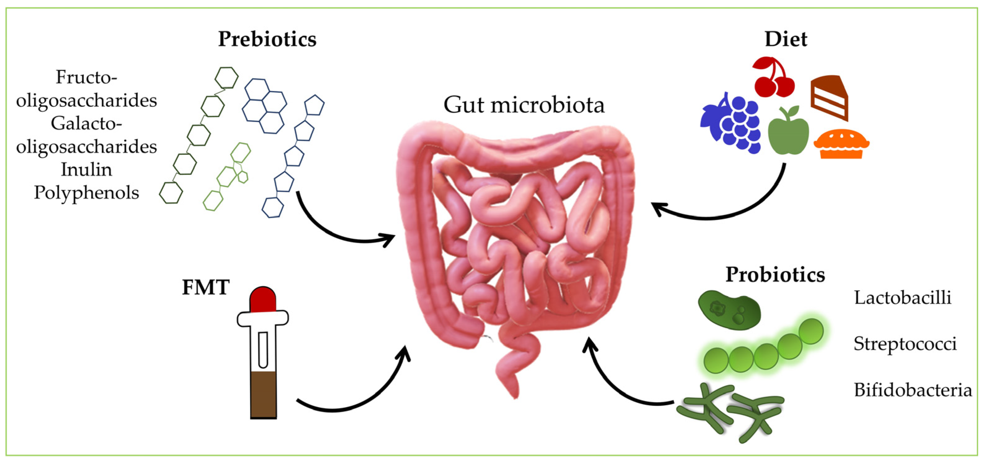
6. Manipulating the Gut Microbiota

The intestinal microbiota is an essential component of who we are, and the mutual dialogue between the microbiota and the host results in lifelong epigenetic programming [133]. The significance of the intestinal microflora in preserving the organism’s normal functioning has become increasingly acknowledged in recent years. As a result, numerous studies have investigated the potential therapeutic effects of microorganisms found in the intestinal microbiome in a myriad of diseases [134]. In most cases, it is unclear whether changes in the microbiota are a cause or consequence of the disease and whether the manipulation of the gut microbiota can help control or even treat the pathological condition [133]. Some lifestyle changes via alternative treatments, including fecal microbiota transplantation (FMT), diet changes, and dietary supplementation with probiotics, prebiotics, and symbiotics, have indicated possible protective effects in microbiota dysbiosis and promotion of healthy microbes, along with minimal long-term unfavorable effects [135] (Figure 2). Nevertheless, existing microbial-targeted treatments, including probiotics, prebiotics, FMT, and specific dietary regimens, yield mixed results that could be more optimal for everyday clinical use [136,137,138].

6.1. Probiotics

Probiotics are defined as live microorganisms that confer health benefits on the host when administered in appropriate amounts [133,139]. Lactobacilli, streptococci, and bifidobacteria are human food’s most common probiotic bacteria. Furthermore, the yeast Saccharomyces boulardii and a strain of E. coli known as the Nissle strain are frequently used [133]. Probiotics stimulate the production of anti-inflammatory cytokines and the secretion of antimicrobial substances, suppress the growth of bacteria, induce an immune response, have an immunomodulating role, and improve the epithelial barrier function [134].
Various studies have shown that distinct probiotic strains or a mixture of strains can be rather helpful in various diseases [133,140]. The use of antibiotics can have long-term consequences on the composition of the intestinal microbiota and, thus, on overall health [133,141]. Conversely, research with probiotics showed fewer distortions of the intestinal microbiota when probiotics were given together with antibiotics [133,142]. Probiotics mainly exert their action in the small intestine, where the concentration of resident microbiota is low, affecting the microbiota’s diversity and richness during their passage [133]. Endogenous bacteria will outnumber probiotic bacteria in the large intestine, but they can still (directly and/or indirectly) impact health [133]. Probiotics support beneficial microorganisms in the small intestine, improve barrier integrity, and reduce nutrient malabsorption and pathology associated with small bowel disease. Treatment with specific strains of probiotics represents a natural and effective approach to restore barrier integrity and eubiosis of the small intestine, resulting in improved health and a reduction in the incidence and severity of small bowel disease [143]. In addition, probiotics (especially lactic acid bacteria) can protect the small intestine by increasing microbial diversity, regulating the expression of proteins involved in homeostasis, and maintaining the integrity of the immune system [144]. Although bile acids and digestive enzymes can affect the viability of probiotics in the small intestine, they are coated with a protective layer, which increases the survival rate of viable probiotics, and more can reach the large intestine. However, as the large intestine has the highest density of bacteria, probiotics encounter resistance from commensal bacteria; as a result, probiotics must compete with the host microbiota for nutrients and adhesion sites to colonize the colonic mucosa and proliferate. Due to their resistance to colonization, most probiotics are excreted from the colon in the stool after oral administration and soon after consumption, so probiotics are undetectable [145]. Probiotics can also act as prebiotics, promoting the growth of specific intestinal microbiota [133]. Metabolic by-products, dead microorganisms, bacterial molecular components, or other nonviable microbial-based products are not, by definition, probiotics, but exhibit probiotic properties [146]. Probiotics can stimulate the growth of part of the intestinal microbiota. For example, some studies have shown that a probiotic strain of Lactobacillus casei increased the concentration of lactobacilli in the stools of young children. Additionally, the Lactococcus lactis strain increases the concentration of bifidobacteria and decreases the concentration of Enterococcus in human-flora-associated rats [147]. Moreover, the levels of commensal bacteria in the ileum of rats correlated with the positive disease outcome of prophylactic probiotic therapy in a rat model of acute pancreatitis [148]. The success of using probiotics in treating IBD can vary, and primarily depends on the strains used and the target subtype of the disease [149]. Probiotics currently available can potentially modulate dysbiosis in IBD patients, but their effects are temporary [136,138,150]. The most ordinary types of probiotics used are Bifidobacterium and Lactobacillus [136,150], which have proven to be effective in UC therapy, and are based on the immunomodulatory and anti-inflammatory effects of probiotics [134]. Visbiome is the most common probiotic cocktail with proven efficacy in UC [135,151]. More specifically, Visbiome contains eight different strains of bacteria, and the most frequently used bacteria are Lactobacilli and Bifidobacteria [135]. Most studies have shown the effectiveness of probiotics in maintaining clinical remission of CD [134]. However, there is insufficient evidence to support probiotics as an adjunctive therapy in CD patients [134,152]. Since the performance of these probiotics is not entirely satisfactory, new candidates for more effective colonizing probiotics—such as combinations of protective resident strains (live biotherapeutic products, LBP)—are emerging [136,153].

6.2. Prebiotics

Prebiotics are nondigestible carbohydrates that are metabolized by resident bacteria and can enhance the composition and metabolic function of beneficial resident bacterial species in the intestine [136,154]. A prebiotic can be defined as a selectively fermented ingredient that stimulates specific changes in the activity or composition of the gut microbiota, providing benefits to the host [133]. Health benefits extend beyond the GIT to the skin, urogenital tract, and lungs [135,155]. Prebiotics are carbohydrate polymers that are not absorbed, as opposed to probiotics (which are living microorganisms) [134]. Prebiotics promote the growth and/or activity of bacteria found in the large intestine and act as growth substrates to selectively increase the number of certain bacteria and/or their activity [133]. Commensal microorganisms ferment them, which leads to a change in the composition of the intestinal microbiome and metabolism [135,155].
Prebiotics include nondigestible carbohydrate sources, such as fructooligosaccharides, inulin, and galactooligosaccharides [134,136,154], but also non-carbohydrate sources such as polyphenols and specific lipids [135,155]. All these compounds promote the growth and metabolic activity of beneficial bacteria found in the digestive tract, such as Lactobacillus and Bifidobacterium species [134]. In addition to the benefits mentioned above, prebiotics stimulate the bacterial production of short-chain fatty acids, such as butyrate, which has immunoregulatory properties. These effects include the suppression of pro-inflammatory cytokines [134,156].
In clinical use, prebiotics may show benefits in treating IBD, but their effects are modest, with conflicting results [136,157]. Thus, F. prausnitzii has been shown to respond to prebiotic supplementation using fructans of mixed chain length [133,158], and decreased numbers of F. prausnitzii have been observed in patients with CD [133].

6.3. Synbiotics

Synbiotics combine selected prebiotics and probiotics to achieve a synergistic effect. Probiotics suppress the development of pathogenic bacteria, while prebiotics stimulate the proliferation of beneficial intestinal microbes and thus synergistically enhance the integrity of the intestinal barrier [135]. The effectiveness of the oral administration of synbiotics is slightly higher than the independent administration of probiotics and prebiotics [134]. Unfortunately, data on the usefulness of synbiotics in patients with IBD are scarce [134,159]. In CD, the use of prebiotics did not lead to a considerable improvement in the patient’s health condition. However, the combined use of prebiotics with Bifidobacterium longum in patients with active CD improved clinical symptoms [134,160].

6.4. Dietary Modifications

It has been shown that there is a connection between a change in usual diet and a diet with specific ingredients that can be risk factors for IBD [161,162], showing that we can manipulate intestinal microbes by intervening in human nutrition [163]. A simple example is the use of specific fiber sources that provide food for the desired intestinal microbes [154,163]. Intestinal microbes adapt to the new food environment, whereby the bacteria in the human intestines contribute to the body’s response to a particular diet [163]. Thus, it was shown that at least two weeks of consuming vegetables rich in inulin fructans in the diet increased the genus Bifidobacterium by 3.8 times [134]. In addition, the intestinal microbiota and intestinal immunity are affected by numerous polyphenols. The large intestine receives a significant amount of unabsorbed polyphenolic compounds, which causes them to interact with the large intestine’s intestinal microbiota, catabolizing, in turn, polyphenols and breaking them down into little fragments [134,164].
Diet has a more significant impact on CD than UC, since the majority of epidemiological research on dietary risk factors has determined an association with CD but not UC [135,165]. Special attention has been paid to the Mediterranean diet in terms of inducing and maintaining CD remission [135]. In addition, diet is an influential lifestyle factor significantly related to the function of the gut microbiota [135]. It also provides valuable insight into developing personalized nutritional strategies to adapt gut microbes in the future [163].

6.5. Fecal Microbiota Transplantation (FMT)

FMT involves collecting feces from a healthy donor and transplanting it into the patient’s gastrointestinal tract [134], replacing the patient’s dysbiotic gut microbiota with microorganisms from healthy donors [149]. FMT’s goal is to restore healthy microbiota [135,166], which is nowadays not only possible, but often a preferred therapeutic alternative in patients with a disturbed intestinal ecosystem. This specific therapeutic method is highly successful in patients with recurrent Clostridioides difficile infection, particularly with strains resistant to standard antibiotic therapy [134,135,136,167].
By triggering many immune-mediated pathways [134,168], FMT can benefit the restoration of intestinal dysbiosis, the production of pro-inflammatory factors [134,169], the reduction of intestinal inflammation, and the promotion of the restoration of intestinal homeostasis [134]. However, numerous inconsistencies in the results of FMT are a consequence of the complex pathogenesis of IBD in relation to CD [135]. Preliminary studies using FMT in patients with UC or CD showed promising results in many cases in achieving and maintaining long-term clinical remission. However, the benefits were more significant in younger patients [134]. Furthermore, the effectiveness of FMT in the IBD patient is still controversial [136,150,167] because FMT can potentially cause grave and life-threatening side effects [136].
Different mixtures of intestinal protective microbial strains or their metabolites may be safer and more adequate than the entire FMT. Therefore, many new LBP formulations are being designed to replace protective bacterial species in IBD patients [136], and novel solutions are being developed in line with precision medicine postulates.

7. Conclusions

The intestinal microbiota comprises various microorganisms, including proteobacteria, bacteria, viruses, fungi, protists, and archaea. The microbiota has a significant role in the GIT, such as participating in the metabolism of various substances and steering the activity of the immune system. Therefore, changes in the composition of the microbiome of the GIT and its function, known as dysbiosis, play a noteworthy role in the development of chronic IDB that entails CD and UC. In order to successfully treat clinical conditions that arise from dysbiosis, it is pivotal to closely investigate the microbiota’s influence and its role in pathogenesis, as well as the currently available therapeutic approaches. In addition, we should be cognizant that adequate probiotic preparations can aid in treating IBD. It is evident that the microbiome is closely linked to a plethora of host phenotypes, so the capability to engineer our commensal microflora will likely be an indispensable element of personalized or precision medicine during this century.

Author Contributions

Conceptualization: J.T. and A.V.; manuscript writing: J.T., T.M., E.P. and I.Š.; updating the text: J.T., M.J. and I.Š.; literature searches: J.T., A.V., T.M., E.P., M.J. and I.Š.; figure drawings: I.Š.; critical reviewing of the manuscript: J.T., A.V., T.M. and I.Š.; organization and editing of the manuscript: J.T. and I.Š. All authors have read and agreed to the published version of the manuscript.

Funding

This research received no external funding.

Institutional Review Board Statement

Not applicable.

Informed Consent Statement

Not applicable.

Data Availability Statement

Not applicable.

Conflicts of Interest

The authors declare no conflict of interest. The funders had no role in the design of the study; in the collection, analyses, or interpretation of data; in the writing of the manuscript; or in the decision to publish the results.

References

  1. Thursby, E.; Juge, N. Introduction to the human gut microbiota. Biochem. J. 2017, 474, 1823–1836. [Google Scholar] [CrossRef]
  2. Sekirov, I.; Russell, S.L.; Antunes, L.C.M.; Finlay, B.B. Gut microbiota in health and disease. Physiol. Rev. 2010, 90, 859–904. [Google Scholar] [CrossRef] [PubMed] [Green Version]
  3. Mizrahi-Man, O.; Davenport, E.R.; Gilad, Y. Taxonomic classification of bacterial 16S rRNA genes using short sequencing reads: Evaluation of effective study designs. PLoS ONE 2013, 8, e53608. [Google Scholar] [CrossRef] [Green Version]
  4. Poretsky, R.; Rodriguez-R, L.M.; Luo, C.; Tsementzi, D.; Konstantinidis, K.T. Strengths and limitations of 16S rRNA gene amplicon sequencing in revealing temporal microbial community dynamics. PLoS ONE 2014, 9, e93827. [Google Scholar] [CrossRef] [PubMed] [Green Version]
  5. Meštrović, T.; Patterson, J. Human Microbiome and Disease. Encycl. Infect. Immun. 2022, 4, 662–673. [Google Scholar]
  6. de Vos, W.M.; Tilg, H.; Van Hul, M.; Cani, P.D. Gut microbiome and health: Mechanistic insights. Gut 2022, 71, 1020–1032. [Google Scholar] [CrossRef] [PubMed]
  7. Dimitrakopoulou, E.I.; Pouliakis, A.; Falaina, V.; Xanthos, T.; Zoumpoulakis, P.; Tsiaka, T.; Sokou, R.; Iliodromiti, Z.; Boutsikou, T.; Iacovidou, N. The Metagenomic and Metabolomic Profile of the Infantile Gut: Can They Be “Predicted” by the Feed Type? Children 2022, 9, 154. [Google Scholar] [CrossRef]
  8. Goodrich, J.K.; Waters, J.L.; Poole, A.C.; Sutter, J.L.; Koren, O.; Blekhman, R.; Beaumont, M.; Van Treuren, W.; Knight, R.; Bell, J.T.; et al. Human genetics shape the gut microbiome. Cell 2014, 159, 789–799. [Google Scholar] [CrossRef] [Green Version]
  9. Lynch, S.V.; Pedersen, O. The Human Intestinal Microbiome in Health and Disease. N. Engl. J. Med. 2016, 375, 2369–2379. [Google Scholar] [CrossRef] [Green Version]
  10. Falony, G.; Joossens, M.; Vieira-Silva, S.; Wang, J.; Darzi, Y.; Faust, K.; Kurilshikov, A.; Bonder, M.J.; Valles-Colomer, M.; Vandeputte, D.; et al. Population-level analysis of gut microbiome variation. Science 2016, 352, 560–564. [Google Scholar] [CrossRef]
  11. Deschasaux, M.; Bouter, K.E.; Prodan, A.; Levin, E.; Groen, A.K.; Herrema, H.; Tremaroli, V.; Bakker, G.J.; Attaye, I.; Pinto-Sietsma, S.J.; et al. Depicting the composition of gut microbiota in a population with varied ethnic origins but shared geography. Nat. Med. 2018, 24, 1526–1531. [Google Scholar] [CrossRef] [PubMed]
  12. Matijašić, M.; Meštrović, T.; Perić, M.; Paljetak, H.; Panek, M.; Bender, D.V.; Kelečić, D.L.; Krznarić; Verbanac, D. Modulating Composition and Metabolic Activity of the Gut Microbiota in IBD Patients. Int. J. Mol. Sci. 2016, 17, 578. [Google Scholar] [CrossRef] [PubMed] [Green Version]
  13. Upadhyay, K.G.; Desai, D.C.; Ashavaid, T.F.; Dherai, A.J. Microbiome and Metabolome in Inflammatory Bowel Disease. J. Gastroenterol. Hepatol. 2022, in press. [Google Scholar] [CrossRef]
  14. Ley, R.E.; Turnbaugh, P.J.; Klein, S.; Gordon, J.I. Microbial ecology: Human gut microbes associated with obesity. Nature 2006, 444, 1022–1023. [Google Scholar] [CrossRef] [PubMed]
  15. Valdes, A.M.; Walter, J.; Segal, E.; Spector, T.D. Role of the gut microbiota in nutrition and health. BMJ 2018, 361, 36–44. [Google Scholar] [CrossRef] [Green Version]
  16. Gill, S.R.; Pop, M.; DeBoy, R.T.; Eckburg, P.B.; Turnbaugh, P.J.; Samuel, B.S.; Gordon, J.I.; Relman, D.A.; Fraser-Liggett, C.M.; Nelson, K.E. Metagenomic analysis of the human distal gut microbiome. Science 2006, 312, 1355–1359. [Google Scholar] [CrossRef] [Green Version]
  17. Rinninella, E.; Raoul, P.; Cintoni, M.; Franceschi, F.; Miggiano, G.A.D.; Gasbarrini, A.; Mele, M.C. What is the Healthy Gut Microbiota Composition? A Changing Ecosystem across Age, Environment, Diet, and Diseases. Microorganisms 2019, 7, 14. [Google Scholar] [CrossRef] [Green Version]
  18. Khosravi, A.; Mazmanian, S.K. Disruption of the gut microbiome as a risk factor for microbial infections. Curr. Opin. Microbiol. 2013, 16, 221–227. [Google Scholar] [CrossRef] [Green Version]
  19. Martínez-García, J.J.; Rainteau, D.; Humbert, L.; Lamaziere, A.; Lesnik, P.; Chamaillard, M. Diurnal Interplay between Epithelium Physiology and Gut Microbiota as a Metronome for Orchestrating Immune and Metabolic Homeostasis. Metabolites 2022, 12, 390. [Google Scholar] [CrossRef]
  20. Talapko, J.; Meštrović, T.; Juzbašić, M.; Tomas, M.; Erić, S.; Horvat Aleksijević, L.; Bekić, S.; Schwarz, D.; Matić, S.; Neuberg, M.; et al. Antimicrobial Peptides-Mechanisms of Action, Antimicrobial Effects and Clinical Applications. Antibiotics 2022, 11, 1417. [Google Scholar] [CrossRef]
  21. Shetty, S.A.; Hugenholtz, F.; Lahti, L.; Smidt, H.; de Vos, W.M. Intestinal microbiome landscaping: Insight in community assemblage and implications for microbial modulation strategies. FEMS Microbiol. Rev. 2017, 41, 182–199. [Google Scholar] [CrossRef] [PubMed]
  22. Brestoff, J.R.; Artis, D. Commensal bacteria at the interface of host metabolism and the immune system. Nat. Immunol. 2013, 14, 676–684. [Google Scholar] [CrossRef] [PubMed] [Green Version]
  23. Kaper, J.B.; Sperandio, V. Bacterial cell-to-cell signaling in the gastrointestinal tract. Infect. Immun. 2005, 73, 3197–3209. [Google Scholar] [CrossRef] [PubMed] [Green Version]
  24. Arumugam, M.; Raes, J.; Pelletier, E.; Le Paslier, D.; Yamada, T.; Mende, D.R.; Fernandes, G.R.; Tap, J.; Bruls, T.; Batto, J.M.; et al. Enterotypes of the human gut microbiome. Nature 2011, 473, 174–180. [Google Scholar] [CrossRef] [Green Version]
  25. Bresser, L.R.F.; de Goffau, M.C.; Levin, E.; Nieuwdorp, M. Gut Microbiota in Nutrition and Health with a Special Focus on Specific Bacterial Clusters. Cells 2022, 11, 3091. [Google Scholar] [CrossRef] [PubMed]
  26. Cui, L.; Morris, A.; Ghedin, E. The human mycobiome in health and disease. Genome Med. 2013, 5, 63. [Google Scholar] [CrossRef] [Green Version]
  27. Matijašić, M.; Meštrović, T.; Paljetak, H.Č.; Perić, M.; Barešić, A.; Verbanac, D. Gut Microbiota beyond Bacteria-Mycobiome, Virome, Archaeome, and Eukaryotic Parasites in IBD. Int. J. Mol. Sci. 2020, 21, 2668. [Google Scholar] [CrossRef] [Green Version]
  28. Cao, Z.; Sugimura, N.; Burgermeister, E.; Ebert, M.P.; Zuo, T.; Lan, P. The gut virome: A new microbiome component in health and disease. EBioMedicine 2022, 81, 104113. [Google Scholar] [CrossRef]
  29. Ruan, W.; Engevik, M.A.; Spinler, J.K.; Versalovic, J. Healthy Human Gastrointestinal Microbiome: Composition and Function After a Decade of Exploration. Dig. Dis. Sci. 2020, 65, 695–705. [Google Scholar] [CrossRef] [Green Version]
  30. McBurney, M.I.; Davis, C.; Fraser, C.M.; Schneeman, B.O.; Huttenhower, C.; Verbeke, K.; Walter, J.; Latulippe, M.E. Establishing What Constitutes a Healthy Human Gut Microbiome: State of the Science, Regulatory Considerations, and Future Directions. J. Nutr. 2019, 149, 1882–1895. [Google Scholar] [CrossRef] [Green Version]
  31. Flint, H.J.; Scott, K.P.; Louis, P.; Duncan, S.H. The role of the gut microbiota in nutrition and health. Nat. Rev. Gastroenterol. Hepatol. 2012, 9, 577–589. [Google Scholar] [CrossRef] [PubMed]
  32. Gupta, V.K.; Paul, S.; Dutta, C. Geography, Ethnicity or Subsistence-Specific Variations in Human Microbiome Composition and Diversity. Front. Microbiol. 2017, 8, 1162. [Google Scholar] [CrossRef] [PubMed] [Green Version]
  33. He, Y.; Wu, W.; Zheng, H.M.; Li, P.; McDonald, D.; Sheng, H.F.; Chen, M.X.; Chen, Z.H.; Ji, G.Y.; Zheng, Z.D.X.; et al. Regional variation limits applications of healthy gut microbiome reference ranges and disease models. Nat. Med. 2018, 24, 1532–1535. [Google Scholar] [CrossRef] [PubMed]
  34. Syromyatnikov, M.; Nesterova, E.; Gladkikh, M.; Smirnova, Y.; Gryaznova, M.; Popov, V. Characteristics of the Gut Bacterial Composition in People of Different Nationalities and Religions. Microorganisms 2022, 10, 1866. [Google Scholar] [CrossRef] [PubMed]
  35. Koliada, A.; Moseiko, V.; Romanenko, M.; Piven, L.; Lushchak, O.; Kryzhanovska, N.; Guryanov, V.; Vaiserman, A. Seasonal variation in gut microbiota composition: Cross-sectional evidence from Ukrainian population. BMC Microbiol. 2020, 20, 100. [Google Scholar] [CrossRef] [Green Version]
  36. Dorelli, B.; Gallè, F.; De Vito, C.; Duranti, G.; Iachini, M.; Zaccarin, M.; Preziosi Standoli, J.; Ceci, R.; Romano, F.; Liguori, G.; et al. Can Physical Activity Influence Human Gut Microbiota Composition Independently of Diet? A Systematic Review. Nutrients 2021, 13, 1890. [Google Scholar] [CrossRef]
  37. Scheiman, J.; Luber, J.M.; Chavkin, T.A.; MacDonald, T.; Tung, A.; Pham, L.D.; Wibowo, M.C.; Wurth, R.C.; Punthambaker, S.; Tierney, B.T.; et al. Meta-omics analysis of elite athletes identifies a performance-enhancing microbe that functions via lactate metabolism. Nat. Med. 2019, 25, 1104–1109. [Google Scholar] [CrossRef]
  38. Lloyd-Price, J.; Abu-Ali, G.; Huttenhower, C. The healthy human microbiome. Genome Med. 2016, 8, 51. [Google Scholar] [CrossRef] [Green Version]
  39. Jandhyala, S.M.; Talukdar, R.; Subramanyam, C.; Vuyyuru, H.; Sasikala, M.; Reddy, D.N. Role of the normal gut microbiota. World J. Gastroenterol. 2015, 21, 8836–8847. [Google Scholar] [CrossRef]
  40. Rothschild, D.; Weissbrod, O.; Barkan, E.; Kurilshikov, A.; Korem, T.; Zeevi, D.; Costea, P.I.; Godneva, A.; Kalka, I.N.; Bar, N.; et al. Environment dominates over host genetics in shaping human gut microbiota. Nature 2018, 555, 210–215. [Google Scholar] [CrossRef]
  41. Das, B.; Nair, G.B. Homeostasis and dysbiosis of the gut microbiome in health and disease. J. Biosci. 2019, 44, 117. [Google Scholar] [CrossRef] [PubMed]
  42. Faith, J.J.; Guruge, J.L.; Charbonneau, M.; Subramanian, S.; Seedorf, H.; Goodman, A.L.; Clemente, J.C.; Knight, R.; Heath, A.C.; Leibel, R.L.; et al. The long-term stability of the human gut microbiota. Science 2013, 341, 1237439. [Google Scholar] [CrossRef] [PubMed] [Green Version]
  43. Hasan, N.; Yang, H. Factors affecting the composition of the gut microbiota, and its modulation. PeerJ 2019, 7, e7502. [Google Scholar] [CrossRef] [PubMed] [Green Version]
  44. David, L.A.; Maurice, C.F.; Carmody, R.N.; Gootenberg, D.B.; Button, J.E.; Wolfe, B.E.; Ling, A.V.; Devlin, A.S.; Varma, Y.; Fischbach, M.A.; et al. Diet rapidly and reproducibly alters the human gut microbiome. Nature 2014, 505, 559–563. [Google Scholar] [CrossRef] [PubMed] [Green Version]
  45. De Filippo, C.; Cavalieri, D.; Di Paola, M.; Ramazzotti, M.; Poullet, J.B.; Massart, S.; Collini, S.; Pieraccini, G.; Lionetti, P. Impact of diet in shaping gut microbiota revealed by a comparative study in children from Europe and rural Africa. Proc. Natl. Acad. Sci. USA 2010, 107, 14691–14696. [Google Scholar] [CrossRef] [Green Version]
  46. Wu, G.D.; Chen, J.; Hoffmann, C.; Bittinger, K.; Chen, Y.Y.; Keilbaugh, S.A.; Bewtra, M.; Knights, D.; Walters, W.A.; Knight, R.; et al. Linking long-term dietary patterns with gut microbial enterotypes. Science 2011, 334, 105–108. [Google Scholar] [CrossRef] [Green Version]
  47. Das, B.; Ghosh, T.S.; Kedia, S.; Rampal, R.; Saxena, S.; Bag, S.; Mitra, R.; Dayal, M.; Mehta, O.; Surendranath, A.; et al. Analysis of the Gut Microbiome of Rural and Urban Healthy Indians Living in Sea Level and High Altitude Areas. Sci. Rep. 2018, 8, 10104. [Google Scholar] [CrossRef] [Green Version]
  48. Vangay, P.; Ward, T.; Gerber, J.S.; Knights, D. Antibiotics, pediatric dysbiosis, and disease. Cell Host Microbe 2015, 17, 553–564. [Google Scholar] [CrossRef] [Green Version]
  49. Ferrer, M.; Méndez-García, C.; Rojo, D.; Barbas, C.; Moya, A. Antibiotic use and microbiome function. Biochem. Pharmacol. 2017, 134, 114–126. [Google Scholar] [CrossRef]
  50. McDonnell, L.; Gilkes, A.; Ashworth, M.; Rowland, V.; Harries, T.H.; Armstrong, D.; White, P. Association between antibiotics and gut microbiome dysbiosis in children: Systematic review and meta-analysis. Gut Microbes 2021, 13, 1870402. [Google Scholar] [CrossRef]
  51. Allen, J.M.; Mailing, L.J.; Niemiro, G.M.; Moore, R.; Cook, M.D.; White, B.A.; Holscher, H.D.; Woods, J.A. Exercise Alters Gut Microbiota Composition and Function in Lean and Obese Humans. Med. Sci. Sports Exerc. 2018, 50, 747–757. [Google Scholar] [CrossRef] [PubMed]
  52. Walter, J.; Ley, R. The human gut microbiome: Ecology and recent evolutionary changes. Annu. Rev. Microbiol. 2011, 65, 411–429. [Google Scholar] [CrossRef] [PubMed] [Green Version]
  53. Bonder, M.J.; Kurilshikov, A.; Tigchelaar, E.F.; Mujagic, Z.; Imhann, F.; Vila, A.V.; Deelen, P.; Vatanen, T.; Schirmer, M.; Smeekens, S.P.; et al. The effect of host genetics on the gut microbiome. Nat. Genet. 2016, 48, 1407–1412. [Google Scholar] [CrossRef] [PubMed]
  54. Lopera-Maya, E.A.; Kurilshikov, A.; van der Graaf, A.; Hu, S.; Andreu-Sánchez, S.; Chen, L.; Vila, A.V.; Gacesa, R.; Sinha, T.; Collij, V.; et al. Effect of host genetics on the gut microbiome in 7738 participants of the Dutch Microbiome Project. Nat. Genet. 2022, 54, 143–151. [Google Scholar] [CrossRef]
  55. Hacquard, S.; Garrido-Oter, R.; González, A.; Spaepen, S.; Ackermann, G.; Lebeis, S.; McHardy, A.C.; Dangl, J.L.; Knight, R.; Ley, R.; et al. Microbiota and Host Nutrition across Plant and Animal Kingdoms. Cell Host Microbe 2015, 17, 603–616. [Google Scholar] [CrossRef] [Green Version]
  56. Zeng, L.Q.; Wei, S.B.; Sun, Y.M.; Qin, W.Y.; Cheng, J.; Mitchelson, K.; Xie, L. Systematic profiling of mRNA and miRNA expression in the pancreatic islets of spontaneously diabetic Goto-Kakizaki rats. Mol. Med. Rep. 2015, 11, 67–74. [Google Scholar] [CrossRef] [Green Version]
  57. Lynch, J.B.; Hsiao, E.Y. Microbiomes as sources of emergent host phenotypes. Science 2019, 365, 1405–1409. [Google Scholar] [CrossRef]
  58. Kumar, T.; Pandey, R.; Chauhan, N.S. Hypoxia Inducible Factor-1α: The Curator of Gut Homeostasis. Front. Cell. Infect. Microbiol. 2020, 10, 227. [Google Scholar] [CrossRef]
  59. Mowat, A.M.I. To respond or not to respond—A personal perspective of intestinal tolerance. Nat. Rev. Immunol. 2018, 18, 405–415. [Google Scholar] [CrossRef] [Green Version]
  60. Belkaid, Y.; Naik, S. Compartmentalized and systemic control of tissue immunity by commensals. Nat. Immunol. 2013, 14, 646–653. [Google Scholar] [CrossRef] [Green Version]
  61. Kawamoto, S.; Maruya, M.; Kato, L.M.; Suda, W.; Atarashi, K.; Doi, Y.; Tsutsui, Y.; Qin, H.; Honda, K.; Okada, T.; et al. Foxp3(+) T cells regulate immunoglobulin a selection and facilitate diversification of bacterial species responsible for immune homeostasis. Immunity 2014, 41, 152–165. [Google Scholar] [CrossRef] [PubMed]
  62. Ichinohe, T.; Pang, I.K.; Kumamoto, Y.; Peaper, D.R.; Ho, J.H.; Murray, T.S.; Iwasaki, A. Microbiota regulates immune defense against respiratory tract influenza A virus infection. Proc. Natl. Acad. Sci. USA 2011, 108, 5354–5359. [Google Scholar] [CrossRef] [PubMed] [Green Version]
  63. Corbitt, N.; Kimura, S.; Isse, K.; Specht, S.; Chedwick, L.; Rosborough, B.R.; Lunz, J.G.; Murase, N.; Yokota, S.; Demetris, A.J. Gut bacteria drive Kupffer cell expansion via MAMP-mediated ICAM-1 induction on sinusoidal endothelium and influence preservation-reperfusion injury after orthotopic liver transplantation. Am. J. Pathol. 2013, 182, 180–191. [Google Scholar] [CrossRef] [PubMed] [Green Version]
  64. Sharon, G.; Sampson, T.R.; Geschwind, D.H.; Mazmanian, S.K. The Central Nervous System and the Gut Microbiome. Cell 2016, 167, 915–932. [Google Scholar] [CrossRef] [PubMed] [Green Version]
  65. Colpitts, S.L.; Kasper, L.H. Influence of the Gut Microbiome on Autoimmunity in the Central Nervous System. J. Immunol. 2017, 198, 596–604. [Google Scholar] [CrossRef] [PubMed] [Green Version]
  66. Macfarlane, S.; Macfarlane, G.T. Regulation of short-chain fatty acid production. Proc. Nutr. Soc. 2003, 62, 67–72. [Google Scholar] [CrossRef]
  67. Devillard, E.; McIntosh, F.M.; Duncan, S.H.; Wallace, R.J. Metabolism of linoleic acid by human gut bacteria: Different routes for biosynthesis of conjugated linoleic acid. J. Bacteriol. 2007, 189, 2566–2570. [Google Scholar] [CrossRef] [Green Version]
  68. Clayton, T.A.; Baker, D.; Lindon, J.C.; Everett, J.R.; Nicholson, J.K. Pharmacometabonomic identification of a significant host-microbiome metabolic interaction affecting human drug metabolism. Proc. Natl. Acad. Sci. USA 2009, 106, 14728–14733. [Google Scholar] [CrossRef] [Green Version]
  69. Bäckhed, F.; Fraser, C.M.; Ringel, Y.; Sanders, M.E.; Sartor, R.B.; Sherman, P.M.; Versalovic, J.; Young, V.; Finlay, B.B. Defining a healthy human gut microbiome: Current concepts, future directions, and clinical applications. Cell Host Microbe 2012, 12, 611–622. [Google Scholar] [CrossRef] [Green Version]
  70. Bodelier, P.L.E. Toward understanding, managing, and protecting microbial ecosystems. Front. Microbiol. 2011, 2, 80. [Google Scholar] [CrossRef] [Green Version]
  71. Lozupone, C.A.; Stombaugh, J.I.; Gordon, J.I.; Jansson, J.K.; Knight, R. Diversity, stability and resilience of the human gut microbiota. Nature 2012, 489, 220–230. [Google Scholar] [CrossRef]
  72. Li, Z.; Quan, G.; Jiang, X.; Yang, Y.; Ding, X.; Zhang, D.; Wang, X.; Hardwidge, P.R.; Ren, W.; Zhu, G. Effects of Metabolites Derived From Gut Microbiota and Hosts on Pathogens. Front. Cell. Infect. Microbiol. 2018, 8, 314. [Google Scholar] [CrossRef] [PubMed] [Green Version]
  73. Ali, A.; Tan, H.Y.; Kaiko, G.E. Role of the Intestinal Epithelium and Its Interaction With the Microbiota in Food Allergy. Front. Immunol. 2020, 11, 604054. [Google Scholar] [CrossRef] [PubMed]
  74. Hollander, D.; Kaunitz, J.D. The “Leaky Gut”: Tight Junctions but Loose Associations? Dig. Dis. Sci. 2020, 65, 1277–1287. [Google Scholar] [CrossRef] [Green Version]
  75. Di Tommaso, N.; Gasbarrini, A.; Ponziani, F.R. Intestinal Barrier in Human Health and Disease. Int. J. Environ. Res. Public Health 2021, 18, 12836. [Google Scholar] [CrossRef]
  76. Di Domenico, M.; Ballini, A.; Boccellino, M.; Scacco, S.; Lovero, R.; Charitos, I.A.; Santacroce, L. The Intestinal Microbiota May Be a Potential Theranostic Tool for Personalized Medicine. J. Pers. Med. 2022, 12, 523. [Google Scholar] [CrossRef]
  77. Zheng, D.; Liwinski, T.; Elinav, E. Interaction between microbiota and immunity in health and disease. Cell Res. 2020, 30, 492–506. [Google Scholar] [CrossRef] [PubMed]
  78. Bruce, A.; Black, M.; Bhattacharya, S. Mode of delivery and risk of inflammatory bowel disease in the offspring: Systematic review and meta-analysis of observational studies. Inflamm. Bowel Dis. 2014, 20, 1217–1226. [Google Scholar] [CrossRef] [PubMed]
  79. Martinez, J.E.; Kahana, D.D.; Ghuman, S.; Wilson, H.P.; Wilson, J.; Kim, S.C.J.; Lagishetty, V.; Jacobs, J.P.; Sinha-Hikim, A.P.; Friedman, T.C. Unhealthy Lifestyle and Gut Dysbiosis: A Better Understanding of the Effects of Poor Diet and Nicotine on the Intestinal Microbiome. Front. Endocrinol. 2021, 12, 667066. [Google Scholar] [CrossRef]
  80. Ramirez, J.; Guarner, F.; Bustos Fernandez, L.; Maruy, A.; Sdepanian, V.L.; Cohen, H. Antibiotics as Major Disruptors of Gut Microbiota. Front. Cell. Infect. Microbiol. 2020, 10, 572912. [Google Scholar] [CrossRef]
  81. Parkin, K.; Christophersen, C.T.; Verhasselt, V.; Cooper, M.N.; Martino, D. Risk Factors for Gut Dysbiosis in Early Life. Microorganisms 2021, 9, 2066. [Google Scholar] [CrossRef]
  82. Zhao, X.; Jiang, L.; Fang, X.; Guo, Z.; Wang, X.; Shi, B.; Meng, Q. Host-microbiota interaction-mediated resistance to inflammatory bowel disease in pigs. Microbiome 2022, 10, 115. [Google Scholar] [CrossRef] [PubMed]
  83. Singh, R.; Zogg, H.; Wei, L.; Bartlett, A.; Ghoshal, U.C.; Rajender, S.; Ro, S. Gut Microbial Dysbiosis in the Pathogenesis of Gastrointestinal Dysmotility and Metabolic Disorders. J. Neurogastroenterol. Motil. 2021, 27, 19–34. [Google Scholar] [CrossRef]
  84. Hou, K.; Wu, Z.X.; Chen, X.Y.; Wang, J.Q.; Zhang, D.; Xiao, C.; Zhu, D.; Koya, J.B.; Wei, L.; Li, J.; et al. Microbiota in health and diseases. Signal Transduct. Target. Ther. 2022, 7, 135. [Google Scholar] [CrossRef] [PubMed]
  85. Zuo, W.; Wang, B.; Bai, X.; Luan, Y.; Fan, Y.; Michail, S.; Sun, F. 16S rRNA and metagenomic shotgun sequencing data revealed consistent patterns of gut microbiome signature in pediatric ulcerative colitis. Sci. Rep. 2022, 12, 6421. [Google Scholar] [CrossRef] [PubMed]
  86. Buret, A.G.; Motta, J.P.; Allain, T.; Ferraz, J.; Wallace, J.L. Pathobiont release from dysbiotic gut microbiota biofilms in intestinal inflammatory diseases: A role for iron? J. Biomed. Sci. 2019, 26, 1. [Google Scholar] [CrossRef]
  87. Elias-Oliveira, J.; Leite, J.A.; Pereira, Í.S.; Guimarães, J.B.; Manso, G.M.D.C.; Silva, J.S.; Tostes, R.C.; Carlos, D. NLR and Intestinal Dysbiosis-Associated Inflammatory Illness: Drivers or Dampers? Front. Immunol. 2020, 11, 1810. [Google Scholar] [CrossRef] [PubMed]
  88. Stojanov, S.; Berlec, A.; Štrukelj, B. The Influence of Probiotics on the Firmicutes/Bacteroidetes Ratio in the Treatment of Obesity and Inflammatory Bowel disease. Microorganisms 2020, 8, 1715. [Google Scholar] [CrossRef]
  89. Santana, P.T.; Rosas, S.L.B.; Ribeiro, B.E.; Marinho, Y.; de Souza, H.S.P. Dysbiosis in Inflammatory Bowel Disease: Pathogenic Role and Potential Therapeutic Targets. Int. J. Mol. Sci. 2022, 23, 3464. [Google Scholar] [CrossRef]
  90. Golińska, E.; Tomusiak, A.; Gosiewski, T.; Wiȩcek, G.; Machul, A.; Mikołajczyk, D.; Bulanda, M.; Heczko, P.B.; Strus, M. Virulence factors of Enterococcus strains isolated from patients with inflammatory bowel disease. World J. Gastroenterol. 2013, 19, 3562–3572. [Google Scholar] [CrossRef]
  91. Yu, L.C.H. Microbiota dysbiosis and barrier dysfunction in inflammatory bowel disease and colorectal cancers: Exploring a common ground hypothesis. J. Biomed. Sci. 2018, 25, 79. [Google Scholar] [CrossRef]
  92. Özdirik, B.; Müller, T.; Wree, A.; Tacke, F.; Sigal, M. The Role of Microbiota in Primary Sclerosing Cholangitis and Related Biliary Malignancies. Int. J. Mol. Sci. 2021, 22, 6975. [Google Scholar] [CrossRef] [PubMed]
  93. Zhou, Y.; Chen, H.; He, H.; Du, Y.; Hu, J.; Li, Y.; Li, Y.; Zhou, Y.; Wang, H.; Chen, Y.; et al. Increased Enterococcus faecalis infection is associated with clinically active Crohn disease. Medicine 2016, 95, e5019. [Google Scholar] [CrossRef]
  94. Brennan, C.A.; Clay, S.L.; Lavoie, S.L.; Bae, S.; Lang, J.K.; Fonseca-Pereira, D.; Rosinski, K.G.; Ou, N.; Glickman, J.N.; Garrett, W.S. Fusobacterium nucleatum drives a pro-inflammatory intestinal microenvironment through metabolite receptor-dependent modulation of IL-17 expression. Gut Microbes 2021, 13, 1987780. [Google Scholar] [CrossRef] [PubMed]
  95. Sultan, S.; El-Mowafy, M.; Elgaml, A.; Ahmed, T.A.E.; Hassan, H.; Mottawea, W. Metabolic Influences of Gut Microbiota Dysbiosis on Inflammatory Bowel Disease. Front. Physiol. 2021, 12, 715506. [Google Scholar] [CrossRef] [PubMed]
  96. Seyedian, S.S.; Nokhostin, F.; Malamir, M.D. A review of the diagnosis, prevention, and treatment methods of inflammatory bowel disease. J. Med. Life 2019, 12, 113–122. [Google Scholar] [CrossRef] [PubMed]
  97. Lee, M.; Chang, E.B. Inflammatory Bowel Diseases (IBD) and the Microbiome-Searching the Crime Scene for Clues. Gastroenterology 2021, 160, 524–537. [Google Scholar] [CrossRef] [PubMed]
  98. Cicero, G.; Ascenti, G.; Bottari, A.; Catanzariti, F.; Blandino, A.; Mazziotti, S. MR enterography: What is next after Crohn’s disease? Jpn. J. Radiol. 2019, 37, 511–517. [Google Scholar] [CrossRef]
  99. Park, J.; Cheon, J.H. Incidence and Prevalence of Inflammatory Bowel Disease across Asia. Yonsei Med. J. 2021, 62, 99–108. [Google Scholar] [CrossRef]
  100. Mak, W.Y.; Zhao, M.; Ng, S.C.; Burisch, J. The epidemiology of inflammatory bowel disease: East meets west. J. Gastroenterol. Hepatol. 2020, 35, 380–389. [Google Scholar] [CrossRef]
  101. Park, S.H. Update on the epidemiology of inflammatory bowel disease in Asia: Where are we now? Intest. Res. 2022, 20, 159–164. [Google Scholar] [CrossRef] [PubMed]
  102. Krzesiek, E.; Kofla-dlubacz, A.; Akutko, K.; Stawarski, A. The Incidence of Inflammatory Bowel Disease in the Paediatric Population in the District of Lower Silesia, Poland. J. Clin. Med. 2021, 10, 3994. [Google Scholar] [CrossRef]
  103. Alshehri, D.; Saadah, O.; Mosli, M.; Edris, S.; Alhindi, R.; Bahieldin, A. Dysbiosis of gut microbiota in inflammatory bowel disease: Current therapies and potential for microbiota-modulating therapeutic approaches. Bosn. J. Basic Med. Sci. 2021, 21, 270–283. [Google Scholar] [CrossRef] [PubMed]
  104. Khan, I.; Ullah, N.; Zha, L.; Bai, Y.; Khan, A.; Zhao, T.; Che, T.; Zhang, C. Alteration of Gut Microbiota in Inflammatory Bowel Disease (IBD): Cause or Consequence? IBD Treatment Targeting the Gut Microbiome. Pathogens 2019, 8, 126. [Google Scholar] [CrossRef] [PubMed] [Green Version]
  105. Akutko, K.; Stawarski, A. Probiotics, Prebiotics and Synbiotics in Inflammatory Bowel Diseases. J. Clin. Med. 2021, 10, 2466. [Google Scholar] [CrossRef]
  106. Zhang, S.L.; Wang, S.N.; Miao, C.Y. Influence of Microbiota on Intestinal Immune System in Ulcerative Colitis and Its Intervention. Front. Immunol. 2017, 8, 1674. [Google Scholar] [CrossRef] [Green Version]
  107. Grenda, T.; Grenda, A.; Domaradzki, P.; Krawczyk, P.; Kwiatek, K. Probiotic Potential of Clostridium spp.-Advantages and Doubts. Curr. Issues Mol. Biol. 2022, 44, 3118–3130. [Google Scholar] [CrossRef]
  108. Gasaly, N.; Hermoso, M.A.; Gotteland, M. Butyrate and the Fine-Tuning of Colonic Homeostasis: Implication for Inflammatory Bowel Diseases. Int. J. Mol. Sci. 2021, 22, 3061. [Google Scholar] [CrossRef]
  109. Beisner, J.; Filipe Rosa, L.; Kaden-Volynets, V.; Stolzer, I.; Günther, C.; Bischoff, S.C. Prebiotic Inulin and Sodium Butyrate Attenuate Obesity-Induced Intestinal Barrier Dysfunction by Induction of Antimicrobial Peptides. Front. Immunol. 2021, 12, 678360. [Google Scholar] [CrossRef]
  110. Salvi, P.S.; Cowles, R.A. Butyrate and the Intestinal Epithelium: Modulation of Proliferation and Inflammation in Homeostasis and Disease. Cells 2021, 10, 1775. [Google Scholar] [CrossRef]
  111. Venegas, D.P.; De La Fuente, M.K.; Landskron, G.; González, M.J.; Quera, R.; Dijkstra, G.; Harmsen, H.J.M.; Faber, K.N.; Hermoso, M.A. Short Chain Fatty Acids (SCFAs)-Mediated Gut Epithelial and Immune Regulation and Its Relevance for Inflammatory Bowel Diseases. Front. Immunol. 2019, 10, 277. [Google Scholar] [CrossRef] [PubMed]
  112. Kikut, J.; Konecka, N.; Ziętek, M.; Kulpa, D.; Szczuko, M. Diet supporting therapy for inflammatory bowel diseases. Eur. J. Nutr. 2021, 60, 2275–2291. [Google Scholar] [CrossRef] [PubMed]
  113. Schierová, D.; Březina, J.; Mrázek, J.; Fliegerová, K.O.; Kvasnová, S.; Bajer, L.; Drastich, P. Gut Microbiome Changes in Patients with Active Left-Sided Ulcerative Colitis after Fecal Microbiome Transplantation and Topical 5-aminosalicylic Acid Therapy. Cells 2020, 9, 2283. [Google Scholar] [CrossRef] [PubMed]
  114. Linares, R.; Francés, R.; Gutiérrez, A.; Juanola, O. Bacterial Translocation as Inflammatory Driver in Crohn’s Disease. Front. cell Dev. Biol. 2021, 9, 703310. [Google Scholar] [CrossRef] [PubMed]
  115. Dordević, D.; Jančíková, S.; Vítězová, M.; Kushkevych, I. Hydrogen sulfide toxicity in the gut environment: Meta-analysis of sulfate-reducing and lactic acid bacteria in inflammatory processes. J. Adv. Res. 2020, 27, 55–69. [Google Scholar] [CrossRef]
  116. Hu, J.; Cheng, S.; Yao, J.; Lin, X.; Li, Y.; Wang, W.; Weng, J.; Zou, Y.; Zhu, L.; Zhi, M. Correlation between altered gut microbiota and elevated inflammation markers in patients with Crohn’s disease. Front. Immunol. 2022, 13, 947313. [Google Scholar] [CrossRef]
  117. López-Siles, M.; Camprubí-Font, C.; Gómez del Pulgar, E.M.; Sabat Mir, M.; Busquets, D.; Sanz, Y.; Martinez-Medina, M. Prevalence, Abundance, and Virulence of Adherent-Invasive Escherichia coli in Ulcerative Colitis, Colorectal Cancer, and Coeliac Disease. Front. Immunol. 2022, 13, 748839. [Google Scholar] [CrossRef]
  118. Paone, P.; Cani, P.D. Mucus barrier, mucins and gut microbiota: The expected slimy partners? Gut 2020, 69, 2232–2243. [Google Scholar] [CrossRef]
  119. Rosenfeld, G.; Bressler, B. Mycobacterium avium paratuberculosis and the etiology of Crohn’s disease: A review of the controversy from the clinician’s perspective. Can. J. Gastroenterol. 2010, 24, 619–624. [Google Scholar] [CrossRef] [Green Version]
  120. Atarashi, K.; Tanoue, T.; Oshima, K.; Suda, W.; Nagano, Y.; Nishikawa, H.; Fukuda, S.; Saito, T.; Narushima, S.; Hase, K.; et al. Treg induction by a rationally selected mixture of Clostridia strains from the human microbiota. Nature 2013, 500, 232–236. [Google Scholar] [CrossRef]
  121. Pittayanon, R.; Lau, J.T.; Leontiadis, G.I.; Tse, F.; Yuan, Y.; Surette, M.; Moayyedi, P. Differences in Gut Microbiota in Patients With vs. Without Inflammatory Bowel Diseases: A Systematic Review. Gastroenterology 2020, 158, 930–946.e1. [Google Scholar] [CrossRef] [PubMed]
  122. Štofilová, J.; Kvaková, M.; Kamlárová, A.; Hijová, E.; Bertková, I.; Guľašová, Z. Probiotic-Based Intervention in the Treatment of Ulcerative Colitis: Conventional and New Approaches. Biomedicines 2022, 10, 2236. [Google Scholar] [CrossRef] [PubMed]
  123. Machiels, K.; Joossens, M.; Sabino, J.; De Preter, V.; Arijs, I.; Eeckhaut, V.; Ballet, V.; Claes, K.; Van Immerseel, F.; Verbeke, K.; et al. A decrease of the butyrate-producing species Roseburia hominis and Faecalibacterium prausnitzii defines dysbiosis in patients with ulcerative colitis. Gut 2014, 63, 1275–1283. [Google Scholar] [CrossRef]
  124. Lopez-Siles, M.; Enrich-Capó, N.; Aldeguer, X.; Sabat-Mir, M.; Duncan, S.H.; Garcia-Gil, L.J.; Martinez-Medina, M. Alterations in the Abundance and Co-occurrence of Akkermansia muciniphila and Faecalibacterium prausnitzii in the Colonic Mucosa of Inflammatory Bowel Disease Subjects. Front. Cell. Infect. Microbiol. 2018, 8, 281. [Google Scholar] [CrossRef] [PubMed]
  125. Li, Q.; Wang, C.; Tang, C.; He, Q.; Li, N.; Li, J. Dysbiosis of Gut Fungal Microbiota is Associated With Mucosal Inflammation in Crohn’s Disease. J. Clin. Gastroenterol. 2014, 48, 513. [Google Scholar] [CrossRef] [Green Version]
  126. Tarris, G.; de Rougemont, A.; Charkaoui, M.; Michiels, C.; Martin, L.; Belliot, G. Enteric Viruses and Inflammatory Bowel Disease. Viruses 2021, 13, 104. [Google Scholar] [CrossRef]
  127. Zuo, T.; Lu, X.J.; Zhang, Y.; Cheung, C.P.; Lam, S.; Zhang, F.; Tang, W.; Ching, J.Y.L.; Zhao, R.; Chan, P.K.S.; et al. Gut mucosal virome alterations in ulcerative colitis. Gut 2019, 68, 1169–1179. [Google Scholar] [CrossRef] [Green Version]
  128. Forbes, J.D.; Van Domselaar, G.; Bernstein, C.N. The Gut Microbiota in Immune-Mediated Inflammatory Diseases. Front. Microbiol. 2016, 7, 1081. [Google Scholar] [CrossRef] [Green Version]
  129. Cococcioni, L.; Panelli, S.; Varotto-Boccazzi, I.; Di Carlo, D.; Pistone, D.; Leccese, G.; Zuccotti, G.V.; Comandatore, F. IBDs and the pediatric age: Their peculiarities and the involvement of the microbiota. Dig. Liver Dis. 2021, 53, 17–25. [Google Scholar] [CrossRef]
  130. Michail, S.; Durbin, M.; Turner, D.; Griffiths, A.M.; Mack, D.R.; Hyams, J.; Leleiko, N.; Kenche, H.; Stolfi, A.; Wine, E. Alterations in the gut microbiome of children with severe ulcerative colitis. Inflamm. Bowel Dis. 2012, 18, 1799–1808. [Google Scholar] [CrossRef]
  131. Takiishi, T.; Fenero, C.I.M.; Câmara, N.O.S. Intestinal barrier and gut microbiota: Shaping our immune responses throughout life. Tissue Barriers 2017, 5, e1373208. [Google Scholar] [CrossRef]
  132. Torun, A.; Hupalowska, A.; Trzonkowski, P.; Kierkus, J.; Pyrzynska, B. Intestinal Microbiota in Common Chronic Inflammatory Disorders Affecting Children. Front. Immunol. 2021, 12, 642166. [Google Scholar] [CrossRef]
  133. Scott, K.P.; Antoine, J.-M.; Midtvedt, T.; van Hemert, S. Manipulating the gut microbiota to maintain health and treat disease. Microb. Ecol. Health Dis. 2015, 26, 25877. [Google Scholar] [CrossRef]
  134. Pavel, F.M.; Vesa, C.M.; Gheorghe, G.; Diaconu, C.C.; Stoicescu, M.; Munteanu, M.A.; Babes, E.E.; Tit, D.M.; Toma, M.M.; Bungau, S. Highlighting the Relevance of Gut Microbiota Manipulation in Inflammatory Bowel Disease. Diagnostics 2021, 11, 1090. [Google Scholar] [CrossRef]
  135. Rashed, R.; Valcheva, R.; Dieleman, L.A. Manipulation of Gut Microbiota as a Key Target for Crohn’s Disease. Front. Med. 2022, 9, 1332. [Google Scholar] [CrossRef]
  136. Mishima, Y.; Sartor, R.B. Manipulating resident microbiota to enhance regulatory immune function to treat inflammatory bowel diseases. J. Gastroenterol. 2020, 55, 4–14. [Google Scholar] [CrossRef] [Green Version]
  137. Nagao-Kitamoto, H.; Kamada, N. Host-microbial Cross-talk in Inflammatory Bowel Disease. Immune Netw. 2017, 17, 1–12. [Google Scholar] [CrossRef] [Green Version]
  138. Sartor, R.B.; Wu, G.D. Roles for Intestinal Bacteria, Viruses, and Fungi in Pathogenesis of Inflammatory Bowel Diseases and Therapeutic Approaches. Gastroenterology 2017, 152, 327–339.e4. [Google Scholar] [CrossRef] [Green Version]
  139. Hill, C.; Guarner, F.; Reid, G.; Gibson, G.R.; Merenstein, D.J.; Pot, B.; Morelli, L.; Canani, R.B.; Flint, H.J.; Salminen, S.; et al. Expert consensus document. The International Scientific Association for Probiotics and Prebiotics consensus statement on the scope and appropriate use of the term probiotic. Nat. Rev. Gastroenterol. Hepatol. 2014, 11, 506–514. [Google Scholar] [CrossRef] [Green Version]
  140. Sanders, M.E. Probiotics: Considerations for human health. Nutr. Rev. 2003, 61, 91–99. [Google Scholar] [CrossRef]
  141. Greenwood, C.; Morrow, A.L.; Lagomarcino, A.J.; Altaye, M.; Taft, D.H.; Yu, Z.; Newburg, D.S.; Ward, D.V.; Schibler, K.R. Early empiric antibiotic use in preterm infants is associated with lower bacterial diversity and higher relative abundance of Enterobacter. J. Pediatr. 2014, 165, 23–29. [Google Scholar] [CrossRef] [PubMed]
  142. Rehman, A.; Heinsen, F.A.; Koenen, M.E.; Venema, K.; Knecht, H.; Hellmig, S.; Schreiber, S.; Ott, S.J. Effects of probiotics and antibiotics on the intestinal homeostasis in a computer controlled model of the large intestine. BMC Microbiol. 2012, 12, 47. [Google Scholar] [CrossRef] [PubMed] [Green Version]
  143. Judkins, T.C.; Archer, D.L.; Kramer, D.C.; Solch, R.J. Probiotics, Nutrition, and the Small Intestine. Curr. Gastroenterol. Rep. 2020, 22, 2. [Google Scholar] [CrossRef] [PubMed] [Green Version]
  144. Mujagic, Z.; De Vos, P.; Boekschoten, M.V.; Govers, C.; Pieters, H.J.H.M.; De Wit, N.J.W.; Bron, P.A.; Masclee, A.A.M.; Troost, F.J. The effects of Lactobacillus plantarum on small intestinal barrier function and mucosal gene transcription; a randomized double-blind placebo controlled trial. Sci. Rep. 2017, 7, 40128. [Google Scholar] [CrossRef]
  145. Han, S.; Lu, Y.; Xie, J.; Fei, Y.; Zheng, G.; Wang, Z.; Liu, J.; Lv, L.; Ling, Z.; Berglund, B.; et al. Probiotic Gastrointestinal Transit and Colonization After Oral Administration: A Long Journey. Front. Cell. Infect. Microbiol. 2021, 11, 102. [Google Scholar] [CrossRef]
  146. Plaza-Diaz, J.; Ruiz-Ojeda, F.J.; Gil-Campos, M.; Gil, A. Mechanisms of Action of Probiotics. Adv. Nutr. 2019, 10, S49. [Google Scholar] [CrossRef] [Green Version]
  147. Bernbom, N.; Licht, T.R.; Brogren, C.H.; Jelle, B.; Johansen, A.H.; Badiola, I.; Vogensen, F.K.; Nørrung, B. Effects of Lactococcus lactis on composition of intestinal microbiota: Role of nisin. Appl. Environ. Microbiol. 2006, 72, 239–244. [Google Scholar] [CrossRef] [Green Version]
  148. Gerritsen, J.; Timmerman, H.M.; Fuentes, S.; van Minnen, L.P.; Panneman, H.; Konstantinov, S.R.; Rombouts, F.M.; Gooszen, H.G.; Akkermans, L.M.A.; Smidt, H.; et al. Correlation between protection against sepsis by probiotic therapy and stimulation of a novel bacterial phylotype. Appl. Environ. Microbiol. 2011, 77, 7749–7756. [Google Scholar] [CrossRef] [Green Version]
  149. Wu, N.; Mah, C.; Koentgen, S.; Zhang, L.; Grimm, M.C.; El-Omar, E.; Hold, G.L. Inflammatory bowel disease and the gut microbiota. Proc. Nutr. Soc. 2021, 80, 424–434. [Google Scholar] [CrossRef] [PubMed]
  150. Basso, P.J.; Saraiva Câmara, N.O.; Sales-Campos, H. Microbial-Based Therapies in the Treatment of Inflammatory Bowel Disease—An Overview of Human Studies. Front. Pharmacol. 2019, 9, 1571. [Google Scholar] [CrossRef] [PubMed] [Green Version]
  151. Bibiloni, R.; Fedorak, R.N.; Tannock, G.W.; Madsen, K.L.; Gionchetti, P.; Campieri, M.; De Simone, C.; Sartor, R.B. VSL#3 probiotic-mixture induces remission in patients with active ulcerative colitis. Am. J. Gastroenterol. 2005, 100, 1539–1546. [Google Scholar] [CrossRef]
  152. Coqueiro, A.Y.; Raizel, R.; Bonvini, A.; Tirapegui, J.; Rogero, M.M. Probiotics for inflammatory bowel diseases: A promising adjuvant treatment. Int. J. Food Sci. Nutr. 2019, 70, 20–29. [Google Scholar] [CrossRef]
  153. O’Toole, P.W.; Marchesi, J.R.; Hill, C. Next-generation probiotics: The spectrum from probiotics to live biotherapeutics. Nat. Microbiol. 2017, 2, 17057. [Google Scholar] [CrossRef] [PubMed] [Green Version]
  154. Gibson, G.R.; Hutkins, R.; Sanders, M.E.; Prescott, S.L.; Reimer, R.A.; Salminen, S.J.; Scott, K.; Stanton, C.; Swanson, K.S.; Cani, P.D.; et al. Expert consensus document: The International Scientific Association for Probiotics and Prebiotics (ISAPP) consensus statement on the definition and scope of prebiotics. Nat. Rev. Gastroenterol. Hepatol. 2017, 14, 491–502. [Google Scholar] [CrossRef] [PubMed] [Green Version]
  155. Valcheva, R.; Dieleman, L.A. Prebiotics: Definition and protective mechanisms. Best Pract. Res. Clin. Gastroenterol. 2016, 30, 27–37. [Google Scholar] [CrossRef] [PubMed]
  156. Singh, N.; Gurav, A.; Sivaprakasam, S.; Brady, E.; Padia, R.; Shi, H.; Thangaraju, M.; Prasad, P.D.; Manicassamy, S.; Munn, D.H.; et al. Activation of Gpr109a, receptor for niacin and the commensal metabolite butyrate, suppresses colonic inflammation and carcinogenesis. Immunity 2014, 40, 128–139. [Google Scholar] [CrossRef] [Green Version]
  157. Knox, N.C.; Forbes, J.D.; Van Domselaar, G.; Bernstein, C.N. The Gut Microbiome as a Target for IBD Treatment: Are We There Yet? Curr. Treat. Options Gastroenterol. 2019, 17, 115–126. [Google Scholar] [CrossRef]
  158. Ramirez-Farias, C.; Slezak, K.; Fuller, Z.; Duncan, A.; Holtrop, G.; Louis, P. Effect of inulin on the human gut microbiota: Stimulation of Bifidobacterium adolescentis and Faecalibacterium prausnitzii. Br. J. Nutr. 2009, 101, 533–540. [Google Scholar] [CrossRef] [Green Version]
  159. Verbeke, K.A.; Boesmans, L.; Boets, E. Modulating the microbiota in inflammatory bowel diseases: Prebiotics, probiotics or faecal transplantation? Proc. Nutr. Soc. 2014, 73, 490–497. [Google Scholar] [CrossRef] [Green Version]
  160. Casellas, F.; Borruel, N.; Torrejón, A.; Varela, E.; Antolin, M.; Guarner, F.; Malagelada, J.R. Oral oligofructose-enriched inulin supplementation in acute ulcerative colitis is well tolerated and associated with lowered faecal calprotectin. Aliment. Pharmacol. Ther. 2007, 25, 1061–1067. [Google Scholar] [CrossRef]
  161. Clooney, A.G.; Eckenberger, J.; Laserna-Mendieta, E.; Sexton, K.A.; Bernstein, M.T.; Vagianos, K.; Sargent, M.; Ryan, F.J.; Moran, C.; Sheehan, D.; et al. Ranking microbiome variance in inflammatory bowel disease: A large longitudinal intercontinental study. Gut 2021, 70, 499–510. [Google Scholar] [CrossRef] [PubMed]
  162. Lewis, J.D.; Abreu, M.T. Diet as a Trigger or Therapy for Inflammatory Bowel Diseases. Gastroenterology 2017, 152, 398–414.e6. [Google Scholar] [CrossRef] [PubMed]
  163. Delzenne, N.M.; Bindels, L.B. Food for thought about manipulating gut bacteria. Nature 2019, 577, 32–34. [Google Scholar] [CrossRef] [PubMed] [Green Version]
  164. Cardona, F.; Andrés-Lacueva, C.; Tulipani, S.; Tinahones, F.J.; Queipo-Ortuño, M.I. Benefits of polyphenols on gut microbiota and implications in human health. J. Nutr. Biochem. 2013, 24, 1415–1422. [Google Scholar] [CrossRef] [PubMed] [Green Version]
  165. Khalili, H.; Håkansson, N.; Chan, S.S.; Chen, Y.; Lochhead, P.; Ludvigsson, J.F.; Chan, A.T.; Hart, A.R.; Olén, O.; Wolk, A. Adherence to a Mediterranean diet is associated with a lower risk of later-onset Crohn’s disease: Results from two large prospective cohort studies. Gut 2020, 69, 1637–1644. [Google Scholar] [CrossRef]
  166. Kelly, C.R.; Kahn, S.; Kashyap, P.; Laine, L.; Rubin, D.; Atreja, A.; Moore, T.; Wu, G. Update on Fecal Microbiota Transplantation 2015: Indications, Methodologies, Mechanisms, and Outlook. Gastroenterology 2015, 149, 223–237. [Google Scholar] [CrossRef] [PubMed] [Green Version]
  167. Vaughn, B.P.; Rank, K.M.; Khoruts, A. Fecal Microbiota Transplantation: Current Status in Treatment of GI and Liver Disease. Clin. Gastroenterol. Hepatol. 2019, 17, 353–361. [Google Scholar] [CrossRef]
  168. Burrello, C.; Garavaglia, F.; Cribiù, F.M.; Ercoli, G.; Lopez, G.; Troisi, J.; Colucci, A.; Guglietta, S.; Carloni, S.; Guglielmetti, S.; et al. Therapeutic faecal microbiota transplantation controls intestinal inflammation through IL10 secretion by immune cells. Nat. Commun. 2018, 9, 5184. [Google Scholar] [CrossRef] [Green Version]
  169. Shen, Z.H.; Zhu, C.X.; Quan, Y.S.; Yang, Z.Y.; Wu, S.; Luo, W.W.; Tan, B.; Wang, X.Y. Relationship between intestinal microbiota and ulcerative colitis: Mechanisms and clinical application of probiotics and fecal microbiota transplantation. World J. Gastroenterol. 2018, 24, 5–14. [Google Scholar] [CrossRef]
Figure 1. The influence of stress on inflammatory bowel disease. Stress promotes the activation of the brain–gut axis, contributing to the development of inflammatory bowel disease (IBD) through dysbiosis, changes in secretion and mobility, disturbance of the intestinal barrier, and the release of inflammatory mediators. CNS—central nervous system.
Microorganisms 10 02405 g001
Figure 2. Schematic representation of different treatments of gut microbiota manipulation. FMT—fecal microbiome transplantation.
Microorganisms 10 02405 g002
Table 1. Types of dysbiosis.
NoDysbiosis TypeReference
1Loss of beneficial bacteria[78]
2Overgrowth of potentially pathogenic bacteria[78]
3Loss of overall bacterial diversity[78]
Table 2. Diseases for which dysbiosis is a crucial factor.
DiseaseReference
Atherosclerosis[79]
Autism[81]
Crohn’s disease[78,79,81]
Diabetes mellitus type 1 and 2[79]
Rheumatoid arthritis[79]
Ulcerative colitis[78,79,81]
Table 3. List of altered bacteria in inflammatory bowel disease.
PhylumGeneraReference
ReducedFirmicutesBacillus[88]
Clostridium[88]
Enterococcus[88]
Lactobacillus[88]
Ruminicoccus[88]
IncreasedBacteroidetesAlistipes[88]
Bacteroides[88]
Parabacteroides[88]
Prevotella[88]
ProteobacteriaEscherichia coli[89]
Klebsiella[89]
Shigella[89]
Publisher’s Note: MDPI stays neutral with regard to jurisdictional claims in published maps and institutional affiliations.

