Article

The Anti-Amoebic Activity of a Peptidomimetic against Acanthamoeba castellanii

1
School of Optometry and Vision Science, University of New South Wales, Sydney 2052, Australia
2
School of Chemical Engineering, University of New South Wales, Sydney 2052, Australia
3
School of Chemistry, University of New South Wales, Sydney 2052, Australia
*
Author to whom correspondence should be addressed.
Microorganisms 2022, 10(12), 2377; https://doi.org/10.3390/microorganisms10122377
Submission received: 11 November 2022 / Revised: 27 November 2022 / Accepted: 29 November 2022 / Published: 30 November 2022
(This article belongs to the Special Issue Advances in Antimicrobial Peptide Engineering)

Abstract

:
Acanthamoeba is a free-living protozoan known to cause keratitis most commonly, especially among contact lens wearers. Treatment of Acanthamoeba keratitis is challenging as Acanthamoeba can encyst from the active form, a trophozoite, into a hibernating cyst that is refractory to antibiotics and difficult to kill; therefore, there is a need for more effective anti-amoebic strategies. In this study, we have evaluated the anti-amoebic activity of the antimicrobial peptide mimic RK-758 against Acanthamoeba castellanii. RK-758 peptidomimetic was subjected to biological assays to investigate its amoebicidal, amoebistatic, anti-encystation, and anti-excystation effects on A. castellanii. The anti-amoebic activity of the peptide mimic RK-758 was compared with chlorhexidine against the Acanthamoeba castellanii ATCC30868 and Acanthamoeba castellanii 044 (a clinical strain) with the concentrations of both ranging from 125 µM down to 7.81 µM. All experiments were performed in duplicate with three independent replicates. The data were represented as mean ± SE and analysed using a two-sample t-test and two-tailed distributions. A p < 0.05 was considered statistically significant. The peptidomimetic RK-758 had anti-Acanthamoeba activity against both trophozoites and cysts in a dose-dependent manner. The RK-758 had amoebicidal and growth inhibitory activities of ≥50% at a concentration between 125 µM and 15.6 µM against the trophozoites of both Acanthamoeba strains. Inhibitory effects on the cyst formation and trophozoite re-emergence from cysts were noted at similar concentrations. Chlorhexidine had 50% activity at 7.81 µM and above against the trophozoites and cysts of both strains. In the haemolysis assay, the RK-758 lysed horse RBCs at concentrations greater than 50 µM whereas lysis occurred at concentrations greater than 125 µM for the chlorhexidine. The peptidomimetic RK-758, therefore, has activity against both the trophozoite and cyst forms of Acanthamoeba and has the potential to be further developed as an anti-microbial agent against Acanthamoeba. RK-758 may also have use as an anti-amoebic disinfectant in contact lens solutions.

1. Introduction

Acanthamoeba is a single-celled eukaryotic microorganism known to cause rare but sight-threatening keratitis, especially among contact lens (CL) wearers [1,2]. Acanthamoeba exists in two forms, the trophozoite is the pathogenic form which is metabolically active and responsible for causing the corneal infection, whereas the cyst form is metabolically dormant or inactive but resistant to disinfectants and antimicrobials [3]. The phenotypic switching between the trophozoite and cyst stages makes it difficult to diagnose and treat this infection [4]. Even if it is diagnosed early, the prognosis is poor due to a lack of effective treatment modalities. The treatment of Acanthamoeba keratitis (AK) remains a challenge and it can lead to corneal toxicity. The current treatment mainly involves topical medications such as chlorhexidine (0.02–0.2%) or polyhexamethylene biguanide (PHMB) alone or in combination with diamidines (propamidine isethionate or hexamidine (0.1% w/v), oral miltefosine, and penetrating keratoplasty in non-responding cases [3,5,6,7]. Furthermore, the therapeutic agents need to be given to patients as topical drops hourly (day and night) for at least the first few days after diagnosis [8]. The treatment of AK may continue for up to 6 months to 1 year or even longer [4,9]. Topical biguanides are the commonly used agents as they can be active against cysts in vivo, unlike many other drugs which have limited cysticidal activity [9]. Even then, Acanthamoeba can be resistant to these normal treatment modalities, allowing the disease to worsen [5]; therefore, there is a need for better and more effective treatment options for this disease.
Antimicrobial peptides (AMPs) are naturally occurring molecules that have gained importance due to their broad-spectrum antimicrobial properties [10]. AMPs have the advantage that it is difficult for microorganisms to develop resistance to them [11]. A total of 3425 antimicrobial peptides have been registered and stored on the antimicrobial database (APD3) as at June 2022 [12,13]. These AMPs can be classified into 18 categories based on their activities [11]. Among them, the majority (85%) are antibacterial peptides, followed by 37% showing antifungal properties, with some of the remainder being potentially useful to treat cancer, having antiviral properties. Antiparasitic AMPs account for approximately 4% (140 out of 3425) [12].
Most AMPs are highly cationic and act by membrane disruption with selective binding to the negatively charged surfaces of pathogens. The most membrane-active AMPs contain both hydrophilic and hydrophobic groups which aggregate at the cell membranes after initial electrostatic interactions with negatively charged lipid groups in prokaryotic membranes, followed by insertion into the cell membrane using their hydrophobic meioties [14]. Cationic AMPs may have a reduced affinity towards eukaryotic cells as these cells generally possess a neutral charge. AMPs are effective against eukaryotic microbes such as yeasts (e.g., Candida sp., Cryptococcus sp., and Saccharomyces sp.) and protozoa (Leishmania sp.). For example, histatin 5 is active against Candida sp. and Aspergillus fumigatus [15], and dermaseptin is active against Leishmania major, Cryptococcus neoformans, and Plasmodium falciparum [16]; however, these naturally occurring AMPs can be difficult to synthesize in copious quantities and they are susceptible to proteolytic degradation [17,18].
The highly cationic peptide, protamine, its derivative, melimine, and magainin are active against Acanthamoeba [10,19,20,21]. Magainin combined with silver nitrate or other antimicrobial agents further enhances the activity against Acanthamoeba trophozoites and cysts [20]. Ceragenin, a cationic steroid antibiotic (CSA)-13 that mimics the activity of endogenous AMPs showed amoebicidal activity in a dose-dependent manner [22]. Additionally, α-helical and β- sheeted AMPs based on trialysin and gomesin, respectively, can permeabilise A. castellanii but these peptides were sensitive to the proteases released by trophozoites [23]. Another α-helical peptide, Ci-MAM-A24, showed activity against A. castellanii by permeabilising the cell membrane at relatively higher concentrations. This AMP was also able to reduce the number of Legionella residing within the Acanthamoeba [24]. Poly-epsilon-lysine (pεK) peptide, alone or covalently attached to hydrogel contact lenses, is active against Acanthamoeba trophozoites and cysts in a phosphate buffer saline or ex vivo corneas [25]. AMPs derived from the antibiotic tyrocidine can have amoebicidal activity and are able to inhibit the encystation of A. castellanii and Naegleria floweri [26]. A synthetic decapeptide (KP) AMP had time-dependent amoebicidal activity against a A. castellanii clinical isolate [27]. Nisin, a natural AMP produced by Lactococcus lactis, can also be active against A. castellanii trophozoites at the end of a 24 h incubation [28]. Moreover, human corneal limbal cells have shown significant upregulation in the gene expression for seven out of eight AMPs studied after exposure to A castellanii trophozoites, indicating a possible role in combating amoebic infection [29].
Peptidomimetics are synthetic compounds designed to mimic the biological function of peptides and overcome the limitations of AMPs, such as their relatively short half-lives in vivo that occurs due to proteolytic degradation, the high cost of their synthesis, and the toxicity of certain AMPs. To mimic the biological function of natural AMPs, peptidomimetic molecules contain amphiphilic, i.e., hydrophobic and hydrophilic backbones with a net positive charge [14]. Studies have shown that these peptidomimetics have an increased stability to enzymatic degradation, improved bioavailability, potent activity against multi-drug resistant bacteria, and synthetic flexibility [30]. Peptidomimetic antibiotic 10, developed from human α- defensin 5, has improved killing activity against Gram-positive and Gram-negative bacteria, including the multi-drug-resistant strains isolated from patients [31]. Novel pyridine-cysteine containing cyclic peptidomimetics have shown high activity against Candida albicans and Gram-negative bacteria such as Pseudomonas aeruginosa, Klebsiella pneumoniae, and Proteus vulgaris [32]. Benzodiazepine-based peptidomimetics had activity against the protozoa, Trypanosoma brucei brucei, which causes sleeping sickness in humans [33]. The current authors have developed various peptidomimetics which are active against antibiotic-resistant bacteria such as Staphylococcus aureus, Escherichia coli [34], and Pseudomonas aeruginosa [35]. Similarly, cholic acid-based AMPs also exhibited high anti-bacterial potency against Gram-negative and Gram-positive bacteria [36]. Peptide mimics of cathelicidin are highly cationic and have shown an efficient and fast (<30 min) killing of methicillin-susceptible S. aureus [37].
Any potential therapeutics for Acanthamoeba must not cause the amoeba to encyst, as this may result in a re-activation/re-infection after the cessation of therapy. The importance of encystment was demonstrated in an Acanthamoeba keratitis worldwide outbreak. This outbreak occurred as the result of contact lens wearers using Complete MoisturePlus (Advance Medical Optics, Santa Ana, California) as a disinfecting solution [38,39]. There was an independent association seen between AK among soft contact lens users and the complete MoisturePlus multi-purpose solution [40]. It was subsequently found, after the contact lens disinfecting solution had been recalled from sale, that a possible reason why this disinfecting solution was associated with the Acanthamoeba keratitis outbreak was that the solution, unlike other disinfectants, caused Acanthamoeba to encyst [41]. A case-control study on AK cases between 2005–2007 in the USA highlighted the importance of promoting healthy contact lens handling, safe hygiene practices among new contact lens users and emphasised the need for standardised anti-Acanthamoeba testing of contact lens solutions [39]. Similarly, the UK had an AK outbreak between 2010–2011 resulting in three times higher incidence rates than between 2004–2009 [2]. Some of the risk factors identified in this outbreak were an oxipol disinfection, contact lenses made of group IV hydrogel contact lens materials (i.e., high water content, ionic hydrogel lenses), poor CL hygiene practice, no or improper hand washing before CL handling, and swimming with contact lenses [2,42]. In the AK infections associated with overnight orthokeratology lenses, the risk factors identified were the use of tap water to clean lenses and cases, the use of homemade saline, and the use of disinfecting solution added to the previously-used remnants of solution in contact lens cases [43,44].
This study evaluated the anti-amoebic activity of an antimicrobial peptidomimetic, RK-758, against Acanthamoeba trophozoites and cysts in comparison with chlorhexidine.

2. Materials and Methods

2.1. Acanthamoeba Culturing and Test Compounds

The Acanthamoeba castellanii strains ATCC30868 and 044 (a clinical isolate) [19] were cultured in a 10 mL protease-peptone yeast glucose (PYG) medium (protease peptone 20 g/L, yeast extract 2 g/L and glucose 18 g/L without additives) [10] in 75 cm2 tissue culture flasks at 32 °C for 5–7 days. Trophozoites were collected once a 90% confluency was achieved by washing and resuspending in 1× phosphate buffered saline (PBS; NaCl 8 g/L, KCL 0.2 g/L, Na2HPO4 1.4 g/L, KH2PO4 0.24 g/L, and pH 7.2) or PYG (depending upon the subsequent assay) by centrifugation at 500 g for 2 min. Cysts were obtained by seeding approximately 5 × 10⁵ trophozoites/mL on non-nutrient agar (NNA) plates (NNA: NaCl 0.012 g/L, MgSO4. 7H2O 0.0004 g/L, CaCl2.6H2O 0.0004 g/L, Na2HPO4 0.0142 g/L, KH2PO4 0.0136 g/L, agar 15 g/L, and pH 6.8) and incubating the plates at 32 °C for 14 days. The cysts were scrapped and washed in PBS by centrifugation at 3500× g for 10 min and resuspended in PBS [45]. The cysts were stored at 4 °C for a maximum of 14 days.
The test compound in this study, peptidomimetic RK-758, was synthesized according to the patents WO2018081869A1 and Australian Provisional Patent Application No. 2021902457, and its chemical structure has been published [35]. The chlorhexidine was sourced from Sigma Aldrich (St. Louis, MO, USA).

2.2. Amoebicidal Assay

Briefly, 5 × 10⁵ trophozoites/mL were incubated with the peptidomimetic RK-758 or chlorhexidine in 24-well plates at concentrations ranging from 125 µM to 7.81 µM in PBS [46]. The chlorhexidine was used as a positive control [47]. PBS alone was used as a negative control. The trophozoites were incubated at 30 °C for 24 h. The number of viable trophozoites was determined by adding 0.1% trypan blue to each well. Dead trophozoites that stained blue and live trophozoites that remained unstained were counted using a Neubauer haemocytometer (Hirschmann, Germany) [46].

2.3. Amoebistatic Assay

Briefly, 2 × 10⁵ trophozoites/mL were incubated with the peptidomimetic RK-758 or chlorhexidine in 24-well plates at concentrations ranging from 125 µM to 7.81 µM in PYG [46]. The PYG medium alone was used as a control. After incubation at 30 °C for 48 h, the number of viable trophozoites was determined using a Neubauer haemocytometer after the addition of 0.1% of trypan blue to each well as mentioned.

2.4. Encystation Assay

To measure the ability of the peptidomimetic RK-758 or chlorhexidine compared to the ability of the trophozoites to encyst, an encystment medium was prepared by adding 50 mM MgCl2 and 10% glucose to 1× PBS by filter sterilization using 0.22 µM membrane filters (Merck, Dublin, Ireland) [48]. The trophozoites (5 × 10⁵ trophozoites/mL) were incubated with either peptidomimetic RK-758 or chlorhexidine in 24-well plates with concentrations ranging from 125 µM to 7.81 µM in the encystment media. The encystment medium alone was used as a control. The trophozoites were incubated at 30 °C for 72 h followed by adding 0.25% (w/v) sodium-dodecyl sulphate to each well to burst the trophozoites leaving the cysts intact [41]. The number of cysts was determined by counting on a Neubauer haemocytometer.

2.5. Excystment Assay

In this assay [45,48], 5 × 10⁵ cysts/mL were incubated with the peptidomimetic RK-758 or chlorhexidine in 24-well plates at concentrations ranging from 125 µM to 7.81 µM in PYG. The PYG medium alone was used as a control. The plates were observed every day at a 10× and 40× magnification to assess the emergence of trophozoites during incubation at 30 °C for 72 h. At the end of the incubation period, the number of trophozoites that re-emerged was counted using a Neubauer haemocytometer.

2.6. Lysis of Horse Red Blood Cells (RBCs)

The haemolytic activities of both the chlorhexidine and peptidomimetic RK-758 were determined using horse red blood cells (HRBCs; Oxid, Australia) as described previously [49,50]. Briefly, the HRBCs were washed three times with PBS at 470× g for 5 min. Chlorhexidine and peptidomimetic RK-758 concentrations ranging from 200 µM to 12.5 µM were added to the washed HRBCs and incubated at 37 °C for 4 h. The PBS was used as a negative control to achieve a 0% lysis. HRBCs in distilled water were used as positive controls to achieve 100% lysis. At the end of the incubation, the cells were pelleted at 1057× g for 5 min, and the supernatant was removed to assess the release of haemoglobin by measuring the OD at 540 nm. The relative OD of the HRBCs treated with the compounds, chlorhexidine and peptidomimetic RK-758, was compared to that of those treated with distilled water and used to determine the relative percentage of haemolysis as follows:
% Haemolysis = (absorbance of test compound) − (absorbance of diluent)
/(absorbance of positive control) − (absorbance of diluent) × 100

2.7. Statistical Analyses

The statistical analyses were performed using the GraphPad Prism 8.4.3 software (GraphPad Software, La Jolla, CA, USA). All the experiments were performed in duplicate with three independent replicates. The data were represented as mean ± SE and analysed using a two-sample t-test and two-tailed distributions. A p < 0.05 was considered statistically significant.

3. Results

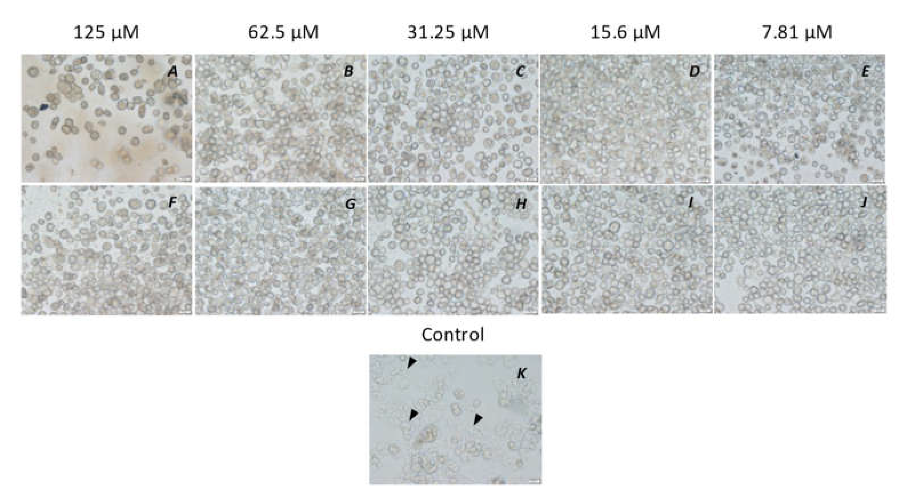
3.1. Amoebicidal Assay

Amoebicidal assays were performed to determine the activity of the peptidomimetic RK-758 in comparison to the chlorhexidine on the viability of A. castellanii ATCC30868 and 044 strains. The number of viable trophozoites was 1.73 × 10⁵ for A. castellanii ATCC30868 and 2.69 × 10⁵ for A. castellanii 044 following a 24 h incubation in PBS alone. The RK-758 caused statistically significant killing of trophozoites between 79%, 87%, and 76% against A. castellanii ATCC30868 (the number of viable trophozoites reduced to 3.58 × 10⁴ (p = 0.03), 2.17 × 10⁴ (p = 0.01), and 1.6 × 10⁴ (p = 0.03), respectively) (Figure 1A), and 98%, 87%, and 57% against A. castellanii 044 (the number of viable trophozoites reduced to 5 × 10³ (p = 0.001), 3.3 × 10⁴ (p = 0.003), and 1.1 × 10⁵ (p = 0.01),respectively) (Figure 1B), at test concentrations ranging between 125 µM, 62.5 µM, and 31.25 µM, respectively, when compared to the PBS alone. Similarly, chlorhexidine caused statistically significant killing of trophozoites by 96%, 81.5%, and 82% against A. castellanii ATCC30868 (the number of viable trophozoites reduced to 6.67 × 10³ (p = 0.01), 3.17 × 10⁴ (p = 0.03), and 3.08 × 10⁴ (p = 0.02), respectively) (Figure 1A), and 98%, 90% and 73% against A. castellanii 044 (the number of viable trophozoites reduced to 5.8 × 10³ (p = 0.001), 2.5 × 10⁴ (p = 0.002), and 7.1 × 10⁴ (p = 0.004),respectively) (Figure 1B), at the same test concentrations when compared to the PBS alone. In addition, chlorhexidine showed statistically significant amoebicidal activity of 62% and 53% against A. castellanii 044 (the number of viable trophozoites reduced to 1 × 10⁵ with p = 0.02, and 1.24 × 10⁵ with p = 0.04, respectively) (Figure 1B) at 15.6 µM and 7.81 µM in comparison to PBS alone. There was no statistically significant difference noted between the activities of the RK-758 and chlorhexidine at each test concentration against both the strains evaluated (p < 0.05).

3.2. Amoebistatic Assay

Growth inhibition assays were performed to determine the amoebistatic activity of the peptidomimetic RK-758 and chlorhexidine against the A. castellanii ATCC30868 and 044 strains. The number of viable trophozoites was enumerated to be 3.08 × 10⁵ for A. castellanii ATCC30868 and 2.17 × 10⁵ for A. castellanii 044 following a 48 h incubation in PYG alone. The RK-758 showed the statistically significant growth inhibition of trophozoites between 97% and 63% against A. castellanii ATCC30868 (the number of viable trophozoites reduced to 9.17 × 10³ at 62.5 µM (p = 0.0008) and 1.13 × 10⁵ at 7.81 µM (p = 0.03) (Figure 2A), and 100% and 85% against A. castellanii 044 (the number of viable trophozoites reduced to 3.08 × 10⁴ at 7.81 µM (p < 0.0001) (Figure 2B), at test concentrations ranging between 125 and 7.81 µM when compared to the PYG alone. Additionally, the chlorhexidine caused a statistically significant growth inhibition against trophozoites between 99% and 83% against A. castellanii ATCC30868 (the number of viable trophozoites reduced to 1.67 × 10³ at 125 µM (p = 0.0007) and 5.25 × 10⁴ at 7.81 µM (p = 0.002) (Figure 2A), and 100% and 98% against A. castellanii 044 (the number of viable trophozoites reduced to 4.17 × 10³ at 7.81 µM, p < 0.001) (Figure 2B), at similar concentrations when compared to the PYG alone. There was no statistically significant difference noted between the activities of the RK-758 and chlorhexidine at each test concentration against both the strains evaluated (p < 0.05), except for the A. castellanii ATCC30868 at 125 µM, where the chlorhexidine had greater activity (p = 0.005).

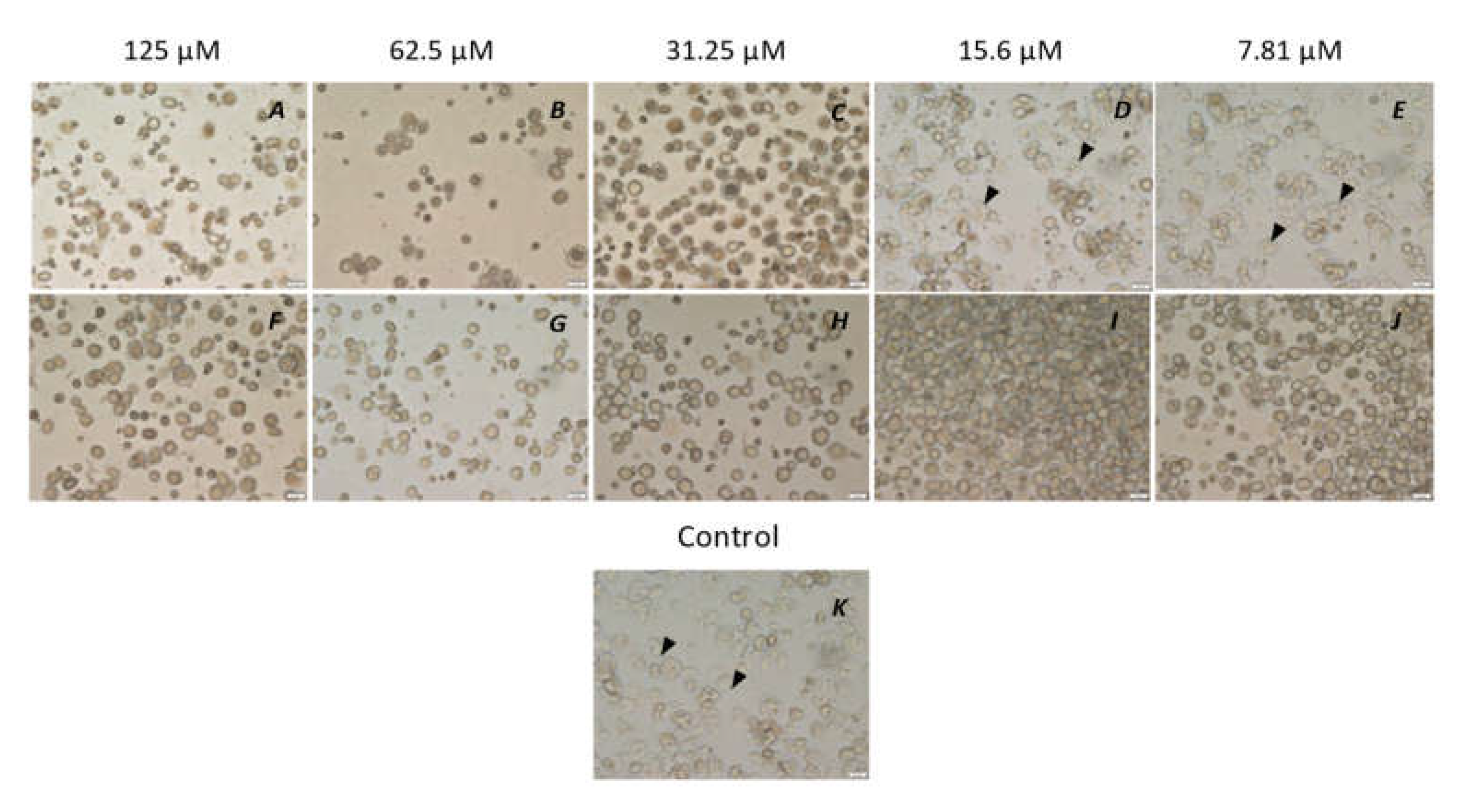
3.3. Encystation Assay

Encystation assays were performed to determine the anti-encystation ability of the chlorhexidine and peptidomimetic RK-758 against A. castellanii ATCC30868 and 044 strains. At the end of the 72 h incubation, 9.75 × 10⁴ cysts of A. castellanii ATCC30868 and 8.75 × 10⁴ cysts of A. castellanii 044 had formed in the encystment medium. The RK-758 inhibited cyst formation by 81% and 68% against A. castellanii ATCC30868 (the number of cysts formed were 1.83 × 10⁴, p = 0.001, and 3 × 10⁴ with p = 0.002, respectively) (Figure 3A) at 125 µM and 62.5 µM test concentrations, respectively, in comparison to the encystment medium. Similarly, RK-758 inhibited cyst formation between 98% and 70% for A. castellanii 044 (the number of cysts formed between 1.67 × 10³ at 125 µM, p = 0.0003 and 2.63 × 10⁴ at 7.81 µM, p =0.005, respectively) (Figure 3B), at test concentrations ranging between 125 and 7.81 µM in comparison to the encystment medium.
Chlorhexidine inhibited cyst formation by between 94% and 54% for A. castellanii ATCC30868 (the number of cysts formed were 5.42 × 10³ at 125 µM, p =0.0002, and 4.42 × 10⁴ at 15.6 µM, p =0.008,respectively) (Figure 2A), at test concentrations ranging between 125 to 15.6 µM in comparison to the encystment medium. Additionally, chlorhexidine inhibited cyst formation between 100% and 75% against A. castellanii 044 (the number of cysts formed were 2.13 × 10⁴ at 7.81 µM, p =0.002) (Figure 2B) at test concentrations ranging from 125 to 7.81 µM in comparison to the encystment medium. There was a statistically significant difference noted between the activities of the RK-758 and chlorhexidine against the A. castellanii ATCC30868 at 62.5 µM and 31.25 µM concentrations (p = 0.003 and p = 0.04), respectively. Similarly, a statistically significant difference was noted between the activities of the RK-758 and chlorhexidine against the A. castellanii 044 at 62.5 µM and 15.6 µM concentrations (p = 0.02 and p = 0.04, respectively).

3.4. Excystation Assay

Excystation assays were conducted to assess the activity of the chlorhexidine and peptidomimetic RK-758 on the re-emergence ability of trophozoites from cysts on the A. castellanii ATCC30868 and 044 strains. At the end of the 72 h incubation, 2.56 × 10⁵ trophozoites of A. castellanii ATCC30868 and 2.52 × 10⁵ trophozoites of A. castellanii 044 had re-emerged in the PYG medium. The peptidomimetic RK-758 inhibited the trophozoites’ re-emergence between 100% and 98% at concentrations ranging from 125 µM to 7.81 µM against A. castellanii ATCC30868 (the number of trophozoites excysted were 2.92 × 10³ at 7.81 µM (p = 0.0008, Figure 4A and Figure 5) in comparison to the PYG medium. Against A. castellanii 044, the peptidomimetic RK-758 inhibited the trophozoites’ re-emergence between 100 and 60% concentrations ranging from 125 µM to 15.6 µM concentrations (the number of trophozoites excysted were 1.00 × 10⁵ at 15.6 µM (p = 0.007, Figure 4B and Figure 6) in comparison to the PYG medium. Similarly, the chlorhexidine inhibited the trophozoite re-emergence by 100% at all the test concentrations between 125 µM and 7.81 µM for A. castellanii ATCC30868 and A. castellanii 044 (p < 0.05, Figure 4A,B) in comparison to the PYG medium.
There was a statistically significant difference between the activities of the RK-758 and chlorhexidine at 7.81 µM against A. castellanii ATCC30868 (p = 0.03) and between the activities of the RK-758 and chlorhexidine against A. castellanii 044 at 15.6 µM and 7.81 µM concentrations (p < 0.001 and p = 0.006, respectively).

3.5. Haemolysis Assay

Chlorhexidine and peptidomimetic RK-758 caused haemolysis in a dose-dependent manner. Chlorhexidine showed a negligible haemolysis between 31.25 µM and 7. 81 µM. The therapeutic index (i.e., haemolytic concentration/antimicrobial concentration) for the chlorhexidine ranged from 16 to 4. The peptidomimetic RK-758 showed haemolysis of < 50% between 31.25 µM and 7.81 µM. The therapeutic index of the RK-758 ranged from 8 to 1.

4. Discussion

This study demonstrated that the peptidomimetic RK-758 was active against both the trophozoite and cyst forms of Acanthamoeba castellanii, most probably due to its cationic charge [19,20,25]. The anti-amoebic effects of any test compounds on both trophozoites and cysts are crucial as the trophozoites can remerge and lead to a relapse of the disease upon stopping treatment [1,4,5]. This may possibly be due to the risk of cysts’ formation in corneal tissue during Acanthamoeba infections [51]. A laboratory study examining the in vitro sensitivity of 23 isolates from 23 patients to 13 different drugs found that PHMB and chlorhexidine were the most successful agents against both trophozoites and cysts [52]. Recently, a retrospective study showed that treatment with biguanides and diamidines resulted in a 64% improvement in the best-corrected visual acuity in patients [5]. A feature that is commonly seen in AK is an initial worsening of inflammation upon starting treatment, and this may possibly be due to the antigens released by the dead organisms [8,53]. The current results show that the peptidomimetic RK-758 possessed amoebicidal and cysticidal activity against both the ATCC and the clinical strain tested, which is essential in tackling an Acanthamoeba infection.
Previous studies have already established that peptidomimetics are highly effective against bacteria [35,36,37], but before this study, the anti-Acanthamoeba effects of peptidomimetics had not been evaluated.
Previously, the naturally occurring antimicrobial peptide protamine had been shown to have amoebicidal activity at 228 µM, and its derivative melimine inhibited Acanthamoeba trophozoites’ adhesion to contact lens at a concentration of 152 µg per lens [10,19]. Additionally, another naturally occurring antimicrobial peptide magainin had minimal inhibitory (90% reduction) and minimal amoebicidal (99.9% reduction) activities between 8 and 16 µM [20,21]. The antimicrobial peptide poly-epsilon-lysine was amoebicidal to trophozoites and cysts at 540 µM producing an 80% death of trophozoites and 76% death of cysts at the end of a 24 h incubation [25]. An AMP derived from tyrocidine, when used at 100 µg/mL and 250 µg/mL, killed 35% and 84% of trophozoites, respectively, at the end of a 24 h incubation and reduced the transformation of trophozoites to cysts by 58% and 93%, respectively, at the end of a 72 h incubation [26]. The exposure of A. castellanii trophozoites to nisin, a bacteriocin AMP, resulted in reductions in cultivable numbers after a 24 h exposure but there was a recovery in the amoeba growth after a 72 h exposure. This may have been due to the lack of a nisin effect on the membrane integrity of the trophozoites [28]. A synthetic decapeptide AMP (called KP) killed 57% of trophozoites at 25 µg/mL after a 24 h incubation [27]. The peptidomimetic RK-758 was effective between 7.81 µM and 62.5 µM against both the strains evaluated. This was somewhat less effective than reported for magainin, but was improved over the reports for protamine and poly-epsilon-lysine. Initial proteolysis assays with trypsin (unpublished) showed that RK-758 was proteolytically stable for 18 h, possibly due to its substituted guanidine end cap. Magainin, however, can be cleaved by proteases [54], which may be one of the reasons it did not outperform the antibiotic ofloxacin in a Phase III clinical trial against bacteria [55].
The current study found that RK-758 performed almost equivalently to the currently used topical treatment, chlorhexidine. Chlorhexidine has previously been reported to have minimum cysticidal activity against nine Acanthamoeba clinical isolates at 3.1 to 25 µg/mL (6 to 49 µM) [56] and the current study showed similar results against both trophozoites and cysts; however, chlorhexidine has been reported to be toxic to the eyes causing corneal irritation and abrasions [57]. Future studies should determine the ocular safety of RK-758 to determine if it has an improved ocular safety profile over chlorhexidine.
A haemolysis assay is often used as a primary measure of the toxicity of antimicrobial peptides [35]. The RK-758 produced minimal haemolysis at 15.6 µM and 7.81 µM, giving a therapeutic index of up to eight. Using LD50 (a dose causing 50% death in mice) as a measure of toxicity, polyhexamethylene biguanide, another commonly used therapeutic for Acanthamoeba keratitis [8,58], has a therapeutic index of 3.2 [59]. However, haemolysis and LD50 are not directly comparable; therefore, experiments directly comparing the therapeutic index of currently used therapies with RK-758 are required. A previous study showed that chlorhexidine at 20 µg/mL (31.97 µM) gave a 20% lysis of rabbit red blood cells after 2 h of incubation [60]. Similarly, the current results showed a negligible lysis of horse red blood cells at a similar concentration (31.25 µM) and a higher concentration (62.5 µM) after 4 h of incubation.
The current study used PBS as a negative control to evaluate the viability of trophozoites in an amoebicidal assay; however, a previous study reported that PBS alone induced 28% encystment after 24 h of incubation [41]. A later study showed only a 4% encystment at the end of a 24 h incubation and no effect on the viability of A. castellanii. The authors suggested that the inclusion of 50 mM of MgCl2 and 10% glucose in the PBS formulation induced encystment [48]. Elsewhere, PBS solutions containing 0.25 ppm PHMB and increasing concentrations of propylene glycol and povidone stimulated encystment in a dose-dependent manner [61]. The current study did not find any encystment during the evaluation of the trophozoite viability.
In summary, the peptidomimetic RK-758 has excellent anti-Acanthamoeba activity on the trophozoites and cysts of A. castellanii ATCC30868 and 044 strains. Its activity is very similar to chlorhexidine, which indicates that RK-758 has the potential to be developed as a new therapeutic agent for the treatment of Acanthamoeba keratitis or a new disinfectant for contact lenses with good activity against Acanthamoeba.

Author Contributions

H.K.P.: Writing—original draft preparation, Methodology, Investigation, Visualization, and Formal analysis. N.A.C.: Visualisation, Writing, Reviewing, and Editing. Z.G. and N.K.: Writing, Reviewing and Editing. M.D.P.W.: Supervision, Methodology, Resources, Writing—Reviewing and Editing. R.K.: Conceptualization, Methodology, Resources, Writing—Reviewing and Editing. All authors have read and agreed to the published version of the manuscript.

Funding

This research was partly funded by NHMRC Ideas grant APP1183597.

Institutional Review Board Statement

Not applicable.

Informed Consent Statement

Not applicable.

Data Availability Statement

The data supporting the conclusions of this article are included within the article and its figures. No large datasets were generated or analysed during the current study.

Acknowledgments

H.K.P. thanks the support from UNSW Sydney Australia for providing a Scientia PhD scholarship for his doctoral degree.

Conflicts of Interest

The authors declare no conflict of interest.

References

  1. Siddiqui, R.; Khan, N.A. Biology and pathogenesis of Acanthamoeba. Parasites Vectors 2012, 5, 6. [Google Scholar] [CrossRef] [PubMed] [Green Version]
  2. Carnt, N.; Hoffman, J.M.; Verma, S.; Hau, S.; Radford, C.F.; Minassian, D.C.; Dart, J.K.G. Acanthamoeba keratitis: Confirmation of the UK outbreak and a prospective case-control study identifying contributing risk factors. Br. J. Ophthalmol. 2018, 102, 1621–1628. [Google Scholar] [CrossRef] [PubMed] [Green Version]
  3. Clarke, B.; Sinha, A.; Parmar, D.N.; Sykakis, E. Advances in the diagnosis and treatment of Acanthamoeba keratitis. J. Ophthalmol. 2012, 2012, 484892. [Google Scholar] [CrossRef] [Green Version]
  4. Szentmary, N.; Daas, L.; Shi, L.; Laurik, K.L.; Lepper, S.; Milioti, G.; Seitz, B. Acanthamoeba keratitis—Clinical signs, differential diagnosis and treatment. J. Curr. Ophthalmol. 2019, 31, 16–23. [Google Scholar] [CrossRef] [PubMed]
  5. List, W.; Glatz, W.; Riedl, R.; Mossboeck, G.; Steinwender, G.; Wedrich, A. Evaluation of Acanthamoeba keratitis cases in a tertiary medical care centre over 21 years. Sci. Rep. 2021, 11, 1036. [Google Scholar] [CrossRef] [PubMed]
  6. Naranjo, A.; Martinez, J.D.; Miller, D.; Tonk, R.; Amescua, G. Systemic miltefosine as an adjunct treatment of progressive Acanthamoeba keratitis. Ocul. Immunol. Inflamm. 2021, 29, 1576–1584. [Google Scholar] [CrossRef]
  7. Kosrirukvongs, P.; Wanachiwanawin, D.; Visvesvara, G.S. Treatment of Acanthamoeba keratitis with chlorhexidine. Ophthalmology 1999, 106, 798–802. [Google Scholar] [CrossRef]
  8. Fanselow, N.; Sirajuddin, N.; Yin, X.T.; Huang, A.J.W.; Stuart, P.M. Acanthamoeba keratitis, pathology, diagnosis and treatment. Pathogens 2021, 10, 323. [Google Scholar] [CrossRef]
  9. Dart, J.K.; Saw, V.P.; Kilvington, S. Acanthamoeba keratitis: Diagnosis and treatment update 2009. Am. J. Ophthalmol. 2009, 148, 487–499.e482. [Google Scholar] [CrossRef]
  10. Dutta, D.; Cole, N.; Kumar, N.; Willcox, M.D. Broad spectrum antimicrobial activity of melimine covalently bound to contact lenses. Investig. Ophthalmol. Vis. Sci. 2013, 54, 175–182. [Google Scholar] [CrossRef]
  11. Huan, Y.; Kong, Q.; Mou, H.; Yi, H. Antimicrobial peptides: Classification, design, application and research progress in multiple fields. Front. Microbiol. 2020, 11, 2559. [Google Scholar] [CrossRef]
  12. Antimicrobial Peptide Database. Available online: https://aps.unmc.edu/ (accessed on 4 November 2022).
  13. Wang, G.; Li, X.; Wang, Z. APD3: The antimicrobial peptide database as a tool for research and education. Nucleic Acids Res. 2016, 44, D1087–D1093. [Google Scholar] [CrossRef] [Green Version]
  14. Kuppusamy, R.; Willcox, M.; Black, D.S.; Kumar, N. Short cationic peptidomimetic antimicrobials. Antibiotics 2019, 8, 44. [Google Scholar] [CrossRef] [PubMed] [Green Version]
  15. Helmerhorst, E.J.; Reijnders, I.M.; van’t Hof, W.; Simoons-Smit, I.; Veerman, E.C.; Amerongen, A.V. Amphotericin B- and fluconazole-resistant Candida spp., Aspergillus fumigatus, and other newly emerging pathogenic fungi are susceptible to basic antifungal peptides. Antimicrob. Agents Chemother. 1999, 43, 702–704. [Google Scholar] [CrossRef] [PubMed] [Green Version]
  16. Feder, R.; Dagan, A.; Mor, A. Structure-activity relationship study of antimicrobial dermaseptin S4 showing the consequences of peptide oligomerization on selective cytotoxicity. J. Biol. Chem. 2000, 275, 4230–4238. [Google Scholar] [CrossRef] [Green Version]
  17. Meiller, T.F.; Hube, B.; Schild, L.; Shirtliff, M.E.; Scheper, M.A.; Winkler, R.; Ton, A.; Jabra-Rizk, M.A. A novel immune evasion strategy of Candida albicans: Proteolytic cleavage of a salivary antimicrobial peptide. PLoS ONE 2009, 4, e5039. [Google Scholar] [CrossRef] [Green Version]
  18. Potempa, J.; Pike, R.N. Corruption of innate immunity by bacterial proteases. J. Innate Immun. 2009, 1, 70–87. [Google Scholar] [CrossRef] [Green Version]
  19. Vijay, A.K.; Bandara, M.; Zhu, H.; Willcox, M.D. Protamine as a potential amoebicidal agent for contact lens disinfection. Optom. Vis. Sci. 2013, 90, 119–124. [Google Scholar] [CrossRef]
  20. Schuster, F.L.; Jacob, L.S. Effects of magainins on ameba and cyst stages of Acanthamoeba polyphaga. Antimicrob. Agents Chemother. 1992, 36, 1263–1271. [Google Scholar] [CrossRef] [Green Version]
  21. Feldman, S.T.; Speaker, M.; Cleveland, P. Effect of magainins on Acanthamoeba castellanii. Rev. Infect. Dis. 1991, 13 (Suppl. S5), S439. [Google Scholar] [CrossRef]
  22. Polat, Z.A.; Savage, P.B.; Genberg, C. In vitro amoebicidal activity of a ceragenin, cationic steroid antibiotic-13, against Acanthamoeba castellanii and its cytotoxic potential. J. Ocul. Pharm. 2011, 27, 1–5. [Google Scholar] [CrossRef] [PubMed]
  23. Sacramento, R.S.; Martins, R.M.; Miranda, A.; Dobroff, A.S.; Daffre, S.; Foronda, A.S.; De Freitas, D.; Schenkman, S. Differential effects of alpha-helical and beta-hairpin antimicrobial peptides against Acanthamoeba castellanii. Parasitology 2009, 136, 813–821. [Google Scholar] [CrossRef] [PubMed]
  24. Schlusselhuber, M.; Humblot, V.; Casale, S.; Methivier, C.; Verdon, J.; Leippe, M.; Berjeaud, J.M. Potent antimicrobial peptides against Legionella pneumophila and its environmental host, Acanthamoeba castellanii. Appl. Microbiol. Biotechnol. 2015, 99, 4879–4891. [Google Scholar] [CrossRef]
  25. Kennedy, S.M.; Deshpande, P.; Gallagher, A.G.; Horsburgh, M.J.; Allison, H.E.; Kaye, S.B.; Wellings, D.A.; Williams, R.L. Amoebicidal activity of poly-epsilon-lysine functionalized hydrogels. Investig. Ophthalmol. Vis. Sci. 2022, 63, 11. [Google Scholar] [CrossRef] [PubMed]
  26. Akbar, N.; Kaman, W.E.; Sarink, M.; Nazmi, K.; Bikker, F.J.; Khan, N.A.; Siddiqui, R. Novel Antiamoebic tyrocidine-derived peptide against brain-eating amoebae. ACS Omega 2022, 7, 28797–28805. [Google Scholar] [CrossRef] [PubMed]
  27. Fiori, P.L.; Mattana, A.; Dessi, D.; Conti, S.; Magliani, W.; Polonelli, L. In vitro acanthamoebicidal activity of a killer monoclonal antibody and a synthetic peptide. J. Antimicrob. Chemother. 2006, 57, 891–898. [Google Scholar] [CrossRef] [Green Version]
  28. De Carvalho Climaco, M.; de Oliveira, Y.L.M.; Ramos, A.C.S.; Ramos-de-Souza, J.; Silva, A.; Jain, S.; Rott, M.B.; Scher, R.; Correa, C.B.; Barbosa, A.A.T.; et al. Nisin induces cell-cycle arrest in free-living amoebae Acanthamoeba castellanii. Acta Parasitol. 2022, 67, 511–517. [Google Scholar] [CrossRef]
  29. Otri, A.M.; Mohammed, I.; Abedin, A.; Cao, Z.; Hopkinson, A.; Panjwani, N.; Dua, H.S. Antimicrobial peptides expression by ocular surface cells in response to Acanthamoeba castellanii: An in vitro study. Br. J. Ophthalmol. 2010, 94, 1523–1527. [Google Scholar] [CrossRef] [Green Version]
  30. Méndez-Samperio, P. Peptidomimetics as a new generation of antimicrobial agents: Current progress. Infect. Drug Resist. 2014, 7, 229–237. [Google Scholar] [CrossRef] [Green Version]
  31. Luo, G.; Zhang, J.; Wang, H.; Sun, Y.; Cheng, B.; Xu, Z.; Zhang, Y.; Li, H.; Lu, W.; Nemeth, E.; et al. Human defensin-inspired discovery of peptidomimetic antibiotics. Proc. Natl. Acad. Sci. USA 2022, 119, e2117283119. [Google Scholar] [CrossRef]
  32. Ibrahim, M.A.; Panda, S.S.; Oliferenko, A.A.; Oliferenko, P.V.; Girgis, A.S.; Elagawany, M.; Küçükbay, F.Z.; Panda, C.S.; Pillai, G.G.; Samir, A.; et al. Macrocyclic peptidomimetics with antimicrobial activity: Synthesis, bioassay, and molecular modeling studies. Org. Biomol. Chem. 2015, 13, 9492–9503. [Google Scholar] [CrossRef] [PubMed] [Green Version]
  33. Di Chio, C.; Previti, S.; Amendola, G.; Cosconati, S.; Schirmeister, T.; Zappalà, M.; Ettari, R. Development of novel benzodiazepine-based peptidomimetics as inhibitors of rhodesain from Trypanosoma brucei rhodesiense. ChemMedChem 2020, 15, 995–1001. [Google Scholar] [CrossRef]
  34. Kuppusamy, R.; Yasir, M.; Yee, E.; Willcox, M.; Black, D.S.; Kumar, N. Guanidine functionalized anthranilamides as effective antibacterials with biofilm disruption activity. Org. Biomol. Chem. 2018, 16, 5871–5888. [Google Scholar] [CrossRef] [PubMed]
  35. Browne, K.; Kuppusamy, R.; Chen, R.; Willcox, M.D.P.; Walsh, W.R.; Black, D.S.; Kumar, N. Bioinspired polydopamine coatings facilitate attachment of antimicrobial peptidomimetics with broad-spectrum antibacterial activity. Int. J. Mol. Sci. 2022, 23, 2952. [Google Scholar] [CrossRef] [PubMed]
  36. Wu, J.; Yu, T.T.; Kuppusamy, R.; Hassan, M.M.; Alghalayini, A.; Cranfield, C.G.; Willcox, M.D.P.; Black, D.S.; Kumar, N. Cholic acid-based antimicrobial peptide mimics as antibacterial agents. Int. J. Mol. Sci. 2022, 23, 4623. [Google Scholar] [CrossRef] [PubMed]
  37. Benjamin, A.B.; Moule, M.G.; Didwania, M.K.; Hardy, J.; Saenkham-Huntsinger, P.; Sule, P.; Nielsen, J.E.; Lin, J.S.; Contag, C.H.; Barron, A.E.; et al. Efficacy of cathelicidin-mimetic antimicrobial peptoids against Staphylococcus aureus. Microbiol. Spectr. 2022, 10, e0053422. [Google Scholar] [CrossRef]
  38. Patel, A.; Hammersmith, K. Contact lens-related microbial keratitis: Recent outbreaks. Curr. Opin. Ophthalmol. 2008, 19, 302–306. [Google Scholar] [CrossRef]
  39. Verani, J.R.; Lorick, S.A.; Yoder, J.S.; Beach, M.J.; Braden, C.R.; Roberts, J.M.; Conover, C.S.; Chen, S.; McConnell, K.A.; Chang, D.C.; et al. National outbreak of Acanthamoeba keratitis associated with use of a contact lens solution, United States. Emerg. Infect. Dis. 2009, 15, 1236–1242. [Google Scholar] [CrossRef]
  40. Joslin, C.E.; Tu, E.Y.; Shoff, M.E.; Booton, G.C.; Fuerst, P.A.; McMahon, T.T.; Anderson, R.J.; Dworkin, M.S.; Sugar, J.; Davis, F.G.; et al. The association of contact lens solution use and Acanthamoeba keratitis. Am. J. Ophthalmol. 2007, 144, 169–180. [Google Scholar] [CrossRef] [Green Version]
  41. Kilvington, S.; Heaselgrave, W.; Lally, J.M.; Ambrus, K.; Powell, H. Encystment of Acanthamoeba during incubation in multipurpose contact lens disinfectant solutions and experimental formulations. Eye Contact Lens. 2008, 34, 133–139. [Google Scholar] [CrossRef]
  42. Radford, C.F.; Lehmann, O.J.; Dart, J.K.; for the National Acanthamoeba Keratitis Study Group. Acanthamoeba keratitis: Multicentre survey in England 1992-6. Br. J. Ophthalmol. 1998, 82, 1387–1392. [Google Scholar] [CrossRef] [PubMed] [Green Version]
  43. Hsu, C.-C.; Kuo, Y.-S.; Lin, P.-Y.; Chen, K.-H. Overnight orthokeratology-associated Acanthamoeba keratitis at a tertiary referral hospital in Taiwan: A retrospective case-control study. J. Chin. Med. Assoc. 2022, 85, 381–387. [Google Scholar] [CrossRef] [PubMed]
  44. Li, W.; Wang, Z.; Qu, J.; Zhang, Y.; Sun, X. Acanthamoeba keratitis related to contact lens use in a tertiary hospital in China. BMC Ophthalmol. 2019, 19, 202. [Google Scholar] [CrossRef] [PubMed] [Green Version]
  45. Narasimhan, S.; Madhavan, H.N.; Therese, K.L. Development and application of an in vitro susceptibility test for Acanthamoeba species isolated from keratitis to polyhexamethylene biguanide and chlorhexidine. Cornea 2002, 21, 203–205. [Google Scholar] [CrossRef]
  46. Baig, A.M.; Iqbal, J.; Khan, N.A. In vitro efficacies of clinically available drugs against growth and viability of an Acanthamoeba castellanii keratitis isolate belonging to the T4 genotype. Antimicrob. Agents Chemother. 2013, 57, 3561–3567. [Google Scholar] [CrossRef] [PubMed] [Green Version]
  47. Anwar, A.; Chi Fung, L.; Anwar, A.; Jagadish, P.; Numan, A.; Khalid, M.; Shahabuddin, S.; Siddiqui, R.; Khan, N.A. Effects of shape and size of cobalt phosphate nanoparticles against Acanthamoeba castellanii. Pathogens 2019, 8, 260. [Google Scholar] [CrossRef] [PubMed] [Green Version]
  48. Aqeel, Y.; Siddiqui, R.; Iftikhar, H.; Khan, N.A. The effect of different environmental conditions on the encystation of Acanthamoeba castellanii belonging to the T4 genotype. Exp. Parasitol. 2013, 135, 30–35. [Google Scholar] [CrossRef]
  49. Sabir, S.; Yu, T.T.; Kuppusamy, R.; Almohaywi, B.; Iskander, G.; Das, T.; Willcox, M.D.P.; Black, D.S.; Kumar, N. Novel seleno- and thio-urea containing dihydropyrrol-2-one analogues as antibacterial agents. Antibiotics 2021, 10, 321. [Google Scholar] [CrossRef]
  50. Yasir, M.; Dutta, D.; Willcox, M.D.P. Mode of action of the antimicrobial peptide Mel4 is independent of Staphylococcus aureus cell membrane permeability. PLoS ONE 2019, 14, e0215703. [Google Scholar] [CrossRef] [Green Version]
  51. Schuster, F.L.; Visvesvara, G.S. Opportunistic amoebae: Challenges in prophylaxis and treatment. Drug Resist. Updates 2004, 7, 41–51. [Google Scholar] [CrossRef]
  52. Elder, M.; Kilvington, S.; Dart, J. A clinicopathologic study of in vivo sensitivity testing and Acanthamoeba keratitis. Investig. Ophthalmol. Vis. Sci. 1994, 35, 1059–1064. [Google Scholar]
  53. Illingworth, C.D.; Cook, S.D. Acanthamoeba keratitis. Surv. Ophthalmol. 1998, 42, 493–508. [Google Scholar] [CrossRef] [PubMed]
  54. Zasloff, M. Magainins, a class of antimicrobial peptides from Xenopus skin: Isolation, characterization of two active forms, and partial cDNA sequence of a precursor. Proc. Natl. Acad. Sci. USA 1987, 84, 5449–5453. [Google Scholar] [CrossRef] [Green Version]
  55. Mookherjee, N.; Anderson, M.A.; Haagsman, H.P.; Davidson, D.J. Antimicrobial host defence peptides: Functions and clinical potential. Nat. Rev. Drug Discov. 2020, 19, 311–332. [Google Scholar] [CrossRef] [PubMed]
  56. Redd, T.K.; Talbott, M.; Cevallos, V.; Lalitha, P.; Seitzman, G.D.; Lietman, T.M.; Keenan, J.D. In vitro comparison of the Acanthamoeba cysticidal activity of povidone iodine, natamycin, and chlorhexidine. Ophthalmol. Sci. 2021, 1, 100025. [Google Scholar] [CrossRef] [PubMed]
  57. Mathers, W.D.; Sutphin, J.E.; Folberg, R.; Meier, P.A.; Wenzel, R.P.; Elgin, R.G. Outbreak of keratitis presumed to Be caused by Acanthamoeba. Am. J. Ophthalmol. 1996, 121, 129–142. [Google Scholar] [CrossRef]
  58. Lim, N.; Goh, D.; Bunce, C.; Xing, W.; Fraenkel, G.; Poole, T.R.; Ficker, L. Comparison of polyhexamethylene biguanide and chlorhexidine as monotherapy agents in the treatment of Acanthamoeba keratitis. Am. J. Ophthalmol. 2008, 145, 130–135. [Google Scholar] [CrossRef]
  59. Asiedu-Gyekye, I.J.; Mahmood, A.S.; Awortwe, C.; Nyarko, A.K. Toxicological assessment of polyhexamethylene biguanide for water treatment. Interdiscip. Toxicol. 2015, 8, 193–202. [Google Scholar] [CrossRef] [Green Version]
  60. Ansel, H.C. Hemolysis of erythrocytes by antibacterial preservatives IV. Hemolytic activity of chlorhexidine diacetate. J. Pharm. Sci. 1967, 56, 616–619. [Google Scholar] [CrossRef]
  61. Kovacs, C.J.; Lynch, S.C.; Rah, M.J.; Millard, K.A.; Morris, T.W. Acanthamoeba encystment: Multifactorial effects of buffers, biocides, and demulcents present in contact lens care solutions. Clin. Ophthalmol. 2015, 9, 1905–1913. [Google Scholar] [CrossRef]
Figure 1. Amoebicidal activity of peptidomimetic RK-758 in comparison with chlorhexidine against Acanthamoeba castellanii ATCC30868 (A) and A. castellanii 044 (B). In brief, 5 × 10⁵ A. castellanii trophozoites were incubated with the peptidomimetics RK-758 and chlorhexidine at 30 °C for 24 h after which the viability was determined by staining with trypan blue using a Neubauer haemocytometer. The results show significant anti-Acanthamoeba activity when compared to the negative control (amoeba alone). * p < 0.05 using a two-sample t-test and two-tailed distribution.
Microorganisms 10 02377 g001
Figure 2. Amoebistatic activity of the peptidomimetic RK-758 in comparison with chlorhexidine against Acanthamoeba castellanii ATCC30868 (A) and A. castellanii 044 (B). In brief, 2 × 10⁵ A. castellanii trophozoites were incubated with the peptidomimetics RK-758 and chlorhexidine at 30 °C for 48 h after which the viability was determined by staining with Trypan blue using a Neubauer haemocytometer. The results showed significant anti-Acanthamoeba activity when compared to the negative control (amoeba alone). ** p < 0.001; * p < 0.05 using a two-sample t-test and two-tailed distribution.
Microorganisms 10 02377 g002
Figure 3. Inhibition of cysts formation by the peptidomimetic RK-758 in comparison with chlorhexidine against Acanthamoeba castellanii ATCC30868 (A) and A. castellanii 044 (B). In brief, 5 × 10⁵ A. castellanii trophozoites were incubated with the peptidomimetics RK-758 and chlorhexidine at 30 °C for 72 h after which cysts were determined by solubilising trophozoites adding 0.25% SDS. The number of cysts was counted using the Neubauer haemocytometer. The results showed significant anti-Acanthamoeba activity when compared to the negative control (amoeba alone). ** p < 0.001; * p < 0.05 using the two-sample t-test and two-tailed distribution.
Microorganisms 10 02377 g003
Figure 4. Inhibition of trophozoites’ re-emergence from cysts by the peptidomimetic RK-758 in comparison with chlorhexidine, against Acanthamoeba castellanii ATCC30868 (A) and A. castellanii 044 (B). In brief, 5 × 10⁵ A. castellanii cysts were incubated with the peptidomimetics RK-758 and chlorhexidine at 30 °C for 72 h after which the trophozoites were determined by counting on a Neubauer haemocytometer. The results showed significant anti-Acanthamoeba activity when compared to the negative control (amoeba alone). ** p < 0.001; * p < 0.05 using a two-sample t-test and two-tailed distribution.
Microorganisms 10 02377 g004
Figure 5. Inhibition of trophozoite emergence from cysts of Acanthamoeba castellanii ATCC30868 by the peptidomimetic RK-758 (AE) in comparison with chlorhexidine (FJ), with concentrations of both ranging from 125 µM to 7.81 µM. In brief, 5 × 10⁵ A. castellanii trophozoites were incubated with the peptidomimetics RK-758 and chlorhexidine at 30 °C for 72 h and observed for the emergence of trophozoites. PYG alone was used as a control (K). PeptidomimeticsRK-758 inhibited the trophozoite emergence between concentrations of 125 µM and 31.25 µM (A–C) whereas the chlorhexidine inhibited their emergence between 125 µM and 7.81 µM (FJ). Arrowheads indicate trophozoites.
Microorganisms 10 02377 g005
Figure 6. Inhibition of trophozoite emergence from cysts of Acanthamoeba castellanii 044 by the peptidomimetic RK-758 (AE) in comparison with chlorhexidine (FJ), with concentrations of both ranging from 125 µM to 7.81 µM. In brief, 5 × 10⁵ A. castellanii trophozoites were incubated with the peptidomimetics RK-758 and chlorhexidine at 30 °C for 72 h and observed for the emergence of trophozoites. PYG alone was used as a control (K). Peptidomimetic RK-758 inhibited the trophozoite emergence between concentrations of 125 µM and 31.25 µM (AC) whereas the chlorhexidine inhibited their emergence between 125 µM and 7.81 µM (FJ). Arrowheads indicate trophozoites.
Microorganisms 10 02377 g006
Publisher’s Note: MDPI stays neutral with regard to jurisdictional claims in published maps and institutional affiliations.

