Article

Gene Regulatory Network of the Noncoding RNA Qrr5 Involved in the Cytotoxicity of Vibrio parahaemolyticus during Infection

1
School of Food Science and Technology, Jiangnan University, Wuxi 214122, China
2
Guangdong Provincial Key Laboratory of Microbial Safety and Health, Ministry of Agriculture and Rural Affairs, Key Laboratory of Agricultural Microbiomics and Precision Application, State Key Laboratory of Applied Microbiology Southern China, Institute of Microbiology, Guangdong Academy of Sciences, Guangzhou 510070, China
*
Authors to whom correspondence should be addressed.
These authors contributed equally to this work.
Microorganisms 2022, 10(10), 2084; https://doi.org/10.3390/microorganisms10102084
Submission received: 13 September 2022 / Revised: 9 October 2022 / Accepted: 19 October 2022 / Published: 21 October 2022
(This article belongs to the Special Issue Foodborne Pathogens: Infections and Pathogenesis)

Abstract

:
Small non-coding RNAs (sRNAs) in bacteria are important regulatory molecules for controlling virulence. In Vibrio spp., Qrr sRNAs are critical for quorum-sensing pathways and regulating the release of some virulence factors. However, the detailed role of Qrr sRNAs in the virulence of Vibrio parahaemolyticus remains poorly understood. In this study, we identified a Vibrio sRNA Qrr5 that positively regulates cytotoxicity and adherence in Caco-2 cells by primarily regulating the T3SS1 gene cluster. A number of 185, 586, 355, and 74 differentially expressed genes (DEGs) detected at 0, 2, 4, and 6 h post-infection, respectively, were mainly associated with ABC transporters and two-component system pathways. The DEGs exhibited a dynamic change in expression at various time points post-infection owing to the deletion of Qrr5. Accordingly, 17 related genes were identified in the co-expression network, and their interaction with Qrr5 was determined based on weighted co-expression network analysis during infection. Taken together, our results provide a comprehensive transcriptome profile of V. parahaemolyticus during infection in Caco-2 cells.

1. Introduction

Small non-coding RNAs (sRNAs) serve as indispensable regulators of many bacterial signaling pathway cascades and pathogenesis [1,2,3]. A novel sRNA, EsrF, can sense high ammonium concentrations in the colon and facilitate bacterial motility and adhesion to the host cell, thereby promoting the pathogenicity of Escherichia coli O157 [4]. Qrr sRNAs are Hfq-dependent trans-encoded sRNAs involved in the regulation of quorum sensing in Vibrio spp. [5,6,7], which positively regulate the expression of AphA, a low-cell-density core regulator that suppresses the expression of LuxR, a high-cell-density core regulator [8,9,10]. In Vibrio harveyi, the Qrr3 sRNA suppresses LuxR through catalytic degradation, LuxM through degradation, and LuxO through a blockade, as well as activating AphA by exposing the ribosome-combining site, whereas the sRNA itself is decomposed [11,12]. However, five RNAs (Qrr1–5) redundantly inhibited the expression of the master regulator SmcR and modulated the expression of virulence factors in cells lacking iron at low cell density and in Vibrio vulnificus at high cell density [13,14]. Fis is a regulator that negatively regulates the expression of the quorum-sensing master regulator OpaR and inhibits capsule polysaccharide (CPS) and biofilm formation, implying that Fis plays a direct role in the quorum sensing, biofilm formation, and metabolism of V. parahaemolyticus. Interestingly, when Fis binds directly to the regulatory regions of Qrr genes, it acts as a positive regulator [15].
Vibrio parahaemolyticus is a gram-negative bacterium generally isolated from estuarine and marine sources, and its infection leads to gastroenteritis related to seafood consumption in humans worldwide [16,17]. Nevertheless, the pathogenesis of V. parahaemolyticus infection has not been completely and explicitly elucidated. The major virulence factors that have been recently characterized include adhesins, flagella, hemolysin, and two type III secretion systems (T3SSs), namely T3SS1 and T3SS2 [18,19]. Every T3SS produces a distinct group of effectors that contributes to virulence by interacting with various host targets and performing various functions. T3SS1 is responsible for cytotoxicity, whereas T3SS2 is primarily responsible for enterotoxicity [20,21,22,23]. Five Qrrs have been identified in V. parahaemolyticus, but only the functions of Qrr2Qrr4 have been elucidated. They activate the target gene AphA at a low density and promote the synthesis of the low-density regulator AphA. They can also act on the target gene OpaR and inhibit the production of the high-density regulator OpaR [24,25]. However, the regulation of toxicity by Qrrs in V. parahaemolyticus has not been directly proven. A previous study reported that AphA can positively regulate the expression of T3SS1 to affect virulence, whereas OpaR negatively regulates the expression of T3SS1 [24,26]. T3SS is an injectable needle-like structure consisting of multiple distinct structural proteins that secrete virulence effectors into host cells to cause cellular damage, and T3SS1 mainly causes a cytotoxic effect on the host [17,25,27]. Therefore, it was speculated that the Qrrs of V. parahaemolyticus might regulate the expression of T3SS1 through the quorum sensing pathway to regulate the pathogenicity of the bacteria in the host. Subsequently, in a transcriptome analysis of V. parahaemolyticus-infected human HeLa cells, it was found that the main effectors of T3SS1 were significantly up-regulated at all time points post-infection (2, 3, 4, 6, and 8 h). However, the expression level of AphA did not change significantly upon infection. Similarly, the expression of OpaR did not change significantly during the early period of infection, and a down-regulated expression trend was observed only during the later period of infection [28,29]. Most studies have focused on the regulation of Qrrs associated with quorum-sensing genes [30,31,32]. These studies indicate that Qrrs of V. parahaemolyticus play a vital role in the regulation of virulence genes. However, the role of Qrrs in the virulence of V. parahaemolyticus remains unclear.
In this study, we detected the relative mRNA expression levels of Qrrs during the infection of Caco-2 cells with V. parahaemolyticus. One of the Qrrs, Qrr5, showed significant changes in expression during the infection. The Qrr5 mutant strain was utilized to infect Caco-2 cells, and transcriptome analyses were performed during infection. We aimed to unveil the mechanism of Qrr sRNAs in regulating virulence factors in V. parahaemolyticus. The findings of this study are expected to promote the development of effective methods to alleviate the toxicity of this pathogen.

2. Materials and Methods

2.1. Bacterial Strains, Plasmid, and Growth Conditions

Vibrio parahaemolyticus RIMD2210633 and derivatives were cultured in 3% NaCl (w/v) alkaline peptone water or on chromogenic Vibrio agar (Huankai, Guangzhou, China) at 37 °C. Escherichia coli strain SM17-λ-pir (Stored in our laboratory, Guangzhou, China) was used for plasmid transformation and conjugation.
The concentrations of antibiotics: ampicillin (100 μg/mL); chloramphenicol (34 μg/mL) for E. coli; chloramphenicol (5 μg/mL) for V. parahaemolyticus.

2.2. Mutant and Complementary Strain Construction

The mutant V. parahaemolyticus strain was constructed by deleting the Qrr5 gene, as previously described using SacB-based allelic exchanges with plasmid pDS132 [33]. In brief, for the construction of ΔQrr5, primer sets of Qrr5-U-F/Qrr5-U-R and Qrr5-D-F/Qrr5-D-R were used to amplify the upstream and downstream sections of Qrr5. A 20-bp overlap was added to each PCR fragment amplified by Qrr5-D-R/Qrr5-U-F, allowing the second fragment of approximately 1200-bp, including a 600-bp fragment upstream and about a 600-bp fragment downstream of Qrr5, using the primers Qrr5-U-F and Qrr5-D-R, respectively. The fused fragment was ligated into SphI- and SacI-digested plasmid pDS132 using the In-Fusion HD Cloning kit (Takara, Beijing, China) according to the protocol. The constructed plasmid was transformed into V. parahaemolyticus cells by E. coli S17-λ-pir. Double cross-over mutants were selected on 10% sucrose LB agar plates. The deletion mutants were verified by PCR and sequencing. The complementation of mutants was constructed using plasmid pBAD33, and the Qrr5 fragment with its promoter was ligated into SacI and XbaI-digested pBAD33. Next, pBAD33-Qrr5 was transformed into the mutant strain using E. coli S17-λ-pir. Primers used for plasmid and mutant construction are listed in Table 1 and Table S1.

2.3. RNA Secondary Structure Prediction

The secondary structure of RNA was predicted with machine learning using mFold software with machine learning from the UNAFold Web at http://www.unafold.org/ (accessed on 12 October 2021) [34].

2.4. Measurement of Growth Curves

To measure growth curves, all strains were inoculated into a 96-well microplate (OD600 = 0.05). Growth curves were obtained by determining the absorbance of each well at 600 nm every 30 min for 24 h at 37 °C utilizing a microplate spectrophotometer (EPOCH2, Bio Tek, Winooski, Vermont, USA), and data were recorded utilizing Gen 5 (EPOCH2).

2.5. Measurement of Cytotoxicity and Adherence

Caco-2 cells were infected with V. parahaemolyticus as previously described [35]. Briefly, Caco-2 cells at approximately 1 × 10⁵ cells/well were incubated in Dulbecco’s modified Eagle medium (DMEM) (Gibco, ThermoFisher Scientific, Shanghai, China) containing 10% (v/v) fetal cow-like serum and 4.5 g/L D-glucose in 12-well culture dishes (Nunc, Roskilde, Denmark) at 37 °C, under anaerobic conditions with 5% CO2. The cytotoxicity of the wild-type, mutant, and complement strains was detected by estimating the activity of cytoplasmic lactate dehydrogenase (LDH) leaking from Caco-2 cells. Caco-2 cells were infected with wild-type, mutant, and complement strains (1 × 10⁷ CFU/mL) under anaerobic conditions for 2, 4, and 6 h (multiplicity of infection, MOI = 100), and an LDH assay was performed to quantify cytotoxicity, in accordance with the manufacturer’s instructions (Solarbio, Beijing, China).
Trypsin-digested Caco-2 cells (105 cells/well) were mixed thoroughly with DMEM- resuspended bacterial solution (107 CFU/mL) at a volume ratio of 1:1 and then co-cultured under anaerobic conditions with 5% CO2 for 1 h. Next, the cells were collected via centrifugation at 1000× g for 10 min and washed twice with phosphate-buffered saline (PBS) (10 mM, pH 7.4) to remove nonadherent bacteria. Then, 500 μL of 0.01% Triton X-100 (Solarbio, Beijing, China) was added to lyse the cells, and finally the cell lysate was gradient-diluted with PBS and counted on plates to obtain the concentration of adherent bacteria. Adherence (%) = (CFUadherence/CFUbefore) × 100%.

2.6. Measurement of Biofilm Formation

Biofilms were established utilizing the method developed by O’Toole and Kolter [36] with minor modifications. In brief, each well of a 96-well polystyrene microliter plate was inoculated with 200 μL of each culture diluted to OD600 = 0.2 in tryptic soy broth supplemented with 3% NaCl. The cells were then incubated at 37 °C without shaking for 48 h. After planktonic cells had been eliminated, biofilms connected to the wall were washed gently with PBS (10 mM, pH 7.4), stained with 230 μL of 0.1% (w/v) crystal violet, and kept in the dark for 20 min. Next, the floating color was eliminated, and the amount of biofilm was evaluated by eluting the crystal violet with 230 μL acetic acid (33%) and estimating the absorbance at 590 nm (OD590).

2.7. RNA-Seq of V. parahaemolyticus after Infection of Caco-2 Cells

Caco-2 cells were infected with V. parahaemolyticus under anaerobic conditions for 2, 4, and 6 h (MOI = 100). The culture broth was then centrifuged at 1000× g for 10 min to remove Caco-2 cells, resulting in a supernatant containing V. parahaemolyticus. Next, the Caco-2 cells were washed 4–5 times with PBS by vigorous shaking to wash off the adherent bacteria, which were then collected, and mixed with the bacteria-containing supernatant in a new tube. The bacterial mixture was then centrifuged at 5800× g for 15 min to obtain the total bacterial pellets. The pellets were washed three times with PBS gently (10 mM, pH 7.4) and all experiments were conducted with three biological replicates. Total RNA was extracted using RNAiso Plus (Taraka, Beijing, China), with DNase I treatment for 30 min, followed by purification with ethanol. A RiboCop rRNA depletion kit (Lexogen, Vienna, Austria) was used to remove rRNA from the samples. Successful removal of rRNA was confirmed using an Agilent 2100 bioanalyzer.
Fragmented mRNA was used as a template to synthesize the first strand of cDNA, and RNase H was used to degrade the RNA strand for preparation of the second strand of cDNA. The purified cDNA was repaired and connected to the sequencing adapter, and the library was completed after purification. AMPure XP reads were screened at approximately 370–420 bp cDNA.

2.8. Reading Mapping and Analysis

Read quality was first examined using FastQC [37]. The original read data from RNA-Seq libraries were then mapped to the V. parahaemolyticus RIMD2210633 genome, using Bowtie2 with standard parameterization. The htseq-count tool was used to quantify the reads aligned to the identified genes using gene annotations by Rockhopper. The calculated uniquely mapped read counts were fed into DESeq2 (version 1.14.1) for the quantitation of significant gene expression with standard parameterization. The differentially expressed genes (DEGs) identified by DESeq2 were filtered using a moderate absolute |log2(Foldchange)| >0 and p-value < 0.05. The read counts were transformed into FPKM to evaluate the gene expression levels. The Kyoto Encyclopedia of Genes and Genomes (KEGG) database was used to annotate the lists of significantly expressed genes with pathway details. The Gene ontology (GO) database predicted the GO functions of the genes in V. parahaemolyticus. Heat maps and boxplots were constructed using the R basic package [38].

2.9. Temporal Analysis

Temporal analysis was performed using Short Time-series Expression Miner (STEM) software according to the protocol at http://www.cs.cmu.edu/~jernst/stem/ (accessed on 20 December 2021) [39].

2.10. Weighted Gene Co-Expression Network Analysis (WGCNA)

A weighted relationship network was developed (with WGCNA) using R package ver.4.1 [40]. After mapping the transcriptome data to the reference transcripts using Kallisto software ver.0.45.0 [41], the transcripts per million (TPM) values of each gene in each sample were calculated. Gene expression analysis was performed for the wild-type and ΔQrr5 groups, and the trend module of the distribution of the associated gene clusters in different groups was established. Given that most related genes should be in the same module, the intersection of the two groups was considered to indicate the related genes. The module to which Qrr5 belongs should be confirmed, and an intersection analysis between the genes of this module and previous genes was conducted. A co-expression network of potential regulatory genes was established considering a co-expression weight greater than 0.5.

2.11. Quantitative Real-Time PCR (qRT-PCR)

qRT-PCR was performed using a two-step qPCR system (LightCycler 96, Roche, Basel, Switzerland) to determine the gene expression level. The extracted RNA was used for reverse transcription to obtain cDNA according to the protocol (Prime Script RT reagent Kit, Takara). TB Green Premix Ex Taq II (Takara) was utilized to carry out the qRT-PCR reaction. The 2−ΔΔCt method was used to assess the fold change in the target genes compared to the housekeeping gene (gyrB). Each qRT-PCR was performed at least three times. Dataset S1 includes the primers used for qRT-PCR.

2.12. Statistical Analysis

A one-way ANOVA was used for comparisons among multiple groups. p-value < 0.01 (**); p-value < 0.05 (*) was considered statistically significant and p-value > 0.05 was non-significant (ns). GraphPad Prism ver.8.0 was used for statistical analysis.

3. Results and Discussion

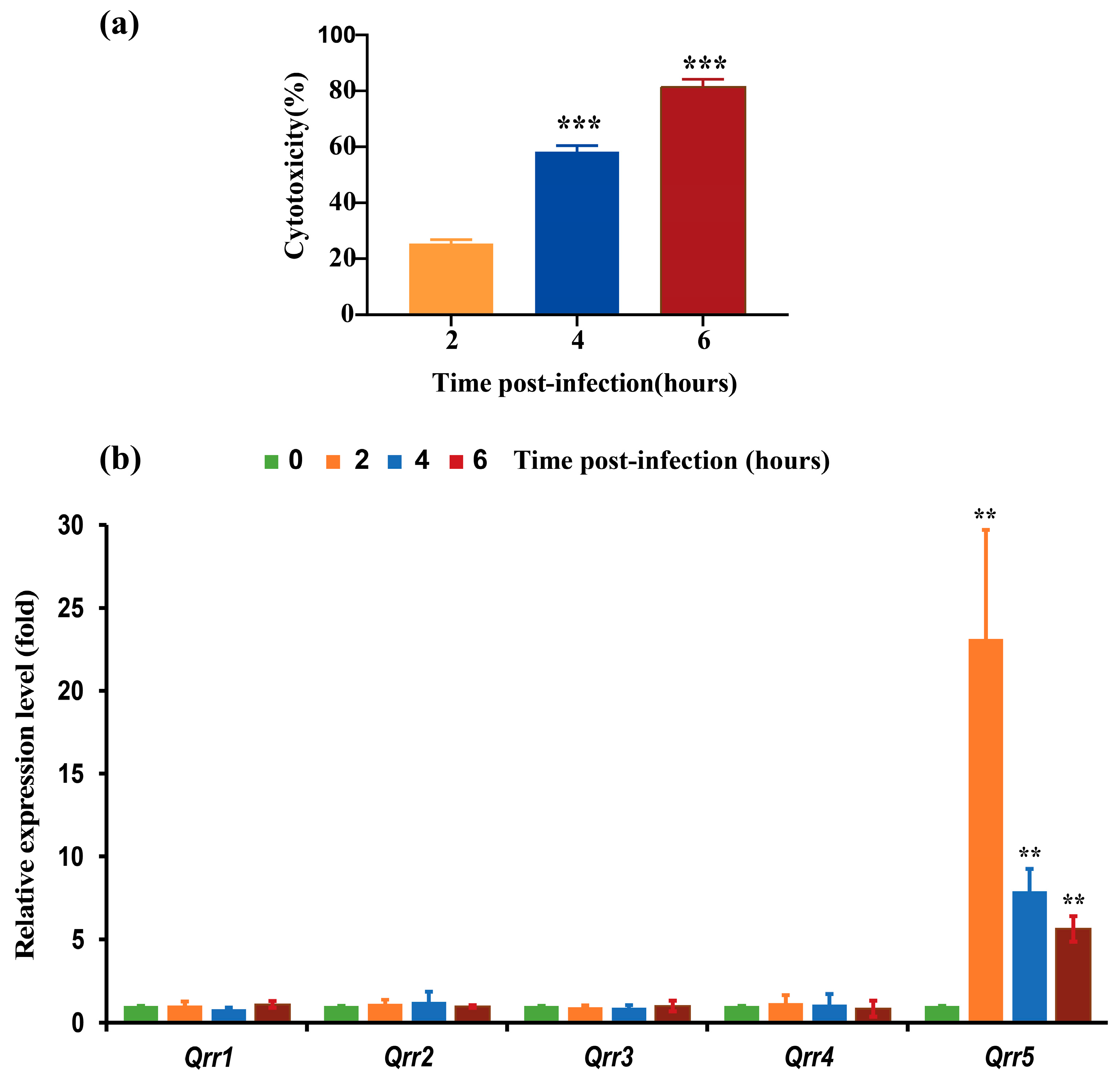
3.1. Expression of Different Qrrs during the Infection of V. parahaemolyticus RIMD2210633 to Caco-2 Cells

It has been found that Qrr1-5 negatively regulate the high cell density quorum-sensing regulator OpaR, which is required for biofilm formation and acts as a repressor of swarming motility [15,33]. However, we found that the cytotoxicity of Caco-2 cells increased with increasing post-infection time, and Qrr5 was significantly upregulated at different post-infection time points compared to Qrr14 (Figure 1a,b). A previous study found that only Qrr5 was significantly upregulated when V. parahaemolyticus infested the host, while the expression of Qrr1Qrr4 showed no change at all time points post-infection, which is completely consistent with our findings [42].
In the functional analysis of Qrr5, we found that Qrr5 was located in chromosome 1 from 1,728,021 to 1,728,129 in V. parahaemolyticus RIMD2210633 (Figure S1). The secondary structure of Qrr5 was portrayed according to its sequence, and the dG was −31.1 (Figure S1).

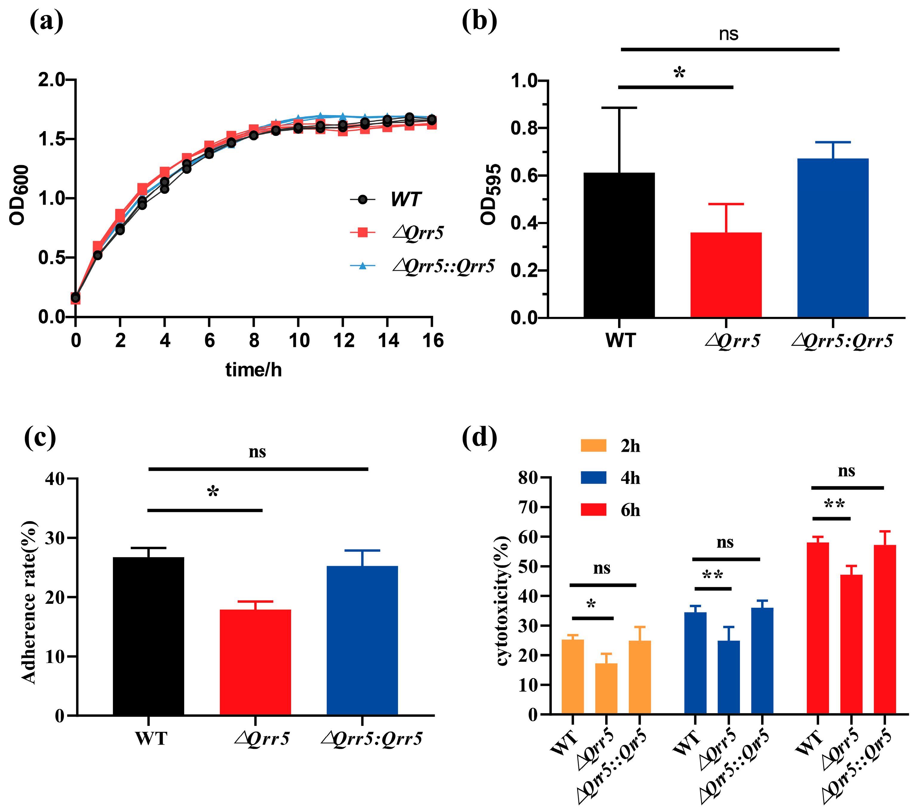
3.2. Qrr5 Is Required for Biofilm Formation, Cytotoxicity, and Adhesion to Caco-2 Cells in V. parahaemolyticus RIMD2210633

To examine the function of Qrr5 in V. parahaemolyticus, Qrr5 was deleted from the V. parahaemolyticus RIMD2210633 genome, and the mutants ΔQrr5 and ΔQrr5::Qrr5 were obtained and verified (Figure S2). In order to compare the difference in bacterial growth, we measured the OD600 after culture for 16 h and found no significant difference between the mutant and wild-type strain (Figure 2a). The biofilm formation of ΔQrr5 was significantly decreased at 48 h compared to that of the wild-type strain (Figure 2b). The adhesion rate of mutant ΔQrr5 to Caco-2 cells was also significantly lower than that of the wild-type (Figure 2c). Moreover, the cytotoxicity of Caco-2 cells infected by mutant ΔQrr5 was significantly attenuated compared to that of the wild-type and ΔQrr5::Qrr5 strains at different time points post-infection (Figure 2d). These data demonstrate that Qrr5 is required for biofilm formation, cytotoxicity, and adhesion to Caco-2 cells in V. parahaemolyticus RIMD2210633.

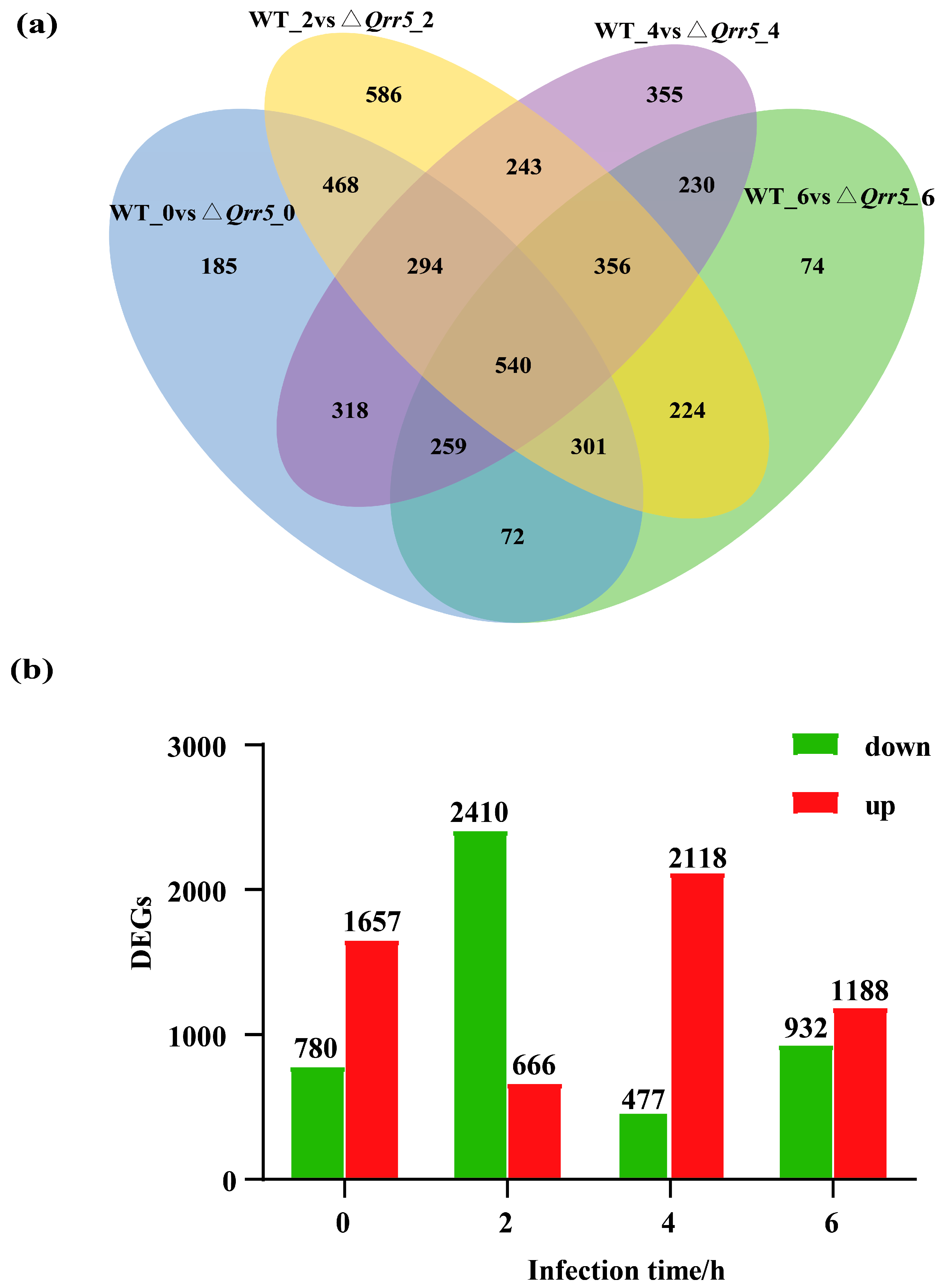
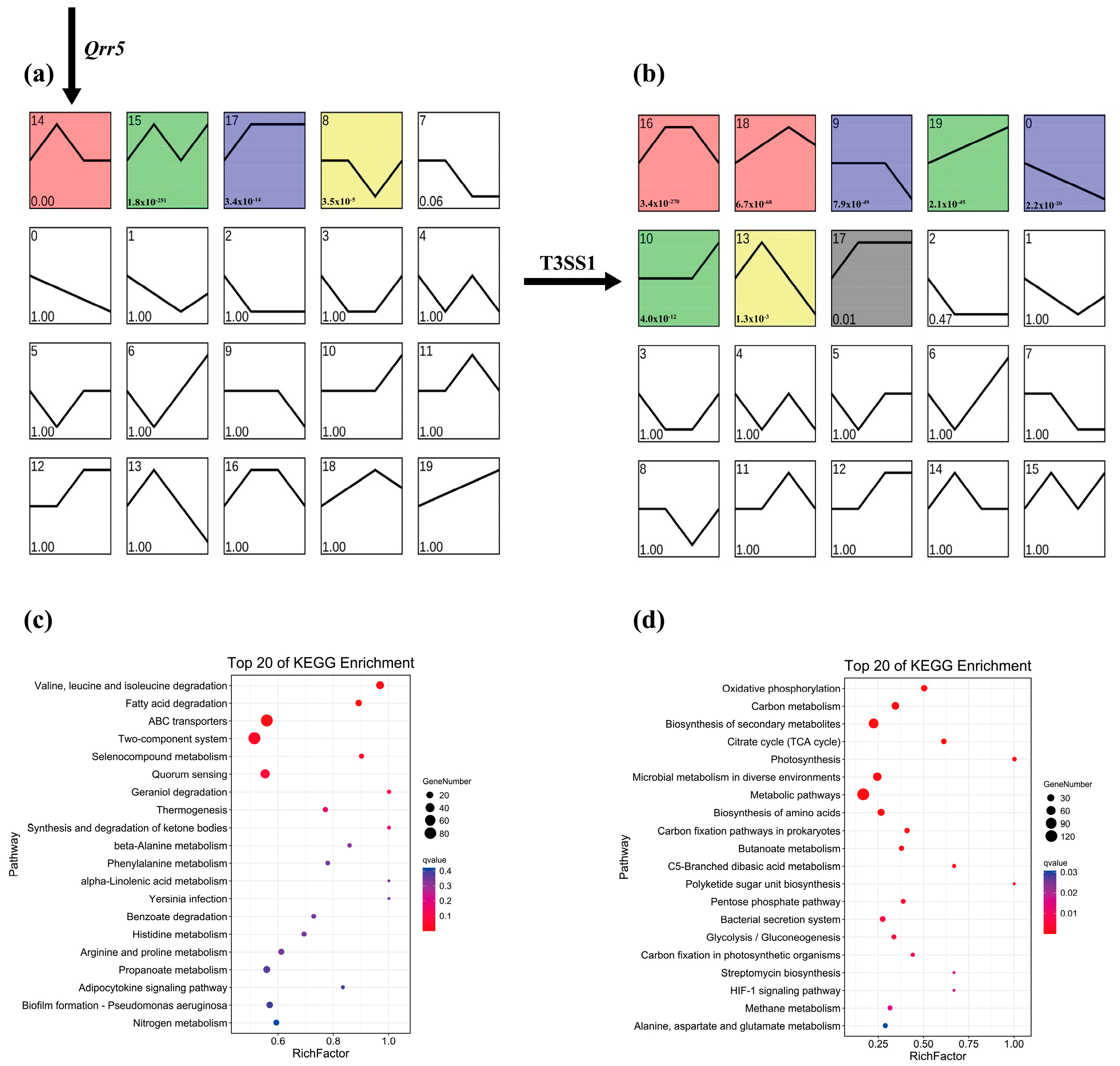
3.3. Untargeted Analysis of Transcriptome Data of ΔQrr5-Mutant V. parahaemolyticus RIMD2210633

To identify alterations in gene expression of V. parahaemolyticus during infection, which could be triggered by the precise regulation of virulence by Qrr5, we set various time points after infection to Caco-2 cells. Using the Deseq2 program, differentially expressed genes between wildtype and Qrr5 mutant strains (log2FC > 1; log2FC < −1 and p-value < 0.05) were identified during various periods (Figures S3 and S4). A core set of 540 common transcripts was found at various time points, and 185, 586, 355, and 74 transcripts were mainly altered at 0, 2, 4, and 6 h, respectively, in mutant ΔQrr5 compared to those in the wildtype strain (Figure 3a). The numbers of downregulated and upregulated genes among all DEGs between the ΔQrr5 and wild-type strains at various time points post-infection. (Figure 3b).

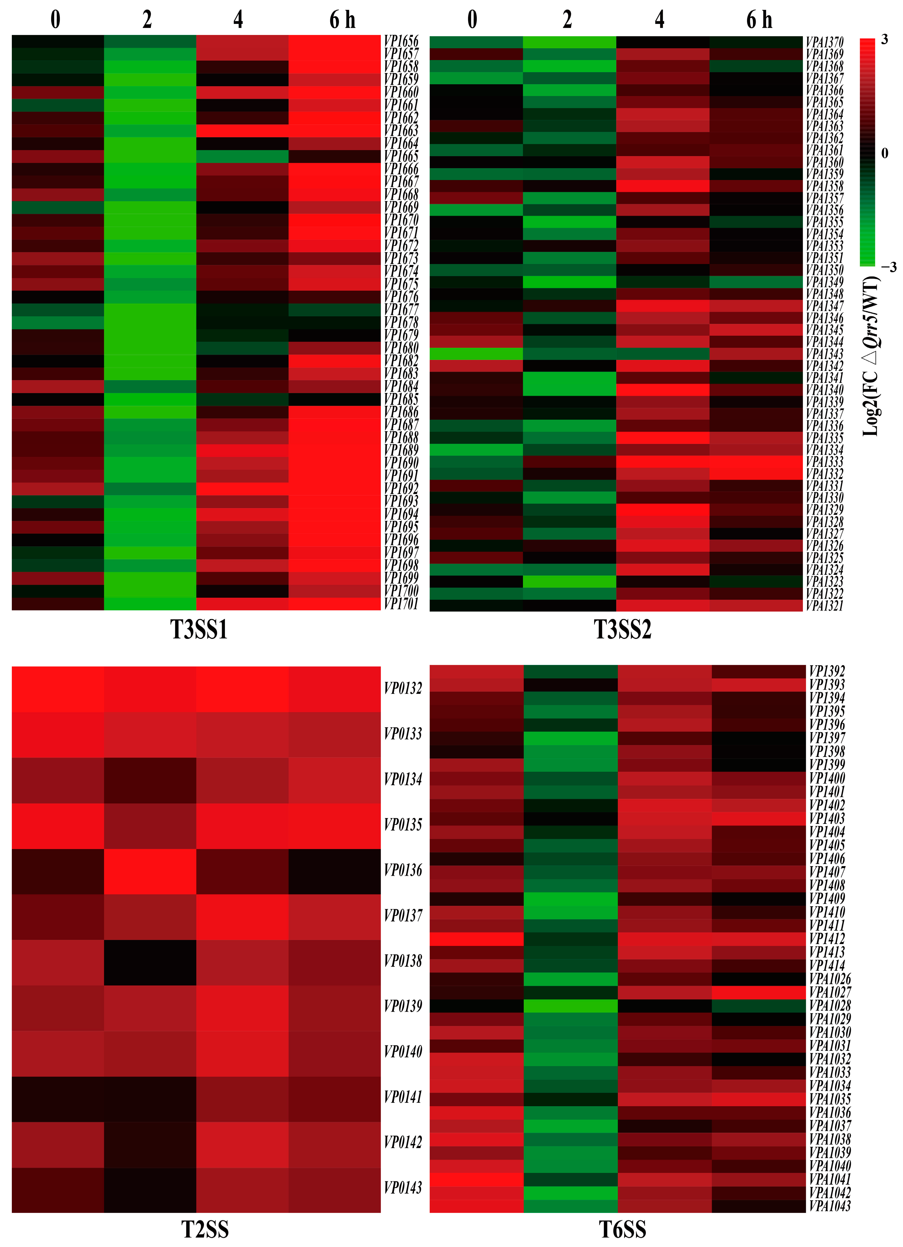
3.4. Targeted Transcriptome Analysis of ΔQrr5-Mutant V. parahaemolyticus RIMD2210633

Bacterial pathogens usually use various strategies to attack mammalian cells, preventing the immune system from responding and causing damage to infected tissue sites [43,44]. Pathogens frequently employ a secretion system to release virulence factors intracellularly into the host cells or extracellularly to interfere with host cell functions and promote bacterial infection [45,46]. Vibrio parahaemolyticus harbors several secretion systems, including T2SS, two T3SSs (T3SS1 and T3SS2), and two T6SSs. All T3SSs are associated with the toxins in mammalian hosts [16]. Reports on the function of T6SSs are limited, but it has been speculated that T6SS2 contributes to bacterial virulence [47,48]. sRNAs are post-transcriptional regulatory molecules that play an indispensable role in adjusting vital processes in bacterial physiology toward host invasion [49,50]. Analysis of the transcripts of V. parahaemolyticus revealed that the deletion of Qrr5 significantly affected the expression of the T3SS1 gene cluster during the infection period, especially at 6 h post-infection (Figure 4). The gene clusters of other secretion systems, such as T3SS2, T6SS, and T2SS, were slightly upregulated during the infection period (Figure 4).

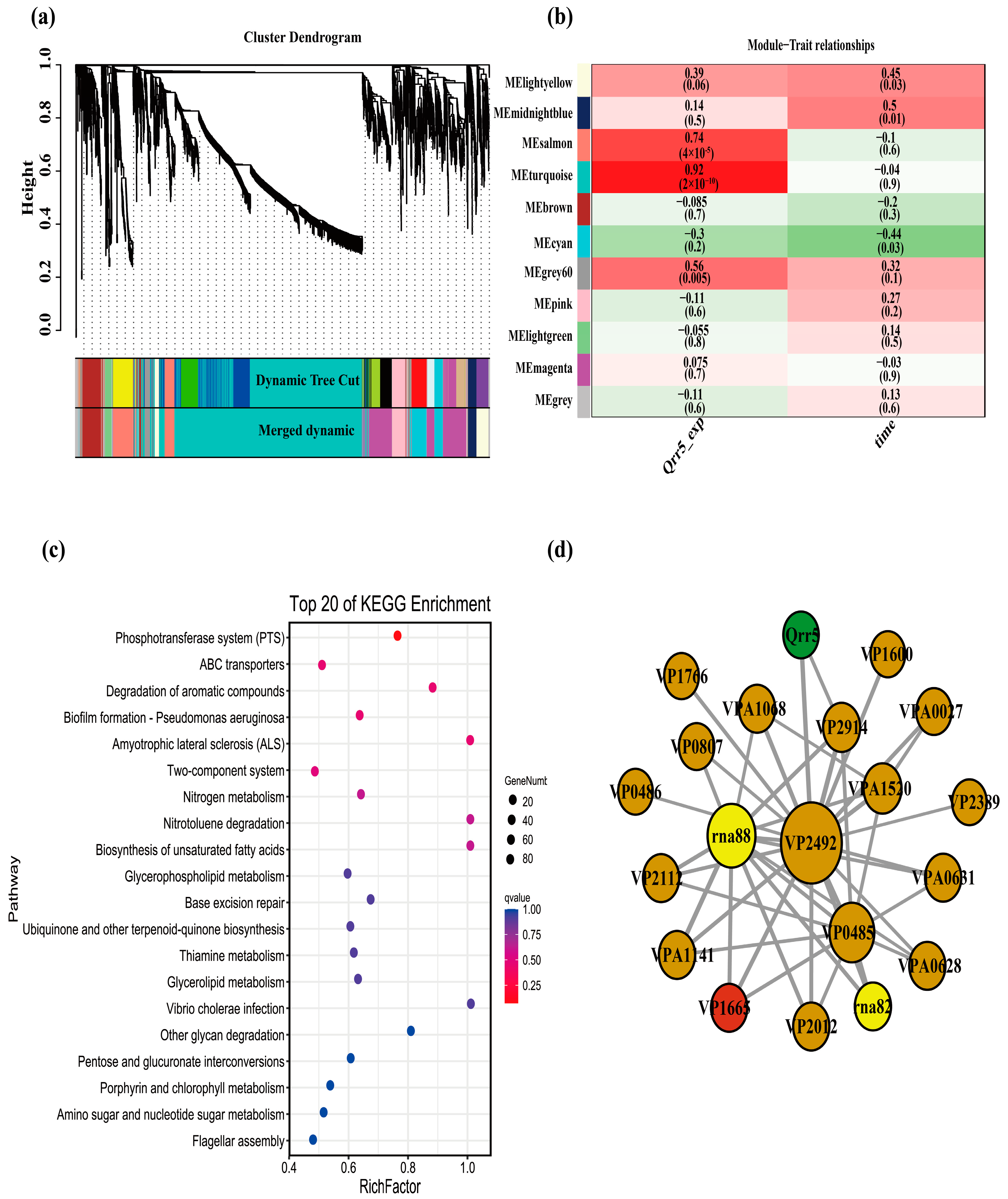
3.5. WGCNA of Transcriptome Data of ΔQrr5-Mutant V. parahaemolyticus RIMD2210633

To obtain an outline of the dynamic variations of DEGs during the process of infection with V. parahaemolyticus, we first performed a trend analysis of all genes in the wild-type and Qrr5 knockout strains separately using STEM to determine the expression trend of T3SS1 genes in wild-type. The results revealed that most T3SS1 genes in the wild-type group were distributed in profiles 14 and 15, whereas most T3SS1 genes in the ΔQrr5 group were distributed in profile 10 (Figure 5a,b). Based on the principle that the genes related to T3SS1 regulation should also be classified into the same profile, the genes in profiles 14 and 15 of the wild-type group were intersected with the genes in profile 10 of the ΔQrr5 group, and 138 genes were obtained. The KEGG pathway enrichment results of all genes in profile 14 (the profile where Qrr5 is located) of the wild-type group were mostly concentrated on ABC transporters and two-component systems (Figure 5c). All genes in profile 10 (the profile where most T3SS1 genes are located) of the ΔQrr5 group mainly focused on the biosynthesis of secondary metabolism (Figure 5d). Subsequently, the expression level of Qrr5 was used as a trait for WGCNA. All genes whose expression met the threshold were divided into 11 gene expression modules (Figure 6a). Qrr5 was located in the turquoise module, which was also the gene module most significantly correlated with Qrr5 expression (Figure 6b). The genes in the turquoise module were intersected with the above 138 genes, and a total of 61 intersecting genes with a potential co-expression relationship with Qrr5 were identified, consistent with the expression trend of the T3SS1 genes. KEGG pathway enrichment analysis of the turquoise module, where Qrr5 is located, showed that ABC transporters and two-component systems were the most related pathways (Figure 6c). Considering a co-expression weight greater than 0.5 as a potential direct regulatory relationship, a direct regulatory gene network of Qrr5 was constructed (Figure 6d). The functions of the co-expression network genes are listed in Table S2. The results showed that VP2914, VP0485, VP2492, and VP1665 exhibited strong interactions with Qrr5, which encodes D-2-hydroxyacid dehydrogenase, TIGR01212 family radical SAM protein, ammonium transporter, and T3SS1 chaperone SycN, respectively. A novel sRNA, EsrF, was found to respond to ammonium transporters to sense high ammonium concentrations in the colon and promote E. coli O157:H7 pathogenicity by enhancing bacterial motility and adhesion to host cells. The coexistence of ammonium transporter and channel mechanisms contributed to the virulence of pathogenic fungi [4,51]. Moreover, the T3SS1 chaperone SycN/YscB interacts with the secreted protein YopN via an N-terminal chaperone-binding domain to subvert the defenses of mammalian hosts in Yersinia pestis, and it was found that they interact with both EsaB and EsaM within the bacterial cells, contributing to the pathogenesis of Edwardsiella tarda [52,53,54]. Our results demonstrate that Qrr5 is an important sRNA in the regulation and the pathogenesis of V. parahaemolyticus. Analysis of the expression levels of Qrrs in this transcriptome revealed that Qrr5 was significantly upregulated at all time points post-infection, whereas Qrr1–4 expression showed no significant changes throughout, which was completely consistent with a previous finding [42]. In summary, Qrr5 regulates cytotoxicity mainly by regulating the T3SS1 gene, as well as participating in the ABC transporters and two-component system pathways during infection.
The expression of genes involved in the gene regulatory network of Qrr5 was further validated by qRT-PCR. The results showed that the expression patterns of these genes at 0, 2, 4, and 6 h post-infection were generally in accordance with those by RNA-Seq. At each post-infection time point, VP1766, VPA1068, VP0485, VP2914, and VP1665 were up-regulated. Moreover, VP2492, VP0486, and VPA0628 were up-regulated at 2 h and down-regulated at 0, 4, and 6 h post-infection (Figure S5).

4. Conclusions

This study reveals that Qrr5 is related to cytotoxicity and can significantly positively regulate virulence gene expression levels in V. parahaemolyticus. This study also elucidates the systematic transcriptomic profile of V. parahaemolyticus mutants during infection of Caco-2 cells. A total of 185, 586, 355, and 74 DEGs were obtained at 0, 2, 4, and 6 h post-infection, respectively, which were mainly related to ABC transporters and two-component system pathways. Using WGCNA, we identified 17 key genes involved in the gene regulatory network associated with Qrr5. These findings highlight the importance of core genes that may be regulated by Qrr5 during infection in Caco-2 cells and provide a basis for further research on the mechanisms of V. parahaemolyticus infection.

Supplementary Materials

The following supporting information can be downloaded at: https://www.mdpi.com/article/10.3390/microorganisms10102084/s1, Figure S1. The genetic map and structure of sRNA Qrr5 in V. parahaemolyticus. Figure S2. Construction and confirmation of ΔQrr5 mutant. Figure S3. Each sample clustering of RNA-Seq data. Figure S4. The scale independence and mean connectivity of RNA-Seq data. Figure S5. Relative expression of several differentially expressed genes (ΔQrr5/wild-type) at the mRNA level determined by qPCR during various infection periods. Table S1. Oligonucleotide Primers. Table S2. The function of genes in the WGCNA co-expression network.

Author Contributions

For research articles with several authors, F.Z., R.P., J.Z., J.W., M.C., L.X., Q.Y., S.W., M.Y., Q.G., Y.D., H.Z. and Q.W. conceived and designed the research; F.Z. performed experiments and R.P. analyzed the data. F.Z. wrote the paper. S.H., R.P. and F.Z. discussed the results and wrote the manuscript. All authors have read and agreed to the published version of the manuscript.

Funding

This work was supported completely by grants from the National Natural Science Foundation of China (No. 31730070; No. 31901796).

Data Availability Statement

Not applicable.

Conflicts of Interest

The authors declare no conflict of interest.

References

  1. Waters, L.S.; Storz, G. Regulatory RNAs in Bacteria. Cell 2009, 136, 615–628. [Google Scholar] [CrossRef] [PubMed] [Green Version]
  2. Hébrard, M.; Kröger, C.; Srikumar, S.; Colgan, A.; Händler, K.; Hinton, J.C.D. sRNAs and the virulence of Salmonella enterica serovar Typhimurium. RNA Biol. 2012, 9, 437–445. [Google Scholar] [CrossRef] [Green Version]
  3. Djapgne, L.; Oglesby, A.G. Impacts of Small RNAs and Their Chaperones on Bacterial Pathogenicity. Front. Cell. Infect. Microbiol. 2021, 11, 604511. [Google Scholar] [CrossRef] [PubMed]
  4. Jia, T.; Liu, B.; Mu, H.; Qian, C.; Wang, L.; Li, L.; Lu, G.; Zhu, W.; Guo, X.; Yang, B.; et al. A Novel Small RNA Promotes Motility and Virulence of Enterohemorrhagic Escherichia coli O157:H7 in Response to Ammonium. mBio 2021, 12. [Google Scholar] [CrossRef] [PubMed]
  5. Lenz, D.H.; Mok, K.C.; Lilley, B.N.; Kulkarni, R.V.; Wingreen, N.S.; Bassler, B.L. The Small RNA Chaperone Hfq and Multiple Small RNAs Control Quorum Sensing in Vibrio harveyi and Vibrio cholerae. Cell 2004, 118, 69–82. [Google Scholar] [CrossRef] [Green Version]
  6. Bardill, J.P.; Zhao, X.; Hammer, B.K. The Vibrio cholerae quorum sensing response is mediated by Hfq-dependent sRNA/mRNA base pairing interactions. Mol. Microbiol. 2011, 80, 1381–1394. [Google Scholar] [CrossRef] [PubMed]
  7. Vincent, H.A.; Henderson, C.A.; Stone, C.M.; Cary, P.D.; Gowers, D.M.; Sobott, F.; Taylor, J.E.; Callaghan, A.J. The low-resolution solution structure of Vibrio cholerae Hfq in complex with Qrr1 sRNA. Nucleic Acids Res. 2012, 40, 8698–8710. [Google Scholar] [CrossRef]
  8. Tu, K.C.; Bassler, B.L. Multiple small RNAs act additively to integrate sensory information and control quorum sensing in Vibrio harveyi. Genes Dev. 2007, 21, 221–233. [Google Scholar] [CrossRef] [Green Version]
  9. Lu, Y.; Rafiq, A.; Zhang, Z.; Aslani, F.; Fijak, M.; Lei, T.; Wang, M.; Kumar, S.; Klug, J.; Bergmann, M.; et al. Uropathogenic Escherichia coli virulence factor hemolysin A causes programmed cell necrosis by altering mitochondrial dynamics. FASEB J. 2018, 32, 4107–4120. [Google Scholar] [CrossRef] [Green Version]
  10. Zhang, Y.; Qiu, Y.; Gao, H.; Sun, J.; Li, X.; Zhang, M.; Xue, X.; Yang, W.; Ni, B.; Hu, L.; et al. OpaR Controls the Metabolism of c-di-GMP in Vibrio parahaemolyticus. Front. Microbiol. 2021, 12, 676436. [Google Scholar] [CrossRef]
  11. Feng, L.; Rutherford, S.T.; Papenfort, K.; Bagert, J.D.; Van Kessel, J.C.; Tirrell, D.A.; Wingreen, N.S.; Bassler, B.L. A Qrr noncoding RNA deploys four different regulatory mechanisms to optimize quorum-sensing dynamics. Cell 2015, 160, 228–240. [Google Scholar] [CrossRef] [PubMed] [Green Version]
  12. Chaparian, R.R.; Ball, A.S.; van Kessel, J.C. Hierarchical Transcriptional Control of the LuxR Quorum-Sensing Regulon of Vibrio harveyi. J. Bacteriol. 2022, 202, e00047-20. [Google Scholar] [CrossRef] [PubMed]
  13. Wen, Y.; Kim, I.H.; Kim, K.-S. Iron- and Quorum-sensing Signals Converge on Small Quorum-regulatory RNAs for Coordinated Regulation of Virulence Factors in Vibrio vulnificus*. J. Biol. Chem. 2016, 291, 14213–14230. [Google Scholar] [CrossRef] [PubMed] [Green Version]
  14. Lee, K.-W.; Wen, Y.; Park, N.-Y.; Kim, K.-S. Quorum sensing and iron-dependent coordinated control of autoinducer-2 production via small RNA RyhB in Vibrio vulnificus. Sci. Rep. 2022, 12, 831. [Google Scholar] [CrossRef]
  15. Tague, J.G.; Regmi, A.; Gregory, G.J.; Boyd, E.F. Fis Connects Two Sensory Pathways, Quorum Sensing and Surface Sensing, to Control Motility in Vibrio parahaemolyticus. Front. Microbiol. 2021, 12. [Google Scholar] [CrossRef]
  16. Letchumanan, V.; Chan, K.-G.; Lee, L.-H. Vibrio parahaemolyticus: A review on the pathogenesis, prevalence, and advance molecular identification techniques. Front. Microbiol. 2014, 5, 705. [Google Scholar] [CrossRef] [Green Version]
  17. Zhang, L.; Orth, K. Virulence determinants for Vibrio parahaemolyticus infection. Curr. Opin. Microbiol. 2013, 16, 70–77. [Google Scholar] [CrossRef]
  18. Wang, J.; Zhan, Y.; Sun, H.; Fu, X.; Kong, Q.; Zhu, C.; Mou, H. Regulation of Virulence Factors Expression During the Intestinal Colonization of Vibrio parahaemolyticus. Foodborne Pathog. Dis. 2022, 19, 169–178. [Google Scholar] [CrossRef]
  19. Li, L.; Meng, H.; Gu, D.; Li, Y.; Jia, M. Molecular mechanisms of Vibrio parahaemolyticus pathogenesis. Microbiol. Res. 2019, 222, 43–51. [Google Scholar] [CrossRef]
  20. Park, K.-S.; Ono, T.; Rokuda, M.; Jang, M.; Okada, K.; Iida, T.; Honda, T. Functional Characterization of Two Type III Secretion Systems of Vibrio parahaemolyticus. Infect. Immun. 2004, 72, 6659–6665. [Google Scholar] [CrossRef]
  21. Piñeyro, P.; Zhou, X.; Orfe, L.H.; Friel, P.J.; Lahmers, K.; Call, D.R. Development of Two Animal Models To Study the Function of Vibrio parahaemolyticus Type III Secretion Systems. Infect. Immun. 2010, 78, 4551–4559. [Google Scholar] [CrossRef] [PubMed] [Green Version]
  22. Plaza, N.; Urrutia, I.M.; Garcia, K.; Waldor, M.K.; Blondel, C.J. Identification of a Family of Vibrio Type III Secretion System Effectors That Contain a Conserved Serine/Threonine Kinase Domain. mSphere 2022, 6, e00599-21. [Google Scholar] [CrossRef]
  23. Erhardt, M.; Namba, K.; Hughes, K.T. Bacterial nanomachines: The flagellum and type III injectisome. Cold Spring Harb. Perspect. Biol. 2010, 2, a000299. [Google Scholar] [CrossRef] [PubMed] [Green Version]
  24. Zhang, Y.; Hu, L.; Qiu, Y.; Osei-Adjei, G.; Tang, H.; Zhang, Y.; Zhang, R.; Sheng, X.; Xu, S.; Yang, W.; et al. QsvR integrates into quorum sensing circuit to control Vibrio parahaemolyticus virulence. Environ. Microbiol. 2019, 21, 1054–1067. [Google Scholar] [CrossRef] [PubMed]
  25. Cornelis, G.R. The type III secretion injectisome. Nat. Reviews. Microbiol. 2006, 4, 811–825. [Google Scholar] [CrossRef]
  26. Wang, L.; Ling, Y.; Jiang, H.; Qiu, Y.; Qiu, J.; Chen, H.; Yang, R.; Zhou, D. AphA is required for biofilm formation, motility, and virulence in pandemic Vibrio parahaemolyticus. Int. J. Food Microbiol. 2013, 160, 245–251. [Google Scholar] [CrossRef]
  27. Zeb, S.; Shah, M.A.; Yasir, M.; Awan, H.M.; Prommeenate, P.; Klanchui, A.; Wren, B.W.; Thomson, N.; Bokhari, H. Type III secretion system confers enhanced virulence in clinical non-O1/non-O139 Vibrio cholerae. Microb. Pathog. 2019, 135, 103645. [Google Scholar] [CrossRef]
  28. Nydam, S.; Shah, D.; Call, D. Transcriptome analysis of Vibrio parahaemolyticus in type III secretion system 1 inducing conditions. Front. Cell. Infect. Microbiol. 2014, 4, 1. [Google Scholar] [CrossRef]
  29. Lian, L.; Li, W.; Xue, T.; Ren, J.; Tang, F.; Liu, Y.; Xue, F.; Dai, J. Comparative transcriptomic analysis provides insights into transcription mechanisms of Vibrio parahaemolyticus T3SS during interaction with HeLa cells. Braz. J. Microbiol. 2022, 53, 289–301. [Google Scholar] [CrossRef]
  30. Liu, H.; Liu, W.; He, X.; Chen, X.; Yang, J.; Wang, Y.; Li, Y.; Ren, J.; Xu, W.; Zhao, Y. Characterization of a cell density-dependent sRNA, Qrr, and its roles in the regulation of the quorum sensing and metabolism in Vibrio alginolyticus. Appl. Microbiol. Biotechnol. 2020, 104, 1707–1720. [Google Scholar] [CrossRef]
  31. Azimi, S.; Klementiev, A.D.; Whiteley, M.; Diggle, S.P. Bacterial Quorum Sensing During Infection. Annu. Rev. Microbiol. 2020, 74, 201–219. [Google Scholar] [CrossRef] [PubMed]
  32. Suresh, S.; Alva, P.P.; Premanath, R. Modulation of quorum sensing-associated virulence in bacteria: Carbohydrate as a key factor. Arch. Microbiol. 2021, 203, 1881–1890. [Google Scholar] [CrossRef]
  33. Zhang, Y.; Qiu, Y.; Tan, Y.; Guo, Z.; Yang, R.; Zhou, D. Transcriptional Regulation of opaR, qrr2–4 and aphA by the Master Quorum-Sensing Regulator OpaR in Vibrio parahaemolyticus. PLoS ONE 2012, 7, e34622. [Google Scholar] [CrossRef] [PubMed]
  34. Zuker, M. Mfold web server for nucleic acid folding and hybridization prediction. Nucleic Acids Res. 2003, 31, 3406–3415. [Google Scholar] [CrossRef] [PubMed]
  35. Zhou, X.; Shah, D.; Konkel, M.; Call, D. Type III secretion system 1 genes in Vibrio parahaemolyticus are positively regulated by ExsA and negatively regulated by ExsD. Mol. Microbiol. 2008, 69, 747–764. [Google Scholar] [CrossRef] [Green Version]
  36. O’Toole, G.A.; Kolter, R. Initiation of biofilm formation in Pseudomonas fluorescens WCS365 proceeds via multiple, convergent signalling pathways: A genetic analysis. Mol. Microbiol. 1998, 28, 449–461. [Google Scholar] [CrossRef] [PubMed]
  37. Wingett, S.; Andrews, S. FastQ Screen: A tool for multi-genome mapping and quality control. F1000Research 2018, 7, 1338. [Google Scholar] [CrossRef]
  38. Wang, L.; Zhang, W.; Li, Q. AssocTests: An R Package for Genetic Association Studies. J. Stat. Softw. 2020, 94, 1–26. [Google Scholar] [CrossRef]
  39. Ernst, J.; Bar-Joseph, Z. STEM: A tool for the analysis of short time series gene expression data. BMC Bioinform. 2006, 7, 191. [Google Scholar] [CrossRef] [Green Version]
  40. Langfelder, P.; Horvath, S. WGCNA: An R package for weighted correlation network analysis. BMC Bioinform. 2008, 9, 559. [Google Scholar] [CrossRef]
  41. Bray, N.L.; Pimentel, H.; Melsted, P.; Pachter, L. Near-optimal probabilistic RNA-seq quantification. Nat. Biotechnol. 2016, 34, 525–527. [Google Scholar] [CrossRef] [PubMed]
  42. Livny, J.; Zhou, X.; Mandlik, A.; Hubbard, T.; Davis, B.M.; Waldor, M.K. Comparative RNA-Seq based dissection of the regulatory networks and environmental stimuli underlying Vibrio parahaemolyticus gene expression during infection. Nucleic Acids Res. 2014, 42, 12212–12223. [Google Scholar] [CrossRef] [PubMed] [Green Version]
  43. Santos, J.C.; Broz, P. Sensing of invading pathogens by GBPs: At the crossroads between cell-autonomous and innate immunity. J. Leukoc. Biol. 2018, 104, 729–735. [Google Scholar] [CrossRef]
  44. Netea, M.G.; Schlitzer, A.; Placek, K.; Joosten, L.A.B.; Schultze, J.L. Innate and Adaptive Immune Memory: An Evolutionary Continuum in the Host’s Response to Pathogens. Cell Host Microbe 2019, 25, 13–26. [Google Scholar] [CrossRef] [Green Version]
  45. Zhang, W.; Xie, R.; Zhang, X.D.; Lee, L.T.O.; Zhang, H.; Yang, M.; Peng, B.; Zheng, J. Organism dual RNA-seq reveals the importance of BarA/UvrY in Vibrio parahaemolyticus virulence. FASEB J. 2020, 34, 7561–7577. [Google Scholar] [CrossRef] [Green Version]
  46. Ning, X.; Sun, L. Gene network analysis reveals a core set of genes involved in the immune response of Japanese flounder (Paralichthys olivaceus) against Vibrio anguillarum infection. Fish Shellfish. Immunol. 2020, 98, 800–809. [Google Scholar] [CrossRef] [PubMed]
  47. Yu, Y.; Fang, L.; Zhang, Y.; Sheng, H.; Fang, W. VgrG2 of type VI secretion system 2 of Vibrio parahaemolyticus induces autophagy in macrophages. Front. Microbiol. 2015, 6. [Google Scholar] [CrossRef] [Green Version]
  48. MacIntyre, D.L.; Miyata, S.T.; Kitaoka, M.; Pukatzki, S. The Vibrio cholerae type VI secretion system displays antimicrobial properties. Proc. Natl. Acad. Sci. USA 2010, 107, 19520–19524. [Google Scholar] [CrossRef] [Green Version]
  49. Babitzke, P.; Romeo, T. CsrB sRNA family: Sequestration of RNA-binding regulatory proteins. Curr. Opin. Microbiol. 2007, 10, 156–163. [Google Scholar] [CrossRef]
  50. Chen, J.; Morita, T.; Gottesman, S. Regulation of Transcription Termination of Small RNAs and by Small RNAs: Molecular Mechanisms and Biological Functions. Front. Cell. Infect. Microbiol. 2019, 9, 201. [Google Scholar] [CrossRef]
  51. Gordon, W.; Sofia, B.A.; Adriana, B.; Giulia, T.; Gaëtan, D.M.; Thomas, H.; Hoskisson, P.A.; Ulrich, Z.; Maria, M.A.; Mélanie, B.; et al. Coexistence of Ammonium Transporter and Channel Mechanisms in Amt-Mep-Rh Twin-His Variants Impairs the Filamentation Signaling Capacity of Fungal Mep2 Transceptors. mBio 2022, 13, e02913-21. [Google Scholar] [CrossRef]
  52. Plano, G.; Joseph, S. The SycN/YscB chaperone-binding domain of YopN is required for the calcium-dependent regulation of Yop secretion by Yersinia pestis. Front. Cell. Infect. Microbiol. 2013, 3. [Google Scholar]
  53. Yi, L.L.; Pin, N.; Bing, Y.H.; Xia, X.H. Regulation of Type III Secretion of Translocon and Effector Proteins by the EsaB/EsaL/EsaM Complex in Edwardsiella tarda. Infect. Immun. 2022, 85, e00322-17. [Google Scholar] [CrossRef] [Green Version]
  54. Plano, G.V.; Schesser, K. The Yersinia pestis type III secretion system: Expression, assembly and role in the evasion of host defenses. Immunol. Res. 2013, 57, 237–245. [Google Scholar] [CrossRef] [PubMed]
Figure 1. (a) The cytotoxicity of Caco-2 infected by Vibrio parahaemolyticus RIMD2210633 during various post-infection time points. (b) The relative mRNA expression of five sRNA Qrrs in V. parahaemolyticus RIMD2210633 during various post-infection time points toward Caco-2 cells. p-value < 0.01 (**); p-value < 0.001 (***).
Microorganisms 10 02084 g001
Figure 2. The phenotypes of ΔQrr5 deletion mutant of Vibrio parahaemolyticus RIMD2210633. (a) The growth curves of wild-type, ΔQrr5 deletion mutant, and the complementary strains were measured at OD600 for 16 h. (b) Biofilm formation of different strains was measured at 595 nm at 48 h by crystal violet staining. (c) The adherence rate of different strains to Caco-2 cells. (d) The cytotoxicity of Caco-2 cells (MOI = 100:1) infected by different strains at 2, 4, and 6 h post-infection. p-value < 0.05 (*); p-value < 0.01 (**); ns: not significant.
Microorganisms 10 02084 g002
Figure 3. The transcription outcomes of V. parahaemolyticus after its infection to Caco-2 cells. (a) Venn diagram portraying the number of significantly expressed genes (log2FC > 1; log2FC < −1 and p-value < 0.05) of wild-type and ΔQrr5 strains across various post-infection time points. (b) The numbers of the down-regulated and up-regulated of all differentially expressed genes in the wild-type and ΔQrr5 strains at various infection time points.
Microorganisms 10 02084 g003
Figure 4. Differential expression of secretion system gene clusters in V. parahaemolyticus mutant ΔQrr5 during the infection period compared to the wild-type strain (including T3SS1, T3SS2, T2SS, and T6SS gene clusters).
Microorganisms 10 02084 g004
Figure 5. The STEM analysis of mutant ΔQrr5 during the infection period compared to wild-type strain by RNA-Seq. (a) Gene trend expression enrichment map of the wild-type group. (b) Gene trend expression enrichment map of ΔQrr5 group. The colors represent the module where the major genes are located, and the white represents the module where fewer genes are located. (c) KEGG pathway enrichment analysis results of all genes in profile 14 (the profile where Qrr5 is located) of the wild-type group trend. (d) KEGG pathway enrichment analysis results of all genes in profile 10 (the profile where most T3SS1 genes are located) of the ΔQrr5 group trend.
Microorganisms 10 02084 g005
Figure 6. The WGCNA analysis of mutant ΔQrr5 during the infection period compared to wild-type strain by RNA-Seq. (a) Hierarchical cluster tree showing eleven modules of co-expressed genes. Each of the genes is represented by a leaf in the tree, and each of the eleven modules by a major tree branch. (b) Module-Qrr5 -expression/times correlations and corresponding P-values. (c) The KEGG pathway enrichment analysis results of the turquoise module where Qrr5 is located by WGCNA. (d) The co-expression network of related genes is regulated by Qrr5. (The red dot was the T3SS1 gene).
Microorganisms 10 02084 g006
Table 1. Bacterial strains and plasmids were used in this study.
Strain or PlasmidCharacteristicsReference
V. parahaemolyticus RIMD 2210633Parent strain, serotype O3:K6[33]
ΔQrr5 mutantRIMD 2210633 ΔQrr5This study
ΔQrr5::Qrr5pBAD33-Qrr5 into ΔQrr5 mutantThis study
E. coli SM17 λpirTpR SmR recA, thi, pro, hsdR-M+RP4:2-Tc:Mu:Km Tn7λpir[33]
pDS132R6K ori, mobRP4 SacB Cmr[33]
pBAD33pBR322 ori, AmprThis study
Publisher’s Note: MDPI stays neutral with regard to jurisdictional claims in published maps and institutional affiliations.

