Article

Performance of Urine Reagent Strips in Detecting the Presence and Estimating the Prevalence and Intensity of Schistosoma haematobium Infection

1
Department of Epidemiology, College of Public Health, University of Nebraska Medical Center, Omaha, NE 68198, USA
2
Aklilu Lemma Institute of Pathobiology, Addis Ababa University, Addis Ababa P.O. Box 1176, Ethiopia
*
Author to whom correspondence should be addressed.
Microorganisms 2022, 10(10), 2062; https://doi.org/10.3390/microorganisms10102062
Submission received: 5 October 2022 / Revised: 16 October 2022 / Accepted: 17 October 2022 / Published: 19 October 2022
(This article belongs to the Special Issue Childhood Infections)

Abstract

:
The performance of the urine reagent strips (URS) in detecting the presence and estimating the intensity of Schistosoma haematobium infection was evaluated using urine filtration microscopy as a reference standard. Urine samples collected from 1288 school-age children living in five villages of the Afar and one village in the Gambella Regional States of Ethiopia between October 2021 and April 2022 were examined using urine filtration and URS. The prevalence of S. haematobium infection was 31.6% based on urine filtration and 32.1% using URS. Using results of the urine filtration as a reference, the sensitivity, specificity, negative predictive values, and accuracy of the URS in detecting S. haematobium egg-positive urine specimens were 73.7%, 87.8%, 87.1%, and 82.8%, respectively. Sensitivity increased significantly with an increase in the urine egg count. Specificity was greater in low prevalence settings and among children aged 5–9 years. The level of hematuria detected was trace (19.1%), weak (30.2%), moderate (36.0%), or high (14.7%). The log odds of showing higher-level hematuria significantly increased as the number of egg counts in urine increased. In conclusion, URS remains good in rapidly screening individuals for S. haematobium infection, but the sensitivity of the test could be lower, particularly when the intensity of the infection is light.

1. Introduction

Human schistosomiasis is one of the neglected tropical parasitic diseases caused by blood flukes of the genus Schistosoma. Six Schistosoma species can infect humans, namely Schistosoma mansoni, S. haematobium, S. japonicum, S. mekongi, S. intercalatum, or S. guineensis [1]. Except for S. haematobium, which causes urogenital schistosomiasis, the rest of the species cause intestinal schistosomiasis. Despite this diversity in schistosome species, most schistosomiasis cases are due to S. mansoni and S. haematobium. Geographically, about 90% of human schistosomiasis cases and an estimated 280,000 deaths occur in sub-Saharan Africa [2]. S. haematobium affects more than 112 million people in the world, mainly in sub-Saharan Africa and the Middle East, causing urogenital schistosomiasis [3,4].
In Ethiopia, urogenital schistosomiasis is endemic in the lowlands of Afar, Gambella, Beneshangul Gumuz, and the Somali Regional States, with varying degrees of infection [5,6]. The prevalence of S. haematobium infection reported among children was 35.9% in those living in Abobo primary school in Gambella Regional State [5], 20.8% in those living in seven villages of Afar Regional State [7], and 47.6% in those living in Hassoba [8]. Another study also reported S. haematobium with a prevalence of 24.2% among children in Abobo village of Gambella Regional State, 5.6% among children in Kumruk village of Beneshangul Gumuz Regional State, and 7.0% among children in Hassoba-Buri village of Afar Regional State [9]. Despite the ongoing preventive treatment of school-age children using praziquantel since 2015 [10], studies done so far reveal that urogenital schistosomiasis remains a public health threat in at least the three regional states of the country [5,6,7,8,9].
Periodic mass drug administration with praziquantel in school-age children to control schistosomiasis is being implemented upon the recommendation by WHO in endemic countries, including Ethiopia. However, in sub-Saharan Africa, children in endemic areas get infected by two years of age, and most remain with chronic infections through their school age and adulthood. As schistosome-infected preschool children are not routinely screened and treated, they constitute a potentially high-risk group for accumulation of morbidity partly because of a lack of child-friendly pediatric praziquantel formulation [11,12].
Mass treatment with praziquantel is expected to change the dynamics of S. haematobium infection, which is measured using diagnostics at the individual, population, and environmental levels [13]. To quantify the burden of and contain the transmission of S. haematobium, the availability of accurate diagnostics remains a top priority. However, there is no gold standard approach universally recommended for diagnosing S. haematobium infection. Although counting the parasite eggs in urine under the microscope has been a standard approach [3,4], the accuracy of the test may vary with the intensity and prevalence of infection, the number of days the sample was collected, and the filtration technique [3,4]. Due to the ongoing control programs that could lead to reduced prevalence and intensity of infection in endemic regions, the sensitivity of urine filtration microscopy may decrease significantly. Diagnosis of samples collected over multiple days may improve the sensitivity of the urine filtration microscopy [2,3,4], but this will incur cost and time.
In the effort to have the best diagnostics for S. haematobium infection, there have been evaluations of tools for the detection of hematuria (urine reagent strips), antigens (circulating anodic or cathodic antigen) or DNA (molecular tests) in urine or antibody released against the parasite in the blood (ELISA) [2,3,4]. Detection of hematuria using urine reagent strips (URS) has been widely used as a proxy for S. haematobium infection. As URS are less expensive, friendly, return results very quickly, and are less affected by the circadian production of schistosome eggs [2,3,4], they are recommended for rapid screening of S. haematobium infection in the field. Detection of hematuria using a reagent strip could particularly be useful for testing S. haematobium infection in school-aged children, as hematuria in children will strongly correlate with urogenital schistosomiasis [14]. However, the sensitivity of the URS in detecting S. haematobium infection showed variation in the prevalence and intensity of infection [7,9,14]. In this study, we examined the performance of the URS in detecting the presence and estimating the prevalence and intensity of S. haematobium infection among school-age children in selected study sites in Afar and the Gambella Regional States of Ethiopia that have been shown to have varied intensity of infection.

2. Methods

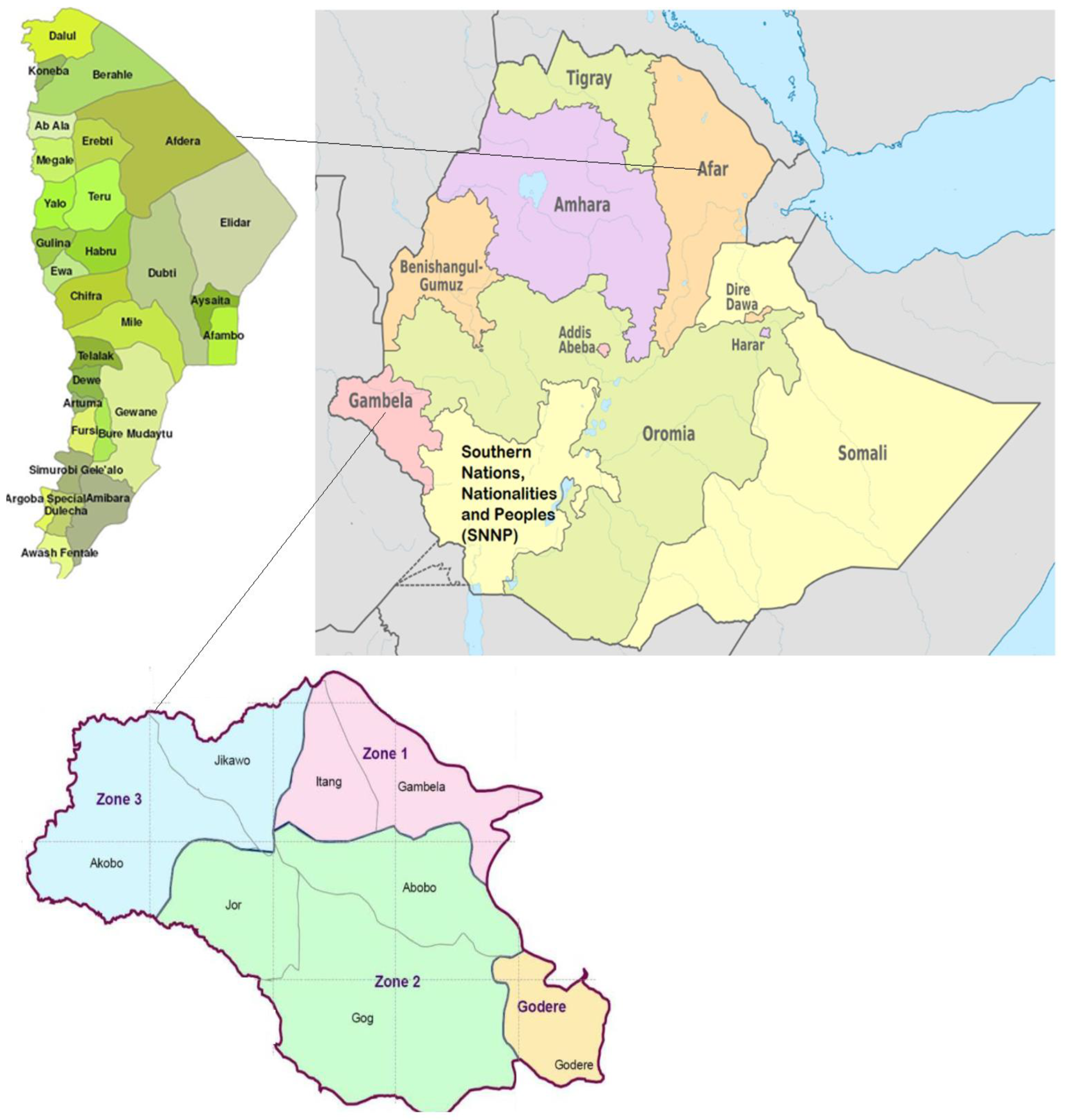
2.1. Study Area and Population

The study was undertaken in Buri, Kusra, Hassoba, Kelhat, and Andada villages in the Afar Regional State, northeastern Ethiopia, and Abobo village in the Gambella Regional State, southwestern Ethiopia (Figure 1). The villages were selected purposively. The mean annual rainfall and temperature for the villages in the Afar are 654 mm3 and 25.6 °C, respectively, while the corresponding values for the village in the Gambella are 68.05 mm3 and 32.38 °C. The majority of the inhabitants in the study areas are pastoralists who live on livestock rearing. Some inhabitants practice small-scale irrigation along the Awash River and live nearby the large-scale irrigation-based cotton farm of the Middle Awash Valley. The study villages were selected based on their endemicity for S. haematobium infection [7,8,9], location near the Awash River or Alwero river (Abobo district), and availability of irrigation canals and swamps. School-age children living in the selected villages from October 2021 to April 2022 were eligible for the study.

2.2. Study Design and Sample Size

A cross-sectional study was carried out from October 2021 to April 2022. The study was part of a project that evaluated the performance of pooled urine testing for estimating the prevalence and intensity of S. haematobium infection. Thus, the total sample size for both regions (n = 1288), specifically for each village, was estimated to address the study’s main goal of pooled urine testing.

2.3. Urine Sample Collection and Examination for Schistosoma haematobium Infection

Urine sample collection and examination were carried out following the procedure employed in a previous study [7]. Each child was asked to bring about 80 mL of urine in a 200 mL capacity labeled plastic container. The urine sample was collected between 10.00 a.m. and 2.00 p.m. About 10 mL of each urine sample was first tested for hematuria as a proxy for S. haematobium infection using Combur 10 Test reagent strips (Human GmbH-Max-Planck-Ring 21, Wiesbaden, Germany) following the manufacturer’s instructions. The results of the strip test were interpreted and recorded as zero (negative), ± (trace), + (weak), ++ (moderate), and +++ (strong) erythrocytes/μL urine or a hemoglobin concentration. Soon after the strip test, the sample was filtered and examined for S. haematobium eggs under a microscope on the spot. Infection intensity was determined as light (1–49 eggs/10 mL urine) or heavy (≥50 eggs/10 mL urine) using the egg count [15]. About 2 drops of 37% formaldehyde solution were added to each 10 mL of a urine sample for later processing at the Medical Parasitology laboratory of Aklilu Lemma Institute of Pathobiology, Addis Ababa University.

2.4. Ethical Consideration

The study obtained ethical approval from the Institutional Review Board (IRB) of the University of Nebraska Medical Center (IRB # 908-19-EP), and Aklilu Lemma Institute of Pathobiology at Addis Ababa University (Ref. No. ALIPB-IRB/10/2012/20). Permission to carry out the study in the villages was also obtained from the regional, zonal, and district health bureaus and village administrations. Written consent was obtained from children’s parents/guardians after the purpose of the study was explained to them. Only children who assented participated in this study. Children found positive for S. haematobium were treated with praziquantel free of charge (40 mg/kg body weight).

2.5. Data Analysis

Data were analyzed using Stata (StataCorp, College Station, TX, USA, v 16). A negative binomial zero-inflated regression model was used to test differences in mean egg count between age groups, gender, villages, and regions where children were living. Chi-square was used to test the binary association of the prevalence of S. haematobium infection with age groups, gender, villages, and regions where children were living. A multivariable logistic regression analysis was used to test the association of demographic factors with S. haematobium infection. The performance of the URS was assessed by calculating the sensitivity, specificity, and negative predictive values, accuracy, and the receiver operating characteristic (ROC) curve using the results of urine filtration as a gold standard. The Kappa test was used to assess the agreement between the URS and urine filtration in detecting the presence and estimating the intensity of infection. Agreement between the two tests was defined as poor (<0.20), fair (0.21–0.40), moderate (0.41–0.60), substantial (0.61–0.80), and perfect (0.81–1.00) based on the kappa values [16]. Ordered regression analysis was used to predict the effect of the egg count in urine on the level of hematuria (negative, trace, weak, moderate, and heavy). Ordered regression analysis was also used to test the relationship between classes of the intensity of infection (negative, light, heavy) and the level of hematuria (negative, trace, weak, moderate, and heavy). p-values were considered significant when less than 0.05.

3. Results

3.1. Prevalence of S. haematobium Infection Using Urine Filtration and Urine Reagent Strips

The demographic characteristics of the study participants and the corresponding prevalence of S. haematobium infections based on urine filtration and the URS methods are summarized in Table 1. Urine samples were collected from 1288 school-age children (mean of age in years ± standard deviation = 9.78 ± 3.45 years; range: 5 to 15 years) living in the Afar (n = 810) and Gambella (n = 478) regions. Children in the Afar region were from Buri, Kusra, Hassoba, Kelhat, and Andada villages, while those in the Gambella region were from the Abobo village. A greater proportion (58.3%) of the children were males. The proportions of children aged 5 to 9 years (51.6%) and 10 to 15 years (48.4%) were similar.
The prevalence of S. haematobium infection among the children was 31.6% on the basis of the urine filtration method and 32.1% using URS. The prevalence of infection determined using the urine filtration method was significantly lower in children aged 5 to 9 years (27.1%) compared to those aged 10 to 15 years (36.4%) (p < 0.01). The odds of infection also significantly increased with an increase in the ages of the children after controlling for the effect of gender and villages in a multivariable logistic regression model (adjusted OR = 1.04, 95% CI 1.01, 1.08, p = 0.019). The prevalence of infection also showed significant differences across the villages (p < 0.01). The prevalence was the highest in Abobo village (43.7%) in the Gambella region and the lowest in Hassoba village (0.76%) in the Afar region. The odds of infection were also greater among children in the Gambella region than those in the Afar region after controlling for the effect of age and gender (adjusted OR = 2.34, 95% CI 1.82, 3.02, p < 0.01). However, the prevalence of infection was comparable between males (32.7%) and females (30.1%) (p = 0.342).
Similarly, the prevalence of infection estimated using the URS was significantly greater among children aged 10 to 15 years than those aged 5 to 9 years (p < 0.01) and those living in Gambella regional states than those in the Afar region (p < 0.01). The URS also showed the highest prevalence of infection among children living in Abobo village (40.6%) and the lowest prevalence among children in Hassoba village (3.1%) (p < 0.01). The difference in the prevalence of infection was not significant between males and females (p = 0.16).

3.2. Intensity of S. haematobium Infection Using Urine Filtration and Urine Reagent Strips

The intensity levels of S. haematobium infection determined using urine filtration and URS methods are summarized in Table 2. The median of S. haematobium egg count per 10 mL of urine was 4.0 (mean = 12.89). There were 407 school-age children with eggs in their urine, of whom the great majority (95.8%) had a light-intensity infection (<50 eggs/10 mL urine) and 4.2% had a heavy-intensity infection (≥50 eggs/10 mL urine). The mean urine egg count (UEC) was significantly greater among males (16.42/10 mL urine) than in females (7.54/10 mL urine) (p < 0.01). The mean UEC was also greater among children living in Afar (17.18/10 mL urine) than those living in Gambella (8.82/10 mL urine) regions (p < 0.01). Similarly, the difference in the mean UEC estimated among children living in different villages was significant (p < 0.01). However, the mean UEC was comparable between children aged 10 to 15 years and those aged 5 to 9 years (3.07/10 mL urine). The difference in the distribution of the classes of the intensity of infection was significant between the Gambella and Afar regions (p < 0.01) and across the villages within the Afar region (p < 0.01).
The majority of the children who tested positive using the URS test had trace (+/−) (19.1%, 79/414), weak (+) (30.2%, 125/414), or moderate (++) (36.0%, 149/414) levels of hematuria. Only 14.7% had high (+++) levels of hematuria. In the multivariable ordered regression analysis, the log odds of showing a higher level of hematuria were greater among males than females (regression coefficient (β) = 0.26, 95% CI = 0.02, 0.49) and those living in Gambella (i.e., Abobo village) than those in the Afar region (five villages) (β = 0.54, 95% CI = 0.30, 0.78). Among children who were living in the Afar region, the log odds of showing higher-level hematuria was lower in those living in Kusra (β = −0.60, 95% CI = −0.95, −0.25), Hassoba (β = −3.04, 95% CI = −4.06, −2.02), Kelhat (β = −0.98, 95% CI = −1.83, −0.13), and Andada (β = −1.45, 95% CI = −2.42, −0.48) compared to those living in Buri. However, the difference in the log odds of showing higher-levels of hematuria was not significant between children aged 5 to 9 years and those aged 10 to 15 years (β = 0.15, 95% CI = −0.09, 0.39, p = 0.234).

3.3. Comparison of the Urine Filtration and the Urine Reagent Strips in Detecting the Presence and Estimating the Intensity of S. haematobium Infection

Table 3 summarizes results comparing the presence and intensity of S. haematobium infection determined using urine filtration and URS. Among the 407 children with at least one egg in their urine based on the urine filtration, 14.0% showed trace (+/−), 18.9% weak (+), 28.0% moderate (++), and 12.8% high level of hematuria by the URS. Children with heavy-intensity infections based on the urine filtration method showed weak (11.76%), moderate (52.94%), or high (35.29%) levels of hematuria via the URS. Among 390 children with light-intensity infections based on urine filtration, 14.6% showed trace, 19.2% weak, 26.9% moderate, and 11.8% heavy levels of hematuria when examine by the URS. However, 27.44% of the children with light-intensity infection using urine filtration showed no hematuria. Close to 13% (114/881) of the children with egg-negative urine using urine filtration were positive for hematuria (2.5% trace, 5.4% weak, 4.0% moderate, 1.0% high).
Assuming results of the urine filtration as a gold standard, the sensitivity, specificity, and negative predictive values of the URS in detecting S. haematobium egg-positive urine specimens were 73.7%, 87.8%, and 87.1%, respectively. The sensitivity of the URS in detecting egg-positive urine specimens increased significantly with an increase in the UEC, 60.3% when UEC was 1–5 per 10 mL urine, 87.0% when UEC was 6–10 per 10 mL urine, 87.9% when UEC was 11–15 per 10 mL urine, and 100% when UEC was ≥16 per 10 mL urine. The specificity (p = 0.015) and negative predictive values (p < 0.01) of the URS in detecting egg-positive urine specimens were significantly greater in Afar (88.6% and 91.9%, respectively) than in the Gambella region (83.6%, 79.2%, respectively). The performance of the URS in predicting S. haematobium egg-negative urine specimens was also greater in those aged 5–9 years (89.9%) than in those of 10–15 years old (85.3%) (p = 0.012). However, the specificity of the URS in detecting egg-negative urine specimens did not show variation by gender (female = 88.0%, male = 86.3%, p = 0.380) and age group (5–9 years = 87.8%, 10–15 years = 86.1%, p = 0.365). Similarly, the sensitivity of the URS in detecting egg-positive urine specimens did not show variation by gender (female = 71.6%, male = 75.1%, p = 0.160), age group (5–9 years = 73.3%, 10–15 years = 74.0%, p = 0.744) and the region where children live (Gambella = 71.8%, Afar = 75.8%, p = 0.095).
The accuracy of the URS in correctly differentiating egg-positive and egg-negative urine specimens was 82.8%. Similarly, the ROC curve, which tested all the cut points and plots the sensitivity and specificity, confirmed that the performance of URS in classifying urine samples as egg positive vs. egg negative was 82.3%. The ROC value increased to 84.5% when the effect of gender, age, and villages on UEC were controlled in the logistic regression model. The odds of detecting hematuria increased significantly with an increase in egg count after controlling for the effect of gender, age, and region where the children live in the logistic regression model (adjusted OR = 1.60; 95% CI = 1.47, 1.73). The percent agreement between urine filtration and URS in detecting the presence of S. haematobium egg in urine samples was 84.4% (kappa = 0.60).
The majority of the samples with weak (61.6%), moderate (76.5%), and high (85.2%) levels of hematuria using the URS also tested positive for S. haematobium egg using urine filtration. Among 61 children with high levels of hematuria based on URS, 10% had heavy-intensity class infections. Of the 149 children who showed moderate levels of hematuria using the URS, 6% had heavy-intensity class infections. However, only 1.6% of the children with a weak level of hematuria using the URS showed heavy intensity class infection in the urine filtration. Urine filtration test confirmed light-intensity infections among 75.4% of children with high levels of hematuria, 70.1% of children with moderate levels of hematuria, 60.0% of children with weak levels of hematuria, 72.2% of children with trace levels of hematuria, and 12.2% of children with no hematuria. The log odds of showing higher levels of hematuria significantly increased by 0.06 units as the number of eggs counted, using the urine filtration methods, increased by one (β = 0.60, 95% CI = 0.04, 0.07). Similarly, the log odds of showing higher levels of hematuria significantly increased as the classes of the intensity of infection determined using the urine egg counts increased (β = 2.89, 95% CI = 2.60, 3.19). After assigning children that showed trace, weak, and moderate levels of hematuria as light-intensity infections and those with high levels of hematuria as heavy-intensity infections, the urine filtration and URS agreed 78.4% of the times in determining the intensity class of infection as light and heavy (kappa = 0.52).

4. Discussion

This study evaluated the performance of the URS in detecting the presence and estimating the intensity of S. haematobium infection using urine filtration as a standard reference. URS showed excellent performance in ruling out S. haematobium egg-negative urine specimens (Specificity = 87.8% and Negative Predictive Value = 87.1%). However, the URS showed moderate sensitivity in accurately detecting S. haematobium egg-positive urine specimens (73.7%). The agreement between the URS and urine filtration was substantial in detecting egg-positive urine specimens (percent agreement = 82.4%) and determining the intensity of S. haematobium infection (percent agreement = 78.4%). Using the urine filtration method as a gold standard, the sensitivity and specificity of the URS in detecting egg-positive urine specimens were 73.1% and 87.7%, respectively.
A review article that combined 74 studies (102,447 participants) also estimated a 75% sensitivity and 87% specificity for detecting hematuria using URS applying a urine filtration microscope as a reference [14]. Two studies conducted in the current study area reported a higher sensitivity (>89%) of URS in detecting S. haematobium infections [7,9]. The decrease in the sensitivity of the URS in identifying individuals infected with S. haematobium in the current study could be due to the low intensity of infections that may have a negligible impact on the amount of blood in the urine, ‘hematuria’. Indeed, close to 96% of infected individuals in the current study had light intensity infection, and the probability of detecting hematuria increased with an increase in the S. haematobium egg count. The chance of detecting higher levels of hematuria also increased with an increase in the intensity of infection. Moreover, variation in the age range of the study subjects (school-age vs. adult), sex-specific participation, local prevalence, ongoing interventions, or treatment programs could contribute to the observed differences in the performance of the URS in detecting S. haematobium egg-positive urine [17].
In sum, the agreement of the URS with the urine filtration method in detecting the presence and estimating the classes of the intensity of S. haematobium infection was substantial (percent agreement = 84.4 and 78.4, respectively). A similar previous study also reported a similar level of agreement (Kappa = 0.64) between the URS and urine filtration in detecting the presence of S. haematobium infection in children [9].
The prevalence of S. haematobium infection was 24.4% in school-age children in the Afar region and 43.7% in the Gambella based on results from the urine filtration method. The prevalence of infection also varied significantly among the villages where the children lived (Ranges: 0.76% to 43.7%). In 2007, a study conducted in one of the study villages (Hassoba) of Afar reported a 47.6% prevalence of S. haematobium infection in children [8]. A study conducted by the current research group in the same village reported a 37% prevalence of S. haematobium infection in children in 2014 [7]. The current prevalence of infection in Hassoba (0.76%) is much lower than the previous reports [7,8]. The decrease in the prevalence of S. haematobium infections in Hassoba over the past two decades might be attributed to the drying out of marshes and ponds around the village that could serve as a habitat for the intermediate host. In addition, repeated preventive treatment in the village could have contributed to the decrease in the prevalence of infection. Moreover, the frequency of children’s contact with the swamps in Hassoba might have been reduced over time due to health education that could increase their awareness about the disease and prevention practices. Furthermore, changes in sanitation practices and safe water supply could have changed the chance of using cercariae-infested water for bathing or drinking in Hassoba.
However, the current prevalence of infection among children in Buri (34.4%) and Abobo (43.7%) was much greater than the prevalence previously reported (33.2–35.9%) [5,9]. A previous study by the team also reported a lower prevalence of infection among children in Buri compared to the current finding (24.9% and 34.4%, respectively) [7]. The increase in the prevalence of S. haematobium infection among children in Buri and Abobo might be attributed to the changes in the water bodies around the villages that serve as a habitat for the intermediate host. Children living in Abobo mainly use the water from the Alwero dam for bathing, washing clothes, and irrigation, which could increase the frequency of contact with cercariae. In addition, disparities in the availability of praziquantel drug at the health posts in Buri and Abobo and differentially less frequent mass deworming programs in these villages could have contributed to the increased prevalence of S. haematobium infections in Buri and Abobo over the past 10 years. Preschool-age children and adults living in the region who are not targets of the mass drug administration program could also serve as a source of infection, contributing to the increased prevalence of infection among school-age children in Buri and Abobo.
The prevalence of infection increased with the age of the children. Children aged at least 10 years old showed a higher prevalence of infection than those aged 5 to 9 years. Studies in Ethiopia [5], Sudan [18], Senegal [19], and Yemen [20] also documented a higher prevalence of urogenital schistosomiasis in children aged 10 to 15 years than in children aged 5 to 9 years. Due to their playing habits and socio-cultural practices, older children may engage more in water contact activities, including swimming, bathing, and fishing, which could increase the chance of exposure to cercariae. Older children may also have increased contact with swamps as they may spend more time outside helping their parents with herding, farming/irrigation activities, and watering cattle, camels, and sheep or goats. The prevalence and intensity of infection also showed significant variation in the villages where children were living. The environmental setting related to swamps and irrigation activities and children’s rate of contact with swamps infested with cercariae could also vary with villages contributing to the observed difference in the prevalence of infection across the villages.
The current results have some relevant public health implications. Although the use of urine filtration for S. haematobium testing requires trained personnel, power, a microscope, a large volume of urine, and a longer time for sample processing and examination, which could limit its application for monitoring and evaluation of control programs and interruption of transmission in endemic regions [21], the URS are cheap and easy to apply, provide results in short time round allowing a large number of tests in a day, and does not require power and trained personnel for use [2,3,4]. However, URS are less sensitive when the intensity of infection is low and prone to false positive results unrelated to urogenital schistosomiasis [2,3,4]. For example, different disease conditions, pregnancy, or menstrual blood could increase hematuria [22,23,24]. Hematuria associated with bladder lesions may persist even after egg excretion stops in the urine [25]. In addition, URS performance may vary with the manufacturing company [26]. Hence, URS may be cost-effective for screening large samples in a population-based survey and estimating community prevalence [27,28]. URS may also be used for individual diagnosis and treatment decisions in specific settings [29,30].
Some limitations could have affected the current results. The number of S. haematobium eggs excreted in urine might not be uniform across the days or within the time hours in a day [31]. On the other hand, urine samples in the current study were collected only on a single day and at different times. This could have increased the chance of missing lightly infected individuals, thereby underestimating the prevalence of infection in the regions. Urine filtration is less sensitive in detecting low-intensity S. haematobium infection [2,3,4,32]. The sensitivity and specificity of the URS could have been lower than 73% and 87%, respectively, had a more sensitive test that detect Schistosoma DNA, including polymerase chain reaction (PCR) been used as a reference standard [33]. Hence, the URS might be more appropriate for screening a population in moderate or high transmission and prevalence settings, but the performance of the test for detecting infection at the individual level could be lower if applied after treatment for monitoring and evaluation of urogenital schistosomiasis control programs [21]. In addition, samples were not collected from some villages in Afar and Gambella where transmission is expected to occur. Thus, the current report may not fully represent the prevalence of infection in the Afar and Gambella regions. Moreover, data on socio-economic characteristics, occupation, and water contact activities that could help better delineate infection risk factors in the regions were not collected. Furthermore, the prevalence of infection in older females might have been overestimated due to menstruation in recent days before the survey.

5. Conclusions

The current study confirmed that URS remains good at rapidly screening individuals for S. haematobium infection, but the sensitivity of the test could be lower, particularly when the intensity of infection is light. Despite differences across the villages, the prevalence of urogenital schistosomiasis among children in the Afar and Gambella regions is moderate (10–50%). Based on WHO recommendations, school-age children in Gambella and Afar could be treated in mass, employing praziquantel every two years to reduce morbidity and control the transmission of the disease in these regions [34,35]. Mass drug administration in Hassoba could be held every three years as the prevalence of infection is low (<10%) [34,35]. The risk of contracting S. haematobium infection may increase in males and older age children.

Author Contributions

Conceptualization, A.D., B.E. and A.A.; methodology, A.D., B.E. and A.A.; software, A.D.; validation, A.D.; formal analysis, A.D.; investigation, A.D., B.E., Y.N. and A.A.; resources, A.D., B.E., Y.N. and A.A.; data curation, A.D. and A.A.; writing—original draft preparation, A.D. and A.A.; writing—review and editing, A.D., B.E. and A.A.; visualization, A.D.; supervision, A.D., B.E., Y.N. and A.A.; project administration, A.D., B.E., Y.N. and A.A.; funding acquisition, A.D., B.E. and A.A. All authors have read and agreed to the published version of the manuscript.

Funding

This research was funded by the College of Public Health at the University of Nebraska Medical Center through Innovation Fund.

Data Availability Statement

The data presented in this study are available on request from the corresponding author. The data are not publicly available due to privacy/ethical issues.

Acknowledgments

We would like to thank health officials and school communities in the Afar and Gembella region for their support during data collection. We also very much acknowledge the study participants without whom this study could not be possible.

Conflicts of Interest

The authors declare no conflict of interest. The funders had no role in the design of the study; in the collection, analyses, or interpretation of data; in the writing of the manuscript, and in the decision to publish the results.

References

  1. Webster, B.L.; Southgate, V.R.; Littlewood, D.T. A revision of the interrelationships of Schistosoma including the recently described Schistosoma guineensis. Int. J. Parasitol. 2006, 36, 947–955. [Google Scholar] [CrossRef] [PubMed]
  2. Gray, D.J.; Ross, A.G.; Li, Y.S.; McManus, D.P. Diagnosis and management of schistosomiasis. Br. Med. J. 2011, 342, d2651. [Google Scholar] [CrossRef] [PubMed] [Green Version]
  3. Mutapi, F.; Maizels, R.; Fenwick, A.; Woolhouse, M. Human schistosomiasis in the post mass drug administration era. Lancet Infect. Dis. 2017, 17, e42–e48. [Google Scholar] [CrossRef] [Green Version]
  4. McManus, D.P.; Dunne, D.W.; Sacko, M.; Utzinger, J.; Vennervald, B.J.; Zhou, X.N. Schistosomiasis. Nat. Rev. Dis. Primers 2018, 4, 13. [Google Scholar] [CrossRef] [PubMed]
  5. Geleta, S.; Alemu, A.; Getie, S.; Mekonnen, Z.; Erko, B. Prevalence of urinary schistosomiasis and associated risk factors among Abobo Primary School children in Gambella Regional State, southwestern Ethiopia: A cross sectional study. Parasit Vectors 2015, 8, 215. [Google Scholar] [CrossRef] [PubMed] [Green Version]
  6. Ali, A.; Erko, B.; Woldemichael, T.; Kloos, H. Epidemiology and Ecology of Health and Disease in Ethiopia; Berhane, Y., Mariam, D.H., Kloos, H., Eds.; Shama Books: Addis Ababa, Ethiopia, 2005; pp. 660–673. [Google Scholar]
  7. Degarege, A.; Mekonnen, Z.; Levecke, B.; Legesse, M.; Negash, Y.; Vercruysse, J.; Erko, B. Prevalence of Schistosoma haematobium Infection among School-Age Children in Afar Area, Northeastern Ethiopia. PLoS ONE 2015, 10, e0133142. [Google Scholar] [CrossRef] [PubMed] [Green Version]
  8. Ayele, B.; Erko, B.; Legesse, M.; Hailu, A.; Medhin, G. Evaluation of circulating cathodic antigen (CCA) strip for diagnosis of urinary schistosomiasis in Hassoba school children, Afar, Ethiopia. Parasite 2008, 15, 69–75. [Google Scholar] [CrossRef] [PubMed] [Green Version]
  9. Deribew, K.; Yewhalaw, D.; Erko, B.; Mekonnen, Z. Urogenital schistosomiasis prevalence and diagnostic performance of urine filtration and urinalysis reagent strip in schoolchildren, Ethiopia. PLoS ONE 2022, 17, e0271569. [Google Scholar] [CrossRef]
  10. Negussu, N.; Mengistu, B.; Kebede, B.; Deribe, K.; Ejigu, E.; Tadesse, G.; Mekete, K.; Sileshi, M. Ethiopia Schistosomiasis and Soil-Transmitted Helminthes Control Programme: Progress and Prospects. Ethiop. Med. J. 2017, 55, 75–80. [Google Scholar] [PubMed]
  11. Ezeamama, A.E.; Bustinduy, A.L.; Nkwata, A.K.; Martinez, L.; Pabalan, N.; Boivin, M.J.; King, C.H. Cognitive deficits and educational loss in children with schistosome infection-A systematic review and meta-analysis. PLoS Negl. Trop. Dis. 2018, 12, e0005524. [Google Scholar] [CrossRef] [PubMed]
  12. Colley, D.G. Morbidity control of schistosomiasis by mass drug administration: How can we do it best and what will it take to move on to elimination? Trop. Med. Health 2014, 42, 25–32. [Google Scholar] [CrossRef] [PubMed] [Green Version]
  13. Stothard, J.R.; Stanton, M.C.; Bustinduy, A.L.; Sousa-Figueiredo, J.C.; Van Dam, G.J.; Betson, M.; Waterhouse, D.; Ward, S.; Allan, F.; Hassan, A.A.; et al. Diagnostics for schistosomiasis in Africa and Arabia: A review of present options in control and future needs for elimination. Parasitology 2014, 141, 1947–1961. [Google Scholar] [CrossRef] [PubMed]
  14. Ochodo, E.A.; Gopalakrishna, G.; Spek, B.; Reitsma, J.B.; van Lieshout, L.; Polman, K.; Lamberton, P.; Bossuyt, P.M.; Leeflang, M.M. Circulating antigen tests and urine reagent strips for diagnosis of active schistosomiasis in endemic areas. Cochrane Database Syst. Rev. 2015, 11, CD009579. [Google Scholar] [CrossRef] [Green Version]
  15. Peters, P.A.; Mahmoud, A.A.; Warren, K.S.; Ouma, J.H.; Siongok, T.K. Field studies of a rapid, accurate means of quantifying Schistosoma haematobium eggs in urine samples. Bull. World Health Organ. 1976, 54, 159–162. [Google Scholar] [PubMed]
  16. Landis, J.R.; Koch, G.G. The measurement of observer agreement for categorical data. Biometrics 1977, 33, 159–174. [Google Scholar] [CrossRef] [PubMed] [Green Version]
  17. King, C.H.; Bertsch, D. Meta-analysis of Urine Heme Dipstick Diagnosis of Schistosoma haematobium Infection, Including Low-Prevalence and Previously-Treated Populations. PLoS Negl. Trop. Dis. 2013, 7, e2431. [Google Scholar] [CrossRef] [PubMed] [Green Version]
  18. Elsiddig, H.A.; Khider, E.; Nour, S.M.; Abdelrafie, A.M.; Mogadam, M.B. Prevalence of urinary Schistosomiasis among schoolchildren in White Nile State, Sudan. Afr. Educ. Res. J. 2019, 7, 29–32. [Google Scholar] [CrossRef]
  19. Senghor, B.; Diallo, A.; Sylla, S.N.; Doucouré, S.; Ndiath, M.O.; Gaayeb, L.; Djuikwo-Teukeng, F.F.; Bâ, C.T.; Sokhna, C. Prevalence and intensity of urinary Schistosomiasis among schoolchildren in the district of Niakhar, region of Fatick, Senegal. Parasit Vectors 2014, 7, 5. [Google Scholar] [CrossRef] [PubMed] [Green Version]
  20. Sady, H.; Al-Mekhlafi, H.M.; Mahdy, M.A.; Lim, Y.A.; Mahmud, R.; Surin, J. Prevalence and associated factors of Schistosomiasis among children in Yemen: Implications for an effective control program. PLoS Negl. Trop. Dis. 2013, 7, e2377. [Google Scholar] [CrossRef] [PubMed] [Green Version]
  21. Diagnostic Target Product Profiles for Monitoring, Evaluation and Surveillance of Schistosomiasis Control Programmes; World Health Organization: Geneva, Switzerland, 2021; Available online: https://www.who.int/publications/i/item/9789240031104 (accessed on 13 October 2022).
  22. Brown, M.A.; Holt, J.L.; Mangos, G.J.; Muray, N.; Curtis, J.; Homer, C. Microscopic hematuria in pregnancy: Relevance to pregnancy outcome. Am. J. Kidney Dis. 2005, 45, 667–673. [Google Scholar] [CrossRef] [PubMed]
  23. McDonald, M.M.; Swagerty, D.; Wetzel, L. Assessment of microscopic hematuria in adults. Am. Fam. Physician 2006, 73, 1748–1754. [Google Scholar] [PubMed]
  24. Hatz, C.; Savioli, L.; Mayombana, C.; Dhunputh, J.; Kisumku, U.M.; Tanner, M. Measurement of schistosomiasis-related morbidity at community level in areas of different endemicity. Bull. World Health Organ. 1990, 68, 777–787. [Google Scholar] [PubMed]
  25. Doehring, E.; Reider, F.; Schmidt-Ehry, G.; Ehrich, J.H.H. Reduction of pathological findings in urine and bladder lesions in infection with Schistosoma haematobium after treatment with praziquantel. J. Infect. Dis. 1985, 152, 807–810. [Google Scholar] [CrossRef] [PubMed]
  26. Ugbomoiko, U.S.; Obiezue, R.N.N.; Ogunniyi, T.A.B.; Ofoezie, I.E. Diagnostic accuracy of different urine dipsticks to detect urinary schistosomiasis: A comparative study in five endemic communities in Osun and Ogun states, Nigeria. J. Helminthol. 2009, 83, 203–209. [Google Scholar] [CrossRef] [PubMed]
  27. Robinson, E.; Picon, D.; Sturrock, H.J.; Sabasio, A.; Lado, M.; Kolaczinski, J.; Brooker, S. The performance of haematuria reagent strips for the rapid mapping of urinary schistosomiasis: Field experience from Southern Sudan. Trop. Med. Int. Health 2009, 14, 1484–1487. [Google Scholar] [CrossRef] [PubMed] [Green Version]
  28. Lengeler, C.; Mshinda, H.; Morona, D.; de Savigny, D. Urinary schistosomiasis-testing with urine filtration and reagent sticks for hematuria provides a comparable prevalence estimate. Acta Trop. 1993, 53, 39–50. [Google Scholar] [CrossRef]
  29. Mott, K.E.; Dixon, H.; Osei-Tutu, E.; England, E.C. Relation between intensity of Schistosoma haematobium infection and clinical haematuria and proteinuria. Lancet 1983, 321, 1005–1008. [Google Scholar] [CrossRef]
  30. Taylor, P.; Chandiwana, S.K.; Matanhire, D. Evaluation of the reagent strip test for haematuria in the control of Schistosoma haematobium infection in schoolchildren. Acta Trop. 1990, 47, 91–100. [Google Scholar] [CrossRef]
  31. Van Etten, L.; Kremsner, P.G.; Krijger, F.W.; Deelder, A.M. Day-to-day variation of egg output and schistosome circulating antigens in urine of Schistosoma haematobium-infected school children from Gabon and follow-up after chemotherapy. Am. J. Trop. Med. Hyg. 1997, 57, 337–341. [Google Scholar] [CrossRef]
  32. Knopp, S.; Ame, S.M.; Hattendorf, J.; Ali, S.M.; Khamis, I.S.; Bakar, F.; Khamis, M.A.; Person, B.; Kabole, F.; Rollinson, D.; et al. Urogenital schistosomiasis elimination in Zanzibar: Accuracy of urine filtration and haematuria reagent strips for diagnosing light intensity Schistosoma haematobium infections. Parasites Vectors 2018, 11, 552. [Google Scholar] [CrossRef]
  33. Weerakoon, K.G.; Gordon, C.A.; McManus, D.P. DNA Diagnostics for Schistosomiasis Control. Trop. Med. Infect. Dis. 2018, 3, 81. [Google Scholar] [CrossRef] [PubMed]
  34. WHO. Schistosomiasis: Progress Report 2001–2011, Strategic Plan 2012–2020; World Health Organization: Geneva, Switzerland, 2017; Available online: https://apps.who.int/iris/handle/10665/78074 (accessed on 24 September 2022).
  35. WHO. Helminth Control in School-Age Children: A Guide for Managers of Control Programmes; World Health Organization: Geneva, Switzerland, 2011; Available online: https://apps.who.int/iris/handle/10665/44671 (accessed on 24 September 2022).
Figure 1. Map of the study areas in Afar and Gambella Regional States, Ethiopia.
Microorganisms 10 02062 g001
Table 1. Prevalence of Schistosoma haematobium infection among school-age children in Afar and Gambella regional states of Ethiopia, October 2021 to April 2022.
VariablesCategoryNumber ExaminedPercent Positive Based on Urine FiltrationPercent Positive Based on Urine Reagent Strips
Age in years5 to 9 66527.128.7
10 to 15 62336.435.8
p-value <0.0010.007
GenderFemale 53830.130.0
Male 75032.733.7
p-value 0.3420.155
VillagesBuri35234.438.9
Kusra25625.426.2
Hassoba1310.763.1
Kelhat3420.620.6
Andada3710.813.5
Abobo47843.740.6
p-value <0.001<0.001
RegionsAfar81024.427.16
Gambella47843.740.59
p-value <0.001<0.001
Table 2. Intensity of Schistosoma haematobium infection among school-age children in Afar and Gambella Regional States of Ethiopia, October 2021 to April 2022.
Urine FiltrationUrine Reagent Strips
Variables Categories Number ExaminedMean UECLight Infections
(%)
Heavy Infections (%)Trace (+/−)Weak (+)Moderate (++)High (+++)
Age (years)5 to 966511.3625.861.205.718.8710.084.06
10 to 1562314.1034.991.446.5810.5913.165.46
p-value0.2790.0010.085
GenderFemale5387.5429.610.566.899.5010.433.17
Male 75016.4230.801.875.609.8712.405.87
p-value<0.010.1060.072
VillagesBuri35215.1832.671.705.6811.9314.207.10
Kusra25623.2325.41.953.9110.947.423.91
Hassoba1311.000.760.002.290.760.000.00
Kelhat342.5720.590.008.825.880.005.88
Andada378.7510.810.000.005.418.110.00
Abobo4788.8242.471.26 |9.0010.4616.115.02
p-value<0.01<0.001<0.001
RegionsAfar81017.1823.091.364.449.268.894.57
Gambella4788.8242.471.269.0010.4616.115.02
p-value<0.01<0.0010.001
Mean UEC (mean urine egg count) = calculated for positive samples.
Table 3. Comparison of the urine filtration microscopy and the urine reagent strips in detecting the presence and estimating the intensity of Schistosoma haematobium infection in Ethiopia, October 2021 to April 2022.
Level of Hematuria Using the Urine Reagent Strips
Egg Count per 10 mL Urine
Using the Urine Filtration
NegativeTrace (+/−)Weak (+)Moderate (++)High (+++)
Negative7672248359
1–59338474016
6–101013152415
11–15434166
16–30024196
31–4901563
≥5000296
Note: values within the table indicate the number of individuals with the specified test results.
Publisher’s Note: MDPI stays neutral with regard to jurisdictional claims in published maps and institutional affiliations.

