More than Garden Variety: Massive Vegetations from Infective Endocarditis


Abstract
:1. Introduction
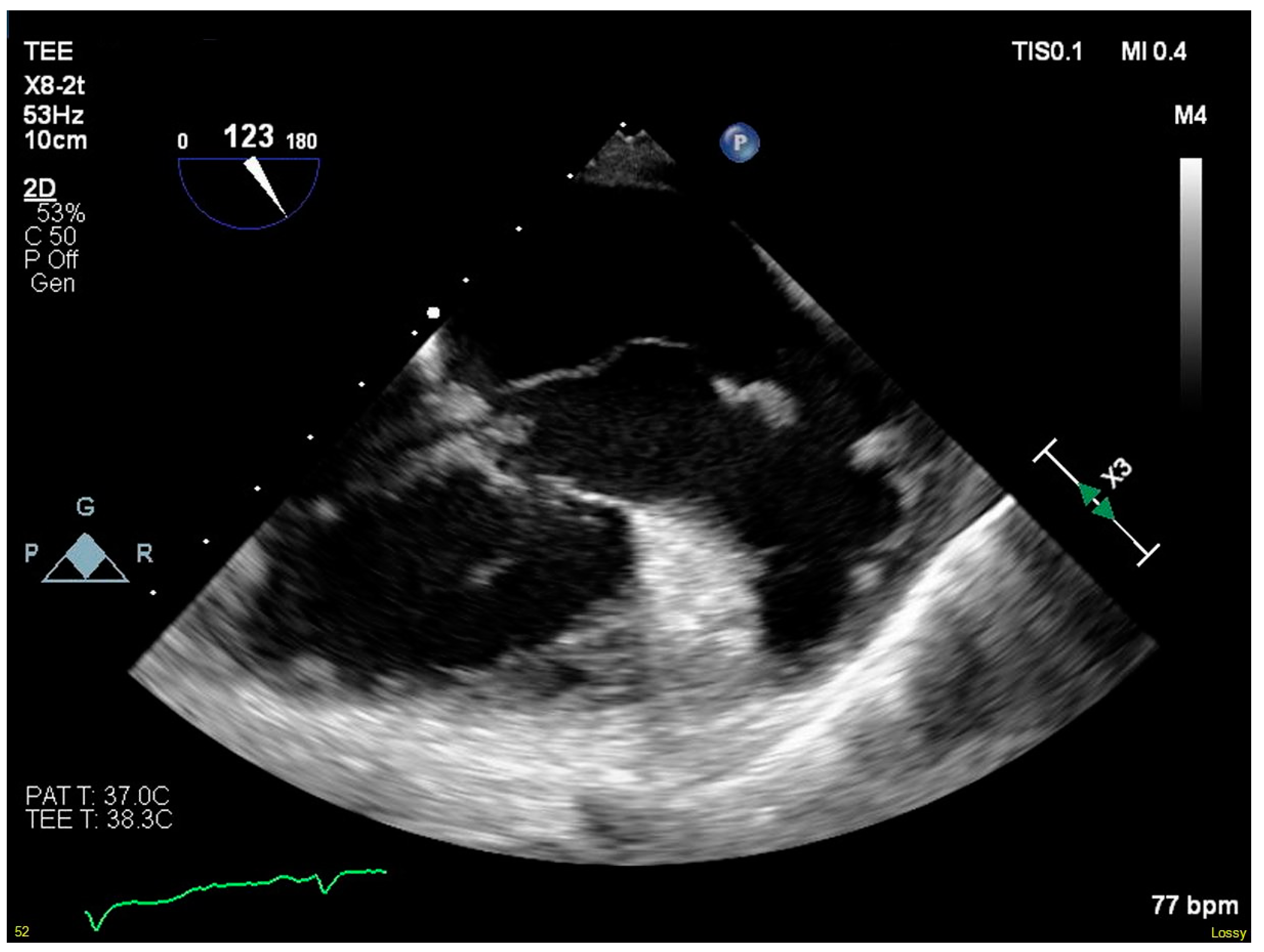
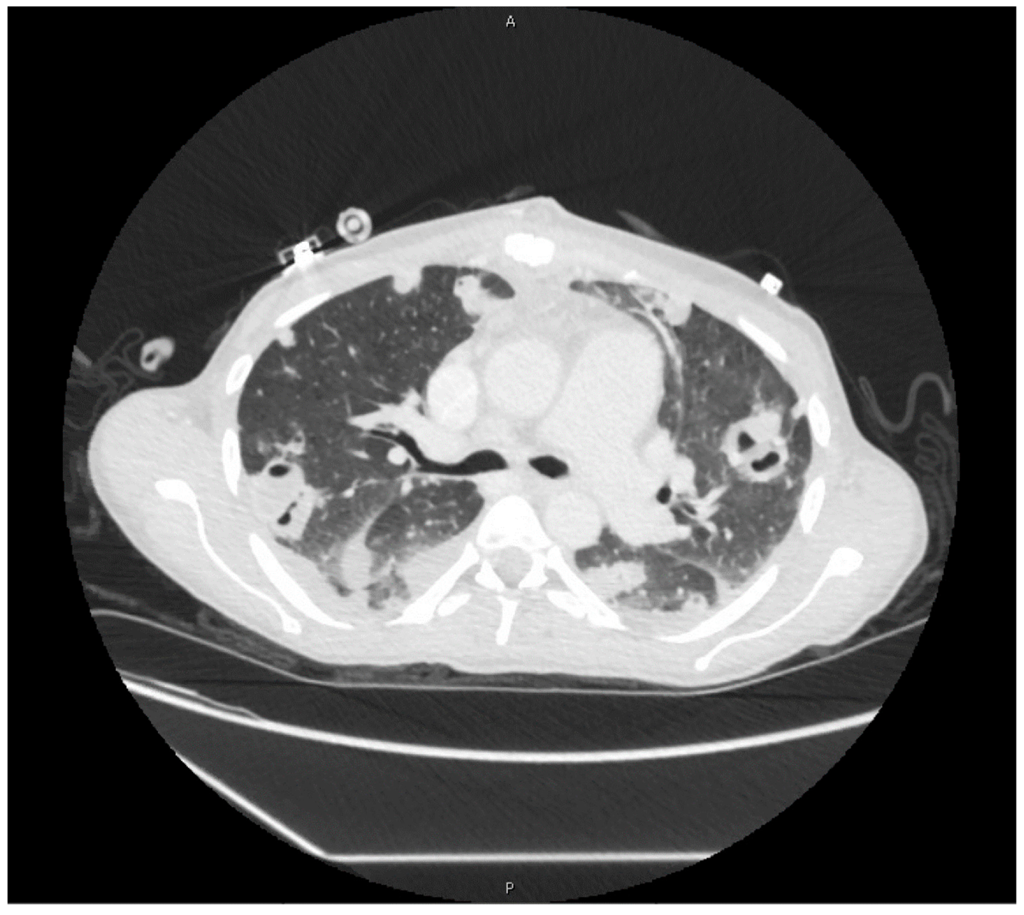
2. Case Presentation
3. Discussion
4. Conclusions
Author Contributions
Funding
Conflicts of Interest
References
- Chambers, H.F.; Bayer, A.S. Native-Valve Infective Endocarditis. N. Engl. J. Med. 2020, 383, 567–576. [Google Scholar] [CrossRef] [PubMed]
- Pettersson, G.B.; Coselli, J.S.; Hussain, S.T.; Griffin, B.; Blackstone, E.H.; Gordon, S.M.; Lemaire, S.A.; Woc-Colburn, L.E. 2016 The American Association for Thoracic Surgery (AATS) consensus guidelines: Surgical treatment of infective endocarditis: Executive summary. J. Thorac. Cardiovasc. Surg. 2017, 153, 1241–1258.e29. [Google Scholar] [CrossRef] [PubMed] [Green Version]
- Baddour, L.M.; Wilson, W.R.; Bayer, A.S.; Fowler, V.G., Jr.; Tleyjeh, I.M.; Rybak, M.J.; Barsic, B.; Lockhart, P.B.; Gewitz, M.H.; Levison, M.E.; et al. Infective Endocarditis in Adults: Diagnosis, Antimicrobial Therapy, and Management of Complications. Circulation 2015, 132, 1435–1486. [Google Scholar] [CrossRef] [PubMed]
- Habib, G.; Lancellotti, P.; Antunes, M.J.; Bongiorni, M.G.; Casalta, J.P.; Del Zotti, F.; Dulgheru, R.; El Khoury, G.; Erba, P.A.; Iung, B.; et al. 2015 ESC Guidelines for the management of infective endocarditis: The Task Force for the Management of Infective Endocarditis of the European Society of Cardiology (ESC). Endorsed by: European Association for Cardio-Thoracic Surgery (EACTS), the European Association of Nuclear Medicine (EANM). Eur. Heart J. 2015, 36, 3075–3128. [Google Scholar] [PubMed]
- Nakatani, S.; Ohara, T.; Ashihara, K.; Izumi, C.; Iwanaga, S.; Eishi, K.; Okita, Y.; Daimon, M.; Kimura, T.; Toyoda, K.; et al. JCS 2017 Guideline on Prevention and Treatment of Infective Endocarditis. Circ. J. 2019, 83, 1767–1809. [Google Scholar] [CrossRef] [PubMed] [Green Version]
- Abubakar, H.; Rashed, A.; Subahi, A.; Yassin, A.S.; Shokr, M.; Elder, M. AngioVac System Used for Vegetation Debulking in a Patient with Tricuspid Valve Endocarditis: A Case Report and Review of the Literature. Case Rep. Cardiol. 2017, 2017, 1–7. [Google Scholar] [CrossRef]
- Leong, R.; Gannon, B.R.; Childs, T.J.; Isotalo, P.A.; Abdollah, H. Aspergillus fumigatus pacemaker lead endocarditis: A case report and review of the literature. Can. J. Cardiol. 2006, 22, 337–340. [Google Scholar] [CrossRef] [Green Version]
- Tanaka, M.; Abe, T.; Hosokawa, S.; Suenaga, Y.; Hikosaka, H. Tricuspid valve Candida endocarditis cured by valve-sparing debridement. Ann. Thorac. Surg. 1989, 48, 857–858. [Google Scholar] [CrossRef]
- Nt, R.; Bray, N.; Wang, H.; Zelnick, K.; Osman, A.; Vicuña, R. Rare infection of implantable cardioverter-defibrillator lead withCandida albicans: Case report and literature review. Ther. Adv. Cardiovasc. Dis. 2014, 8, 193–201. [Google Scholar]
- Chopra, T.; Dhar, S.; Sobel, J.D.; Afonso, L.; Bhargava, A.; Kumar, S.; Chopra, A. Candida kefyr Endocarditis in a Patient with Hypertrophic Obstructive Cardiomyopathy. Am. J. Med. Sci. 2010, 339, 188–189. [Google Scholar] [CrossRef]
- Davis, W.A.; Isner, J.M.; Bracey, A.W.; Roberts, W.C.; Garagusi, V.F. Disseminated petriellidium boydii and pacemaker endocarditis1. Am. J. Med. 1980, 69, 929–932. [Google Scholar] [CrossRef]
- Miyata, E.; Satoh, S.; Inokuchi, K.; Aso, A.; Kimura, Y.; Yokoyama, S.; Mori, E.; Nakamura, T.; Matsumoto, T.; Fujino, Y.; et al. Three fatal cases of rapidly progressive infective endocarditis caused by Staphylococcus aureus: One case with huge vegetation. Circ. J. 2007, 71, 1488–1491. [Google Scholar] [CrossRef] [PubMed] [Green Version]
- Machado, M.N.; Nakazone, M.A.; Takakura, I.T.; Silva, C.M.P.D.C.; Maia, L.N. Spontaneous Bacterial Pericarditis and Coronary Sinus Endocarditis Caused by Oxacillin-Susceptible Staphylococcus aureus. Case Rep. Med. 2010, 2010, 1–3. [Google Scholar] [CrossRef] [PubMed]
- Tomaszuk-Kazberuk, A.; Sobkowicz, B.; Hirnle, T.; Lewczuk, A.; Sawicki, R.; Musiał, W. Giant right ventricular mural vegetation mimicking a cardiac tumour. Kardiol. Pol. 2011, 69, 587–589. [Google Scholar] [PubMed]
- Bernal, J.M.; Gonzalez, I.M.; Miralles, P.J. Prophylactic resection of a tricuspid valve vegetation in infective endocarditis. Int. J. Cardiol. 1986, 12, 255–257. [Google Scholar] [CrossRef]
- Straumann, E.; Stulz, P.; Jenzer, H.R. Tricuspid Valve Endocarditis in the Drug Addict: A Reconstructive Approach (“Vegetectomy”). Thorac. Cardiovasc. Surg. 1990, 38, 291–294. [Google Scholar] [CrossRef] [PubMed]
- Scarvelis, D.; Malcolm, I. Embolization of a huge tricuspid valve bacterial vegetation. J. Am. Soc. Echocardiogr. 2002, 15, 185–187. [Google Scholar] [CrossRef]
- Birsic, G.W.; Fulcher, J.W.; Fiester, S.E. Embolization of Endocardial Vegetation with Stroke Presentation. Am. J. Forensic Med. Pathol. 2019, 40, 72–76. [Google Scholar] [CrossRef]
- Iwama, T.; Shigematsu, S.; Asami, K.; Kubo, I.; Kitazume, H.; Tanabe, S.; Matsunaga, Y. Tricuspid Valve Endocarditis with Large Vegetations in a Non-Drug Addict without Underlying Cardiac Disease. Intern. Med. 1996, 35, 203–206. [Google Scholar] [CrossRef] [Green Version]
- Cheng, H.-L.; Lin, W.-C.; Shih, P.-Y.; Huang, C.-H.; Hsu, Y.-C.; Yie, J.-C.; Chen, S.-Y.; Lin, C.-P. Streptococcus agalactiae infective endocarditis with large vegetation in a patient with underlying protein S deficiency. Infection 2012, 41, 247–250. [Google Scholar] [CrossRef]
- Koushi, K.; Kitani, K.; Takahashi, A. A large vegetation at the tricuspid valve with gas image. Asian Cardiovasc. Thorac. Ann. 2014, 24, 488. [Google Scholar] [CrossRef] [PubMed]
- Montenegro-Sá, F.; Guardado, J.; Antunes, A.; Morais, J. A rare late finding in corrected tetralogy of Fallot: A case report. Eur. Heart J. Case Rep. 2018, 2, yty060. [Google Scholar] [CrossRef] [PubMed]
- Ghosh, P.K.; Miller, H.I.; Vidne, B.A. Mitral obstruction in bacterial endocarditis. Br. Heart J. 1985, 53, 341–344. [Google Scholar] [CrossRef] [PubMed]
- Moorthy, K.; Prakash, R.; Aronow, W.S. Echocardiographic appearance of aortic valve vegetations in bacterial endocarditis due to actinobacillus actinomycetemcomitans. J. Clin. Ultrasound 1977, 5, 49–51. [Google Scholar] [CrossRef]
- Bamrah, V.S.; Williams, G.W.; Hughes, C.V.; Rose, H.D.; Tristani, F.E. Haemophilus parainfluenzae mitral valve vegetation without hemodynamic abnormality. Am. J. Med. 1979, 66, 543–546. [Google Scholar] [CrossRef]
- Carruthers, M.M. Endocarditis due to enteric bacilli other than Salmonellae: Case reports and literature review. Am. J. Med. Sci. 1977, 273, 203–211. [Google Scholar] [CrossRef]
- Salsano, A.; Sportelli, E.; Borile, S.; Santini, F. Proteus mirabilis bioprosthetic tricuspid valve endocarditis with massive right ventricular vegetation: A new entity in the prosthetic valve endocarditis aetiology. Eur. J. Cardio Thorac. Surg. 2016, 50, 581–582. [Google Scholar] [CrossRef] [Green Version]


| Year and Location of Report | Age/Sex | Pathogen | Area of Involvement | Maximum Length of Vegetation (cm) | Surgical Intervention | Pathogen-Specific Antimicrobial Therapy | Length of Pathogen-Specific Antimicrobial Therapy | Outcome |
|---|---|---|---|---|---|---|---|---|
| 2006/Canada [7] | 71/M | Aspergillus fumigatus | pacemaker lead, superior vena cava stent | 6 | none | empiric only | not reported | death |
| 1989/Japan [8] | 22/F | Candida albicans | native tricuspid | 4 | vegetectomy, tricuspid valve debridement | amphotericin B → amphotericin B/miconazole/5-fluorocytosine | not reported | success |
| 2014/USA [9] | 60/F | C. albicans | ICD lead | 4.5 | ICD extraction, vegetectomy | micafungin → fluconazole | 2 weeks → 6 weeks | relapse |
| 2010/USA [10] | 74/F | Candida kefyr | native mitral | 7 | none | micafungin → fluconazole | 10 days → 6 weeks | success |
| 1980/USA [11] | 62/F | Petriellidium boydii | pacemaker lead | 4 | none | not reported | not reported | death |
| Our case | 39/F | MRSA | native tricuspid | 5.6 | tricuspid valvectomy | vancomycin | 48 days | success |
| 2007/Japan [12] | 64/M | MSSA | prosthetic mitral | 7 | none | cefazolin/gentamycin | <1 week | death |
| 2010/Brazil [13] | 44/M | MSSA | coronary sinus | 4 | vegetectomy, drainage of pyopericardium | oxacillin | not reported | success |
| 2011/Poland [14] | 20/M | MSSA | right ventricular free wall | 5 | vegetectomy | fluoroquinolone → vancomycin | not reported | success |
| 1986/Spain [15] | 22/M | Staphylococcus aureus | native tricuspid | 7 | vegetectomy | cloxacillin sodium/tobramycin | ≥6 weeks | success |
| 1990/Switzerland [16] | 24/F | S. aureus | native tricuspid | 4 | vegetectomy, tricuspid valvuloplasty | flucloxacillin/gentamycin | 56 days | success |
| 2002/Canada [17] | 30/F | S. aureus | native tricuspid | 4.7 | tricuspid valvectomy, right pulmonary artery thromboendarterectomy | not reported | not reported | success |
| 2019/USA [18] | 37/F | S. aureus | native mitral and tricuspid | 10 | none | none | none | death |
| 1996/Japan [19] | 77/M | gamma-Streptococcus | native tricuspid | 5 | tricuspid valve replacement | penicillin G/gentamycin → penicillin G/cefotiam | 6 weeks → 1 month | success |
| 2013/Taiwan [20] | 81/F | Streptococcus agalactiae | native mitral | 4.2 | mitral valve replacement | penicillin G | not reported | death |
| 2016/Japan [21] | 53/M | S. agalactiae | native tricuspid | 4 | tricuspid valve replacement | not reported | not reported | not reported |
| 2018/Portugal [22] | 37/M | Streptococcus mitis | prosthetic pulmonic | 9 | pulmonic valve replacement | amoxicillin/gentamycin → vancomycin/gentamycin | 4 days → 5 weeks | success |
| 2017/USA [6] | 33/F | Streptococcus pyogenes | native tricuspid | 4.2 | percutaneous extraction of vegetation | penicillin G/clindamycin | 6 weeks | success |
| 1985/Israel [23] | 47/M | Streptococcus viridans | native mitral | 4 | mitral valve replacement | vancomycin | 15 days | death |
| 1977/USA [24] | 57/M | Aggregatibacter actinomycetemcomitans | native aortic | 5 | aortic valve replacement | cefalotin | not reported | death |
| 1979/USA [25] | 51/M | Haemophilus parainfluenzae | native mitral | 4.5 | none | ampicillin → chloramphenicol | 6 weeks → 9 days | death |
| 1977/USA [26] | 82/M | Escherichia coli | native mitral | 4 | none | ampicillin → cefalotin → cefalotin/gentamycin | 2 days → 7 days → 5 days | death |
| 2016/Italy [27] | 43/F | Proteus mirabilis | prosthetic tricuspid | 9 | tricuspid valve re-replacement | not reported | not reported | not reported |
Publisher’s Note: MDPI stays neutral with regard to jurisdictional claims in published maps and institutional affiliations. |
© 2020 by the authors. Licensee MDPI, Basel, Switzerland. This article is an open access article distributed under the terms and conditions of the Creative Commons Attribution (CC BY) license (http://creativecommons.org/licenses/by/4.0/).
Share and Cite
Radcliffe, C.; Oen-Hsiao, J.; Grant, M. More than Garden Variety: Massive Vegetations from Infective Endocarditis. Pathogens 2020, 9, 998. https://doi.org/10.3390/pathogens9120998
Radcliffe C, Oen-Hsiao J, Grant M. More than Garden Variety: Massive Vegetations from Infective Endocarditis. Pathogens. 2020; 9(12):998. https://doi.org/10.3390/pathogens9120998
Chicago/Turabian StyleRadcliffe, Christopher, Joyce Oen-Hsiao, and Matthew Grant. 2020. "More than Garden Variety: Massive Vegetations from Infective Endocarditis" Pathogens 9, no. 12: 998. https://doi.org/10.3390/pathogens9120998