Next Issue
Volume 10, January
Previous Issue
Volume 9, November


Pathogens, Volume 9, Issue 12 (December 2020) – 100 articles

Cover Story (view full-size image): 
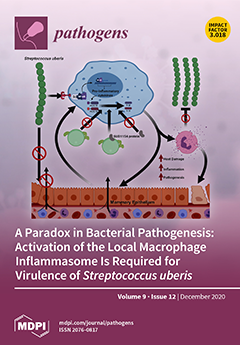
Streptococcus uberis is a common cause of intramammary infection and mastitis in dairy cattle, but a commensal at other body sites. Virulence is dependent on high-level colonisation, and during early pathogenesis, this relies on the cell-surface protein, sub1154. This protein is necessary for transcriptionally independent activation of the NLRP3 inflammasome in primed mammary macrophages. The consequent inflammatory response in vivo produces a bactericidal environment but also results in damage to host tissue. The dependency of colonisation on sub1154 can be reconciled in a model in which the bacterial growth-promoting effect of host damage outweighs inhibition due to the bactericidal activity. This leads to the conclusion that, paradoxically, this bacterium only colonises like a pathogen because the host responds to it like a pathogen. View this paper
  • Issues are regarded as officially published after their release is announced to the table of contents alert mailing list.
  • You may sign up for e-mail alerts to receive table of contents of newly released issues.
  • PDF is the official format for papers published in both, html and pdf forms. To view the papers in pdf format, click on the "PDF Full-text" link, and use the free Adobe Reader to open them.
Order results
Result details
Section
Select all
Export citation of selected articles as:
13 pages, 1811 KiB  
Article
Pantoea ananatis, A New Bacterial Pathogen Affecting Wheat Plants (Triticum L.) in Poland
by Krzysztof Krawczyk, Beata Wielkopolan and Aleksandra Obrępalska-Stęplowska
Pathogens 2020, 9(12), 1079; https://doi.org/10.3390/pathogens9121079 - 21 Dec 2020
Cited by 14 | Viewed by 4924
Abstract
Wheat (Triticum aestivum) is one of the most economically important crops in the world. During the routine monitoring of wheat pest, the cereal leaf beetle (CLB, Oulema melanopus, Coleoptera, Chrysomelidae), in the Greater Poland region, it was observed that some leaves [...] Read more.
Wheat (Triticum aestivum) is one of the most economically important crops in the world. During the routine monitoring of wheat pest, the cereal leaf beetle (CLB, Oulema melanopus, Coleoptera, Chrysomelidae), in the Greater Poland region, it was observed that some leaves wounded by CLB also displayed brownish lesions with clear margins and yellow halo, disease symptoms resembling a bacterial infection. The aim of this study was therefore to investigate those symptoms to establish a causal agent of the disease. The identification based on the results of the Biolog’s Gen III system, 16S rRNA, and gyrB genes sequencing, revealed the presence of eight strains of Pantoea ananatis bacteria. Four strains were derived from wheat leaves (Ta024, Ta027, Ta030, Ta046), and four from the CLB’s oral secretion (OUC1, OUD2, OUF2, and OUG1). They shared the nucleotide identity ranging from 99 to 100% to P. ananatis strains deposited in the GenBank database. Additionally, the multi-locus sequence analysis (MLSA) of concatenated sequences of partial atpD, fusA, gyrB, rplB, and rpoB genes was performed. All P. ananatis strains isolated in Poland, grouped into one cluster supported with high bootstrap value. Pathogenicity tests performed on four varieties of wheat plants have identified P. ananatis strains as a causal agent of wheat disease. To our knowledge, this is the first report of P. ananatis affecting wheat plants. Full article
(This article belongs to the Special Issue Plant Pathogenic Bacteria in Crops)
Show Figures

Figure 1

19 pages, 1410 KiB  
Article
Computational Analysis of African Swine Fever Virus Protein Space for the Design of an Epitope-Based Vaccine Ensemble
by Albert Ros-Lucas, Florencia Correa-Fiz, Laia Bosch-Camós, Fernando Rodriguez and Julio Alonso-Padilla
Pathogens 2020, 9(12), 1078; https://doi.org/10.3390/pathogens9121078 - 21 Dec 2020
Cited by 15 | Viewed by 4356
Abstract
African swine fever virus is the etiological agent of African swine fever, a transmissible severe hemorrhagic disease that affects pigs, causing massive economic losses. There is neither a treatment nor a vaccine available, and the only method to control its spread is by [...] Read more.
African swine fever virus is the etiological agent of African swine fever, a transmissible severe hemorrhagic disease that affects pigs, causing massive economic losses. There is neither a treatment nor a vaccine available, and the only method to control its spread is by extensive culling of pigs. So far, classical vaccine development approaches have not yielded sufficiently good results in terms of concomitant safety and efficacy. Nowadays, thanks to advances in genomic and proteomic techniques, a reverse vaccinology strategy can be explored to design alternative vaccine formulations. In this study, ASFV protein sequences were analyzed using an in-house pipeline based on publicly available immunoinformatic tools to identify epitopes of interest for a prospective vaccine ensemble. These included experimentally validated sequences from the Immune Epitope Database, as well as de novo predicted sequences. Experimentally validated and predicted epitopes were prioritized following a series of criteria that included evolutionary conservation, presence in the virulent and currently circulating variant Georgia 2007/1, and lack of identity to either the pig proteome or putative proteins from pig gut microbiota. Following this strategy, 29 B-cell, 14 CD4+ T-cell and 6 CD8+ T-cell epitopes were selected, which represent a starting point to investigating the protective capacity of ASFV epitope-based vaccines. Full article
(This article belongs to the Special Issue Animal Vector-Borne Diseases)
Show Figures

Figure 1

11 pages, 2425 KiB  
Article
Analysis of Risk Factors for African Swine Fever in Lombardy to Identify Pig Holdings and Areas Most at Risk of Introduction in Order to Plan Preventive Measures
by Silvia Bellini, Alessandra Scaburri, Marco Tironi and Stefania Calò
Pathogens 2020, 9(12), 1077; https://doi.org/10.3390/pathogens9121077 - 21 Dec 2020
Cited by 4 | Viewed by 1922
Abstract
In 2019, the area of the European Union (EU) affected by African swine fever (ASF) expanded progressively in a southwestern direction from Baltic and eastern countries. The disease can severely affect and disrupt regional and international trade of pigs and pork products with [...] Read more.
In 2019, the area of the European Union (EU) affected by African swine fever (ASF) expanded progressively in a southwestern direction from Baltic and eastern countries. The disease can severely affect and disrupt regional and international trade of pigs and pork products with serious socioeconomic damages to the pig industry. Lombardy is one of the most important European pig producers and the introduction of ASF into the pig population could adversely affect the entire sector. A study was carried out to identify the farms and territories in the region most at risk of ASF introduction to plan preventive measures. The pig holdings were identified through a descriptive analysis of pig movements and Social Network Analysis (SNA), while, for the identification of the most exposed municipalities, an assessment of risk factors was carried out using the ranking of summed scores attributed to the Z-score. From the analysis, it was found that 109 municipalities and 297 pig holdings of the region were potentially more at risk, and these holdings were selected for target surveillance. This information was provided to veterinary authority to target surveillance in pig farms, in order to early detect a possible incursion of ASF and prevent its spread. Full article
(This article belongs to the Special Issue African Swine Fever Virus Infection)
Show Figures

Figure 1

20 pages, 1771 KiB  
Article
Prevalence, Intensity, and Correlates of Soil-Transmitted Helminth Infections among School Children after a Decade of Preventive Chemotherapy in Western Rwanda
by Joseph Kabatende, Michael Mugisha, Lazare Ntirenganya, Abbie Barry, Eugene Ruberanziza, Jean Bosco Mbonigaba, Ulf Bergman, Emile Bienvenu and Eleni Aklillu
Pathogens 2020, 9(12), 1076; https://doi.org/10.3390/pathogens9121076 - 21 Dec 2020
Cited by 17 | Viewed by 3456
Abstract
Preventive chemotherapy (PC) is a WHO-recommended core intervention measures to eliminate Soil-Transmitted Helminths (STH) as a public health problem by 2020, defined as a reduction in prevalence to <1% of moderate or high-intensity infection. We conducted a cross-sectional study to investigate the prevalence, [...] Read more.
Preventive chemotherapy (PC) is a WHO-recommended core intervention measures to eliminate Soil-Transmitted Helminths (STH) as a public health problem by 2020, defined as a reduction in prevalence to <1% of moderate or high-intensity infection. We conducted a cross-sectional study to investigate the prevalence, intensity, and correlates of STH after a decade of PC in Rwanda. A total of 4998 school children (5–15 years old) from four districts along Lake Kivu in the western province were screened for STH using Kato-Katz. The overall prevalence of Soil-transmitted helminths among school children was 77.7% (range between districts = 54% to 92%). Trichirus trichiura was the most common STH (66.8%, range between districts = 23% to 88.2%), followed by Ascaris lumbricoides (49.9%, range between district = 28.5% to 63.3%) and hookworms (1.9%, range between districts = 0.6% to 2.9%). The prevalence of single, double and of triple parasite coinfection were 48.6%, 50.3%, and 1.1%, respectively. The overall prevalence of moderate or high-intensity infection for Trichirus trichiura and Ascaris lumbricoides was 7.1% and 13.9, respectively. Multivariate logistic regression model revealed that male sex, district, stunting, and schistosomiasis coinfection as significant predictors of STH infection. Despite a decade of PC implementation, STH remain a significant public health problem in Rwanda. Full article
(This article belongs to the Section Human Pathogens)
Show Figures

Figure 1

14 pages, 295 KiB  
Article
Validation of a Novel Commercial ELISA Test for the Detection of Antibodies against Coxiella burnetii
by Salvatore Ledda, Cinzia Santucciu, Valentina Chisu and Giovanna Masala
Pathogens 2020, 9(12), 1075; https://doi.org/10.3390/pathogens9121075 - 21 Dec 2020
Viewed by 2163
Abstract
Q fever is a zoonosis caused by Coxiella burnetii, a Gram-negative pathogen with a complex life cycle and a high impact on public and animal health all over the world. The symptoms are indistinguishable from those belonging to other diseases, and the [...] Read more.
Q fever is a zoonosis caused by Coxiella burnetii, a Gram-negative pathogen with a complex life cycle and a high impact on public and animal health all over the world. The symptoms are indistinguishable from those belonging to other diseases, and the disease could be symptomless. For these reasons, reliable laboratory tests are essential for an accurate diagnosis. The aim of this study was to validate a novel enzyme-linked immunosorbent assay (ELISA) test, named the Chorus Q Fever Phase II IgG and IgM Kit (DIESSE, Diagnostica Senese S.p.A), which is performed by an instrument named Chorus, a new device in medical diagnostics. This diagnostic test is employed for the detection of antibodies against C. burnetii Phase II antigens in acute disease. Our validation protocol was performed according to the Italian Accreditation Body (ACCREDIA) (Regulation UNI CEI EN ISO/IEC 17025:2018 and 17043:2010), OIE (World Organization for Animal Health), and Statement for Reporting Studies of Diagnostic Accuracy (STARD). Operator performance was evaluated along with the analytical specificity and sensitivity (ASp and ASe) and diagnostic accuracy of the kit, with parameters such as diagnostic specificity and sensitivity (DSp and DSe) and positive and negative predictive values (PPV and NPV), in addition to the repeatability. According to the evaluated parameters, the diagnostic ELISA test was shown to be suitable for validation and commercialization as a screening method in human sera and a valid support for clinical diagnostics. Full article
(This article belongs to the Special Issue Q Fever)
14 pages, 4867 KiB  
Article
TaqMan Assays for Simultaneous Detection of Bacillus anthracis and Bacillus cereus biovar anthracis
by Diansy Zincke, Michael H. Norris, Odalis Cruz, Berzhan Kurmanov, W. Scott McGraw, David J. Daegling, John Krigbaum, Thi Thu Ha Hoang, Kamil Khanipov, Georgiy Golovko, Ted Hadfield and Jason K. Blackburn
Pathogens 2020, 9(12), 1074; https://doi.org/10.3390/pathogens9121074 - 21 Dec 2020
Cited by 5 | Viewed by 2434
Abstract
Anthrax is a worldwide zoonotic disease caused by the spore-forming bacterium Bacillus anthracis. Primarily a disease of herbivores, human infections often result from direct contact with contaminated animal products (cutaneous and inhalational anthrax) or through consumption of infected meat (gastrointestinal anthrax). The [...] Read more.
Anthrax is a worldwide zoonotic disease caused by the spore-forming bacterium Bacillus anthracis. Primarily a disease of herbivores, human infections often result from direct contact with contaminated animal products (cutaneous and inhalational anthrax) or through consumption of infected meat (gastrointestinal anthrax). The genetic near neighbor, Bacillus cereus biovar anthracis (Bcbva), causes an anthrax-like illness in the wildlife and livestock of west and central Africa due to the presence and expression of B. anthracis-specific virulence factors in this background. While Bcbva infections have not been reported in humans, a recent seroprevalence study detected Bcbva antibodies in the rural population around Taï National Park. This work describes the development of new TaqMan multiplex PCRs for the simultaneous detection of B. anthracis and Bcbva. The assays are designed to amplify Ba-1, capB, and lef markers in B. anthracis and genomic island IV (GI4), capB, and lef in Bcbva. Our assays allow for the rapid discrimination of B. anthracis and Bcbva and will provide insights into the molecular epidemiology of these two important pathogens that share an overlapping geographical range in west and central Africa. Full article
(This article belongs to the Section Animal Pathogens)
Show Figures

Figure 1

7 pages, 233 KiB  
Review
Animal and Human Vaccines against West Nile Virus
by Juan-Carlos Saiz
Pathogens 2020, 9(12), 1073; https://doi.org/10.3390/pathogens9121073 - 21 Dec 2020
Cited by 23 | Viewed by 3580
Abstract
West Nile virus (WNV) is a widely distributed enveloped flavivirus transmitted by mosquitoes, which main hosts are birds. The virus sporadically infects equids and humans with serious economic and health consequences, as infected individuals can develop a severe neuroinvasive disease that can even [...] Read more.
West Nile virus (WNV) is a widely distributed enveloped flavivirus transmitted by mosquitoes, which main hosts are birds. The virus sporadically infects equids and humans with serious economic and health consequences, as infected individuals can develop a severe neuroinvasive disease that can even lead to death. Nowadays, no WNV-specific therapy is available and vaccines are only licensed for use in horses but not for humans. While several methodologies for WNV vaccine development have been successfully applied and have contributed to significantly reducing its incidence in horses in the US, none have progressed to phase III clinical trials in humans. This review addresses the status of WNV vaccines for horses, birds, and humans, summarizing and discussing the challenges they face for their clinical advance and their introduction to the market. Full article
(This article belongs to the Special Issue West Nile Virus Infection)
10 pages, 1995 KiB  
Review
Molecular Epidemiology of HIV-1 in African Countries: A Comprehensive Overview
by Marta Giovanetti, Massimo Ciccozzi, Cristina Parolin and Alessandra Borsetti
Pathogens 2020, 9(12), 1072; https://doi.org/10.3390/pathogens9121072 - 21 Dec 2020
Cited by 25 | Viewed by 5428
Abstract
The human immunodeficiency virus type 1 (HIV-1) originated in non-human primates in West-central Africa and continues to be a major global public health issue, having claimed almost 33 million lives so far. In Africa, it is estimated that more than 20 million people [...] Read more.
The human immunodeficiency virus type 1 (HIV-1) originated in non-human primates in West-central Africa and continues to be a major global public health issue, having claimed almost 33 million lives so far. In Africa, it is estimated that more than 20 million people are living with HIV/Acquired Immunodeficiency Syndrome (AIDS) and that more than 730,000 new HIV-1 infections still occur each year, likely due to low access to testing. The high genetic variability of HIV-1, due to a fast replication cycle and high mutation rate, may cause the generation of many viral variants in a single infected patient during a single day. Therefore, the active monitoring and characterization of the HIV-1 subtypes and recombinant forms circulating through African countries poses a significant challenge to more specific diagnoses, treatments, care, and intervention strategies. In this review, a concise characterization of all the subtypes and recombinant forms circulating in Africa is presented to highlight the magnitude of the HIV-1 threat among the African countries and to understand virus genetic diversity and dispersion dynamics better. Full article
(This article belongs to the Special Issue Reviews from Section "Human Pathogens")
Show Figures

Figure 1

14 pages, 2841 KiB  
Article
The Control of Zoonotic Soil-Transmitted Helminthoses Using Saprophytic Fungi
by Cándido Viña, María Isabel Silva, Antonio Miguel Palomero, Mathilde Voinot, María Vilá, José Ángel Hernández, Adolfo Paz-Silva, Rita Sánchez-Andrade, Cristiana Filipa Cazapal-Monteiro and María Sol Arias
Pathogens 2020, 9(12), 1071; https://doi.org/10.3390/pathogens9121071 - 21 Dec 2020
Cited by 6 | Viewed by 2299
Abstract
Soil-transmitted helminths (STHs) are parasites transmitted through contact with soil contaminated with their infective eggs/larvae. People are infected by exposure to human-specific species or animal species (zoonotic agents). Fecal samples containing eggs of Ascaris suum or Lemurostrongylus sp. were sprayed with spores of [...] Read more.
Soil-transmitted helminths (STHs) are parasites transmitted through contact with soil contaminated with their infective eggs/larvae. People are infected by exposure to human-specific species or animal species (zoonotic agents). Fecal samples containing eggs of Ascaris suum or Lemurostrongylus sp. were sprayed with spores of the soil saprophytic filamentous fungi Clonostachys rosea (CR) and Trichoderma atrobrunneum (TA). The antagonistic effect was assessed by estimating the viability of eggs and their developmental rate. Compared to the controls (unexposed to fungi), the viability of the eggs of A. suum was halved in CR and decreased by two thirds in TA, while the viability of the eggs of Lemurostrongylus sp. was reduced by one quarter and one third in CR and TA treatments, respectively. The Soil Contamination Index (SCI), defined as the viable eggs that attained the infective stage, reached the highest percentages for A. suum in the controls after four weeks (66%), with 21% in CL and 11% in TA. For Lemurostrongylus sp., the values were 80%, 49%, and 41% for control, CR and TA treatments, respectively. We concluded that spreading spores of C. rosea or T. atrobrunneum directly onto the feces of animal species represents a sustainable approach under a One Health context to potentially reduce the risk of zoonotic STHs in humans. Full article
(This article belongs to the Special Issue Prevention of Soil-Borne Parasites)
Show Figures

Graphical abstract

17 pages, 3205 KiB  
Article
Country-Wide qPCR Based Assessment of Plasmodiophora brassicae Spread in Agricultural Soils and Recommendations for the Cultivation of Brassicaceae Crops in Poland
by Anna Czubatka-Bieńkowska, Joanna Kaczmarek, Katarzyna Marzec-Schmidt, Anna Nieróbca, Agnieszka Czajka and Małgorzata Jędryczka
Pathogens 2020, 9(12), 1070; https://doi.org/10.3390/pathogens9121070 - 20 Dec 2020
Cited by 11 | Viewed by 2610
Abstract
Clubroot is a damaging disease of oilseed rape and vegetable brassicas worldwide, caused by the soil-borne protist Plasmodiophora brassicae Wor. Due to the long life of resting spores, the assessment of the pathogen abundance in agricultural fields can serve as a guideline for [...] Read more.
Clubroot is a damaging disease of oilseed rape and vegetable brassicas worldwide, caused by the soil-borne protist Plasmodiophora brassicae Wor. Due to the long life of resting spores, the assessment of the pathogen abundance in agricultural fields can serve as a guideline for disease control at the country-wide level or the regional scale. Between 2013 and 2019, we collected 431 soil samples from fields cultivated with Brassicaceae crops throughout 16 provinces of Poland. The samples were subjected to qPCR based analysis of P. brassicae DNA concentration. From these data, the spore loads and gene copies g−1 soil were calculated and used to produce an assessment of the current clubroot risk potential at a country-wide and regional scale. The country-wide map, showing the spread of the pathogen in agricultural soils, was made using ArcGis software package implementing the interpolation with the Inverse Distance Weight method. The calculation of gene copies specific to P. brassicae helped to formulate the recommendations for farmers in respect to the cultivation guidelines. It showed a high risk of yield losses in defined regions of north, south-west and central Poland and an urgent need to undertake intensive preventative measures. Full article
Show Figures

Figure 1

15 pages, 1858 KiB  
Article
In vitro Interactions between Streptococcus intermedius and Streptococcus salivarius K12 on a Titanium Cylindrical Surface
by Carla Vacca, Maria Paola Contu, Cecilia Rossi, Maria Laura Ferrando, Cornelio Blus, Serge Szmukler-Moncler, Alessandra Scano and Germano Orrù
Pathogens 2020, 9(12), 1069; https://doi.org/10.3390/pathogens9121069 - 20 Dec 2020
Cited by 10 | Viewed by 3648
Abstract
Peri-implantitis is a steadily rising disease and is caused by oral bacterial pathogens able to form biofilm on implant surfaces and peri-implant tissues, making antibiotics treatment less effective. The use of commercial probiotics against oral pathogens could serve as an alternative to prevent [...] Read more.
Peri-implantitis is a steadily rising disease and is caused by oral bacterial pathogens able to form biofilm on implant surfaces and peri-implant tissues, making antibiotics treatment less effective. The use of commercial probiotics against oral pathogens could serve as an alternative to prevent biofilm formation. Streptococcus intermedius is one of the early colonizers of biofilm formation in dental implants. The aim of this study was to model the interaction between S. intermedius and Streptococcus salivarius strain K12, a probiotic bacterium producing bacteriocins. S. intermedius was co-cultured with S. salivarius K12 in an in vitro model simulating the biofilm formation in a dental implant composed by a titanium cylinder system. Biofilm formation rate was assessed by Real-Time PCR quantification of bacterial count and expression levels of luxS gene, used in response to cell density in the biofilm. Biofilm formation, bacteriocin production, luxS expression patterns were found to be already expressed within the first 12 h. More importantly, S. salivarius K12 was able to counter the biofilm formation in a titanium cylinder under the tested condition. In conclusion, our dental implant model may be useful for exploring probiotic-pathogen interaction to find an alternative to antibiotics for peri-implantitis treatment. Full article
(This article belongs to the Section Human Pathogens)
Show Figures

Figure 1

8 pages, 203 KiB  
Communication
Retrospective Study on the Occurrence of Antibodies against Coxiella burnetii in Dogs from Central Italy
by Valentina Virginia Ebani
Pathogens 2020, 9(12), 1068; https://doi.org/10.3390/pathogens9121068 - 20 Dec 2020
Cited by 6 | Viewed by 1749
Abstract
Coxiella burnetii, a cause of infection in humans and several animal species, is transmitted through inhalations and oral route but also tick bites. Its spreading in ruminants has been largely investigated, whereas data about the occurrence of this infection in canine population are [...] Read more.
Coxiella burnetii, a cause of infection in humans and several animal species, is transmitted through inhalations and oral route but also tick bites. Its spreading in ruminants has been largely investigated, whereas data about the occurrence of this infection in canine population are scanty. In this retrospective study, blood serum samples of 516 dogs were tested by indirect immunofluorescence assay to detect antibodies against C. burnetii; 42 (8.13%) were positive with titers ranging from 1:64 to 1:512. The highest seroprevalences were detected in dogs aged > 5 years, employed in hunting activity and living in a peri-urban/rural environment. Diagnosis for C. burnetii infection should be always carried out in bitches with reproductive disorders. Moreover, in view of the zoonotic impact of this infection, asymptomatic dogs exposed to ticks’ bites and/or to contact with infected farm animals should be checked, too. Full article
(This article belongs to the Section Animal Pathogens)
10 pages, 2224 KiB  
Brief Report
Performance of Commercially Available Rapid Serological Assays for the Detection of SARS-CoV-2 Antibodies
by Anwar M. Hashem, Rowa Y. Alhabbab, Abdullah Algaissi, Mohamed A. Alfaleh, Sharif Hala, Turki S. Abujamel, M-Zaki ElAssouli, Afrah A. AL-Somali, Fadwa S. Alofi, Asim A. Khogeer, Almohanad A. Alkayyal, Ahmad Bakur Mahmoud, Naif A. M. Almontashiri and Arnab Pain
Pathogens 2020, 9(12), 1067; https://doi.org/10.3390/pathogens9121067 - 19 Dec 2020
Cited by 6 | Viewed by 2671
Abstract
The coronavirus disease 2019 (COVID-19) pandemic, caused by the severe acute respiratory syndrome coronavirus 2 (SARS-CoV-2), continues to spread globally. Although several rapid commercial serological assays have been developed, little is known about their performance and accuracy in detecting SARS-CoV-2-specific antibodies in COVID-19 [...] Read more.
The coronavirus disease 2019 (COVID-19) pandemic, caused by the severe acute respiratory syndrome coronavirus 2 (SARS-CoV-2), continues to spread globally. Although several rapid commercial serological assays have been developed, little is known about their performance and accuracy in detecting SARS-CoV-2-specific antibodies in COVID-19 patient samples. Here, we have evaluated the performance of seven commercially available rapid lateral flow immunoassays (LFIA) obtained from different manufacturers, and compared them to in-house developed and validated ELISA assays for the detection of SARS-CoV-2-specific IgM and IgG antibodies in RT-PCR-confirmed COVID-19 patients. While all evaluated LFIA assays showed high specificity, our data showed a significant variation in sensitivity of these assays, which ranged from 0% to 54% for samples collected early during infection (3–7 days post symptoms onset) and from 54% to 88% for samples collected at later time points during infection (8–27 days post symptoms onset). Therefore, we recommend prior evaluation and validation of these assays before being routinely used to detect IgM and IgG in COVID-19 patients. Moreover, our findings suggest the use of LFIA assays in combination with other standard methods, and not as an alternative. Full article
(This article belongs to the Section Human Pathogens)
Show Figures

Figure 1

19 pages, 363 KiB  
Review
Recent Advances in the Pursuit of an Effective Acinetobacter baumannii Vaccine
by Patrick S. Gellings, Ashley A. Wilkins and Lisa A. Morici
Pathogens 2020, 9(12), 1066; https://doi.org/10.3390/pathogens9121066 - 19 Dec 2020
Cited by 37 | Viewed by 4501
Abstract
Acinetobacter baumannii has been a major cause of nosocomial infections for decades. The absence of an available vaccine coupled with emerging multidrug resistance has prevented the medical community from effectively controlling this human pathogen. Furthermore, the ongoing pandemic caused by SARS-CoV-2 has increased [...] Read more.
Acinetobacter baumannii has been a major cause of nosocomial infections for decades. The absence of an available vaccine coupled with emerging multidrug resistance has prevented the medical community from effectively controlling this human pathogen. Furthermore, the ongoing pandemic caused by SARS-CoV-2 has increased the risk of hospitalized patients developing ventilator-associated pneumonia caused by bacterial opportunists including A. baumannii. The shortage of antibiotics in the development pipeline prompted the World Health Organization to designate A. baumannii a top priority for the development of new medical countermeasures, such as a vaccine. There are a number of important considerations associated with the development of an A. baumannii vaccine, including strain characteristics, diverse disease manifestations, and target population. In the past decade, research efforts have revealed a number of promising new immunization strategies that could culminate in a safe and protective vaccine against A. baumannii. In this review, we highlight the recent progress in the development of A. baumannii vaccines, discuss potential challenges, and propose future directions to achieve an effective intervention against this human pathogen. Full article
(This article belongs to the Special Issue Reviews from Section "Human Pathogens")
14 pages, 1558 KiB  
Article
Clinical Biofilm Ring Test® Reveals the Potential Role of β-Lactams in the Induction of Biofilm Formation by P. aeruginosa in Cystic Fibrosis Patients
by Elodie Olivares, Jason Tasse, Stéphanie Badel-Berchoux, Christian Provot, Gilles Prévost and Thierry Bernardi
Pathogens 2020, 9(12), 1065; https://doi.org/10.3390/pathogens9121065 - 19 Dec 2020
Cited by 5 | Viewed by 2329
Abstract
Biofilms are characterized by high tolerance to antimicrobials. However, conventional antibiograms are performed on planktonic microorganisms. Through the clinical Biofilm Ring Test® (cBRT), initially aimed to measure the adhesion propensity of bacteria, we discerned a variable distribution of biofilm-producer strains among P. [...] Read more.
Biofilms are characterized by high tolerance to antimicrobials. However, conventional antibiograms are performed on planktonic microorganisms. Through the clinical Biofilm Ring Test® (cBRT), initially aimed to measure the adhesion propensity of bacteria, we discerned a variable distribution of biofilm-producer strains among P. aeruginosa samples isolated from expectorations of cystic fibrosis (CF) patients. Despite a majority of spontaneous adherent isolates, few strains remained planktonic after 5 h of incubation. Their analysis by an adapted protocol of the cBRT revealed an induction of the biofilm early formation by sub-inhibitory doses of β-lactams. Microscopic observations of bacterial cultures stained with Syto 9/Propidium Iodide (PI) confirmed the ability of antimicrobials to increase either the bacterial biomass or the biovolume occupied by induced sessile cells. Finally, the cBRT and its derivatives enabled to highlight in a few hours the potential inducer property of antibiotics on bacterial adhesion. This phenomenon should be considered carefully in the context of CF since patients are constantly under fluctuating antimicrobial treatments. To conclude, assays derived from the Biofilm Ring Test® (BRT) device, not only define efficient doses preventing biofilm formation, but could be useful for the antimicrobial selection in CF, to avoid inducer molecules of the early biofilm initiation. Full article
(This article belongs to the Special Issue Pseudomonas aeruginosa Pathogenesis)
Show Figures

Figure 1

6 pages, 501 KiB  
Brief Report
Investigation and Follow-Up of a Staphylococcal Food Poisoning Outbreak Linked to the Consumption of Traditional Hand-Crafted Alm Cheese
by Virginia Filipello, Emanuela Bonometti, Massimo Campagnani, Irene Bertoletti, Angelo Romano, Fabio Zuccon, Chiara Campanella, Marina Nadia Losio and Guido Finazzi
Pathogens 2020, 9(12), 1064; https://doi.org/10.3390/pathogens9121064 - 19 Dec 2020
Cited by 10 | Viewed by 2625
Abstract
Staphylococcal food poisoning (SFP) is one of the most important foodborne diseases. This work describes a SFP event linked to the consumption of alm cheese and involved three people belonging to the same family. Leftovers of the consumed cheese, samples from the grocery [...] Read more.
Staphylococcal food poisoning (SFP) is one of the most important foodborne diseases. This work describes a SFP event linked to the consumption of alm cheese and involved three people belonging to the same family. Leftovers of the consumed cheese, samples from the grocery store and the producing alm were collected and tested for Coagulase positive staphylococci (CPS) enumeration and for the presence of staphylococcal enterotoxins (SEs). Isolates were typed with MLST, spa typing, and tested for SEs and methicillin resistance genes. An in vitro test evaluated SEs production in relation to bacterial growth. The presence of CPS and SEs was detected in all cheese samples and all isolates belonged to the same methicillin sensitive ST8/t13296 strain harbouring sed, ser and sej genes. The in vitro test showed the production of enterotoxins started from 105 CFU/mL. The farmer was prescribed with corrective actions that led to eradication of the contaminating strain. Full article
(This article belongs to the Special Issue Staphylococcus Infections in Humans and Animals)
Show Figures

Figure 1

11 pages, 822 KiB  
Article
In Vitro Anthelminthic Efficacy of Aqueous Pomegranate (Punica granatum L.) Extracts against Gastrointestinal Nematodes of Sheep
by Fabio Castagna, Domenico Britti, Manuela Oliverio, Antonio Bosco, Sonia Bonacci, Giuseppe Iriti, Monica Ragusa, Vincenzo Musolino, Laura Rinaldi, Ernesto Palma and Vincenzo Musella
Pathogens 2020, 9(12), 1063; https://doi.org/10.3390/pathogens9121063 - 18 Dec 2020
Cited by 22 | Viewed by 3098
Abstract
The worldwide increased difficulty to counteract gastrointestinal nematode (GIN) infection in sheep, due to progressing anthelmintic resistance, has led to the evaluation of other alternative helminth control options, mainly from plants. The anthelmintic efficacy of an aqueous Punica granatum macerate was evaluated in [...] Read more.
The worldwide increased difficulty to counteract gastrointestinal nematode (GIN) infection in sheep, due to progressing anthelmintic resistance, has led to the evaluation of other alternative helminth control options, mainly from plants. The anthelmintic efficacy of an aqueous Punica granatum macerate was evaluated in sheep naturally infected by GIN in southern Italy. The macerate was chemically characterized by chromatographic analysis coupled with high-resolution mass spectrometry (LC/HRMS) and an aliquot was concentrated to obtain a dry extract. A part was characterized, the remaining washed with methanol to obtain an insoluble residue and methanol phase. In the methanol fraction, the quantitatively predominant gallic acid was purified to obtain the pure molecule. The three fractions thus obtained were used for in vitro studies (i.e., egg hatch test) to verify anthelmintic efficacy. For this purpose, fecal samples were collected from sheep naturally infected by GINs. Fractions were diluted in H2O/DMSO 0.5% at 1.00, 0.5, 0.25, 0.125, 0.05, and 0.005 mg/mL concentrations. Thiabendazole (0.25 and 0.5 mg/mL) and deionized water were used as positive and negative controls, respectively. Egg hatch test results indicated that all fractions caused a significant (p < 0.05) egg hatch inhibition within 48 h of exposure highlighting a high (>82%) efficacy in vitro at all tested doses. Maximal egg hatching inhibition effect was exhibited by the methanol fraction (99.3% and 89.3% at 1 and 0.005 mg/mL concentrations), followed by the insoluble residue and gallic acid (94.7% and 85.3% and 94.0% and 82.7% at 1 and 0.005 mg/mL, respectively). The current study validated the anthelmintic potential of traditional P. granatum macerate against GIN infection in sheep, thus highlighting the role of gallic acid as principal component and justifying a need to undertake further in vivo studies on these ethno-veterinary remedies. Full article
(This article belongs to the Special Issue Animal Parasitic Diseases)
Show Figures

Figure 1

20 pages, 10165 KiB  
Review
Biofilm Formation as a Complex Result of Virulence and Adaptive Responses of Helicobacter pylori
by Paweł Krzyżek, Rossella Grande, Paweł Migdał, Emil Paluch and Grażyna Gościniak
Pathogens 2020, 9(12), 1062; https://doi.org/10.3390/pathogens9121062 - 18 Dec 2020
Cited by 40 | Viewed by 6601
Abstract
Helicobacter pylori is a bacterium that is capable of colonizing a host for many years, often for a lifetime. The survival in the gastric environment is enabled by the production of numerous virulence factors conditioning adhesion to the mucosa surface, acquisition of nutrients, [...] Read more.
Helicobacter pylori is a bacterium that is capable of colonizing a host for many years, often for a lifetime. The survival in the gastric environment is enabled by the production of numerous virulence factors conditioning adhesion to the mucosa surface, acquisition of nutrients, and neutralization of the immune system activity. It is increasingly recognized, however, that the adaptive mechanisms of H. pylori in the stomach may also be linked to the ability of this pathogen to form biofilms. Initially, biofilms produced by H. pylori were strongly associated by scientists with water distribution systems and considered as a survival mechanism outside the host and a source of fecal-oral infections. In the course of the last 20 years, however, this trend has changed and now the most attention is focused on the biomedical aspect of this structure and its potential contribution to the therapeutic difficulties of H. pylori. Taking into account this fact, the aim of the current review is to discuss the phenomenon of H. pylori biofilm formation and present this mechanism as a resultant of the virulence and adaptive responses of H. pylori, including morphological transformation, membrane vesicles secretion, matrix production, efflux pump activity, and intermicrobial communication. These mechanisms will be considered in the context of transcriptomic and proteomic changes in H. pylori biofilms and their modulating effect on the development of this complex structure. Full article
Show Figures

Graphical abstract

13 pages, 1736 KiB  
Article
Genomic Profiling for Piroplasms in Feeding Ixodid Ticks in the Eastern Cape, South Africa
by Olusesan Adeyemi Adelabu, Benson Chuks Iweriebor, Anthony Ifeanyi Okoh and Larry Chikwelu Obi
Pathogens 2020, 9(12), 1061; https://doi.org/10.3390/pathogens9121061 - 18 Dec 2020
Viewed by 2035
Abstract
Importation of tick-infected animals and the uncontrollable migration of birds and wild animals across borders can lead to geographical expansion and redistribution of ticks and pathogen vectors, thus leading to the emergence and re-emergence of tick-borne diseases in humans and animals. Comparatively, little [...] Read more.
Importation of tick-infected animals and the uncontrollable migration of birds and wild animals across borders can lead to geographical expansion and redistribution of ticks and pathogen vectors, thus leading to the emergence and re-emergence of tick-borne diseases in humans and animals. Comparatively, little is known about the occurrence of piroplasms in ixodid ticks in the Eastern Cape, South Africa, thus necessitating this study, which is aimed at detecting piroplasms (Theileria and Babesia) from feeding tick samples collected from cattle, sheep, and goats in selected sites in the Eastern Cape, South Africa. A total of 1200 feeding ixodid ticks collected from farm animals at selected homesteads were first subjected to molecular identification using mitochondrial 12S ribosomal RNA (rRNA) gene by PCR and were further tested for the presence of piroplasms through amplification of the 18S rRNA gene via nested-PCR followed by sequencing of the PCR products. The results indicated that 853 (71.1%) corresponded to the genus Rhipicephalus, 335 (27.9%) corresponded to genus Amblyomma, and 12 (1%) corresponded to genus Haemaphysalis. Amblyomma hebraeum and Rhipicephalus appendiculatus were the most common identified ticks from this study. The 18S rRNA nested-PCR revealed that 44 (3.7%) samples were confirmed positive for Theileria. A homology search for the generated sequences revealed a high percentage identity of 98–98.9% similarity to T. buffeli, T. orientalis, and T. sergenti in the GenBank. Based on the results obtained herein, we conclude that there is a big diversity of Theileria species; therefore, we suggest that this research should cover more geographical areas in order to reveal the true prevalence of this pathogen in the studied area because this will be a great step in the possible prevention of an outbreak that could have devastating effects on livestock production and human health in both the studied areas and South Africa at large. Full article
(This article belongs to the Section Ticks)
Show Figures

Figure 1

14 pages, 1389 KiB  
Article
Autoantibody Profiling in Plasma of Dengue Virus–Infected Individuals
by Hoa Thi My Vo, Veasna Duong, Sowath Ly, Quan-Zhen Li, Philippe Dussart and Tineke Cantaert
Pathogens 2020, 9(12), 1060; https://doi.org/10.3390/pathogens9121060 - 18 Dec 2020
Cited by 6 | Viewed by 2166
Abstract
Dengue is an arboviral disease caused by dengue virus (DENV) with high prevalence in tropical and sub-tropical regions. Autoimmune syndromes following dengue can be observed in long term follow up. Anti-DENV antibodies are cross-reactive with surface antigens on endothelial cells or platelets and [...] Read more.
Dengue is an arboviral disease caused by dengue virus (DENV) with high prevalence in tropical and sub-tropical regions. Autoimmune syndromes following dengue can be observed in long term follow up. Anti-DENV antibodies are cross-reactive with surface antigens on endothelial cells or platelets and could be involved in the pathogenesis of dengue. However, no studies have analyzed the autoantibody repertoire and its roles in dengue pathogenesis. Hence, we aimed to describe the autoantibody profile in dengue patients with different disease severities. We utilized a protein array with 128 putative autoantigens to screen for IgM and IgG reactivity in plasma obtained from healthy donors (n = 8), asymptomatic individuals infected with DENV (n = 11) and hospitalized dengue patients (n = 21). Even though the patient cohort is small, we show that 80 IgM and 6 IgG autoantibodies were elevated in DENV infected patients compared to age-matched healthy donors. Individuals undergoing a primary DENV infection showed higher amounts of IgG autoantibodies, not IgM autoantibodies, compared to individuals undergoing secondary infection. No differences were observed between asymptomatic and hospitalized dengue patients. Nineteen autoantibodies, which react against several coagulation and complement components, correlated with platelet counts in severe dengue patients. This current study provides a framework to explore a possible role of candidate autoantibodies in dengue immunopathogenesis. Full article
(This article belongs to the Special Issue Emerging and Re-emerging Arboviruses)
Show Figures

Figure 1

19 pages, 1143 KiB  
Review
Epstein–Barr Virus—Oral Bacterial Link in the Development of Oral Squamous Cell Carcinoma
by Daniela Núñez-Acurio, Denisse Bravo and Francisco Aguayo
Pathogens 2020, 9(12), 1059; https://doi.org/10.3390/pathogens9121059 - 18 Dec 2020
Cited by 8 | Viewed by 3470
Abstract
Oral squamous cell carcinoma (OSCC) is the most common type of oral cancer. Its development has been associated with diverse factors such as tobacco smoking and alcohol consumption. In addition, it has been suggested that microorganisms are risk factors for oral carcinogenesis. Epstein–Barr [...] Read more.
Oral squamous cell carcinoma (OSCC) is the most common type of oral cancer. Its development has been associated with diverse factors such as tobacco smoking and alcohol consumption. In addition, it has been suggested that microorganisms are risk factors for oral carcinogenesis. Epstein–Barr virus (EBV), which establishes lifelong persistent infections and is intermittently shed in the saliva, has been associated with several lymphomas and carcinomas that arise in the oral cavity. In particular, it has been detected in a subset of OSCCs. Moreover, its presence in patients with periodontitis has also been described. Porphyromonas gingivalis (P. gingivalis) is an oral bacterium in the development of periodontal diseases. As a keystone pathogen of periodontitis, P. gingivalis is known not only to damage local periodontal tissues but also to evade the host immune system and eventually affect systemic health. Persistent exposure to P. gingivalis promotes tumorigenic properties of oral epithelial cells, suggesting that chronic P. gingivalis infection is a potential risk factor for OSCC. Given that the oral cavity serves as the main site where EBV and P. gingivalis are harbored, and because of their oncogenic potential, we review here the current information about the participation of these microorganisms in oral carcinogenesis, describe the mechanisms by which EBV and P. gingivalis independently or synergistically can collaborate, and propose a model of interaction between both microorganisms. Full article
(This article belongs to the Section Human Pathogens)
Show Figures

Figure 1

13 pages, 952 KiB  
Review
Current Developments in the Epidemiology and Control of Enzootic Bovine Leukosis as Caused by Bovine Leukemia Virus
by Paul C. Bartlett, Vickie J. Ruggiero, Holden C. Hutchinson, Casey J. Droscha, Bo Norby, Kelly R. B. Sporer and Tasia M. Taxis
Pathogens 2020, 9(12), 1058; https://doi.org/10.3390/pathogens9121058 - 18 Dec 2020
Cited by 30 | Viewed by 4735
Abstract
Enzootic Bovine Leukosis (EBL) caused by the bovine leukemia virus (BLV) has been eradicated in over 20 countries. In contrast, the U.S. and many other nations are experiencing increasing prevalence in the absence of efforts to control transmission. Recent studies have shown that [...] Read more.
Enzootic Bovine Leukosis (EBL) caused by the bovine leukemia virus (BLV) has been eradicated in over 20 countries. In contrast, the U.S. and many other nations are experiencing increasing prevalence in the absence of efforts to control transmission. Recent studies have shown that BLV infection in dairy cattle has a greater impact beyond the long-recognized lymphoma development that occurs in <5% of infected cattle. Like other retroviruses, BLV appears to cause multiple immune system disruptions, affecting both cellular and humoral immunity, which are likely responsible for increasingly documented associations with decreased dairy production and decreased productive lifespan. Realization of these economic losses has increased interest in controlling BLV using technology that was unavailable decades ago, when many nations eradicated BLV via traditional antibody testing and slaughter methods. This traditional control is not economically feasible for many nations where the average herd antibody prevalence is rapidly approaching 50%. The ELISA screening of cattle with follow-up testing via qPCR for proviral load helps prioritize the most infectious cattle for segregation or culling. The efficacy of this approach has been demonstrated in at least four herds. Breeding cattle for resistance to BLV disease progression also appears to hold promise, and several laboratories are working on BLV vaccines. There are many research priorities for a wide variety of disciplines, especially including the need to investigate the reports linking BLV and human breast cancer. Full article
(This article belongs to the Collection Bovine Leukemia Virus Infection)
Show Figures

Figure 1

18 pages, 2532 KiB  
Article
Differential Mortality and High Viral Load in Naive Pacific Oyster Families Exposed to OsHV-1 Suggests Tolerance Rather than Resistance to Infection
by M. Victoria Agnew, Carolyn S. Friedman, Christopher Langdon, Konstantin Divilov, Blaine Schoolfield, Benjamin Morga, Lionel Degremont, Arun K. Dhar, Peter Kirkland, Brett Dumbauld and Colleen A. Burge
Pathogens 2020, 9(12), 1057; https://doi.org/10.3390/pathogens9121057 - 17 Dec 2020
Cited by 9 | Viewed by 3071
Abstract
Pacific oysters, Crassostrea gigas, are one of the most productive aquaculture species in the world. However, they are threatened by the spread of Ostreid herpesvirus-1 (OsHV-1) and its microvariants (collectively “µvars”), which cause mass mortalities in all life stages of Pacific oysters [...] Read more.
Pacific oysters, Crassostrea gigas, are one of the most productive aquaculture species in the world. However, they are threatened by the spread of Ostreid herpesvirus-1 (OsHV-1) and its microvariants (collectively “µvars”), which cause mass mortalities in all life stages of Pacific oysters globally. Breeding programs have been successful in reducing mortality due to OsHV-1 variants following viral outbreaks; however, an OsHV-1-resistant oyster line does not yet exist in the United States (US), and it is unknown how OsHV-1 µvars will affect US oyster populations compared to the current variant, which is similar to the OsHV-1 reference, found in Tomales Bay, CA. The goals of this study were to investigate the resistance of C. gigas juveniles produced by the Molluscan Broodstock Program (MBP) to three variants of OsHV-1: a California reference OsHV-1, an Australian µvar, and a French µvar. This is the first study to directly compare OsHV-1 µvars to a non-µvar. The survival probability of oysters exposed to the French (FRA) or Australian (AUS) µvar was significantly lower (43% and 71%, respectively) than to the reference variant and controls (96%). No oyster family demonstrated resistance to all three OsHV-1 variants, and many surviving oysters contained high copy numbers of viral DNA (mean ~3.53 × 10⁸). These results indicate that the introduction of OsHV-1 µvars could have substantial effects on US Pacific oyster aquaculture if truly resistant lines are not achieved, and highlight the need to consider resistance to infection in addition to survival as traits in breeding programs to reduce the risk of the spread of OsHV-1 variants. Full article
(This article belongs to the Section Animal Pathogens)
Show Figures

Figure 1

20 pages, 913 KiB  
Review
Extracellular Vesicles: Roles in Human Viral Infections, Immune-Diagnostic, and Therapeutic Applications
by Ayodeji O. Ipinmoroti and Qiana L. Matthews
Pathogens 2020, 9(12), 1056; https://doi.org/10.3390/pathogens9121056 - 17 Dec 2020
Cited by 33 | Viewed by 3692
Abstract
Membrane-bound vesicles that are released from cells are increasingly being studied as a medium of intercellular communication, as these act to shuttle functional proteins, such as lipids, DNA, rRNA, and miRNA, between cells during essential physiological processes. Extracellular vesicles (EVs), most commonly exosomes, [...] Read more.
Membrane-bound vesicles that are released from cells are increasingly being studied as a medium of intercellular communication, as these act to shuttle functional proteins, such as lipids, DNA, rRNA, and miRNA, between cells during essential physiological processes. Extracellular vesicles (EVs), most commonly exosomes, are consistently produced by virus-infected cells, and they play crucial roles in mediating communication between infected and uninfected cells. Notably, pathophysiological roles for EVs have been established in various viral infections, including human immune deficiency virus (HIV), coronavirus (CoV), and human adenovirus (HAdv). Retroviruses, such as HIV, modulate the production and composition of EVs, and critically, these viruses can exploit EV formation, secretion, and release pathways to promote infection, transmission, and intercellular spread. Consequently, EV production has been investigated as a potential tool for the development of improved viral infection diagnostics and therapeutics. This review will summarize our present knowledge of EV–virus relationships, focusing on their known roles in pathophysiological pathways, immunomodulatory mechanisms, and utility for biomarker discovery. This review will also discuss the potential for EVs to be exploited as diagnostic and treatment tools for viral infection. Full article
Show Figures

Graphical abstract

10 pages, 1197 KiB  
Communication
Inosine Pranobex Significantly Decreased the Case-Fatality Rate among PCR Positive Elderly with SARS-CoV-2 at Three Nursing Homes in the Czech Republic
by Jiří Beran, Marian Špajdel, Věra Katzerová, Alena Holoušová, Jan Malyš, Jana Finger Rousková and Jiří Slíva
Pathogens 2020, 9(12), 1055; https://doi.org/10.3390/pathogens9121055 - 16 Dec 2020
Cited by 20 | Viewed by 14276 | Correction
Abstract
During the COVID-19 pandemic, the elderly population has been disproportionately affected, especially those in nursing homes (NH). Inosine pranobex (IP) has been previously demonstrated to be effective in treating acute viral respiratory infections. In three NH experiencing the SARS-CoV-2 virus epidemic, we started [...] Read more.
During the COVID-19 pandemic, the elderly population has been disproportionately affected, especially those in nursing homes (NH). Inosine pranobex (IP) has been previously demonstrated to be effective in treating acute viral respiratory infections. In three NH experiencing the SARS-CoV-2 virus epidemic, we started treatment with IP as soon as clients tested PCR+. In Litovel, CZ, the difference in case-fatality rate (CFR) for the PCR+ group using vs. not using IP was statistically significant, and the odds ratio (OR) was 7.2. When comparing all those taking IP in the three NH vs. the non-drug PCR+ group in Litovel, the odds ratio was lower for all three NH, but still significant at 2.9. The CFR in all three tested NHs, age range 75–84, compared to the CFR in all NHs in the Czech Republic, was significantly reduced (7.5% vs. 18%) (OR: 2.8); there was also a significant difference across all age groups (OR: 1.7). In our study with 301 residents, the CFR was significantly reduced (OR: 2.8) to 11.9% (17/142) in comparison to a study in Ireland with 27.6% (211/764). We think the effect of IP was significant in this reduction; nevertheless, these are preliminary results that need larger-scale trials on COVID-19 patients. Full article
(This article belongs to the Section Immunological Responses and Immune Defense Mechanisms)
Show Figures

Figure 1

12 pages, 698 KiB  
Article
Prevalence of Aggregatibacter actinomycetemcomitans and Periodontal Findings among 14 to 15-Year Old Danish Adolescents: A Descriptive Cross-Sectional Study
by Anne Birkeholm Jensen, Flemming Isidor, Marianne Lund, Michael Væth, Anders Johansson, Niels Nørskov Lauritsen and Dorte Haubek
Pathogens 2020, 9(12), 1054; https://doi.org/10.3390/pathogens9121054 - 16 Dec 2020
Cited by 9 | Viewed by 1952
Abstract
Aggregatibacter actinomycetemcomitans (Aa) is a keystone pathogen associated with periodontitis in adolescents. The knowledge on the prevalence of Aa and periodontitis among adolescents in Northern Europe is sparse. A total of 525 14- to 15-year-old adolescents from the municipality of Aarhus, [...] Read more.
Aggregatibacter actinomycetemcomitans (Aa) is a keystone pathogen associated with periodontitis in adolescents. The knowledge on the prevalence of Aa and periodontitis among adolescents in Northern Europe is sparse. A total of 525 14- to 15-year-old adolescents from the municipality of Aarhus, Denmark, underwent a full-mouth clinical examination. Plaque score (PS), bleeding on probing (BOP), probing pocket depth (PPD), and clinical attachment loss (CAL) were recorded. Subgingival plaque samples (SPS) and stimulated saliva samples (SSS) were collected and analyzed for the presence of JP2 and non-JP2 genotypes of Aa using real-time PCR. A total of 70 (13.3%) individuals were positive for Aa, with 17 found in SPS, 19 in SSS, and 35 in both. The highly leukotoxic JP2 genotype of Aa was not detected. The individuals positive for Aa in both SPS and SSS had poorer periodontal outcomes (PPD and CAL) than individuals without Aa and individuals carrying Aa in either SPS or SSS only. In conclusion, 13% of 14- to 15-year-old Danish adolescents were positive for Aa, and the presence of Aa in both SPS and SSS was associated with poorer periodontal outcomes. Full article
(This article belongs to the Section Human Pathogens)
Show Figures

Figure 1

20 pages, 2481 KiB  
Article
Simultaneous Quantification of Vibrio metoecus and Vibrio cholerae with Its O1 Serogroup and Toxigenic Subpopulations in Environmental Reservoirs
by Tania Nasreen, Nora A. S. Hussain, Mohammad Tarequl Islam, Fabini D. Orata, Paul C. Kirchberger, Rebecca J. Case, Munirul Alam, Stephanie K. Yanow and Yann F. Boucher
Pathogens 2020, 9(12), 1053; https://doi.org/10.3390/pathogens9121053 - 16 Dec 2020
Cited by 8 | Viewed by 2539
Abstract
Vibrio metoecus is a recently described aquatic bacterium and opportunistic pathogen, closely related to and often coexisting with Vibrio cholerae. To study the relative abundance and population dynamics of both species in aquatic environments of cholera-endemic and cholera-free regions, we developed a multiplex [...] Read more.
Vibrio metoecus is a recently described aquatic bacterium and opportunistic pathogen, closely related to and often coexisting with Vibrio cholerae. To study the relative abundance and population dynamics of both species in aquatic environments of cholera-endemic and cholera-free regions, we developed a multiplex qPCR assay allowing simultaneous quantification of total V. metoecus and V. cholerae (including toxigenic and O1 serogroup) cells. The presence of V. metoecus was restricted to samples from regions that are not endemic for cholera, where it was found at 20% of the abundance of V. cholerae. In this environment, non-toxigenic O1 serogroup V. cholerae represents almost one-fifth of the total V. cholerae population. In contrast, toxigenic O1 serogroup V. cholerae was also present in low abundance on the coast of cholera-endemic regions, but sustained in relatively high proportions throughout the year in inland waters. The majority of cells from both Vibrio species were recovered from particles rather than free-living, indicating a potential preference for attached versus planktonic lifestyles. This research further elucidates the population dynamics underpinning V. cholerae and its closest relative in cholera-endemic and non-endemic regions through culture-independent quantification from environmental samples. Full article
Show Figures

Figure 1

16 pages, 2733 KiB  
Review
COVID-19 and Acute Kidney Injury: A Systematic Review and Meta-Analysis
by Fabrizio Fabrizi, Carlo M. Alfieri, Roberta Cerutti, Giovanna Lunghi and Piergiorgio Messa
Pathogens 2020, 9(12), 1052; https://doi.org/10.3390/pathogens9121052 - 15 Dec 2020
Cited by 52 | Viewed by 5267
Abstract
Background: coronavirus disease 2019 (COVID-19), caused by severe acute respiratory syndrome—coronavirus-2 (SARS-CoV-2)—is an ongoing pandemic with high morbidity and mortality rates. Preliminary evidence suggests that acute kidney injury (AKI) is uncommon in patients with COVID-19 and associated with poor outcomes. Study aims and [...] Read more.
Background: coronavirus disease 2019 (COVID-19), caused by severe acute respiratory syndrome—coronavirus-2 (SARS-CoV-2)—is an ongoing pandemic with high morbidity and mortality rates. Preliminary evidence suggests that acute kidney injury (AKI) is uncommon in patients with COVID-19 and associated with poor outcomes. Study aims and design: we performed a systematic review of the literature with a meta-analysis of clinical studies to evaluate the frequency of AKI and dialysis requirement in patients who underwent hospitalization due to COVID-19. The incidence of AKI according to the death risk was calculated in these patients. The random-effects model of DerSimonian and Laird was adopted, with heterogeneity and stratified analyses. Results: thirty-nine clinical studies (n = 25,566 unique patients) were retrieved. The pooled incidence of AKI was 0.154 (95% CI, 0.107; 0.201; p < 0.0001) across the studies. Significant heterogeneity was found (p = 0.0001). The overall frequency of COVID-19-positive patients who underwent renal replacement therapy (RRT) was 0.043 (95% CI, 0.031; 0.055; p < 0.0001); no publication bias was found (Egger’s test, p = 0.11). The pooled estimate of AKI incidence in patients with severe COVID-19 was 0.53 (95% CI, 0.427; 0.633) and heterogeneity occurred (Q = 621.08, I2 = 97.26, p = 0.0001). According to our meta-regression, age (p < 0.007) and arterial hypertension (p < 0.001) were associated with AKI occurrence in hospitalized COVID-19 positive patients. The odds ratio (OR) for the incidence of AKI in deceased COVID-19 positive patients was greater than among survivors, 15.4 (95% CI, 20.99; 11.4; p < 0.001). Conclusions: AKI is a common complication in hospitalized COVID-19 positive patients. Additional studies are under way to assess the risk of AKI in COVID-19 patients and to deepen the mechanisms of kidney injury. Full article
(This article belongs to the Section Human Pathogens)
Show Figures

Figure 1

38 pages, 7555 KiB  
Article
Biological and Genomic Characterization of a Novel Jumbo Bacteriophage, vB_VhaM_pir03 with Broad Host Lytic Activity against Vibrio harveyi
by Gerald N. Misol, Jr., Constantina Kokkari and Pantelis Katharios
Pathogens 2020, 9(12), 1051; https://doi.org/10.3390/pathogens9121051 - 15 Dec 2020
Cited by 22 | Viewed by 4284
Abstract
Vibrio harveyi is a Gram-negative marine bacterium that causes major disease outbreaks and economic losses in aquaculture. Phage therapy has been considered as a potential alternative to antibiotics however, candidate bacteriophages require comprehensive characterization for a safe and practical phage therapy. In this [...] Read more.
Vibrio harveyi is a Gram-negative marine bacterium that causes major disease outbreaks and economic losses in aquaculture. Phage therapy has been considered as a potential alternative to antibiotics however, candidate bacteriophages require comprehensive characterization for a safe and practical phage therapy. In this work, a lytic novel jumbo bacteriophage, vB_VhaM_pir03 belonging to the Myoviridae family was isolated and characterized against V. harveyi type strain DSM19623. It had broad host lytic activity against 31 antibiotic-resistant strains of V. harveyi, V. alginolyticus, V. campbellii and V. owensii. Adsorption time of vB_VhaM_pir03 was determined at 6 min while the latent-phase was at 40 min and burst-size at 75 pfu/mL. vB_VhaM_pir03 was able to lyse several host strains at multiplicity-of-infections (MOI) 0.1 to 10. The genome of vB_VhaM_pir03 consists of 286,284 base pairs with 334 predicted open reading frames (ORFs). No virulence, antibiotic resistance, integrase encoding genes and transducing potential were detected. Phylogenetic and phylogenomic analysis showed that vB_VhaM_pir03 is a novel bacteriophage displaying the highest similarity to another jumbo phage, vB_BONAISHI infecting Vibrio coralliilyticus. Experimental phage therapy trial using brine shrimp, Artemia salina infected with V. harveyi demonstrated that vB_VhaM_pir03 was able to significantly reduce mortality 24 h post infection when administered at MOI 0.1 which suggests that it can be an excellent candidate for phage therapy. Full article
Show Figures

Figure 1

22 pages, 10934 KiB  
Article
Piscine Orthoreovirus-1 Isolates Differ in Their Ability to Induce Heart and Skeletal Muscle Inflammation in Atlantic Salmon (Salmo salar)
by Øystein Wessel, Elisabeth F. Hansen, Maria K. Dahle, Marta Alarcon, Nina A. Vatne, Ingvild B. Nyman, Karen B. Soleim, Kannimuthu Dhamotharan, Gerrit Timmerhaus, Turhan Markussen, Morten Lund, Håvard Aanes, Magnus Devold, Makoto Inami, Marie Løvoll and Espen Rimstad
Pathogens 2020, 9(12), 1050; https://doi.org/10.3390/pathogens9121050 - 14 Dec 2020
Cited by 28 | Viewed by 5021
Abstract
Piscine orthoreovirus 1 (PRV-1) is the causative agent of heart and skeletal muscle inflammation (HSMI) in farmed Atlantic salmon (Salmo salar). The virus is widespread in Atlantic salmon and was present in Norway long before the first description of HSMI in [...] Read more.
Piscine orthoreovirus 1 (PRV-1) is the causative agent of heart and skeletal muscle inflammation (HSMI) in farmed Atlantic salmon (Salmo salar). The virus is widespread in Atlantic salmon and was present in Norway long before the first description of HSMI in 1999. Furthermore, in Canada the virus is prevalent in farmed Atlantic salmon but HSMI is not and Canadian isolates have failed to reproduce HSMI experimentally. This has led to the hypothesis that there are virulence differences between PRV-1 isolates. In this study we performed a dose standardized challenge trial, comparing six PRV-1 isolates, including two Norwegian field isolates from 2018, three historical Norwegian isolates predating the first report of HSMI and one Canadian isolate. The Norwegian 2018 isolates induced lower viral protein load in blood cells but higher plasma viremia. Following peak replication in blood, the two Norwegian 2018 isolates induced histopathological lesions in the heart consistent with HSMI, whereas all three historical Norwegian and the Canadian isolates induced only mild cardiac lesions. This is the first demonstration of virulence differences between PRV-1 isolates and the phenotypic differences are linked to viral proteins encoded by segment S1, M2, L1, L2 and S4. Full article
Show Figures

Figure 1

Previous Issue
Next Issue
Back to TopTop