Review

NF-κB as an Important Factor in Optimizing Poxvirus-Based Vaccines against Viral Infections

by
Justyna Struzik
* and
Lidia Szulc-Dąbrowska
Division of Immunology, Department of Preclinical Sciences, Institute of Veterinary Medicine, Warsaw University of Life Sciences-SGGW, Ciszewskiego 8, 02-786 Warsaw, Poland
*
Author to whom correspondence should be addressed.
Pathogens 2020, 9(12), 1001; https://doi.org/10.3390/pathogens9121001
Submission received: 19 October 2020 / Revised: 24 November 2020 / Accepted: 27 November 2020 / Published: 29 November 2020
(This article belongs to the Special Issue Feature Papers of Vaccines and Therapeutic Developments)

Abstract

:
Poxviruses are large dsDNA viruses that are regarded as good candidates for vaccine vectors. Because the members of the Poxviridae family encode numerous immunomodulatory proteins in their genomes, it is necessary to carry out certain modifications in poxviral candidates for vaccine vectors to improve the vaccine. Currently, several poxvirus-based vaccines targeted at viral infections are under development. One of the important aspects of the influence of poxviruses on the immune system is that they encode a large array of inhibitors of the nuclear factor kappa-light-chain-enhancer of activated B cells (NF-κB), which is the key element of both innate and adaptive immunity. Importantly, the NF-κB transcription factor induces the mechanisms associated with adaptive immunological memory involving the activation of effector and memory T cells upon vaccination. Since poxviruses encode various NF-κB inhibitor proteins, before the use of poxviral vaccine vectors, modifications that influence NF-κB activation and consequently affect the immunogenicity of the vaccine should be carried out. This review focuses on NF-κB as an essential factor in the optimization of poxviral vaccines against viral infections.

1. Introduction

Poxviridae is a family of dsDNA viruses. It is divided into two subfamilies: Chordopoxvirinae, the viruses of vertebrates, and Entomopoxvirinae, the viruses of insects. The Chordopoxvirinae subfamily includes 18 genera: Avipoxvirus, Capripoxvirus, Centapoxvirus, Cervidopoxvirus, Crocodylidopoxvirus, Leporipoxvirus, Macropopoxvirus, Molluscipoxvirus, Mustelpoxvirus, Orthopoxvirus, Oryzopoxvirus, Parapoxvirus, Pteropopoxvirus, Salmonpoxvirus, Sciuripoxvirus, Suipoxvirus, Vespertilionpoxvirus, and Yatapoxvirus [1]. Poxviruses are represented by numerous human and animal pathogens. Among them, variola virus (VARV) orthopoxvirus, a human pathogen, is the causative agent of smallpox, a disease that had caused over 300 million deaths worldwide by the late 1970s before the global smallpox eradication program was completed. In the global smallpox eradication program, vaccinia virus (VACV), a zoonotic pathogen belonging to the Orthopoxvirus genus, was used [2,3]. Other members of the Poxviridae family, such as orf virus (ORFV) and goatpoxvirus (GTPV), which represent the Parapoxvirus and Capripoxvirus genera, respectively, may also serve as vaccines and are described in this review.
With the exception of parapoxviruses, poxvirus virions have a brick shape. The virions of parapoxviruses are cocoon-shaped. The virions of parapoxvirus and other members of the Poxviridae family have dimensions of 260 × 160 nm and 350 × 250 nm, respectively [4,5]. Depending on the number of membranes surrounding the virion, two infectious forms of poxviruses are observed. Mature virus (MV), which contains a tubular nucleocapsid surrounded by a biconcave core wall and proteinaceous lateral bodies, is enclosed by a single proteolipid membrane bilayer. In turn, extracellular virus (EV) is composed of MV surrounded by an additional membrane derived from an early endosome or the trans-Golgi. This membrane is acquired by the virus during exocytosis [6].
The genome of poxviruses ranges from 130 to 300 kbp. The largest genome can be observed among avipoxviruses, whereas the smallest can be observed in parapoxviruses [4,5]. The genes encoding the open reading frames (ORFs) linked to virus replication, such as those essential for the nucleic acid synthesis and structural components of the virion, are located within the conserved central region of the poxvirus genome. These genes encode DNA polymerase, DNA ligase, DNA-dependent RNA polymerase, as well as the enzymes involved in capping and polyadenylation of mRNAs, and thymidine kinase (TK). The genes flanking the central region of the poxvirus genome encode numerous proteins that determine the host range and virulence and are responsible for modulating the immune response of the host. The two DNA strands of poxvirus genome are joined together by covalent linkage at both ends, where inverted terminal repetitions (ITRs), which are long tandem repeated nucleotide sequences flanking the genome, are present [5].
The ORFs present at the terminal poxviral genome mainly target the innate immune response mechanisms of the host via modulation of the antiviral signaling pathways. One of the innate antiviral pathways modulated by poxviruses is the stimulator of interferon (IFN) genes (STING) pathway that senses the viral dsDNA. Cytosolic sensors of viral DNA, namely cyclic GMP–AMP synthase (cGAS), DNA-dependent protein kinase (DNA-PK), and IFN-γ-inducible protein 16 (IFI16), activate the STING adaptor protein, and this protein, in turn, activates tumor necrosis factor (TNF) receptor (TNFR)-associated factor (TRAF) nuclear factor kappa-light-chain-enhancer of activated B cells (NF-κB) (TANK)-binding kinase 1 (TBK1)–IFN regulatory factor 3 (IRF3) and inhibitor κB (IκB) kinase (IKK)–NF-κB pathways which are crucial for antiviral response. These pathways induce the synthesis of immune defense molecules, such as proinflammatory cytokines and IFNs. It has been demonstrated that VACV encodes C4 and C16 proteins which may antagonize DNA–PK and thus impair cytokine response and IRF3 inhibition. C16, which acts upstream of STING, inhibits its activation. In addition, B2 VACV has been found to target cGMP. C4 may also inhibit NF-κB activation. Importantly, the inhibition of NF-κB by poxviruses may be multidirectional and occur downstream of STING activation [7,8].
Deletion of B2 or the antagonist of VACV DNA-PK could be beneficial for vaccine development. Thus, studies on DNA sensing pathways may shed light on the potential therapeutic strategies. Knowing that certain VACV proteins, such as K1 and A55, prevent the nuclear translocation of NF-κB or NF-κB heterodimer processing, it should be considered that they may also inhibit STING-induced NF-κB activation [7]. Taken together, studying the modification of NF-κB and other related cellular signaling pathways by VACV and other poxviruses may help in finding novel options for the modification of vaccine vectors.

2. Poxviruses as Candidates for Vaccine Vector Design

Poxviruses are transmitted via mucosal, respiratory, and parenteral routes [9]. Although these viruses may enter various cell types, only the cells supporting their full replication cycle can be considered as permissive for the infection. The genomes of many poxviruses share similar sequences; however, during their evolution, the loss or truncation of certain genes, which confer the full replication cycle of the poxviruses, influenced their host range [9,10,11].
Poxviruses serve as good vaccine vectors due to the fact that a large sequence of up to 25 kbp encoding viral, bacterial, parasitic, and tumor antigens can be introduced into their genomes. These heterologous antigens are aimed at triggering antibody response and inducing cytotoxic T lymphocytes (CTLs) to confer immunity. As mentioned earlier, poxviral vectors can infect different types of cells [12,13,14]. The lifecycle of poxviruses takes place in the cell cytoplasm, within the cytoplasmic compartments called viral factories. Poxviral pathogens encode factors needed for DNA replication, transcription, mRNA processing, and cytoplasmic redox systems. However, like other viruses, poxviruses are fully dependent on host ribosomes, which are required for mRNA translation [15,16,17,18,19,20,21,22,23,24,25].
One of the advantages that make poxviruses good vaccine vectors is that the cytoplasmic replication cycle of these vectors eliminates the risk of integration into the host genome and persistence within the host. Importantly, poxviral vaccines are easy to store, especially when freeze-dried. The thermostability of these vaccines can also be ensured by using sugar-glass technology. Additionally, the cost of poxviral vaccines is low and their administration is needle-free [12,13,14].
Although poxviruses are regarded as promising vaccine tools, certain challenges limit the design of poxviral vaccines. When using VACV and other poxvirus-based vaccines, it is desirable to achieve enhanced immunogenicity and/or virus attenuation. This is particularly important for improving the safety profile of the vaccine. Due to the abundance of immunomodulatory genes and cellular targets of the poxviruses, which remain unrevealed, there are still many opportunities for virus modification in order to improve the vaccine efficacy by inducing stronger immunological memory. In addition, reduction of dosage and administration regimes would be beneficial as well [7]. One of the strategies employing poxvirus vaccines is prime-boost vaccination, in which poxviral vectors that enhance T cell responses as boosters are combined with other vectors. On the other hand, when used as primers with protein and adjuvant, poxviruses improve the B cell responses. Furthermore, the optimization of the antigen expression is based on mosaic immunogen sequences [13].
When modifying poxviral vaccine vectors, the immunomodulatory genes should be removed in order to enhance immunogenicity [13]. Poxviruses, which express a wide range of host response modifiers influencing cellular signaling pathways involved in immunity and inflammation, share multiple mechanisms of host evasion. Since Poxviridae family members encode a number of cellular signaling inhibitors, this review describes the influence of poxvirus-based vaccines on the NF-κB transcription factor [9]. Several data indicate the importance of NF-κB in the development of poxvirus-derived vaccines. These are based on both veterinary and human antiviral vaccines. Therefore, in this review, we focus on the benefits of certain modifications of poxviral vaccine vectors and how these modifications can affect NF-κB signaling in different cells and hosts and the possible mechanisms of immune response modulation that can be shared by individual poxvirus genera. We describe the VACV-, ORFV-, and GTPV-based vaccines, which can be used against viral infections.

3. Poxviral Vectors for Vaccine Applications
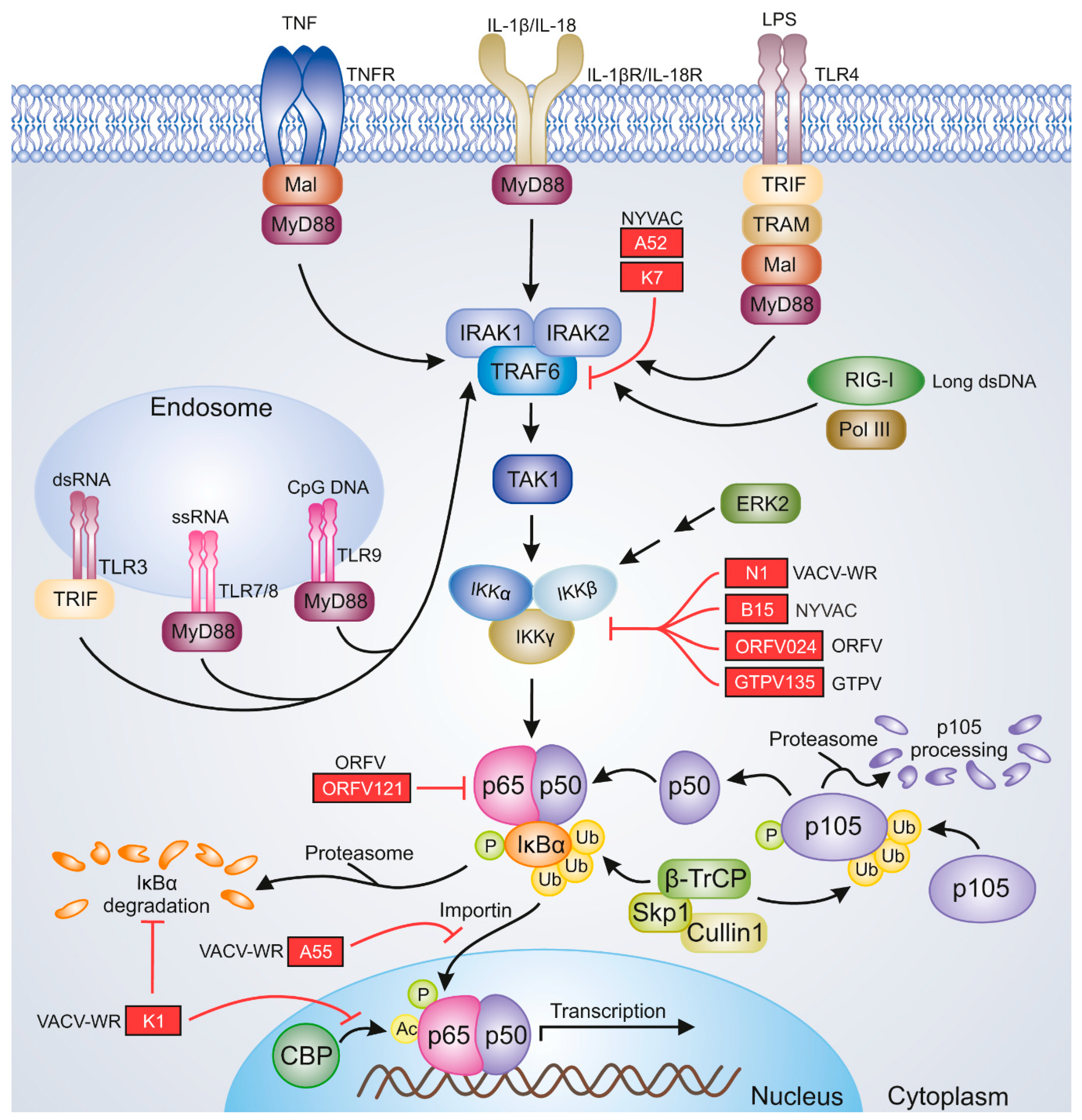
The vaccine used in the global smallpox eradication program was based on several strains of VACV. For instance, in the United States, the New York City Board of Health (known as NYCBH) and Lister strains were used for vaccination against smallpox, while in Europe, Lister, Bern, Paris, and Copenhagen (called VACV-COP) strains were applied. The first-generation antismallpox vaccines were propagated in the skin of calf and other animals, while the second-generation VACV-based vaccines were grown in tissue culture and chicken embryos instead of live animals. Unfortunately, the vaccines generated in cell cultures are not sufficiently safe. Therefore, the use of second-generation antismallpox vaccines is limited [3]. Recently, it has been shown that chicken embryonic stem cells (cESCs) may serve as an alternative source for the propagation of poxvirus vaccine vectors. The idea behind the use of cESCs rather than mammalian ESCs is linked to ethical issues and safety concerns, the most important of which is that cESCs lack transforming oncogenes or adventitious agents [26].
Despite the eradication of smallpox, the risk of reoccurring VARV infections remains to be eliminated because of the bioterrorist threat or the possible de novo synthesis of the virus. Therefore, the pathogenesis of orthopoxvirus diseases is still of interest to researchers [27,28]. Although VACV-based vaccines used in the global smallpox eradication program were effective, some adverse effects of vaccination, including postvaccinal encephalitis, generalized vaccinia, progressive vaccinia, and eczema vaccination, were observed in immunocompromised individuals and patients with skin conditions. To overcome these, modified VACV Ankara (MVA)—Bavarian Nordic (MVA-BN), a third-generation attenuated antismallpox vaccine—has been introduced. This vaccine is approved in Canada (Imvamune) and the European Union (Imvanex) [29,30].
An important concern related to smallpox is not only the fear of its reoccurrence but also the occurrence of new zoonotic orthopoxviral infections and the disappearance of antismallpox immunity, which confers cross-protection against other orthopoxviral diseases. Zoonotic poxviral infections are caused by VACV, monkeypox virus (MPXV), cowpox virus (CPXV), camelpox virus (CMLV) orthopoxviruses, bovine papular stomatitis virus (BPSV), ORFV, pseudocowpox virus (PCPV) parapoxviruses, Yaba monkey tumor virus (YMTV), and tanapox virus (TPV) yatapoxviruses. Therefore, antiviral drugs and vaccines against poxviral diseases are still under development [31,32,33,34,35,36,37,38,39,40].
The currently used VACV-based vaccines, which are nonreplicating and attenuated, such as MVA-BN-vectored encephalitic alphavirus vaccine, target the biothreat viruses. Another VACV-derived vaccine, RABORAL V-RG, an antirabies vaccine expressing the rabies virus (RABV) glycoprotein gene (V-RG), has been used in Europe and North America to vaccinate foxes and raccoons [30]. Additionally, VACV-based vaccine candidates have been demonstrated to protect against emerging viral diseases, such as chikungunya virus (CHIKV) disease [41,42] and yellow fever [43] in preclinical animal models. The Sementis Copenhagen Vector (SCV) is a new multiplication-defective VACV-COP-derived vaccine vector with targeted deletion of D13L gene encoding D13 protein that is essential for viral assembly. This new vector has recently been successfully tested in nonhuman primates as a vaccine against Zika and chikungunya [41].
The present status of clinical trials employing VACV against viral diseases is shown in Table 1. Other poxviruses that can be used as vaccine vectors belong to Avipoxvirus [44,45], Capripoxvirus [46], Leporipoxvirus [47], Parapoxvirus [48], and Suipoxvirus [49] genera.

4. NF-κB Signaling

One of the key factors involved in the proper induction of antiviral immunity is NF-κB. It constitutes a family of dimeric transcription factors, which regulate the expression of numerous genes involved in the cell cycle, apoptosis, and immunity. The NF-κB family consists of five proteins: RelA/p65, RelB, c-Rel, NFκB1 p105/p50, and NFκB2 p100/p52. The NF-κB dimer that is most commonly detected in the cytoplasm of unstimulated cells is composed of RelA and p50 subunits [50]. The RelA/p50 heterodimer remains in the cytoplasm due to the activity of IκBα, which masks the nuclear localization sequences (NLSs) of NF-κB [51]. The classical NF-κB signaling pathway is induced by proinflammatory cytokines such as interleukin-1β (IL-1β), IL-18, and TNF-α and various ligands of pattern recognition receptors (PRRs), which are represented by retinoic acid-inducible gene-I (RIG-I) and Toll-like receptors (TLRs). In the NF-κB signaling cascade, the cellular receptors cooperate with adapter molecules and induce the cellular pathways that activate the transcriptionally active dimers [52]. Upon the stimulation of NF-κB signaling, transforming growth factor (TGF)-β-activated kinase 1 (ΤAΚ1) activates IKK, the IKKβ subunit of which triggers IκBα phosphorylation at Ser32 and Ser36. This event results in the recognition of IκBα by the E3 ubiquitin ligase complex composed of β-transducin repeat-containing proteins: S-phase kinase-associated protein 1 (Skp1)–Cullin 1–F-box (SCFβ−TrCP). Conjugation of phosphorylated IκBα with K48-linked polyubiquitin chains of Lys 48 of ubiquitin by SCFβ−TrCP results in 26S proteasome-mediated IκBα degradation and the release of RelA/p50 dimers. These dimers translocate to the nucleus, where they bind DNA and initiate the transcription of target genes. E3 ubiquitin ligase complex is also involved in p105 proteasomal processing to p50 [50,53,54] (Figure 1). On the other hand, the noncanonical NF-κB signaling triggered by the members of the TNF superfamily leads to the activation of NF-κB-inducing kinase (NIK), which then activates IKKα. IKKα, in turn, phosphorylates the C-terminal portion of p100 precursor protein, which retains RelB in the cytoplasm due to its IκB activity. Following the phosphorylation of p100 at Ser866 and Ser870, IκB-like C-terminal portions of this protein are ubiquitinated, leading to the generation of a p52 active NF-κB subunit. RelB/p52 dimers translocate to the nucleus and initiate the transcription of target genes [55]. In general, the canonical NF-κB signaling is responsible for the regulation of innate immunity [56], whereas the noncanonical NF-κB activation pathway regulates the adaptive immune responses. However, there exist regulatory mechanisms for these two signaling pathways as well as for the crosstalk between them [55,57]. The modulation of NF-κB signaling is attributed to viral pathogens, one excellent example of which is the viruses belonging to the Poxviridae family encoding multiple immunomodulatory proteins; these proteins affect the components of NF-κB signaling and therefore disrupt the antiviral innate response [52,58]. Selected NF-κB inhibitors of VACV, ORFV, and GTPV, which may be relevant to the efficacy of poxviral vaccines, are shown in Figure 1.

5. NF-κB in Optimization of Poxviral Vaccines

5.1. Vaccinia Virus

VACV is a pathogen whose origin or natural host has not been identified so far. It was believed that VACV infections occur due to the spread of vaccine strains into new wild hosts. However, it is now obvious that VACV is transmitted via peridomestic rodents, which infect wild animals and cows. In humans, VACV can be transmitted via infected animals and is observed in milkers in areas where the virus circulates, especially in Asian and South American countries, such as Brazil. Furthermore, VACV infection of humans occurs via their direct contact with the crusts containing viral particles, which results in the formation of skin focal lesions on the hands and forearms [18,37,59]. After a few days, the focal lesions form pustules leading to edema and erythema. Ulcerated and necrotic lesions appear after a maximum of 12 days of VACV infection, following which crusts develop. In 4 weeks, the lesions disappear, but local lymphadenopathy can be observed for 20 days. Alternatively, a systemic infection manifested by fever, headache, and muscle ache develops immediately after the appearance of lesions [60].

5.1.1. Modified VACV Ankara

VACV is regarded as a universal vaccine carrier. However, the use of replicating VACV as an antismallpox vaccine has led to severe adverse effects in individuals with an immunocompromised immune system or skin disorders. Currently, MVA obtained by 570 passages of chorioallantois VACV Ankara (CVA) strain in chicken embryo fibroblasts remains an excellent alternative to traditional antismallpox vaccines [61]. MVA does not replicate in human cells but displays good immunogenicity as well as a good safety profile in vivo. In addition, MVA can be successfully used in immunocompromised humans [12,15,32,62,63,64,65,66]. The ongoing clinical trials on MVA-vectored vaccines targeted at viral diseases are shown in Table 1.
MVA is a good candidate for an effective and safe vaccine vector. Nevertheless, its immunogenicity can be enhanced to improve the efficacy of the vaccine, for which the introduction of immune-stimulating genes into MVA and reinsertion of certain VACV genes to obtain a replication-competent virus are considered beneficial [67].
Despite the expression of A46 (Toll/IL-1-receptor signaling interference protein), B16 (IL-1-binding protein (IL-1BP)), and Κ7 (B cell lymphoma 2 (Bcl-2)-like protein), which inhibit NF-κB signaling, MVA stimulates NF-κB [61,68,69]. Studies on NF-κB signaling in MVA-infected cells have shown that early MVA protein expression in human 293T fibroblasts activates the phosphorylation of extracellular signal-regulated kinase 2 (ERK2), which, in turn, mediates the activation of NF-κB [70]. Further research revealed that MVA triggers NF-κB activation via VACV growth factor (VGF), which interacts with the epidermal growth factor receptor (EGFR). Moreover, in 293T cells and Hacat keratinocytes infected by MVA deprived of an early C11R gene encoding VGF, reduced activation of ERK2 and NF-κB was observed. Since keratinocytes are the immediate target of the vaccine, the prosurvival role of NF-κB would be beneficial to stimulate cells for a long duration for optimal induction of immune response before cell lysis occurs [71].
Other studies on MVA revealed that, during the early phase of viral replication in human embryonic kidney 293 cells that were transformed with large T antigen (HEK 293T), IκBα degradation occurs before the initiation of viral replication. It has been shown that in Chinese hamster ovary (CHO), HEK 293T, and rabbit kidney 13 (RK13) cells, IκBα degradation can be inhibited by the expression of CP77, a CPXV Brighton Red (CPXV-BR) early host-range gene [72].
CP77-encoded protein is one of the ankyrin (ANK) repeat proteins [73]. In general, ANK repeats, consisting of 30–34 amino acid residues, are involved in protein–protein, protein–sugar, or protein–lipid interactions. ANK repeat proteins take part in cellular signaling, vesicular trafficking, cell cycle control, and inflammation, are responsible for cytoskeleton integrity, and regulate transcription. They are present in both eukaryotic and prokaryotic cells, such as intracellular bacteria [74].
ANK repeat proteins are not commonly expressed by viruses. However, within the Chordopoxvirinae subfamily of poxviruses, only three species of different genera lack ANK proteins. In poxviruses, these proteins are composed of multiple ANK motifs, which start from the N-terminus. The ANK motifs are followed by non-ANK linker sequence. In turn, the C-terminus has the homolog of cellular F-box sequence. The cellular F-box sequence interacts with ubiquitin ligase E3 complexes, thus allowing ubiquitination and subsequently 26S proteasome-mediated protein degradation. ANK proteins of poxviruses belonging to Avipoxvirus, Parapoxvirus, Orthopoxvirus, and the Leporipoxvirus supergroup genera may either bind to Skp1 or interact with cellular proteins, thus acting as inhibitors of cellular signaling, such as NF-κB [75]. Some poxviral ANK repeat proteins, including VACV K1 [76] and myxoma virus (MYXV) M150 [77], may act as nuclear NF-κB inhibitors.
Furthermore, certain poxviral ANK repeat proteins, in which the F-box domain is absent, serve as host-range proteins [75]. These are represented by VACV K1 protein encoded by K1L gene [78]. It has been shown that MVA expressing the VACV Western Reserve (VACV-WR) gene K1L, which prevents IκBα degradation [79] and p65 acetylation [76], potentially inhibits IκBα degradation in HEK 293T cells. Importantly, in mouse embryonic fibroblasts (MEFs), human or mouse dsRNA-activated protein kinase R (PKR) is crucial for IκBα degradation. It can be assumed that the induction of PKR by MVA may stimulate immune response and impair virus replication, which is critical for the safety and efficacy of the vaccine [72].
Furthermore, stimulation of immune response is necessary for the prolonged presentation of antigens and delayed viral clearance. The insertion of a VACV-CVA 5.2-kb region containing apoptosis inhibitor and ANK repeat protein gene M1L [80] and NF-κB inhibitors encoded by M2L (mitogen-activated protein kinase kinase (MEK)/ERK and NF-κB inhibitor) and K1L (PKR and NF-κB inhibitor) genes into MVA decreases both apoptosis and virus-mediated NF-κB activation in antigen-presenting cells (APCs) in vivo. Moreover, VACV-specific CD8+ T cell response is diminished in vivo after treatment with MVA/5.2 kb compared to MVA. These results contradict the findings observed in vitro, in which MVA/5.2 kb displays an immunostimulatory effect [67].

5.1.2. New York VACV

Another VACV-based vaccine vector, the New York VACV strain (NYVAC), derived from the VACV-COP strain, is deprived of 18 ORFs, including the host-range K1L gene, encoding an IFN antagonist protein and NF-κB inhibitor. It has been shown that both MVA and NYVAC-infected HeLa cells display enhanced expression of NF-κB protein, degradation of IκBα, and secretion of IL-6. Furthermore, NYVAC upregulates certain NF-κB-responsive genes, including activating transcription factor 3 (ATF3). It can be concluded that ATF3 may act pro-apoptotically during NYVAC infection. However, the ectopic expression of VACV-WR K1L gene in NYVAC-infected cells induced apoptosis but inhibited NF-κB. This indicates that K1 does not prevent apoptosis in NYVAC-infected cells. Both induction of apoptosis and inhibition of NF-κB may not be favorable for the NYVAC replication cycle; however, apoptosis occurs at the late stage of the viral lifecycle when the replication is completed. These events are important for the generation of immune response and vector clearance [81]. Interestingly, although both MVA and NYVAC stimulate NF-κB, A52R gene is present in NYVAC, but not in MVA. The absence of A52, a Bcl-2-like protein, which binds IL-1 receptor (IL-1R)-associated kinase 2 (IRAK2) and thus inhibits NF-κB, is likely to upregulate the TLR3 pathway in MVA-infected cells. Since A52 binds to IRAK2 and TNFR-associated factor 6 (TRAF6), which are important for TLR signaling, upregulation of TRAF6 and downregulation of IL-1α and IL-1β were observed in MVA-infected immature human monocyte-derived dendritic cells (MDDCs), but not in NYVAC-infected cells [82].
NYVAC has been proposed as a candidate for a human immunodeficiency virus (HIV) vaccine. NYVAC-C ΔA52R ΔB15R ΔK7R, and NYVAC deletion mutant lacking A52R, B15R (encoding B14 protein that binds IKKβ), and K7R (encoding TRAF6 and IRAK2-binding protein) has been used to express HIV type 1 (HIV-1) envelope (Env) glycoprotein 120 (gp120) and GPN (Gag-Pol-Nef) clade C antigens. In mice, NYVAC-C ΔA52R ΔB15R ΔK7R induced the activation of chemokines/cytokines and migration of Nα and Nβ neutrophils to the infection site. This effect was accompanied by an increase in T cell response toward HIV antigens. The activation of virus-specific CD8+ T cells was triggered by Nβ neutrophils displaying an APC-like phenotype [83]. Further analyses of mice models infected with NYVAC mutants (NYVAC-C ΔA52R, NYVAC-C ΔA52R ΔK7R, or NYVAC-C ΔA52R ΔB15R) showed that the infection increased the number of CD11c+ major histocompatibility complex (MHCII)+-positive DCs in mice. Deletion of A52R gene influenced the migration of DCs, whereas double-gene deletion affected the migration of both DCs and neutrophils. Finally, the deletion of all A52R, B15R, and K7R genes not only enhanced the migration of DCs, neutrophils, and natural killer (NK) cells but also influenced chemokine release. In addition, NYVAC-C ΔA52R ΔB15R, NYVAC-C ΔA52R ΔK7R, and NYVAC-C Δ3 (triple-deletion) mutants induced CTLs. Among the double-deletion mutants, NYVAC-C ΔA52R ΔB15R not only induced a strong T CD8+ response but was also effective in the induction of IgG. These studies demonstrate that double or triple deletion of NF-κB inhibitors from NYVAC enhances both T cell-specific and humoral anti-HIV responses. The induction of Gag- and Pol-specific CD8+ T lymphocytes by NYVAC mutants showed that NYVAC-based vectors are promising anti-HIV vaccine candidates [84].

5.1.3. VACV Western Reserve

Another VACV strain that can be used as a vaccine vector is VACV-WR. When modifying the VACV-WR genome, single-gene deletions are more beneficial than the deletions of multiple genes, which may decrease the immunogenicity of the vaccine [85]. One of the candidate genes that can be deleted from VACV-WR is N1L, encoding a Bcl-like inhibitor of NF-κB, which prevents NF-κB activation by proinflammatory cytokines including TNF-α or IL-1β [86]. Studies on intradermal murine model infection with VACV-WR devoid of N1L gene have shown that NF-κB is essential for CD8+ T cell memory and, consequently, for the efficacy of vaccines. N1 is an early VACV protein that inhibits apoptosis. Therefore, its mutation or deletion reduces the virulence of VACV and, at the same time, enhances CD8+ T cell response, which is desirable for antiviral protection induced by the vaccine [87]. VACV-WR vaccine can also be modified by the deletion of K1L NF-κB inhibitor. In mice models, K1-deficient virus induced VACV-specific CD8+ T cell response and prevented lethal VACV infections, despite silencing the innate immune response. Above all, at day one postinfection, the deletion mutant did not induce the expression of NF-κB-regulated genes, such as Nfkbia and Tnf. However, Ifna4, Il7, and Nfkb2, which are only partially controlled by NF-κB, were downregulated [88].
Recently, the importance of VACV-WR BTB-BACK-Kelch (BBK)-like protein, A55, in NF-κB modulation has been described. A55 is an NF-κB inhibitor that disturbs the p65–importin interaction and thus prevents the transcription of NF-κB-regulated genes and impairs inflammatory response. Especially, NF-κB-regulated cytokines and inflammation influence the proliferation and development of effector and memory T cells. As expected, the deletion of the A55R gene from VACV-WR resulted in the enhancement of CD8+ T cell memory, and the vaccine displayed increased immunogenicity and protected the mice challenged with VACV intranasally [89].

5.2. Orf Virus

ORFV, a virus belonging to the Parapoxvirus genus, is the causative agent of orf disease—highly contagious ecthyma. In humans, orf is an enzootic and self-limiting disease manifested as pustular dermatitis, which spontaneously resolves within 3 to 6 weeks. The ORFV infections caused in humans are frequently observed in Asia and Africa. In sheep and goats, these infections cause scabby mouth disease, which is characterized by high morbidity in infected sheep worldwide. ORFV may also infect cats, reindeers, camels, serows, and musk oxen. Mortality due to ORFV infections is rarely associated with secondary infections and aspiration pneumonia. In general, orf disease is a threat to kids and lambs and may cause farms to suffer economic losses [90,91,92,93,94].
The use of ORFV as a vaccine vector may constitute a novel strategy to vaccinate both permissive and nonpermissive hosts against orf, which is an alternative to the current attenuated vaccines that are inefficient or insufficiently safe. The immunomodulatory properties of ORFV, as well as its ability to replicate in various hosts, make it a good vaccine candidate. Moreover, preclinical studies have confirmed these properties of inactivated ORFV [91]. Due to the fact that ORFV does not spread systemically or neutralize antibody production, it can be successfully used as a vaccine vector for repeat immunizations [95]. The highly attenuated anticontagious ecthyma vaccine strain D1701 derived from ORFV protects sheep for 4–6 months [96,97]. When adapted to Vero cells, D1701-V can be used to deliver target genes into the vegf-e gene for the construction of a vaccine against pseudorabies virus (PRV), the causative agent of Aujeszky’s disease [98,99,100]. D1701-V-VP1 expressing capsid protein VP1 has been used to immunize rabbits against rabbit hemorrhagic disease virus (RHDV) [101]. Another recombinant D1701-V vector, D1701-V-RabG expressing the RABV glycoprotein, has been designed as a new antirabies vaccine for companion animals and tested on murine, dog, and cat models [97]. D1701-V-HAh5n expressing H5 hemagglutinin has also been proposed as a vaccine against avian influenza virus H5N1 and tested on mice models [102]. In addition, DNA vaccines expressing ORFV011 EEV envelope phospholipase and ORFV059 immunodominant envelope antigen F1L protein have shown enhanced immunogenicity and triggered lasting immunity in mouse models [103].

ORFV-IA82

Thus far, several ORFV-encoded proteins capable of inhibiting NF-κB signaling have been identified, including ORFV024 [104], ORFV002 [105,106], ORFV121 [107], ORFV073 [108], and ORFV119 [109]. Recently, ORFV020, a dsRNA-binding IFN resistance protein, displaying dsRNA adenosine deaminase activity, has been described. ORFV020 is a counterpart of VACV-WR E3, which inhibits the activation of PKR and NF-κB. Moreover, it belongs to viral IFN (VIR) resistance proteins that inhibit IFN-mediated antiviral response. Therefore, ORFV expressing ORFV020 is resistant to the activity of IFN type I and type II. Considering the conservative nature of ORFV isolates, the deletion or mutation of the E3L counterpart may be ideal for vaccine construction [110].
ORFV strain ORFV-IA82 is used as a vaccine against porcine epidemic diarrhea. The ORFV-PEDV-S virus expressing spike (S) proteins of porcine epidemic diarrhea virus (PEDV) was constructed using the ORFV121 locus insertion site. This site encodes a unique parapoxviral NF-κB inhibitor, which blocks the phosphorylation of p65 and its nuclear translocation [107]. Immunization of pigs with ORFV-PEDV-S induced the production of neutralizing antibodies and PEDV-specific serum IgA and IgG. Importantly, ORFV-PEDV-S ensured protection from the clinical outcomes of the infection. Reduced virus shedding was also found upon the immunization of infected animals [111]. Furthermore, ORFV-PEDV-S vaccine has been shown to induce passive immunity in newborn piglets [112].
In addition, ORFV-IA82 can be used as an antirabies vaccine. The gene ORFV024 encoding a unique parapoxviral inhibitor of IKK kinase and IκBα degradation [89] was used as an insertion site for the RABV glycoprotein (G) gene. Similarly, ORFV121 gene encoding NF-κB inhibitor has been used as an insertion site for G-encoding gene. Immunization of pigs and cattle with ORFVΔ024RABV-G or ORFVΔ121RABV-G resulted in the induction of neutralizing antibodies. Of these, the ORFVΔ121 mutant was more immunogenic [113].

5.3. Goatpox Virus

GTPV, a member of the Capripoxvirus genus, is a sheep pathogen and the causative agent of the goatpox disease. Goatpox is transmitted via aerosols and insects and causes systemic infections in goats and sheep, which manifest as fever, enlargement of lymph nodes and skin, and respiratory and gastrointestinal lesions. Goatpox disease is also a source of economic loss to domestic ruminant farms. In general, GTPV is an economically important capripoxvirus in central Asia, North Africa, the Middle East, and India [114,115,116]. At present, only attenuated vaccines are available for GTPV and other capripoxviruses [117]. The capripoxvirus-based vaccines, which are obtained by serial passages, confer protective immunity for 1 year after vaccination. Capripoxviruses can also be used as vectors for vaccines against diseases caused by ruminant pathogens, such as bluetongue, Rift Valley fever, peste des petits ruminants, or rinderpest. Certain GTPV strains, such as Isiolo and Kedong, which infect goats, sheep, and cattle, are used as a universal vaccine against capripox diseases [46]. Gorgan strain-based vaccines protect cattle against lumpy skin disease. Caprivac (Jordan Bio-Industries Centre, JOVAC) is one of the vaccines used against goatpox in cattle in the Middle East [118].

GTPV-AV41

The existing GTPV vaccine, GTPV-AV41, contains an attenuated strain obtained by passages of GTPV-AV40 strain in the testis cells of goats and sheep. Unfortunately, it may cause generalized skin lesions and miscarriages in vaccinated animals, and thus, its use hinders distinguishing between vaccinated and infected ones [119]. Therefore, modifications of the virus are needed to improve the vaccine. It is worth noticing that the inactivation of NF-κB-related genes has been observed among capripoxvirus vaccine strains. For instance, in the GTPV Gorgan vaccine, a 1.6-kbp deletion led to the inactivation of GTPV_144 and GTPV_145 genes. GTPV_144 is a counterpart of VACV-COP A55R, encoding Kelch repeat and BTB domain-containing protein 1, while GTPV_145 is related to VACV-COP B4R, encoding ANK repeat protein [117]. Mutations in these two genes are common in capripoxviruses and are typical for vaccine strains. Since A55 inhibits CD8+ T cell memory, the deletion of A55R gene or its GTPV counterpart may improve the immunogenicity of the vaccine [89]. GTPV_145 encodes an ANK repeat protein, a counterpart of B4 VACV-COP and EVM154 proteasome inhibitor of ectromelia virus, which interacts with Skp1 and conjugated ubiquitin and subsequently inhibits IκBα degradation. It is believed that that EVM154 may be involved in virus spread and its depletion may cause attenuation of the virus [120,121].
One of the insertion site candidates for GTPV-based vaccines is the 135 ORF containing an early gene, which is not essential for the viral replication in vitro and in vivo. The 135 gene encodes an 18-kDa protein, which inhibits NF-κB and apoptosis. The GTPV135 protein is a counterpart of a Bcl-like VACV-WR N1 protein. The 135 gene is a host innate immune response inhibitor and may therefore serve as an insertion site for live attenuated dual vaccines instead of the tk locus insertion site. Interestingly, the GTPV AV41 vaccine strain expressing the hemagglutinin protein of peste des petis ruminants virus (PPRV), whose gene was inserted in the ORF135 insertion site, displayed a stronger antibody neutralization response than the strain with a tk insertion site [122]. To improve GTPV-AV41 and prevent the side effects of the vaccine, it may be necessary to perform modifications based on the deletion of the nonessential gene. For instance, deletion of the viral tk gene and ORF8–18 may be beneficial for the vaccine. Among ORF8–18 homologs of VACV, the following NF-κB inhibitors can be found: ORF12, which encodes ANK repeat protein and a counterpart of B4, and ORF15, encoding a homolog of VACV IL-18BP, C12. ORF16, in turn, encodes an EGF-liked growth factor, a C11 VACV counterpart, which may activate NF-κB. In vaccinated animals, the attenuated vaccine GTPV-TK-ORF vector allowed maintaining immunogenicity and increased safety compared to wild-type GTPV-AV41. The vaccine also induced the production of neutralizing antibodies and GTPV-specific antibodies, as well as the release of IFN-γ in goats. Hence, removal of nonessential genes that are linked to apoptosis inhibition and immune modulation is considered as a factor that may improve the efficacy of the vaccine [123].

6. Conclusions

Generation of effective immune response and immunological memory, as well as safety, is the main concern in vaccine development. When employing virus-based vaccines, it is necessary to ensure both the complete replication cycle of the virus and proper induction of immunological memory for determining the vaccine efficiency. The loss of viral immunomodulatory proteins may affect these parameters, thus influencing efficiency. Since poxviruses modulate the activation of immune cells by affecting the NF-κB-mediated apoptosis regulation, inflammation, and immunological memory, discovering new mechanisms of NF-κB inhibition and cellular targets of poxviruses may help modify vaccine candidates to improve the efficacy of poxvirus-based vaccines and the immunological memory generated by them.

Author Contributions

J.S. contributed to conceptualization and writing (original draft preparation, review, and editing). L.S.-D. contributed to conceptualization and writing (figure preparation, review, and editing). All authors have read and agree to the published version of the manuscript.

Funding

This work was funded by National Science Centre, Poland, grant number UMO-2015/19/D/NZ6/02873.

Conflicts of Interest

The authors declare no conflict of interest. The funders had no role in the design of the study; in the collection, analyses, or interpretation of data; in the writing of the manuscript; or in the decision to publish the results.

Abbreviations

VARVvariola virus
VACVvaccinia virus
ORFVorf virus
GTPVgoatpox virus
MVmature virus
EVextracellular virus
ORFopen reading frame
TKthymidine kinase
ITRinverted terminal repetition
IFNinterferon
STINGstimulator of IFN genes
cGAScyclic GMP-AMP synthase
DNA-PKDNA-dependent protein kinase
IFI16IFN-γ-inducible protein 16
TNFtumor necrosis factor
TNFRTNF receptor
TRAFTNFR-associated factor
NF-κBnuclear factor kappa-light-chain-enhancer of activated B cells
TANKTRAF family member-associated NF-κB activator
TBK1TANK-binding kinase
IRF3IFN regulatory factor 3
IκBinhibitor κB
IKKIκB kinase
CTLcytotoxic T lymphocyte
NYCBHNew York City Board of Health
VACV-COPVACV Copenhagen strain
cESCchicken embryonic stem cell
MVAmodified VACV Ankara
MVA-BNmodified VACV Ankara - Bavarian Nordic
MPXVmonkeypox virus
CPXVcowpox virus
CMLVcamelpox virus
BPSVbovine papular stomatitis virus
PCPVpseudocowpox virus
YMTVYaba monkey tumor virus
TPVtanapox virus
RABVrabies virus
V-RGRABV glycoprotein gene
CHIKVchikungunya virus
SCVSementis Copenhagen Vector
Adadenovirus
Ad26Ad serotype 26
Ad26.HPV16Ad26 HPV16 vaccine
Ad26.HPV18 Ad26 HPV18 vaccine
Ad26.Mos.HIVAd26-mosaic-HIV vaccine
Ad26.Mos4.HIVAd26-mosaic 4-HIV vaccine
Ad26.ZEBOVhuman Ad26 expressing the Ebola virus Mayinga variant gp
AIDSacquired immune deficiency syndrome
ATIanalytical treatment interruption
bNAbsbroadly neutralizing HIV-1 antibodies
ChAdChimpanzee adenovirus
ChAd155ChAd serotype 155
ChAd155-hIi-HBVChAd HBV vaccine
ChAd3-hliNSmutChAd3 encoding NSmut linked to hli
ChAdOx1replication-deficient ChAd vector derived from isolate Y25
ChAdOx1.HTIChAdOx1 expressing HTI
ChAdV63ChAd serotype 63
ChAdV63.HIVconsvChAdV63 expressing HIVconsv
CMVcytomegalovirus
DNA.HTIplasmid DNA expressing HTI
gpglycoprotein
gp140 DPgp140 drug product
GS-9620vesatolimod
HBchepatitis B core antigen
HBc-HBs/AS01B-4HBV vaccine
HBshepatitis B surface antigen
HBVhepatitis B virus
HCThematopoietic cell transplantation
HIVhuman immunodeficiency virus
HIV-1HIV type 1
HIVconsHIV conserved antigenic regions
hlihuman invariant chain
HPVhuman papillomavirus
HPV16/18human papillomavirus type 16/18
HTIHIVACAT T cell immunogen
IL-12interleukin-12
M1matrix protein
MenACWYmeningococcal ACWY-tetanus toxoid conjugate vaccine
MVA.HIVconsvMVA expressing HIVconsv
MVA.HPV16/18MVA HPV16/18 vaccine
MVA.HTIMVA expressing HTI
MVA.tHIVconsv3MVA-based T-cell vaccine expressing novel HIV-1 immunogens
MVA.tHIVconsv4MVA-based T-cell vaccine expressing novel HIV-1 immunogens
MVA62BMVA component—encoding HIV-1 Gag, protease, reverse transcriptase, and envelope gp160
MVA-BN-FiloMVA-BN-Filo vector
MVA-HBVMVA HBV vaccine
MVA-hliNSmutMVA encoding NSmut linked to hli
MVA-mosaicMVA mosaic HIV vaccine
MVA-NP + M1MVA encoding NP and M1
NPnucleoprotein
NSmutHCV nonstructural immunogen
p24CE + p55gagDNA vaccines expressing p24CE and p55gag immunogens
rVSVΔG-ZEBOV-GPrecombinant VSV–Zaire Ebola virus gp
TLR9Toll-like receptor 9
VRC07, 10-1074anti-HIV-1 bNAbs
VSVvesicular stomatitis virus
NLSnuclear localization sequence
IL-1βinterleukin-1β
IL-18interleukin-18
TNF-αtumor necrosis factor-α
PRRpattern recognition receptor
RIG-Iretinoic acid-inducible gene-I
TGF-βtransforming growth factor-β
TAK1TGF-β-activated kinase 1
Skp1S-phase kinase-associated protein 1
SCFSkp1-Cul1-F-box
β-TrCPβ-transducin repeat-containing protein
NIKNF-κB-inducing kinase
Acacetyl group
CBPCREB-binding protein
CpGcytosine–guanine dinucleotide
ERK2extracellular signal-regulated kinase 2
IKKαIκB kinase α
IKKβIκB kinase β
IKKγIκB kinase γ
IL-18RIL-18 receptor
IL-1βRIL-1β receptor
IRAK1IL-1R-associated kinase 1
IRAK2IL-1R-associated kinase 2
LPSlipopolysaccharide
MalMyD88-adapter-like
MyD88myeloid differentiation primary response gene 88
Pphosphate group
Pol IIIpolymerase III
TLR3Toll-like receptor 3
TLR4Toll-like receptor 4
TLR7Toll-like receptor 7
TLR8Toll-like receptor 8
TRAMTRIF-related adapter molecule
TRIFToll-IL-1R-domain-containing adapter-inducing interferon-β
UbUb-ubiquitin moieties
VACV-WRVACV Western Reserve strain
β-TrCPβ-transducin repeat-containing protein
CVAchorioallantois VACV Ankara strain
IL-1BPIL-1 binding protein
Bcl-2B-cell lymphoma 2
VGFVACV growth factor
EGFRepidermal growth factor receptor
HEK 293Thuman embryonic kidney 293 cells transformed with large T antigen
CHOChinese hamster ovary cells
RK13rabbit kidney 13 cells
CPXV-BRCPXV Brighton Red strain
ANKankyrin repeat
MYXVmyxoma virus
MEFsmouse embryonic fibroblasts
PKRprotein kinase R
MEKmitogen-activated protein kinase kinase
APCantigen presenting cell
ATF3activating transcription factor 3
MDDCmonocyte-derived dendritic cell
gp120glycoprotein 120
GPNGag-Pol-Nef
MHCIImajor histocompatibility complex class II
NKnatural killer cell
BBKBTB-BACK-Kelch
PRVpseudorabies virus
RHDVrabbit hemorrhagic disease virus
D1701-V-RabGrecombinant D1701 ORFV strain expressing RABV glycoprotein
D1701-V-HAh5nrecombinant D1701 ORFV strain expressing H5 hemagglutinin
VIRviral IFN resistance
PEDVporcine epidemic diarrhea virus
Sspike protein
ORFV-PEDV-S
ORFVΔ024RABV-GORFV Δ024 mutant expressing RABV glycoprotein
ORFVΔ121RABV-GORFV Δ121 mutant expressing RABV glycoprotein
PPRVpeste des petis ruminants virus

References

  1. Virus Taxonomy: 2019 Release. Available online: https://talk.ictvonline.org/ (accessed on 20 November 2020).
  2. Babkin, I.V.; Babkina, I.N. The origin of the variola virus. Viruses 2015, 7, 1100–1112. [Google Scholar] [CrossRef] [PubMed] [Green Version]
  3. Shchelkunova, G.A.; Shchelkunov, S.N. 40 Years without Smallpox. Acta Naturae 2017, 9, 4–12. [Google Scholar] [CrossRef] [PubMed]
  4. Buller, R.M.L. Poxviruses. In Infectious Diseases, 4th ed.; Cohen, J., Powderly, W.G., Opal, S.M., Eds.; Elsevier: Amsterdam, The Netherlands, 2017; pp. 1452–1457.e1. [Google Scholar] [CrossRef]
  5. Burrell, C.J.; Howard, C.R.; Murphy, F.A. (Eds.) Poxviruses. In Fenner and White’s Medical Virology, 5th ed.; Academic Press: London, UK, 2017; pp. 229–236. [Google Scholar] [CrossRef]
  6. Condit, R.C.; Moussatche, N. The vaccinia virus E6 protein influences virion protein localization during virus assembly. Virology 2015, 482, 147–156. [Google Scholar] [CrossRef] [PubMed] [Green Version]
  7. El-Jesr, M.; Teir, M.; Maluquer de Motes, C. Vaccinia virus activation and antagonism of cytosolic DNA sensing. Front. Immunol. 2020, 11, 568412. [Google Scholar] [CrossRef] [PubMed]
  8. Lu, Y.; Zhang, L. DNA-sensing antiviral innate immunity in poxvirus infection. Front. Immunol. 2020, 11, 1637. [Google Scholar] [CrossRef] [PubMed]
  9. Reynolds, M.G.; Guagliardo, S.A.J.; Nakazawa, Y.J.; Doty, J.B.; Mauldin, M.R. Understanding orthopoxvirus host range and evolution: From the enigmatic to the usual suspects. Curr. Opin. Virol. 2018, 28, 108–115. [Google Scholar] [CrossRef]
  10. McFadden, G. Poxvirus tropism. Nat. Rev. Microbiol. 2005, 3, 201–213. [Google Scholar] [CrossRef]
  11. Haller, S.L.; Peng, C.; McFadden, G.; Rothenburg, S. Poxviruses and the evolution of host range and virulence. Infect. Genet. Evol. 2014, 21, 15–40. [Google Scholar] [CrossRef] [Green Version]
  12. Okeke, M.I.; Okoli, A.S.; Diaz, D.; Offor, C.; Oludotun, T.G.; Tryland, M.; Bøhn, T.; Moens, U. Hazard characterization of modified vaccinia virus Ankara vector: What are the knowledge gaps? Viruses 2017, 9, 318. [Google Scholar] [CrossRef] [Green Version]
  13. García-Arriaza, J.; Esteban, M. Enhancing poxvirus vectors vaccine immunogenicity. Hum. Vaccines Immunother. 2014, 10, 2235–2244. [Google Scholar] [CrossRef] [Green Version]
  14. Prow, N.A.; Jimenez Martinez, R.; Hayball, J.D.; Howley, P.M.; Suhrbier, A. Poxvirus-based vector systems and the potential for multi-valent and multi-pathogen vaccines. Expert Rev. Vaccines 2018, 17, 925–934. [Google Scholar] [CrossRef] [PubMed]
  15. Walsh, S.R.; Wilck, M.B.; Dominguez, D.J.; Zablowsky, E.; Bajimaya, S.; Gagne, L.S.; Verrill, K.A.; Kleinjan, J.A.; Patel, A.; Zhang, Y.; et al. Safety and immunogenicity of modified vaccinia Ankara in hematopoietic stem cell transplant recipients: A randomized, controlled trial. J. Infect. Dis. 2013, 207, 1888–1897. [Google Scholar] [CrossRef] [PubMed] [Green Version]
  16. Moss, B. Poxviridae: The viruses and their replication. In Fields Virology, 5th ed.; Knipe, D.M., Howley, P.M., Griffin, D.E., Lamb, R.A., Martin, M.A., Roizman, B., Straus, S.E., Eds.; Lippincott Williams & Wilkins: Philadelphia, PA, USA, 2007; pp. 2905–2946. [Google Scholar]
  17. Van Vliet, K.; Mohamed, M.R.; Zhang, L.; Villa, N.Y.; Werden, S.J.; Liu, J.; McFadden, G. Poxvirus proteomics and virus-host protein interactions. Microbiol. Mol. Biol. Rev. 2009, 73, 730–749. [Google Scholar] [CrossRef] [PubMed] [Green Version]
  18. Smith, G.L.; Talbot-Cooper, C.; Lu, Y. How does vaccinia virus interfere with interferon? Adv. Virus Res. 2018, 100, 355–378. [Google Scholar] [CrossRef]
  19. Meade, N.; DiGiuseppe, S.; Walsh, D. Translational control during poxvirus infection. Wiley Interdiscip. Rev. RNA 2019, 10, e1515. [Google Scholar] [CrossRef]
  20. Mercer, J.; Knébel, S.; Schmidt, F.I.; Crouse, J.; Burkard, C.; Helenius, A. Vaccinia virus strains use distinct forms of macropinocytosis for host-cell entry. Proc. Natl. Acad. Sci. USA 2010, 107, 9346–9351. [Google Scholar] [CrossRef] [Green Version]
  21. Odom, M.R.; Hendrickson, R.C.; Lefkowitz, E.J. Poxvirus protein evolution: Family wide assessment of possible horizontal gene transfer events. Virus Res. 2009, 144, 233–249. [Google Scholar] [CrossRef]
  22. Laliberte, J.P.; Moss, B. Lipid membranes in poxvirus replication. Viruses 2010, 2, 972–986. [Google Scholar] [CrossRef] [Green Version]
  23. Schmidt, F.I.; Bleck, C.K.; Mercer, J. Poxvirus host cell entry. Curr. Opin. Virol. 2012, 2, 20–27. [Google Scholar] [CrossRef]
  24. Moss, B. Poxvirus DNA replication. Cold Spring Harb. Perspect. Biol. 2013, 5, a010199. [Google Scholar] [CrossRef] [Green Version]
  25. Liu, L.; Cooper, T.; Howley, P.M.; Hayball, J.D. From crescent to mature virion: Vaccinia virus assembly and maturation. Viruses 2014, 6, 3787–3808. [Google Scholar] [CrossRef] [PubMed] [Green Version]
  26. Giotis, E.S.; Montillet, G.; Pain, B.; Skinner, M.A. Chicken embryonic-stem cells are permissive to poxvirus recombinant vaccine vectors. Genes 2019, 10, 237. [Google Scholar] [CrossRef] [PubMed] [Green Version]
  27. Hughes, A.L.; Irausquin, S.; Friedman, R. The evolutionary biology of poxviruses. Infect. Genet. Evol. 2010, 10, 50–59. [Google Scholar] [CrossRef] [PubMed] [Green Version]
  28. Pittman, P.R.; Hahn, M.; Lee, H.S.; Koca, C.; Samy, N.; Schmidt, D.; Hornung, J.; Weidenthaler, H.; Heery, C.R.; Meyer, T.P.H.; et al. Phase 3 efficacy trial of modified vaccinia Ankara as a vaccine against smallpox. N. Engl. J. Med. 2019, 381, 1897–1908. [Google Scholar] [CrossRef]
  29. Voigt, E.A.; Kennedy, R.B.; Poland, G.A. Defending against smallpox: A focus on vaccines. Expert Rev. Vaccines 2016, 15, 1197–1211. [Google Scholar] [CrossRef] [Green Version]
  30. Nagata, L.P.; Irwin, C.R.; Hu, W.G.; Evans, D.H. Vaccinia-based vaccines to biothreat and emerging viruses. Biotechnol. Genet. Eng. Rev. 2018, 34, 107–121. [Google Scholar] [CrossRef]
  31. Shchelkunov, S.N. An increasing danger of zoonotic orthopoxvirus infections. PLoS Pathog. 2013, 9, e1003756. [Google Scholar] [CrossRef] [Green Version]
  32. Albarnaz, J.D.; Torres, A.A.; Smith, G.L. Modulating vaccinia virus immunomodulators to improve immunological memory. Viruses 2018, 10, 101. [Google Scholar] [CrossRef] [Green Version]
  33. Beer, E.M.; Rao, V.B. A systematic review of the epidemiology of human monkeypox outbreaks and implications for outbreak strategy. PLoS Negl. Trop. Dis. 2019, 13, e0007791. [Google Scholar] [CrossRef] [Green Version]
  34. Yinka-Ogunleye, A.; Aruna, O.; Dalhat, M.; Ogoina, D.; McCollum, A.; Disu, Y.; Mamadu, I.; Akinpelu, A.; Ahmad, A.; Burga, J.; et al. Outbreak of human monkeypox in Nigeria in 2017–18: A clinical and epidemiological report. Lancet Infect. Dis. 2019, 19, 872–879. [Google Scholar] [CrossRef]
  35. de Assis, F.L.; Vinhote, W.M.; Barbosa, J.D.; de Oliveira, C.H.; de Oliveira, C.M.; Campos, K.F.; Silva, N.S.; Trindade Gde, S.; Abrahão, J.S.; Kroon, E.G. Reemergence of vaccinia virus during zoonotic outbreak, Pará State, Brazil. Emerg. Infect. Dis. 2013, 19, 2017–2020. [Google Scholar] [CrossRef] [PubMed]
  36. Abrahão, J.S.; Campos, R.K.; Trindade Gde, S.; Guimarães da Fonseca, F.; Ferreira, P.C.; Kroon, E.G. Outbreak of severe zoonotic vaccinia virus infection, Southeastern Brazil. Emerg. Infect. Dis. 2015, 21, 695–698. [Google Scholar] [CrossRef] [PubMed]
  37. Peres, M.G.; Bacchiega, T.S.; Appolinário, C.M.; Vicente, A.F.; Mioni, M.S.R.; Ribeiro, B.L.D.; Fonseca, C.R.S.; Pelícia, V.C.; Ferreira, F.; Oliveira, G.P.; et al. Vaccinia virus in blood samples of humans, domestic and wild mammals in Brazil. Viruses 2018, 10, 42. [Google Scholar] [CrossRef] [PubMed] [Green Version]
  38. Antwerpen, M.H.; Georgi, E.; Nikolic, A.; Zoeller, G.; Wohlsein, P.; Baumgärtner, W.; Peyrefitte, C.; Charrel, R.; Meyer, H. Use of Next Generation Sequencing to study two cowpox virus outbreaks. PeerJ. 2019, 7, e6561. [Google Scholar] [CrossRef] [PubMed] [Green Version]
  39. Khalafalla, A.I.; Abdelazim, F. Human and dromedary camel infection with camelpox virus in Eastern Sudan. Vector Borne Zoonotic Dis. 2017, 17, 281–284. [Google Scholar] [CrossRef] [PubMed]
  40. Oliveira, G.P.; Rodrigues, R.A.L.; Lima, M.T.; Drumond, B.P.; Abrahão, J.S. Poxvirus host range genes and virus-host spectrum: A critical review. Viruses 2017, 9, 331. [Google Scholar] [CrossRef] [PubMed] [Green Version]
  41. Prow, N.A.; Liu, L.; McCarthy, M.K.; Walters, K.; Kalkeri, R.; Geiger, J.; Koide, F.; Cooper, T.H.; Eldi, P.; Nakayama, E.; et al. The vaccinia virus based Sementis Copenhagen Vector vaccine against Zika and chikungunya is immunogenic in non-human primates. NPJ Vaccines 2020, 5, 44. [Google Scholar] [CrossRef]
  42. Zhang, Y.; Han, J.C.; Jing, J.; Liu, H.; Zhang, H.; Li, Z.H.; Jin, N.Y.; Lu, H.J. Construction and immunogenicity of recombinant vaccinia virus vaccine against Japanese encephalitis and chikungunya viruses infection in mice. Vector Borne Zoonotic Dis. 2020, 20, 788–796. [Google Scholar] [CrossRef]
  43. Julander, J.G.; Testori, M.; Cheminay, C.; Volkmann, A. Immunogenicity and protection after vaccination with a modified vaccinia virus Ankara-vectored yellow fever vaccine in the hamster model. Front. Immunol. 2018, 9, 1756. [Google Scholar] [CrossRef] [Green Version]
  44. Garanzini, D.; Del Médico-Zajac, M.P.; Calamante, G. Development of recombinant canarypox viruses expressing immunogens. Methods Mol. Biol. 2017, 1581, 15–28. [Google Scholar] [CrossRef]
  45. Townsend, D.G.; Trivedi, S.; Jackson, R.J.; Ranasinghe, C. Recombinant fowlpox virus vector-based vaccines: Expression kinetics, dissemination and safety profile following intranasal delivery. J. Gen. Virol. 2017, 98, 496–505. [Google Scholar] [CrossRef] [PubMed] [Green Version]
  46. Liu, F.; Zhang, H.; Liu, W. Construction of recombinant capripoxviruses as vaccine vectors for delivering foreign antigens: Methodology and application. Comp. Immunol. Microbiol. Infect. Dis. 2019, 65, 181–188. [Google Scholar] [CrossRef] [PubMed]
  47. Reemers, S.; Peeters, L.; van Schijndel, J.; Bruton, B.; Sutton, D.; van der Waart, L.; van de Zande, S. Novel trivalent vectored vaccine for control of myxomatosis and disease caused by classical and a new genotype of rabbit haemorrhagic disease virus. Vaccines 2020, 8, 441. [Google Scholar] [CrossRef] [PubMed]
  48. Reguzova, A.; Ghosh, M.; Müller, M.; Rziha, H.J.; Amann, R. Orf virus-based vaccine vector D1701-V induces strong CD8+ T cell response against the transgene but not against ORFV-derived epitopes. Vaccines 2020, 8, 295. [Google Scholar] [CrossRef] [PubMed]
  49. Fan, H.J.; Lin, H.X. Recombinant swinepox virus for veterinary vaccine development. In Vaccine Technologies for Veterinary Viral Diseases. Methods in Molecular Biology; Brun, A., Ed.; Humana Press: New York, NY, USA, 2016; Volume 1349, pp. 163–175. [Google Scholar] [CrossRef]
  50. Zhang, Q.; Lenardo, M.J.; Baltimore, D. 30 Years of NF-κB: A blossoming of relevance to human pathobiology. Cell 2017, 168, 37–57. [Google Scholar] [CrossRef] [Green Version]
  51. Beg, A.A.; Ruben, S.M.; Scheinman, R.I.; Haskill, S.; Rosen, C.A.; Baldwin, A.S., Jr. IκB interacts with the nuclear localization sequences of the subunits of NF-κB: A mechanism for cytoplasmic retention. Genes Dev. 1992, 6, 1899–1913. [Google Scholar] [CrossRef] [Green Version]
  52. Brady, G.; Bowie, A.G. Innate immune activation of NFκB and its antagonism by poxviruses. Cytokine Growth Factor Rev. 2014, 25, 611–620. [Google Scholar] [CrossRef]
  53. Traenckner, E.B.; Pahl, H.L.; Henkel, T.; Schmidt, K.N.; Wilk, S.; Baeuerle, P.A. Phosphorylation of human IκB-α on serines 32 and 36 controls IκB-α proteolysis and NF-κB activation in response to diverse stimuli. EMBO J. 1995, 14, 2876–2883. [Google Scholar] [CrossRef]
  54. Collins, P.E.; Mitxitorena, I.; Carmody, R.J. The ubiquitination of NF-κB subunits in the control of transcription. Cells 2016, 5, 23. [Google Scholar] [CrossRef] [Green Version]
  55. Sun, S.C. The non-canonical NF-κB pathway in immunity and inflammation. Nat. Rev. Immunol. 2017, 17, 545–558. [Google Scholar] [CrossRef]
  56. Liu, T.; Zhang, L.; Joo, D.; Sun, S.C. NF-κB signaling in inflammation. Signal Transduct. Target. Ther. 2017, 2, 17023. [Google Scholar] [CrossRef] [PubMed] [Green Version]
  57. Gray, C.M.; Remouchamps, C.; McCorkell, K.A.; Solt, L.A.; Dejardin, E.; Orange, J.S.; May, M.J. Noncanonical NF-κB signaling is limited by classical NF-κB activity. Sci. Signal. 2014, 7, ra13. [Google Scholar] [CrossRef] [PubMed] [Green Version]
  58. Mohamed, M.R.; McFadden, G. NFκB inhibitors: Strategies from poxviruses. Cell Cycle 2009, 8, 3125–3132. [Google Scholar] [CrossRef] [PubMed] [Green Version]
  59. Lima, M.T.; Oliveira, G.P.; Afonso, J.; Souto, R.; de Mendonça, C.L.; Dantas, A.; Abrahao, J.S.; Kroon, E.G. An update on the known host range of the Brazilian vaccinia virus: An outbreak in buffalo calves. Front. Microbiol. 2019, 9, 3327. [Google Scholar] [CrossRef] [Green Version]
  60. Silva, D.C.; Moreira-Silva, E.A.; Gomes, J.D.A.S.; Fonseca, F.G.; Correa-Oliveira, R. Clinical signs, diagnosis, and case reports of Vaccinia virus infections. Braz. J. Infect. Dis. 2010, 14, 129–134. [Google Scholar] [CrossRef]
  61. Meisinger-Henschel, C.; Schmidt, M.; Lukassen, S.; Linke, B.; Krause, L.; Konietzny, S.; Goesmann, A.; Howley, P.; Chaplin, P.; Suter, M.; et al. Genomic sequence of chorioallantois vaccinia virus Ankara, the ancestor of modified vaccinia virus Ankara. J. Gen. Virol. 2007, 88, 3249–3259. [Google Scholar] [CrossRef]
  62. von Sonnenburg, F.; Perona, P.; Darsow, U.; Ring, J.; von Krempelhuber, A.; Vollmar, J.; Roesch, S.; Baedeker, N.; Kollaritsch, H.; Chaplin, P. Safety and immunogenicity of modified vaccinia Ankara as a smallpox vaccine in people with atopic dermatitis. Vaccine 2014, 32, 5696–5702. [Google Scholar] [CrossRef]
  63. Zitzmann-Roth, E.M.; von Sonnenburg, F.; de la Motte, S.; Arndtz-Wiedemann, N.; von Krempelhuber, A.; Uebler, N.; Vollmar, J.; Virgin, G.; Chaplin, P. Cardiac safety of Modified Vaccinia Ankara for vaccination against smallpox in a young, healthy study population. PLoS ONE 2015, 10, e0122653. [Google Scholar] [CrossRef] [Green Version]
  64. Overton, E.T.; Stapleton, J.; Frank, I.; Hassler, S.; Goepfert, P.A.; Barker, D.; Wagner, E.; von Krempelhuber, A.; Virgin, G.; Meyer, T.P.; et al. Safety and immunogenicity of modified vaccinia Ankara-Bavarian Nordic smallpox vaccine in vaccinia-naive and experienced human immunodeficiency virus-infected individuals: An open-label, controlled clinical phase II trial. Open Forum Infect. Dis. 2015, 2, ofv040. [Google Scholar] [CrossRef]
  65. Mothe, B.; Climent, N.; Plana, M.; Rosàs, M.; Jiménez, J.L.; Muñoz-Fernández, M.Á.; Puertas, M.C.; Carrillo, J.; Gonzalez, N.; León, A.; et al. Safety and immunogenicity of a modified vaccinia Ankara-based HIV-1 vaccine (MVA-B) in HIV-1-infected patients alone or in combination with a drug to reactivate latent HIV-1. J. Antimicrob. Chemother. 2015, 70, 1833–1842. [Google Scholar] [CrossRef] [Green Version]
  66. Jackson, L.A.; Frey, S.E.; El Sahly, H.M.; Mulligan, M.J.; Winokur, P.L.; Kotloff, K.L.; Campbell, J.D.; Atmar, R.L.; Graham, I.; Anderson, E.J.; et al. Safety and immunogenicity of a modified vaccinia Ankara vaccine using three immunization schedules and two modes of delivery: A randomized clinical non-inferiority trial. Vaccine 2017, 35, 1675–1682. [Google Scholar] [CrossRef] [PubMed]
  67. Ryerson, M.R.; Shisler, J.L. Characterizing the effects of insertion of a 5.2 kb region of a VACV genome, which contains known immune evasion genes, on MVA immunogenicity. Virus Res. 2018, 246, 55–64. [Google Scholar] [CrossRef] [PubMed]
  68. Volz, A.; Sutter, G. Modified vaccinia virus Ankara: History, value in basic research, and current perspectives for vaccine development. Adv. Virus Res. 2017, 97, 187–243. [Google Scholar] [CrossRef] [PubMed]
  69. McCoy, L.E.; Fahy, A.S.; Chen, R.A.; Smith, G.L. Mutations in modified virus Ankara protein 183 render it a non-functional counterpart of B14, an inhibitor of nuclear factor κB activation. J. Gen. Virol. 2010, 91, 2216–2220. [Google Scholar] [CrossRef]
  70. Martin, S.; Shisler, J.L. Early viral protein synthesis is necessary for NF-κB activation in modified vaccinia Ankara (MVA)-infected 293 T fibroblast cells. Virology 2009, 390, 298–306. [Google Scholar] [CrossRef] [Green Version]
  71. Martin, S.; Harris, D.T.; Shisler, J. The C11R gene, which encodes the vaccinia virus growth factor, is partially responsible for MVA-induced NF-κB and ERK2 activation. J. Virol. 2012, 86, 9629–9639. [Google Scholar] [CrossRef] [Green Version]
  72. Lynch, H.E.; Ray, C.A.; Oie, K.L.; Pollara, J.J.; Petty, I.T.; Sadler, A.J.; Williams, B.R.; Pickup, D.J. Modified vaccinia virus Ankara can activate NF-κB transcription factors through a double-stranded RNA-activated protein kinase (PKR)-dependent pathway during the early phase of virus replication. Virology 2009, 391, 177–186. [Google Scholar] [CrossRef] [Green Version]
  73. Chang, S.J.; Hsiao, J.C.; Sonnberg, S.; Chiang, C.T.; Yang, M.H.; Tzou, D.L.; Mercer, A.A.; Chang, W. Poxvirus host range protein CP77 contains an F-box-like domain that is necessary to suppress NF-κB activation by tumor necrosis factor α but is independent of its host range function. J. Virol. 2009, 83, 4140–4152. [Google Scholar] [CrossRef] [Green Version]
  74. Islam, Z.; Nagampalli, R.S.K.; Fatima, M.T.; Ashraf, G.M. New paradigm in ankyrin repeats: Beyond protein-protein interaction module. Int. J. Biol. Macromol. 2018, 109, 1164–1173. [Google Scholar] [CrossRef]
  75. Herbert, M.H.; Squire, C.J.; Mercer, A.A. Poxviral ankyrin proteins. Viruses 2015, 7, 709–738. [Google Scholar] [CrossRef]
  76. Bravo Cruz, A.G.; Shisler, J.L. Vaccinia virus K1 ankyrin repeat protein inhibits NF-κB activation by preventing RelA acetylation. J. Gen. Virol. 2016, 97, 2691–2702. [Google Scholar] [CrossRef] [PubMed]
  77. Camus-Bouclainville, C.; Fiette, L.; Bouchiha, S.; Pignolet, B.; Counor, D.; Filipe, C.; Gelfi, J.; Messud-Petit, F. A virulence factor of myxoma virus colocalizes with NF-κB in the nucleus and interferes with inflammation. J. Virol. 2004, 78, 2510–2516. [Google Scholar] [CrossRef] [PubMed] [Green Version]
  78. Li, Y.; Meng, X.; Xiang, Y.; Deng, J. Structure function studies of vaccinia virus host range protein K1 reveal a novel functional surface for ankyrin repeat proteins. J. Virol. 2010, 84, 3331–3338. [Google Scholar] [CrossRef] [PubMed] [Green Version]
  79. Shisler, J.L.; Jin, X.L. The vaccinia virus K1L gene product inhibits host NF-κB activation by preventing IκBα degradation. J. Virol. 2004, 78, 3553–3560. [Google Scholar] [CrossRef] [Green Version]
  80. Ryerson, M.R.; Richards, M.M.; Kvansakul, M.; Hawkins, C.J.; Shisler, J.L. Vaccinia virus encodes a novel inhibitor of apoptosis that associates with the apoptosome. J. Virol. 2017, 91, e01385-17. [Google Scholar] [CrossRef] [Green Version]
  81. Guerra, S.; López-Fernández, L.A.; Pascual-Montano, A.; Nájera, J.L.; Zaballos, A.; Esteban, M. Host response to the attenuated poxvirus vector NYVAC: Upregulation of apoptotic genes and NF-κB-responsive genes in infected HeLa cells. J. Virol. 2006, 80, 985–998. [Google Scholar] [CrossRef] [Green Version]
  82. Guerra, S.; Nájera, J.L.; González, J.M.; López-Fernández, L.A.; Climent, N.; Gatell, J.M.; Gallart, T.; Esteban, M. Distinct gene expression profiling after infection of immature human monocyte-derived dendritic cells by the attenuated poxvirus vectors MVA and NYVAC. J. Virol. 2007, 81, 8707–8721. [Google Scholar] [CrossRef] [Green Version]
  83. Di Pilato, M.; Mejías-Pérez, E.; Zonca, M.; Perdiguero, B.; Gómez, C.E.; Trakala, M.; Nieto, J.; Nájera, J.L.; Sorzano, C.O.; Combadière, C.; et al. NFκB activation by modified vaccinia virus as a novel strategy to enhance neutrophil migration and HIV-specific T-cell responses. Proc. Natl Acad. Sci. USA 2015, 112, E1333–E1342. [Google Scholar] [CrossRef] [Green Version]
  84. Di Pilato, M.; Mejías-Pérez, E.; Sorzano, C.O.S.; Esteban, M. Distinct roles of vaccinia virus NF-κB inhibitor proteins A52, B15, and K7 in the immune response. J. Virol. 2017, 91, e00575-17. [Google Scholar] [CrossRef] [Green Version]
  85. Sumner, R.P.; Ren, H.; Ferguson, B.J.; Smith, G.L. Increased attenuation but decreased immunogenicity by deletion of multiple vaccinia virus immunomodulators. Vaccine 2016, 34, 4827–4834. [Google Scholar] [CrossRef]
  86. Maluquer de Motes, C.; Cooray, S.; Ren, H.; Almeida, G.M.; McGourty, K.; Bahar, M.W.; Stuart, D.I.; Grimes, J.M.; Graham, S.C.; Smith, G.L. Inhibition of apoptosis and NF-κB activation by vaccinia protein N1 occur via distinct binding surfaces and make different contributions to virulence. PLoS Pathog. 2011, 7, e1002430. [Google Scholar] [CrossRef] [PubMed] [Green Version]
  87. Ren, H.; Ferguson, B.J.; Maluquer de Motes, C.; Sumner, R.P.; Harman, L.E.; Smith, G.L. Enhancement of CD8(+) T-cell memory by removal of a vaccinia virus nuclear factor-κB inhibitor. Immunology 2015, 145, 34–49. [Google Scholar] [CrossRef] [PubMed] [Green Version]
  88. Bravo Cruz, A.G.; Han, A.; Roy, E.J.; Guzmán, A.B.; Miller, R.J.; Driskell, E.A.; O’Brien, W.D., Jr.; Shisler, J.L. Deletion of the K1L gene results in a vaccinia virus that is less pathogenic due to muted innate immune responses, yet still elicits protective immunity. J. Virol. 2017, 91, e00542-17. [Google Scholar] [CrossRef] [PubMed] [Green Version]
  89. Pallett, M.A.; Ren, H.; Zhang, R.Y.; Scutts, S.R.; Gonzalez, L.; Zhu, Z.; Maluquer de Motes, C.; Smith, G.L. Vaccinia Virus BBK E3 ligase adaptor A55 targets importin-dependent NF-κB activation and inhibits CD8+ T-cell memory. J. Virol. 2019, 93, e00051-19. [Google Scholar] [CrossRef] [PubMed] [Green Version]
  90. da Costa, R.A.; Cargnelutti, J.F.; Schild, C.O.; Flores, E.F.; Riet-Correa, F.; Giannitti, F. Outbreak of contagious ecthyma caused by Orf virus (Parapoxvirus ovis) in a vaccinated sheep flock in Uruguay. Braz. J. Microbiol. 2019, 50, 565–569. [Google Scholar] [CrossRef]
  91. Wang, R.; Wang, Y.; Liu, F.; Luo, S. Orf virus: A promising new therapeutic agent. Rev. Med. Virol. 2019, 29, e2013. [Google Scholar] [CrossRef] [PubMed] [Green Version]
  92. Bergqvist, C.; Kurban, M.; Abbas, O. Orf virus infection. Rev. Med. Virol. 2017, 27, 1–9. [Google Scholar] [CrossRef]
  93. Bala, J.A.; Balakrishnan, K.N.; Abdullah, A.A.; Mohamed, R.; Haron, A.W.; Jesse, F.F.A.; Noordin, M.M.; Mohd-Azmi, M.L. The re-emerging of orf virus infection: A call for surveillance, vaccination and effective control measures. Microb. Pathog. 2018, 120, 55–63. [Google Scholar] [CrossRef]
  94. Small, S.; Cresswell, L.; Lovatt, F.; Gummery, E.; Onyango, J.; McQuilkin, C.; Wapenaar, W. Do UK sheep farmers use orf vaccine correctly and could their vaccination strategy affect vaccine efficacy? Vet. Rec. 2019, 185, 305. [Google Scholar] [CrossRef] [Green Version]
  95. Rziha, H.J.; Rohde, J.; Amann, R. Generation and Selection of Orf Virus (ORFV) Recombinants. Methods Mol. Biol. 2016, 1349, 177–200. [Google Scholar] [CrossRef]
  96. Cottone, R.; Büttner, M.; Bauer, B.; Henkel, M.; Hettich, E.; Rziha, H.J. Analysis of genomic rearrangement and subsequent gene deletion of the attenuated Orf virus strain D1701. Virus Res. 1998, 56, 53–67. [Google Scholar] [CrossRef]
  97. Amann, R.; Rohde, J.; Wulle, U.; Conlee, D.; Raue, R.; Martinon, O.; Rziha, H.J. A new rabies vaccine based on a recombinant ORF virus (parapoxvirus) expressing the rabies virus glycoprotein. J. Virol. 2013, 87, 1618–1630. [Google Scholar] [CrossRef] [PubMed] [Green Version]
  98. Rziha, H.J.; Büttner, M.; Müller, M.; Salomon, F.; Reguzova, A.; Liable, D.; Amann, R. Genomic characterization of orf virus strain D1701-V (Parapoxvirus) and development of novel sites for multiple transgene expression. Viruses 2019, 11, 127. [Google Scholar] [CrossRef] [PubMed] [Green Version]
  99. Fischer, T.; Planz, O.; Stitz, L.; Rziha, H.J. Novel recombinant parapoxvirus vectors induce protective humoral and cellular immunity against lethal herpesvirus challenge infection in mice. J. Virol. 2003, 77, 9312–9323. [Google Scholar] [CrossRef] [Green Version]
  100. van Rooij, E.M.; Rijsewijk, F.A.; Moonen-Leusen, H.W.; Bianchi, A.T.; Rziha, H.J. Comparison of different prime-boost regimes with DNA and recombinant Orf virus based vaccines expressing glycoprotein D of pseudorabies virus in pigs. Vaccine 2010, 28, 1808–1813. [Google Scholar] [CrossRef]
  101. Rohde, J.; Schirrmeier, H.; Granzow, H.; Rziha, H.J. A new recombinant Orf virus (ORFV, Parapoxvirus) protects rabbits against lethal infection with rabbit hemorrhagic disease virus (RHDV). Vaccine 2011, 29, 9256–9264. [Google Scholar] [CrossRef]
  102. Rohde, J.; Amann, R.; Rziha, H.J. New Orf virus (Parapoxvirus) recombinant expressing H5 hemagglutinin protects mice against H5N1 and H1N1 influenza a virus. PLoS ONE 2013, 8, e83802. [Google Scholar] [CrossRef] [Green Version]
  103. Zhao, K.; He, W.; Gao, W.; Lu, H.; Han, T.; Li, J.; Zhang, X.; Zhang, B.; Wang, G.; Su, G.; et al. Orf virus DNA vaccines expressing ORFV 011 and ORFV 059 chimeric protein enhances immunogenicity. Virol. J. 2011, 8, 562. [Google Scholar] [CrossRef] [Green Version]
  104. Diel, D.G.; Delhon, G.; Luo, S.; Flores, E.F.; Rock, D.L. A novel inhibitor of the NF-κB signaling pathway encoded by the parapoxvirus orf virus. J. Virol. 2010, 84, 3962–3973. [Google Scholar] [CrossRef] [Green Version]
  105. Diel, D.G.; Luo, S.; Delhon, G.; Peng, Y.; Flores, E.F.; Rock, D.L. A nuclear inhibitor of NF-κB encoded by a poxvirus. J. Virol. 2011, 85, 264–275. [Google Scholar] [CrossRef] [Green Version]
  106. Ning, Z.; Zheng, Z.; Hao, W.; Duan, C.; Li, W.; Wang, Y.; Li, M.; Luo, S. The N terminus of orf virus-encoded protein 002 inhibits acetylation of NF-κB p65 by preventing Ser(276) phosphorylation. PLoS ONE. 2013, 8, e58854. [Google Scholar] [CrossRef] [PubMed] [Green Version]
  107. Diel, D.G.; Luo, S.; Delhon, G.; Peng, Y.; Flores, E.F.; Rock, D.L. Orf virus ORFV121 encodes a novel inhibitor of NF-κB that contributes to virus virulence. J. Virol. 2011, 85, 2037–2049. [Google Scholar] [CrossRef] [PubMed] [Green Version]
  108. Khatiwada, S.; Delhon, G.; Nagendraprabhu, P.; Chaulagain, S.; Luo, S.; Diel, D.G.; Flores, E.F.; Rock, D.L. A parapoxviral virion protein inhibits NF-κB signaling early in infection. PLoS Pathog. 2017, 13, e1006561. [Google Scholar] [CrossRef] [PubMed]
  109. Nagendraprabhu, P.; Khatiwada, S.; Chaulagain, S.; Delhon, G.; Rock, D.L. A parapoxviral virion protein targets the retinoblastoma protein to inhibit NF-κB signaling. PLoS Pathog. 2017, 13, e1006779. [Google Scholar] [CrossRef] [PubMed] [Green Version]
  110. Karki, M.; Kumar, A.; Arya, S.; Ramakrishnan, M.A.; Venkatesan, G. Poxviral E3L ortholog (Viral Interferon resistance gene) of orf viruses of sheep and goats indicates species-specific clustering with heterogeneity among parapoxviruses. Cytokine 2019, 120, 15–21. [Google Scholar] [CrossRef]
  111. Hain, K.S.; Joshi, L.R.; Okda, F.; Nelson, J.; Singrey, A.; Lawson, S.; Martins, M.; Pillatzki, A.; Kutish, G.F.; Nelson, E.A.; et al. Immunogenicity of a recombinant parapoxvirus expressing the spike protein of Porcine epidemic diarrhea virus. J. Gen. Virol. 2016, 97, 2719–2731. [Google Scholar] [CrossRef]
  112. Joshi, L.R.; Okda, F.A.; Singrey, A.; Maggioli, M.F.; Faccin, T.C.; Fernandes, M.H.V.; Hain, K.S.; Dee, S.; Bauermann, F.V.; Nelson, E.A.; et al. Passive immunity to porcine epidemic diarrhea virus following immunization of pregnant gilts with a recombinant orf virus vector expressing the spike protein. Arch. Virol. 2018, 163, 2327–2335. [Google Scholar] [CrossRef]
  113. Martins, M.; Joshi, L.R.; Rodrigues, F.S.; Anziliero, D.; Frandoloso, R.; Kutish, G.F.; Rock, D.L.; Weiblen, R.; Flores, E.F.; Diel, D.G. Immunogenicity of ORFV-based vectors expressing the rabies virus glycoprotein in livestock species. Virology 2017, 511, 229–239. [Google Scholar] [CrossRef]
  114. Tulman, E.R.; Afonso, C.L.; Lu, Z.; Zsak, L.; Sur, J.H.; Sandybaev, N.T.; Kerembekova, U.Z.; Zaitsev, V.L.; Kutish, G.F.; Rock, D.L. The genomes of sheeppox and goatpox viruses. J. Virol. 2002, 76, 6054–6061. [Google Scholar] [CrossRef] [Green Version]
  115. Bowden, T.R.; Babiuk, S.L.; Parkyn, G.R.; Copps, J.S.; Boyle, D.B. Capripoxvirus tissue tropism and shedding: A quantitative study in experimentally infected sheep and goats. Virology 2008, 371, 380–393. [Google Scholar] [CrossRef] [Green Version]
  116. Tuppurainen, E.S.M.; Venter, E.H.; Shisler, J.L.; Gari, G.; Mekonnen, G.A.; Juleff, N.; Lyons, N.A.; De Clercq, K.; Upton, C.; Bowden, T.R.; et al. Review: Capripoxvirus diseases: Current status and opportunities for control. Transbound. Emerg. Dis. 2017, 64, 729–745. [Google Scholar] [CrossRef] [PubMed]
  117. Biswas, S.; Noyce, R.S.; Babiuk, L.A.; Lung, O.; Bulach, D.M.; Bowden, T.R.; Boyle, D.B.; Babiuk, S.; Evans, D.H. Extended sequencing of vaccine and wild-type capripoxvirus isolates provides insights into genes modulating virulence and host range. Transbound. Emerg. Dis. 2019. [Google Scholar] [CrossRef] [PubMed]
  118. Gari, G.; Abie, G.; Gizaw, D.; Wubete, A.; Kidane, M.; Asgedom, H.; Bayissa, B.; Ayelet, G.; Oura, C.A.; Roger, F.; et al. Evaluation of the safety, immunogenicity and efficacy of three capripoxvirus vaccine strains against lumpy skin disease virus. Vaccine 2015, 33, 3256–3261. [Google Scholar] [CrossRef] [PubMed]
  119. Zheng, M.; Jin, N.; Liu, Q.; Huo, X.; Li, Y.; Hu, B.; Ma, H.; Zhu, Z.; Cong, Y.; Li, X.; et al. Immunogenicity and protective efficacy of Semliki forest virus replicon-based DNA vaccines encoding goatpox virus structural proteins. Virology 2009, 391, 33–43. [Google Scholar] [CrossRef] [PubMed] [Green Version]
  120. Burles, K.; Irwin, C.R.; Burton, R.L.; Schriewer, J.; Evans, D.H.; Buller, R.M.; Barry, M. Initial characterization of vaccinia virus B4 suggests a role in virus spread. Virology 2014, 456–457, 108–120. [Google Scholar] [CrossRef] [PubMed] [Green Version]
  121. Burles, K.; van Buuren, N.; Barry, M. Ectromelia virus encodes a family of Ankyrin/F-box proteins that regulate NFκB. Virology 2014, 468–470, 351–362. [Google Scholar] [CrossRef] [Green Version]
  122. Zhang, M.; Sun, Y.; Chen, W.; Bu, Z. The 135 Gene of goatpox virus encodes an inhibitor of NF-κB and apoptosis and may serve as an improved insertion site to generate vectored live vaccine. J. Virol. 2018, 92, e00190-18. [Google Scholar] [CrossRef] [Green Version]
  123. Zhu, Y.; Li, Y.; Bai, B.; Fang, J.; Zhang, K.; Yin, X.; Li, S.; Li, W.; Ma, Y.; Cui, Y. Construction of an attenuated goatpox virus AV41 strain by deleting the TK gene and ORF8-18. Antivir. Res. 2018, 157, 111–119. [Google Scholar] [CrossRef]
Figure 1. Poxviral inhibitors of NF-κB signaling. The image represents selected viral proteins that block NF-κB activation. The proteins shown in the figure are described in the text. Black pointing arrows indicate activation; red blunt arrows indicate inhibition. Ac, acetyl group; CBP, CREB-binding protein; CpG, cytosine–guanine dinucleotide; ERK2, extracellular signal-regulated kinase 2; GTPV, goatpox virus; IKKα, IκB kinase α; IKKβ, IκB kinase β; IKKγ, IκB kinase γ; IL-1β, interleukin 1β; IL-18, interleukin 18; IL-18R, IL-18 receptor; IL-1βR, IL-1β receptor; IRAK1, IL-1R-associated kinase 1; IRAK2, IL-1R-associated kinase 2; IκBα, inhibitor κBα; LPS, lipopolysaccharide; Mal, MyD88-adapter-like; MyD88, myeloid differentiation primary response gene 88; NYVAC, vaccinia virus New York strain; ORFV, orf virus; P, phosphate group; Pol III, polymerase III; RIG-I, retinoic acid-inducible gene; Skp1, S-phase kinase-associated protein 1; TAK1, transforming growth factor (TGF)β-activated kinase 1; TLR3, Toll-like receptor 3; TLR4, Toll-like receptor 4; TLR7, Toll-like receptor 7; TLR8, Toll-like receptor 8; TLR9, Toll-like receptor 9; TNF, tumor necrosis factor; TNFR, TNF receptor; TRAF6, TNFR-associated factor 6; TRAM, TRIF-related adapter molecule; TRIF, Toll-IL-1R-domain-containing adapter-inducing interferon-β; Ub, Ub-ubiquitin moieties; VACV-WR, vaccinia virus Western Reserve strain; β-TrCP, β-transducin repeat-containing protein.
Pathogens 09 01001 g001
Table 1. Summary of ongoing clinical trials on VACV-based vaccines against viral diseases.
InterventionsConditionsStatusStudy
ID Number
Vaccination
with ACAM2000
Smallpox vaccine
adverse reaction
Phase 4NCT02443623
ImvamuneMPXV infectionPhase 3NCT02977715
MVA-NP+M1InfluenzaPhase 2NCT03880474
Ad26.Mos.HIV
MVA-mosaic
gp140 DP
Healthy
(HIV prevention)
Phase 1
Phase 2
NCT02315703
DNA.HTI + MVA.HTIHIVPhase 1NCT03204617
ChAdOx1.HTI + MVA.HTI
Ad26.Mos4.HIVHIVPhase 1NCT03307915
MVA-mosaic
Clade C gp140 + Mosaic gp140
MVA.tHIVconsv3HIV-1Phase 1NCT03844386
MVA.tHIVconsv4
DNA.HTIHIV-1Phase 1NCT04385875
MVA.HTI
ChAdOx1.HTI
IL-12 adjuvanted p24CE DNA primeHIV/AIDSPhase 1
Phase 2
NCT04357821
IL-12 adjuvanted DNA boost (p24CE + p55gag)
MVA/HIV62B (MVA62B) boost
single dose of two bNAbs with a TLR9 agonist
ATI with single dose of VRC07 and 10-1074
RaltegravirHIVPhase 2NCT02336074
Vorinostat
ChAdV63.HIVconsv
MVA.HIVconsv
ChAdOx1.HTIHIV/AIDSPhase 2NCT04364035
MVA.HTI
GS-9620
Ad26.HPV16HPVPhase 1
Phase 2
NCT03610581
Ad26.HPV18
MVA.HPV16/18
ChAd155-hIi-HBVHepatitis B, chronicPhase 1NCT03866187
HBc-HBs/AS01B-4
MVA-HBV
ChAd3-hliNSmutHepatitis CPhase 1NCT03688061
MVA-hliNSmut
Multi-peptideHCT patients previously infected with CMVPhase 2NCT02506933
CMV-MVA vaccine
Multi-antigenCMV-positivePhase 1NCT03354728
CMV-MVA vaccineHCT recipientPhase 2
Multi-peptideStem cell donors vaccinationPhase 2NCT03560752
CMV-MVA vaccine
Multi-peptideCMV-positivePhase 2NCT04060277
CMV-MVA vaccineHCT recipient
Ad26.ZEBOVEbolaPhase 2NCT02876328
MVA-BN-Filo
rVSVΔG-ZEBOV-GP
rVSV boost
Ad26.ZEBOV/MVA-BN-FiloEbolaPhase 2NCT04028349
Ad26.ZEBOVEbolaPhase 2NCT03929757
MVA-BN-Filo
MenACWY
Ad26.ZEBOV vaccineEbolaPhase 2NCT04186000
Ad26.ZEBOVHemorrhagic feverPhase 3NCT02661464
MVA-BN-FiloEbola
Ad26.ZEBOVEbolaPhase 3NCT04152486
MVA-BN-Filo
Ad26.ZEBOVEbolaPhase 3NCT04228783
MVA-BN-Filo
Abbreviations: Ad, adenovirus; Ad26, Ad serotype 26; Ad26.HPV16, Ad26 HPV16 vaccine; Ad26.HPV18, Ad26 HPV18 vaccine; Ad26.Mos.HIV, Ad26-mosaic-HIV vaccine; Ad26.Mos4.HIV, Ad26-mosaic 4-HIV vaccine; Ad26.ZEBOV, human Ad26 expressing the Ebola virus Mayinga variant gp; AIDS, acquired immune deficiency syndrome; ATI, analytical treatment interruption; bNAbs, broadly neutralizing HIV-1 antibodies; ChAd, Chimpanzee adenovirus; ChAd155, ChAd serotype 155; ChAd155-hIi-HBV, ChAd HBV vaccine; ChAd3-hliNSmut, ChAd3 encoding NSmut linked to hli; ChAdOx1, replication-deficient ChAd vector derived from isolate Y25; ChAdOx1.HTI, ChAdOx1 expressing HTI; ChAdV63, ChAd serotype 63; ChAdV63.HIVconsv, ChAdV63 expressing HIVconsv; CMV, cytomegalovirus; DNA.HTI, plasmid DNA expressing HTI; gp, glycoprotein; gp140 DP, gp140 drug product; GS-9620, vesatolimod; HBc, hepatitis B core antigen; HBc-HBs/AS01B-4, HBV vaccine; HBs, hepatitis B surface antigen; HBV, hepatitis B virus; HCT, hematopoietic cell transplantation; HIV, human immunodeficiency virus; HIV-1, HIV type 1; HIVcons, HIV conserved antigenic regions; hli, human invariant chain; HPV, human papillomavirus; HPV16/18, human papillomavirus type 16/18; HTI, HIVACAT T cell immunogen; IL-12, interleukin-12; M1, matrix protein; MenACWY, meningococcal ACWY-tetanus toxoid conjugate vaccine; MPXV, monkeypox virus; MVA, modified vaccinia virus Ankara; MVA.HIVconsv, MVA expressing HIVconsv; MVA.HPV16/18, MVA HPV16/18 vaccine; MVA.HTI, MVA expressing HTI; MVA.tHIVconsv3, MVA.tHIVconsv4, MVA-based T-cell vaccines expressing novel HIV-1 immunogens; MVA62B, MVA component encoding HIV-1 Gag, protease, reverse transcriptase, and envelope protein gp160; MVA-BN, MVA—Bavarian Nordic; MVA-BN-Filo, MVA-BN-Filo vector; MVA-HBV, MVA HBV vaccine; MVA-hliNSmut, MVA encoding NSmut linked to hli; MVA-mosaic, MVA mosaic HIV vaccine; MVA-NP + M1, MVA encoding NP and M1; NP, nucleoprotein; NSmut, HCV nonstructural immunogen; p24CE + p55gag, DNA vaccines expressing p24CE and p55gag immunogens; rVSVΔG-ZEBOV-GP, recombinant VSV–Zaire Ebola virus gp; TLR9, Toll-like receptor 9; VRC07, 10-1074, anti-HIV-1 bNAbs; VSV, vesicular stomatitis virus.
Publisher’s Note: MDPI stays neutral with regard to jurisdictional claims in published maps and institutional affiliations.

