Review

History and Current Status of Mediterranean Spotted Fever (MSF) in the Crimean Peninsula and Neighboring Regions along the Black Sea Coast

by
Muniver T. Gafarova
1,* and
Marina E. Eremeeva
2,*
1
S.I. Georgievsky Medical Academy (Academic Unit), V.I. Vernadsky Crimean Federal University, 295051 Simferopol, Russia
2
Jiann-Ping Hsu College of Public Health, Georgia Southern University, Statesboro, GA 30458, USA
*
Authors to whom correspondence should be addressed.
Pathogens 2023, 12(9), 1161; https://doi.org/10.3390/pathogens12091161
Submission received: 29 July 2023 / Revised: 24 August 2023 / Accepted: 29 August 2023 / Published: 14 September 2023
(This article belongs to the Special Issue Advances in Spotted Fever)

Abstract

:
Mediterranean spotted fever (MSF) is a tick-borne rickettsiosis caused by Rickettsia conorii subspecies conorii and transmitted to humans by Rhipicephalus sanguineus ticks. The disease was first discovered in Tunisia in 1910 and was subsequently reported from other Mediterranean countries. The first cases of MSF in the former Soviet Union were detected in 1936 on the Crimean Peninsula. This review summarizes the historic information and main features of MSF in that region and contemporary surveillance and control efforts for this rickettsiosis. Current data pertinent to the epidemiology of the disease, circulation of the ticks and distribution of animal hosts are discussed and compared for each of the countries in the Black Sea basin where MSF occurs.

1. Introduction

Mediterranean spotted fever (MSF) is a tick-borne disease caused by Rickettsia conorii subsp. conorii. Conor and Bruch described this disease as a highly febrile illness with dermal spots (fièvre boutonneuse) that they observed in seven patients in Tunisia in 1910 [1]. In the following years, a similar disease was diagnosed in other regions surrounding the Mediterranean Sea, including Spain, Italy, Greece and the southern border of France. Interest expanded in 1925 after Olmer identified a cluster of similar cases in Marseille, France [2]. In 1927, Boinet and Pieri described the clinical hallmark of the illness, a “tache noire” or eschar, the skin lesion which develops at the site of an infected tick attachment [3]. Experimental work by Durand and Conseil and Blanc and Caminopetros demonstrated that the brown dog tick Rhipicephalus sanguineus serves both as the vector of the disease and reservoir of the agent [4,5]. The etiological agent of MSF was named Rickettsia conorii in 1932. With the most useful nomenclature of the Rickettsia species, R. conorii sensu stricto is called R. conorii subspecies conorii and it belongs to the R. conorii complex together with three other subspecies [6]. Strain Malish 7 is the reference type isolate which defines the prototypical characteristics of the etiological agent.
MSF is now known to be endemic in areas extending throughout the countries of the Mediterranean, Black and Caspian Seas; to Sub Saharan, Central and South Africa; the Middle East; the Indian subcontinent; and to China [7,8,9,10]. Depending on the region, the prevalence and clinical manifestations of the disease typically diagnosed as MSF vary substantially. This is largely because local (autochthonous) populations of ticks differ in their ecological associations and repertoire of circulating SFGR [11,12], and the co-occurrence of other subspecies of the R. conorii complex, including the R. conorii subspecies israelensis, R. conorii subspecies caspia and R. conorii subspecies indica, which can be transmitted and maintained by Rhipicephalus spp. ticks [13,14,15,16,17]. The varied genetic traits of people in these regions can also affect the presentation of these diseases [18,19,20].
The aim of this review is to examine the specific features and current trends of MSF in countries located in the Black Sea littoral, with special focus on the endemic situation, epidemiology and clinical features of MSF in the Crimean Peninsula.

2. Historic Summary of MSF in Crimea

The first cases of MSF were identified in Crimea (city of Sevastopol) in 1936 during a parasitology expedition headed by A. Ya. Alymov [21]. These observations and subsequent work by Andreev produced a detailed description of the clinical illness and its epidemiological characteristics, which were consistent with earlier observations by French authors [22]. Most importantly, these clinical observations were corroborated by laboratory isolation of rickettsiae from the blood of a patient and ticks collected from the patient’s dogs [22]. Close contact with Rhipicephalus sanguineus-infested dogs was noted for each patient. Interesting details were noted in two instances: (1) a pediatric case occurring after a child removed ticks from a neighbor’s dogs and (2) a case in a man occurring after he slept with his dog during a hunting trip. Both cases resulted in the onset of MSF 5 days after these exposures.
Subsequent passive surveillance and active investigations identified 27 new cases of MSF in other parts of Crimea, including Yalta, Evpatoria, Kerch, Dzhankoy and Simferopol, during 1936–1938. Eight cases of MSF were recorded by the Sevastopol Department of Infectious Diseases in 1936, and seven patients each in 1937 and 1938 [23].
No formal disease surveillance activities were conducted during and immediately after World War II, although according to anecdotal sources, numerous cases of MSF occurred during that period of time. In the following decade of 1947–1957, 52 cases of MSF were identified in Crimea [24,25].
During 1958–1960, a comprehensive campaign consisting of epidemiological surveillance, veterinary and environmental tick control and dog owner education was implemented to mitigate the occurrence of MSF in Sevastopol and its vicinity [25,26]. These concerted efforts resulted in almost complete elimination of cases of MSF, which were only rarely identified in areas designated as control sites for this campaign. This systematic approach to tick control was in place for almost 40 subsequent years and resulted in only sporadic cases of MSF, with an annual incidence ranging from 0.4 to 1.44 per 100 thousand population [24,26].

3. History of MSF in Other Regions of the Black Sea Basin (1931–1990s)

The geographic locations of the countries with Black Sea littoral regions are depicted in Figure 1.
In 1947, Zdrodovskii and Golinevich investigated the endemic foci of MSF on the Caucasian coast of the Black Sea (city of Sukhumi) and described the isolation of an R. conorii strain from R. sanguineus collected from a dog in a patient’s household [27]. The isolate had biological properties consistent with virulent strains of Rickettsia, as it caused 7 days of fever and periorchitis in guinea pigs and fever and lymphadenopathy in rhesus macaques. Clinical features of contemporary cases of MSF diagnosed along the Caucasus coast of the Black Sea were summarized by Avetisova [28].
In Romania, the first reported outbreak of boutonneuse fever occurred during the summer of 1931 in Constanta (Constanza district of the southern Romania area that includes the Danube delta and Black Sea coast); it involved 34 individuals [29,30]. MSF has been diagnosed in Romania since 1910; 11 cases were identified prior to 1930 and an additional 4 cases in 1932–1934 [30]. This is an interesting fact, since Romania is not a Mediterranean country. The disease was probably overlooked in the following years and during World War II until a 1948 outbreak in the capital city of Bucharest that continued until 1951 with 89 cases [30]. Both the 1931 and 1948 outbreaks were linked to the presence of dogs parasitized by R. sanguineus, and an SFG Rickettsia isolate was recovered from the blood of one of the patients from Bucharest [29,30]. In subsequent years through the end of the 20th century, only sporadic cases and community-centered outbreaks were reported (cited in [31]).
Recognition of the first clinical case of MSF in Bulgaria is attributed to the work conducted by I. Vaptzarov in 1948, followed by the subsequent isolation of a regional strain of R. conorii in 1956 by Shindarov (cited in [32]). This episode occurred in the Plovdiv area, the inland district of Bulgaria; however, MSF was subsequently recognized along the Black Sea coast, including the Varna and Burgas districts, as well as in other parts of the country. The dynamics of MSF are thought to have two waves in Bulgaria; the first started in 1948 and continued until 1970 with 240 total MSF cases reported, including sporadic cases and small group outbreaks (cited in [32]). No MSF cases were reported during 1970–1992, which was attributed to effective control of stray dogs, vector control and changes in animal farming and agriculture practices. The second wave of MSF in Bulgaria started in 1993 when an initial ten cases were confirmed using microimmunofluorescence; it reached over 10,000 cases by 2004, with the largest incidence occurring in the south-eastern and southern parts of the country, but with sporadic cases diagnosed across the country and outside the main endemic areas [32,33].
Turkey has an unusual geographic position, with its northern border touching the Black Sea and its southern and western borders connecting to the Mediterranean Sea. MSF has been described in Turkey since 1938; however, it was not considered to be a public health issue due to its low incidence, morbidity, and mortality (cited in [21,34]). Only sporadic cases were reported during the 1970s and 1980s. Cases of MSF diagnosed based on clinical and epidemiological features have been described in Bursa, Edirne and Istanbul since 1987 and, most recently, with the help of serological and molecular diagnostic methods in the Ankara, Antalya and Trakya regions [35,36,37,38]. The occurrence of MSF in the Trakya region and Ankara was confirmed via isolation of Rickettsia conorii conorii from skin and PCR detection of R. conorii DNA in the skin and blood of patients [37,39].

4. Epidemiological Status of MSF in Crimea and Other Countries in the Black Sea Region (1990–2021)

Since the 1990s, economic instability in the region and the lack of tick and veterinary surveillance and control efforts have led to a large increase in roaming stray dogs with massive tick infestations in Crimea. This situation caused the re-emergence of old and the emergence of new foci of MSF throughout the Crimean Peninsula which affected the cities of Evpatoria, Yalta, Alushta, Sudak, Feodosia, Kerch, Saki, Black Sea, Simferopol, Bakhchisarai, Leninsky and other districts. Available data about formally reported cases during the last 25 years of passive surveillance indicated a peak in cases occurred in 2000 followed by a deep plunge during 2003–2014 and an apparent continuous upswing until cases declined during the COVID-19 outbreak (Figure 2). This decline in reporting and diagnosis of tick-borne diseases during the COVID-19 epidemic has been reported in other countries worldwide [40,41,42].
Whether these numbers reflect the actual epidemiological situation and impact of MSF on the local population is not known; however, it is probable that the true prevalence of this infection in the region is largely underreported. This speculation is based on an 11.1% seroprevalence to the R. sibirica antigen detected using a complement fixation test among 350 healthy blood donors in Crimea [43]. To better understand these findings, it would require another serological survey using more state-of-the-art diagnostic tests such as ELISA, MIF and Western blotting and cross-absorption assays using multiple antigens from Rickettsia found in this region [44].
The observed increase in the number of MSF cases in Crimea in recent decades is consistent with the large number of local residents seeking medical care following tick bites (Figure 3). Although it appears that there are no age-related differences in tick-bite incidence, there were statistically significant higher numbers of adult visits in 2014 and 2015 (p < 0.001), while in 2020 there were more records of tick attachment(s) to children. Most of these bites occurred during early May through the end of June. The incidence of travel cases of MSF in tourists visiting Crimea is not known, since those infections are not formally captured and, with few exceptions, are probably largely missed [45].
MSF is considered to be a wide-spread rickettsisosis in 21st century Bulgaria and occurs along both the coastal and inland districts, with the highest incidence reported from south-eastern and southern areas of the country including the Burgas, Varna, Slivan, Stara Zagora, Haskovo, Jambol, Plovdiv and Pazardjik regions [32]. Early in the 21st century, the total annual number of reported cases of MSF exceeded 1500 and its incidence rate was higher than in endemic countries situated in the Mediterranean basin, like Croatia or Algeria; however, a continuous decline was detected during the following years, with the lowest incidence observed during the COVID-19 outbreak and 2021–2022 (Figure 4) [46]. MSF is diagnosed in all age groups, with milder clinical forms diagnosed in children and moderate to severe forms of illness in adults and especially elderly patients [47,48,49]. MSF cases occur from March throughout November with peak numbers in July–August. In contrast to cases during 1940–1950, urban residents are more frequently affected and contact with dogs and tick contact (removing, squashing and/or being bitten) are among the top risk factors [32,49,50]. The MSF fatality rate in Bulgaria varied depending on the region: it was reported to be 0.9% (n = 902) in the Pazardzhic region in 1996–2003, 1.47% (n = 885) in the Varna region among cases diagnosed during 1994–2003 and 3.46% (n = 774) during 1993–2003 in the Plovdiv region [49,50,51]; only one fatal MSF case was diagnosed in 2022. Both the occurrence of severe disease and lethal outcomes were associated with advanced age of the patient, underlying health conditions, delayed doctor visits and/or inadequate treatment and patient management; these are common issues with patients suffering from rickettsial diseases [49,52]. Lack of familiarity with MSF among physicians was another factor that complicated patient care at the onset of the second wave of MSF in Bulgaria; this is again a common problem affecting the proper diagnosis and management of rickettsioses [53].
According to the Romanian National Institute of Public Health (https://www.cnscbt.ro/, accessed on 26 June 2023), the highest incidence of 4.42 cases per 100,000 was in 2001 [56]. This situation mostly impacted two southern coastal districts, Constanta and Tulcea, which experienced 10-fold (in 2000) and 8-fold (in 2005) increases in MSF case numbers, respectively, compared with the rest of the country [56]. These regions remained the primary sites of MSF in Romania through 2016 (the last year surveillance data were available from the CNSCBT annual reports), although the overall country-level incidence rate decreased by 2005 and remains low at 0.3 cases per 100,000 [56]. According to the Romanian national surveillance system, 80% of reported MSF cases were confirmed via serologic testing of paired sera, and the remaining cases were classified as probable based on a single serum test result [31]. Both rural and urban residents become infected, and individuals aged 45 years and older are most often infected. Clinical manifestation is mild in 58% of cases, although two deaths were reported during 2000–2008 in Constanta [57]. MSF cases occurred from April through November with the highest incidence in May through August, consistent with the questing behavior of R. sanguineus [58,59,60]. Most patients reported tick exposure, or contacts with dogs and other tick-infested peridomestic animals, thus corroborating observations from other countries.
Since MSF is not a reportable condition in Turkey, its incidence and morbidity may only be evaluated based on case reports. Clinical manifestations are very similar to those reported in other countries in the Black Sea basin [35,36,61], with pancytopenia being one atypical symptom [62]. Elderly people and individuals with concomitant hypertension or diabetes mellitus are at higher risk for severe illness and death [35]. MSF may be significantly under-recognized and underdiagnosed in Turkey due to the significant resemblance of its severe forms to severe sepsis [38] and viral hemorrhagic fevers, particularly Crimean Congo Hemorrhagic fever [35,61].

5. Description of Main Clinical Features of MSF in Crimea

The clinical manifestations of MSF in Crimea most often match descriptions of this disease in other endemic locations (Table 1). These consist of the onset of an acute febrile illness after a 5–10-day incubation period (post-tick exposure) with a body temperature increase to 39–40 °C which lasts for 3 to 10 days, headache, joint pain, development of an eschar (72% of patients) and regional lymphadenopathy (32%) [21,22,23]. A polymorphic, maculo-papular and papular-petechial rash, with spots 2–10 mm in diameter, develops on days 2 to 4 of illness and persists for up to 4 days. When first apparent, spots are pink, but eventually become dark red and even cyanotic in color in the later stage of the illness. The whole body is affected, including the development of a rash on the palms and soles in 80% of individuals. Typically, the disease course is relatively mild, but elderly individuals are most often severely affected and present with very high fever. According to the early observations, most patients do not develop classic “Typhus-like” neurological signs [21,22,23]. According to Alymov, patients recover in a few days after their temperature returns to normal [21], a remarkable outcome since these observations were made before antibiotics were used for the treatment of rickettsioses. The rash disappears starting on about day 7 of illness, but its traces can be seen up to day 75 after the onset of the disease and associated elevated pigmentation can be observed even longer in some individuals. Pigmentation developed at the site of the eschar can persist for 2–3 years after complete convalescence.
Recent clinical cases of MSF in Crimea often have more severe morbidity and moderate (83.2%) and severe (24.6%) courses of infection and more frequent occurrence of atypical clinical manifestations [63,64,65] (Table 1). The first formally recorded fatal cases due to R. conorii in Crimea were diagnosed in 1996 as a part of MSF outbreak investigation in the Saki region [66]; 31 cases were identified, including 2 fatalities involving elderly patients. Although access to modern laboratory diagnostic methods is generally not available for most clinical diagnostic labs, 7 out 14 Crimea MSF cases were confirmed with PCR in 2014, and all cases were mild to medium severity [67]. Only one severe illness was diagnosed among fourteen MSF cases reported in 2015 [67]. According to recent observations, a severe course of MSF and prolonged recovery are commonly observed among Crimean patients aged over 50 years with preexisting heart ischemia and a history of myocardial infarction, atrial fibrillation, heart failure and chronic obstructive bronchitis, as well as in individuals suffering from chronic alcoholism and homeless persons [63,64]. The significance of poor cardiac health and associated symptoms for poor outcomes from MSF was also noted by the original investigators of MSF in Crimea [21,22,23]; this correlation is consistent with the classic observations reported by Olmer [2].

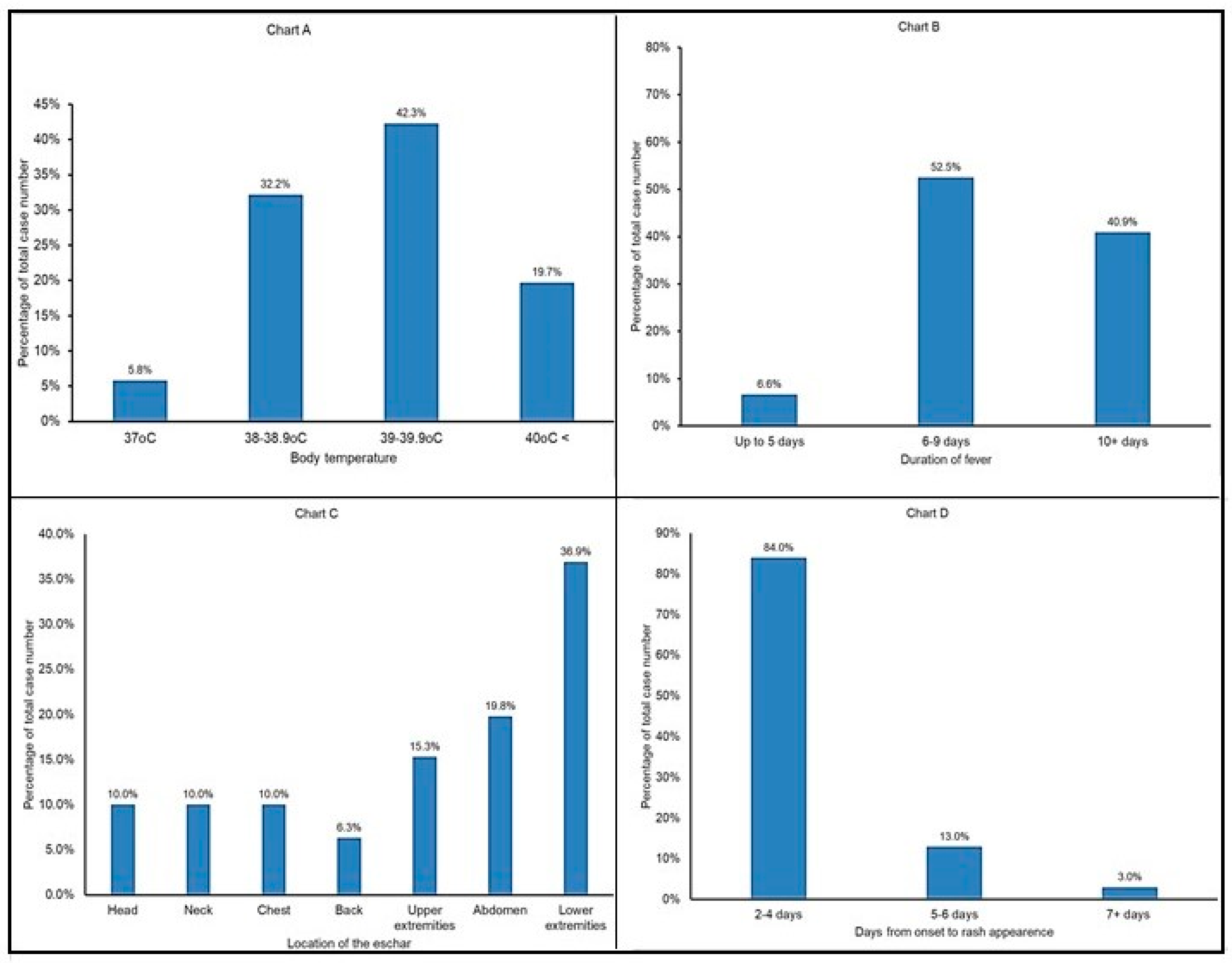
6. Analysis of Recent Clinical Cases of MSF in Crimea

A summary of the most recent MSF cases in Crimea identified five main stages of the illness, including the incubation period, pre-exanthema stage, exanthema and full-blown disease, followed by a convalescence period [63,64]. The incubation period lasts, on average, for 5–7 days (range from 2 to 15 days); in 27 individuals with good recall of finding and removal of ticks, the incubation period averaged 6.3 ± 0.2 days. The onset is acute and characterized by a sharp increase in the body temperature to 39–40 °C with chills in most patients (Figure 5A). In 75.2% of the patients, their temperature remained stable before therapy was started; 15.3% of the patients experienced only morning temperature spikes and 9.5% suffered from intermittent fever. Fever lasted between one and two weeks in most patients depending upon the time of admission to the hospital and start of the treatment (Figure 5B), and it decreased after 4–6 days of treatment.
Eighty-three percent of the patients had a headache which had a persisting diffuse character and was moderate in half of the individuals. In some patients, the headache radiated to the periorbital region, and was accompanied by dizziness, photophobia and sleep disturbance. Myalgia was observed in half of the patients; its manifestations varied from very intense, impeding movement, to moderate, with muscle weakness and aches throughout the body. One-third had arthralgias, predominantly symmetrical without dysfunction of the joints. In 25% of patients, arthralgia and myalgia appeared earlier in the joints and muscles closest to the tick attachment site, but it was especially pronounced when the eschar was in the lower extremities. Headache, myalgia and arthralgia resolved within 5–7 days of antibiotic, antipyretic and intravenous fluid therapy in most patients, although in approximately one-third of the patients, headache and arthralgia persisted for an additional 2–3 weeks following convalescence.
An eschar was found in 81% of the patients; the lower extremities and abdominal areas were the most common locations of the eschar, followed by the upper extremities and upper parts of the body (Figure 5C); multiple eschars were present in some instances (Figure 6). In addition, unilateral catarrhal conjunctivitis was present in 1.8% of cases. A total of 23% of the patients with an eschar had regional lymphadenitis but only 2.2% patients had enlarged lymph nodes nearest to the eschar site. Inguinal lymph nodes of the corresponding side were enlarged when an eschar was present on the lower extremities or in the lower abdominal region. Enlarged axillary lymph nodes occurred when an eschar was present on the skin of the chest, upper back or upper limbs. Cervical and submandibular lymph node enlargement was detected when the eschar was on the scalp or neck. These observations were in good agreement with historic reports of MSF in Crimea [21,22,64,65].
The development of eschars occurred in several steps consistent with the stage of the disease. At admission to the hospital (on average, 6.3 ± 0.2 days of illness), it manifested as a superficial necrotic area of s kin of 0.5 to 2.0 cm in diameter covered with a thin hemorrhagic crust (scab) tightly welded to the underlying tissues; bleeding typically occurred if this scab was lifted. Necrosis in 105 (76.6%) patients was located on an infiltrated base, around which a zone of hyperemia was present in the form of a ring 1 to 5 cm in diameter. The eschar area was generally painless on palpation, but some patients reported subjective feelings of burning, itching and mild soreness. Typically, the scab would come off within 3–4 days following a temperature drop, and the exposed wound would epithelialize without a scar within 5–7 days. Ring-like skin peeling, and subsequent pigmentation would develop at this hyperemia area of the eschar and last at least for 2–3 months after convalescence.
According to our observations, 96.3% of MSF patients had a rash which developed within 2–4 days following the onset of the illness (Figure 5D and Figure 7). Typically, it first appears on the body trunk and lower extremities, and then spreads to the rest of the body, including the face (78.8%) palms and soles (69%). At the peak of the disease, most patients had a puffy pale face (in 5% of cases face hyperemia was noted), shiny eyes, injected sclera and their skin was moist and hot to the touch. A combination of these symptoms was consistent with the clinical profile of MSF described earlier in Crimea [22]. Data presented in earlier studies also indicated that a rash develops in most cases; its prevalence varied from 100% in a 1952 study to 77.4–80% in 2009 [64,65].
In total, 73% of the patients had maculopapular rash, and roseola-papular rush was present in 23.3% of patients; the few remaining patients had no skin lesions. The rash first appears in the form of rosaceae, which then transforms within 1–2 days into macula and papules in 1–2 days. Their color varies from pale pink to brownish red with a cyanotic tint. Moderately abundant rash was observed in 40% of the patients and only single lesions were found in 10% of patients, typically localized on the exterior surfaces of the limbs, an observation consistent with the data of other authors [21,22,65,68]. At the peak of disease, 3.7% of patients had enanthema on the soft palate in the form of few petechia-like lesions. The rash started to resolve with a drop in temperature, on average after 4–5 days of antibiotic therapy. Individual lesions faded, decreased in size and the surface of the skin became smoother. Areas with petechiae and papules could remain pigmented for 3–4 weeks after convalescence.
Organ system involvement, particularly the respiratory and cardiovascular systems, was most remarkable at the peak of the illness and reflected changes due to interstitial inflammatory processes underlying progressive pathological changes due to rickettsia-caused vasculitis [69]. According to our observations, on admission, patients complained of a loss of appetite (58.3%), sore throat, nasal congestion, dry cough, shortness of breath, moderate chest pain (3.6%) and palpitations (1.5%). Hepatomegaly was diagnosed in 42.3% of patients. As the disease progressed, patients developed a bradycardia accompanied by hypotension in one-third of the cases. These symptoms are consistent with clinical manifestations of myocarditis and other cardio-vascular conditions developed as a part of disease progression [70,71]; however, they may be also the result of preexisting heart conditions in elderly patients and individuals with a history of ischemic heart disease and myocardial infarction. Different from the original reports of MSF in Crimea, 31.3% of patients suffered from bradypsychia (slow reaction) and bradilalia (speech was affected), but these conditions improved after 3–4 days of therapy. Severe forms of neurological disorder(s) were noted in two patients who had night-time hallucinations at the peak of the fever, and one individual experienced a short-term loss of consciousness which was interpreted as a manifestation of “typhoid status”.

7. Vector and Host Associations of Rickettsia conorii in Crimea and Other Countries in the Black Sea Region

The brown dog tick, Rhipicephalus sanguineus Latreille, is the vector and reservoir of R. conorii conorii [5]. Numerous studies were conducted in Crimea to determine the distribution and host associations of this tick [72,73,74,75,76,77]. In-depth study and evaluation of diverse species of wild and domestic animals identified dogs, Canis familiaris, as the main vertebrate host of R. sanguineus; however, this tick can feed on other animals [72,74,76]. According to studies from the 1960s conducted in Crimea, R. sanguineus was found mostly on dogs (abundance index [number of ticks per animal] up to 114.0), but it was present infrequently on cows (0.05–1.7), sheep (0.17) and goats (0.1) [74]. This tick will also infest cats, foxes and other wild canids [72].
The life cycle of Rh. sanguineus consists of four stages—eggs and three active parasitic stages (larva, nymph and the adults, male and female) that require a bloodmeal [11,12]. Rh. sanguineus is a one-host tick feeding on one species of animal in the larval, nymphal and adult stages; gravid females leave the host for the ground prior to laying eggs. Once on the ground, the female starts oviposition in 7–10 days, and larvae will hatch after 36–47 days. Larvae will molt into nymphs 7–9 days following a bloodmeal, and nymphs will molt into adults 12–13 days after engorgement. The entire life cycle lasts 3 months, although these events may be accelerated or delayed depending on environmental temperature and humidity [11,12]. In Crimea, adult Rh. sanguineus are typically found on dogs from March to September, with peak infestations observed in May–June and August–September [74,76]; nymphs are rare in April and abundant in July–August. Rh. sanguineus can overwinter as larvae, nymphs or adults. This tick can also establish persisting in-house infestations and survive in internal and external crevices [78,79,80]; however, this type of behavior has not been reported in Crimea.
Naïve Rh. sanguineus can acquire R. conorii conorii at any life stage as a part of the bloodmeal when feeding on a rickettsiemic dog or another animal, or from another tick through co-feeding [12,81]. Infected ticks will transmit rickettsiae to a naïve animal during feeding. Dogs are highly susceptible to R. conorii and remain infectious to ticks for at least a month post-infection [81]; however, poor breeds are more prone to rickettsial infection and differ in severity of R. conorii infection [82].
Once a tick is infected, R. conorii establishes a systemic infection and is transmitted transtadially and transovarially; however, the net effect on the infected tick is detrimental [83]. The exposure of Rh. sanguineus to R. conorii conorii strain Malish during experimental feeding infection produced lasting harmful effects on their survival [84,85], including a reduction in engorged larva-to-nymph molting success and a decreased survival in questing nymphs. Furthermore, the R. conorii infection rate was low in surviving ticks. However, significant variations were reported pertinent to the degree of damage caused by different R. conorii subspecies [86,87]; these phenomena may reflect unique properties of various Rickettsia isolates and how well they propagate in the ovary of a particular tick [88].
Historically, other tick species have also been identified as vectors of R. conorii [7,8]; however, retrospectively, many of these associations may be misleading because they were established before molecular studies became widely available for ecological and epidemiological investigations of individual rickettsial agents and tick population genetics. In this regard, the difficulties inherent in identifying various genotypes of Rhipicephalus sanguineus sensu lato should be emphasized [89], as well as complications in accurately speciating engorged immature ticks and interpreting the test results [90]. In African countries, R. conorii conorii has been detected in Rhipicephalus simus and Haemaphysalis leachi [91].

8. Contemporary Tick Surveillance Efforts in Countries Adjacent to the Black Sea

Contemporary acarological studies conducted in the Crimea Peninsula during the last decade indicated the occurrence of diverse tick species in different ecological settings [92,93,94]. Nidicolous R. sanguineus is mostly present in the peridomestic environment [75,76]. In addition to R. sanguineus infesting dogs and sometimes cats and foxes, Dermacentor marginatus, D. reticulatus, Ixodes ricinus, Hyalomma marginatum, Haemaphysalis punctana and R. bursa were collected in various ecological settings in Crimea [92,93,94]. In one location, 28% (n = 57) of R. sanguineus tested positive for R. conorii or R. massiliae [92]. A follow-up study tested 1972 ticks collected across the Peninsula, and 20.3% of 305 R. sanguineus tested positive via SFGR gltA screening PCR [93]. Subsequent sequencing identified DNA of R. conorii and R. massiliae in 36.4% and 9.2%, respectively, of PCR-positive brown dog ticks [93]. The same study detected R. conorii DNA in one Hyalomma marginatum [93]. Other ticks originating from different Crimean ecosystems included H. punctata (31.5% gltA positive, n = 1062), H. marginatum (34.5%, n = 139), D. marginatus (52.7%, n = 110), D. reticulatus (26.3%, n = 5) and Ixodes ricinus (13.9%, n = 47) [93]. SFGR prevalence ranged from the highest (50.6%) in ticks collected in the eastern steppe zone of the Peninsula to 12% in the west steppes, and only 4.5% in the mountain forest zone and southern coastal area [93]. Most importantly, six species of SFGR previously unknown in Crimea were identified, including R. sibirica subsp. mongolotimonae, R. slovaca, R. aeschlimannii, R. monacensis, R. helvetica and R. raoultii [93,94], all of which can cause human infection [7]. These pathogens had very focal distributions, suggesting they were restricted to specific habitats of their main animal hosts and/or established bird migration routes in Crimea [93]. These SFG rickettsiae cause varying human clinical responses; thus, some variations in clinical manifestations of local MSF cases may actually be due to infections with different Rickettsia species than R. conorii conorii. This can only be confirmed with molecular detection and identification of the SFG rickettsia agent causing each clinical rickettsial infection.
The distribution of MSF cases does not extend north of Crimea, despite the broad occurrence of brown dog ticks along Black Sea shores [72]. The most northern occurrence of Rh. sanguineus was documented at 48oN in Ukraine by collection of two females from a dog in a Kiev Park [72] and from grass in proximity to a railroad in the Kiev-Sviatoshynsky district [95]; however, these findings are considered to be incidental and may possibly have originated from a pet that had previously travelled to the endemic areas. There is also an unusual report of R. conorii DNA in one Ixodes ricinus [96]; however, this finding is based on a conserved fragment of an SFGR gltA gene and needs further confirmation.
Rhipicephalus sanguineus also occurs along the eastern shore of the Black Sea [97]; however, there are no data pertinent to the detection and identification of SFGR or reporting rickettsioses in these areas. An MSF outbreak was only once formally recognized and investigated in the City of Sukhumi in 1947; the R. conorii strain M-1 was isolated as part of that investigation [27,98], and later identified as a unique genotype of R. conorii conorii [99,100]. Nowadays, R. conorii is only rarely found in dog ticks in Georgia [101]. In contrast, surveillance of other species of questing ticks in these areas identified a similar repertoire of SFGR as those found in Crimea beyond R. conorii, including R. helvetica, R. slovaca, R. monacensis, R. raoulti, R. aeschlimanii and R. massiliae, with some regional variations depending on the tick species tested [95,101].
As stated in Section 3 above, MSF due to R. conorii has been formally diagnosed and confirmed using molecular methods in Turkey and R. sanguineus is identified among their human-biting ticks [35]; however, acarological studies conducted in the last 20 years are not consistent with the idea that MSF is a common illness in this country [102,103,104,105,106,107,108]. We identified only one case-report providing indirect evidence for the role of R. sanguineus in the transmission of R. conorii in Turkey [109]. Depending on the study, researchers have collected up to 12 different species of ticks parasitizing humans, domestic and wild animals and questing ticks from different areas of Turkey [102,103,104,105,106,107,108,110,111]; however, only a few collections included R. sanguineus [102,110,111]. These studies described the detection and identification of nine SFGR of known pathogenicity for humans and several SFGR with unknown virulence in different areas of Turkey, including Istanbul, Central Anatolia, the Yozgat province, Corum province and others [103,104,108,112]. Infrequent findings of R. conorii and some unusual tick associations may need further investigation and confirmatory study using a larger collection of ticks and simultaneous molecular speciation of ticks together with multigene characterization of the Rickettsia species detected. Molecular characterization of SFGR infections associated with R. sanguineus-infested domestic dogs will need to be performed to confirm the occurrence of a natural cycle of MSF in Turkey.
There are several old reports describing diverse hard ticks in Bulgaria and much work has dealt with Lyme disease and tick-borne encephalitis [113,114,115]. Rhipicephalus sanguineus is identified as the vector of R. conorii [32,33,116], and isolation of R. conorii has been reported (cited in [32]). Studies conducted in 1965–1967 reported 18.1% of R. sanguineus tested positive for R. conorii; this was probably established by inoculating laboratory animals with suspensions of homogenized ticks since the tick hemolymph test for rickettsial studies was not introduced until 1970 [117]. The most recent study summarized the results of testing 1780 specimens of six species of Ixodid ticks, including R. sanguineus, collected from regions endemic and non-endemic for MSF [32]. The average rate of positivity for SFGR was 22.8%, with no significant differences between infected ticks originating from MSF endemic (23.4%, n = 1052) and non-endemic (~22%, n = 780) areas, although the positivity rate for R. sanguineus was not reported as a separate variable [32,33,116]. A recent acarological investigation focused on ticks originating from the Strandja Nature Park from the Black Sea region of Bulgaria identified R. monacensis and R. helvetica in I. ricinus from vegetation, dogs and goats, and R. aeschlimannii in Hy. anatolicum (n = 1), Hy. excavatum (n = 2), Hy. marginatum (n = 4) and Rhipicephalus spp. (n = 1) from dogs and cattle. Only 10 R. sanguineus were collected from vegetation and they tested negative for SFGR, thus suggesting that R. conorii may have only a very focal presence among geographically dispersed populations of brown dog ticks in Bulgaria or may be associated with imported pets.
There are only infrequent findings of R. conorii in Romania, including its detection in 0.8% (n = 120) of R. sanguineus collected from owned dogs seen at a veterinary clinic in Bucharest [59] and in 1.9% (n = 53) of questing H. punctata from an urban site in Cluj-Napoca [118]. Rh sanguineus is found on dogs, cattle, sheep and wildlife [60,119]. Its typical habitat is within the southern lowlands of Romania, although R. sanguineus has been found in the Transylvania basin and northeastern parts of the country [119]. In recent years, it was also observed that the steppe tick Rhipicephalus rossicus is common and is the dominant species infesting dogs in the areas of southeastern Romania typically known as the classic habitat for the brown dog tick [120,121]. Pathogen carriage and the vectorial capacity of R. rossicus has not yet been determined for many tick-borne organisms; however, it should be noted that this tick may be commonly misidentified as R. sanguinieus, especially when engorged ticks are collected from dogs [89,119]. As for other ticks parasitizing humans, peridomestic and wildlife animals, their species diversity is similar to other countries situated in the Black Sea region. These ticks frequently tested positive for R. helvetica and R. monacensis (Ixodes spp.), R. raoultii and R. slovaca (Dermacentor spp.), R. aeschlimanii (Hyalomma spp.) and R. massiliae [58,118,122]; however, there are big variations in the reported SFGR positivity rate and prevalence of individual Rickettsia species detected depending upon the source of the ticks and molecular targets used.

9. Implications of Other Rickettsia and Rickettsioses Sympatric to R. conorii conorii and MSF in Black-Sea-Area Countries

An analysis of publications reporting acarological surveillance efforts in Crimea and neighboring territories indicates that endemic areas traditionally thought to be places for the occurrence and circulation of R. conorii and, thus, cases of MSF have been diagnosed in very diverse and complicated ecosystems with the co-circulation of multiple tick species carrying different SFGR [32,37,58,59,93,108]. Of course, R. sanguineus widely occupies a unique peridomestic niche both in rural and urban environments. However, there are multiple plausible scenarios of encroachment of these other natural habitats and ticks which can permit encounters with humans and their pet animals. At present, these systems probably exist in a condition close to their natural equilibrium with only infrequent contact with humans; however, numerous environmental and anthropogenic factors can contribute to their introduction to humans and their pets.
There are only a few reports of diagnosis of non-R. conorii infection in the countries surrounding the Black Sea; they are limited to PCR-diagnosed R. sibirica mongolotimonae and R. slovaca cases in Turkey [123,124], a serologically diagnosed R. slovaca-associated case of lymphadenopathy in Bulgaria [125] and R. massiliae, R. slovaca and R. slovaca-R. raoultii cases in Romania [126]. A serological IFA test using paired sera is the gold standard method for the diagnosis of SFG rickettsioses, and when used in conjunction with Western blotting analysis it may provide unequivocal identification of the etiological agent [44,52]; however, only a limited repertoire of antigens is commonly used for routine laboratory diagnosis in most countries. Clinical symptoms and a patient’s exposure history may provide additional clues to distinguish and diagnose other SFGR rickettsioses [Table 2]; however, these clues may be overlooked, particularly when physician familiarity is low [53]. These situations have several implications. Generic diagnosis of SFGR rickettsiosis without identifying its etiology may not make a significant difference from a clinical point of view and patient management; however, it will not contribute to accumulating specific information about the spectrum of SFGR responsible for human morbidity in each region. This situation may also negatively affect vector control programs since different SFGR are carried by different tick species with diverse habitats and animal host associations. On the other hand, when a case of MSF or another spotted fever group rickettsiosis is misdiagnosed as another illness, it may contribute to unnecessary morbidity and mortality among local residents [38,61,127,128]. The Black Sea coast is an important tourist destination for all the surrounding countries, and many travel-related cases may be overlooked [31].
As has been already mentioned above, some MSF cases in Turkey may be misdiagnosed as viral hemorrhagic fevers [52]. Moreover, there is a much longer list of clinical entities that may be confused with rickettsioses. These include upper respiratory tract infection, pneumonia, urinary tract infection, viral gastroenteritis, non-rickettsial bacterial sepsis, thrombotic thrombocytopenic purpura, idiopathic vasculitis and viral or bacterial meningoencephalitis [52]. Therefore, it takes an astute physician to distinguish these presentations and initiate correct empiric treatment to prevent poor patient outcomes. Systemic evaluation of patients’ symptoms, a detailed clinical picture and epidemiological clues together with family and travel history and known exposure and risk factors should be performed together with reviewing standard laboratory tests (total blood count, peripheral blood smear, blood chemistry and hepatic function panel) to implement a proper patient management plan. Doxycycline is the drug of choice for treatment of all tick-borne rickettsial diseases in patients of all ages, including children aged <8 years [16,52]. Prompt favorable response to it has been suggested as a useful clinical clue for rickettsioses. Clarithromycin and azithromycin can also be acceptable therapeutic alternatives to tetracyclines for children aged <8 years with Mediterranean spotted fever [152]; however, the same trial did not evaluate therapeutic effects on other SFG rickettsiae. Prophylactic treatment for rickettsial diseases in persons who have had recent tick bites and are not ill is not recommended [153]. It is also not recommended to prescribe antibiotics to asymptomatic individuals seropositive for SFGR regardless of their past treatment history [154].
A recent systematic review estimated that diagnosis of MSF based only on clinical symptoms misses 57.9% (6922/11,956) of the total patients presenting with an eschar and 81.6% (6922/8478) of patients when the eschar is absent or overlooked [155]. To ensure adequate patient management, proper laboratory confirmatory diagnosis is necessary, which is no longer challenging for SFG rickettsioses. Molecular methods can provide a definitive etiological diagnosis of rickettsiosis; however, clinical specimens must be collected within a very narrow period of time and prior to starting antibiotic therapy because the rickettsial DNA load in human blood depends on the Rickettsia species and severity of the illness [156,157]. Furthermore, rickettsial quantity in patients suffering from Rocky Mountain spotted fever appears to have a circadian rhythm, so the largest numbers are present during early morning hours [158]; however, similar observations for other rickettsioses are not available. Therefore, a skin biopsy of the eschar or skin rash is considered a more informative and reliable specimen for confirmatory diagnosis of rickettsioses [52,159]. Testing eschar scabs and eschar swabs is recommended to minimize invasive procedures associated with skin biopsies, which are not as acceptable for pediatric patients or for eschars on sensitive areas of the skin [160,161,162]. The utility of this approach has been demonstrated by confirming acute infections due to R. conorii, R. slovaca and R. sibirica mongolotimae [160,161].
The use of real-time PCR is now a common practice for detecting rickettsial DNA in clinical specimens. The use of multiplex PCR permits better specificity and accuracy of the diagnosis, especially when multiple etiological agents are suspected. A combination of broad-spectrum SYBR-Green PCR assay targeting the Rickettsia OmpB gene and R. conorii-specific ompA TaqMan was evaluated for differential diagnosis of MSF in endemic areas where other pathogenic SFGR are present [163]; the analytical sensitivity of both assays was <10 DNA copies. This approach was similar to another duplex PCR that combines pan-Rickettsia targets such as the 16S rRNA gene or 23S rRNA gene and group-specific gltA or another genus-specific gene like the hypothetical protein A1G_04230 to conduct differential detection of typhus group rickettsiae and R. rickettsii, respectively, and other Rickettsia species [164,165]. A reverse transcription PCR assay targeting the more stable and higher copy number rickettsial 23S rRNA gene permitted a 100-fold increase in analytical sensitivity compared with DNA-based detection methods and had comparable performance for testing banked patient samples [166]. However, a negative result cannot exclude clinical disease.
It is expected that the broader availability of nucleic acid-based methods and their use in regular clinical practice will improve routine diagnosis of spotted fever group rickettsioses and permit more effective treatment of these diseases in the region. Droplet digital PCR is one of the possible approaches and may be most useful for multiplex testing [167]. Next-generation sequencing provides powerful methodology permitting relatively quick identification of the etiological agent, especially for patients with atypical clinical manifestations and unclear epidemiologic evidence of a tick bite or exposure; however, this platform is not fully validated with rickettsioses and may pose challenges to complete identification when only a small number of Rickettsia reads are generated and other abundant microbial flora are present in the clinical sample [9,168]. Inexpensive Oxford Nanopore long-read technology can be used for genome sequencing of Rickettsia [169]. This less expensive technology is also proposed to be compatible with point-of-care diagnostics in resource-limited settings. Other commercial NGS-based methods may be useful for the detection of Rickettsia and diagnosis of rickettsioses [170]. Metagenomic sequencing of ticks permits one to identify a range of tick-borne pathogens present in a particular area, including known pathogens and those that have not been previously associated with human disease [171,172,173]

10. Conclusions and Highlights

MSF is an endemic disease for the Crimean Peninsula. For 85 years, it has been diagnosed annually along the Black Sea coast and manifests in sporadic cases or outbreak forms. MSF is a seasonal disease, with the majority of cases occurring in the spring and summer months during the period of peak tick activity. The incidence rate depends on the population of brown dog ticks, the number of abandoned and stray dogs and inconsistent animal and vector control measures. In recent years, moderate to severe clinical manifestations were the most common and presented with the classic clinical triad of fever, eschar and rash. Persons in all age groups are affected, but elderly people with underlying health conditions often have the most malignant course of the illness. The epidemiological, ecological and clinical features of MSF in Crimea are very similar to other regions in the Black Sea basin. The presence of diverse species of human-biting ticks in many eco-systems and the circulation of numerous species of Rickettsia known to cause spotted fever group rickettsioses with a wide range of clinical manifestations is challenging for both physicians and public health officials. An effort to utilize new rickettsial molecular diagnostic platforms and assays for routine clinical practice and vector surveys will provide a better basis for future improvements in treatment and a reduction in all of the spotted fever rickettsioses occurring in this region.

Author Contributions

Summary of clinical and epidemiological data: M.T.G.; M.T.G. and M.E.E. conceptualized the review and wrote and edited the manuscript. All authors have read and agreed to the published version of the manuscript.

Funding

This research received no external funding.

Institutional Review Board Statement

Not applicable.

Informed Consent Statement

Informed consent has been obtained from the patient(s) to publish this paper.

Data Availability Statement

The data presented in this study are available on request from the corresponding author.

Acknowledgments

We thank Gregory A. Dasch for helpful discussion and suggestions for the manuscript. We apologize to many of our colleagues, whose work could not be cited due to the format and length of this manuscript.

Conflicts of Interest

The authors declare no conflict of interest.

References

  1. Conor, A.; Bruch, A. Une fièvre éruptive observée en Tunisie. Bull. Soc. Pathol. Exot. Filial. 1910, 8, 492–496. (In French) [Google Scholar]
  2. Olmer, D. Sur une infection épidémique, avec exantheme de nature indéterminée. Mars. Med. 1925, 22, 1291–1293. (In French) [Google Scholar]
  3. Boinet, M.M.; Pieri, J. Epidémies d’exantheme infectieux de nature indéterminée, observées sur le littoral méditerranéen. Presse Medicale 1927, 89, 173–182. (In French) [Google Scholar]
  4. Blanc, G.; Caminopetros, J. Etudes épidémiologiques et expérimentales sur la fièvre boutonneuse, faites à l’Institut Pasteur d’Athènes. Arch. Inst. Pasteur Tunis 1932, 20, 343–394. (In French) [Google Scholar]
  5. Durand, P.; Conseil, E. Transmission expérimentale de la fièvre boutonneuse par Rhipicephalus sanguineus. Comptes Rendus Hebdomadaires des Séances de l’Académie des Sciences 1930, 190, 1244–1246. (In French) [Google Scholar]
  6. Zhu, Y.; Fournier, P.-E.; Eremeeva, M.; Raoult, D. Proposal to create subspecies of Rickettsia conorii based on multi-locus sequence typing and an emended description of Rickettsia conorii. BMC Microbiol. 2005, 5, 11. [Google Scholar] [CrossRef] [PubMed]
  7. Parola, P.; Paddock, C.D.; Socolovschi, C.; Labruna, M.B.; Mediannikov, O.; Kernif, T.; Abdad, M.Y.; Stenos, J.; Bitam, I.; Fournier, P.-E.; et al. Update on tick-borne rickettsioses around the world: A geographic approach. Clin. Microbiol. Rev. 2013, 26, 657–702. [Google Scholar] [CrossRef]
  8. Rovery, C.; Brouqui, P.; Raoult, D. Questions on Mediterranean spotted fever a century after its discovery. Emerg. Infect. Dis. 2008, 14, 1360–1367. [Google Scholar] [CrossRef]
  9. Xu, N.; Gai, W.; Zhang, Y.; Wang, W.; Wang, G.; Dasch, G.A.; Eremeeva, M.E. Confirmation of Rickettsia conorii subspecies indica infection by next-generation sequencing, Shandong, China. Emerg. Infect. Dis. 2021, 27, 2691–2694. [Google Scholar] [CrossRef]
  10. Gu, X.L.; Wang, R.; Zhou, C.-M.; Cui, J.-T.; Li, Z.-M.; Jiang, Z.-Z.; Li, B.; Peng, Q.-M.; Zhang, W.-K.; Han, H.-J.; et al. Natural Mediterranean Spotted fever foci, Qingdao, China. Emerg. Infect. Dis. 2022, 28, 2524–2527. [Google Scholar] [CrossRef]
  11. Gray, J.; Dantas-Torres, F.; Estrada-Peña, A.; Levin, M. Systematics and ecology of the brown dog tick, Rhipicephalus sanguineus. Ticks Tick Borne Dis. 2013, 4, 171–180. [Google Scholar] [CrossRef] [PubMed]
  12. Dantas-Torres, F.; Otranto, D. Rhipicephalus sanguineus (Brown dog tick). Trends Parasitol. 2022, 38, 993–994. [Google Scholar] [CrossRef] [PubMed]
  13. Torina, A.; de Mera, I.G.F.; Alongi, A.; Mangold, A.J.; Blanda, V.; Scarlata, F.; Di Marco, V.; de la Fuente, J. Rickettsia conorii Indian tick typhus strain and R. slovaca in humans, Sicily. Emerg. Infect. Dis. 2012, 18, 1008–1010. [Google Scholar] [CrossRef]
  14. de Sousa, R.; Ismail, N.; Doria-Nobrega, S.; Costa, P.; Abreu, T.; Francca, A.; Amaro, M.; Proencca, P.; Brito, P.; Poccas, J.; et al. The presence of eschars, but not greater severity, in Portuguese patients infected with Israeli spotted fever. Ann. N. Y. Acad. Sci. 2005, 1063, 197–202. [Google Scholar] [CrossRef] [PubMed]
  15. Chisu, V.; Masala, G.; Foxi, C.; Socolovschi, C.; Raoult, D.; Parola, P. Rickettsia conorii israelensis in Rhipicephalus sanguineus ticks, Sardinia, Italy. Ticks Tick Borne Dis. 2014, 5, 446–448. [Google Scholar] [CrossRef]
  16. Brouqui, P.; Parola, P.; Fournier, P.E.; Raoult, D. Spotted fever rickettsioses in southern and eastern Europe. FEMS Immunol. Med. Microbiol. 2007, 49, 2–12. [Google Scholar] [CrossRef]
  17. Renvoisé, A.; Delaunay, P.; Blanchouin, E.; Cannavo, I.; Cua, E.; Socolovschi, C.; Parola, P.; Raoult, D. Urban family cluster of spotted fever rickettsiosis linked to Rhipicephalus sanguineus infected with Rickettsia conorii subsp. caspia and Rickettsia massiliae. Ticks Tick Borne Dis. 2012, 3, 389–392. [Google Scholar] [CrossRef]
  18. Piras, M.A.; Calia, G.; Saba, F.; Gakis, C.; Andreoni, G. Glucose-6-phosphate dehydrogenase deficiency in male patients with Mediterranean spotted fever in Sardinia. J. Infect. Dis. 1983, 147, 607–608. [Google Scholar] [CrossRef]
  19. Raoult, D.; Lena, D.; Perrimont, H.; Gallais, H.; Walker, D.H.; Casanova, P. Hemolysis with Mediterranean spotted fever and glucose-6-phosphate-dehydrogenase deficiency. Trans. R. Soc. Trop. Med. Hyg. 1986, 80, 961–962. [Google Scholar] [CrossRef]
  20. Chaumentin, G.; Zenone, T.; Bibollet, C.; Denoyel, G.A.; Boibieux, A.; Biron, F.; Peyramond, D. Malignant boutonneuse fever and polymyalgia rheumatica: A coincidental association? Infection 1997, 25, 320–322. [Google Scholar] [CrossRef]
  21. Alymov, A.Y. Marseille fever. Soviet Med. 1939, 13, 30–33. (In Russian) [Google Scholar]
  22. Andreev, M.F. Clinical and epidemiological observations of the Marseille spotted fever. Clin. Med. 1941, 19, 80–88. (In Russian) [Google Scholar]
  23. Andreev, M.F. A case of Marseille fever observed in winter. Ter. Arkhiv 1940, 18, 208–215. (In Russian) [Google Scholar]
  24. Pakshin, M.F.; Nikitin, A.M. Data from a study of a natural focus of Marseilles fever in one of the Crimean districts over a 40-year period (1947–1986). Med. Parazitol. 1988, 4, 46–48. (In Russian) [Google Scholar]
  25. Pakshin, M.F.; Pestenko, V.I.; Kostetskii, N.V. On the epidemiology of Marseilles fever in the Crimea and means for its eradication. Zhurnal Mikrobiol. Epidemiol. Immunobiol. 1960, 31, 61–64. (In Russian) [Google Scholar]
  26. Kulagin, S.M.; Tarasevich, I.V.; Rubakin, P.E.; Nikitina, M.; Krupina, Z.N. On the eradication of Marseilles fever. Certain observations on Marseilles fever in Sevastopol. Zhurnal Mikrobiol. Epidemiol. Immunobiol. 1960, 31, 117–121. (In Russian) [Google Scholar]
  27. Zdrodovskii, P.; Golinevitch, E.M. Experimental observations of Marseille fever. In Rickettsiae and Rickettsioses; Zdrodovskii, P., Ed.; USSR Academy of Medical Science: Moscow, Russia, 1948; pp. 216–224. (In Russian) [Google Scholar]
  28. Avetisova, A.S. Observations of Marseille fever. Zhurnal Mikrobiol. Epidemiol. Immunobiol. 1952, 12, 26–31. (In Russian) [Google Scholar]
  29. Combiesco, D. Sur une épidémie de fièvre boutonneuse observée à Constantza-Roumanie. Arch. Roum. Pathol. Exp. Microbiol. 1948, 14, 99–112. (In French) [Google Scholar]
  30. Combiescu, D.; Dumitrescu, N.; Russ, M.; Dinculescu, M. Consideraţii epidemiologice asurpa unor cazuri de febră butonoasă ivite in ultimii 41 de ani. Cultivarea Rickettsiei conori şi caracterele tulpinii izolate dintr’un focar autohton de febră butonoasă. Stud. Cercet. Inframicrobiol. Microbiol. Parazitol. 1953, IV, 99–107. (In Romanian) [Google Scholar]
  31. Pitigoi, D.; Olaru, I.D.; Badescu, D.; Rafila, A.; Arama, V.; Hristea, A. Mediterranean spotted fever in southeastern Romania. Biomed. Res. Int. 2013, 2013, 395806. [Google Scholar] [CrossRef]
  32. Alexandrov, E.; Alexandrova, D.; Kamarinchev, B.; Kantardjiev, T.; Dimitrova, Z. Marseilles fever (Mediterranean spotted fever) in Bulgaria-state and problems. In Contemporary State of the Rickettsioses in the World and in Bulgaria; Alexandrov, E., Kazar, J., Hechemy, K., Kantardjiev, T., Eds.; Prof. Marin Drinov Academic Publishing House: Sofia, Bulgaria, 2007; pp. 80–93. [Google Scholar]
  33. Alexandrov, E.G.; Mitov, D.G.; Kamarinchev, B.D.; Bogdanov, N.R. Current features of Mediterranean spotted fever in Bulgaria. In Rickettsiae and Rickettsial Diseases at the Turn of the Third Millenium; Raoult, D., Brouqui, P., Eds.; Elsevier: Paris, France, 1999; pp. 279–281. [Google Scholar]
  34. Rehacek, J.; Tarasevich, I.V. Rickettsia conorii. In Acari-Borne Rickettsiae and Rickettsioses in Eurasia; Rehacek, J., Tarasevich, I.V., Eds.; Veda Publishing House of the Slovak Academy of Sciences: Bratislava, Slovakia, 1988; pp. 14–48. [Google Scholar]
  35. Kuloglu, F.; Rolain, J.M.; Akata, F.; Eroglu, C.; Celik, A.D.; Parola, P. Mediterranean spotted fever in the Trakya region of Turkey. Ticks Tick Borne Dis. 2012, 3, 298–304. [Google Scholar] [CrossRef] [PubMed]
  36. Mert, A.; Ozaras, R.; Tabak, F.; Bilir, M.; Ozturk, R. Mediterranean spotted fever: A review of fifteen cases. J. Dermatol. 2006, 33, 103–107. [Google Scholar] [CrossRef] [PubMed]
  37. Celebi, B.; Yesilyurt, M.; Kilic, S. A Rickettsia case caused by Rickettsia conorii. Mikrobiyol. Bul. 2018, 52, 431–438. [Google Scholar] [CrossRef] [PubMed]
  38. Uzunlulu, M.; Gönenç, I.; Kaya, S.; Selvi, E.; Arslan, F. A case of Mediterranean spotted fever mimicking severe sepsis. J. Vector Borne Dis. 2022, 59, 298–301. [Google Scholar] [CrossRef] [PubMed]
  39. Kuloglu, F.; Rolain, J.M.; Fournier, P.E.; Akata, F.; Tugrul, M.; Raoult, D. First isolation of Rickettsia conorii from humans in the Trakya (European) region of Turkey. Eur. J. Clin. Microbiol. Infect. Dis. 2004, 23, 609–614. [Google Scholar] [CrossRef]
  40. McCormick, D.W.; Kugeler, K.J.; Marx, G.E.; Jayanthi, P.; Dietz, S.; Mead, P.; Hinckley, A.F. Effects of COVID-19 pandemic on reported Lyme Disease, United States, 2020. Emerg. Infect. Dis. 2021, 27, 2715–2717. [Google Scholar] [CrossRef]
  41. Sulik, M.; Toczylowski, K.; Grygorczuk, S. Epidemiology of tick-borne encephalitis in Poland (2010–2019) and the impact of the COVID-19 pandemic on the notified incidence of the disease. Przegl. Epidemiol. 2021, 75, 76–85. [Google Scholar] [CrossRef]
  42. Piotrowski, M.; Rymaszewska, A. The impact of a pandemic COVID-19 on the incidence of borreliosis in Poland. Acta Parasitol. 2022, 67, 1007–1009. [Google Scholar] [CrossRef]
  43. Pen’kovskaya, N.A. Epidemiological features of Mediterranean spotted fever in the Crimea at the modern stage. Krims’kiy Ter. Zhurnal 2014, 1, 140–145. (In Russian) [Google Scholar]
  44. Eremeeva, M.E.; Shpynov, S.N.; Tokarevich, N.K. Modern approaches to laboratory diagnosis of rickettsial diseases. Russ. J. Infect. Immun. 2014, 4, 113–134. [Google Scholar] [CrossRef]
  45. Zvereva, N.N.; Saifullin, M.A.; Karan, L.S.; Bazarova, M.V.; Saifullin, R.F.; Smetanina, S.V. The case of tick-borne spotted fever in a child arriving from the Crimea. Det. Infektsii 2018, 17, 69–72. [Google Scholar] [CrossRef]
  46. Vladimirova, N.; Minkova, A.; Bogdanov, N.R.; Ivanova, S.; Petkova, K.; Getsova, Z. Acute Infectious Diseases in Bulgaria in 2022 (Basic Epidemiological Indicators); Department of Epidemiology, National Center for Infectious and Parasitic Diseases: Sofia, Bulgaria, 2022; pp. 1–54. Available online: https://www.ncipd.org/index.php?option=com_content&view=featured&Itemid=1086&lang=bg (accessed on 26 June 2023). (In Bulgarian)
  47. Baltadzhiev, I.; Kevorkyan, A.; Popivanova, N. Mediterranean spotted fever in child and adult patients: Investigation from an endemic region in Bulgaria. Cent. Eur. J. Public Health 2020, 28, 187–192. [Google Scholar] [CrossRef] [PubMed]
  48. Pishmisheva, M.; Stoycheva, M.; Vatev, N.; Semerdjieva, M. Mediterranean spotted fever in children in the Pazardjik region, South Bulgaria. Pediatr. Infect. Dis. J. 2014, 33, 542–544. [Google Scholar] [CrossRef] [PubMed]
  49. Nenova, M. Mediterranean spotted fever in Varna region. In Contemporary State of the Rickettsioses in the World and in Bulgaria; Alexandrov, E., Kazar, J., Hechemy, K., Kantardjiev, T., Eds.; Prof. Marin Drinov Academic Publishing House: Sofia, Bulgaria, 2007; pp. 94–106. [Google Scholar]
  50. Popivanova, N.; Baltadzhiev, I.; Zaprianov, Z. Mediterranean spotted fever in the Plovdiv region of Bulgaria. In Contemporary State of the Rickettsioses in the World and in Bulgaria; Alexandrov, A., Kazar, J., Hechemy, K., Kantardjiev, T., Eds.; Prof. Marin Drinov Academic Publishing House: Sofia, Bulgaria, 2007; pp. 116–129. [Google Scholar]
  51. Pishmisheva, P. Mediterranean spotted fever in Pazardzhic region-clinic, paraclinic, treatment, specific forms of rickettsiosis, epidemiology. In Contemporary State of the Rickettsioses in the World and in Bulgaria; Alexandrov, A., Kazar, J., Hechemy, K., Kantardjiev, T., Eds.; Prof. Marin Drinov Academic Publishing House: Sofia, Bulgaria, 2007; pp. 107–115. [Google Scholar]
  52. Biggs, H.M.; Behravesh, C.B.; Bradley, K.K.; Dahlgren, F.S.; Drexler, N.A.; Dumler, J.S.; Folk, S.M.; Kato, C.Y.; Lash, R.R.; Levin, M.L.; et al. Diagnosis and management of tickborne rickettsial diseases: Rocky Mountain spotted fever and other spotted fever group rickettsioses, ehrlichioses, and anaplasmosis-United States. MMWR Recomm. Rep. 2016, 65, 1–44. [Google Scholar] [CrossRef]
  53. O’Reilly, M.; Paddock, C.; Elchos, B.; Goddard, J.; Childs, J.; Currie, M. Physician knowledge of the diagnosis and management of Rocky Mountain spotted fever: Mississippi, 2002. Ann. N. Y. Acad. Sci. 2003, 990, 295–301. [Google Scholar] [CrossRef]
  54. National Center for Infectious and Parasitic Diseases. Epidemiological Bulletin; National Center for Infectious and Parasitic Diseases: Sofia, Bulgaria, 2022; Available online: https://www.ncipd.org/index.php?option=com_content&view=featured&Itemid=730&lang=en (accessed on 26 June 2023).
  55. Macrotrends. Bulgaria Population 1950–2023. Macrotrends. Available online: https://www.macrotrends.net/countries/BGR/bulgaria/population (accessed on 25 June 2023).
  56. National Centre for Surveillance and Control of Communicable Diseases [Institutul Naţional de Sanatate Publica]. Report of Communicable Diseases; National Centre for Surveillance and Control of Communicable Diseases [Institutul Naţional de Sanatate Publica]: Bucharest, Romania, 2016; pp. 70–73. Available online: http://www.cnscbt.ro/ (accessed on 26 June 2023).
  57. Serban, R.; Pistol, A.; Neguţ, M.; Cucuiu, R. Rickettsia conorii infection in Romania, 2000–2008. Bacteriol. Virusol. Parazitol. Epidemiol. 2009, 54, 177–183. [Google Scholar]
  58. Andersson, M.O.; Marga, G.; Banu, T.; Dobler, G.; Chitimia-Dobler, L. Tick-borne pathogens in tick species infesting humans in Sibiu County, central Romania. Parasitol. Res. 2018, 117, 1591–1597. [Google Scholar] [CrossRef]
  59. Ionita, M.; Silaghi, C.; Mitrea, I.L.; Edouard, S.; Parola, P.; Pfister, K. Molecular detection of Rickettsia conorii and other zoonotic spotted fever group rickettsiae in ticks, Romania. Ticks Tick Borne Dis. 2016, 7, 150–153. [Google Scholar] [CrossRef]
  60. Mihalca, A.D.; Dumitrache, M.O.; Sándor, A.D.; Magdaş, C.; Oltean, M.; Györke, A.; Matei, I.A.; Ionică, A.; D’amico, G.; Cozma, V.; et al. Tick parasites of rodents in Romania: Host preferences, community structure and geographical distribution. Parasites Vectors 2012, 5, 266. [Google Scholar] [CrossRef]
  61. Sengoz, G.; Yildirim, F.; Yasar, K.K.; Tozalgan, U.; Aydin, O.A. Fifty-six cases with Mediterranean spotted fever: Evaluation of tick-borne spotted diseases in Turkey. Turkderm-Turk. Arch. Dermatol. Venerol. 2009, 43, 139–143. [Google Scholar]
  62. Ozkan, A.; Ozkalemkas, F.; Ali, R.; Karadogan, S.; Ozkocaman, V.; Ozcelik, T.; Tunali, A. Mediterranean spotted fever: Presentation with pancytopenia. Am. J. Hematol. 2006, 81, 646–647. [Google Scholar] [CrossRef] [PubMed]
  63. Gafarova, M.T. Marseilles Fever (Epidemiology, Clinic and Diagnosis); Tarpan: Simferopol, Ukraine, 2004; p. 98. [Google Scholar]
  64. Gafarova, M.T.; Verbenets, E.A.; Achkasova, T.A.; Schomoylov, D.K.; Midikari, A.C. Epidemiology and clinical features of Marseille fever in Crimea. Infect. Dis. News Opin. Educ. 2017, 2, 61–66. (In Russian) [Google Scholar]
  65. Verbenets, E.A. Comparative characteristics of clinico-epdemiological features of Marseille during time-course of disease. Tauride Med. Biol. Bull. 2009, 12, 23–26. (In Russian) [Google Scholar]
  66. Klymchuk, M.D.; Dorofeev, Y.O.; Lezhentsev, B.M.; Tovpinec, N.N. An outbreak of tick-borne rickettsosis (Marseille fever) in the Crimea. Infect. Bolezn. 1999, 3, 10–12. (In Ukranian) [Google Scholar]
  67. Popova, A.Y.; Kulichenko, A.N.; Maletskaya, O.V.; Vasilenko, N.F.; Shaposhnikova, L.I.; Kotenev, E.S.; Dubyansky, V.M.; Volynkina, A.S.; Lisitskaya, Y.V.; Samarina, I.V.; et al. Epidemiologic situation by natural-foci infections in the Crimea Federal District in 2014–2015. Zhurnal Mikrobiol. Epidemiol. Immunobiol. 2016, 93, 62–69. [Google Scholar] [CrossRef]
  68. Verbenets, E.A. Modern Epidemiological and Clinical Features of Marseilles Fever at the Present Stage, Optimization of Epidemiological Surveillance. Abstract of the Dissertation for the Degree of Candidate of Medical Sciences: 14.02.02 Verbenets Elena Anatolyevna; L. V. Gromashevsky Institute of Epidemiology and Infectious Diseases: Kiev, Ukraine, 2013. (In Ukranian) [Google Scholar]
  69. Mansueto, P.; Vitale, G.; Cascio, A.; Seidita, A.; Pepe, I.; Carroccio, A.; di Rosa, S.; Rini, G.B.; Cillari, E.; Walker, D.H. New insight into immunity and immunopathology of rickettsial diseases. Clin. Dev. Immunol. 2012, 2012, 967852. [Google Scholar] [CrossRef]
  70. Colomba, C.; Siracusa, L.; Trizzino, M.; Gioè, C.; Giammanco, A.; Cascio, A. Myocarditis in Mediterranean spotted fever: A case report and a review of the literature. JMM Case Rep. 2016, 3, e005039. [Google Scholar] [CrossRef] [PubMed]
  71. Doyle, A.; Bhalla, K.S.; Jones, J.M.; Ennis, D.M. Myocardial involvement in rocky mountain spotted fever: A case report and review. Am. J. Med. Sci. 2006, 332, 208–210. [Google Scholar] [CrossRef]
  72. Akimov, I.A.; Nebogatkin, I.V. Ticks of the genus Rhipicephalus (Acari, Ixodidae) and their distribution in Ukraine. Vestn. Zool. 2013, 47, e28–e34. [Google Scholar] [CrossRef]
  73. Klyushkina, E.A. About the tick Rhipicephalus sanguineus Latreille—A carrier of Marseille fever. J. Microbiol. 1966, 11, 146. (In Russian) [Google Scholar]
  74. Klyushkina, E.A. Distribution and biology of tick Rh. sanguineus Latr. in Crimea. Med. Parazitol. Parazitic Dis. 1968, 46–50. (In Russian) [Google Scholar]
  75. Evstafiev, I.L.; Pyshkin, V.B.; Evstafief, A.I.; Tovpinets, N.N. Electronic databases and geoinformation technologies in environmental and biomedical research. Cult. Peoples Black Sea Region. 2005, 7–10. (In Russian) [Google Scholar]
  76. Evstafiev, I.L.; Tovpinets, N.N. Rhipicephalus sanguineus (Ixodidae) in the Crimea: Environmental and epizootological aspects. Vestn. Zool. 2002, 36, 85–91. (In Russian) [Google Scholar]
  77. Nebogatkin, I.V.; Tovpinets, N.N. The outbreak of bloody tick Rhipicephalus sanguineus (Ixodide) on the Kerch Peninsula. Vestn. Zool. 1997, 81. (In Russian) [Google Scholar]
  78. Demma, L.J.; Traeger, M.S.; Nicholson, W.L.; Paddock, C.D.; Blau, D.M.; Eremeeva, M.E.; Dasch, G.A.; Levin, M.L.; Singleton, J.; Zaki, S.R.; et al. Rocky Mountain spotted fever from an unexpected tick vector in Arizona. N. Engl. J. Med. 2005, 353, 587–594. [Google Scholar] [CrossRef] [PubMed]
  79. Hansford, K.M.; Pietzsch, M.E.; Cull, B.; Medlock, J.M.; Wall, R. Overwintering of the brown dog tick in residential properties in England–raising awareness. Vet. Rec. 2015, 177, 156. [Google Scholar] [CrossRef] [PubMed]
  80. Uspensky, I.; Ioffe-Uspensky, I. The dog factor in brown dog tick Rhipicephalus sanguineus (Acari: Ixodidae) infestations in and near human dwellings. Int. J. Med. Microbiol. 2002, 291 (Suppl. S33), 156–163. [Google Scholar] [CrossRef]
  81. Levin, M.L.; Killmaster, L.F.; Zemtsova, G.E. Domestic dogs (Canis familiaris) as reservoir hosts for Rickettsia conorii. Vector-Borne Zoonotic Dis. 2012, 12, 28–33. [Google Scholar] [CrossRef]
  82. Solano-Gallego, L.; Kidd, L.; Trotta, M.; Di Marco, M.; Caldin, M.; Furlanello, T.; Breitschwerdt, E. Febrile illness associated with Rickettsia conorii infection in dogs from Sicily. Emerg. Infect. Dis. 2006, 12, 1985–1988. [Google Scholar] [CrossRef]
  83. Parola, P.; Socolovschi, C.; Raoult, D. Deciphering the relationships between Rickettsia conorii conorii and Rhipicephalus sanguineus in the ecology and epidemiology of Mediterranean spotted fever. Ann. N. Y. Acad. Sci. 2009, 1166, 49–54. [Google Scholar] [CrossRef]
  84. Levin, M.L.; Killmaster, L.; Eremeeva, M.; Dasch, G. Effects of Rickettsia conorii infection on the survival of Rhipicephalus sanguineus ticks. Clin. Microbiol. Infect. 2009, 15 (Suppl. S2), 277–278. [Google Scholar] [CrossRef] [PubMed]
  85. Socolovschi, C.; Bitam, I.; Raoult, D.; Parola, P. Transmission of Rickettsia conorii conorii in naturally infected Rhipicephalus sanguineus. Clin. Microbiol. Infect. 2009, 15 (Suppl. S2), 319–321. [Google Scholar] [CrossRef] [PubMed]
  86. Levin, M.L.; Killmaster, L.; Zemtsova, G.; Grant, D.; Mumcuoglu, K.Y.; Eremeeva, M.E.; Dasch, G.A. Incongruent effects of two isolates of Rickettsia conorii on the survival of Rhipicephalus sanguineus ticks. Exp. Appl. Acarol. 2009, 49, 347–359. [Google Scholar] [CrossRef] [PubMed]
  87. Socolovschi, C.; Matsumoto, K.; Brouqui, P.; Raoult, D.; Parola, P. Experimental infection of Rhipicephalus sanguineus with Rickettsia conorii conorii. Clin. Microbiol. Infect. 2009, 15 (Suppl. S2), 324–325. [Google Scholar] [CrossRef] [PubMed]
  88. Eremeeva, M.E.; Dasch, G.A. Challenges posed by tick-borne rickettsiae: Eco-epidemiology and public health implications. Front. Public Health 2015, 3, 55. [Google Scholar] [CrossRef]
  89. Dantas-Torres, F.; Latrofa, M.S.; Annoscia, G.; Giannelli, A.; Parisi, A.; Otranto, D. Morphological and genetic diversity of Rhipicephalus sanguineus sensu lato from the New and Old Worlds. Parasites Vectors 2013, 6, 213. [Google Scholar] [CrossRef]
  90. Kent, R.J. Molecular methods for arthropod bloodmeal identification and applications to ecological and vector-borne disease studies. Mol. Ecol. Resour. 2009, 9, 4–18. [Google Scholar] [CrossRef]
  91. Beati, L.; Kelly, P.J.; Matthewman, L.A.; Mason, P.R.; Raoult, D. Prevalence of rickettsia-like organisms and spotted fever group rickettsiae in ticks (Acari: Ixodidae) from Zimbabwe. J. Med. Entomol. 1995, 32, 787–792. [Google Scholar] [CrossRef]
  92. Alieva, E.E.; Bondarenko, E.I.; Maliy, K.D.; Shvalov, A.N.; Verbenets, E.A.; Gafarova, M.T. The role of Rhipicephalus sanguineus mites parasitizing on dogs in the spread of tick-borne rickettsiosis pathogens in Sevastopol. New Microbes New Infect. 2020, 36, 100704. [Google Scholar] [CrossRef]
  93. Gafarova, M.T.; Bondarenko, E.I.; Maliy, K.D.; Alieva, E.E.; Evstafiev, I.L.; Tovpinec, N.N.; Malaya, N.K.; Kubyshkin, A.V. Prevalence of causative agents of transmissive tick-borned ricketsious in the Crimean Peninsula. Klin. Lab. Diagn. Russ. Clin. Lab. Diagn. 2022, 67, 170–176. [Google Scholar] [CrossRef]
  94. Kartashov, M.Y.; Tikhonov, S.N.; Mikryukova, T.P.; Kovalenko, I.S.; Ternovoi, V.A.; Barinova, O.Y.; Netesov, S.V. Genotyping of rickettsial isolates circulating in the Crimean Peninsula. Mol. Genet. Microbiol. Virol. 2018, 33, 110–117. [Google Scholar] [CrossRef]
  95. Rogovskyy, A.S.; Nebogatkin, I.V.; Scoles, G.A. Ixodid ticks in the megapolis of Kyiv, Ukraine. Ticks Tick Borne Dis. 2017, 8, 99–102. [Google Scholar] [CrossRef] [PubMed]
  96. Rogovskyy, A.; Batool, M.; Gillis, D.C.; Holman, P.J.; Nebogatkin, I.V.; Rogovska, Y.V.; Rogovskyy, M.S. Diversity of Borrelia spirochetes and other zoonotic agents in ticks from Kyiv, Ukraine. Ticks Tick Borne Dis. 2018, 9, 404–409. [Google Scholar] [CrossRef]
  97. Livanova, N.N.; Fomenko, N.V.; Akimov, I.A.; Ivanov, M.J.; Tikunova, N.V.; Armstrong, R.; Konyaev, S.V. Dog survey in Russian veterinary hospitals: Tick identification and molecular detection of tick-borne pathogens. Parasites Vectors 2018, 11, 591. [Google Scholar] [CrossRef] [PubMed]
  98. Golinevitch, H. A propos de la différenciation de quelques rickettsies du groupe de la fièvre poupree à tiques. Arch. Inst. Pasteur Tunis 1960, 37, 13–22. (In French) [Google Scholar]
  99. Eremeeva, M.E.; Balayeva, N.M.; Ignatovich, V.F.; Raoult, D. Proteinic and genomic identification of spotted fever group rickettsiae isolated in the former USSR. J. Clin. Microbiol. 1993, 31, 2625–2633. [Google Scholar] [CrossRef]
  100. Fournier, P.E.; Zhu, Y.; Ogata, H.; Raoult, D. Use of highly variable intergenic spacer sequences for multispacer typing of Rickettsia conorii strains. J. Clin. Microbiol. 2004, 42, 5757–5766. [Google Scholar] [CrossRef]
  101. Sukhiashvili, R.; Zhgenti, E.; Khmaladze, E.; Burjanadze, I.; Imnadze, P.; Jiang, J.; John, H.S.; Farris, C.M.; Gallagher, T.; Obiso, R.J.; et al. Identification and distribution of nine tick-borne spotted fever group Rickettsiae in the Country of Georgia. Ticks Tick Borne Dis. 2020, 11, 101470. [Google Scholar] [CrossRef]
  102. Christova, I.; Van de Pol, J.; Yazar, S.; Velo, E.; Schouls, L. Identification of Borrelia burgdorferi sensu lato, Anaplasma and Ehrlichia species, and spotted fever group rickettsiae in ticks from Southeastern Europe. Eur. J. Clin. Microbiol. Infect. Dis. 2003, 22, 535–542. [Google Scholar] [CrossRef]
  103. Karasartova, D.; Gureser, A.S.; Gokce, T.; Celebi, B.; Yapar, D.; Keskin, A.; Celik, S.; Ece, Y.; Erenler, A.K.; Usluca, S.; et al. Bacterial and protozoal pathogens found in ticks collected from humans in Corum province of Turkey. PLoS Negl. Trop. Dis. 2018, 12, e0006395. [Google Scholar] [CrossRef]
  104. Keskin, A.; Bursali, A.; Keskin, A.; Tekin, S. Molecular detection of spotted fever group rickettsiae in ticks removed from humans in Turkey. Ticks Tick Borne Dis. 2016, 7, 951–953. [Google Scholar] [CrossRef]
  105. Orkun, O. Comprehensive screening of tick-borne microorganisms indicates that a great variety of pathogens are circulating between hard ticks (Ixodoidea: Ixodidae) and domestic ruminants in natural foci of Anatolia. Ticks Tick Borne Dis. 2022, 13, 102027. [Google Scholar] [CrossRef] [PubMed]
  106. Orkun, O.; Cakmak, A. Molecular identification of tick-borne bacteria in wild animals and their ticks in Central Anatolia, Turkey. Comp. Immunol. Microbiol. Infect. Dis. 2019, 63, 58–65. [Google Scholar] [CrossRef] [PubMed]
  107. Orkun, O.; Karaer, Z.; Çakmak, A.; Nalbantoğlu, S. Spotted fever group rickettsiae in ticks in Turkey. Ticks Tick Borne Dis. 2014, 5, 213–218. [Google Scholar] [CrossRef]
  108. Orkun, O.; Karaer, Z.; Çakmak, A.; Nalbantoğlu, S. Identification of tick-borne pathogens in ticks feeding on humans in Turkey. PLoS Negl. Trop. Dis. 2014, 8, e3067. [Google Scholar] [CrossRef] [PubMed]
  109. Oztoprak, N.; Celebi, G.; Aydemir, H.; Pişkin, N.; Bektaş, S.; Koca, R.; Kuloğlu, F. Mediterranean spotted fever due to contact with dog-tick. Mikrobiyoloji Bul. 2008, 42, 701–706. [Google Scholar]
  110. Polat, E.; Altinkum, S.M.; Bagdatli, Y.; Baykara, O. The tick fauna in Istanbul, Turkey, from 2013 to 2017 and identification of their pathogens by multiplex PCR: An epidemiological study. Exp. Appl. Acarol. 2021, 84, 825–834. [Google Scholar] [CrossRef]
  111. Gargili, A.; Palomar, A.M.; Midilli, K.; Portillo, A.; Kar, S.; Oteo, J.A. Rickettsia species in ticks removed from humans in Istanbul, Turkey. Vector Borne Zoonotic Dis. 2012, 12, 938–941. [Google Scholar] [CrossRef]
  112. Orkun, O.; Çakmak, A.; Nalbantoğlu, S.; Karaer, Z. Molecular detection of a novel Babesia sp. and pathogenic spotted fever group rickettsiae in ticks collected from hedgehogs in Turkey: Haemaphysalis erinacei, a novel candidate vector for the genus Babesia. Infect. Genet. Evol. 2019, 69, 190–198. [Google Scholar] [CrossRef]
  113. Christova, I.; Dimitrov, H.; Trifonova, I.; Gladnishka, T.; Mitkovska, V.; Stojanova, A.; Taseva, E.; Ivanova, V. Detection of human tick-borne pathogens in rodents from Bulgaria. Acta Zool. Bulg. 2012, 64 (Suppl. S4), 111–114. [Google Scholar]
  114. Christova, I.; Schouls, L.; van de Pol, I.; Park, J.; Panayotov, S.; Lefterova, V.; Kantardjiev, T.; Dumler, J.S. High prevalence of granulocytic Ehrlichiae and Borrelia burgdorferi sensu lato in Ixodes ricinus ticks from Bulgaria. J. Clin. Microbiol. 2001, 39, 4172–4174. [Google Scholar] [CrossRef] [PubMed]
  115. Gladnishka, T.K.; Tasseva, E.I.; Christova, I.S.; Nikolov, M.A.; Lazarov, S.P. Detection of Borrelia burgdorferi sensu lato, Anaplasma phagocytophilum and spotted fever group rickettsiae in ticks from the region of Sofia, Bulgaria (Acari: Parasitiformes: Ixodidae). Eur. Arachnol. Acta Zool. Bulg. 2005, 58 (Suppl. S1), 339–343. [Google Scholar]
  116. Nader, J.; Król, N.; Pfeffer, M.; Ohlendorf, V.; Marklewitz, M.; Drosten, C.; Junglen, S.; Obiegala, A. The diversity of tick-borne bacteria and parasites in ticks collected from the Strandja Nature Park in south-eastern Bulgaria. Parasites Vectors 2018, 11, 165. [Google Scholar] [CrossRef] [PubMed]
  117. Burgdorfer, W. Hemolymph test. A technique for detection of rickettsiae in ticks. Am. J. Trop. Med. Hyg. 1970, 19, 1010–1014. [Google Scholar] [CrossRef]
  118. Borsan, S.D.; Ionică, A.M.; Galon, C.; Toma-Naic, A.; Peştean, C.; Sándor, A.D.; Moutailler, S.; Mihalca, A.D. High diversity, prevalence, and co-infection rates of tick-borne pathogens in ticks and wildlife hosts in an urban area in Romania. Front. Microbiol. 2021, 12, 645002. [Google Scholar] [CrossRef]
  119. Mihalca, A.D.; Dumitrache, M.O.; Magdaş, C.; Gherman, C.M.; Domşa, C.; Mircean, V.; Ghira, I.V.; Pocora, V.; Ionescu, D.T.; Barabási, S.S.; et al. Synopsis of the hard ticks (Acari: Ixodidae) of Romania with update on host associations and geographical distribution. Exp. Appl. Acarol. 2012, 58, 183–206. [Google Scholar] [CrossRef]
  120. Dumitrache, M.O.; Kiss, B.; Dantas-Torres, F.; Latrofa, M.S.; D’Amico, G.; Sándor, A.D.; Mihalca, A.D. Seasonal dynamics of Rhipicephalus rossicus attacking domestic dogs from the steppic region of southeastern Romania. Parasites Vectors 2014, 7, 97. [Google Scholar] [CrossRef]
  121. Sándor, A.D.; Dumitrache, M.O.; D’Amico, G.; Kiss, B.J.; Mihalca, A.D. Rhipicephalus rossicus and not R. sanguineus is the dominant tick species of dogs in the wetlands of the Danube Delta, Romania. Vet. Parasitol. 2014, 204, 430–432. [Google Scholar] [CrossRef]
  122. Mărcuţan, I.D.; Kalmár, Z.; Ionică, A.M.; D’Amico, G.; Mihalca, A.D.; Vasile, C.; Sándor, A.D. Spotted fever group rickettsiae in ticks of migratory birds in Romania. Parasites Vectors 2016, 9, 294. [Google Scholar] [CrossRef]
  123. Kuscu, F.; Orkun, O.; Ulu, A.; Kurtaran, B.; Komur, S.; Inal, A.S.; Erdogan, D.; Tasova, Y.; Aksu, H.S.Z. Rickettsia sibirica mongolitimonae infection, Turkey, 2016. Emerg. Infect. Dis. 2017, 23, 1214–1216. [Google Scholar] [CrossRef]
  124. Emiroglu, M.; Celebi, B.; Alkan, G.; Yilmaz, Y. The first human case of Rickettsia slovaca from Turkey. Ticks Tick Borne Dis. 2021, 12, 101755. [Google Scholar] [CrossRef]
  125. Komitova, R.; Lakos, A.; Aleksandrov, A.; Christova, I.; Murdjeva, M. A case of tick-transmitted lymphadenopathy in Bulgaria associated with Rickettsia slovaca. Scand. J. Infect. Dis. 2003, 35, 213. [Google Scholar] [CrossRef] [PubMed]
  126. Zaharia, M.; Popescu, C.P.; Florescu, S.A.; Ceausu, E.; Raoult, D.; Parola, P.; Socolovschi, C. Rickettsia massiliae infection and SENLAT syndrome in Romania. Ticks Tick Borne Dis. 2016, 7, 759–762. [Google Scholar] [CrossRef]
  127. Weinberger, M.; Keysary, A.; Sandbank, J.; Zaidenstein, R.; Itzhaki, A.; Strenger, C.; Leitner, M.; Paddock, C.D.; Eremeeva, M.E. Fatal Rickettsia conorii subsp. israelensis infection, Israel. Emerg. Infect. Dis. 2008, 14, 821–824. [Google Scholar] [CrossRef] [PubMed]
  128. Demeester, R.; Claus, M.; Hildebrand, M.; Vlieghe, E.; Bottieau, E. Diversity of life-threatening complications due to Mediterranean spotted fever in returning travelers. J. Travel Med. 2010, 17, 100–104. [Google Scholar] [CrossRef] [PubMed]
  129. Parola, P.; Rovery, C.; Rolain, J.M.; Brouqui, P.; Davoust, B.; Raoult, D. Rickettsia slovaca and R. raoultii in tick-borne rickettsioses. Emerg. Infect. Dis. 2009, 15, 1105–1108. [Google Scholar] [CrossRef]
  130. Igolkina, Y.; Rar, V.; Krasnova, E.; Filimonova, E.; Tikunov, A.; Epikhina, T.; Tikunova, N. Occurrence and clinical manifestations of tick-borne rickettsioses in Western Siberia: First Russian cases of Rickettsia aeschlimannii and Rickettsia slovaca infections. Ticks Tick Borne Dis. 2022, 13, 101927. [Google Scholar] [CrossRef]
  131. Santibáñez, S.; Portillo, A.; Ibarra, V.; Santibáñez, P.; Metola, L.; García-García, C.; Palomar, A.M.; Cervera-Acedo, C.; Alba, J.; Blanco, J.R.; et al. Epidemiological, clinical, and microbiological characteristics in a large series of patients affected by Dermacentor-Borne-Necrosis-Erythema-Lymphadenopathy from a unique centre from Spain. Pathogens 2022, 11, 528. [Google Scholar] [CrossRef]
  132. Dong, Z.; Yang, Y.; Wang, Q.; Xie, S.; Zhao, S.; Tan, W.; Yuan, W.; Wang, Y. A case with neurological abnormalities caused by Rickettsia raoultii in northwestern China. BMC Infect. Dis. 2019, 19, 796. [Google Scholar] [CrossRef]
  133. Li, H.; Zhang, P.-H.; Huang, Y.; Du, J.; Cui, N.; Yang, Z.-D.; Tang, F.; Fu, F.-X.; Cui, X.-M.; Fan, Y.-D.; et al. Isolation and identification of Rickettsia raoultii in human cases: A surveillance study in 3 medical centers in China. Clin. Infect. Dis. 2018, 66, 1109–1115. [Google Scholar] [CrossRef]
  134. Hocquart, M.; Drouet, H.; Levet, P.; Raoult, D.; Parola, P.; Eldin, C. Cellulitis of the face associated with SENLAT caused by Rickettsia slovaca detected by qPCR on scalp eschar swab sample: An unusual case report and review of literature. Ticks Tick Borne Dis. 2019, 10, 1142–1145. [Google Scholar] [CrossRef] [PubMed]
  135. Selmi, M.; Bertolotti, L.; Tomassone, L.; Mannelli, A. Rickettsia slovaca in Dermacentor marginatus and tick-borne lymphadenopathy, Tuscany, Italy. Emerg. Infect. Dis. 2008, 14, 817–820. [Google Scholar] [CrossRef] [PubMed]
  136. Yang, M.H.; Jia, Y.; Dong, Z.; Zhang, Y.; Xie, S.; Liu, Q.; Wang, Y. Rickettsia aeschlimannii infection in a woman from Xingjiang, Northwestern China. Vector-Borne Zoonotic Dis. 2022, 22, 55–57. [Google Scholar] [CrossRef]
  137. Tosoni, A.; Mirijello, A.; Ciervo, A.; Mancini, F.; Rezza, G.; Damiano, F.; Cauda, R.; Gasbarrini, A.; Addolorato, G. Human Rickettsia aeschlimannii infection: First case with acute hepatitis and review of the literature. Eur. Rev. Med. Pharmacol. Sci. 2016, 20, 2630–2633. [Google Scholar] [PubMed]
  138. Mokrani, N.; Parola, P.; Tebbal, S.; Dalichaouche, M.; Aouati, A.; Raoult, D. Rickettsia aeschlimannii infection, Algeria. Emerg. Infect. Dis. 2008, 14, 1814–1815. [Google Scholar] [CrossRef]
  139. Pretorius, A.M.; Birtles, R.J. Rickettsia aeschlimannii: A new pathogenic spotted fever group rickettsia, South Africa. Emerg. Infect. Dis. 2002, 8, 874. [Google Scholar] [CrossRef]
  140. Lu, M.; Li, F.; Liao, Y.; Shen, J.-J.; Xu, J.-M.; Chen, Y.-Z.; Li, J.-H.; Holmes, E.C.; Zhang, Y.-Z. Epidemiology and diversity of Rickettsiales bacteria in humans and animals in Jiangsu and Jiangxi provinces, China. Sci. Rep. 2019, 9, 13176. [Google Scholar] [CrossRef]
  141. Tijsse-Klasen, E.; Sprong, H.; Pandak, N. Co-infection of Borrelia burgdorferi sensu lato and Rickettsia species in ticks and in an erythema migrans patient. Parasites Vectors 2013, 6, 347. [Google Scholar] [CrossRef]
  142. de Sousa, R.; Dos Santos, M.L.; Cruz, C.; Almeida, V.; Garrote, A.R.; Ramirez, F.; Seixas, D.; Manata, M.J.; Maltez, F. Rare case of rickettsiosis caused by Rickettsia monacensis, Portugal, 2021. Emerg. Infect. Dis. 2022, 28, 1068–1071. [Google Scholar] [CrossRef]
  143. Koetsvelda, J.; Tijsse-Klasen, E.; Herremans, T.; Hovius, J.W.; Sprong, H. Serological and molecular evidence for spotted fever group Rickettsia and Borrelia burgdorferi sensu lato co-infections in the Netherlands. Ticks Tick-Borne Dis. 2016, 7, 371–377. [Google Scholar] [CrossRef]
  144. Eldin, C.; Virgili, G.; Attard, L.; Edouard, S.; Viale, P.; Raoult, D.; Parola, P. Rickettsia massiliae infection after a tick bite on the eyelid. Travel Med. Infect. Dis. 2018, 26, 66–68. [Google Scholar] [CrossRef] [PubMed]
  145. Revilla-Marti, P.; Cecilio-Irazola, Á; Gayán-Ordás, J.; Sanjoaquín-Conde, I.; Linares-Vicente, J.A.; Oteo, J.A. Acute myopericarditis associated with tickborne Rickettsia sibirica mongolitimonae. Emerg. Infect. Dis. 2017, 23, 2091–2093. [Google Scholar] [CrossRef] [PubMed]
  146. Loarte, M.D.C.; Melenotte, C.; Cassir, N.; Cammilleri, S.; Dory-Lautrec, P.; Raoult, D.; Parola, P. Rickettsia mongolitimonae encephalitis, Southern France, 2018. Emerg. Infect. Dis. 2020, 26, 362–364. [Google Scholar] [CrossRef] [PubMed]
  147. Echevarria-Zubero, R.; Porras-López, E.; Campelo-Gutiérrez, C.; Rivas-Crespo, J.C.; Lucas, A.M.-D.; Cobo-Vázquez, E. Lymphangitis-associated rickettsiosis by Rickettsia sibirica mongolitimonae. J. Pediatr. Infect. Dis. Soc. 2021, 10, 797–799. [Google Scholar] [CrossRef] [PubMed]
  148. Angelakis, E.; Richet, H.; Raoult, D. Rickettsia sibirica mongolitimonae infection, France, 2010–2014. Emerg. Infect. Dis. 2016, 22, 880–882. [Google Scholar] [CrossRef] [PubMed]
  149. Nouchi, A.; Monsel, G.; Jaspard, M.; Jannic, A.; Angelakis, E.; Caumes, E. Rickettsia sibirica mongolitimonae infection in a woman travelling from Cameroon: A case report and review of the literature. J. Travel Med. 2017, 25, 1–3. [Google Scholar] [CrossRef]
  150. Fournier, P.E.; Allombert, C.; Supputamongkol, Y.; Caruso, G.; Brouqui, P.; Raoult, D. Aneruptive fever associated with antibodies to Rickettsia helvetica in Europe and Thailand. J. Clin. Microbiol. 2004, 42, 816–818. [Google Scholar] [CrossRef]
  151. Baseri, N.; Salehi-Vaziri, M.; Mostafavi, E.; Amiri, F.B.; Latifian, M.; Stenos, J.; Esmaeili, S. Investigation of Rickettsia conorii in patients suspected of having Crimean-Congo hemorrhagic fever. Pathogens 2022, 11, 973. [Google Scholar] [CrossRef]
  152. Cascio, A.; Colomba, C.; Antinori, S.; Paterson, D.L.; Titone, L. Clarithromycin versus azithromycin in the treatment of Mediterranean spotted fever in children: A randomized controlled trial. Clin. Infect. Dis. 2002, 34, 154–158. [Google Scholar] [CrossRef]
  153. Kenyon, R.H.; Williams, R.G.; Oster, C.N.; Pedersen, C.E. Prophylactic treatment of Rocky Mountain spotted fever. J. Clin. Microbiol. 1978, 8, 102–104. [Google Scholar] [CrossRef]
  154. Clements, M.L.; Dumler, J.S.; Fiset, P.; Wisseman, C.L.; Snyder, M.J., Jr.; Levine, M.M. Serodiagnosis of Rocky Mountain spotted fever: Comparison of IgM and IgG enzyme-linked immunosorbent assays and indirect fluorescent antibody test. J. Infect. Dis. 1983, 148, 876–880. [Google Scholar] [CrossRef] [PubMed]
  155. van Eekeren, L.E.; de Vries, S.G.; Wagenaar, J.F.P.; Spijker, R.; Grobusch, M.P.; Goorhuis, A. Under-diagnosis of rickettsial disease in clinical practice: A systematic review. Travel Med. Infect. Dis. 2018, 26, 7–15. [Google Scholar] [CrossRef] [PubMed]
  156. Leitner, M.; Rzotkiewicz, S.; Yitzhaki, S.; Keysary, A. Polymerase chain reaction-based diagnosis of Mediterranean spotted fever in serum and tissue samples. Am. J. Trop. Med. Hyg. 2002, 67, 166–169. [Google Scholar] [CrossRef] [PubMed]
  157. Mendes do Nascimento, E.M.; Colombo, S.; Nagasse-Sugahara, T.; Angerami; Resende, M.; da Silva, L.; Katz, G.; dos Santos, F. Evaluation of PCR-based assay in human serum samples for diagnosis of fatal cases of spotted fever group rickettsiosis. Clin. Microbiol. Infect. 2009, 15 (Suppl. S2), 232–234. [Google Scholar] [CrossRef]
  158. Kato, C.; Chung, I.; Paddock, C. Estimation of Rickettsia rickettsii copy number in the blood of patients with Rocky Mountain spotted fever suggests cyclic diurnal trends in bacter[a]emia. Clin. Microbiol. Infect. 2016, 22, 394–396. [Google Scholar] [CrossRef]
  159. Brouqui, P.; Bacellar, F.; Baranton, G.; Birtles, R.; Bjoërsdorff, A.; Blanco, J.; Caruso, G.; Cinco, M.; Fournier, P.; Francavilla, E.; et al. Guidelines for the diagnosis of tick-borne bacterial diseases in Europe. Clin. Microbiol. Infect. 2004, 10, 1108–1132. [Google Scholar] [CrossRef]
  160. Bechah, Y.; Socolovschi, C.; Raoult, D. Identification of rickettsial infections by using cutaneous swab specimens and PCR. Emerg. Infect. Dis. 2011, 17, 83–86. [Google Scholar] [CrossRef]
  161. Mouffok, N.; Socolovschi, C.; Renvoise, A.; Parola, P.; Raoult, D. Diagnosis of rickettsioses from eschar swab samples, Algeria. Emerg. Infect. Dis. 2011, 17, 1968–1969. [Google Scholar] [CrossRef] [PubMed]
  162. Wang, J.M.; Hudson, B.J.; Watts, M.R.; Karagiannis, T.; Fisher, N.J.; Anderson, C.; Roffey, P. Diagnosis of Queensland tick typhus and African tick bite fever by PCR of lesion swabs. Emerg. Infect. Dis. 2009, 15, 963–965. [Google Scholar] [CrossRef]
  163. Blanda, V.; D’Agostino, R.; Giudice, E.; Randazzo, K.; La Russa, F.; Villari, S.; Vullo, S.; Torina, A. New real-time PCRs to differentiate Rickettsia spp. and Rickettsia conorii. Molecules 2020, 25, 4431. [Google Scholar] [CrossRef]
  164. Kato, C.Y.; Chung, I.H.; Robinson, L.K.; Austin, A.L.; Dasch, G.A.; Massung, R.F. Assessment of real-time PCR assay for detection of Rickettsia spp. and Rickettsia rickettsii in banked clinical samples. J. Clin. Microbiol. 2013, 51, 314–317. [Google Scholar] [CrossRef]
  165. Giulieri, S.; Jaton, K.; Cometta, A.; Trellu, L.T.; Greub, G. Development of a duplex real-time PCR for the detection of Rickettsia spp. and typhus group rickettsia in clinical samples. FEMS Immunol. Med. Microbiol. 2012, 64, 92–97. [Google Scholar] [CrossRef]
  166. Chung, I.H.; Robinson, L.K.; Stewart-Juba, J.J.; Dasch, G.A.; Kato, C.Y. Analytically sensitive Rickettsia species detection for laboratory diagnosis. Am. J. Trop. Med. Hyg. 2022, 106, 1352–1357. [Google Scholar] [CrossRef]
  167. Wichianchot, S.; Hongsrichan, N.; Maneeruttanarungroj, C.; Pinlaor, S.; Iamrod, K.; Purisarn, A.; Donthaisong, P.; Karanis, P.; Nimsuphan, B.; Rucksaken, R. A newly developed droplet digital PCR for Ehrlichia canis detection: Comparisons to conventional PCR and blood smear techniques. J. Vet. Med. Sci. 2022, 84, 831–840. [Google Scholar] [CrossRef]
  168. Xu, Q.; Zhang, W.; Wang, Q.; Fu, X.; Han, J.; Huang, Y. Detection of co-infection with Orientia tsutsugamushi and hemorrhagic fever with renal syndrome by next-generation sequencing. Int. J. Neurosci. 2023, 133, 701–704. [Google Scholar] [CrossRef] [PubMed]
  169. Elliott, I.; Batty, E.M.; Ming, D.; Robinson, M.T.; Nawtaisong, P.; de Cesare, M.; Newton, P.N.; Bowden, R. Oxford Nanopore MinION sequencing enables rapid whole genome assembly of Rickettsia typhi in a resource-limited setting. Am. J. Trop. Med. Hyg. 2020, 102, 408–414. [Google Scholar] [CrossRef]
  170. Stafford, I.A.; Centeno, F.H.; Al Mohajer, M.; Parkerson, G.; Woc-Colburn, L.; Burgos-Lee, A.J.; Rac, M.; Dunn, J.; Muldrew, K. Successful detection of unrecognized Rickettsia typhi in pregnancy using cell-free next-generation sequencing. Case Rep. Obstet. Gynecol. 2020, 2020, 6767351. [Google Scholar] [CrossRef] [PubMed]
  171. Chaorattanakawee, S.; Korkusol, A.; Tippayachai, B.; Promsathaporn, S.; Poole-Smith, B.K.; Takhampunya, R. Amplicon-based next generation sequencing for rapid identification of Rickettsia and ectoparasite species from entomological surveillance in Thailand. Pathogens 2021, 10, 215. [Google Scholar] [CrossRef] [PubMed]
  172. Greay, T.L.; Evasco, K.L.; Evans, M.L.; Oskam, C.L.; Magni, P.A.; Ryan, U.M.; Irwin, P.J. Illuminating the bacterial microbiome of Australian ticks with 16S and Rickettsia-specific next-generation sequencing. Curr. Res. Parasitol. Vector Borne Dis. 2021, 1, 100037. [Google Scholar] [CrossRef]
  173. Dasch, G.A.; Ramaiah, A.; Holmes, Z.C.; Zambrano, M.L.; Shirey, T.B. Use of the Ion Torrent PGM for determining the genomic sequences of Francisella and Coxiella-like endosymbionts and Rickettsia directly from hard ticks. In Contemporary Acarology; Skvarla, M.J., Ochoa, R., Rodrigues, J.C.V., Hutcheson, H.J., Eds.; Springer Verlag: Berlin/Heidelberg, Germany, 2017; Chapter 1; Volume 1, pp. 1–35. [Google Scholar] [CrossRef]
Figure 1. Map depicting the Black Sea coast and neighboring countries. Symbols indicate reported clinical cases diagnosed as MSF and detection and/or isolation of Rickettsia conorii conorii from Rhipicephalus sanguineus in corresponding regions. Figure was generated using counter map from https://d-maps.com/m/mediterranean/mernoire/mernoire10.gif (accessed on 25 June 2023).
Pathogens 12 01161 g001
Figure 2. Incidence of MSF in Crimean Peninsula. Original data.
Pathogens 12 01161 g002
Figure 3. Known records of people seeking medical care due to illness after a tick bite in Crimea. Primary Y-axis indicates number of people seeking medical care and secondary Y-axis shows percent distribution among adult and pediatric patients. Original data.
Pathogens 12 01161 g003
Figure 4. Incidence of MSF in Bulgaria. Data were obtained from the website of the National Center for Infectious and Parasitic Diseases, Sofia, Bulgaria [54]. Population estimates were obtained from [55].
Pathogens 12 01161 g004
Figure 5. Summary of the clinical symptoms in recent cases of MSF in Crimea. (A) Frequency of body temperature, (B) duration of fever, (C) location of eschar, (D) days from disease onset to rash development.
Pathogens 12 01161 g005
Figure 6. Multiple eschars on skin of a patient clinically diagnosed with Mediterranean spotted fever (Photo by M.T. Gafarova with patient consent).
Pathogens 12 01161 g006
Figure 7. Maculopapular rash on the skin of patients clinically diagnosed with Mediterranean spotted fever (Photo by M.T. Gafarova with patient consents).
Pathogens 12 01161 g007
Table 1. Clinical findings and symptoms of MSF in Crimea and other countries in the Black Sea Basin.
Symptom or Clinical FindingMSF in CrimeaMSF in BulgariaMSF in TurkeyMSF in Romania
Alymov 1939 & Andreev 1941 [21,22]Gafarova 2004 [63]Popivanova et al., 2007 [50]Pishmisheva et al., 2014 a [48]Baltadzhiev et al., 2020 b [47]Mert et al., 2006 [36]Kuloglu et al., 2012 [35]Pitigoi et al., 2013 [31]
Number of cases observedUnknown137774257 (1253)54415128171
Incubation period 5–102–15 (5–7 average)3–10< days1–14 days (3.2 days average)5–7 days0–6 daysNR
Acute onsetyesyesyesyesyesyesyesyes
Fever100% (39–40 °C)100% (39–40 °C)99.24%98.8%99.14–100%100% (39.5–40 °C)100%99.4%
Fever (duration)10–15 days5–10 days3–7< days2.5 d (average)NR7–14 days1–8 days
Headache 83%33.69%18.7%
(61.93%)
33.62–34.11%87%66.9%43.1% (n = 151)
Myalgia (arthralgia) 54.7%74.13%40.08%
(78.77%)
74.13–68.23%93%60.3%43.4% (n = 152) d
Malaise (adynamia) 50% NR68.31–54.11%NRNRNR
Arthralgia 29.2% NR 100%NRNR
Chills yes67.39%NR67.45–60%100%NRNR
Eschar72%81%77.04%72.76%
(67.2%)
76.93–73.68%13%70.3%67.3%
Lymphadenitis32%23.3% 13.3%Not observedNRNRNR
Painful lymph nodes16%45%Mostly absent54.09% (10.93%)39.2% aNRNRNR
Persisting pigmentation in the area of eschar and/or rash2–3 years2–3 monthsYes [after rash}NRNRNRNRNR
Rash, onset 2–4 days2–7< days3–5 daysNR3–5 days2nd day of fever12 h to 10 daysNR
Rash, duration, days1–4 days4–5 days NRNRNRNRNR
Rash appearancePolymorphic, maculopapular to papulo-petechialPolymorphic: maculopapular (73%) to roseo-papular (23.3%)Papular (49.9%), maculopapular (50.09%) and hemorrhagic 915.69%)Maculopapular aMaculopapularMaculopapular, also petechiae in 4 casesMaculopapular and petechialMaculopapular or purpuric
Rash, occurrence80–100%77.4%99.27%100% (98.2%)100%100%95.3% (10.6%)98.2%
Rash, locationTrunk to palm and solesTrunk to face, palms and solesPalms and soles, sometimes face and headFace, palms and solesFace, body trunk and limbs including palms and feetLimbs and trunk (centripetal type)Palms and solesNR
Hypotension36%21.8%NRNRNRNRNRNR
Hepatomegaly40%42.3% (n = 58)60.29%83.27%
(78.93%)
46.44–23.68%13%11.6%NR
Splenomegaly32%2.2%46.44%Yes a113.84–63.15% b13%5.6%NR
Euphoria64%NRNR NRNRNRNR
Depression16%NR NRNRNRNR
Insomnia80%NR NRNRNRNR
Severity of disease
Severe 12%14.6%33.85%2.7%29.5% a0%10.9%0%
Average|Moderate severity20%83.2%56.68%97.3%41.16–32.79100%89.1%100%
Decreased appetite (anorexia) 58.3%57.35%NR57.32–70.58%NRNRNR
Nausea (vomiting) 14.4%11.29% (17.66%)NR11.2 (17.67)
52.94 (50.98)%
NRNRNR
Abdominal pain++6.73%NR6.68–23.52%NRNRNR
Diarrhea++1.82%NR1.72–5.88%NRNRNR
Sore throatNoNo0.36%NR0.43–8.23%20% cNR16.8% (n = 149) d
ConjunctivitisNR16% (n = 20)5.1%NR 20%19.8%
Laboratory findings:
Leukopenia++3.86%NRNR b13%12.7%NR
Monocytosis++NRNRNR bNRNRNR
Leucocytosis+11.3%9.62%NRNR b74%32%31.8% (n = 170)
Thrombocytopemia_11.03%↓ most casesNRNR b33%52.5%50.9% (n = 159)
Erythrocyte precipitation rate↑, 72%84.5% (n = 137) NRNR b↑, 100% (n = 7)NR↑, 55.1% (n = 138)
↑ C-reactive protein NR 98%NR
Proteinuria32%29.9% NR NRNRNR
Elevation of liver enzymes (AST and/or ALT) AST: 34.3% ALT: 72.7%58.1% (54.46%)>50% a1 60%72%78.5% (n = 158)
Fatality<2%None9.44% [adults]0%3.5% (4.1%) bNone1.6%0%
a This study reports observations of 257 children and 1253 adults, with most details provided for pediatric patients; one pediatric patient presented with necrotic rash. It is stated that splenomegaly most occurred in children and that more than half of the cases had increased aminotransferase values; however, exact prevalence is not reported. First number corresponds to observations in pediatric patients, and second numbers are observations in adults [48]. b This study by Baltadzhiev et al. [47] reports observations of MSF in 464 adults and 85 children; table includes data for both age groups, respectively. Enlarged and painful lymph nodes were observed only in pediatric patients. Indicates observations of combined enlarged liver and spleen. Briefly, 29.5% includes severe (88 cases) and malignant (55) forms and 19 lethal cases. Fatality was calculated per total number of cases and for adults only (in brackets). Details of laboratory parameters are not reported; however, it is indicated that they were consistent with MSF. c Authors also reported hyperemia of tonsils and pharynx in 4 (27%) patients [36]. d Arthralgia was identified in 23.7% (n = 152) and reported as a separate symptom. Reported respiratory symptoms occurred in 16.8% of 149 patients examined; however, specific clinical manifestations are not described [31].
Table 2. Clinical signs and symptoms of rickettsial diseases caused by Rickettsia species sympatric to R. conorii conorii in the Black Sea region a.
Feature or SymptomEtiological Agent
R. raoultiiR. slovacaR. aeschlimaniiR. monacensisR. massiliaeR. sibirica mongolotimonae
Number of cases b721431012956
Case geographyFrance, Slovakia, Poland, Russia, China, Romania, SpainFrance, Slovakia, Italy, Germany, Hungary, Spain, Poland, Romania, RussiaSouth Africa, Greece, Algeria, Italy, Morocco, Russia, ChinaSpain, Italy, Portugal, Netherlands, South Korea, ChinaItaly, France, Romania, Greece, Tunisia, ArgentinaFrance, Spain, Portugal, Greece, Egypt, Algeria, Sri Lanka, Turkey
Primary tick vectorDermacentor sp.Dermacentor sp.Hyalomma sp.Ixodes sp.Rhipicephalus sp.Hyalomma sp., Rhipicephalus sp.
Prevalence in ticks c1.8–58%1.7–24.3%0.8–77.2%0.5–57%2–92%4–8%
Fever37% (n = 70)21.8% (n = 133)80% (39–40 °C)100% (38–40 °C)100%100%
Eschar20.3% (n = 69)40.5% (n = 116)80%75% (n = 8)100%94.6%
Rash5.6%8% (n = 124)60%87.5% (n = 8)80%73%
Rash, typeNot specified in reportsFrequently not specified in reportsMaculopapularMacular, maculopapular or erythematousMaculopapular to purpuric rashMaculopapular
Headache25%53% d50%75% (n = 8)44%Reported very rarely
Lymphadenopathy32.9% (n = 70)60.8% (n = 120)NR50% (n = 8)22%57.7% (n = 26)
LymphangitisNRNRNRNRNR37.5%
Complications, atypical symptomsPulmonary edema, lethargy,Alopecia, asthenia, cellulitis of the faceAcute hepatitis, arthritis, retina hemorrhageNRAcute vision loss, seizure, myalgiaEncephalitis, myopericarditis, retinal vasculitis
Co-infectionTick-borne encephalitis virus, B. miyamotoi, Tacheng tick virus, R. slovaca Coxiella burnetii, R. raoultiiB. burgdorferiB. burgdorferi,
O. tsutsugamushi
NRNR
References[129,130,131,132,133][7,126,129,130,131,134,135][130,136,137,138,139][140,141,142,143][144][123,145,146,147,148,149]
a Clinical signs and symptoms are summarized based on clinical case reports published in English language peer-reviewed journals. Cases due to R. helvetica were not included due to relatively mild, self-limited illness and only a very limited number of molecularly confirmed clinical cases in the peer-reviewed literature [7,143,150,151]. NR, not reported. b Patient number is based on clinical case reports and reports of molecular testing of clinical samples without detailed clinical history. c The prevalence in ticks varies based on the geographic region, individual study design and characteristics of the tick collections, and tools used for laboratory testing and may be different from the numbers reported herein. d Reported according to the summary presented in [7].
Disclaimer/Publisher’s Note: The statements, opinions and data contained in all publications are solely those of the individual author(s) and contributor(s) and not of MDPI and/or the editor(s). MDPI and/or the editor(s) disclaim responsibility for any injury to people or property resulting from any ideas, methods, instructions or products referred to in the content.

