Article

Kudoa septempunctata Spores Cause Acute Gastroenteric Symptoms in Mouse and Musk Shrew Models as Evidenced In Vitro in Human Colon Cells

1
Division of Vectors and Parasitic Diseases, Korea Disease Control and Prevention Agency, 187 Osongsaenmyeong2-ro, Osong-eup, Heungdeok-gu, Cheongju 28159, Republic of Korea
2
College of Medicine, Chung-Ang University, Seoul 06974, Republic of Korea
3
Department of Medical Research Center for Bioreaction to Reactive Oxygen Species, Biomedical Science Institute, School of Medicine, Graduate School, Kyung Hee University, Seoul 02447, Republic of Korea
4
Convergence Research Center for Insect Vectors, Incheon National University, Incheon 22012, Republic of Korea
*
Authors to whom correspondence should be addressed.
Pathogens 2023, 12(5), 739; https://doi.org/10.3390/pathogens12050739
Submission received: 17 April 2023 / Revised: 15 May 2023 / Accepted: 18 May 2023 / Published: 20 May 2023
(This article belongs to the Special Issue Advances in Parasitic Diseases—Second Edition)

Abstract

:
Kudoa septempunctata is a myxosporean parasite that infects the trunk muscles of olive flounder (Paralichthys olivaceus) and has been reported to cause foodborne illnesses in humans. However, the molecular mechanisms underlying K. septempunctata spore toxicity remain largely unknown. In this study, the gastroenteropathy of K. septempunctata was examined in human colon adenocarcinoma cells as well as experimental mice inoculated with spores. We found that K. septempunctata decreased transepithelial resistance and disrupted epithelial tight junctions by deleting ZO-1 in Caco-2 monolayers. Additionally, serotonin (5-HT), an emetic neurotransmitter, was increased in K. septempunctata-inoculated cells. In vivo, K. septempunctata spores induced diarrhea in suckling mice (80% in ddY and 70% in ICR mice), with a minimum provocative dose of 2 × 10⁵ K. septempunctata spores. In house musk shrews, K. septempunctata induced emesis within 1 h and induced serotonin secretion in the intestinal epithelium. In conclusion, K. septempunctata may induce diarrhea and emesis by increasing intestinal permeability and serotonin secretion.

1. Introduction

Kudoa septempunctata (Myxosporea: Multivalvulida) is a myxosporean, which is primarily found in the trunk muscles of olive flounders (Paralichthys olivaceus). It has been found to occasionally cause transient diarrhea and vomiting in humans in Japan [1,2,3,4,5]. K. septempunctata spores have six or seven shell valves and polar capsules. K. septempunctata can be genetically classified into ST1, ST2, and ST3 groups; both ST1 and ST2 genotypes are typically found in Japan, whilst ST3 is dominant in the Republic of Korea [2]. The initial case report described K. septempunctata as the causative agent of food poisoning in Japan, and it has previously been identified as a neglected foodborne pathogen that can cause food poisoning [1,4,5,6,7,8]. However, the pathogenic mechanism of this myxosporean is not yet entirely understood.
The pathogenicity of K. septempunctata has previously been investigated using experimental mouse models, including house musk shrews (Suncus murinus) and suckling mice. The symptoms, including diarrhea and emesis, were transient. The lag phase was found to be only 1–20 h after feeding with olive founder fillets containing K. septempunctata spores [5]. The pathogenicity of K. septempunctata was first observed in experiments using suckling ddY mice. Other past studies have reported no pathological changes in adult BALB/c and ddY suckling mice that had been fed K. septempunctata spores of genotype ST3 [9,10,11].
The pathogenicity of K. septempunctata, as determined through in vitro assays using Kudoa spores, remains unclear [12,13,14]. The toxicity of K. septempunctata has been verified by measuring the tight junction barrier integrity of Caco-2 cells [12]. The sporoplasm of K. septempunctata has been shown to quickly invade the cytoplasm of Caco-2 cells. Invasion results in the rapid decrease in transepithelial electrical resistance (TEER) [15]. Another previous study reported the immune-promoting effects of K. septempunctata in HT29 cells [16]. Conversely, another study reported that K. septempunctata spores did not cause inflammation in Caco-2 cells [13]. Furthermore, Yamamoto et al. insisted that K. septempunctata damaged the human intestinal epithelium to increase the production of IL-8 and serotonin (5-HT), which likely resulted in emesis associated with K. septempunctata invasion [17].
To identify the pathogenic mechanism of K. septempunctata, here, we analyzed the permeability of human intestinal epithelial cells inoculated with K. septempunctata alongside the pathogenicity of K. septempunctata in suckling mice and house musk shrews.

2. Materials and Methods

2.1. Experimental Animals and Ethics

House musk shrews (Suncus murinus, 8 weeks old) were purchased from JLA Inc. (Naritashi, Japan). The animal protocol used in this study was reviewed and approved based on ethical procedures and scientific care by the Korea Disease Control & Prevention Institutional Animal Care and Use Committee (KDCA-IACUC; Permit Number KDCA-02-19-2A). House musk shrew care and protocols were carried out in accordance with the Korean Disease Control guidelines for the Care and Use of Laboratory Animals. Additionally, specific pathogen-free, pregnant ddY mice were purchased from Keari Co., Ltd. (Wakayama, Japan) and bred at the Laboratory Animal Facility at Chung-Ang University (Seoul, Republic of Korea). Meanwhile, ICR suckling mice were purchased from YoungBio Ltd. (Seongnam-si, Republic of Korea). All animal experiments were performed in accordance with the Chung-Ang University Guide for the Care and Use of Laboratory Animals (Permit Number:2020-00015).

2.2. Kudoa septempunctata Collection

Olive flounders were purchased from fishery farms on Jeju Island, with the flounders being subsequently examined for K. septempunctata spores using rapid diagnostic kits (Kudoa Rapido A; Solpotoo Co., Ltd., Jeju, Korea) by following the manufacturer’s instructions. K. septempunctata -infected olive flounder muscles (>106 spores/g) were then minced in phosphate-buffered saline (PBS) before being centrifuged at 2000× g for 10 min. The pellet was then resuspended in PBS and placed in a 15–30% Percoll gradient cushion (Sigma, St. Louis, MO, USA) within a 15 mL conical tube. After centrifugation at 1500× g for 30 min at 20 °C, the resulting pellets of Kudoa spores were washed by resuspension in 50 mL of PBS and then spun at 1500× g for 30 min at 20 °C. Finally, spores were collected in 1–2 mL of PBS and subsequently counted using a hematocytometer [3]. The spores were then stored at –70 °C until further use. Additionally, the spore suspensions for the inactivation were heated at 95 °C for 10 min to inactivate the spores [5].

2.3. Correlative Three-Dimensional (3-D) Quantitative Phase Imaging (QPI)

For 3-D QPI, K. septempunctata was sub-cultured in a microscopic dish (Tomodish, Tomocube Inc., Daejeon, Republic of Korea) of #1.5H thickness with a 50 mm diameter glass bottom. The 3-D QPI of live K. septempunctata was obtained using commercial holotomography (HT-2H, Tomocube Inc., Daejeon, Republic of Korea), which is based on Mach–Zehnder interferometry equipped with a digital micromirror device (DMD). A coherent monochromatic laser (λ = 532 nm) was divided into two paths, a reference and a sample beam, using a 2 × 2 single-mode fiber coupler. A 3-D refractive index (RI) tomogram was then reconstructed using multiple 2-D holographic images acquired under 49 illumination conditions, normal incidence, and 48 azimuthally symmetric directions with a polar angle of 64.5. The DMD was used to control the angle of the illuminating beam impinging on the sample [18]. The diffracted beams from the sample were subsequently collected using a high numerical aperture (NA) objective lens (NA = 1.2, UPLSAPO 60XW, Olympus, Tokyo, Japan). The off-axis hologram was also recorded using a CMOS image sensor (FL3-U3-13Y3MC; FLIR Systems). A total of 33 2-D sections within a 5 μm range were acquired by moving the focus along the z-axis with a step size of 156 nm, immediately after acquiring a 3-D QPI image. Deconvolution of the reconstructed 3-D fluorescence images was performed using commercial software (AutoQuant X3, Media Cybernetics, Rockville, MD, USA). Additionally, the visualization of 3-D RI maps was carried out using commercial software (TomoStudioTM, Tomocube Inc., Daejeon, Republic of Korea).

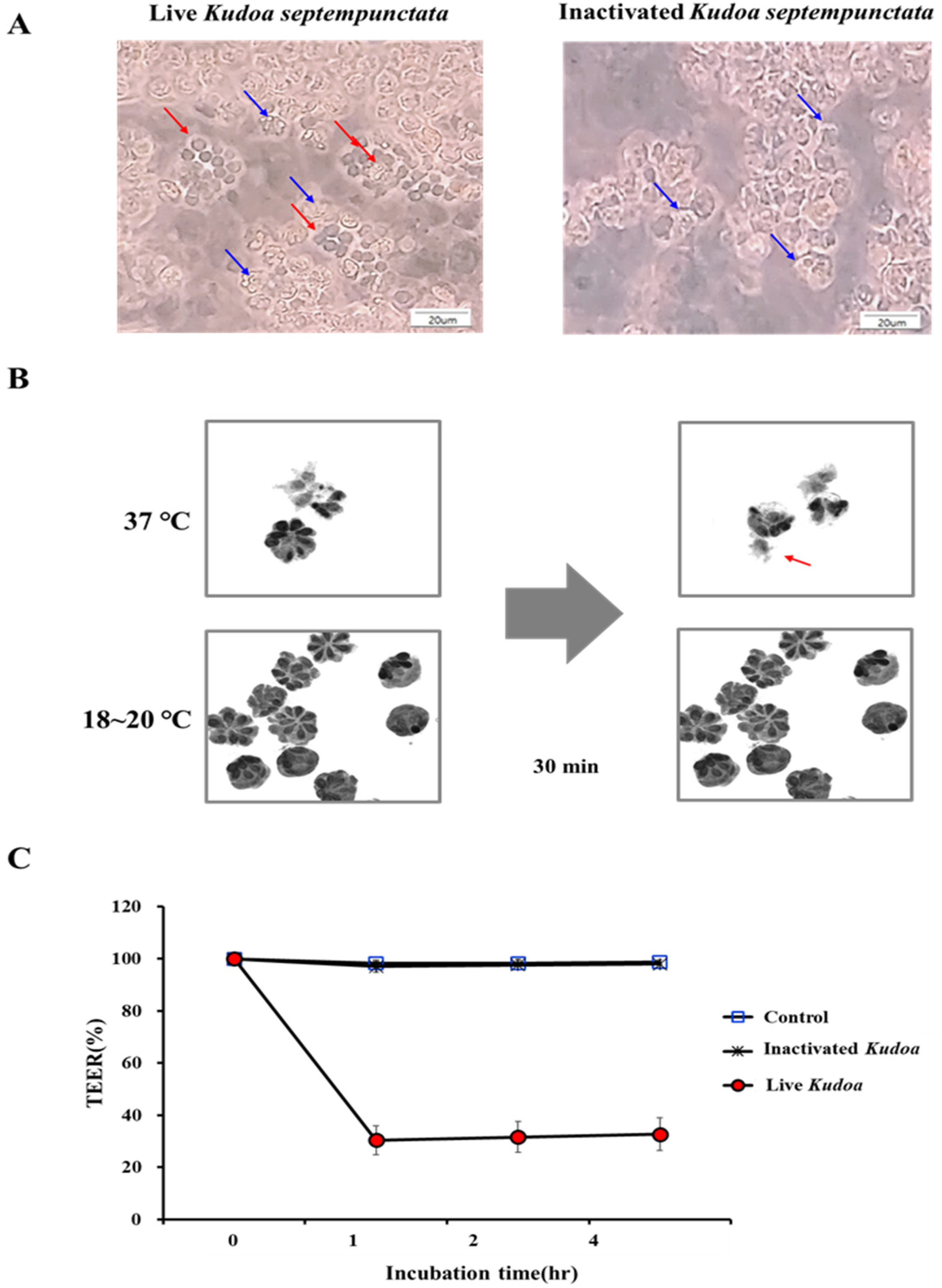
2.4. TEER Assay for Spore Activity

Human intestinal cell line Caco-2 cells (ATCC, HTB37; Manassas, VA, USA) were used to assess the activity of K. septempunctata spores collected from olive flounder muscles. For this, the Caco-2 cells were maintained in Dulbecco’s modified Eagle medium (Gibco, St. Louis, MO, USA) supplemented with 10% FBS, 100 U/mL penicillin, and 100 μg/mL streptomycin (Gibco, St. Louis, MO, USA) at 37 °C in a 5% CO2 incubator. For these assays, 24-well plates with collagen-coated insert membranes (Corning, New York, NY, USA) were used. Caco-2 cells were seeded at 2 × 10⁵ cells/well on Biocoat cell culture inserts (pore size of 0.4 μm). TEER was then measured using a Millicell ERS-2 (Millipore, Billerica, MA, USA) to determine the state of differentiation and cell monolayer integrity, with the inserts with TEER > 1000 Ω·cm2 being subjected to downstream experiments. Purified K. septempunctata spores (5 × 10⁵ spores/well) were then added to cell culture inserts, with TEER being measured every hour. Heat-inactivated spores (equivalent to 5 × 10⁵ spores/well) were also inoculated as a control in cell culture inserts, with TEER again being measured.

2.5. Immunofluorescence Assay on Zonula occludens-1 (ZO-1) in Cell-Line Cells

The Caco-2 or HCA-7 cell monolayers treated with K. septempunctata live spores (5 × 10⁵ spores/well) or the heat-inactivated spores (equivalent to 5 × 10⁵ spores/well) were incubated for 4 h at 37 °C. The treated cells were fixed in 4% formaldehyde solution (Sigma–Aldrich, St. Louis, MO, USA) overnight at 4 °C and then washed with PBS. After permeabilization with 0.1% Triton X-100 (Sigma–Aldrich, St. Louis, MO, USA) for 5 min, the cells were blocked with 1% BSA for 30 min before being incubated with Zonula occludens-1-FITC conjugated antibody (1:200, Invitrogen, Waltham, MA, USA) overnight at 4 °C in humidified chambers. The labelled cells were then washed in PBS and observed under a fluorescence microscope (Olympus, Tokyo, Japan).

2.6. Assay on the Serotonin Production of Caco-2 Cells

After TEER measurement, Caco-2 cell culture media were collected [17], whilst the concentration of serotonin (5-hydroxytryptamine, 5-HT) was measured using an ELISA kit (IBL, Minneapolis, MN, USA), in accordance with the manufacturer’s instructions.

2.7. Suckling Mouse Provocation for Diarrheal Response

All suckling mice were handled throughout the experiment, received from a delivery, stabilized for 1 h, administered the spores, and then observed for diarrhea in a walk-in-incubator that was maintained at 27 °C and 50% humidity. Suckling mice (4–5 days old, ddY strain) were separated from their mothers 2 h prior to administration of the spores and randomly divided into four groups (n = 10): K. septempunctata, 2 × 10⁶ and 2 × 10⁵ spores; heat-inactivated K. septempunctata, 2 × 10⁶ spores; and PBS only, as a negative control.
ICR suckling mice (4–5 days old) were used to explore whether K. septempunctata spores could provoke diarrhea in this mouse strain. ICR suckling mice were directly delivered from the animal supplier early in the morning on the day of the experiment before being randomly divided into six groups (n = 5 or 10) as follows: K. septempunctata 2 × 10⁶, 2 × 10⁵, 2 × 10⁴, 2 × 10³, 2 × 10² spores; heat-inactivated K. septempunctata, 2 × 10⁶ spores; and PBS only, as a negative control.
The spores were then suspended in 100 μL of PBS before being administered directly into the stomach of suckling mice using a 1 mL syringe and needle capped with a polyethylene capillary tube (Supplementary Figure S1A,B). All suckling mice in this experiment were laid individually on a paper towel in a plastic weigh boat under incandescent lamps in a walk-in-incubator at a temperature of 27 °C and 50% humidity. The ambient temperature of the suckling mice was maintained at 31–35 °C in this study (Supplementary Figure S1C). We also observed and recorded mouse movements, defecation, fecal form, and diarrhea over a time course of 4 h.

2.8. Experimental Design for the House Musk Shrews to Determine Emetic Response to K. septempunctata

House musk shrews (8 weeks old, n = 25) were randomly divided into four groups: Heat-inactivated K. septempunctata 1 × 10⁸ spores (n = 8); K. septempunctata 1 × 10⁷ spores (n = 8); K. septempunctata 1 × 10⁸ spores (n = 7); and PBS alone as a negative control. The spores were suspended in 500 μL PBS and orally administered using a ball-pointed needle at room temperature (18–20 °C). We then observed the shrews for 40 min and recorded their responses, including emesis and nausea.

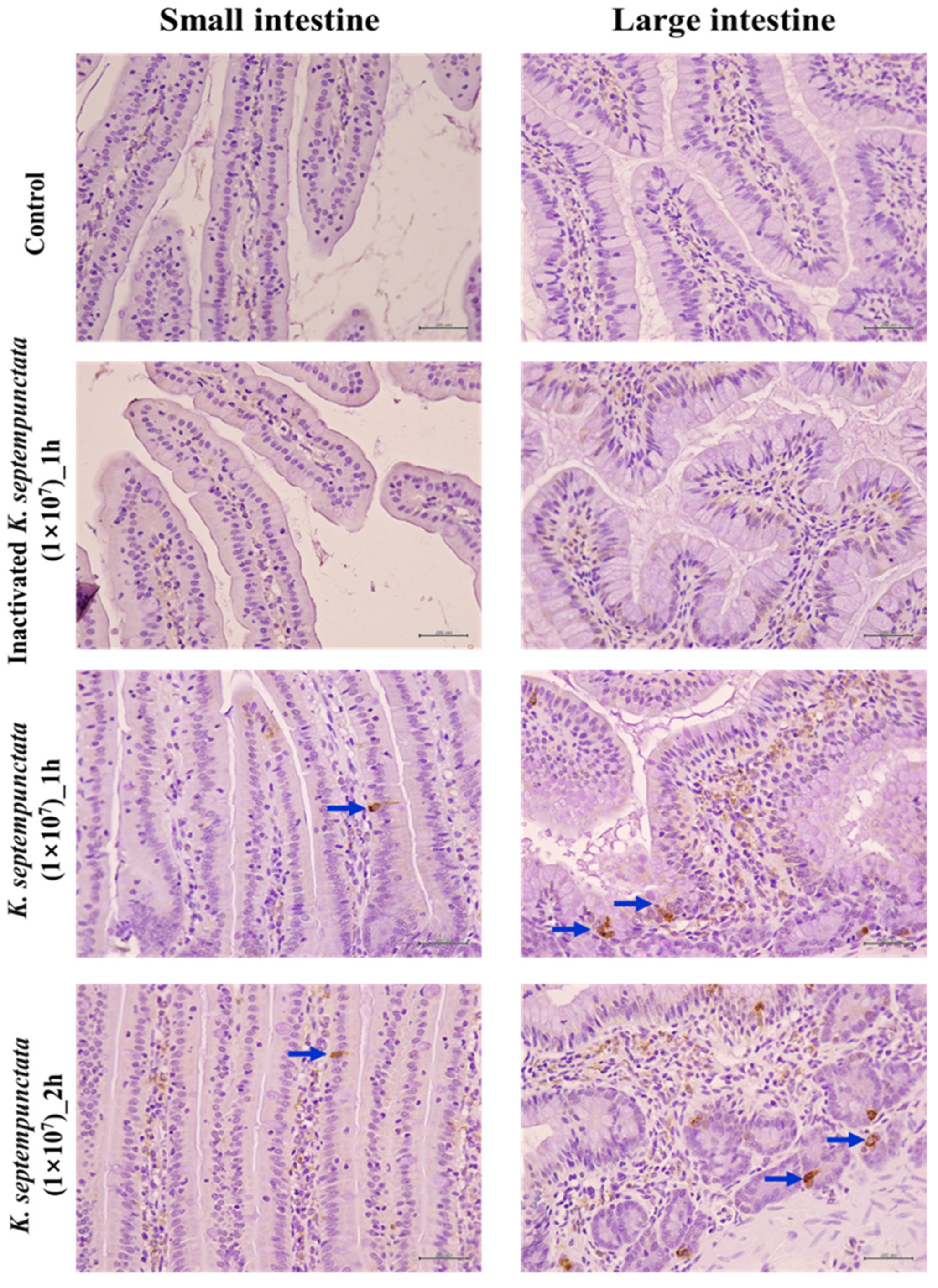
2.9. Immunohistochemical Localization of Serotonin in the Intestinal Tissues

The small and large intestines of house musk shrews were both resected 1–2 h after oral administration of the spores and fixed using the Swiss roll method [19]. Paraffin blocks were sectioned at 5–6 μm thickness and deparaffinized. For immunohistochemical staining, ribbons were rehydrated, with endogenous peroxidases being quenched with 3% H2O2 in methanol for 30 min in the dark. After washing with PBS for 20 min, the ribbons were blocked with 1% bovine serum albumin (Sigma–Aldrich, St. Louis, MO, USA) for 20 min. The ribbons were then incubated with primary antibodies at the following dilutions in blocking buffer: Mouse anti-serotonin (1:250, LSBio, Seattle, WA, USA), anti-c-fos (1:100, Abcam, Cambridge, UK), anti-mast cell tryptase (1:200, Abcam), anti-chromogranin A (1:400, Abcam), and anti-CaMKI (1:50, Abcam). After washing in PBS, the ribbons were incubated with the secondary antibody, horseradish peroxidase-conjugated anti-mouse IgG (1:500, Cell Signaling, Danvers, MA, USA). After washing, the color was developed using a substrate, DAB (3,3′-diaminobenzidine tetrahydrochloride), before being counter-stained with hematoxylin.

2.10. Statistical Analysis

Student’s t-tests were used to evaluate the statistical significance of the differences between individual experimental groups. Differences were considered statistically significant at p < 0.05.

3. Results

3.1. Kudoa septempunctata Sporoplasm Altered the Transepithelial Electrical Resistance of the Caco-2 Cell Monolayer

A cell monolayer permeability assay was performed to examine whether K. septempunctata damaged the Caco-2 cell junctions. When added to the Caco-2 cell monolayers, the live K. septempunctata spores released sporoplasm, whilst the heat-inactivated K. septempunctata spores did not (Figure 1A). In addition, it was observed as a result of holotomographic microscopy that the sporoplasm of K. septempunctata was released within 30 min after exposure to 37 °C (the temperature that the spores are exposed to in the stomach and intestines during human infection) but were not released at room temperature (18–20 °C) (Figure 1B). TEER across the Caco-2 cell monolayer was measured as an index of permeability change. The live K. septempunctata spores substantially decreased TEER by 70%, 1 h after inoculation, indicating an increase in the permeability of the Caco-2 cell monolayer (Figure 1C). These results indicated that K. septempunctata could interfere with the maintenance of TEER of Caco-2 cell monolayers through releasing sporoplasm at 37 °C.

3.2. K. septempunctata Disturbed Tight Junction ZO-1 Protein Arrangement

ZO-1 protein decreased within the monolayer of intestinal epithelial cells (Caco-2 and HCA-7) treated with live K. septempunctata spores after 4 h (Figure 2). There was no difference observed in the expression of ZO-1 protein in intestinal epithelial cells that had been treated with heated K. septempunctata spores. The results subsequently revealed that K. septempunctata sporoplasm was invasive and increased the permeability of the intestinal epithelium by disturbing the ZO-1 protein arrangement in the tight junction.

3.3. Diarrheal Responses of ddY and ICR Suckling Mice to K. septempunctata

To confirm whether K. septempunctata caused diarrhea, spores were orally administered to ddY suckling mice. The K. septempunctata 1 × 10⁵ spore group produced diarrhea 1.3 times in 40% of the mice (Table 1) for 119.0 ± 32.8 min. In contrast, the 2 × 10⁶ K. septempunctata group spores provoked diarrhea 3.3 times in 80% of the ddY suckling mice (p < 0.001) for 115.0 ± 23.9 min. The duration of diarrhea was similar between the two groups, although the frequency of diarrhea was increased in the high-dose group (2 × 10⁶).
In ICR suckling mice, the K. septempunctata 2 × 10⁶ spore group provoked diarrhea 4.3 times in 100% of the mice, whilst the 2 × 10⁵ spore group resulted in diarrhea 2.4 times in 80% of the mice (p < 0.001) (Table 2). In the 2 × 10⁴ and 2 × 10³ spore groups, one of the five mice had diarrhea once. Meanwhile, in the 2 × 10² spore group, the mice did not exhibit diarrhea or acute gastrointestinal symptoms (Table 2). These results indicated that high doses of K. septempunctata spores produced diarrhea one to four times in ddY and ICR suckling mice at an ambient temperature of 31–35 °C.

3.4. Emetic Response to K. septempunctata in House Musk Shrews

Emetic responses were induced in three of the four house musk shrews that were administered the K. septempunctata 1 × 10⁸ spores (Table 3). It took 30.0 ± 5.8 min to induce emesis, with an average of 4.0 ± 1.2 emetic reactions. In the 1 × 10⁷ spore group, emesis was induced in two of four house musk shrews with 3.0 ± 1.0 emetic reactions in 23.5 ± 2.5 min. However, no emetic reactions were observed in the heat-inactivated K. septempunctata group.

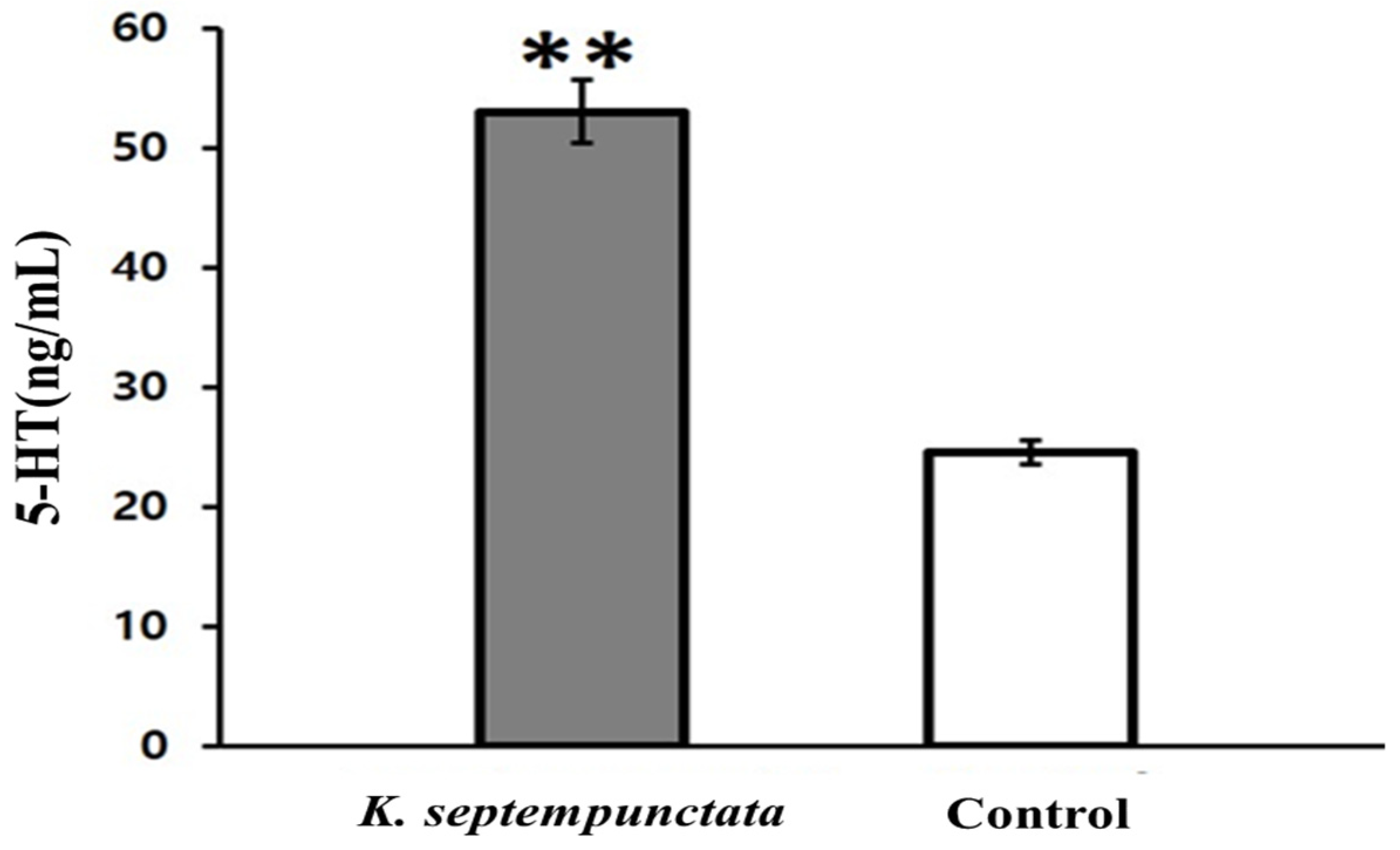
3.5. Serotonin (5-HT) Produced In Vitro and In Vivo by K. septempunctata

An additional experiment was conducted to determine whether serotonin, which plays an important role in inducing emesis, was secreted from intestinal epithelial cells, Caco-2 cells, by K. septempunctata (Figure 3). The concentration of serotonin secretions increased after 4 h of treatment with K. septempunctata in Caco-2 cells compared to the control group (p < 0.01). To confirm the mechanism of serotonin-induced vomiting, the induction of serotonin secretion was confirmed in the intestinal tissues of house musk shrews that had been orally administered K. septempunctata. Furthermore, serotonin secretion was induced more both in the small and large intestines of the house musk shrews than in the control and heat-inactivated K. septempunctata groups (Figure 4).

4. Discussion

The food-borne parasite, K. septempunctata, can parasitize humans accidentally when raw or undercooked parasitized fish are consumed. Additionally, altered intestinal permeability plays a significant role in the worsening of the clinical manifestations in patients where hostile reactions to food have been demonstrated [20,21,22]. The habit of eating raw fish therefore poses a high risk to the integrity of the intestinal mucosa [5,7,23]. The sporoplasm released from K. septempunctata spores contributes to gastrointestinal symptoms and occasionally results in concurrent inflammatory symptoms [6,16,24,25].
Intestinal barrier dysfunction associated with gastrointestinal symptoms influences both the sensitization and effector phases of toxicity. Caco-2 cells have been extensively used as an in vitro model for the evaluation of intestinal permeability considering their structural and functional similarity to the mature intestinal epithelium [26,27,28]. The K. septempunctata spores were found to have rapidly decreased TEER (within 1 h), thus indicating an increase in the permeability of the Caco-2 cell monolayer. The early decrease in TEER observed in Caco-2 monolayers could be explained by the release of sporoplasm from K. septempunctata. In this study, the release of sporoplasm from K. septempunctata spores was observed on the surface of Caco-2 cells within 1 h of inoculation. Several myxosporean parasites have previously been observed to release sporoplasm into the host intestine [12,29]. The sporoplasm of K. septempunctata quickly invaded Caco-2 cells, reaching the basolateral side of Caco-2 cells within 30 min of infection. This rapid invasion corresponded to a reduction in TEER alongside a short lag phase before the emergence of clinical symptoms. In addition, the release of sporoplasm at 37 °C (human body temperature) could be suggested as a reason for the acute diarrhea and emesis resulting from K. septempunctata infection. Our results confirmed that the K. septempunctata sporoplasm invasion could increase the permeability of the human intestinal epithelium and cause acute diarrhea.
Diarrheal diseases caused by enteric infections can result in impaired epithelial barrier function and the dysregulation of fluid and ion transport procedures [30,31,32,33]. Several enteric pathogens have established specific strategies to change or disrupt this structure as part of pathogenesis, resulting in either pathogen invasion or consequences, such as diarrhea [30,34]. Previous reports have shown that diarrhea-inducing parasites were associated with an increased permeability of the intestinal epithelium, alongside decreased tight junction (TJ) proteins, such as occludin and ZO-1 [28,35,36]. The TJ complex is composed of transmembrane proteins, including occludin, junctional adhesion proteins, and members of the claudin family. ZO-1 interacts with the intracellular protein complex that attaches to structural elements of the cytoskeleton [37]. ZO-1 is a key player in the interaction of occludin with other TJ junction proteins. Our results provide evidence that the release of K. septempunctata sporoplasm interfered with its interaction with ZO-1 and other TJ proteins, thus inducing diarrhea.
There have been several previous reports investigating whether K. septempunctata spores were able to provoke diarrhea in suckling mice. For example, Jang et al. reported that K. septempunctata did not infect the gastrointestinal tract or induce diarrhea in ddY suckling mice [9]. Meanwhile, Kawai et al. suggested that K. septempunctata induced watery stools and elevated fluid accumulation in suckling mice [5]. In the present study, K. septempunctata induced diarrhea in ICR and ddY suckling mice. An explanation for these varying experimental results for suckling rats as a result of Kudoa infection could be due to the experimental temperature conditions. The digestive physiology of suckling mice was observed to decrease at a room temperature of 21 °C, although the body temperature of suckling mice was maintained at normal levels at 31–35 °C, whilst digestive physiology continued to function normally in response to K. septempunctata infection. In addition, a minimal-dose assay for diarrhea-inducing K. septempunctata in suckling ICR mice was conducted. The 2 × 10⁶ and 2 × 10⁵ spore groups each induced diarrhea in 100% and 80% of ICR suckling mice, respectively, whilst the 2 × 10⁴ and 2 × 10³ spore groups induced diarrhea in only 20% of ICR suckling mice. Additionally, Kawai et al. found that 1 × 10⁵ K. septempunctata spores induced diarrhea in ddY suckling mice. The results for the minimum dose required to induce diarrhea in suckling mice provide evidence for the estimated intake threshold of K. septempunctata for the occurrence of symptoms in humans being 7.2 × 10⁷ spores/g of fish filet [24].
The pathophysiological basis of the derivation of emesis by K. septempunctata remains poorly understood. The luminal enterochromaffin (EC) cells “taste” and “sense” the luminal contents and can release mediators such as serotonin to activate ENS, as well as extrinsic vagal afferents to the brain. The stimulation of the extrinsic vagal and spinal afferent fibers by serotonin slows gastric emptying, pancreatic secretion, satiation, pain, and discomfort, alongside nausea and emesis [38]. Serotonin is also present in the secretory granules of EC cells, which are highly abundant in the duodenum and comprise the single largest enteroendocrine cell population [39,40]. The main depository of serotonin in the body is the mucosa of the gastrointestinal tract [41], although the details of gut serotonin remain unclear [42]. Rotavirus has previously been shown to stimulate human EC cells in the gut, causing serotonin release, which activates vagal afferents and the brain stem emesis center, a reaction cascade associated with emesis [43]. It has been previously reported that the oral administration of K. septempunctata to house musk shrews induced serotonin production in the intestines [44]. In this study, serotonin increased in response to the K. septempunctata cascade in Caco-2 cells and in the small and large intestinal tissues of house musk shrews. Serotonin is a physiological mediator that stimulates the emesis center in the brain. It has been suggested that serotonin produced in the intestine may trigger disease-associated emesis [45,46,47]. Additionally, serotonin released from EC cells acts as a pro- and anti-inflammatory molecule, which is strongly supported by the evidence. IL-8 and serotonin have both been reported to cause inflammation and emesis associated with K. septempunctata invasion via AP-1 upregulation and the induction of the MAPK/NF-kappa B pathway [17]. In the current study, serotonin induced by K. septempunctata spores played a role in emesis in house musk shrews.
Based on the above results, it is proposed that the K. septempunctata spores released the sporoplasm at 37 °C decreased TEER in the intestinal epithelium, which consequently transuded exudate into the intestinal lumen and caused watery diarrhea in suckling mice. The spores also induced serotonin production in the intestine, which in turn mediated emesis in house musk shrews.

Supplementary Materials

The following supporting information can be downloaded at https://www.mdpi.com/article/10.3390/pathogens12050739/s1, Figure S1: Administration of Kudoa septempunctata spores and observations in suckling mice, Figure S2: House musk shrews administered with 2 × 10⁷ Kudoa septempunctata spores.

Author Contributions

Conceived the study: J.-W.J. and S.-J.H.; designed experiments: S.-H.H., J.-Y.K., S.-O.L., S.-J.H., H.-I.L. and J.-W.J.; performed experiments: S.-H.H., J.-Y.K. and S.-O.L.; data analysis: S.-H.H., J.-Y.K. and S.-O.L.; reagents/materials/analysis tools: S.-H.H., J.-Y.K. and S.-O.L.; wrote the manuscript: S.-H.H., J.-W.J. and S.-J.H. All authors have read and agreed to the published version of the manuscript.

Funding

This work was supported by grants from the Korean Disease Control and Prevention Agency (KDCA:4837-311-210-13 and KDCA-2019-P4402-00).

Institutional Review Board Statement

Korea Disease Control & Prevention Institutional Animal Care and Use Committee (KDCA-IACUC; Permit Number KDCA-02-19-2A), Chung-Ang University Guide for the Care and Use of Laboratory Animals (Permit Number: 2020-00015).

Informed Consent Statement

Not applicable.

Data Availability Statement

Not applicable.

Conflicts of Interest

The authors have no conflicting interest.

References

  1. Matsukane, Y.; Sato, H.; Tanaka, S.; Kamata, Y.; Sugita-Konishi, Y. Kudoa septempunctata n. sp. (Myxosporea: Multivalvulida) from an Aquacultured Olive Flounder (Paralichthys olivaceus) Imported from Korea. Parasitol. Res. 2010, 107, 865–872. [Google Scholar] [CrossRef] [PubMed]
  2. Takeuchi, F.; Ogasawara, Y.; Kato, K.; Sekizuka, T.; Nozaki, T.; Sugita-Konishi, Y.; Ohnishi, T.; Kuroda, M. Genetic Variants of Kudoa septempunctata (Myxozoa: Multivalvulida), a Flounder Parasite Causing Foodborne Disease. J. Fish Dis. 2016, 39, 667–672. [Google Scholar] [CrossRef] [PubMed]
  3. Chase, J.C.; Dawson-Coates, J.A.; Haddow, J.D.; Stewart, M.H.; Haines, L.R.; Whitaker, D.J.; Ken, M.L.; Olafson, R.W.; Pearson, T.W. Analysis of Kudoa thyrsites (Myxozoa: Myxosporea) Spore Antigens Using Monoclonal Antibodies. Dis. Aquat. Organ. 2001, 45, 121–129. [Google Scholar] [CrossRef] [PubMed]
  4. Harada, T.; Kawai, T.; Jinnai, M.; Ohnishi, T.; Sugita-Konishi, Y.; Kumeda, Y. Detection of Kudoa septempunctata 18S Ribosomal DNA in Patient Fecal Samples from Novel Food-Borne Outbreaks Caused by Consumption of Raw Olive Flounder (Paralichthys olivaceus). J. Clin. Microbiol. 2012, 50, 2964–2968. [Google Scholar] [CrossRef]
  5. Kawai, T.; Sekizuka, T.; Yahata, Y.; Kuroda, M.; Kumeda, Y.; Iijima, Y.; Kamata, Y.; Sugita-Konishi, Y.; Ohnishi, T. Identification of Kudoa septempunctata as the Causative Agent of Novel Food Poisoning Outbreaks in Japan by Consumption of Paralichthys olivaceus in Raw Fish. Clin. Infect. Dis. 2012, 54, 1046–1052. [Google Scholar] [CrossRef]
  6. Iwashita, Y.; Kamijo, Y.; Nakahashi, S.; Shindo, A.; Yokoyama, K.; Yamamoto, A.; Omori, Y.; Ishikura, K.; Fujioka, M.; Hatada, T.; et al. Food Poisoning Associated with Kudoa septempunctata. J. Emerg. Med. 2013, 44, 943–945. [Google Scholar] [CrossRef]
  7. Suzuki, J.; Murata, R.; Yokoyama, H.; Sadamasu, K.; Kai, A. Detection Rate of Diarrhoea-Causing Kudoa Hexapunctata in Pacific Bluefin Tuna Thunnus orientalis from Japanese Waters. Int. J. Food Microbiol. 2015, 194, 1–6. [Google Scholar] [CrossRef]
  8. Yokoyama, H.; Funaguma, N.; Kobayashi, S. In Vitro Inactivation of Kudoa septempunctata Spores Infecting the Muscle of Olive Flounder Paralichthys olivaceus. Foodborne Pathog. Dis. 2016, 13, 21–27. [Google Scholar] [CrossRef]
  9. Jang, Y.; Ahn, M.; Bang, H.; Kang, B. Effects of Kudoa septempunctata Genotype ST3 Isolate from Korea on ddY Suckling Mice. Parasite 2016, 23, 18. [Google Scholar] [CrossRef]
  10. Ahn, M.; Woo, H.; Kang, B.; Jang, Y.; Shin, T. Effect of Oral Administration of Kudoa septempunctata Genotype ST3 in Adult BALB/C Mice. Parasite 2015, 22, 35. [Google Scholar] [CrossRef]
  11. Guo, Q.; Jia, L.; Qin, J.; Li, H.; Gu, Z. Myxozoans and Our Dinner Table: Pathogenicity Studies of Myxobolus Honghuensis (Myxosporea: Bivalvulida) Using a Suckling Mice Model. Foodborne Pathog. Dis. 2015, 12, 653–660. [Google Scholar] [CrossRef] [PubMed]
  12. Ohnishi, T.; Kikuchi, Y.; Furusawa, H.; Kamata, Y.; Sugita-Konishi, Y. Kudoa septempunctata Invasion Increases the Permeability of Human Intestinal Epithelial Monolayer. Foodborne Pathog. Dis. 2013, 10, 137–142. [Google Scholar] [CrossRef] [PubMed]
  13. Ahn, M.; Ko, H.J.; Kim, J.; Jang, Y.; Shin, T. Evaluation of the Inflammatory Response to Kudoa septempunctata Genotype ST3 Isolated from Olive Flounder (Paralichthys olivaceus) in Caco-2 Cells. Parasite 2018, 25, 12. [Google Scholar] [CrossRef] [PubMed]
  14. Ohnishi, T.; Oyama, R.; Furusawa, H.; Ohba, N.; Kamata, Y.; Sugita-Konishi, Y. Kudoa septempunctata Was Recognised by Toll-Like Receptor 2 Produced by a RAW 264 Macrophage-Like Cell Line. Food Addit. Contam. Part A Chem. Anal. Control Expo. Risk Assess. 2013, 30, 1365–1369. [Google Scholar] [CrossRef]
  15. Srinivasan, B.; Kolli, A.R.; Esch, M.B.; Abaci, H.E.; Shuler, M.L.; Hickman, J.J. TEER Measurement Techniques for In Vitro Barrier Model Systems. J. Lab. Autom. 2015, 20, 107–126. [Google Scholar] [CrossRef]
  16. Shin, J.H.; Yang, J.P.; Seo, S.H.; Kim, S.G.; Kim, E.M.; Ham, D.W.; Shin, E.H. Immune-Triggering Effect of the Foodborne Parasite Kudoa septempunctata through the C-Type Lectin Mincle in HT29 Cells. BMB Rep. 2020, 53, 478–483. [Google Scholar] [CrossRef]
  17. Yamamoto, S.; Kawano, F.; Yokoyama, H.; Kobayashi, S. Effects of Kudoa septempunctata Infections in a Human Intestinal Epithelial Model (Caco-2): A DNA Microarray Study. Biosci. Biotechnol. Biochem. 2020, 84, 1030–1038. [Google Scholar] [CrossRef]
  18. Shin, S.; Kim, K.; Yoon, J.; Park, Y. Active Illumination Using a Digital Micromirror Device for Quantitative Phase Imaging. Opt. Lett. 2015, 40, 5407–5410. [Google Scholar] [CrossRef]
  19. Fedchenko, N.; Reifenrath, J. Different Approaches for Interpretation and Reporting of Immunohistochemistry Analysis Results in the Bone Tissue—A Review. Diagn. Pathol. 2014, 9, 221. [Google Scholar] [CrossRef]
  20. Samadi, N.; Klems, M.; Untersmayr, E. The Role of Gastrointestinal Permeability in Food Allergy. Ann. Allergy Asthma Immunol. 2018, 121, 168–173. [Google Scholar] [CrossRef]
  21. König, J.; Wells, J.; Cani, P.D.; García-Ródenas, C.L.; MacDonald, T.; Mercenier, A.; Whyte, J.; Troost, F.; Brummer, R.J. Human Intestinal Barrier Function in Health and Disease. Clin. Transl. Gastroenterol. 2016, 7, e196. [Google Scholar] [CrossRef] [PubMed]
  22. Ventura, M.T.; Polimeno, L.; Amoruso, A.C.; Gatti, F.; Annoscia, E.; Marinaro, M.; Di Leo, E.; Matino, M.G.; Buquicchio, R.; Bonini, S.; et al. Intestinal Permeability in Patients with Adverse Reactions to Food. Dig. Liver Dis. 2006, 38, 732–736. [Google Scholar] [CrossRef] [PubMed]
  23. Kim, J.J.; Ryu, S.; Lee, H. Foodborne Illness Outbreaks in Gyeonggi Province, Korea, Following Seafood Consumption Potentially Caused by Kudoa septempunctata between 2015 and 2016. Osong Public Health Res. Perspect. 2018, 9, 66–72. [Google Scholar] [CrossRef] [PubMed]
  24. Yahata, Y.; Sugita-Konishi, Y.; Ohnishi, T.; Toyokawa, T.; Nakamura, N.; Taniguchi, K.; Okabe, N. Kudoa septempunctata-Induced Gastroenteritis in Humans after Flounder Consumption in Japan: A Case-Controlled Study. Jpn. J. Infect. Dis. 2015, 68, 119–123. [Google Scholar] [CrossRef]
  25. Tachibana, T.; Watari, T. Kudoa septempunctata Infection: An Underdiagnosed Pathogen of Acute Gastrointestinal Symptoms. QJM Int. J. Med. 2020, 113, 43–44. [Google Scholar] [CrossRef]
  26. Lytton, S.D.; Fischer, W.; Nagel, W.; Haas, R.; Beck, F.X. Production of Ammonium by Helicobacter pylori Mediates Occludin Processing and Disruption of Tight Junctions in Caco-2 Cells. Microbiology 2005, 151, 3267–3276. [Google Scholar] [CrossRef]
  27. Dickman, K.G.; Hempson, S.J.; Anderson, J.; Lippe, S.; Zhao, L.; Burakoff, R.; Shaw, R.D. Rotavirus Alters Paracellular Permeability and Energy Metabolism in Caco-2 Cells. Am. J. Physiol. Gastrointest. Liver Physiol. 2000, 279, G757–G766. [Google Scholar] [CrossRef]
  28. Carballeda-Sangiao, N.; Sánchez-Alonso, I.; Navas, A.; Arcos, S.C.; de Palencia, P.F.; Careche, M.; González-Muñoz, M. Anisakis simplex Products Impair Intestinal Epithelial Barrier Function and Occludin and Zonula occludens-1 Localisation in Differentiated Caco-2 Cells. PLOS Negl. Trop. Dis. 2020, 14, e0008462. [Google Scholar] [CrossRef]
  29. El-Matbouli, M.; Hoffmann, R.W. Light and Electron Microscopic Studies on the Chronological Development of Myxobolus cerebralis to the Actinosporean Stage in Tubifex Tubifex. Int. J. Parasitol. 1998, 28, 195–217. [Google Scholar] [CrossRef]
  30. Guttman, J.A.; Finlay, B.B. Tight Junctions as Targets of Infectious Agents. Biochim. Biophys. Acta 2009, 1788, 832–841. [Google Scholar] [CrossRef]
  31. Viswanathan, V.K.; Hodges, K.; Hecht, G. Enteric Infection Meets Intestinal Function: How Bacterial Pathogens Cause Diarrhoea. Nat. Rev. Microbiol. 2009, 7, 110–119. [Google Scholar] [CrossRef] [PubMed]
  32. Field, M. Intestinal Ion Transport and the Pathophysiology of Diarrhea. J. Clin. Investig. 2003, 111, 931–943. [Google Scholar] [CrossRef] [PubMed]
  33. Hoque, K.M.; Chakraborty, S.; Sheikh, I.A.; Woodward, O.M. New Advances in the Pathophysiology of Intestinal Ion Transport and Barrier Function in Diarrhea and the Impact on Therapy. Expert Rev. Anti. Infect. Ther. 2012, 10, 687–699. [Google Scholar] [CrossRef] [PubMed]
  34. Vogelmann, R.; Amieva, M.R.; Falkow, S.; Nelson, W.J. Breaking into the Epithelial Apical-Junctional Complex--News from Pathogen Hackers. Curr. Opin. Cell Biol. 2004, 16, 86–93. [Google Scholar] [CrossRef]
  35. Kumar, A.; Chatterjee, I.; Anbazhagan, A.N.; Jayawardena, D.; Priyamvada, S.; Alrefai, W.A.; Sun, J.; Borthakur, A.; Dudeja, P.K. Cryptosporidium parvum Disrupts Intestinal Epithelial Barrier Function via Altering Expression of Key Tight Junction and Adherens Junction Proteins. Cell. Microbiol. 2018, 20, e12830. [Google Scholar] [CrossRef]
  36. Buret, A.G.; Mitchell, K.; Muench, D.G.; Scott, K.G. Giardia lamblia Disrupts Tight Junctional ZO-1 and Increases Permeability in Non-transformed Human Small Intestinal Epithelial Monolayers: Effects of Epidermal Growth Factor. Parasitology 2002, 125, 11–19. [Google Scholar] [CrossRef]
  37. Chelakkot, C.; Ghim, J.; Ryu, S.H. Mechanisms Regulating Intestinal Barrier Integrity and Its Pathological Implications. Exp. Mol. Med. 2018, 50, 1–9. [Google Scholar] [CrossRef]
  38. Rao, M.; Gershon, M.D. The Bowel and beyond: The Enteric Nervous System in Neurological Disorders. Nat. Rev. Gastroenterol. Hepatol. 2016, 13, 517–528. [Google Scholar] [CrossRef]
  39. Bertrand, P.P.; Kunze, W.A.; Furness, J.B.; Bornstein, J.C. The Terminals of Myenteric Intrinsic Primary Afferent Neurons of the Guinea-Pig Ileum Are Excited by 5-Hydroxytryptamine Acting at 5-hydroxytryptamine-3 Receptors. Neuroscience 2000, 101, 459–469. [Google Scholar] [CrossRef]
  40. Hansen, M.B.; Witte, A.B. The Role of Serotonin in Intestinal Luminal Sensing and Secretion. Acta Physiol. 2008, 193, 311–323. [Google Scholar] [CrossRef]
  41. Spohn, S.N.; Mawe, G.M. Non-conventional Features of Peripheral Serotonin Signalling—The Gut and Beyond. Nat. Rev. Gastroenterol. Hepatol. 2017, 14, 412–420. [Google Scholar] [CrossRef] [PubMed]
  42. Hansen, M.B. The Enteric Nervous System I: Organisation and Classification. Pharmacol. Toxicol. 2003, 92, 105–113. [Google Scholar] [CrossRef] [PubMed]
  43. Hagbom, M.; Istrate, C.; Engblom, D.; Karlsson, T.; Rodriguez-Diaz, J.; Buesa, J.; Taylor, J.A.; Loitto, V.M.; Magnusson, K.E.; Ahlman, H.; et al. Rotavirus Stimulates Release of Serotonin (5-HT) from Human Enterochromaffin Cells and Activates Brain Structures Involved in Nausea and Vomiting. PLOS Pathog. 2011, 7, e1002115. [Google Scholar] [CrossRef] [PubMed]
  44. Sugita-Konishi, Y.; Sato, H.; Ohnishi, T. Novel Foodborne Disease Associated with Consumption of Raw Fish, Olive Flounder (Paralichthys olivaceus). J. Food Saf. 2014, 2, 141–150. [Google Scholar] [CrossRef]
  45. Terry, N.; Margolis, K.G. Serotonergic Mechanisms Regulating the GI Tract: Experimental Evidence and Therapeutic Relevance. Handb. Exp. Pharmacol. 2017, 239, 319–342. [Google Scholar] [CrossRef]
  46. Margolis, K.G.; Stevanovic, K.; Li, Z.; Yang, Q.M.; Oravecz, T.; Zambrowicz, B.; Jhaver, K.G.; Diacou, A.; Gershon, M.D. Pharmacological Reduction of Mucosal but Not Neuronal Serotonin Opposes Inflammation in Mouse Intestine. Gut 2014, 63, 928–937. [Google Scholar] [CrossRef]
  47. Kim, J.J.; Wang, H.; Terc, J.D.; Zambrowicz, B.; Yang, Q.M.; Khan, W.I. Blocking Peripheral Serotonin Synthesis by Telotristat Etiprate (LX1032/LX1606) Reduces Severity of Both Chemical- and Infection-Induced Intestinal Inflammation. Am. J. Physiol. Gastrointest. Liver Physiol. 2015, 309, G455–G465. [Google Scholar] [CrossRef]
Figure 1. Release of sporoplasm from Kudoa septempunctata spores and the alteration of transepithelial electrical resistance (TEER) in Caco-2 cell monolayers. (A) The K. septempunctata spores (blue arrow) were added to the Caco-2 cell monolayer and incubated at 37 °C for 1 h. Sporoplasm released is indicated (red arrows). (B) A sporoplasm releasing (red arrow) from a K. septempunctata spore was observed under a holotomographic microscope 30 min after incubation at 37 °C. (C) TEER decrease in the Caco-2 cell monolayer by live K. septempunctata spores. Values are means ± standard deviation; measurements in triplicate.
Pathogens 12 00739 g001
Figure 2. Zonula occuludens-1 (ZO-1) localized by immunofluorescent staining. ZO-1 expression revealed altered matrices in Caco-2 and HCA-7 cells incubated with K. septempunctata for 4 h. Black arrows indicate K. septempunctata sporoplasm on cells. Red arrows indicate disturbing the ZO-1 proteins. BF, bright field.
Pathogens 12 00739 g002
Figure 3. Serotonin (5-HT) produced and secreted from Caco-2 cells. K. septempunctata spores were added to the culture medium and cultured for 4 h. Serotonin levels were determined in the culture supernatants. Data represent the mean ± standard deviation (SD) of three independent replicates. ** Statistically significant difference between treated and untreated cells (p < 0.01).
Pathogens 12 00739 g003
Figure 4. Localization of serotonin in the epithelia of the small and large intestines of house musk shrews administered 1 × 10⁷ K. septempunctata spores/mouse. Blue arrows indicate serotonin-producing cells; scale bar = 200 nm.
Pathogens 12 00739 g004
Table 1. Diarrheal response of ddY suckling mice provoked by Kudoa septempunctata spores.
K. septempunctata Spores Administered.No. of
Diarrhea/Mouse Tested
No. of
Diarrheal
Incidents
Duration of Diarrhea
(min)
Live 2 × 1068/10 ***3.3115.0 ± 23.9
Live 1 × 1054/101.3119.0 ± 32.8
Heat-inactivated 2 × 1060/10__
PBS1/101254
*** Differences between the treated and untreated groups; statistical significance, p < 0.001. Value represents the mean ± standard deviation.
Table 2. Diarrheal response of ICR suckling mice to Kudoa septempunctata spores.
K. septempunctata Spores
Administered
No. of Diarrhea/Mouse TestedNo. of Diarrheal Incidentsp-Value
Live 2 × 10610/104.30.0001 ***
Live 2 × 1058/102.40.0006 ***
Live 2 × 1041/50.40.555
Live 2 × 1031/50.40.555
Live 2 × 10²⁰/50-
Heat-inactivated 2 × 1060/50.1-
PBS1/100.1-
*** Differences between the treated and untreated groups; statistical significance, p < 0.001.
Table 3. Kudoa septempunctata spore-induced emetic responses in house musk shrews.
K. septempunctata Spores
Administered
No. of Musk Shrews (Emesis/Tested)No. of
Emetic Events
Duration of
Emesis (min)
Live 1 × 1076/7 ***4.0 ± 1.230.0 ± 5.8
Live 1 × 1064/83.0 ± 1.023.5 ± 2.5
Heat-inactivated 1 × 1070/800
PBS0/200
*** Differences between the treated and untreated groups; statistical significance, p < 0.001. Values represent the mean ± standard deviation.
Disclaimer/Publisher’s Note: The statements, opinions and data contained in all publications are solely those of the individual author(s) and contributor(s) and not of MDPI and/or the editor(s). MDPI and/or the editor(s) disclaim responsibility for any injury to people or property resulting from any ideas, methods, instructions or products referred to in the content.

