
Non-Endemic Leishmaniases Reported Globally in Humans between 2000 and 2021—A Comprehensive Review


Abstract
:1. Introduction
2. Search Strategy, Eligibility, and Review
- -
- Leishmania (Leishmania) subgenus: L. mexicana complex (L. mexicana, L. amazonensis, L. venezuelensis); L. donovani complex (L. donovani, L. infantum (=L. chagasi); L. tropica complex (L. tropica, L. killicki, L. aethiopica); L. major;
- -
- Leishmania (Mundinia) subgenus: L. martiniquensis; L. “siamensis”;
- -
- Leishmania (Viannia) subgenus: L. braziliensis complex (L. braziliensis, L. peruviana); L. guyanensis complex (L. guyanensis, L. panamensis); L. lainsoni; L. naiffi.
3. Results and Discussion
3.1. Human Leishmania Infection and Leishmaniasis
3.1.1. Visceral Leishmaniasis

| Description | VL | CL/MCL/ML | ||||
|---|---|---|---|---|---|---|
| Frequency Patients Tested | Frequency Tested Positive | References | Frequency Patients Tested | Frequency Tested Positive | References | |
| Microscopy | 84.8% (291/343) | 91.8% (302/329) | 63.4% (1072/1690) | 84.3% (904/1072) | ||
| Bone marrow | 84.5% (246/291) | 91.5% (225/246) | [12,17,19,20,24,26,27,29,31,32,35,38,40,42,43,44,46,47,49,51,53,55,57,58,59,60,61,62,63,64,65,66,68,199,200,201,202,203,205,207,209,210,211,212,213,214,215,216,217,218,219,249,250,251,256,257,258,260,261,262] | NR | NA | NA |
| Liver | 10.3% (30/291) | 90.0% (27/30) | [17,21,26,32,36,202,216,257,261] | NR | NA | NA |
| Spleen | 8.6% (25/291) | 92.0% (23/25) | [16,17,23,38,206,216,252,257,261] | NR | NA | NA |
| Lymph node | 1.7% (5/291) | 100% (5/5) | [35,37,198,261] | 0.1% (1/1072) | 100% (1/1) | [82] |
| Blood | 1.0% (3/291) | 100% (3/3) | [21,24,29] | NR | NA | NA |
| Skin | 1.0% (3/291) | 100% (3/3) | [66,257,261] | 96.7% (1037/1072) | 84.2% (873/1037) a | [7,11,12,20,27,30,38,66,69,70,71,72,73,75,77,78,79,80,81,82,84,85,86,87,88,89,90,91,92,95,96,97,98,99,100,101,103,104,105,106,110,113,115,116,117,119,120,121,122,123,124,126,127,128,129,131,134,137,139,140,141,143,144,145,147,148,149,150,151,152,153,156,157,159,160,162,163,164,165,166,167,168,169,170,171,174,175,176,179,181,182,183,184,185,186,187,191,194,195,196,197,221,222,224,225,226,227,228,229,230,231,234,235,236,237,238,239,240,241,242,243,244,246,247,254,255,263,268,271,275,279,280,281] |
| Other | 5.5% (16/291) b | 93.8% (15/16) | [32] | NR | NA | NA |
| Mucosa c | NR | NA | NA | 3.1% (33/1072) | 90.9% (30/33) | [11,26,30,38,76,82,107,108,109,111,114,130,135,146,152,158,161,180,190,223,233,245] |
| Serology | 68.8% (236/343) | 95.2% (318/334) | 6.8% (115/1690) d | 69.1% (85/123) | ||
| DAT | 45.1% (97/215) | 96.9% (94/97) | [16,38,42,52,257,259,261] | 6.1% (6/99) | 66.7% (4/6) | [103,158] |
| rK39 | 44.2% (95/215) | 93.7% (89/95) | [38,52,60,204,210,211,213,257,259,261,262,281] | 17.2% (17/99) | 64.7% (11/17) | [114,119,158,174,190,277] |
| IFAT | 41.4% (89/215) | 94.4% (84/89) | [18,19,20,25,29,32,33,40,43,44,47,59,198,202,203,212,216,250,257] | 73.7% (73/99) | 71.2% (52/73) | [11,71,80,89,101,104,107,119,129,135,138,143,179,226] |
| ELISA | 13.0% (28/215) | 92.9% (26/28) | [18,20,33,37,40,41,44,47,68,199,203,250] | 7.1% (7/99) | 57.1% (4/7) | [70,89,135,144,170,176,179] |
| WB | 1.9% (4/215) | 100% (4/4) | [25,68,203,256] | 4.0% (4/99) | 100% (4/4) | [104,121,135] |
| PCR | 51.6% (177/343) | 94.1% (206/219) | 84.9% (1435/1690) | 97.2% (1398/1438) | ||
| Bone marrow | 83.7% (118/141) | 96.6% (114/118) | [12,20,21,22,24,26,27,29,32,33,40,42,44,45,47,48,49,50,51,52,57,58,59,61,62,63,64,65,66,68,199,209,210,211,212,214,215,250,253,260] | NR | NA | NA |
| Blood | 30.5% (43/141) | 93.0% (40/43) | [18,20,24,25,29,32,41,44,48,60,61,62,66,202,217,250,260] | 0.1% (2/1435) | 100% (2/2) | [20] |
| Liver | 7.1% (10/141) | 90.0% (9/10) | [21,26,32,48,202] | NR | NA | NA |
| Spleen | 4.3% (6/141) | 100% (6/6) | [16,23,54,59,66,206] | NR | NA | NA |
| Other | 2.8% (4/141) e | 100% (4/4) | [28,29,48,204] | NR | NA | NA |
| Lymph node | 1.4% (2/141) | 100% (2/2) | [37,56] | 0.1% (2/1435) | 100% (2/2) | [82,180] |
| Mucosa c | NR | NA | NA | 4.0% (57/1435) | 96.5% (55/57) | [11,30,38,76,77,82,107,109,111,114,129,130,133,135,142,146,158,161,173,179,180,190,223,233,245,273] |
| Skin | NR | NA | NA | 96.0% (1378/1435) | 97.2% (1340/1378) f | [7,9,11,12,20,26,28,30,38,45,61,65,66,70,75,76,77,78,79,82,83,87,89,91,92,93,94,95,96,97,98,100,102,104,105,106,107,109,110,111,114,116,117,119,121,122,123,124,126,127,128,129,130,132,133,134,135,136,137,138,139,140,141,142,143,144,145,146,147,149,150,151,153,156,157,158,159,161,162,164,165,166,167,168,170,171,172,173,174,175,176,177,178,179,180,181,182,185,186,187,188,189,190,191,192,193,194,195,196,197,223,225,226,227,229,230,231,233,235,236,238,239,240,241,242,243,244,245,246,247,255,266,268,271,273,275,278] |
| Culture | 17.5% (60/343) | 76.2% (48/63) | 48.7% (823/1690) | 86.8% (714/823) | ||
| Bone marrow | 76.1% (35/46) | 88.6% (31/35) | [12,18,21,27,29,32,44,51,63,181,199,209,250] | NR | NA | NA |
| Spleen | 19.6% (9/46) | 100% (9/9) | [16,206] | NR | NA | NA |
| Lymph node | 4.3% (2/46) | 100% (2/2) | [11,198] | 0.2% (2/823) | 100% (2/2) | [82,180] |
| Blood | 2.2% (1/46) | 100% (1/1) | [29] | NR | NA | NA |
| Liver | 2.2% (1/46) | 100% (1/1) | [21] | NR | NA | NA |
| Other | 2.2% (1/46) g | 100% (1/1) | [32] | NR | NA | NA |
| Mucosa | NR | NA | NA | 1.8% (15/823) | 86.7% (13/15) | [12,43,73,94,111,207,222,236,259] |
| Skin | NR | NA | NA | 98.2% (808/823) | 86.5% (699/808) h | [7,9,12,28,30,69,70,71,72,73,75,77,80,81,82,85,87,89,90,91,95,97,98,100,104,110,116,117,119,120,121,122,123,127,128,129,131,134,137,145,148,149,150,156,159,163,166,169,175,176,181,183,187,221,226,227,228,234,235,242,247,263,266,268,271,275,278] |
| Species/complex | ||||||
| L. donovani complex | 98.8% (167/169) | NA | [9,10,11,16,20,21,22,24,25,26,27,28,29,32,33,37,40,44,45,47,48,49,51,52,54,57,59,60,61,62,63,64,66,68,198,199,202,206,209,210,211,212,214,256,260,261] | See Figure 3 | NA | NA |
| L. infantum + L. major | 0.6% (1/169) | NA | NA | NR | NA | NA |
| L. subgenus Mundinia | 0.6% (1/169) | NA | NA | NR | NA | NA |

3.1.2. Cutaneous, Mucocutaneous and Mucosal Leishmaniasis
3.1.3. Clinical Aspects and Management of Cutaneous Leishmaniasis
- -
- L. donovani complex: predominantly single lesions; involving mostly the head/neck and upper limbs, often non-ulcerative (especially plaques/crusts);
- -
- L. major: predominantly multiple lesions; involving mostly the limbs, mostly ulcerative;
- -
- L. tropica: single or multiple lesions; predominantly in the head/neck and upper limbs, often non-ulcerative.
- -
- L. aethiopica: 13 cases reported [7,10,11,119,132,133,136,247,275,277], six of them imported from Ethiopia [10,11,136,247]; only three cases described lesions [119,132,247]: two of them as single lesions in the face or trunk [121,247] and one as multiple lesions in the face [132]; in two patients the lesions were papular/nodular [119,247] and in one it was a plaque/crust [132]; management strategy was described in six cases (one no treatment, [247], two topical with intralesional antimonial and cryotherapy, [277], and three systemic with miltefosine or LAmB, [119,132,133]). Relapses were not reported in any of the cases.
- -
- L. braziliensis complex: predominantly single lesions; most often in the lower and upper limbs; commonly ulcerative; more frequently associated with lymphadenopathy;
- -
- L. guyanensis complex: predominantly multiple lesions; involving mostly the upper limbs; ulcerative; often associated with lymphadenopathy;
- -
- L. mexicana complex: predominantly single lesions; mostly in the head/neck; mostly ulcerative, but also frequently plaques/crusts.
- -
- L. naiffi: nine cases were reported [12,69,106,133,136], two from French Guiana [69,136], 2 from Surinam [106] one from Brazil [133] and four from unspecified countries of Latin America; only three cases described lesions (2/3 single, 1/3 multiple; 1/3 upper limbs, 1/3 lower limbs, 1/3 face; 1/3 ulcer, 2/3 plaque/crust, [16,108]); of the three cases where management strategy was described [108], only one was treated, with topical paromomycin [133], and no failures/relapses were reported;
- -
- L. lainsoni: only one case reported [69], imported from Brazil; multiple ulcerated lesions in the upper limb; no treatment description available;
- -
- L. martiniquensis: only one case reported [69], imported from the French Caribbean; multiple papular lesions, in the upper limbs; classified as PKDL in the original article; no treatment description available.
3.1.4. Particular Aspects of Mucocutaneous and Mucosal Leishmaniasis
3.1.5. Particular Aspects in Immunosuppressed Patients
4. Conclusions
- Over 10,000 non-endemic cases of human leishmaniasis were reported in the literature from 2000–2021, most commonly CL, followed by VL and ML/MCL.
- VL resulted from travel to Europe in most cases; approximately half of the patients were children or elderly; fever, hepatosplenomegaly and pancytopenia were the most common findings; atypical presentations such as isolated lymphadenopathy, gastrointestinal and pulmonary involvement were described; the diagnosis was commonly made by microscopic examination of bone marrow biopsy/aspiration and/or serology; L. donovani complex was implied in almost all cases; LAmB was the drug most often used for treatment.
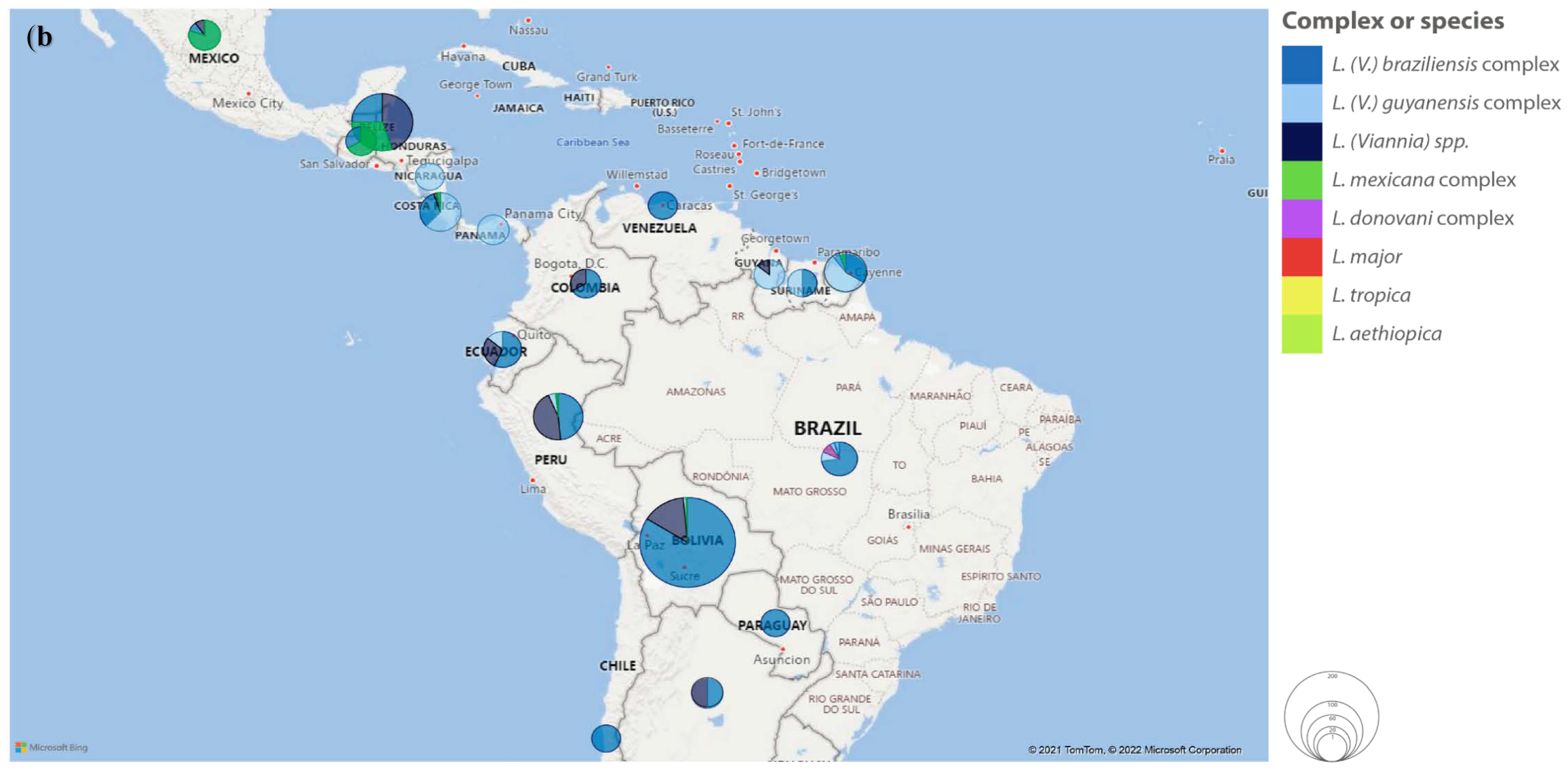
- Most CL cases were diagnosed in refugees from the Middle East, migrants from Latin America and South Asia and military personnel deployed in Asia; diagnosis relied on skin scraping and/or biopsy, with positivity rates higher for PCR than microscopy or culture; L. tropica and L. major were the two most common species in the Old World, while L. braziliensis complex and L. guyanensis complex were predominant in the New World; the number, type and location of lesions differed between species/complexes, as well as the therapeutic strategies used and the relapse rates reported; L. aethiopica, L. naiffi, L. lainsoni and L. martiniquensis infections were rarely described.
- MCL was reported in younger individuals, infected in the New World, most often by L. braziliensis complex; nasal mucosa was more often involved, and lymphadenopathy was common; the time between cutaneous and mucosal lesions varied from simultaneous to fifty years.
- ML was diagnosed mostly in older patients, infected in the Old World, most often by L. donovani complex; oral and laryngeal mucosa involvement was frequently described.
- Immunosuppressed patients represented a significant share of ML and VL cases; the two most common causes for immunosuppression were HIV/AIDS infection and chronic therapy, where anti-TNFα drugs represented the largest group; relapse/failure rates were higher in these patients.
- Non-endemic leishmaniasis represents an individual health problem, especially for refugees and immunosuppressed people; but also, a public health concern, related to the risk of introduction of the disease in new areas.
Supplementary Materials
Author Contributions
Funding
Institutional Review Board Statement
Informed Consent Statement
Conflicts of Interest
References
- Akhoundi, M.; Downing, T.; Votýpka, J.; Kuhls, K.; Lukeš, J.; Cannet, A.; Ravel, C.; Marty, P.; Delaunay, P.; Kasbari, M.; et al. Leishmania Infections: Molecular Targets and Diagnosis. Mol. Aspects Med. 2017, 57, 1–29. [Google Scholar] [CrossRef] [PubMed]
- Solano-Gallego, L.; Mirá, G.; Koutinas, A.; Cardoso, L.; Pennisi, M.G.; Ferrer, L.; Bourdeau, P.; Oliva, G.; Baneth, G. LeishVet Guidelines for the Practical Management of Canine Leishmaniosis. Parasit Vectors 2011, 4, 86. [Google Scholar] [CrossRef] [PubMed]
- WHO Global Health Observatory: Leishmaniasis. Available online: https://www.who.int/data/gho/data/themes/topics/gho-ntd-leishmaniasis (accessed on 2 March 2022).
- Leishmaniasis. Available online: https://www.who.int/news-room/fact-sheets/detail/leishmaniasis (accessed on 2 March 2022).
- Word Health Organization (WHO). Manual on Case Management and Surveillance of the Leishmaniases in the Who European Region; Word Health Organization (WHO): Geneva, Switzerland, 2017. [Google Scholar]
- Lachaud, L.; Dedet, J.P.; Marty, P.; Faraut, F.; Buffet, P.; Gangneux, J.P.; Ravel, C.; Bastien, P.; Working Group for the Notification of Human Leishmanioses in France. Surveillance of Leishmaniases in France, 1999 to 2012. Euro. surveill. 2013, 18, 20534. [Google Scholar] [CrossRef] [PubMed]
- Wall, E.C.; Watson, J.; Armstrong, M.; Chiodini, P.L.; Lockwood, D.N. Epidemiology of Imported Cutaneous Leishmaniasis at the Hospital for Tropical Diseases, London, United Kingdom: Use of Polymerase Chain Reaction to Identify the Species. Am. J. Trop. Med. Hyg. 2012, 86, 115–118. [Google Scholar] [CrossRef]
- Weitzel, T.; Mühlberger, N.; Jelinek, T.; Schunk, M.; Ehrhardt, S.; Bogdan, C.; Arasteh, K.; Schneider, T.; Kern, W.V.; Fätkenheuer, G.; et al. Imported Leishmaniasis in Germany 2001–2004: Data of the SIMPID Surveillance Network. Eur. J. Clin. Microbiol. Infect. Dis. 2005, 24, 471–476. [Google Scholar] [CrossRef]
- Fernández-Arévalo, A.; Ballart, C.; Muñoz-Basagoiti, J.; Basarte, L.; Lobato, G.; Arnau, A.; Abras, A.; Tebar, S.; Llovet, T.; Lami, P.; et al. Autochthonous and Imported Tegumentary Leishmaniasis in Catalonia (Spain): Aetiological Evolution in the Last Four Decades and Usefulness of Different Typing Approaches Based on Biochemical, Molecular and Proteomic Markers. Transbound. Emerg. Dis. 2021, 69, 1404–1418. [Google Scholar] [CrossRef]
- di Muccio, T.; Scalone, A.; Bruno, A.; Marangi, M.; Grande, R.; Armignacco, O.; Gradoni, L.; Gramiccia, M. Epidemiology of Imported Leishmaniasis in Italy: Implications for a European Endemic Country. PLoS ONE 2015, 10, e0129418. [Google Scholar] [CrossRef]
- Söbirk, S.K.; Inghammar, M.; Collin, M.; Davidsson, L. Imported Leishmaniasis in Sweden 1993-2016. Epidemiol. Infect. 2018, 146, 1267–1274. [Google Scholar] [CrossRef]
- Bart, A.; van Thiel, P.P.; de Vries, H.J.; Hodiamont, C.J.; van Gool, T. Imported Leishmaniasis in the Netherlands from 2005 to 2012: Epidemiology, Diagnostic Techniques and Sequence-Based Species Typing from 195 Patients. Euro. Surveill. 2013, 18, 20544. [Google Scholar] [CrossRef]
- Boggild, A.K.; Caumes, E.; Grobusch, M.P.; Schwartz, E.; Hynes, N.A.; Libman, M.; Connor, B.A.; Chakrabarti, S.; Parola, P.; Keystone, J.S.; et al. Cutaneous and Mucocutaneous Leishmaniasis in Travellers and Migrants: A 20-Year GeoSentinel Surveillance Network Analysis. J. Travel Med. 2019, 26, taz055. [Google Scholar] [CrossRef]
- Flores-Figueroa, J.; Okhuysen, P.C.; von Sonnenburg, F.; Dupont, H.L.; Libman, M.D.; Keystone, J.S.; Hale, D.C.; Burchard, G.; Han, P.V.; Wilder-Smith, A.; et al. Patterns of Illness in Travelers Visiting Mexico and Central America: The GeoSentinel Experience. Clin. Infect. Dis 2011, 53, 523–531. [Google Scholar] [CrossRef]
- Guery, R.; Walker, S.L.; Harms, G.; Neumayr, A.; van Thiel, P.; Gangneux, J.P.; Clerinx, J.; Söbirk, S.K.; Visser, L.; Lachaud, L.; et al. Clinical Diversity and Treatment Results in Tegumentary Leishmaniasis: A European Clinical Report in 459 Patients. PLoS Negl. Trop. Dis. 2021, 15, e0009863. [Google Scholar] [CrossRef]
- Boussery, G.; Boelaert, M.; van Peteghem, J.; Ejikon, P.; Henckaerts, K. Visceral Leishmaniasis (Kala-Azar) Outbreak in Somali Refugees and Kenyan Shepherds, Kenya. Emerg. Infect. Dis. 2001, 7, 603–604. [Google Scholar] [CrossRef]
- Chalupa, P.; Vanista, J.; Nohynkova, M. The Review of Imported Visceral Leishmaniosis in the Czech Republic. Bratisl. Lek. Listy. 2001, 102, 84–91. [Google Scholar]
- Mégarbane, B.; Bruneel, F.; Cazals-Hatem, D.; Adle-Biassette, H.; Houze, S.; Wolff, M.; Régnier, B. A Strange Disease of the Plateaux. Visceral Leishmaniasis. Rev. Med. Interne. 2001, 22 (Suppl. S2), 219–222. [Google Scholar] [CrossRef]
- Iqbal, J.; Hira, P.R.; Saroj, G.; Philip, R.; Al-Ali, F.; Madda, P.J.; Sher, A. Imported Visceral Leishmaniasis: Diagnostic Dilemmas and Comparative Analysis of Three Assays. J. Clin. Microbiol. 2002, 40, 475–479. [Google Scholar] [CrossRef]
- Harms, G.; Schönian, G.; Feldmeier, H. Leishmaniasis in Germany. Emerg. Infect. Dis. 2003, 9, 872–875. [Google Scholar] [CrossRef]
- Centers for Disease Control and Prevention (CDC). Two Cases of Visceral Leishmaniasis in U.S. Military Personnel—Afghanistan, 2002–2004. MMWR Morb. Mortal. Wkly. Rep. 2004, 53, 265–268. [Google Scholar]
- Williams, M.J.; Korsheed, S.; Goddard, A.F.; Venkatesan, P. Hepatosplenomegaly in a Patient Returning from Spain. Br. J. Hosp. Med. 2006, 67, 670. [Google Scholar] [CrossRef]
- Visser, L.; Verweij, J. Clinical Reasoning and Decision-Making in Practice. A Young Boy with Fever, Pancytopenia and an Enlarged Spleen. Ned. Tijdschr. Geneeskd. 2006, 150, 2169–2170. [Google Scholar]
- Agteresch, H.J.; van’t Veer, M.B.; Cornelissen, J.J.; Sluiters, J.F. Visceral Leishmaniasis after Allogeneic Hematopoietic Stem Cell Transplantation. Bone Marrow Transpl. 2007, 40, 391–393. [Google Scholar] [CrossRef] [PubMed]
- Hoffmann-Tonn, K.; Geis, S.; Peckelsen, C.; Berna, G.; Fleischmann, E.; Bretzel, G.; Löscher, T. Acute Febrile Disease with Splenomegaly and Pancytopenia. A 66-Year-Old Greek Patient with a Prosthetic Mitral Valve. Internist 2007, 48, 731–736. [Google Scholar] [CrossRef] [PubMed]
- Weisser, M.; Khanlari, B.; Terracciano, L.; Arber, C.; Gratwohl, A.; Bassetti, S.; Hatz, C.; Battegay, M.; Flückiger, U. Visceral Leishmaniasis: A Threat to Immunocompromised Patients in Non-Endemic Areas? Clin. Microbiol. Infect. 2007, 13, 751–753. [Google Scholar] [CrossRef] [PubMed]
- Hagenah, G.C.; Wündisch, T.; Eckstein, E.; Zimmermann, S.; Holst, F.; Grimm, W.; Neubauer, A.; Lohoff, M. Sepsis-like Disease in an Immunocompromised Patient with a Travel History to Mallorca. Internist 2007, 48, 727–730. [Google Scholar] [CrossRef]
- Stark, D.; van Hal, S.; Lee, R.; Marriott, D.; Harkness, J. Leishmaniasis, an Emerging Imported Infection: Report of 20 Cases from Australia. J. Travel Med. 2008, 15, 351–354. [Google Scholar] [CrossRef]
- Beltrame, A.; Arzese, A.; Camporese, A.; Rorato, G.; Crapis, M.; Tarabini-Castellani, G.; Boscutti, G.; Pizzolitto, S.; Calianno, G.; Matteelli, A.; et al. Acute Renal Failure Due to Visceral Leishmaniasis by Leishmania infantum Successfully Treated with a Single High Dose of Liposomal Amphotericin B. J. Travel Med. 2008, 15, 358–360. [Google Scholar] [CrossRef]
- Pérez-Ayala, A.; Norman, F.; Pérez-Molina, J.A.; Herrero, J.M.; Monge, B.; López-Vélez, R. Imported Leishmaniasis: A Heterogeneous Group of Diseases. J. Travel Med. 2009, 16, 395–401. [Google Scholar] [CrossRef]
- Neghina, R.; Neghina, A.M.; Merkler, C.; Marincu, I.; Moldovan, R.; Iacobiciu, I. Importation of Visceral Leishmaniasis in Returning Romanian Workers from Spain. Travel Med. Infect. Dis. 2009, 7, 35–39. [Google Scholar] [CrossRef]
- Buonomano, R.; Brinkmann, F.; Leupin, N.; Boscacci, R.; Zimmermann, A.; Müller, N.; Fux, C.A. Holiday Souvenirs from the Mediterranean: Three Instructive Cases of Visceral Leishmaniasis. Scand. J. Infect. Dis. 2009, 41, 777–781. [Google Scholar] [CrossRef]
- Schmid, M.B.; Leichsenring, M.; Keller, C.; Hegasy, G. Pancytopenia, Fever, and Splenomegaly in a 2-Year-Old Boy. Dtsch. Med. Wochenschr. 2009, 134, 1274–1277. [Google Scholar] [CrossRef]
- Robibaro, B.; Funk, G.C.; Dekan, G.; Demetriou, D.; Ziesche, R.; Winkler, S.; Block, L.H. Unusual Microbes in Asthma Exacerbation: Alcaligenes xylosoxidans and Leishmania. Eur. Respir. J. 2009, 33, 1216–1219. [Google Scholar] [CrossRef]
- Warich-Eitel, S.; Rauthe, S.; Eck, M. Gastric Leishmaniasis. A Rare Type of Gastritis. Pathologe 2010, 31, 205–207. [Google Scholar] [CrossRef]
- Auyeung, P.; French, M.A.; Hollingsworth, P.N. Immune Restoration Disease Associated with Leishmania donovani Infection Following Antiretroviral Therapy for HIV Infection. J. Microbiol. Immunol. Infect. 2010, 43, 74–76. [Google Scholar] [CrossRef]
- Ignatius, R.; Loddenkemper, C.; Woitzik, J.; Schneider, T.; Harms, G. Localized Leishmanial Lymphadenopathy: An Unusual Manifestation of Leishmaniasis in a Traveler in Southern Europe. Vector Borne Zoonotic Dis. 2011, 11, 1213–1215. [Google Scholar] [CrossRef]
- Moore, E.M.; Lockwood, D.N. Leishmaniasis. Clin. Med. 2011, 11, 492–497. [Google Scholar] [CrossRef]
- Benes, J.; Kabelková, M.; Holecková, P.; Kozák, T.; Benesová, K.; Mikulenková, D.; Nohýnková, E. Visceral Leishmaniasis (Two Case Reports). Klin. Mikrobiol. Infekc. Lek. 2012, 18, 43–47. [Google Scholar]
- Posch, C.; Walochnik, J.; Gschnait, A.; Feichtinger, H.; Rappersberger, K. Kala Azar-Lethal Course of Visceral Leishmaniasis. Synchronous Infection with Leishmania donovani/infantum Complex and Leishmania major in a Patient after Mediterranean Vacation. Hautarzt 2012, 63, 947–951. [Google Scholar] [CrossRef]
- Poeppl, W.; Herkner, H.; Tobudic, S.; Faas, A.; Auer, H.; Mooseder, G.; Burgmann, H.; Walochnik, J. Seroprevalence and Asymptomatic Carriage of Leishmania Spp. in Austria, a Non-Endemic European Country. Clin. Microbiol. Infect. 2013, 19, 572–577. [Google Scholar] [CrossRef]
- van Raalte, D.; Wesselius, H.; Klerk, G. Unexpected Diagnosis of Visceral Leishmaniasis in a Patient Presenting with an Infected ICD Lead. Neth. J. Med. 2014, 72, 146–148. [Google Scholar]
- Watkins, E.R.; Shamasunder, S.; Cascino, T.; White, K.L.; Katrak, S.; Bern, C.; Schwartz, B.S. Visceral Leishmaniasis-Associated Hemophagocytic Lymphohistiocytosis in a Traveler Returning from a Pilgrimage to the Camino de Santiago. J. Travel Med. 2014, 21, 429–432. [Google Scholar] [CrossRef]
- Bode, S.F.N.; Bogdan, C.; Beutel, K.; Behnisch, W.; Greiner, J.; Henning, S.; Jorch, N.; Jankofsky, M.; Jakob, M.; Schmid, I.; et al. Hemophagocytic Lymphohistiocytosis in Imported Pediatric Visceral Leishmaniasis in a Nonendemic Area. J. Pediatr. 2014, 165, 147–153. [Google Scholar] [CrossRef]
- Roberts, T.; Barratt, J.; Sandaradura, I.; Lee, R.; Harkness, J.; Marriott, D.; Ellis, J.; Stark, D. Molecular Epidemiology of Imported Cases of Leishmaniasis in Australia from 2008 to 2014. PLoS ONE 2015, 10, e0119212. [Google Scholar] [CrossRef]
- Koster, K.L.; Laws, H.J.; Troeger, A.; Meisel, R.; Borkhardt, A.; Oommen, P.T. Visceral Leishmaniasis as a Possible Reason for Pancytopenia. Front. Pediatr. 2015, 3, 59. [Google Scholar] [CrossRef]
- Schleenvoigt, B.T.; Ignatius, R.; Baier, M.; Schneider, T.; Weber, M.; Hagel, S.; Forstner, C.; Pletz, M.W. Development of Visceral Leishmaniasis in an HIV(+) Patient upon Immune Reconstitution Following the Initiation of Antiretroviral Therapy. Infection 2016, 44, 115–119. [Google Scholar] [CrossRef]
- Eichenberger, A.; Buechi, A.E.; Neumayr, A.; Hatz, C.; Rauch, A.; Huguenot, M.; Diamantis-Karamitopoulou, E.; Staehelin, C. A Severe Case of Visceral Leishmaniasis and Liposomal Amphotericin B Treatment Failure in an Immunosuppressed Patient 15 Years after Exposure. BMC Infect. Dis. 2017, 17, 81. [Google Scholar] [CrossRef]
- Asbury, K.; Seville, M.T.; Pritt, B.; Scotch, A.; Rosenthal, A.; Grys, T.E.; Kelemen, K. Closing the Brief Case: The Unexpected Souvenir. J. Clin. Microbiol. 2018, 56, e01414–e01417. [Google Scholar] [CrossRef]
- Mahendiran, T.; Doolub, G.; Nisbet, A. Fever in a Returning Traveller: Visceral Leishmaniasis Triggering Haemophagocytic Lymphohistiocytosis. BMJ Case Rep. 2018, 2018, bcr2018224775. [Google Scholar] [CrossRef]
- Haque, L.; Villanueva, M.; Russo, A.; Yuan, Y.; Lee, E.J.; Topal, J.; Podoltsev, N. A Rare Case of Visceral Leishmaniasis in an Immunocompetent Traveler Returning to the United States from Europe. PLoS Negl. Trop. Dis. 2018, 12, e0006727. [Google Scholar] [CrossRef]
- Johnson, S.M.; Gilmour, K.; Samarasinghe, S.; Bamford, A. Haemophagocytic Lymphohistiocytosis Complicating Visceral Leishmaniasis in the UK: A Case for Detailed Travel History, a High Index of Suspicion and Timely Diagnostics. BMJ Case Rep. 2019, 12, e228307. [Google Scholar] [CrossRef]
- Schaper, A.S.; Cremer, M.; Stackelberg, A.; Kallinich, T. Visceral Leishmaniasis in a Toddler Returning from Vacation in Southern Europe Presenting with Pancytopenia and Fever. Klin. Padiatr. 2019, 231, 269–270. [Google Scholar] [CrossRef]
- Blomberg, B.; Müller, K.E.; Helgeland, L.; Fladeby, C.; Mørch, K. A Man in His 80s with Arthritis and Persistent Fever. Tidsskr. Nor. Laegeforen. 2019, 139. [Google Scholar] [CrossRef]
- Douse, D.M.; Goldstein, R.S.; Montgomery, D.J.; Sinnott, M. Gastric Leishmaniasis in the Setting of HIV/AIDS Infection at Community Hospital in Southeastern United States. Access Microbiol. 2019, 1, e000045. [Google Scholar] [CrossRef] [PubMed]
- Schmutz, M.; Schaller, T.; Kubuschok, B.; Fleischmann, C.; Hirschbühl, K.; Dintner, S.; Häckel, T.; Märkl, B.; Trepel, M.; Claus, R. Periodic Fever and Pancytopenia in a 35-Year-Old Patient. Internist 2019, 60, 1305–1310. [Google Scholar] [CrossRef] [PubMed]
- Williams, E.; Isles, N.S.; Seemann, T.; Kilpatrick, T.; Grigg, A.; Leroi, M.; Howden, B.P.; Kwong, J.C. Case Report: Confirmation by Metagenomic Sequencing of Visceral Leishmaniasis in an Immunosuppressed Returned Traveler. Am. J. Trop. Med. Hyg. 2020, 103, 1930–1933. [Google Scholar] [CrossRef]
- Camilleri, M.; Richards, H.; Pomplun, S.; Wilson, A.; Checkley, A.; Rabin, N. Leishmaniasis as an Unusual Cause of Pancytopenia in a Patient Receiving Immunomodulatory Therapy for Myeloma. Br. J. Haematol. 2020, 190, 305. [Google Scholar] [CrossRef]
- Glans, H.; Hertting, O. Leishmaniasis a Neglected but Serious Tropical Disease. Lakartidningen 2020, 117, 19256. [Google Scholar]
- Offenbacher, R.; Rybinski, B.; Joseph, T.; Rahmani, N.; Boucher, T.; Weiser, D.A. An 8-Year-Old Boy With Fever, Splenomegaly, and Pancytopenia. Pediatrics 2020, 146, e20192372. [Google Scholar] [CrossRef]
- Aissaoui, N.; Hamane, S.; Gits-Muselli, M.; Petit, A.; Benderdouche, M.; Denis, B.; Alanio, A.; Dellière, S.; Bagot, M.; Bretagne, S. Imported Leishmaniasis in Travelers: A 7-Year Retrospective from a Parisian Hospital in France. BMC Infect. Dis. 2021, 21, 953. [Google Scholar] [CrossRef]
- Schwetz, V.; Trummer, C.; Friedl, C.; Beham-Schmid, C.; Kulnik, R.; Wölfler, A.; Horvath, K.; Wunsch, S.; Prattes, J.; Zollner-Schwetz, I.; et al. Visceral Leishmaniasis in a Patient with Diabetes Mellitus Type 2 and Discrete Bicytopenia. Clin. Case Rep. 2017, 6, 78–81. [Google Scholar] [CrossRef]
- Lübbert, C.; Opitz, B.M.; Harms-Zwingenberger, G.; Nietsch, H.H. Fever, Pancytopenia, and Splenomegaly 8 Months after a Trip to Majorca Island (Spain). Med. Klin 2008, 103, 29–35. [Google Scholar] [CrossRef]
- Hamilton, A.; Kelleher, A.; Marriott, D. A Case of Severe Visceral Leishmaniasis Resulting from Travel to Greece. BMJ Case Rep. 2009, 2009, bcr06.2009.2036. [Google Scholar] [CrossRef]
- Herremans, T.; Pinelli, E.; Casparie, M.; Nozari, N.; Roelfsema, J.; Kortbeek, L. Increase of Imported Leishmaniasis in the Netherlands: A Twelve Year Overview (1996–2007). Int. Health 2010, 2, 42–46. [Google Scholar] [CrossRef]
- Schwartz, T.; Jensenius, M.; Blomberg, B.; Fladeby, C.; Mæland, A.; Pettersen, F.O. Imported Visceral Leishmaniasis and Immunosuppression in Seven Norwegian Patients. Trop. Dis. Travel Med. Vaccines 2019, 5, 16. [Google Scholar] [CrossRef]
- Tzani, M.; Barrasa, A.; Vakali, A.; Georgakopoulou, T.; Mellou, K.; Pervanidou, D. Surveillance Data for Human Leishmaniasis Indicate the Need for a Sustainable Action Plan for Its Management and Control, Greece, 2004 to 2018. Euro. Surveill. 2021, 26, 2000159. [Google Scholar] [CrossRef]
- Dujardin, A.; de La Blanchardière, A.; Dina, J.; Stefic, K.; Ravel, C.; Bonhomme, J.; Verdon, R.; Fournier, A.L. Case Report: Leishmania and HIV Co-Diagnosis: How to Understand Medical History? Front. Immunol. 2021, 12, 669723. [Google Scholar] [CrossRef]
- Tan, H.H.; Wong, S.S.; Ong, B.H. Cutaneous Leishmaniasis: A Report of Two Cases Seen at a Tertiary Dermatological Centre in Singapore. Singap. Med. J. 2000, 41, 179–181. [Google Scholar]
- Brecelj, M.; Pikelj, F.; Gubenšek, F.; Anderluh, G. Polymerase Chain Reaction as a Diagnostic Tool for Detecting Leishmania. Infection 2000, 28, 111–113. [Google Scholar] [CrossRef]
- Manfredi, R.; di Bari, M.A.; Calza, L.; Chiodo, F. American Cutaneous Leishmaniasis as a Rare Imported Disease in Europe: A Case Report Favourably Treated with Antimonial Derivatives. Eur. J. Epidemiol. 2001, 17, 793–795. [Google Scholar] [CrossRef]
- Sotiropoulos, G.; Wilbur, B. Two Cases of Cutaneous Leishmaniasis Presenting to the Emergency Department as Chronic Ulcers. J. Emerg. Med. 2001, 20, 353–356. [Google Scholar] [CrossRef]
- Mackowiak, P.A.; Hatchette, T.F.; Green, P.; Schlech, W.F.; Haase, D.A.; Miller, R.; Haldane, D.J.M. Lesion on the Arm of a Returning Traveler. Cutaneous Leishmaniasis. Clin. Infect. Dis. 2001, 33, 815. [Google Scholar] [CrossRef]
- Galioto, P.; Fornaro, V. A Case of Mucocutaneous Leishmaniasis. Ear. Nose. Throat J. 2002, 81, 46–48. [Google Scholar] [CrossRef]
- Scope, A.; Trau, H.; Anders, G.; Barzilai, A.; Confino, Y.; Schwartz, E. Experience with New World Cutaneous Leishmaniasis in Travelers. J. Am. Acad. Dermatol. 2003, 49, 672–678. [Google Scholar] [CrossRef]
- Scope, A.; Trau, H.; Bakon, M.; Yarom, N.; Nasereddin, A.; Schwartz, E. Imported Mucosal Leishmaniasis in a Traveler. Clin. Infect. Dis. 2003, 37, 83–87. [Google Scholar] [CrossRef]
- Lawn, S.D.; Yardley, V.; Vega-Lopez, F.; Watson, J.; Lockwood, D.N. New World Cutaneous Leishmaniasis in Returned Travellers: Treatment Failures Using Intravenous Sodium Stibogluconate. Trans. R. Soc. Trop. Med. Hyg. 2003, 97, 443–445. [Google Scholar] [CrossRef]
- Haider, S.; Boutross-Tadross, O.; Radhi, J.; Momar, N. Cutaneous Ulcer in a Man Returning from Central America. CMAJ 2003, 4, 591. [Google Scholar]
- Bormann, G.; William, T.; Schulz, A.; Marsch, W.; Gaber, G. American Cutaneous Leishmaniasis: Special Features in Diagnosis and Therapy. Dtsch. Med. Wochenschr. 2003, 128, 2065–2068. [Google Scholar] [CrossRef]
- Costa, J.W.; Milner, D.A.; Maguire, J.H. Mucocutaneous Leishmaniasis in a US Citizen. Oral. Sur.g Oral. Med. Oral. Pathol. Oral. Radiol. Endod. 2003, 96, 573–577. [Google Scholar] [CrossRef]
- el Hajj, L.; Thellier, M.; Carrière, J.; Bricaire, F.; Danis, M.; Caumes, E. Localized Cutaneous Leishmaniasis Imported into Paris: A Review of 39 Cases. Int. J. Dermatol. 2004, 43, 120–125. [Google Scholar] [CrossRef]
- Lawn, S.D.; Whetham, J.; Chiodini, P.L.; Kanagalingam, J.; Watson, J.; Behrens, R.H.; Lockwood, D.N.J. New World Mucosal and Cutaneous Leishmaniasis: An Emerging Health Problem among British Travellers. QJM 2004, 97, 781–788. [Google Scholar] [CrossRef]
- Morelli, P.; Gianelli, E.; Calattini, S.; Corbellino, M.; Antinori, S.; Meroni, L. Itraconazole Can Be Effective in the Treatment of Sporotrichoid Leishmaniasis. J. Travel Med. 2004, 11, 328–330. [Google Scholar] [CrossRef]
- Cardenas, G.A.; Gonzalez-Serva, A.; Cohen, C. Multiple Leg Ulcers in a Traveler. Cleve Clin. J. Med. 2004, 71, 109–112. [Google Scholar] [CrossRef] [PubMed]
- Storer, E.; Wayte, J. Cutaneous Leishmaniasis in Afghani Refugees. Australas. J. Derm. 2005, 46, 80–83. [Google Scholar] [CrossRef] [PubMed]
- Raschke, A.; Wetzig, T.; Paasch, U.; Grunewald, S.; Simon, J.C. Firm Blue-Red Solid Nodules on the Forehead of a 39-Year-Old Patient. J. Dtsch. Dermatol. Ges. 2005, 3, 919–920. [Google Scholar] [CrossRef] [PubMed]
- Loo, W.J.; Chan, S.K.; Rytina, E.; Lockwoood, D.N.J.; Sterling, J.C.; Todd, P. Five Cases of Cutaneous Leishmaniasis in Cambridge, U.K. Br. J. Dermatol. 2005, 153, 1076–1078. [Google Scholar] [CrossRef]
- Antonovich, D.D.; Callen, J.P.; Paniker, P.U. No Walk in the Park. Am. J. Med. 2005, 118, 715–716. [Google Scholar] [CrossRef]
- Mateo, M.; Cruz, I.; Flores, M.D.; López-Vélez, R. Slowly Progressing Skin Ulcers Following a Stay in Costa Rica. Enferm. Infecc. Y Microbiol. Clin. 2005, 23, 243–244. [Google Scholar] [CrossRef]
- Zeegelaar, J.E.; Steketee, W.H.; van Thiel, P.P.A.M.; Wetsteyn, J.C.F.M.; Kager, P.A.; Faber, W.R. Changing Pattern of Imported Cutaneous Leishmaniasis in the Netherlands. Clin. Exp. Dermatol. 2005, 30, 1–5. [Google Scholar] [CrossRef]
- Scarisbrick, J.J.; Chiodini, P.L.; Watson, J.; Moody, A.; Armstrong, M.; Lockwood, D.; Bryceson, A.; Vega-López, F. Clinical Features and Diagnosis of 42 Travellers with Cutaneous Leishmaniasis. Travel Med. Infect. Dis. 2006, 4, 14–21. [Google Scholar] [CrossRef]
- Vinetz, J.M.; Soong, L. Leishmania Mexicana Infection of the Eyelid in a Traveler to Belize. Braz. J. Infect. Dis. 2007, 11, 149–152. [Google Scholar] [CrossRef]
- Ismailjee, S.B.; Bernstein, J.M.; Burdette, S.D. Bite the Hand That Sprays You. Skinmed 2006, 5, 296–299. [Google Scholar] [CrossRef]
- Ather, S.; Chan, D.S.; Leaper, D.J.; Harding, K.G. Case Report and Literature Review of Leishmaniasis as a Cause of Leg Ulceration in the United Kingdom. J. Wound Care 2006, 15, 389–391. [Google Scholar] [CrossRef]
- Morizot, G.; Delgiudice, P.; Caumes, E.; Laffitte, E.; Marty, P.; Dupuy, A.; Sarfati, C.; Hadj-Rabia, S.; Darie, H.; le Guern, A.S.; et al. Healing of Old World Cutaneous Leishmaniasis in Travelers Treated with Fluconazole: Drug Effect or Spontaneous Evolution? Am. J. Trop. Med. Hyg. 2007, 76, 48–52. [Google Scholar] [CrossRef]
- Schnedl, J.; Auer, H.; Fischer, M.; Tomaso, H.; Pustelnik, T.; Mooseder, G. Cutaneous Leishmaniasis—An Import from Belize. Wien. Klin. Wochenschr. 2007, 119, 102–105. [Google Scholar] [CrossRef]
- Campos-Muñoz, L.; Quesada-Cortés, A.; Martín-Díaz, M.A.; Rubio-Flores, C.; de Lucas-Laguna, R. Leishmania braziliensis: Report of a Pediatric Imported Case With Response to Liposomal Amphotericin B. Actas Dermosifiliogr. 2007, 98, 42–44. [Google Scholar] [CrossRef]
- Konecny, P.; Stark, D.J. An Australian Case of New World Cutaneous Leishmaniasis. Med. J. Aust. 2007, 186, 315–317. [Google Scholar] [CrossRef]
- González-Llavona, B.; Biosca-Echenique, G.; Soto-Díaz, A.; Naranjo-Díaz, M.J.; Espadafor-López, B.; García-Mellado, V. Cutaneous Leishmaniasis in a Senegal Patient. Actas Dermo. Sifiliogr. 2007, 98, 54–58. [Google Scholar] [CrossRef]
- Solomon, M.; Baum, S.; Barzilai, A.; Scope, A.; Trau, H.; Schwartz, E. Liposomal Amphotericin B in Comparison to Sodium Stibogluconate for Cutaneous Infection Due to Leishmania braziliensis. J. Am. Acad. Dermatol. 2007, 56, 612–616. [Google Scholar] [CrossRef]
- Delgado, O.; Silva, S.; Coraspe, V.; Rivas, M.A.; Rodriguez-Morales, A.J.; Navarro, P.; Franco-Paredes, C. Cutaneous Leishmaniasis Imported from Colombia to Northcentral Venezuela: Implications for Travel Advice. Travel Med. Infect. Dis. 2008, 6, 376–379. [Google Scholar] [CrossRef]
- Schleucher, R.D.; Zanger, P.; Gaessler, M.; Knobloch, J. Successful Diagnosis and Treatment 50 Years after Exposure: Is Mucocutaneous Leishmaniasis Still a Neglected Differential Diagnosis? J. Travel Med. 2008, 15, 466–467. [Google Scholar] [CrossRef]
- Ahmed, Z.; Chowdhury, S.; Bhuiyan, S. Cutaneous Leishmaniasis. Mymensingh Med. J. 2009, 18, 260–263. [Google Scholar]
- Berens-Riha, N.; Fleischmann, E.; Pratlong, F.; Bretzel, G.; von Sonnenburg, F.; Löscher, T. Cutaneous Leishmaniasis (Leishmania tropica) in a German Tourist after Travel to Greece. J. Travel Med. 2009, 16, 220–222. [Google Scholar] [CrossRef]
- Holmes, W.J.M.; Tehrani, H.; Liew, S. Cutaneous Leishmaniasis: A Diagnosis of Suspicion. J. Hand Surg. Eur. Vol. 2009, 34, 555–556. [Google Scholar] [CrossRef]
- van der Snoek, E.M.; Lammers, A.M.; Kortbeek, L.M.; Roelfsema, J.H.; Bart, A.; Jaspers, C.A.J.J. Spontaneous Cure of American Cutaneous Leishmaniasis Due to Leishmania naiffi in Two Dutch Infantry Soldiers. Clin. Exp. Dermatol. 2009, 34, e889–91. [Google Scholar] [CrossRef]
- González, M.; Benito, F.; García, L.; Iglesias, A. Mucocutaneous Leishmaniasis: An Imported Illness with ENT Repercussions. Acta Otorrinolaringol. Esp. 2009, 60, 298–300. [Google Scholar] [CrossRef]
- Jeddi, F.; Caumes, E.; Thellier, M.; Jauréguiberry, S.; Mazier, D.; Buffet, P.A. Drug Hypersensitivity Syndrome Induced by Meglumine Antimoniate. Am. J. Trop. Med. Hyg. 2009, 80, 939–940. [Google Scholar] [CrossRef]
- Santangeli, L.; McCluney, N.A.; Hathorn, I.; Shakeel, M.; Anderson, C. Leishmaniasis Presenting to the Otolaryngologist: A Rare but Important Cause of Persistent Hoarseness. J. Laryngol. Otol. 2009, 123, 1181–1183. [Google Scholar] [CrossRef]
- Mulvaney, P.; Aram, G.; Maggiore, P.R.; Kutzner, H.; Carlson, J.A. Delay in Diagnosis: Trauma-and Coinfection-Related Cutaneous Leishmaniasis Because of Leishmania guyanensis Infection. J. Cutan. Pathol. 2009, 36, 53–60. [Google Scholar] [CrossRef]
- Leitner, V.; Weingast, J.; Harmankaya, K.; Walochnik, J.; Pehamberger, H.; Petzelbauer, P.; Auer, H.; Binder, M. Leishmaniasis in the Tongue of an Immunocompetent Man. Am. J. Trop. Med. Hyg. 2010, 82, 597–599. [Google Scholar] [CrossRef] [PubMed]
- van Thiel, P.P.A.M.; Leenstra, T.; Kager, P.A.; de Vries, H.J.; van Vugt, M.; van der Meide, W.F.; Bart, A.; Zeegelaar, J.E.; van der Sluis, A.; Schallig, H.D.F.H.; et al. Miltefosine Treatment of Leishmania major Infection: An Observational Study Involving Dutch Military Personnel Returning from Northern Afghanistan. Clin. Infect. Dis. 2010, 50, 80–83. [Google Scholar] [CrossRef] [PubMed]
- Marovt, M.; Kokol, R.; Stanimirović, A.; Miljković, J. Cutaneous Leishmaniasis: A Case Report. Acta Dermatovenerol. Alp. Pannonica. Adriat. 2010, 19, 41–43. [Google Scholar] [PubMed]
- Mazumder, S.A.; Pandey, S.; Brewer, S.C.; Baselski, V.S.; Weina, P.J.; Land, M.A.; Fleckenstein, J.M. Lingual Leishmaniasis Complicating Visceral Disease. J. Travel Med. 2010, 17, 212–214. [Google Scholar] [CrossRef]
- Crogan, J.; Gunasekera, H.; Wood, N.; Sheikh, M.; Isaacs, D. Management of Old World Cutaneous Leishmaniasis in Refugee Children. Pediatr. Infect. Dis. J. 2010, 29, 357–359. [Google Scholar] [CrossRef]
- Harms, G.; Scherbaum, H.; Reiter-Owona, I.; Stich, A.; Richter, J. Treatment of Imported New World Cutaneous Leishmaniasis in Germany. Int. J. Dermatol. 2011, 50, 1336–1342. [Google Scholar] [CrossRef]
- Cannella, A.P.; Nguyen, B.M.; Piggott, C.D.; Lee, R.A.; Vinetz, J.M.; Mehta, S.R. A Cluster of Cutaneous Leishmaniasis Associated with Human Smuggling. Am. J. Trop. Med. Hyg. 2011, 84, 847–850. [Google Scholar] [CrossRef]
- Solomon, M.; Benenson, S.; Baum, S.; Schwartz, E. Tropical Skin Infections among Israeli Travelers. Am. J. Trop. Med. Hyg. 2011, 85, 868–872. [Google Scholar] [CrossRef]
- Zanger, P.; Kötter, I.; Raible, A.; Gelanew, T.; Schönian, G.; Kremsner, P.G. Case Report: Successful Treatment of Cutaneous Leishmaniasis Caused by Leishmania aethiopica with Liposomal Amphothericin B in an Immunocompromised Traveler Returning from Eritrea. Am. J. Trop. Med. Hyg. 2011, 84, 692–694. [Google Scholar] [CrossRef]
- Zaghi, D.; Panosian, C.; Gutierrez, M.A.; Gregson, A.; Taylor, E.; Ochoa, M.T. New World Cutaneous Leishmaniasis: Current Challenges in Diagnosis and Parenteral Treatment. J. Am. Acad. Dermatol. 2011, 64, 587–592. [Google Scholar] [CrossRef]
- Mougin, B.; Avenel-Audran, M.; Hasseine, L.; Martin, L.; Cottin, J.; Pomares, C.; Delaunay, P.; Marty, P.; Ravel, C.; Chabasse, D.; et al. A Cutaneous Ulcer Resulting from Mycobacterium ulcerans–Leishmania braziliensis Coinfection in South America. Am. J. Trop. Med. Hyg. 2011, 85, 897–899. [Google Scholar] [CrossRef]
- Blaylock, J.M.; Wortmann, G.W. A Case Report and Literature Review of “Chiclero’s Ulcer”. Travel Med. Infect. Dis. 2012, 10, 275–278. [Google Scholar] [CrossRef]
- Kelly, P.; Baudry, T.; Peyron, F. Imported Cutaneous Leishmaniasis in a Short-Term Traveler Returning from Central Mali-The Role of PCR. Travel Med. Infect. Dis. 2012, 10, 97–100. [Google Scholar] [CrossRef]
- Terhorst, D.; Blume-Peytavi, U.; Schönian, G.; Schewe, C.; Haas, N.; Burbach, G.J. Leishmaniasis: A Reminder in the Face of Forgotten Travel. J. Pediatr. 2012, 161, 966. [Google Scholar] [CrossRef]
- Wise, E.S.; Armstrong, M.S.; Watson, J.; Lockwood, D.N. Monitoring Toxicity Associated with Parenteral Sodium Stibogluconate in the Day-Case Management of Returned Travellers with New World Cutaneous Leishmaniasis. PLoS Negl. Trop. Dis. 2012, 6, e1688. [Google Scholar] [CrossRef]
- Poeppl, W.; Oeser, C.; Grabmeier-Pfistershammer, K.; Walochnik, J.; Burgmann, H. Clinical Findings and Management of Imported Cutaneous Leishmaniasis: Report of 14 Cases from Austria. Travel Med. Infect. Dis. 2013, 11, 90–94. [Google Scholar] [CrossRef]
- Fathi, R.; Fathi, A. A Case of Cutaneous Leishmaniasis Found in Indiana. Derm. Online J. 2013, 19, 18982. [Google Scholar] [CrossRef]
- Demers, E.; Forrest, D.M.; Weichert, G.E. Cutaneous Leishmaniasis in a Returning Traveller. CMAJ 2013, 185, 681–683. [Google Scholar] [CrossRef]
- Eichner, S.; Thoma-Uszynski, S.; Herrgott, I.; Sebald, H.; Debus, A.; Tsianakas, A.; Ehrchen, J.; Harms, G.; Simon, M.; Sunderkötter, C.; et al. Clinical Complexity of Leishmania (Viannia) braziliensis Infections amongst Travelers. Eur. J. Derm. 2013, 23, 218–223. [Google Scholar] [CrossRef]
- Teemul, T.A.; Giles-Lima, M.; Williams, J.; Lester, S.E. Laryngeal Leishmaniasis: Case Report of a Rare Infection. Head Neck 2013, 35, E277–E279. [Google Scholar] [CrossRef]
- Veraldi, S.; Tavecchio, S. Gifts from the Tropics. Int. J. Derm. 2013, 52, 896–897. [Google Scholar] [CrossRef]
- Neumayr, A.L.C.; Morizot, G.; Visser, L.G.; Lockwood, D.N.J.; Beck, B.R.; Schneider, S.; Bellaud, G.; Cordoliani, F.; Foulet, F.; Laffitte, E.A.; et al. Clinical Aspects and Management of Cutaneous Leishmaniasis in Rheumatoid Patients Treated with TNF-α Antagonists. Travel Med. Infect. Dis. 2013, 11, 412–420. [Google Scholar] [CrossRef] [PubMed]
- Mosimann, V.; Neumayr, A.; Hatz, C.; Blum, J.A. Cutaneous Leishmaniasis in Switzerland: First Experience with Species-Specific Treatment. Infection 2013, 41, 1177–1182. [Google Scholar] [CrossRef] [PubMed]
- Larréché, S.; Launay, G.; Weibel Galluzzo, C.; Bousquet, A.; Eperon, G.; Pilo, J.E.; Ravel, C.; Chappuis, F.; Dupin, M.; Mérens, A. Cluster of Zoonotic Cutaneous Leishmaniasis (Leishmania major) in European Travelers Returning from Turkmenistan. J. Travel Med. 2013, 20, 400–402. [Google Scholar] [CrossRef] [PubMed]
- Ehlert, N.; Seilmaier, M.; Guggemos, W.; Löscher, T.; Meurer, A.; Wendtner, C.M. Severe Oral Mucositis in a Patient with HIV Infection. Dtsch. Med. Wochenschr. 2013, 138, 1601–1605. [Google Scholar] [CrossRef] [PubMed]
- Lavergne, R.A.; Iriart, X.; Martin-Blondel, G.; Chauvin, P.; Menard, S.; Fillaux, J.; Cassaing, S.; Roques-Malecaze, C.; Arnaud, S.; Valentin, A.; et al. Contribution of Molecular Diagnosis to the Management of Cutaneous Leishmaniasis in Travellers. Clin. Microbiol. Infect. 2014, 20, O528–O530. [Google Scholar] [CrossRef]
- van Hees, C.; van Hellemond, J.; den Boer, M. A Boy with an Unexpected Souvenir from Morocco. Ned. Tijdschr. Voor Geneeskd. 2014, 158, A7606. [Google Scholar]
- Ntais, P.; Christodoulou, V.; Tsirigotakis, N.; Dokianakis, E.; Dedet, J.P.; Pratlong, F.; Antoniou, M. Will the Introduction of Leishmania tropica MON-58, in the Island of Crete, Lead to the Settlement and Spread of This Rare Zymodeme? Acta Trop. 2014, 132, 125–130. [Google Scholar] [CrossRef]
- Barry, M.A.; Koshelev, M.V.; Sun, G.S.; Grekin, S.J.; Stager, C.E.; Diwan, A.H.; Wasko, C.A.; Murray, K.O.; Woc-Colburn, L. Cutaneous Leishmaniasis in Cuban Immigrants to Texas Who Traveled through the Darién Jungle, Panama. Am. J. Trop. Med Hyg. 2014, 91, 345–347. [Google Scholar] [CrossRef]
- Raghunath, R.S.; Yong, A.S.W.; Igali, L.; Tan, E.; Lockwood, D. Cutaneous Leishmaniasis in a Returning UK Traveller. Postgrad. Med. J. 2014, 90, 540–541. [Google Scholar] [CrossRef]
- Trufant, J.; Lewin, J.; Christopher, H.; Meehan, S.; Pomeranz, M. New World Cutaneous Leishmaniasis. Derm. Online J. 2014, 20, 13030. [Google Scholar] [CrossRef]
- Downing, C.P.; Woc-Colburn, L.; Tyring, S.K. Nasal Erythema and Crusting after a Trip to the Venezuelan Rainforest. JAMA 2014, 312, 1250–1251. [Google Scholar] [CrossRef]
- Siah, T.; Lavender, T.; Charlton, F.; Wahie, S.; Schwab, U. An Unusual Erysipelas-like Presentation. Derm. Online J. 2014, 20, 21255. [Google Scholar] [CrossRef]
- Ito, K.; Takahara, M.; Ito, M.; Oshiro, M.; Takahashi, K.; Uezato, H.; Imafuku, S. An Imported Case of Cutaneous Leishmaniasis Caused by Leishmania (Leishmania) donovani in Japan. J. Dermatol. 2014, 41, 926–928. [Google Scholar] [CrossRef]
- Rahman, H.; Razzak, M.; Chanda, B.; Bhaskar, K.; Mondal, D. Cutaneous Leishmaniasis in an Immigrant Saudi Worker: A Case Report. J. Health Popul. Nutr. 2014, 32, 372–376. [Google Scholar]
- Nadler, C.; Enk, C.D.; Leon, G.T.; Samuni, Y.; Maly, A.; Czerninski, R. Diagnosis and Management of Oral Leishmaniasis—Case Series and Literature Review. J. Oral. Maxillofac. Surg. 2014, 72, 927–934. [Google Scholar] [CrossRef]
- Bailey, M.S.; Langman, G. Misdiagnosis of Cutaneous Leishmaniasis and Recurrence after Surgical Excision. J. R Army Med. Corps 2014, 160, 314–316. [Google Scholar] [CrossRef]
- Calderaro, A.; Montecchini, S.; Rossi, S.; Gorrini, C.; Dell’Anna, M.L.; Piccolo, G.; Medici, M.C.; Arcangeletti, M.C.; Chezzi, C.; de Conto, F. A 22-Year Survey of Leishmaniasis Cases in a Tertiary-Care Hospital in an Endemic Setting. Int. J. Environ. Res. Public Health 2014, 11, 2834–2845. [Google Scholar] [CrossRef]
- Tan, E.M.; Marcelin, J.R.; Virk, A. Photo Quiz: A 24-Year-Old Traveler with an Insect Bite and Rash. Clin. Infect. Dis. 2015, 61, 1314. [Google Scholar] [CrossRef]
- Cohen, J.; Saavedra, A.; Sax, P.; Lipworth, A. Pink Plaque on the Arm of a Man after a Trip to Mexico: Cutaneous Leishmaniasis. Dermatol. Online J. 2015, 21, 13030. [Google Scholar] [CrossRef]
- Vanbrabant, P.; van den Broucke, S.; Soentjens, P. Cutaneous Ulcer after a Stay in the Tropics. Neth. J. Med. 2015, 73, 44. [Google Scholar]
- Wollina, U.; Koch, A.; Schönlebe, J.; Tchernev, G.; Chokoeva, A.; Lotti, T. Non-Healing Facial Lesions: Cutaneous Old World Leishmaniasis in Dresden. J. Biol. Regul. Homeost. Agents 2015, 29, 99–102. [Google Scholar]
- Menten, K.; Soentjens, P.; Caenepeel, P.; Lemkens, P. Mucocutaneous Leishmaniasis of the Nose: A Case Report. B-ENT 2015, 11, 77–80. [Google Scholar]
- Crovetto-Martínez, R.; Aguirre-Urizar, J.M.; Orte-Aldea, C.; Araluce-Iturbe, I.; Whyte-Orozco, J.; Crovetto-De La Torre, M.A. Mucocutaneous Leishmaniasis Must Be Included in the Differential Diagnosis of Midline Destructive Disease: Two Case Reports. Oral. Surg. Oral. Med. Oral. Pathol. Oral. Radiol. 2015, 119, e20–e26. [Google Scholar] [CrossRef] [PubMed]
- Roberts, R.M.; Mukherjee, J.; Phillips, D. Laryngeal Leishmaniasis in a Patient Taking Inhaled Corticosteroids. BMJ Case Rep. 2016, 2016, bcr2016215444. [Google Scholar] [CrossRef] [PubMed]
- Zhang, M.; Liu, F.; Liu, H.B.; Hu, W.X.; Sang, H. Imported Cutaneous Leishmaniasis Caused by Leishmania major in a Chinese Laborer Who Worked in Saudi Arabia. An. Bras. Dermatol. 2016, 91, 365–367. [Google Scholar] [CrossRef]
- Bradshaw, S.; Litvinov, I.V. Dermal Leishmaniasis in a 25-Year-Old Syrian Refugee. CMAJ 2017, 189, E1397. [Google Scholar] [CrossRef]
- Patel, T.A.; Scadding, G.K.; Phillips, D.E.; Lockwood, D.N. Case Report: Old World Mucosal Leishmaniasis: Report of Five Imported Cases to the Hospital for Tropical Diseases, London, United Kingdom. Am. J. Trop. Med. Hyg. 2017, 97, 1116–1119. [Google Scholar] [CrossRef]
- Goodrich, E.S.; Sears, S.C.; Sorrells, T.; Radike, J.K.; Miladi, A.; Glass, J.S. A Case of Cutaneous Leishmaniasis guyanensis Mimicking Otitis Externa. Mil. Med. 2017, 182, e1969–e1972. [Google Scholar] [CrossRef]
- Basher, A.; Nath, P.; Dey, T.; Sayeed, A.A.; Faiz, M.A.; Chowdhury, F.R. Cutaneous Leishmaniasis in Immigrant Workers Returning to Bangladesh—An Emerging Problem. Travel Med. Infect. Dis. 2017, 19, 62–63. [Google Scholar] [CrossRef]
- Harrison, N.; Walochnik, J.; Ramsebner, R.; Veletzky, L.; Lagler, H.; Ramharter, M. Case Report: Progressive Perforation of the Nasal Septum Due to Leishmania major: A Case of Mucosal Leishmaniasis in a Traveler. Am. J. Trop. Med. Hyg. 2017, 96, 653–655. [Google Scholar] [CrossRef]
- van der Snoek, E.M.; Couwenberg, S.M.; Stijnis, C.; Kortbeek, L.M.; Schadd, E.M. Two Cases of Cutaneous Leishmaniasis in Dutch Military Personnel Treated with Oral Miltefosine. J. R Army Med. Corps 2017, 163, 68–70. [Google Scholar] [CrossRef]
- Challener, D.; Abu Saleh, O. A Traveler’s Unwanted Souvenir. Am. J. Med. 2018, 131, e137–e138. [Google Scholar] [CrossRef]
- Knöpfel, N.; Noguera-Morel, L.; Azorin, D.; Sanz, F.; Torrelo, A.; Hernández-Martín, A. Cutaneous Leishmania Tropica in Children: Report of Three Imported Cases Successfully Treated with Liposomal Amphotericin B. J. Eur. Acad. Dermatol. Venereol. 2018, 32, e8–e10. [Google Scholar] [CrossRef] [PubMed]
- Clegg, A.; Hall, C.; Lockwood, D. An Ulcer That Will Not Heal. BMJ 2018, 362, k3042. [Google Scholar] [CrossRef] [PubMed]
- Montalvo, A.M.; Fraga, J.; Blanco, O.; González, D.; Monzote, L.; Soong, L.; Capó, V. Imported Leishmaniasis Cases in Cuba (2006–2016): What Have We Learned. Trop. Dis. Travel Med. Vaccines 2018, 4, 7. [Google Scholar] [CrossRef]
- Imai, K.; Tarumoto, N.; Amo, K.; Takahashi, M.; Sakamoto, N.; Kosaka, A.; Kato, Y.; Mikita, K.; Sakai, J.; Murakami, T.; et al. Non-Invasive Diagnosis of Cutaneous Leishmaniasis by the Direct Boil Loop-Mediated Isothermal Amplification Method and MinIONTM Nanopore Sequencing. Parasitol. Int. 2018, 67, 34–37. [Google Scholar] [CrossRef]
- Islam, S. Rapidly Progressing Facial Leishmaniasis: Effective Treatment with Liposomal Amphotericin B and a Review of the Management of Old World Cutaneous Leishmaniasis. Paediatr. Int. Child Health 2018, 38, 158–161. [Google Scholar] [CrossRef]
- Michelerio, A.; Barruscotti, S.; Bossi, G.; Brazzelli, V. Pediatric Old World Cutaneous Leishmaniasis Treated with Oral Fluconazole: A Case Series. Pediatr. Dermatol. 2018, 35, 384–387. [Google Scholar] [CrossRef]
- Navarrete-Dechent, C.; Cevallos, C.; Jercic, M.I.; Saldias-Fuentes, C.; González, S.; Labarca, J. Liposomal Amphotericin B Treatment of Cutaneous Leishmaniasis Caused by L. braziliensis: An Imported Case Report. Rev. Chil. Infectol. 2018, 35, 612–616. [Google Scholar] [CrossRef]
- Imessaoudene, L.; Jacobs, C.; Hunt, W. Cutaneous Leishmaniasis in a Globetrotting Explorer. BMJ Case Rep. 2019, 12. [Google Scholar] [CrossRef]
- Hijawi, K.J.F.; Hijjawi, N.S.; Ibbini, J.H. Detection, Genotyping, and Phylogenetic Analysis of Leishmania Isolates Collected from Infected Jordanian Residents and Syrian Refugees Who Suffered from Cutaneous Leishmaniasis. Parasitol. Res. 2019, 118, 793–805. [Google Scholar] [CrossRef]
- Solomon, M.; Sahar, N.; Pavlotzky, F.; Barzilai, A.; Jaffe, C.L.; Nasereddin, A.; Schwartz, E. Mucosal Leishmaniasis in Travelers with Leishmania braziliensis Complex Returning to Israel. Emerg. Infect. Dis. 2019, 25, 642–648. [Google Scholar] [CrossRef]
- Khan, M.A.A.; Chowdhury, R.; Nath, R.; Hansen, S.; Nath, P.; Maruf, S.; Abd El Wahed, A.; Mondal, D. Imported Cutaneous Leishmaniasis: Molecular Investigation Unveils Leishmania major in Bangladesh. Parasit Vectors 2019, 12. [Google Scholar] [CrossRef]
- Taxy, J.B.; Goldin, H.M.; Dickie, S.; Cibull, T. Cutaneous Leishmaniasis: Contribution of Routine Histopathology in Unexpected Encounters. Am. J. Surg. Pathol. 2019, 43, 195–200. [Google Scholar] [CrossRef]
- Fernández-Figueroa, E.A.; Sánchez-Montes, S.; Miranda-Ortíz, H.; Mendoza-Vargas, A.; Cervantes-Sarabia, R.; Cárdenas-Ovando, R.A.; Ruiz-Remigio, A.; Becker, I. Relevance of Epidemiological Surveillance in Travelers: An Imported Case of Leishmania tropica in Mexico. Rev. Inst. Med. Trop. S. Paulo 2020, 62, 1–5. [Google Scholar] [CrossRef]
- Crone, C.G.; Helleberg, M. Cutaneous Leishmaniasis with Secondary Mucosal Disease in a Traveller Due to Leishmania (Viannia) braziliensis. J. Travel Med. 2020, 27. [Google Scholar] [CrossRef]
- Mann, S.; Phupitakphol, T.; Davis, B.; Newman, S.; Suarez, J.A.; Henao-Martínez, A.; Franco-Paredes, C. Case Report: Cutaneous Leishmaniasis Due to Leishmania (Viannia) panamensis in Two Travelers Successfully Treated with Miltefosine. Am. J. Trop. Med. Hyg. 2020, 103, 1081–1084. [Google Scholar] [CrossRef]
- Basile, G.; Cristofaro, G.; Locatello, L.G.; Vellere, I.; Piccica, M.; Bresci, S.; Maggiore, G.; Gallo, O.; Novelli, A.; di Muccio, T.; et al. Refractory Mucocutaneous Leishmaniasis Resolved with Combination Treatment Based on Intravenous Pentamidine, Oral Azole, Aerosolized Liposomal Amphotericin B, and Intralesional Meglumine Antimoniate. Int. J. Infect. Dis. 2020, 97, 204–207. [Google Scholar] [CrossRef]
- Murray, H.W.; Eiras, D.P.; Kirkman, L.A.; Chai, R.L.; Caplivski, D. Case Report: Mucosal Leishmaniasis in New York City. Am. J. Trop. Med. Hyg. 2020, 102, 1319–1322. [Google Scholar] [CrossRef]
- Talas, J.; Mielcarek, K.; Wu, J.; Brunner, M.; Steinhoff, M.; Zouboulis, C.C. Cutaneous Leishmaniasis in Germany-Still a Travel-Related Disease. Der Hautarzt Z. Fur Dermatol. Venerol. Und Verwandte Geb. 2022, 73, 146–151. [Google Scholar] [CrossRef]
- Abadir, A.; Patel, A.; Haider, S. Systemic Therapy of New World Cutaneous Leishmaniasis: A Case Report and Review Article. Can. J. Infect. Dis. Med Microbio.l 2010, 21, e79–e83. [Google Scholar] [CrossRef]
- Madke, B.; Kharkar, V.; Chikhalkar, S.; Mahajan, S.; Khopkar, U. Successful Treatment of Multifocal Cutaneous Leishmaniasis with Miltefosine. Indian J. Dermatol. 2011, 56, 587–590. [Google Scholar] [CrossRef]
- Shin, J.Y.; Lee, Y.B.; Cho, B.K.; Park, H.J. New World Cutaneous Leishmaniasis Treated with Intralesional Injection of Pentavalent Antimony. Ann. Dermatol. 2013, 25, 80–83. [Google Scholar] [CrossRef] [PubMed]
- Kuna, A.; Gajewski, M.; Bykowska, M.; Pietkiewicz, H.; Olszański, R.; Myjak, P. Imported Cutaneous Leishmaniasis: A 13-Year Experience of a Polish Tertiary Center. Postepy Derm. Alergol. 2019, 36, 104–111. [Google Scholar] [CrossRef] [PubMed]
- Tsai, P.H.; Chen, Y.T.; Liau, J.Y.; Huang, M.H.; Hsu, H.M.; Yeong, E.K.; Hung, C.C. Molecular Diagnosis and Therapy for Cutaneous Leishmaniasis of a Returned Traveler from Mexico. J. Microbiol. Immunol. Infect 2021, 54, 1154–1158. [Google Scholar] [CrossRef] [PubMed]
- Kitano, H.; Sanjoba, C.; Goto, Y.; Iwamoto, K.; Kitagawa, H.; Nomura, T.; Omori, K.; Shigemoto, N.; Hide, M.; Matsumoto, Y.; et al. Complicated Cutaneous Leishmaniasis Caused by an Imported Case of Leishmania tropica in Japan: A Case Report. Trop. Med. Health 2021, 49, 20. [Google Scholar] [CrossRef]
- Bizri, N.A.; Alam, W.; Khoury, M.; Musharrafieh, U.; Ghosn, N.; Berri, A.; Bizri, A.R. The Association Between the Syrian Crisis and Cutaneous Leishmaniasis in Lebanon. Acta Parasitol. 2021, 66, 1240–1245. [Google Scholar] [CrossRef]
- Eldin, C.; L’Ollivier, C.; Ranque, S.; Gautret, P.; Parola, P. “Chiclero’s Ulcer” Due to Leishmania mexicana in Travelers Returning from Central America: A Case Report and Review of the Literature. Pathogens 2021, 10, 1112. [Google Scholar] [CrossRef]
- Chong, G.L.M.; Ong, D.S.; de Mendonça Melo, M.; van Hellemond, J.J. Painful and Swollen Tongue: Mucosal Leishmaniasis Due to Leishmania infantum. Int. J. Infect. Dis. 2021, 113, 109–112. [Google Scholar] [CrossRef]
- Iannone, M.; Oranges, T.; Dini, V.; Romanelli, M.; Janowska, A. Wound Management Strategy for Treatment of Localized Cutaneous Leishmaniasis Using the TIME Framework. Wounds 2021, 33, E6–E9. [Google Scholar]
- Guery, R.; Henry, B.; Martin-Blondel, G.; Rouzaud, C.; Cordoliani, F.; Harms, G.; Gangneux, J.P.; Foulet, F.; Bourrat, E.; Baccard, M.; et al. Liposomal Amphotericin B in Travelers with Cutaneous and Muco-Cutaneous Leishmaniasis: Not a Panacea. PLoS Negl. Trop. Dis. 2017, 11, e0006094. [Google Scholar] [CrossRef]
- Lescure, F.; Bonnard, P.; Chandenier, J.; Schmit, J.; Douadi, Y. Atypical Cutaneous Leishmaniasis. Presse. Med. 2002, 31, 259–261. [Google Scholar]
- Spinicci, M.; Zammarchi, L.; Gramiccia, M.; di Muccio, T.; Bartolozzi, D.; Corsi, P.; Trotta, M.; Bartoloni, A. Effective Meglumine Antimoniate Intralesional Therapy for Chiclero’s Ulcer Refractory to Systemic Liposomal Amphotericin B. J. Travel Med. 2021, 28, 1–2. [Google Scholar] [CrossRef]
- Çizmeci, Z.; Karakuş, M.; Karabela, Ş.N.; Erdoğan, B.; Güleç, N. Leishmaniasis in Istanbul; A New Epidemiological Data about Refugee Leishmaniasis. Acta Trop. 2019, 195, 23–27. [Google Scholar] [CrossRef]
- Crowe, A.; Slavin, J.; Stark, D.; Aboltins, C. A Case of Imported Leishmania infantum Cutaneous Leishmaniasis; an Unusual Presentation Occurring 19 Years after Travel. BMC Infect. Dis. 2014, 14, 597. [Google Scholar] [CrossRef]
- Darling, M.; Reichenberg, J.; Gavino, A. New World Cutaneous Leishmaniasis: Obstacles in Initiating Treatment with Sodium Stibogluconate in 2 Travelers from Texas-PubMed. J. Drugs Dermatol. 2013, 12, 476–478. [Google Scholar]
- Harms, G.; Zenk, J.; Martin, S.; Kokozidou, M.; Püschel, W.; Bienzle, U.; Seitz, H.M. Localized Lymphadenopathy Due to Leishmanial Infection. Infection 2001, 29, 355–356. [Google Scholar] [CrossRef]
- Martín-Sánchez, J.; Navarro-Mari, J.M.; Pasquau-Liaǹo, J.; Salomón, O.D.; Morillas-Márquez, F. Visceral Leishmaniasis Caused by Leishmania infantum in a Spanish Patient in Argentina: What Is the Origin of the Infection? Case Report. BMC Infect. Dis. 2004, 4, 20. [Google Scholar] [CrossRef]
- Meijer, M.-J.; Kuiper-Kramer, E.; Overbosch, D.; Groeneveld, P.H.P. Fever from the USA or Portugal? Neth. J. Med. 2005, 63, 367. [Google Scholar]
- El-Ramli, R.; Koulaouzidis, A. An Unusual Cause for Anaemia. J. Gastrointestin Liver Dis. 2008, 17, 353. [Google Scholar]
- Khanlari, B.; Bodmer, M.; Terracciano, L.; Heim, M.H.; Fluckiger, U.; Weisser, M. Hepatitis with Fibrin-Ring Granulomas. Infection 2008, 36, 381–383. [Google Scholar] [CrossRef]
- Seilmaier, M.; Hecht, A.; Guggemos, W.; Adorf, D.; Gehbauer, G. Pancytopenia, Hepatosplenomegaly and Dry Cough after Breast Cancer. Dtsch. Med. Wochenschr. 2009, 134, 1269–1273. [Google Scholar] [CrossRef]
- Hicks, L.; Kant, P.; Tay, P.H.; Vincini, V.; Schuster, H.; Rotimi, O.; Maughan, N.; Jordan, C.; Moss, S.; Everett, S.; et al. Visceral Leishmaniasis Presenting with Intestinal Failure: A Case Report and Literature Review. Eur. J. Gastroenterol. Hepatol. 2009, 21, 117–122. [Google Scholar] [CrossRef]
- Peeters, E.; Verhulst, S.; Wojciechowski, M.; Vlieghe, E.; Jorens, P.; van Marck, V.; Ramet, J.; de Dooy, J. Visceral Leishmaniasis in a Child Infected with the Human Immunodeficiency Virus in a Non-Endemic Region. J. Trop. Pediatr. 2011, 57, 493–495. [Google Scholar] [CrossRef]
- Skram, M.K.; Bjering, S.; Hermansen, N.O.; Dini, L.; Hellebostad, M. A 15-Month-Old Girl with Fever and Pancytopenia. Tidsskr. Nor. Laegeforen. 2011, 131, 2482–2486. [Google Scholar] [CrossRef]
- Mccusker, L.; Platt, J. An Unusual Case of Anaemia in an Octogenarian. Age Ageing 2012, 41, 696–698. [Google Scholar] [CrossRef]
- Cuperis, F.; Oosterwijk, P.; Vos, A.; Ramijn, J.; van Dobbenburgh, A.; Bisseling, T. Visceral Leishmaniasis: Not Only a Tropical Disease. Ned. Tijdschr. Geneeskd. 2013, 157, A5958. [Google Scholar]
- Besada, E.; Njålla, R.J.; Nossent, J.C. Imported Case of Visceral Leishmaniasis Presenting as Pancytopenia in a Norwegian Patient Treated with Methotrexate and Etanercept for Psoriasis Arthritis. Rheumatol. Int. 2013, 33, 2687–2689. [Google Scholar] [CrossRef]
- Trück, J.; Rampton, C.; Kelly, S.J.; Kelly, D. Visceral Leishmaniasis in an Infant Following a Holiday Trip to Spain. BMJ Case Rep. 2015, 2015. [Google Scholar] [CrossRef]
- Alcheikh, A.; Lynar, S.; Brown, C.; Lee, A.; Bryant, C. Unusual Case of Splenomegaly and Pancytopenia in a Returned Traveller. Intern. Med. J. 2017, 47, 1325–1326. [Google Scholar] [CrossRef]
- Adamczick, C.; Dierig, A.; Welzel, T.; Schifferli, A.; Blum, J.; Ritz, N. Double Trouble: Visceral Leishmaniasis in Twins after Traveling to Tuscany—A Case Report. BMC Infect. Dis. 2018, 18, 495. [Google Scholar] [CrossRef]
- Depaquit, J.; Kaltenbach, M.L.; Gay, F. Visceral Leishmaniasis in Traveler to Guyana Caused by Leishmania Siamensis, London, UK. Emerg. Infect. Dis. 2018, 24, 1599–1600. [Google Scholar] [CrossRef]
- Theodosiou, A.; Hiew, H.; Petridou, C. An Acute Presentation of Haemophagocytic Lymphohistiocytosis Due to Visceral Leishmaniasis in a British Adult Returning Traveller. Acute Med. 2019, 18, 184–188. [Google Scholar] [CrossRef] [PubMed]
- Ahmed, A.A.; Li, W.; Livingston, R.A. Travelling with Visceral Leishmaniasis. Clin. Microbiol. Infect. 2020, 26, 454–455. [Google Scholar] [CrossRef] [PubMed]
- Vécsei, A.; Kastner, U.; Trebo, M.; Kornmüller, R.; Picher, O.; Schratzberger-Vécsei, E.; Gadner, H. Pediatric Visceral Leishmaniasis in Austria: Diagnostic Difficulties in a Non-Endemic Region. Wien. Klin. Wochenschr. 2001, 113, 102–106. [Google Scholar] [PubMed]
- Kaae, J.; Nørgaard, P.; Himmelstrup, B. Visceral Leishmaniasis Diagnosed in a Patient with MALT Lymphoma. Eur. J. Intern. Med. 2007, 18, 235–237. [Google Scholar] [CrossRef]
- Suvajdžić, N.; Pavlović, M.; Mišić, S.; Čemerikić, V.; Atkinson, H.D.A.; Čolović, M. Secondary Myelofibrosis in Visceral Leishmaniasis—Case Report. Haematologia 2001, 31, 167–171. [Google Scholar] [CrossRef]
- Minakaran, N.; Soorma, T.; Ladhani, S.N. Visceral Leishmaniasis in a UK Toddler Following a Short Trip to a Popular Holiday Destination in Spain. Case Rep. Infect. Dis. 2014, 2014, 1–3. [Google Scholar] [CrossRef]
- Rayatt, S.S.; Moss, A.L.H. Cutaneous Leishmaniasis. Br. J. Plast. Surg. 2000, 53, 443–445. [Google Scholar] [CrossRef]
- ME, W.; LC, L. Photo Quiz. Healthy Student with a Papule. New World Cutaneous Leishmaniasis. Clin. Infect. Dis. 2001, 33, 816. [Google Scholar] [CrossRef]
- Loo, W.J.; Lishman, S.C.; Lockwood, D.N.J.; Todd, P.M. Case 2. Cutaneous Leishmaniasis, Leishmania Amastigotes. Clin. Exp. Dermatol. 2003, 28, 567–568. [Google Scholar] [CrossRef]
- Clayton, R.; Grabczynska, S. Mucocutaneous Leishmaniasi.is Presenting as Facial Cellulitis. J. Laryngol. Otol. 2005, 119, 567–569. [Google Scholar] [CrossRef]
- Mirzabeigi, M.; Farooq, U.; Baraniak, S.; Dowdy, L.; Ciancio, G.; Vincek, V. Reactivation of Dormant Cutaneous Leishmania Infection in a Kidney Transplant Patient. J. Cutan. Pathol. 2006, 33, 701–704. [Google Scholar] [CrossRef]
- Darné, S.; Sinclair, S.A. A Sandfly in Surrey? A Case of Cutaneous Leishmaniasis in the United Kingdom without History of Recent Travel to an Endemic Area. Clin. Exp. Dermatol. 2006, 31, 155–156. [Google Scholar] [CrossRef]
- Flaig, M.J.; Rupec, J.; Ruzicka, T.; Rupec, R.A. Topical Treatment of Persistent Cutaneous Leishmaniasis with Paromomycin. Hautarzt 2007, 58, 689–692. [Google Scholar] [CrossRef]
- Herrmann, A.; Wohlrab, J.; Sudeck, H.; Burchard, G.D.; Marsch, W.C. Chronic Lupoid Leishmaniasis. A Rare Differential Diagnosis in Germany for Erythematous Infiltrative Facial Plaques. Hautarzt 2007, 58, 256–260. [Google Scholar] [CrossRef]
- Mings, S.; Beck, J.C.; Davidson, C.; Ondo, A.L.; Shanler, S.D.; Berman, J. Cutaneous Leishmaniasis with Boggy Induration and Simultaneous Mucosal Disease. Am. J. Trop. Med. Hyg. 2009, 80, 3–5. [Google Scholar] [CrossRef]
- Faber, W.R.; Wonders, J.; Jensema, A.J.; Chocholova, E.; Kager, P.A. Cutaneous Leishmaniasis with Lymphadenopathy Due to Leishmania donovani. Clin. Exp. Dermatol. 2009, 34, e196–e198. [Google Scholar] [CrossRef]
- Stewardson, A.J.; Leder, K.; Torresi, J.; Johnson, D.F. Two Cases of Old World Cutaneous Leishmaniasis in Australian Travelers Visiting Morocco. J. Travel Med. 2010, 17, 278–280. [Google Scholar] [CrossRef]
- Merino, P.; González, F.; Herreros, B.; Picazo, J. Traveler with Multiple Skin Lesions of Six Months’ Evolution. Enferm. Infecc. Y Microbiol. Clin. 2010, 28, 323–324. [Google Scholar] [CrossRef]
- Poeppl, W.; Walochnik, J.; Pustelnik, T.; Auer, H.; Mooseder, G. Cutaneous Leishmaniasis after Travel to Cyprus and Successful Treatment with Miltefosine. Am. J. Trop. Med. Hyg. 2011, 84, 562–565. [Google Scholar] [CrossRef]
- Neumayr, A.L.C.; Walter, C.; Stoeckle, M.; Braendle, N.; Glatz, K.; Blum, J.A. Successful Treatment of Imported Mucosal Leishmania infantum Leishmaniasis with Miltefosine after Severe Hypokalemia under Meglumine Antimoniate Treatment. J. Travel Med. 2012, 19, 124–126. [Google Scholar] [CrossRef]
- Schwartz, R.A.; Kapila, R.; McElligott, S.C.; Atkin, S.H.; Lambert, W.C. Cutaneous Leishmaniasis and Rickettsial African Tick-Bite Fever: A Combination of Exotic Traveler’s Diseases in the Same Patient. Int. J. Dermatol. 2012, 51, 960–963. [Google Scholar] [CrossRef]
- Newlove, T.; Robinson, M.; Meehan, S.A.; Pomerantz, R. Old World Cutaneous Leishmaniasis. Derm. Online J. 2012, 18, 32. [Google Scholar] [CrossRef]
- Ondriska, F.; Bukovinova, P.; Votypka, J.; Nohynkova, E.; Boldis, V. Imported New World Cutaneous Leishmaniasis in a Traveller from Slovakia. Bratisl. Lek. Listy. 2015, 116, 203–206. [Google Scholar] [CrossRef]
- Montalvo, A.M.; de Armas, Y.; Fraga, J.; Blanco, O.; Menéndez, R.; Montoto, V.; Capó de Paz, V. Molecular and Histological Tools to Diagnose an Imported Case of American Cutaneous Leishmaniasis in Cuba. Int. J. Dermatol. 2015, 54, 1175–1179. [Google Scholar] [CrossRef]
- Basnett, A.; Nguyen, T.A.; Cannavino, C.; Krakowski, A.C. Ablative Fractional Laser Resurfacing with Topical Paromomycin as Adjunctive Treatment for a Recalcitrant Cutaneous Leishmaniasis Wound. Lasers. Surg. Med. 2015, 47, 788–791. [Google Scholar] [CrossRef]
- Mongkolrattanothai, K.; Nadipuram, S.M.; Krakowski, A.C.; Stone, M.M.; Krogstad, P.; Lehman, D. Leishmaniasis Gone Viral: Social Media and an Outbreak of Cutaneous Leishmaniasis. Pediatr. Dermatol. 2016, 33, e276–e277. [Google Scholar] [CrossRef]
- Kuilder, J.S.; Wismans, P.J.; Baerveldt, E.M.; van Hellemond, J.J.; de Mendonça Melo, M.; van Genderen, P.J.J. Leishmania Major Cutaneous Leishmaniasis in 3 Travelers Returning from Israel to the Netherlands. Emerg. Infect. Dis. 2016, 22, 2022–2024. [Google Scholar] [CrossRef]
- Colson, F.; Boufflette, N.; Somja, J.; Hayette, M.; Nikkels, A. Image of the Month: Travel Souvenir. Rev. Med. Liege 2017, 72, 429–431. [Google Scholar]
- Schüürmann, M.; Kunz, M.; Lübbert, C. Oriental Sore (Old-World Cutaneous Leishmaniasis). Dtsch. Arztebl. Int. 2017, 114, 754. [Google Scholar] [CrossRef]
- Ganjaei, K.G.; Lawton, K.; Gaur, S. Cutaneous Leishmaniasis in an American Adolescent Returning From Israel. J. Pediatric. Infect. Dis. Soc. 2018, 7, E178–E181. [Google Scholar] [CrossRef]
- Watson, I.T.; Patel, R.C.; Jahan-Tigh, R.R. Extensive, Multifocal Cutaneous Leishmaniasis Presenting with Varied Morphologies in an Immunosuppressed Patient. Am. J. Trop. Med. Hyg. 2019, 101, 281–282. [Google Scholar] [CrossRef] [PubMed]
- Suqati, A.A.; Pudszuhn, A.; Hofmann, V.M. Mucocutaneous Leishmaniasis: Case Report and Literature Review of a Rare Endonasal Infection. Pan. Afr. Med. J. 2020, 36, 292. [Google Scholar] [CrossRef] [PubMed]
- van Kesteren, L.; Maniewski, U.; Bottieau, E.; Cnops, L.; Huits, R. Cutaneous Leishmaniasis in Syrian Refugee Children: A Case Series. Pediatr. Infect. Dis. J. 2020, 39, E154–E156. [Google Scholar] [CrossRef] [PubMed]
- Gilkey, T.; Ulman, C.A.; McGwire, B.; Plaza, J.A. Cutaneous Leishmania aethiopica Diagnosed in the United States. Dermatol. Online J. 2021, 27. [Google Scholar] [CrossRef]
- Update: Cutaneous Leishmaniasis in U.S. Military Personnel—Southwest/Central Asia, 2002–2004. Available online: https://www.cdc.gov/mmwr/preview/mmwrhtml/mm5312a4.htm (accessed on 13 July 2022).
- Révész, T.; Wolfs, T.F.; Kardos, G.; Van Furth, A.M. Visceral Leishmaniasis: Also Beware of These Deceptive Microbes in Non-Endemic Countries! Arch. Dis. Child. 2001, 84, 373b–3373. [Google Scholar] [CrossRef]
- Epple, H.J.; Harms, G.; Notter, M.; Husack, R.; Zeitz, M.; Schneider, T. HIV Positive Patient with Pancytopenia and Massive Splenomegaly. Internist 2003, 44, 1031–1036. [Google Scholar] [CrossRef]
- Ju, O.; Grove, D.I.; Jaksic, W.J.; Dart, G.W. Visceral Leishmaniasis: A Trip to the Greek Islands Is Not Always Idyllic. Med. J. Aust. 2004, 181, 446–447. [Google Scholar] [CrossRef]
- Moore, J.; Brown, K. Sun, Sangria and Sandflies: Leishmaniasis in an Immunosuppressed Patient Returning from Spain. Travel Med. Infect. Dis. 2013, 11, 119–122. [Google Scholar] [CrossRef]
- Polley, S.D.; Watson, J.; Chiodini, P.L.; Lockwood, D.N.J. Visceral Leishmaniasis in Traveler to Guyana Caused by Leishmania Siamensis, London, UK. Emerg. Infect. Dis. 2018, 24, 155–156. [Google Scholar] [CrossRef]
- Ellner, K.; Darie, H.; Goesch, J.; Consigny, P.H. 61-Year-Old Woman with Formation of Nodules after a Tropical Journey. Dtsch. Med. Wochenschr. 2012, 137, 2263–2264. [Google Scholar] [CrossRef]
- Nasri, J.; Cajacob, L.; Wirz, E.; Ruf, M.T.; Blum, J.; Mühleisen, B.; Navarini, A.A.; Maul, L.v. Pre-Ulcerative Leishmaniasis Mimicking Chilblains in a Returning Traveller from Southern Europe. J. Eur. Acad. Dermatol. Venereol. 2021, 35, e503–e505. [Google Scholar] [CrossRef]
- Stefaniak, J.; Malgorzata, P.; Kacprzak, E.; Skoryna-Karcz, B. Visceral Leishmaniasis. Przegl. Epidemiol. 2003, 57, 341–348. [Google Scholar]
- Malik, A.N.J.; John, L.; Bryceson, A.D.M.; Lockwood, D.N.J. Changing Pattern of Visceral Leishmaniasis, United Kingdom, 1985–2004. Emerg. Infect. Dis. 2006, 12, 1257–1259. [Google Scholar] [CrossRef]
- Aardema, H.; Sijpkens, Y.W.J.; Visser, L.G. Pancytopenia in a Simultaneous Pancreas and Kidney Transplant Recipient: An Unexpected Cause—A Case of Visceral Leishmaniasis in a Transplant Recipient. Clin. Nephrol. 2009, 71, 460–462. [Google Scholar] [CrossRef]
- El-Moamly, A.; El-Sweify, M.; Hafeez, M. Performance of RK39 Immunochromatography and Freeze-Dried Direct Agglutination Tests in the Diagnosis of Imported Visceral Leishmaniasis. Parasitol. Res. 2012, 110, 349–354. [Google Scholar] [CrossRef]
- Ehehalt, U.; Schunk, M.; Jensenius, M.; van Genderen, P.J.J.; Gkrania-Klotsas, E.; Chappuis, F.; Schlagenhauf, P.; Castelli, F.; Lopez-Velez, R.; Parola, P.; et al. Leishmaniasis Acquired by Travellers to Endemic Regions in Europe: A EuroTravNet Multi-Centre Study. Travel Med. Infect. Dis. 2014, 12, 167–172. [Google Scholar] [CrossRef]
- Fletcher, K.; Issa, R.; Lockwood, D.N.J. Visceral Leishmaniasis and Immunocompromise as a Risk Factor for the Development of Visceral Leishmaniasis: A Changing Pattern at the Hospital for Tropical Diseases, London. PLoS ONE 2015, 10, e0121418. [Google Scholar] [CrossRef]
- Dakić, Z.; Nikolić, A.; Lavadinović, L.; Pelemiš, M.; Klun, I.; Dulović, O.; Milošević, B.; Stevanović, G.; Ofori-Belić, I.; Poluga, J.; et al. Imported Parasitic Infections in Serbia. Eur. J. Microbiol. Immunol. 2011, 1, 80–85. [Google Scholar] [CrossRef]
- Lange, C.G.; Salata, R.A. Cutaneous Leishmaniasis. Med. Klin. 2001, 96, 561–562. [Google Scholar] [CrossRef]
- Schönian, G.; Nasereddin, A.; Dinse, N.; Schweynoch, C.; Schallig, H.D.F.H.; Presber, W.; Jaffe, C.L. PCR Diagnosis and Characterization of Leishmania in Local and Imported Clinical Samples. Diagn. Microbiol. Infect. Dis. 2003, 47, 349–358. [Google Scholar] [CrossRef]
- Stienlauf, S.; Segal, G.; Sidi, Y.; Schwartz, E. Epidemiology of Travel-Related Hospitalization. J. Travel Med. 2005, 12, 136–141. [Google Scholar] [CrossRef]
- Royer, M.; Crowe, M. American Cutaneous Leishmaniasis: A Cluster of 3 Cases during Military Training in Panama. Arch. Pathol. Lab. Med. 2002, 126, 471. [Google Scholar] [CrossRef]
- Bailey, M.S. Cutaneous Leishmaniasis in British Troops Following Jungle Training in Belize. Travel Med. Infect. Dis. 2011, 9, 253–254. [Google Scholar] [CrossRef]
- Yang, Y.-T.; Zhang, M.; Gao, C.-H.; Shi, F.; Guan, L.-R.; Wang, J.-Y. Detection and Species Identification of Two Imported Cases of Cutaneous Leishmaniasis. Zhongguo Ji Sheng Chong Xue Yu Ji Sheng Chong Bing Za Zhi 2011, 29, 461–464. [Google Scholar]
- van Thiel, P.P.A.M.; Zeegelaar, J.E.; van Gool, T.; Faber, W.R.; Kager, P.A. Cutaneous Leishmaniasis in Three Dutch Military Cohorts Following Jungle Training in Belize. Travel Med. Infect. Dis. 2011, 9, 153–160. [Google Scholar] [CrossRef]
- Boggild, A.K.; Geduld, J.; Libman, M.; Ward, B.J.; McCarthy, A.; Hajek, J.; Ghesquiere, W.; Vincelette, J.; Kuhn, S.; Freedman, D.; et al. Travel-Acquired Infections in Canada: CanTravNet 2011–2012. Can. Commun. Dis. Rep. 2014, 40, 313–325. [Google Scholar] [CrossRef]
- Zhao, Y.L.; Li, S.H.; Zhu, X.; Zhou, R.M.; Yang, C.Y.; Liu, Y.; Niu, Y.N.; Lu, D.L.; Zhang, H.W.; Zhao, D.Y. Investigating the Aggregation of Imported Cutaneous Leishmaniasis in Henan, Central China. Biomed. Environ. Sci. 2021, 34, 247–249. [Google Scholar] [CrossRef]
- Field, V.; Gautret, P.; Schlagenhauf, P.; Burchard, G.D.; Caumes, E.; Jensenius, M.; Castelli, F.; Gkrania-Klotsas, E.; Weld, L.; Lopez-Velez, R.; et al. Travel and Migration Associated Infectious Diseases Morbidity in Europe, 2008. BMC Infect. Dis. 2010, 10, 330. [Google Scholar] [CrossRef] [PubMed]
- Silgado, A.; Armas, M.; Sánchez-Montalvá, A.; Goterris, L.; Ubals, M.; Temprana-Salvador, J.; Aparicio, G.; Chicharro, C.; Serre-Delcor, N.; Ferrer, B.; et al. Changes in the Microbiological Diagnosis and Epidemiology of Cutaneous Leishmaniasis in Real-Time PCR Era: A Six-Year Experience in a Referral Center in Barcelona. PLoS Negl. Trop. Dis. 2021, 15, e0009884. [Google Scholar] [CrossRef] [PubMed]
- Saroufim, M.; Charafeddine, K.; Issa, G.; Khalifeh, H.; Habib, R.H.; Berry, A.; Ghosn, N.; Rady, A.; Khalifeh, I. Ongoing Epidemic of Cutaneous Leishmaniasis among Syrian Refugees, Lebanon. Emerg. Infect. Dis. 2014, 20, 1712–1715. [Google Scholar] [CrossRef] [PubMed]
- Özbilgin, A.; Gencoglan, G.; Tunali, V.; Çavuş, İ.; Yıldırım, A.; Gündüz, C.; Harman, M. Refugees at the Crossroads of Continents: A Molecular Approach for Cutaneous Leishmaniasis Among Refugees in Turkey. Acta Parasitol. 2020, 65, 136–143. [Google Scholar] [CrossRef]
- Monteiro, W.M.; Neitzke-Abreu, H.C.; Ferreira, M.E.M.D.C.; de Melo, G.C.; Barbosa, M.D.G.V.; Lonardoni, M.V.C.; Silveira, T.G.V.; Teodoro, U. Population Mobility and Production of American Tegumentary Leishmaniasis in the State of Paraná, Southern Brazil. Rev. Soc. Bras. Med. Trop. 2009, 42, 509–514. [Google Scholar] [CrossRef]
- Morizot, G.; Kendjo, E.; Mouri, O.; Thellier, M.; Pérignon, A.; Foulet, F.; Cordoliani, F.; Bourrat, E.; Laffitte, E.; Alcaraz, I.; et al. Travelers with Cutaneous Leishmaniasis Cured without Systemic Therapy. Clin. Infect. Dis. 2013, 57, 370–380. [Google Scholar] [CrossRef]
- Mohammadi, A.; Khamesipour, A.; Khatami, A.; Javadi, A.; Nassiri-Kashani, M.; Firooz, A.; Dowlati, Y.; Behnia, M.; Eskandari, S. Cutaneous Leishmaniasis in Suspected Patients Referred to the Center for Research and Training in Skin Diseases and Leprosy, Tehran, Iran from 2008 to 2011. Iran. J. Parasitol. 2013, 8, 430–436. [Google Scholar]
- Centers for Disease Control and Prevention (CDC) Update: Cutaneous Leishmaniasis in U.S. Military Personnel—Southwest/Central Asia, 2002–2004. MMWR Morb. Mortal. Wkly. Rep. 2004, 264–265.
- Rawlins, T.T.S.C. American Cutaneous Leishmaniasis in Guyana, South America. Ann. Trop. Med. Parasitol. 2001, 95, 245–251. [Google Scholar] [CrossRef]
- Alcântara de Castro, E.A.; Thomaz Soccol, V.; Membrive, N.; Luz, E. Epidemiological and Clinical Study of 332 Cases of Cutaneous Leishmaniasis in the North of Parana State from 1993 to 1998. Rev. Soc. Bras. Med. Trop. 2002, 35, 445–452. [Google Scholar] [CrossRef]
- Ahluwalia, S.; Lawn, S.D.; Kanagalingam, J.; Grant, H.; Lockwood, D.N.J. Mucocutaneous Leishmaniasis: An Imported Infection among Travellers to Central and South America. BMJ 2004, 329, 842–844. [Google Scholar] [CrossRef]
- Gradoni, L. Epidemiological Surveillance of Leishmaniasis in the European Union: Operational and Research Challenges. Euro Surveill. 2013, 18, 20539. [Google Scholar] [CrossRef]
- Blum, J. LeishMan: Harmonising Diagnostic and Clinical Management of Leishmaniasis in Europe. Euro Surveill. 2013, 18, 20538. [Google Scholar] [CrossRef]
- Zijlstra, E.E.; El-Hassan, A.M. Leishmaniasis in Sudan. Visceral Leishmaniasis. Trans. R Soc. Trop. Med. Hyg. 2001, 95 (Suppl. S1), S27–S58. [Google Scholar] [CrossRef]
- Pagliano, P.; Rossi, M.; Rescigno, C.; Altieri, S.; Coppola, M.G.; Gramiccia, M.; Scalone, A.; Gradoni, L.; Faella, F. Mediterranean Visceral Leishmaniasis in HIV-Negative Adults: A Retrospective Analysis of 64 Consecutive Cases (1995–2001). J. Antimicrob. Chemother. 2003, 52, 264–268. [Google Scholar] [CrossRef]
- Gupta, N.; Kant, K.; Mirdha, B. Clinical and Laboratory Analysis of Patients with Leishmaniasis: A Retrospective Study from a Tertiary Care Center in New Delhi. Iran. J. Parasitol. 2017, 12, 632–637. [Google Scholar]
- Brum, N.F.F.; Coelho, J.S.; Carvalho, L.S.; Vieira, M.N.O.; Bentes, A.A.; Carellos, E.V.M.; Diniz, L.M.O.; Carvalho, A.L.; Romanelli, R.M.d.C. Hemophagocytic Lymphohistiocytosis and Visceral Leishmaniasis in Children: A Series of Cases and Literature Review. Rev. Paul. Pediatr. 2021, 40, e2020269. [Google Scholar] [CrossRef]
- Manomat, J.; Leelayoova, S.; Bualert, L.; Tan-ariya, P.; Siripattanapipong, S.; Mungthin, M.; Naaglor, T.; Piyaraj, P. Prevalence and Risk Factors Associated with Leishmania Infection in Trang Province, Southern Thailand. PLoS Negl. Trop. Dis. 2017, 11, e0006095. [Google Scholar] [CrossRef]
- Karamian, M.; Motazedian, M.H.; Mehrabani, D.; Gholami, K. Leishmania major Infection in a Patient with Visceral Leishmaniasis: Treatment with Amphotericin B. Parasitol. Res. 2007, 101, 1431–1434. [Google Scholar] [CrossRef]
- Ravel, C.; Cortes, S.; Pratlong, F.; Morio, F.; Dedet, J.-P.; Campino, L. First Report of Genetic Hybrids between Two Very Divergent Leishmania Species: Leishmania infantum and Leishmania major. Int. J. Parasitol. 2006, 36, 1383–1388. [Google Scholar] [CrossRef]
- Babiker, A.M.; Ravagnan, S.; Fusaro, A.; Hassan, M.M.; Bakheit, S.M.; Mukhtar, M.M.; Cattoli, G.; Capelli, G. Concomitant Infection with Leishmania donovani and L. major in Single Ulcers of Cutaneous Leishmaniasis Patients from Sudan. J. Trop. Med. 2014, 2014. [Google Scholar] [CrossRef] [PubMed]
- Badirzadeh, A.; Mohebali, M.; Sabzevari, S.; Ghafoori, M.; Arzamani, K.; Seyyedin, M.; Hashemi, S.A. Case Report: First Coinfection Report of Mixed Leishmania infantum/Leishmania major and Human Immunodeficiency Virus-Acquired Immune Deficiency Syndrome: Report of a Case of Disseminated Cutaneous Leishmaniasis in Iran. Am. J. Trop. Med. Hyg. 2018, 98, 122–125. [Google Scholar] [CrossRef]
- al Balushi, A.; Khamis, F.; Klaassen, C.H.W.; Gangneux, J.P.; van Hellemond, J.J.; Petersen, E. Double Infection With Leishmania tropica and L. major in an HIV Patient Controlled With High Doses of Amphotericin B. Open Forum Infect. Dis. 2018, 5. [Google Scholar] [CrossRef]
- Antinori, S.; Cascio, A.; Parravicini, C.; Bianchi, R.; Corbellino, M. Leishmaniasis among Organ Transplant Recipients. Lancet Infect. Dis 2008, 8, 191–199. [Google Scholar] [CrossRef]
- Diro, E.; Lynen, L.; Ritmeijer, K.; Boelaert, M.; Hailu, A.; van Griensven, J. Visceral Leishmaniasis and HIV Coinfection in East Africa. PLoS Negl. Trop. Dis. 2014, 8. [Google Scholar] [CrossRef] [PubMed]
- WHO Guideline for the Treatment of Visceral Leishmaniasis in HIV Co-Infected Patients in East Africa and South-East Asia [Internet]-PubMed. Available online: https://pubmed.ncbi.nlm.nih.gov/35763584/ (accessed on 19 July 2022).
- UNHCR-Global Trends in Forced Displacement-2020. Available online: https://www.unhcr.org/60b638e37/unhcr-global-trends-2020 (accessed on 3 March 2022).
- Saab, M.; el Hage, H.; Charafeddine, K.; Habib, R.H.; Khalifeh, I. Diagnosis of Cutaneous Leishmaniasis: Why Punch When You Can Scrape? Am. J. Trop. Med. Hyg. 2015, 92, 518. [Google Scholar] [CrossRef] [PubMed]
- Zanetti, A.D.S.; Sato, C.M.; Longhi, F.G.; Ferreira, S.M.B.; Espinosa, O.A. Diagnostic Accuracy of Enzyme-Linked Immunosorbent Assays to Detect Anti-Leishmania Antibodies in Patients with American Tegumentary Leishmaniasis: A Systematic Review. Rev. Inst. Med. Trop. S. Paulo 2019, 61, e42. [Google Scholar] [CrossRef]
- Maroli, M.; Feliciangeli, M.D.; Bichaud, L.; Charrel, R.N.; Gradoni, L. Phlebotomine Sandflies and the Spreading of Leishmaniases and Other Diseases of Public Health Concern. Med. Vet. Entomol. 2013, 27, 123–147. [Google Scholar] [CrossRef]
- Du, R.; Hotez, P.J.; Al-Salem, W.S.; Acosta-Serrano, A. Old World Cutaneous Leishmaniasis and Refugee Crises in the Middle East and North Africa. PLoS Negl. Trop. Dis. 2016, 10, e0004545. [Google Scholar] [CrossRef]
- van der Werf, J.; Derrough, T.; Duffell, E.; Pharris, A.; Suk, J.; De, H.; Gomes, C.; Mårdh, O.; Velasco Muñoz, C.; Causevic, S.; et al. Public Health Guidance on Screening and Vaccination for Infectious Diseases in Newly Arrived Migrants within the EU/EEA; European Centre for Disease Prevention and Control: Solna, Sweden, 2018. [Google Scholar] [CrossRef]
- Badenes, M.; Aguilar, A.; Massagué, C.; Carrasco, C.; de Gispert, B.; Fernández, J.; Roca, C.; Roura, S.; Sànchez, R.; Solsona, L.; et al. Guiad’atenció Primària al Pacient Immigrant Segona Edició. 2011. Available online: http://gestor.camfic.cat/Uploads/ITEM_413_EBLOG_1916.pdf. (accessed on 19 July 2022).
- Vergel, C.; Palacios, R.; Cadena, H.; Posso, C.J.; Valderrama, L.; Perez, M.; Walker, J.; Travi, B.L.; Saravia, N.G. Evidence for Leishmania (Viannia) Parasites in the Skin and Blood of Patients Before and After Treatment. J. Infect. Dis. 2006, 194, 503–511. [Google Scholar] [CrossRef]
- European Centre for Disease Prevention and Control (ECDC). Phlebotomus sergenti-Current Known Distribution: March 2022. Available online: https://www.ecdc.europa.eu/en/publications-data/phlebotomus-sergenti-current-known-distribution-march-2022 (accessed on 19 July 2022).
- European Centre for Disease Prevention and Control (ECDC). Phlebotomus perniciosus-Current Known Distribution: January 2019. Available online: https://www.ecdc.europa.eu/en/publications-data/phlebotomus-perniciosus-current-known-distribution-january-2019 (accessed on 3 March 2022).
- Bongiorno, G.; di Muccio, T.; Bianchi, R.; Gramiccia, M.; Gradoni, L. Laboratory Transmission of an Asian Strain of Leishmania tropica by the Bite of the Southern European Sand Fly Phlebotomus Perniciosus. Int. J. Parasitol. 2019, 49, 417–421. [Google Scholar] [CrossRef]
- Vaselek, S.; Volf, P. Experimental Infection of Phlebotomus perniciosus and Phlebotomus tobbi with Different Leishmania tropica Strains. Int. J. Parasitol. 2019, 49, 831–835. [Google Scholar] [CrossRef]
- Antoniou, M.; Gramiccia, M.; Molina, R.; Dvorak, V.; Volf, P. The Role of Indigenous Phlebotomine Sandflies and Mammals in the Spreading of Leishmaniasis Agents in the Mediterranean Region. Euro. Surveill. 2013, 18, 20540. [Google Scholar] [CrossRef]
- Fotakis, E.A.; Giantsis, I.A.; Avgerinou, A.; Kourtidis, S.; Agathaggelidou, E.; Kapoula, C.; Dadakou, G.; Vontas, J.; Chaskopoulou, A. Identification of Leishmania Species in Naturally Infected Sand Flies from Refugee Camps, Greece. Emerg. Infect. Dis. 2019, 25, 361–364. [Google Scholar] [CrossRef]
- Campino, L.; Maia, C. The Role of Reservoirs: Canine Leishmaniasis. In Drug Resistance in Leishmania Parasites–Consequences, Molecular Mechanism and Possible Treatments; Ponte-Sucre, A., Padron-Nieves, M., Diaz, E., Eds.; Springer: Berlin/Heidelberg, Germany, 2018; pp. 59–83. [Google Scholar]
- Faiman, R.; Abbasi, I.; Jaffe, C.; Motro, Y.; Nasereddin, A.; Schnur, L.F.; Torem, M.; Pratlong, F.; Dedet, J.P.; Warburg, A. A Newly Emerged Cutaneous Leishmaniasis Focus in Northern Israel and Two New Reservoir Hosts of Leishmania major. PLoS Negl. Trop. Dis. 2013, 7, e2058. [Google Scholar] [CrossRef]
- Svárovská, A.; Ant, T.H.; Seblová, V.; Jecná, L.; Beverley, S.M.; Volf, P. Leishmania major Glycosylation Mutants Require Phosphoglycans (Lpg2-) but Not Lipophosphoglycan (Lpg1-) for Survival in Permissive Sand Fly Vectors. PLoS Negl. Trop. Dis. 2010, 4, e580. [Google Scholar] [CrossRef]
- Seblova, V.; Myskova, J.; Hlavacova, J.; Votypka, J.; Antoniou, M.; Volf, P. Natural Hybrid of Leishmania infantum/L. donovani: Development in Phlebotomus tobbi, P. perniciosus and Lutzomyia longipalpis and Comparison with Non-Hybrid Strains Differing in Tissue Tropism. Parasit Vectors 2015, 8, 605. [Google Scholar] [CrossRef]
- Campino, L.; Cortes, S.; Dionísio, L.; Neto, L.; Afonso, M.O.; Maia, C. The First Detection of Leishmania major in Naturally Infected Sergentomyia minuta in Portugal. Mem. Inst. Oswaldo. Cruz. 2013, 108, 516–518. [Google Scholar] [CrossRef]
- Pereira, A.; Parreira, R.; Cristóvão, J.M.; Castelli, G.; Bruno, F.; Vitale, F.; Campino, L.; Maia, C. Phylogenetic Insights on Leishmania Detected in Cats as Revealed by Nucleotide Sequence Analysis of Multiple Genetic Markers. Infect. Genet. Evol. 2020, 77, 104069. [Google Scholar] [CrossRef]
- Antoniou, M.; Haralambous, C.; Mazeris, A.; Pratlong, F.; Dedet, J.P.; Soteriadou, K. Leishmania donovani Leishmaniasis in Cyprus. Lancet. Infect. Dis. 2008, 8, 6–7. [Google Scholar] [CrossRef]
- Rogers, M.B.; Downing, T.; Smith, B.A.; Imamura, H.; Sanders, M.; Svobodova, M.; Volf, P.; Berriman, M.; Cotton, J.A.; Smith, D.F. Genomic confirmation of hybridisation and recent inbreeding in a vector-isolated Leishmania population. PLoS Genet. 2014, 10, e1004092. [Google Scholar] [CrossRef]
- WHO. Control of the Leishmaniasis: Report of the Who Expert Committee on the Control of Leishmaniases; WHO Technical Report Series; WHO: Geneva, Switzerland, 2010; ISBN 9789241209496. [Google Scholar]
- Kato, H.; Cáceres, A.G.; Gomez, E.A.; Tabbabi, A.; Mizushima, D.; Yamamoto, D.S.; Hashiguchi, Y. Prevalence of Genetically Complex Leishmania Strains With Hybrid and Mito-Nuclear Discordance. Front. Cell Infect. Microbiol. 2021, 11, 625001. [Google Scholar] [CrossRef]
- Volf, P.; Benkova, I.; Myskova, J.; Sadlova, J.; Campino, L.; Ravel, C. Increased Transmission Potential of Leishmania major/Leishmania infantum Hybrids. Int. J. Parasitol. 2007, 37, 589–593. [Google Scholar] [CrossRef]
- van Griensven, J.; Carrillo, E.; López-Vélez, R.; Lynen, L.; Moreno, J. Leishmaniasis in Immunosuppressed Individuals. Clin. Microbiol. Infect. 2014, 20, 286–299. [Google Scholar] [CrossRef]
- Singh, O.P.; Tiwary, P.; Kushwaha, A.K.; Singh, S.K.; Singh, D.K.; Lawyer, P.; Rowton, E.; Chaubey, R.; Singh, A.K.; Rai, T.K.; et al. Xenodiagnosis to Evaluate the Infectiousness of Humans to Sandflies in an Area Endemic for Visceral Leishmaniasis in Bihar, India: A Transmission-Dynamics Study. Lancet Microbe 2021, 2, e23–e31. [Google Scholar] [CrossRef]
- Gómez, P.N.; Hidalgo, I.C.; de La Paz Casas Hidalgo, M.; Sánchez, R.Á.; Delgado, A.R.; Cabeza-Barrera, J. Cutaneous Leishmaniasis Associated with TNF-α Blockers: A Case Report. Eur. J. Hosp. Pharm. 2019, 26, 233–234. [Google Scholar] [CrossRef]
- Bosch-Nicolau, P.; Ubals, M.; Salvador, F.; Sánchez-Montalvá, A.; Aparicio, G.; Erra, A.; de Salazar, P.M.; Sulleiro, E.; Molina, I. Leishmaniasis and Tumor Necrosis Factor Alpha Antagonists in the Mediterranean Basin. A Switch in Clinical Expression. PLoS Negl. Tro.p Dis. 2019, 13, e0007708. [Google Scholar] [CrossRef]
- Surveillance, Prevention and Control of Leishmaniases in the European Union and Its Neighbouring Countries. Available online: https://www.ecdc.europa.eu/en/publications-data/surveillance-prevention-control-leishmaniases-European-Union-and-neighbouring-countries (accessed on 19 July 2022).


| Description | Frequency | References |
|---|---|---|
| Clinical signs | ||
| Fever | 92.5% (172/186) | [16,17,18,19,20,21,22,23,25,27,29,30,31,32,33,36,38,40,43,44,46,47,48,49,50,51,52,53,54,56,57,59,60,62,63,64,66,68,199,200,202,203,207,208,209,210,211,212,214,215,216,218,219,249,250,251,252,256,258,259,260] |
| Hepatosplenomegaly | 82.3% (153/186) | [16,17,18,19,20,21,22,23,25,27,29,30,31,32,33,36,38,40,43,44,46,47,48,49,50,51,52,53,54,56,57,59,60,62,63,64,66,68,199,200,202,203,207,208,209,210,211,212,214,215,216,218,219,249,250,251,252,256,258,259,260] |
| Constitutional signs | 42.5% (79/186) | [35,48,55,217,252,259] |
| Gastrointestinal signs | 15.6% (29/186) | [19,32,35,48,55,56,204,217,251,256,259] |
| Respiratory signs | 18.3% (34/186) | [19,31,34,46,50,203,211,251,259] |
| Skin and mucosal haemorrhage | 12.4% (23/186) | [35,48,55,217,252,259] |
| Lymphadenopathy | 10.2% (19/186) | [35,37,38,56,59,198,205,213,250,256,259] |
| Skin lesions | 4.8% (9/186) | [19,31,35,40,55,66,204,252,257] |
| Genitourinary signs | 1.6% (3/186) | [19,29,64] |
| Edema | 1.1% (2/186) | [35,218] |
| Ocular lesions | 0.5% (1/186) | [35] |
| Laboratory findings | ||
| Pancytopenia | 64.0% (80/125) | [17,20,21,22,23,24,25,27,30,31,32,33,35,36,38,40,42,43,46,47,49,50,51,52,53,54,56,57,58,59,60,62,63,66,68,200,201,202,203,206,209,210,212,214,215,216,217,218,219,249,250,251,256,258] |
| Bicytopenia | 23.2% (29/125) a | [17,20,29,44,55,59,64,199,205,253] |
| Monocytopenia | 10.4% (13/125) b | [19,20,31,44,48,204,207,216,252] |
| Elevated liver enzymes | 16.0% (20/125) | [19,21,22,32,36,43,48,50,51,59,62,64,202,203,212] |
| Hyperglobulinemia | 11.2% (14/125) | [21,22,25,32,59,199,201,216,250,251,256] |
| Hypoalbuminemia | 6.4% (8/125) | [21,22,29,46,201,204,210] |
| Renal failure | 1.6% (2/125) | [39,48] |
| Others | 20.0% (25/125) c | [17,26,44,49,50,59,68,206,214,218] |
| Treatment Strategy | Frequency of Total Cases | Frequency of Failure/Relapse Cases | References |
|---|---|---|---|
| Systemic | 100% (256/256) | 13.7% (35/256) | |
| Amphotericin B | 74.8% (184/246) | 12.0% (22/184) | [17,20,21,22,198,200,249,250,251] |
| Pentavalent antimonials | 22.0% (54/246) | 18.5% (10/54) | [8,16,18,19,32,40,44,216,256,257,260,261] |
| Miltefosine | 0.8% (2/246) | 50.0% (1/2) | [8,58] |
| Pentamidine | 0.8% (2/246) | 50.0% (1/2) | [17,257] |
| Paromomycin | 0.8% (2/246) | 50.0% (1/2) | [257] |
| Combination | 0.8% (2/246) a | NR | [36,50] |
| Local | NR | NA | NA |
| None | NR | NA | NA |
| Treatment Strategy | Total Cases | L. donovani Complex | L. major | L. tropica | ||||||||
|---|---|---|---|---|---|---|---|---|---|---|---|---|
| Frequency of Cases | Frequency of Failure/Relapse Cases | References | Frequency of Cases | Frequency of Failure/Relapse Cases | References | Frequency of Cases | Frequency of Failure/Relapse Cases | References | Frequency of Cases | Frequency of Failure/Relapse Cases | References | |
| Systemic | 55.7% (282/506) | 20.2% (57/282) | 53.4% (47/88) | 17.0% (8/47) | 76.3% (87/114) | 19.5% (17/87) | 58.5% (24/41) | 12.5% (3/24) | ||||
| Azole | 28.7% (81/282) | 40.7% (33/81) | [15,85,86,95,115,126,127,133,134,152,153,169,185,230,235,246] | 10.6% (5/47) | 100% (5/5) | [158,166,187] | 31.0% (27/87) | 33.3% (9/27) | [95,116,169] | 37.5% (9/24) | 22.2% (2/9) | [122,160,166,207,219,222] |
| Miltefosine | 29.8% (84/282) | 935% (8/84) | [15,112,132,133,155,158,160,162,180,183,232,240,255,260] | 31.9% (15/47) | 6.7% (1/15) | [132,133,162,229,232,255] | 42.5% (37/87) | 10.8% (4/37) | [128,240,255] | NR | NA | NA |
| Amphotericin B | 24.5% (69/282) | 23.2% (16/69) | [18,20,28,111,114,119,126,130,132,133,135,160,164,168,187,190,196,244,245,260] | 42.5% (20/47) | 10.0% (2/20) | [20,28,66,111,130,132,133,135,190,192,196,245] | 11.5% (10/87) | 40.0% (4/10) | [28,126,161,192] | 62.5% (15/24) | 6.7% (1/15) | [28,164,168,187,192,244] |
| Antimonials (intramuscular) | 13.8% (39/282) | NR | [15,94,109,132,133,143,156,230,233] | 10.6% (5/47) | NR | [109,132,133,143,233] | 2.3% (2/87) | NR | [133,156] | NR | NA | NA |
| Combination | 1.8% (5/282) a | NR | [30,174,185] | NR | NR | NA | 4.6% (4/87) b | NR | [174,185] | NR | NA | NA |
| Pentamidine | 1.4% (4/282) | NR | [20] | 2.1% (1/47) | NR | [20] | 8.0% (7/87) | NR | [81,135] | NR | NA | NA |
| Local | 34.0% (172/506) | 7.0% (12/172) | 25.0% (22/88) | 9.1% (2/22) | 20.2% (23/114) | 8.7% (2/23) | 36.6% (15/41) | 26.7% (4/15) | ||||
| Antimonials (intralesional) | 50.6% (87/172) | 8.0% (7/87) | [15,20,30,83,85,87,132,133,144,145,164,167,172,176,231,246,260] | 36.4% (8/22) | NR | [20,87,132,144] | 8.7% (2/23) | 50% (1/2) | [30,83] | 46.7% (7/15) | 42.9% (3/7) | [133,145,164,167,176,246] |
| Paromomycin | 20.3% (35/172) | 8.6% (3/35) | [15,20,104,124,133,226,227,239,243,260] | 22.7% (5/22) | 20.0% (1/5) | [124,133,227] | 69.6% (16/23) | NR | [166,190,202] | 40.0% (6/15) | 16.7% (1/6) | [20,104,133,226] |
| Combination | 18.6% (32/172) c | 6.3% (2/32) | [15,123] | NR | NA | NA | 4.3% (1/23) d | 100% (1/1) | [123] | NR | NA | NA |
| Cryotherapy, thermotherapy, excision | 10.5% (18/172) | NR | [15,126,133,138,157,242,260] | 40.9% (9/22) | 11.1% (1/9) | [126,133,182] | 17.4% (4/23) | NR | [126,133] | 13.3% (2/15) | NR | [138,157] |
| None | 9.5% (48/506) | 8.3% (4/65) | [15,20,87,115,126,171,240,247,260] | 21.6% (19/88) | NR | [20,126,260] | 2.6% (3/114) | NR | [126,171,240] | 2.4% (1/41) | NR | [87] |
| Systemic + local | 0.8% (4/506) e | [126,133,238] | NR | NA | NA | 0.9% (1/114) f | 100% (1/1) | [238] | 2.4% (1/41) g | NR | [133] |
| Treatment Strategy | Total Cases | L. braziliensis Complex | L. Guyanensis Complex | L. mexicana Complex | ||||||||
|---|---|---|---|---|---|---|---|---|---|---|---|---|
| Frequency of Cases | Frequency of Failure/Relapse Cases | References | Frequency of Cases | Frequency of Failure/Relapse Cases | References | Frequency of Cases | Frequency of Failure/Relapse Cases | References | Frequency of Cases | Frequency of Failure/Relapse Cases | References | |
| Systemic | 86.4% (521/603) | 12.5% (52/521) | 88.8% (199/224) | 12.7% (25/199) | 85.1% (40/47) | 25.0% (10/47) | 60.0% (9/15) | 33.3% (3/9) | ||||
| Antimonials (intramuscular) | 53.7% (280/521) | 7.1% (20/280) | [15,20,30,69,71,72,73,75,76,77,78,79,80,82,87,89,92,100,101,105,107,108,116,120,125,133,142,165,166,173,223,263,266,269] | 40.1% (81/199) | 8.6% (7/81) | [30,75,76,79,87,89,100,105,107,116,133,142,166,173,221,223,263] | 22.5% (9/40) | NR | [73,120,133,266] | 11.1% (1/9) | 100% (1/1) | [92] |
| Amphotericin B | 28.4% (148/521) | 16.9% (25/148) | [15,28,61,77,98,100,116,120,121,128,139,149,154,159,163,166,167,170,173,177,194,224,237] | 52.3% (104/199) | 14.8% (12/81) | [20,28,98,116,121,133,167,170,173,177] | 35.0% (14/40) | 42.9% (6/14) | [117,120,139,149,159,163] | 66.7% (6/9) | 33.3% (2/6) | [28,128,166,194] |
| Pentamidine | 10.4% (26/521) | 11.5% (3/26) | [15,61,96,133,151,241] | 0.5 (1/199) | NR | [133] | 17.5% (7/40) | NR | [133,151,241] | NR | NA | NA |
| Miltefosine | 8.4% (44/521) | 34.1% (15/44) | [15,93,116,126,129,133,140,162,173,178,180,228] | 6.0% (12/199) | 41.7% (5/12) | [116,129,162,173,180,228] | 15.0% (6/40) | 33.3% (2/6) | [93,126,133,178] | NR | NA | NA |
| Azole | 2.1% (11/521) | 9.1% (1/11) | [15,20,30,61,74,88,97,133] | 0.5% (1/199) | 100% (1/1) | [97] | 10.0% (4/40) | 50.0% (2/4) | [74,133] | 11.1% (1/9) | NR | [20] |
| Combination | 2.3% (12/521) a | 8.3% (1/12) | [15,30,84,102,132,179,185,186] | NR | NA | NA | NR | NA | NA | 11.1% (1/9) b | NR | [186] |
| Local | 7.0% (42/603) | 4.7% (2/42) | 0.4% (1/224) | 100% (1/1) | 6.4% (3/47) | NR | 6.7% (1/15) | NR | ||||
| Antimonials (intralesional) | 45.2% (19/42) | 5.3% (1/19) | [15,30,61,87,133,146,236,269] | NR | NA | NA | 66.7% (2/3) | NR | [133,236] | 100% (1/1) | NR | [87] |
| Combination | 23.8% (10/42) | NR | [125,132] | NR | NA | NA | NR | NA | NA | NR | NA | NA |
| Paromomycin | 11.9% (5/42) | 20.0% (1/5) | [15,133] | 100% (1/1) | 100% (1/1) | [133] | NR | NA | NA | NR | NA | NA |
| Cryotherapy, thermotherapy, excision | 9.1% (8/42) | NR | [110,185,191,193,269] | NR | NA | NA | 33.3% (1/3) | NR | [110] | NR | NA | NA |
| None | 5.6% (34/603) | 2.9% (1/34) | [15,106,110,122,126,133,150,173,189] | 9.8% (22/224) | NR | [173] | NR | NA | NA | 26.7% (4/15) | 25.0% (1/4) | [122,133,150,189] |
| Systemic + local | 1.0% (6/603) c | 33.3% (2/6) | [30,61,96,146,147,182,184] | 0.9% (2/224) | NR | [30,146] | 2.1% (1/47) | NR | [182] | 6.7% (1/15) | NR | [147] |
| Description | Frequency in MCL | References | Frequency in ML | References |
|---|---|---|---|---|
| Sex | ||||
| Male | 74.6% (47/63) | [14,30,38,65,74,77,80,82,102,107,108,142,146,153,173,177,179,180] | 78.8% (26/33) | [30,76,109,111,114,130,132,135,154,155,158,161,173,180,190] |
| Female | 25.4% (16/63) | [20,65,82,173,223,228,245] | 21.2% (7/33) | [20,30,158,233] |
| Median age (range) | 37 years (17 to 75 years) | [9,11,14,20,30,38,65,74,77,80,82,102,107,108,142,146,173,177,179,180,223,228,272] | 64 years (24 to 84 years) | [20,30,76,82,109,111,114,130,132,135,154,155,158,161,173,180,190,233] |
| Region of infection | ||||
| New World | 80.6% (58/72) | [9,11,12,20,30,65,74,77,80,82,102,107,108,142,146,173,177,179,180,223,228,272] | 23.1% (9/39) | [30,76,82,133,154,173] |
| South America | 89.5% (51/57) | [9,11,12,20,30,65,77,80,82,102,107,108,142,146,173,177,179,180,228,272] | 87.5% (7/8) | [30,76,82,154,173] |
| Central America | 10.5% (6/57) | [14,74,82,223] | 12.5% (1/8) | [154] |
| Old World | 19.4% (14/72) | [6,11,153,245] | 76.9% (30/39) | [20,26,30,109,111,114,130,132,133,135,158,161,180,190,233,273] |
| Europe and Central Asia | 85.7% (12/14) | [11,153,245] | 85.7% (24/28) | [20,26,109,111,130,132,133,135,158,190,233,273] |
| Subsaharan Africa | 7.1% (1/14) | [11] | NR | NA |
| South Asia | 7.1% (1/14) | [6] | 3.6% (1/28) | [30] |
| Middle East | NR | NA | 7.1% (2/28) | [114,180] |
| North Africa | NR | NA | 3.6% (1/28) | [161] |
| Species/complex | ||||
| L. braziliensis complex | 67.3% (33/49) | [12,20,30,102,107,129,142,146,173,223,228] | 20.0% (6/30) | [30,76,133,173] |
| L. (Viannia) sp. | 16.3% (8/49) | [38,77,82] | 3.3% (1/30) | [82] |
| L. donovani complex | 12.2% (6/49) | [153,245] | 70.0% (21/30) | [20,26,109,111,130,132,133,135,158,190,233,273] |
| L. guyanensis complex | 4.1% (2/49) | [74] | NR | NA |
| L. major | NR | NA | 3.3% (1/30) | [161] |
| L. tropica | NR | NA | 3.3% (1/30) | [180] |
| Mucosal location | ||||
| Nasal only | 68.4% (26/38) | [30,38,74,77,80,82,102,107,142,173,177,179,223,245] | 37.0% (10/27) | [30,76,132,133,154,158,161,180] |
| Oral and nasal | 15.8% (6/38) | [108,146,173] | NR | NA |
| Oral only | 12.2% (2/38) | [129,173] | 29.6% (8/27) a | [111,114,133,135,173,190,233] |
| Pharyngeal | 7.9% (3/38) | [82,180,228] | NR | NA |
| Laryngeal | 2.6% (1/38) | [30] | 29.6% (8/27) | [30,82,109,130,155,158] |
| Mucosal signs/symptoms | ||||
| Nasal obstruction | 57.9% (22/38) | [38,74,82,102,107,108,173] | 21.1% (4/19) | [154,158,180] |
| Nasal discharge | 34.2% (13/38) | [38,74,82,102,107,108,173] | 15.8% (3/19) | [76] |
| Localized edema | 18.4% (7/38) | [77,80,82,102,107,223] | 36.8% (7/) | [26,76,109,111,114,159] |
| Epistaxis | 10.5% (4/38) | [38,107] | 10.5% (2/19) | [154,161] |
| Palate perforation | 7.9% (3/38) | [108,146] | 10.5% (2/19) | [154,233] |
| Odynophagia | 7.9% (3/38) | [82,180,228] | 5.3% (1/19) | [82] |
| Cough | 2.6% (1/38) | [179] | 5.3% (1/19) | [158] |
| Dysphagia | 2.6% (1/38) | [179] | 10.5% (2/19) | [111,130] |
| Hoarseness | 2.6% (1/38) | [179] | 42.1% (8/19) | [30,82,109,130,155,158] |
| Nasal septum perforation | 2.6% (1/38) | [107] | 10.5% (2/19) | [154,161] |
| Lymphadenopathy/lymphangitis | 11.1% (10/90) | [38,82,108,129,142,177,228] | 2.6% (1/19) | [158] |
| Description | Frequency in VL | References | Frequency in CL | References | Frequency in MCL | References | Frequency in ML | References |
|---|---|---|---|---|---|---|---|---|
| Cases | 21.7% (117/540) | [8,10,20,24,26,27,28,32,34,35,36,38,39,40,42,47,48,54,55,57,58,61,66,68,202,203,204,205,207,208,209,217,250,252,257,258,260,261] | 0.7% (70/9642) | [8,15,20,28,30,66,119,126,132,133,192,224,260,277] | 12.2% (11/90) | [8,15,26,153,192,245] | 41.0% (16/39) | [15,20,30,132,135,158,190] |
| Immunosuppressive condition | ||||||||
| HIV | 49.1% (56/114) | [8,10,20,26,28,32,35,36,47,55,61,66,68,205,250,257,261] | 14.8% (8/54) | [28,30,260,277] | 44.4% (4/9) | [8,192] | 9.1% (1/11) | [135] |
| Therapy | 25.4% (29/114) | 44.4% (24/54) | 33.3% (3/9) | 72.7% (8/11) | ||||
| Methotrexate (±steroid) | 34.5% (10/29) | [20,40,54,66,208,260,261] | 4.2% (1/24) | [20] | NR | NA | 12.5% (1/8) | [158] |
| Steroid only | 24.1% (7/29) | [26,34,54,202,252,260,261] | NR | NA | NR | NA | 50.0% (4/8) | [30,158] |
| Anti-TNFα (±other drugs) | 20.7% (6/29) a | [66,209,261] | 58.3% (14/24) b | [119,132] | 33.3% (1/3) c | [245] | 25.0% (2/8) d | [165,194] |
| Azatioprine (±steroid) | 10.3% (3/29) | [27,48,261] | 4.2% (1/24) | [126] | NR | NA | NR | NA |
| Unspecified | 6.9% (2/29) | [10] | 33.3% (8/24) | [66,192] | 66.7% (2/3) | [192] | 12.5% (1/8) | [20] |
| Other monoclonal antibody | 3.4% (1/29) e | [57] | NR | NA | NR | NA | NR | NA |
| Malignancy | 12.3% (14/114) f | [20,26,39,58,61,66,203,217,257,258,261] | NR | NA | 11.1% (1/9) g | [26,153] | 18.2% (2/11) h | [158,190] |
| Diabetes mellitus | 10.5% (12/114) | [25,26,38,42,181,202,204,207,261] | 38.9% (21/54) | [129,277] | 11.1% (1/9) | [179] | NR | NA |
| Transplant | 2.6% (3/114) i | [24,257,260] | 1.9% (1/54) j | [224] | NR | NA | NR | NA |
| Region of infection | ||||||||
| Old World | 98.2% (112/114) | [8,10,20,24,26,27,28,32,34,36,38,39,40,42,47,48,54,57,58,61,66,202,203,205,207,208,209,217,250,252,257,258,260,261] | 91.3% (42/46) l | [8,15,20,26,28,30,66,119,126,132,133,135,153,158,190,192,245,260] | ||||
| New World | 1.8% (2/114) | [55,204] | 8.7% (4/46) l | [8,15,133,192,224] | ||||
| Species/complex | ||||||||
| L. donovani complex | 100% (60/60) | [10,20,24,26,27,28,40,47,48,54,57,58,61,66,68,202,204,209,250,260,261] | 88.9% (24/27) l | [20,66,132,135,153,158,190,245,260] | ||||
| L. aethiopica | NR | NA | 7.4% (2/27) l | [119,132] | ||||
| L. braziliensis complex | NR | NA | 3.7% (1/27) l | [26] | ||||
| Treatment strategy | ||||||||
| Systemic | 100% (37/37) k | [8,20,24,26,27,32,34,35,36,39,40,47,48,54,55,58,61,66,68,202,203,204,205,208,209,217,250,252,257,260] | 92.6% (25/27) l | [20,30,66,119,126,132,135,153,158,190,192,224,245] | ||||
| Local | NR | NA | 7.4% (2/27) l | |||||
| Relapses | 21.6% (8/37) | [10,32,35,47,55,58,66,252,257,260] | 22.2% (6/27) l | |||||
Publisher’s Note: MDPI stays neutral with regard to jurisdictional claims in published maps and institutional affiliations. |
© 2022 by the authors. Licensee MDPI, Basel, Switzerland. This article is an open access article distributed under the terms and conditions of the Creative Commons Attribution (CC BY) license (https://creativecommons.org/licenses/by/4.0/).
Share and Cite
Rocha, R.; Pereira, A.; Maia, C. Non-Endemic Leishmaniases Reported Globally in Humans between 2000 and 2021—A Comprehensive Review. Pathogens 2022, 11, 921. https://doi.org/10.3390/pathogens11080921
Rocha R, Pereira A, Maia C. Non-Endemic Leishmaniases Reported Globally in Humans between 2000 and 2021—A Comprehensive Review. Pathogens. 2022; 11(8):921. https://doi.org/10.3390/pathogens11080921
Chicago/Turabian StyleRocha, Rafael, André Pereira, and Carla Maia. 2022. "Non-Endemic Leishmaniases Reported Globally in Humans between 2000 and 2021—A Comprehensive Review" Pathogens 11, no. 8: 921. https://doi.org/10.3390/pathogens11080921