Article

Evidence of Virulent Multi-Drug Resistant and Biofilm-Forming Listeria Species Isolated from Various Sources in South Africa

by
Christ-Donald Kaptchouang Tchatchouang
1,
Justine Fri
1,
Peter Kotsoana Montso
1,
Giulia Amagliani
2,
Giuditta Fiorella Schiavano
3,
Madira Coutlyne Manganyi
4,
Giulia Baldelli
2,
Giorgio Brandi
2 and
Collins Njie Ateba
1,*
1
Food Security and Safety Focus Area, Faculty of Natural and Agricultural Sciences, North-West University, Mmabatho 2735, South Africa
2
Department of Biomolecular Sciences, University of Urbino Carlo Bo, 61029 Urbino, PU, Italy
3
Department of Humanities, University of Urbino Carlo Bo, 61029 Urbino, PU, Italy
4
Department of Biological and Environmental Sciences, Faculty of Natural Sciences, Walter Sisulu University, Mthatha 5117, South Africa
*
Author to whom correspondence should be addressed.
Pathogens 2022, 11(8), 843; https://doi.org/10.3390/pathogens11080843
Submission received: 9 June 2022 / Revised: 15 July 2022 / Accepted: 18 July 2022 / Published: 27 July 2022

Abstract

:
Listeriosis is a foodborne disease caused by Listeria monocytogenes species and is known to cause severe complications, particularly in pregnant women, young children, the elderly, and immunocompromised individuals. The aim of this study was to investigate the presence of Listeria species in food and water using both biochemical and species-specific PCR analysis. L. monocytogenes isolates were further screened for the presence of various antibiotic resistance, virulence, and biofilm-forming determinants profiles using phenotypic and genotypic assays. A total of 207 samples (composed of meat, milk, vegetables, and water) were collected and analyzed for presence of L. monocytogenes using species specific PCR analysis. Out of 267 presumptive isolates, 53 (19.85%) were confirmed as the Listeria species, and these comprised 26 L. monocytogenes, 3 L. innocua, 2 L. welshimeri, and 1 L. thailandensis. The remaining 21 Listeria species were classified as uncultured Listeria, based on 16SrRNA sequence analysis results. A large proportion (76% to 100%) of the L. monocytogenes were resistant to erythromycin (76%), clindamycin (100%), gentamicin (100%), tetracycline (100%), novobiocin (100%), oxacillin (100%), nalidixic acid (100%), and kanamycin (100%). The isolates revealed various multi-drug resistant (MDR) phenotypes, with E-DA-GM-T-NO-OX-NA-K being the most predominant MDR phenotypes observed in the L. monocytogenes isolates. The virulence genes prfA, hlyA, actA, and plcB were detected in 100%, 68%, 56%, and 20% of the isolates, respectively. In addition, L. monocytogenes isolates were capable of forming strong biofilm at 4 °C (%) after 24 to 72 h incubation periods, moderate for 8% isolates at 48 h and 20% at 72 h (p < 0.05). Moreover, at 25 °C and 37 °C, small proportions of the isolates displayed moderate (8–20%) biofilm formation after 48 and 72 h incubation periods. Biofilm formation genes flaA and luxS were detected in 72% and 56% of the isolates, respectively. These findings suggest that proper hygiene measures must be enforced along the food chain to ensure food safety.

1. Introduction

Listeria species are Gram-positive bacteria that belong to Listeriacaeae family. They are widely distributed in nature and capable of causing listeriosis in humans and animals [1]. In terms of pathogenicity, L. monocytogenes and L. ivanovii are the most pathogenic species, and they are responsible for several outbreaks in humans [2]. Their ubiquitous nature allows them to strive in various environments, particularly water, soil, plants, animals, silage, sewage, and food processing environments [3]. In the food industry, L. monocytogenes is regarded as a significant pathogen of clinical importance and responsible for food contamination worldwide [4]. Despite this, listeriosis is underreported, especially in developing countries, and this is a serious concern, given that it accounts for nearly 20–30% mortality in pregnant women, newborn babies, immunocompromised patient, and the elderly [5]. In humans, symptoms include vomiting, flu-like illness, diarrhoea, fever, and abdominal pain [6]. Furthermore, pregnant women are reporting symptoms such as headaches, chills, dizziness, and gastroenteritis, which can lead to stillbirth, early delivery, abortion, or septicemia in the child [7]. In addition, listeriosis may lead to other fatal conditions, such as encephalitis and rhombencephalitis, and these may result to meningoencephalitis, conjunctivitis, pneumonia, and meningitis, especially in infants [8,9,10].
When compared to other common bacterial species, L. monocytogenes does not cause foodborne diseases as frequently. Because of the severity of its effects on vulnerable persons, as well as the potential to produce disproportionally high levels of morbidity and death in patients, it is a pathogen of great public health concern. [11]. Moreover, an increasing trend in the occurrence of human invasive listeriosis, even in developed countries, which have advanced public health facilities, is a cause for concern [12]. Several studies have reported the presence of Listeria spp. in food and food products, including vegetables, pre-cooked or frozen meat, poultry, pork, fresh produce, dairy products, seafood, and ready-to-eat (RTE) foods [13,14,15,16]. Owing to current lifestyles, individuals rely heavily on ready-to-eat (RTE) and ready-to-cook (RTC) food products that have undergone minimal processing, in order to meet their nutritional requirements [17,18]. These bacteria have the potential to increase the risks of food contamination by bacteria pathogens like Listeria species, with serious consequences for public health.
Recently, the United States Food and Drug Administration (FDA) published an announcement for several companies reporting a recall of various food, such as enoki mushrooms (Enoki mushroom was imported by Shanghai Finc Food Co., Ltd. Supplier fron Shanghai, China), cream sauce products, salads, fish and pickled fish products, seafood mushroom, jalapeno cream cheese, spicy queso dip, and queso, due to the potential risk of L. monocytogenes contamination [19,20,21]. Several countries, such as the United States of America [22], the European Union (Austria, Denmark, Finland, Sweden, the United Kingdom, Australia, and Spain), Japan, and China have experienced massive listeriosis outbreaks [15]. In 2018, South Africa reported the world’s most severe listeriosis outbreak associated with contamination of the processed meat product (polony) and characterized by a gradual increase in cases of the disease from January 2017 to July 2018. Out of the 1060 laboratory-confirmed cases of listeriosis reported by the National Institute of Communicable Diseases (NICD), 216 deaths were recorded [15]. Massive fatalities, with a projected economic cost to the tune of USD 260 million for lethality and USD 10.4 million for hospitalization, were recorded in South Africa [23]. In addition, the company that was implicated as a source of contamination with L. monocytogenes suffered an estimated loss of USD 15 million, due to a recall and temporary ban to the production and selling of certain meat products (polony) [23].
The prudent use of antimicrobial agents is critical in the effective management of the infections caused by L. monocytogenes, especially for an organism that has historically been susceptible to a number of antimicrobial agents [11]. Although the Listeria species are susceptible to quinolones, monobactams, fosfomycin, and broad-spectrum cephalosporins [2], and beta-lactam antibiotics (penicillin, ampicillin), with or without gentamicin, are generally recommended for the treatment of listeriosis. Alternative treatments include vancomycin and trimethoprim/sulfamethoxazole in patients who are allergic to penicillin [24]. However, recent studies have reported the occurrence of antimicrobial resistance (AMR) [25,26] and biofilm-forming L. monocytogenes [27,28], including those circulating in food-producing animals [29], food processing environments [30,31], the food chain [11], and healthcare facilities [32]. It is also worth noting that the current life-span of antimicrobial agents has reduced over time [33]. Furthermore, it is well-documented that biofilms contribute to an increase in antibiotic resistance, since biofilm-forming bacteria have been reported to be more resistant to antibiotics, when compared to their planktonic counterparts [34]. The biofilm structure (extracellular polymeric substance) enhances the ability of microbial cells to withstand and survive chemical compounds, including those used in food industry [35,36]. For this reason, understanding the pathogenicity of L. monocytogenes, through the determination of the presence of virulence genes that enable entry, proliferation, and spread in host cells, is critical [37,38,39]. Amongst them, the inlAB internalisation locus, Listeria pathogenicity island-1 (LIPI-1), hpt intracellular growth locus, InlB, prfA, plcA, hly, mpl, actA, and plcB, genes have been associated with virulence in L. monocytogenes [40,41,42,43,44,45]. Some studies have also reported a reciprocal association between antibiotic resistance and increased bacterial virulence and transmission [46,47]. Therefore, the aim of the current study was to investigate the occurrence, AMR, virulence, and biofilm-forming potentials of Listeria sp. isolated from various sources in the North West province of South Africa.

2. Results

2.1. Detection of Listeria spp.

Out of the 207 samples screened, a total of 267 isolates were presumptively positive for Listeria spp., based on the morphological and biochemical characteristics (Gram-positive rods, catalase-positive, beta hemolysis, H2S-negative, gas-negative, oxidase-negative, methyl red-positive, and esculin hydrolysis) of the isolates. Of these, 53 (19.85%) were confirmed positive for the genus Listeria spp. using PCR analysis. These constituted 14 (5.24%) from water samples, 36 (13.48%) from food samples and 3 (1.12%) from fecal samples. Based on species specific PCR assays, 25 (47.17%) isolates were confirmed as L. monocytogenes, 14 (26.41%) from water samples, 3 (1.12%) from vegetables, and 8 (15.09%) from meat/meat products) (Table 1). However, the 16S rRNA sequence BLAST search results revealed that of the 53 Listeria spp. isolates sequenced, 26 were identified as L. monocytogenes, while the others were 3 L. innocua, 2 L. welshimeri, 1 L. thailandensis, and 21 uncultured Listeria spp.

2.2. Antibiotic Resistance

2.2.1. Antibiotic Resistant Profiles of L. monocytogenes Strains

A total of 25 L. monocytogenes isolate were subjected to antimicrobial susceptibility test against 12 antibiotics. The results revealed that 62.5% to 100% of L. monocytogenes isolates were resistant to most of the antibiotics, including ampicillin, trimethoprim and sulfamethoxazole, erythromycin, and norfloxacin. Twenty-four isolates were resistant to kanamycin. Most of the isolates were found to be resistant to a total of seven antibiotics (kanamycin, novobiocin, tetracycline, oxacillin, clindamycin, nalidixic acid, and gentamicin) tested. The isolates from water samples were more resistant than those isolated from food samples. Table 2 and Table 3 showed the proportion of the AMR phenotypes of the isolates. Thirteen MAR phenotype patterns were observed in L. monocytogenes isolates, with E-DA-GM-T-NOV-OX-NA-K being the most frequently detected phenotype. The MAR indices ranged from 0.6 to 0.9, with a mean of 0.74 (Table 3).

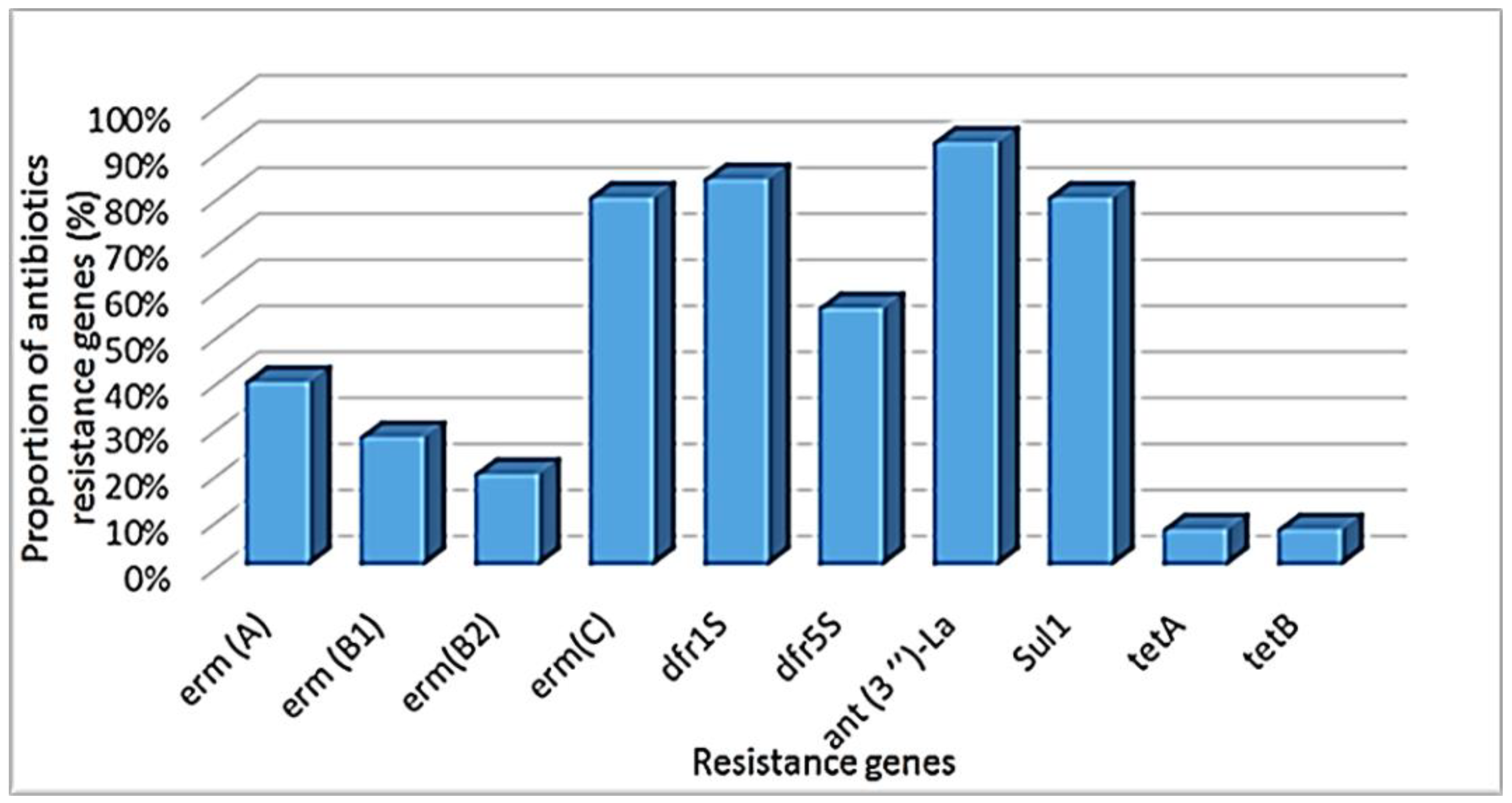
2.2.2. Antibiotic Resistance Genes

A total of 25 L. monocytogenes isolate were screened for the presence of AMR determinants. Out of the twenty AMR genes tested, 10 were detected in 23 isolates. The highest detections were ant (3″)-la (92%, 23/25), dfr1S (84%, 21/25), sul1 (80%, 20/25), and ermC (80%, 20/25), while the lowest were tetA and tetB at 8% (2/25) (Figure 1).

2.3. Proportion of Virulence Genes Detection in L. monocytogenes

Out of eight virulence genes, five (prfA, actA, hlyA, inlC, inlJ) were detected in 25 L. monocytes by PCR. The prfA was the most frequently (100%) detected gene in all L. monocytogenes isolates when compared to the hlyA (68%, n = 17) and actA (56%, n = 14) genes. Only five isolates harbored the plcA gene. Out of 25 L. monocytogenes isolates, 3 from food samples carried 8 virulence genes. In general, 5 virulence genes (actA, hlyA, inlC, inlJ, prfA) were most often detected in this study (Figure S1).

2.4. Biofilm-Forming Potential of L. monocytogenes Isolates

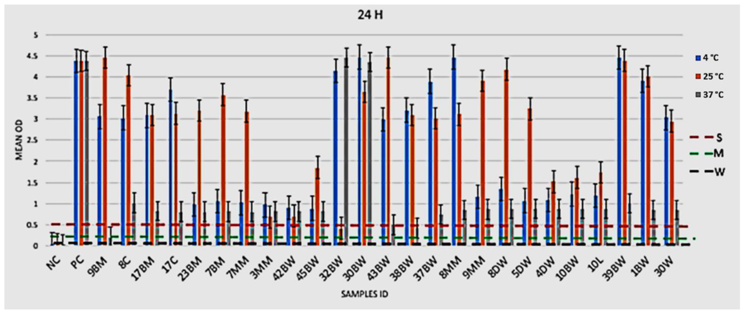
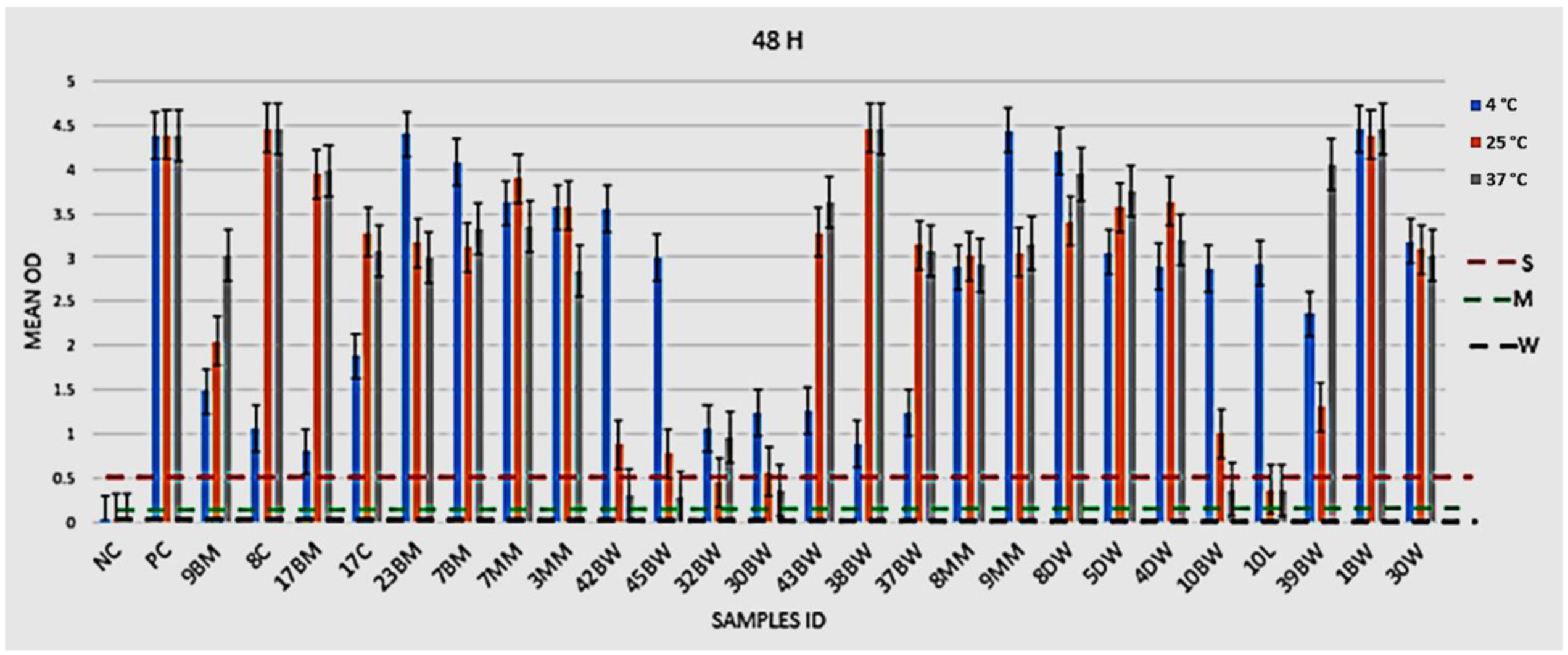
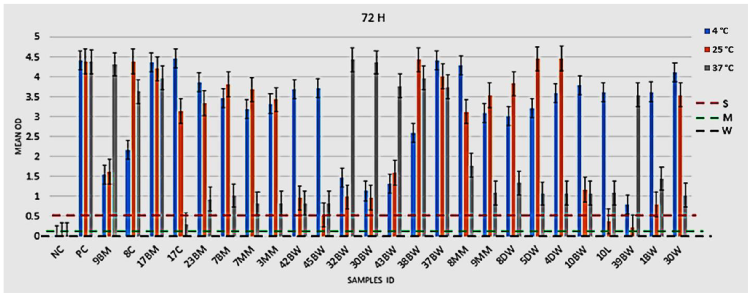
2.4.1. Phenotypic Assessment of Biofilm Formation

The microtiter plate assay was employed to assess the ability of L. monocytogenes isolates to form biofilm at different temperatures (4, 25, and 37 °C) over 24, 48, and 72 h periods. Based on biofilm formation patterns, the isolates were grouped either as non-adherent, weakly, moderately, or strongly adherent strains (Table 4). The results showed that all the isolates (n = 25) exhibited strong biofilm attachments, following 24, 48, and 72 h incubation periods at 4 °C. At 25 °C, 2 (8%) L. monocytogenes isolates produced moderate biofilm, while 23 (92%) formed a strong biofilm attachment, following 24 and 72 h incubation periods. Strong biofilm formation was demonstrated by 20 (80%) isolates at 37 °C after a 48 h incubation period. At 37 °C, the production of biofilm was strong after a 24 h incubation period, and the biofilm production of some isolates decreased after 72 h (p < 0.05), as shown in (Appendix A) Figure A1, Figure A2 and Figure A3.

2.4.2. Biofilm Formation Genes

PCR was utilized to determine the presence of genes associated with biofilm formation. Out of 25 L. monocytogenes isolates, 14 were positives for luxS and flaA (56% and 72%, respectively). A total of 9 (36%) isolates from food samples carried luxS gene followed by 5 (20%) from water and food samples harboring flaA gene. A total of 9 isolates (5 from food samples and 4 from water samples) were positive for both flaA and luxS genes (Figure S2).

3. Discussion

L. monocytogenes is capable of causing a sporadic disease, termed listeriosis, in humans and animals that results in very high hospitalization and fatalities [48]. Listeriosis is amongst the most problematic food-borne diseases in several countries, and it is associated with contamination at all stages in the food production process [49,50,51,52]. This pathogen is a psychrophile and can grow at refrigerated temperatures [53,54,55]. Due to this feature, it is very difficult to inactivate and curb the proliferation of L. monocytogenes cells in RTE food. This, therefore, indicates the need for continuous monitoring of this pathogen, especially in the food industry [56]. The main objective of this study was to assess the presence of Listeria sp. from various sources that are linked to the food chain, especially after South Africa recorded the world’s most severe outbreak of listeriosis in 2017 [57]. The results obtained in this study show that the occurrence of Listeria spp. was higher in food products (13.48%) than water (5.24%) and cattle feces (1.12%) samples. L. monocytogenes was detected in water and food. Interestingly, L. monocytogenes was most often detected in meat and meat products (eight; 15.09%), when compared to vegetables (three; 5.66%). These findings are a cause for concern, given the fact that listeriosis outbreaks have been associated with meat and poultry products, such as hot dogs [58], RTE pork meats [59], and turkey meat products [60]. In addition, L. monocytogenes was most frequently isolated in packing houses that processed frozen (41.3%), fresh-cut vegetables (7.9%) [61,62] and vegetables (9.5%) [63], mushrooms (1.6%) [64], and prepackaged salads and canned vegetables (5.4%) [65]. Food products tested in this study, such as cucumber and lettuce, may have been contaminated with L. monocytogenes as a result of soil contact. This pathogen may be introduced into food processing plants by human carriers (workers) and animals through faecal shedding [66,67]. The risks that these bacteria (L. monocytogenes) provide to public health are highlighted by their ability to survive for years, or even decades, in food processing facilities, as well as their ability to assist in food contamination during processing, handling, and packaging and the ease of foodborne transmission to consumers. Moreover, the detection of L. monocytogenes, including other species, such as L. ivanovii, L. grayi, and L. innocua, in wastewater effluents in a treatment facility in South Africa clearly indicates the potential to contaminate the environment. This may also increase the routes of transmission and dissemination of L. monocytogenes to foods [68].
Another objective of this study was to determine the antimicrobial resistant profiles of L. monocytogenes isolated from different sources within the food chain. In this study, large proportions of L. monocytogenes isolates were resistant to the majority of the antibiotics (62.5% to 100%). These results are consistent with another study, which reported high (90.74%) resistant L. monocytogenes isolates against nalidixic acid [69]. MAR (Multi-antibiotic resistance is a bacterium that is resistant to several antibiotics) were most prominent with nalidixic acid/cloxacillin (35.2%), nalidixic acid/colistin (31.5%), and cloxacillin/colistin/nalidixic acid (29.6%) [70]. However, a low (5.6%) AMR pattern was shown in chloramphenicol/nitrofurantin/cotrimoxazole [61]. In this study, the MAR phenotype E-DA-GM-T-NOV-OX-NA-K appeared in three different locations on different samples, which could be due to the use of antibiotics in those area. The continuous misuse of antibiotics led to the development of antibiotic resistance, specifically multi-drug resistant (MDR, which is an isolate that is resistant to at least one antibiotic in three or more drug classes) strains [71,72]. Notably, a high MAR index (Mean = 0.74) was observed in this study, and these findings were higher than that of the previous study [73]. This suggest that the isolates originate from a high-risk environment. Furthermore, the results obtained in this study revealed that L. monocytogenes harbored antimicrobial resistant genes ant(3″)-la (92%), dfr1S (84%), sul1 (80%), and ermC (80%), while the lowest were tetA and tetB at 8%). Contradictory to our findings, Heidarzadeh et al. reported tetM was detected at high rates (70%) of tetracycline-resistant isolates [70]. Approximately, 27.3% isolates carried dfrD. The ermB gene was detected in 83.3% of the erythromycin-resistant isolates [70]. Although antibiotic resistance in L. monocytogenes was rare, over the last two decades, there has been an increasing trend in resistance to one or multiple common antibiotics found in both clinical and environmental sources. In general, resistance genes are handed along by transferable plasmids and conjugative transposons, and L. monocytogenes resistant to antibiotics is no exception. Despite the fact that the study was unable to prove a direct transmission link between resistant genes in L. monocytogenes, our findings revealed several resistance genes in water and food samples.
A further objective of the study was to determine the presence of virulence genes in L. monocytogenes. The detection of prfA (100%), hlyA (68%), and actA (56%) genes indicates the potential pathogenicity of the isolates and their potential to cause foodborne infections. In addition, plcB genes were detected in five isolates. In contrast to our findings, Du et al. [74] found plcB gene in all isolates, whereas the inlB, actA, plcA, and iap genes were found in 71.4–90.5% of the isolates. Several studies have shown that the prfA gene in L. monocytogenes encodes a protein that induces the transcription of the listeriolysin gene (lisA) [75,76,77,78]. This signifies the prfA gene in the pathogenesis of listeriosis. Furthermore, the prfA gene in L. monocytogenes possessed two important crucial functions: response to environmental stress conditions and capacity to infect humans [75].
Biofilm formation, as well as the adaptation to stress, induced by cold temperatures and sub-lethal concentrations of disinfectants, coupled with the potential to inhabit ecological niches in facilities and equipment that are difficult to clean, have been identified as key contributors to the persistence of L. monocytogenes in the food chain [79]. Biofilm formation or ‘glycocalyx’ facilitates the adherence of bacteria to surfaces and plays a significant role in the success of pathogenic bacteria; thus, the prevention of adhesion could be an effective way to combat infections and cross-contamination [80]. This study evaluated the biofilm formation potentials of 25 L. moncytogenes strains isolated from different sources. L. monocytogenes has the potential to multiply within a wide range of temperatures (2–45 °C); due to that, three various temperatures were evaluated, which comprised 37 °C (representing the optimum growth temperature range between 30 and 37 °C), 25 °C (representing the room temperature), and 4 °C (which stand for refrigeration temperature of foods in retail) [15,81,82,83]. Our findings reveal that strong biofilm attachments were formed at 4 °C for 24, 48, and 72 h in all isolates (n = 25; 100%), followed by moderate biofilm formation for two (8%) isolates at 48 h and five (20%) L. monocytogenes isolates at 72 h. Similar results by Lado et al. [81] showed that L. monocytogenes isolates produced a strong biofilm community at 10 and 37 °C. All L. monocytogenes isolates utilized in this study were from food sources, as well as food processing equipment.
In addition, PCR-based biofilm formation genes showed that 18 out of 25 (72%) were amplified for flaA, followed by 14 (56%) for luxS. A total of nine (36%) isolates from food samples were found to carry the gene luxS, followed by five (20%) from water and food samples harboring the flaA gene. In a recent study, it was proven that L. monocytogenes biofilm was produced at optimum of neutral pH and 37 °C; supplementary data showed temperature as a key player in L. monocytogenes biofilm formation. It was concluded that the impacts of various factors on biofilm production are strain-dependent [74]. In Egypt, particularly Sohag city, 295 samples of ice cream, vaginal swabs, stool, and the urine of aborted women were collected and examined for the virulence ability of L. monocytogenes. Biofilm investigation was conducted by PCR detection of biofilm genes, such as luxS and flaA. The luxS gene was detected at 81.8%, followed by flaA at 72.7% in ice cream. In aborted women, the luxS gene amplified 81.8%, followed by flaA (63.6%), which contradicted our results [84]. In another study, Hassanien and Shaker assessed the biofilm potential of L. monocytogenes and showed that out of 38 isolates amplified, 23 (60.52%) and 1 (2.63%) were positive to luxS and flaA genes respectively [85].
Over the past two decades, researchers have shown the impact of biofilm in the food processing industries. These biofilms contribute to cross contamination by protecting and harboring harmful or spoilage microorganisms, including L. monocytogenes [86]. Antibiotic resistance is higher in the context of biofilm development than in planktonic cells [35,87]. Hence, this poses a problem in the control of L. monocytogenes from food sources, water, and food processing surfaces, as well as equipment.

4. Materials and Methods

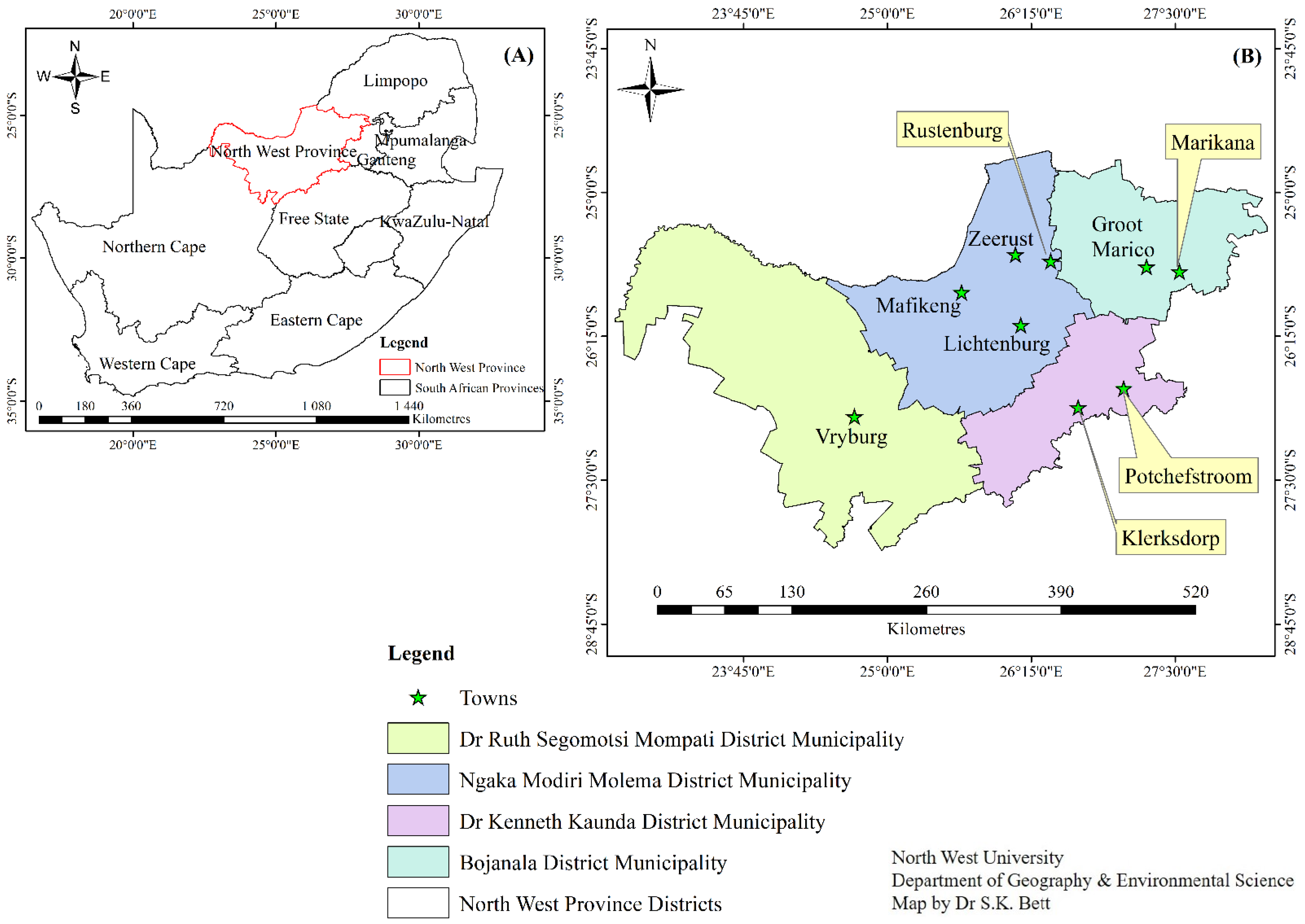
4.1. Sample Area and Sample Collection

A total of 207 samples were collected randomly from different sources (water, meat, vegetable, milk, and cattle feces) around the North West province in South Africa, between June 2018 and March 2020. The samples comprised 59 water (42 from boreholes and 17 from rivers) and 47 meat (12 wors (a South African traditional home-made sausage that is commercially available), 10 Russian (a South African sausage-shaped packed meat with a ‘strong meaty flavour and an intense smoky finish), 10 mince, and 15 red meat) samples. In addition, 23 vegetable samples (comprising 6 carrots, 5 cucumber, 4 lettuce, 4 cabbage, and 4 spinach), 34 milk (22 pasteurized and 12 unpasteurized), and 44 cattle feces samples were collected. The water samples were collected from eight sites, including Mafikeng, Potchefstroom, Klerksdorp, Zeerust, Vryburg, Marikana, Lichtenburg, and Groot Marco (Figure 2). For the collection of cattle feces, aseptic procedures were followed, and samples were obtained directly from the rectum of the animals, using sterile arm-length gloves, which were changed for each sample collected. The food samples were collected from the shelves of different shops and displayed for consumption by the consumers. Non-pasteurized milk was collected from dairy farms during the milking of cattle and distributed into sterile 250 mL bottles. All samples were transported on ice to the laboratory and processed within 24 h of collection. Figure 2 shows the different areas from which samples were collected.

4.2. Bacterial Strains

The reference strains L. monocytogenes (ATCC 19115) and L. ivanovii (ATCC 19119) were used as positive controls, while Escherichia coli (ATCC 25922) was used as negative control in the study. These American-type culture collection strains were purchased from Davies Diagnostics, Randburg, South Africa.

4.3. Isolation of Listeria spp.

To process the fecal, meat, and vegetable samples, 25 g of each sample was homogenized in 225 mL of half-Fraser broth (HFB, Acumedia, Lansing, MI, USA), while, for milk samples, 25 mL of each sample was transferred into HFB. Aliquots of 100 mL of water samples were filtered through a 0.45-μm Whatman filter paper (Whatman® glass microfiber GS filter paper, Separation Scientific SA (Pty) Ltd., Johannesburg, South Africa) using a vacuum pump machine (model N035AN.18, Freiburg, Germany). The membrane filters were inoculated in 225 mL of HFB and incubated aerobically at 30 °C for 24 h. Following incubation, aliquots of 100 µL from each sample were transferred into 10 mL of Fraser broth (Acumedia) and incubated at 30 °C for 24 h. Tubes with black cultures, resulting from the hydrolysis of esculin, were considered to be presumptively positive for Listeria species, and 100 µL aliquots were spread-plated on Listeria isolation medium (Oxford formulation, Acumedia) [88]. The plates were incubated at 30 °C for 24–48 h. Pure isolates with greenish-grey morphologies and a concave center surrounded by a black halo (due to hydrolysis of esculin) were selected for further identification tests. Two to three colonies were picked from each plate and subjected to Gram staining and biochemical testing, such as the fermentation of carbohydrates (mannitol, rhamnose, dextrose, maltose, and xylose), motility, catalase activity, and β-hemolysis [89]. Samples with positive β-hemolysis and catalase tests fermented most sugars, including rhamnose, dextrose, and maltose, but not mannitol or xylose, were identified as Listeria spp. and sent to molecular identification for confirmation.

4.4. Molecular Characterizations of Listeria Species Using PCR Analysis

4.4.1. Extraction of Chromosomal DNA

Genomic DNA of each presumptive isolate was extracted using a commercial Genomic DNATM Tissue Miniprep Kit (Zymo Research, Irvine, CA, USA), following the manufacturer’s instructions; the quantification of the DNA was performed using a NanoDropTM 1000 spectrophotometer (Thermo Fischer Scientific, Waltham, MA, USA).

4.4.2. Identification of Listeria spp., Virulence Genes, Antibiotic Resistance Genes and Biofilm Genes Using PCR (Polymerase Chain Reaction) Technique

Except otherwise mentioned, all PCR reagents used in this study were from Fermentas (Waltham, MA, USA), and all primers were synthesized by Inqaba Biotechnical (Pty) Ltd. (Pretoria, South Africa). DNA was subjected to bacterial 16S rRNA gene amplification, and the amplicons were sent to Inqaba Biotechnical (Pty) Ltd. (Pretoria, South Africa) for the 16S rRNA sequencing identification. The prs gene was also used for the detection of Listeria spp., and the primers (jograyi, lin0464, liv22228, lmo1030, iseelin and lwe1801) were used for the detection of Listeria specific species (Table S1). DNA from L. monocytogenes ATCC 19115 and L. ivanovii ATCC 19119 were used as positive controls, while nuclease-free water was used as a negative control. The C1000 TouchTM DNA thermal cycler (Bio-Rad, Singapore) was used to perform the amplifications. PCR primer sequences, target genes, amplicon sizes, and cycling conditions are shown in Table S1.
PCR was used to detect the actin (actA), p60 (iap), phosphatidylinositol phospholipase C (plcA, plcB), internalin (inla, inlb, inlc, inlj), regulatory (prfA), and hemolysin (hlyA) virulence genes, as previously described [90,91,92]. Amplification conditions of each PCR reaction are listed in Table S2.
The isolates confirmed to be Listeria spp. that were presenting resistance to more than two antibiotics were considered as multi-drug resistant (MDR) and were tested for harboring the erm(A), erm(B1), erm(B2), erm(C), erm(T), blaTEM, drf1, drf5, drf12, dfr1S, dfr5S, dfr7s, dfr12s, dfr17s, tetA, tetB, ant(3’’)-la, sul1, sul2 and pse1 antibiotics resistance genes. The primer sequences, PCR cycling conditions, and amplicon sizes are indicated in Table S3.
PCR was utilized to detect the biofilm-forming genes luxS and flaA of L. monocytogenes (Table S4). Each PCR reaction mix was constituted by 12.5 µL of 2X DreamTaq green master mix, 11 µL of RNase nuclease-free PCR water, 0.5 µM of each primer (forward and reverse), and 1 µL of template DNA, for total of 25 µL standard volume. The cycling conditions of the PCR are stated in Tables S1–S4.
All PCR amplicons were separated by electrophoresis on a 2% (w/v) agarose gel comprising 0.001 µg/mL ethidium bromide. A 100 bp DNA molecular weight marker (Fermentas) was incorporated in each gel run. Electrophoresis was conducted at 60 Volt for 120 min and the images captured using a ChemiDoc Imaging System (Bio-Rad, UK).

4.5. Antimicrobial Susceptibility Test

The Kirby-Bauer disc diffusion technique was used to determine the antimicrobial susceptibility profiles of the isolates [93]. A total of 12 antibiotics (Mast Diagnostics, UK) were used, including ampicillin (AMP), 10 µg; erythromycin (E), 15 µg; kanamycin (K), 30 µg; norfloxacin (NOR), 10 µg; novobiocin (NB), 30 µg; tetracycline (TET), 30 µg; oxacillin (OX), 5 µg; clindamycin (DM), 2 µg; nalidixic acid (NA), 30 µg; gentamicin (GM), 15 µg; meropenem (MEM), 10 µg; and trimethoprim and sulfamethoxazole (SXT), 1.25 + 23.75 µg. The selection of these antibiotics was based on the treatment regimen for listeriosis or their extensive use as prophylactics in beef and dairy cattle farming in South Africa. Overnight pure cultures were prepared in 15mL TSB (tryptic soy broth) and incubated at 37 °C for 24 h. The bacterial suspension was adjusted by performing serial dilution until reaching 106 cells/mL on the McFarland standards scale of 0.5. After 24 h, the bacterial cell suspension were spread on Mueller Hinton agar (Lasec SA (Pty) Ltd, Johannesburg, South Africa) plates using a sterile swab, the plates were incubated aerobically for 24 h at 37 °C, and the diameter of the zones of inhibition around the discs were measured [94]. The results were interpreted based on the antibiotic growth inhibition zone diameter critical values, as recommended by the Clinical Laboratory Standards Institute (CLSI) guidelines [95], in order to classify the isolates as sensitive, intermediate, or resistant to a particular antibiotic. For quality control, L. monocytogenes ATCC 19115 and L. ivanovii ATCC 19119, were used as reference.

4.6. Biofilm Formation

Phenotypic Detection

Biofilm formation by L. monocytogenes was determined using the microtiter plate assay described by Bertrand et al. [96] and Mahm [97] with slight modifications. L. monocytogenes strains were sub-cultured in tryptic soy broth (TSB) supplemented with 0.6% yeast extract (TSBYE) and incubated at 37 °C overnight. A 10 µL aliquot of each overnight culture at 105 CFU was used to inoculate 96-well plates containing 190µL of brain–heart infusion (BHI) broth (Lasec SA (Pty) Ltd, Johannesburg, South Africa) per well. Each isolate was loaded in triplicates. Two positive controls, Pseudomonas aeruginosa (strong biofilm producer) and L. monocytogenes ATCC 19115 were included, while a negative control well contained 200 µL of BHI only. For all samples, plates were incubated at three different temperatures (4, 25, and 37 °C) for 24, 48, and 72 h. After incubation, the plates were rinsed three times with PBS (300 µL, pH 7.4) to remove unattached microbial cells, and excess PBS was removed by blotting the plate using a clean paper towel and allowed to dry. The dye of crystal violet (0.1%, 200 µL) was added to each well to stain the biofilms formed by the cells on well bottom and incubated at room temperature for one hour. The wells were rinsed three times with 300 µL of PBS, and aliquots of 95% ethanol (200 µL) were added to each well for stain fixation, with incubation at room temperature for an hour. The microplate reader (Shenzhen Heales Technology Development Co., Ltd., Shenzhen, China) was used to read the optical density at 630 nm. The cut-off value of the negative control (ODc) was determined as three standard deviations (SD) above the mean OD of the negative control. As stipulated by Stepanovi’c et al. (2000), biofilm formation was categorized into four groups, according to the ODs obtained: OD ≤ ODc, non-adherent; ODc < OD ≤ 2 x ODc, weak biofilm formation; 2 x ODc < OD ≤ 4 x ODc, moderate biofilm formation; and 4 x ODc < OD, strong biofilm formation.

5. Conclusions

The current study revealed the occurrence of Listeria spp. from various sample types. Our finding showed that L. monocytogenes carried virulence genes and was resistant to several antibiotics. In addition, further investigation indicated strong biofilm formation by L. monocytogenes strains. This clearly suggests that L. monocytogenes have all the features that contribute to pathogenicity, while being difficult to control, due to the resistance. Hence, effective alternative measures (such as greater attention to good manufacturing and hygiene practices throughout the food production, distribution, and storage chains, including in the home environment) are needed to control and prevent resistant L. monocytogenes and reduce the risk of listeriosis.

Supplementary Materials

The following supporting information can be downloaded at: https://www.mdpi.com/article/10.3390/pathogens11080843/s1, Figure S1: PCR amplification of virulence genes (M: Marker, PC: Positive control, NC: Negative control, 1–8: isolates amplification of virulence genes); Figure S2: PCR amplification of biofilm genes (M: Marker, CN: Negative control, 1: Positive control, 2–7: isolates amplification of flaA gene, 8–13: isolates amplification of LuxS gene, 14: Positive control, 15: negative control); Table S1: List of oligonucleotide primer sequences and PCR conditions used for the identification of Listeria spp.; Table S2: List of oligonucleotide primer sequences and PCR conditions used for detection of virulence genes in L. monocytogenes; Table S3. List of oligonucleotide primer sequences and PCR conditions used for detection of antibiotic resistance genes in L. monocytogenes; Table S4. List of oligonucleotide primer sequences and PCR conditions used for detection of genes associated with biofilm formation in L. monocytogenes; Refs [74,96,97,98,99,100,101,102,103,104,105,106,107,108,109] are cited in the supplementary materials.

Author Contributions

C.-D.K.T. collected samples, executed the laboratory experiment, analyzed the data, and drafted the first version of the manuscript. J.F., M.C.M., P.K.M., G.B. (Giulia Baldelli), G.B. (Giorgio Brandi), G.F.S., G.A. and C.N.A. contributed to the review and finalization of the manuscript. C.N.A., G.A., P.K.M., J.F., G.B. (Giulia Baldelli), G.B. (Giorgio Brandi), G.F.S. and G.A. conceptualized, supervised, finalized the article, and approved the manuscript. All authors have read and agreed to the published version of the manuscript.

Funding

Authors appreciate the North-West University for financial assistance. Additional funding was awarded by Inqaba Biotechnical Industries (Pty) Ltd (INQABA BIOTEC SeedIT 2018).

Institutional Review Board Statement

The North-west University ethics committee has approved the study protocol (FNASREC-NWU-00733-18-A9-2021).

Informed Consent Statement

Not applicable.

Data Availability Statement

The data presented in this study are available on request from the corresponding author.

Acknowledgments

Authors acknowledge the North-West University postgraduate bursary and the SEEDIT 2018 from Inqaba Biotechnical Industries (Pty) Ltd. for the financial support.

Conflicts of Interest

The authors declare no conflict of interest.

Appendix A

Figure A1. Biofilm formation of L. monocytogenes at 24 h/4 °C, 25 °C, 7 °C. Negative control (NC), positive control (PC), beef meat (BM), borehole water (BW), cucumber (C), dam water (DW), lettuce (L), mince meat (MM), strong biofilm (S), moderate biofilm (M), weak biofilm (W), numbers in (sample ID): sample number.
Pathogens 11 00843 g0a1
Figure A2. Biofilm formation of L. monocytogenes at 48 h/ 4 °C, 25 °C, 37 °C. Negative control (NC), positive control (PC), beef meat (BM), borehole water (BW), cucumber (C), dam water (DW), lettuce (L), mince meat (MM), strong biofilm (S), moderate biofilm (M), weak biofilm (W), numbers in (sample ID): sample number.
Pathogens 11 00843 g0a2
Figure A3. Biofilm formation of L. monocytogenes at 72 h/ 4 °C, 25 °C, 37 °C. Negative control (NC), positive control (PC), beef meat (BM), borehole water (BW), cucumber (C), dam water (DW), lettuce (L), mince meat (MM), strong biofilm (S), moderate biofilm (M), weak biofilm (W), numbers in (sample ID): sample number.
Pathogens 11 00843 g0a3

References

  1. McLauchlin, J.; Mitchell, R.; Smerdon, W.; Jewell, K. Listeria monocytogenes and listeriosis: A review of hazard characterisation for use in microbiological risk assessment of foods. Int. J. Food Microbiol. 2004, 92, 15–33. [Google Scholar] [CrossRef]
  2. Troxler, R.; von Graevenitz, A.; Funke, G.; Wiedemann, B.; Stock, I. Natural antibiotic susceptibility of Listeria species: L. grayi, L. innocua, L. ivanovii, L. monocytogenes, L. seeligeri and L. welshimeri strains. Clin. Microbiol. Infect. 2000, 6, 525–535. [Google Scholar] [CrossRef] [PubMed] [Green Version]
  3. Møretrø, T.; Langsrud, S. Listeria monocytogenes: Biofilm formation and persistence in food-processing environments. Biofilms 2004, 1, 107–121. [Google Scholar] [CrossRef]
  4. Liu, D. Identification, subtyping and virulence determination of Listeria monocytogenes, an important foodborne pathogen. J. Med Microbiol. 2006, 55, 645–659. [Google Scholar] [CrossRef] [PubMed] [Green Version]
  5. David, D.J.V.; Cossart, P. Recent advances in understanding Listeria monocytogenes infection: The importance of subcellular and physiological context. F1000Research 2017, 6, 1126. [Google Scholar] [CrossRef] [PubMed] [Green Version]
  6. Linscott, A.J. Food-Borne Illnesses. Clin. Microbiol. Newsl. 2011, 33, 41–45. [Google Scholar] [CrossRef]
  7. Wang, Z.; Tao, X.; Liu, S.; Zhao, Y.; Yang, X. An Update Review on Listeria Infection in Pregnancy. Infect. Drug Resist. 2021, 14, 1967–1978. [Google Scholar] [CrossRef]
  8. Mateus, T.; Silva, J.; Maia, R.; Teixeira, P. Listeriosis during Pregnancy: A Public Health Concern. ISRN Obstet. Gynecol. 2013, 2013, 851712. [Google Scholar] [CrossRef] [Green Version]
  9. Kitada, T.; Kadoba, K.; Watanabe, R.; Koyama, T.; Nakayama, Y.; Taki, M.; Yukawa, S.; Odani, K.; Morinobu, A. Listeriosis presenting with fever, arthralgia, elevated liver enzymes, and hyperferritinaemia in pregnancy: A critical mimicker of adult-onset Still’s disease. Scand. J. Rheumatol. 2021, 51, 78–80. [Google Scholar] [CrossRef]
  10. Ntuli, N.; Wadula, J.; Nakwa, F.; Thomas, R.; Van Kwawegen, A.; Sepeng, L.; Seake, K.; Kgwadi, D.; Sono, L.; Ondongo-Ezhet, C.; et al. Characteristics and Outcomes of Neonates With Blood Stream Infection Due to Listeria monocytogenes. Pediatr. Infect. Dis. J. 2021, 40, 917–921. [Google Scholar] [CrossRef]
  11. Allen, K.J.; Wałecka-Zacharska, E.; Chen, J.C.; Katarzyna, K.-P.; Devlieghere, F.; Van Meervenne, E.; Osek, J.; Wieczorek, K.; Bania, J. Listeria monocytogenes—An examination of food chain factors potentially contributing to antimicrobial resistance. Food Microbiol. 2016, 54, 178–189. [Google Scholar] [CrossRef]
  12. Authority, E.F.S.; Prevention, E.C.F.D. Control, The European Union summary report on trends and sources of zoonoses, zoonotic agents and food-borne outbreaks in 2014. EFSA J. 2015, 13, 4329. [Google Scholar]
  13. Marshall, B.M.; Levy, S.B. Food Animals and Antimicrobials: Impacts on Human Health. Clin. Microbiol. Rev. 2011, 24, 718–733. [Google Scholar] [CrossRef] [Green Version]
  14. Kawacka, I.; Olejnik-Schmidt, A.; Schmidt, M.; Sip, A. Effectiveness of Phage-Based Inhibition of Listeria monocytogenes in Food Products and Food Processing Environments. Microorganisms 2020, 8, 1764. [Google Scholar] [CrossRef]
  15. Tchatchouang, C.-D.K.; Fri, J.; De Santi, M.; Brandi, G.; Schiavano, G.F.; Amagliani, G.; Ateba, C.N. Listeriosis Outbreak in South Africa: A Comparative Analysis with Previously Reported Cases Worldwide. Microorganisms 2020, 8, 135. [Google Scholar] [CrossRef] [Green Version]
  16. Munshi, M.; Sharma, M.; Deb, S. Viability of High-Pressure Technology in the Food Industry. In Handbook of Research on Food Processing and Preservation Technologies: Emerging Techniques for Food Processing, Quality, and Safety Assurance; Apple Academic Press: Palm Bay, FL, USA, 2021; Volume 5, p. 51. [Google Scholar]
  17. Patel, D.; Rathod, R. Ready-to-eat food perception, food preferences and food choice—A theoretical discussion. Int. J. Multidiscip. Res. Dev. 2017, 3, 198–205. [Google Scholar]
  18. Chaudhury, R. Determinants of consumer behavior in buying RTE foods. J. Bus. Retail. Manag. Res. 2010, 5, 76–86. [Google Scholar]
  19. Food Safety News. Company Recalls Imported Mushrooms after State Test Finds Listeria Monocytogenes. News Desk on 20 March 2022. Available online: https://www.foodsafetynews.com/2022/03/company-recalls-imported-mushrooms-after-state-test-finds-Listeria-monocytogenes/ (accessed on 4 April 2022).
  20. Food Drug Administration. Banner Smoked Fish Expands Recalls Smoked Fish Products, Salads, Pickled Fish Products, and Cream Sauce Products Because of Possible Health Risk. Company Announcement Date: 3 June 2021. Available online: https://www.fda.gov/safety/recalls-market-withdrawals-safety-alerts/banner-smoked-fish-expands-recalls-smoked-fish-products-salads-pickled-fish-products-and-cream-sauce (accessed on 7 April 2022).
  21. Food Drug Administration. Outbreak Investigation of Listeria Monocytogenes: Enoki Mushrooms (March 2020). Available online: https://www.fda.gov/food/outbreaks-foodborne-illness/outbreak-investigation-Listeria-monocytogenes-enoki-mushrooms-march-2020 (accessed on 7 April 2022).
  22. US FDA Guidance for Industry. Estimating the Maximum Safe Starting Dose in Initial Clinical Trials for Therapeutics in Adult Healthy Volunteers. Pharmacology and Toxicology. 2005. Available online: http://www.fda.gov/cder/guidance/index.htm (accessed on 25 April 2017).
  23. Olanya, O.M.; Hoshide, A.K.; Ijabadeniyi, O.A.; Ukuku, D.O.; Mukhopadhyay, S.; Niemira, B.A.; Ayeni, O. Cost estimation of listeriosis (Listeria monocytogenes) occurrence in South Africa in 2017 and its food safety implications. Food Control 2019, 102, 231–239. [Google Scholar] [CrossRef]
  24. Jones, E.M.; MacGowan, A.P. Antimicrobial chemotherapy of human infection due to Listeria monocytogenes. Eur. J. Clin. Microbiol. 1995, 14, 165–175. [Google Scholar] [CrossRef]
  25. Walsh, D.; Duffy, G.; Sheridan, J.; Blair, I.; McDowell, D. Antibiotic resistance among Listeria, including Listeria monocytogenes, in retail foods. J. Appl. Microbiol. 2001, 90, 517–522. [Google Scholar] [CrossRef]
  26. Shamloo, E.; Hosseini, H.; Moghadam, Z.A.; Larsen, M.H.; Haslberger, A.; Alebouyeh, M. Importance of Listeria monocyto-genes in food safety: A review of its prevalence, detection, and antibiotic resistance. Iran. J. Vet. Res. 2019, 20, 241. [Google Scholar]
  27. Borucki, M.K.; Peppin, J.D.; White, D.; Loge, F.; Call, D.R. Variation in Biofilm Formation among Strains of Listeriamonocytogenes. Appl. Environ. Microbiol. 2003, 69, 7336–7342. [Google Scholar] [CrossRef] [Green Version]
  28. Rodríguez-López, P.; Rodríguez-Herrera, J.J.; Vázquez-Sánchez, D.; Cabo, M.L. Current Knowledge on Listeria monocytogenes Biofilms in Food-Related Environments: Incidence, Resistance to Biocides, Ecology and Biocontrol. Foods 2018, 7, 85. [Google Scholar] [CrossRef] [PubMed] [Green Version]
  29. Bennani, H.; Mateus, A.; Mays, N.; Eastmure, E.; Stärk, K.D.C.; Häsler, B. Overview of Evidence of Antimicrobial Use and Antimicrobial Resistance in the Food Chain. Antibiotics 2020, 9, 49. [Google Scholar] [CrossRef] [Green Version]
  30. Gray, J.A.; Chandry, P.S.; Kaur, M.; Kocharunchitt, C.; Bowman, J.P.; Fox, E.M. Characterisation of Listeria monocytogenes food-associated isolates to assess environmental fitness and virulence potential. Int. J. Food Microbiol. 2021, 350, 109247. [Google Scholar] [CrossRef]
  31. Jordan, K.; Hunt, K.; Lourenco, A.; Pennone, V. Listeria monocytogenes in the Food Processing Environment. Curr. Clin. Microbiol. Rep. 2018, 5, 106–119. [Google Scholar] [CrossRef]
  32. Ferri, M.; Ranucci, E.; Romagnoli, P.; Giaccone, V. Antimicrobial resistance: A global emerging threat to public health systems. Crit. Rev. Food Sci. Nutr. 2015, 57, 2857–2876. [Google Scholar] [CrossRef]
  33. El Salabi, A.; Walsh, T.; Chouchani, C. Extended spectrum β-lactamases, carbapenemases and mobile genetic elements responsible for antibiotics resistance in Gram-negative bacteria. Crit. Rev. Microbiol. 2012, 39, 113–122. [Google Scholar] [CrossRef]
  34. Beceiro, A.; Tomás, M.; Bou, G. Antimicrobial Resistance and Virulence: A Successful or Deleterious Association in the Bacterial World? Clin. Microbiol. Rev. 2013, 26, 185–230. [Google Scholar] [CrossRef] [Green Version]
  35. Manganyi, M.; Regnier, T.; Olivier, E. Antimicrobial activities of selected essential oils against Fusarium oxysporum isolates and their biofilms. S. Afr. J. Bot. 2015, 99, 115–121. [Google Scholar] [CrossRef]
  36. Gonçalves, I.R.; Dantas, R.C.C.; Ferreira, M.L.; Batistão, D.W.D.F.; Gontijo-Filho, P.P.; Ribas, R.M. Carbapenem-resistant Pseudomonas aeruginosa: Association with virulence genes and biofilm formation. Braz. J. Microbiol. 2016, 48, 211–217. [Google Scholar] [CrossRef] [Green Version]
  37. Velge, P.; Bottreau, E.; Van-Langendonck, N.; Kaeffer, B. Cell proliferation enhances entry of Listeria monocytogenes into intestinal epithelial cells by two proliferation-dependent entry pathways. J. Med. Microbiol. 1997, 46, 681–692. [Google Scholar] [CrossRef] [Green Version]
  38. Cossart, P. Invasion of mammalian cells by Listeria monocytogenes: Functional mimicry to subvert cellular functions. Trends Cell Biol. 2003, 13, 23–31. [Google Scholar] [CrossRef]
  39. Chatterjee, S.S.; Hossain, H.; Otten, S.; Kuenne, C.; Kuchmina, K.; Machata, S.; Domann, E.; Chakraborty, T.; Hain, T. Intracellular Gene Expression Profile of Listeria monocytogenes. Infect. Immun. 2006, 74, 1171–1180. [Google Scholar] [CrossRef] [Green Version]
  40. Kreft, J.; Vázquez-Boland, J.A. Regulation of virulence genes in Listeria. Int. J. Med Microbiol. 2001, 291, 145–157. [Google Scholar] [CrossRef]
  41. Volokhov, D.V.; Duperrier, S.; Neverov, A.A.; George, J.; Buchrieser, C.; Hitchins, A.D. The Presence of the Internalin Gene in Natural Atypically Hemolytic Listeria innocua Strains Suggests Descent from L. monocytogenes. Appl. Environ. Microbiol. 2007, 73, 1928–1939. [Google Scholar] [CrossRef] [Green Version]
  42. De las Heras, A.; Cain, R.J.; Bielecka, M.K.; Vazquez-Boland, J.A. Regulation of Listeria virulence: PrfA master and commander. Curr. Opin. Microbiol. 2011, 14, 118–127. [Google Scholar] [CrossRef]
  43. Quereda, J.J.; Morón-García, A.; Palacios-Gorba, C.; Dessaux, C.; Portillo, F.G.-D.; Pucciarelli, M.G.; Ortega, A.D. Pathogenicity and virulence of Listeria monocytogenes: A trip from environmental to medical microbiology. Virulence 2021, 12, 2509–2545. [Google Scholar] [CrossRef]
  44. Vázquez-Boland, J.A.; Domínguez-Bernal, G.; González-Zorn, B.; Kreft, J.; Goebel, W. Pathogenicity islands and virulence evolution in Listeria. Microbes Infect. 2001, 3, 571–584. [Google Scholar] [CrossRef]
  45. Schiavano, G.F.; Ateba, C.N.; Petruzzelli, A.; Mele, V.; Amagliani, G.; Guidi, F.; De Santi, M.; Pomilio, F.; Blasi, G.; Gattuso, A.; et al. Whole-Genome Sequencing Characterization of Virulence Profiles of Listeria monocytogenes Food and Human Isolates and In Vitro Adhesion/Invasion Assessment. Microorganisms 2021, 10, 62. [Google Scholar] [CrossRef]
  46. Mukherjee, K.; Raju, R.; Fischer, R.; Vilcinskas, A. Galleria Mellonella as a Model Host to Study Gut Microbe Homeostasis and Brain Infection by the Human Pathogen Listeria Monocytogenes. Yellow Biotechnol. 2013, 135, 27–39. [Google Scholar] [CrossRef]
  47. Jian, Z.; Zeng, L.; Xu, T.; Sun, S.; Yan, S.; Yang, L.; Huang, Y.; Jia, J.; Dou, T. Antibiotic resistance genes in bacteria: Occurrence, spread, and control. J. Basic Microbiol. 2021, 61, 1049–1070. [Google Scholar] [CrossRef]
  48. EFSA; ECDC. European Food Safety Authority (EFSA); European Centre for Disease Prevention and Control (ECDC). The European Union summary report on trends and sources of zoonoses, zoonotic agents and food-borne outbreaks in 2017. EFSA J. 2018, 16, e05500. [Google Scholar] [CrossRef]
  49. Muchaamba, F.; Eshwar, A.K.; Stevens, M.J.A.; von Ah, U.; Tasara, T. Variable Carbon Source Utilization, Stress Resistance, and Virulence Profiles Among Listeria monocytogenes Strains Responsible for Listeriosis Outbreaks in Switzerland. Front. Microbiol. 2019, 10, 957. [Google Scholar] [CrossRef] [PubMed] [Green Version]
  50. Matle, I.; Mbatha, K.R.; Lentsoane, O.; Magwedere, K.; Morey, L.; Madoroba, E. Occurrence, serotypes, and characteristics of Listeria monocytogenes in meat and meat products in South Africa between 2014 and 2016. J. Food Saf. 2019, 39, e12629. [Google Scholar] [CrossRef]
  51. Matle, I.; Mafuna, T.; Madoroba, E.; Mbatha, K.R.; Magwedere, K.; Pierneef, R. Population Structure of Non-ST6 Listeria monocytogenes Isolated in the Red Meat and Poultry Value Chain in South Africa. Microorganisms 2020, 8, 1152. [Google Scholar] [CrossRef]
  52. Rip, D.; Gouws, P.A. PCR–Restriction Fragment Length Polymorphism and Pulsed-Field Gel Electrophoresis Characterization of Listeria monocytogenes Isolates from Ready-to-Eat Foods, the Food Processing Environment, and Clinical Samples in South Africa. J. Food Prot. 2020, 83, 518–533. [Google Scholar] [CrossRef]
  53. Parichanon, P.; Sattayakhom, A.; Matan, N.; Matan, N. Antimicrobial activity of lime oil in the vapour phase against Listeria monocytogenes on ready-to-eat salad during cold storage and its possible mode of action. Food Control 2022, 132, 108486. [Google Scholar] [CrossRef]
  54. Makumbe, H.; Tabit, F.; Dlamini, B. Prevalence, Molecular Identification, Antimicrobial Resistance, and Disinfectant Susceptibility of Listeria innocua Isolated from Ready-to-Eat Foods Sold in Johannesburg, South Africa. J. Food Qual. Hazards Control 2021, 8, 131–139. [Google Scholar] [CrossRef]
  55. Keet, R.; Rip, D. Listeria monocytogenes isolates from Western Cape, South Africa exhibit resistance to multiple antibiotics and contradicts certain global resistance patterns. AIMS Microbiol. 2021, 7, 40–58. [Google Scholar] [CrossRef]
  56. Archer, D.L. The evolution of FDA’s policy on Listeria monocytogenes in ready-to-eat foods in the United States. Curr. Opin. Food Sci. 2018, 20, 64–68. [Google Scholar] [CrossRef]
  57. Thomas, J.; Govender, N.; McCarthy, K.M.; Erasmus, L.K.; Doyle, T.J.; Allam, M.; Ismail, A.; Ramalwa, N.; Sekwadi, P.; Ntshoe, G.; et al. Outbreak of Listeriosis in South Africa Associated with Processed Meat. N. Engl. J. Med. 2020, 382, 632–643. [Google Scholar] [CrossRef]
  58. Mazzotta, A.S.; Gombas, D.E. Heat Resistance of an Outbreak Strain of Listeria monocytogenes in Hot Dog Batter. J. Food Prot. 2001, 64, 321–324. [Google Scholar] [CrossRef]
  59. WHO. Listeriosis–Spain [Internet]. Cited 7 August 2020. Available online: https://www.who.int/csr/don/16-september-2019-listeriosis-spain/en/ (accessed on 4 May 2022).
  60. Gelbíčová, T.; Zobaníková, M.; Tomáštíková, Z.; Van Walle, I.; Ruppitsch, W.; Karpíšková, R. An outbreak of listeriosis linked to turkey meat products in the Czech Republic, 2012–2016. Epidemiol. Infect. 2018, 146, 1407–1412. [Google Scholar] [CrossRef] [Green Version]
  61. Pappelbaum, K.; Grif, K.; Heller, I.; Würzner, R.; Hein, I.; Ellerbroek, L.; Wagner, M. Monitoring Hygiene On- and At-Line Is Critical for Controlling Listeria monocytogenes during Produce Processing. J. Food Prot. 2008, 71, 735–741. [Google Scholar] [CrossRef]
  62. Alvarez-Ordóñez, A.; Leong, D.; Hunt, K.; Scollard, J.; Butler, F.; Jordan, K. Production of safer food by understanding risk factors for L. monocytogenes occurrence and persistence in food processing environments. J. Food Saf. 2018, 38, e12516. [Google Scholar] [CrossRef]
  63. Leong, D.; NicAogáin, K.; Luque-Sastre, L.; McManamon, O.; Hunt, K.; Alvarez-Ordóñez, A.; Scollard, J.; Schmalenberger, A.; Fanning, S.; O’Byrne, C.; et al. A 3-year multi-food study of the presence and persistence of Listeria monocytogenes in 54 small food businesses in Ireland. Int. J. Food Microbiol. 2017, 249, 18–26. [Google Scholar] [CrossRef]
  64. Viswanath, P.; Murugesan, L.; Knabel, S.J.; Verghese, B.; Chikthimmah, N.; Laborde, L.F. Incidence of Listeria monocytogenes and Listeria spp. in a Small-Scale Mushroom Production Facility. J. Food Prot. 2013, 76, 608–615. [Google Scholar] [CrossRef]
  65. Gianfranceschi, M.; Gattuso, A.; Tartaro, S.; Aureli, P. Incidence of Listeria monocytogenes in food and environmental samples in Italy between 1990 and 1999: Serotype distribution in food, environmental and clinical samples. Eur. J. Epidemiol. 2002, 18, 1001–1006. [Google Scholar] [CrossRef]
  66. Grif, K.; Patscheider, G.; Dierich, M.P.; Allerberger, F. Incidence of Fecal Carriage of Listeria monocytogenes in Three Healthy Volunteers: A One-Year Prospective Stool Survey. Eur. J. Clin. Microbiol. 2003, 22, 16–20. [Google Scholar] [CrossRef]
  67. Castro, H.; Jaakkonen, A.; Hakkinen, M.; Korkeala, H.; Lindström, M. Occurrence, Persistence, and Contamination Routes of Listeria monocytogenes Genotypes on Three Finnish Dairy Cattle Farms: A Longitudinal Study. Appl. Environ. Microbiol. 2018, 84, e02000-17. [Google Scholar] [CrossRef] [Green Version]
  68. Odjadjare, E.E.O.; Okoh, A.I. Prevalence and distribution of Listeria pathogens in the final effluents of a rural wastewater treatment facility in the Eastern Cape Province of South Africa. World J. Microbiol. Biotechnol. 2009, 26, 297–307. [Google Scholar] [CrossRef]
  69. Enurah, L.U.; Aboaba, O.O.; Nwachukwu, S.C.U.; Nwosuh, C.I. Antibiotic resistant profiles of food (fresh raw milk) and en-vironmental (abattoir effluents) isolates of Listeria monocytogenes from the six zones of Nigeria. Appl. Microbiol. Biotechnol. 2019, 2, 124–132. [Google Scholar] [CrossRef]
  70. Heidarzadeh, S.; Pourmand, M.R.; Hasanvand, S.; Pirjani, R.; Afshar, D.; Noori, M.; Dallal, M.M.S. Antimicrobial Susceptibility, Serotyping, and Molecular Characterization of Antibiotic Resistance Genes in Listeria monocytogenes Isolated from Pregnant Women with a History of Abortion. Iran. J. Public Health 2021, 50, 170–179. [Google Scholar] [CrossRef] [PubMed]
  71. Morvan, A.; Moubareck, C.; Leclercq, A.; Hervé-Bazin, M.; Bremont, S.; Lecuit, M.; Courvalin, P.; Le Monnier, A. Antimicrobial Resistance of Listeria monocytogenes Strains Isolated from Humans in France. Antimicrob. Agents Chemother. 2010, 54, 2728–2731. [Google Scholar] [CrossRef] [PubMed] [Green Version]
  72. Makhubalo, K.; Manganyi, M.; Kumar, A.; Mbewe, M.; Ateba, C.N. Phenotypic and Genetic Characterization of Sorbitol-Fermenting Escherichia coli O157: H7 Isolated from Retail Beef and Mince Beef. J. Hum. Ecol. 2016, 56, 20–30. [Google Scholar] [CrossRef]
  73. Tshitshi, L.; Manganyi, M.C.; Montso, P.K.; Mbewe, M.; Ateba, C.N. Extended Spectrum Beta-Lactamase-Resistant Determinants among Carbapenem-Resistant Enterobacteriaceae from Beef Cattle in the North West Province, South Africa: A Critical Assessment of Their Possible Public Health Implications. Antibiotics 2020, 9, 820. [Google Scholar] [CrossRef] [PubMed]
  74. Du, X.-J.; Zhang, X.; Wang, X.-Y.; Su, Y.; Li, P.; Wang, S. Isolation and characterization of Listeria monocytogenes in Chinese food obtained from the central area of China. Food Control 2016, 74, 9–16. [Google Scholar] [CrossRef]
  75. Gaballa, A.; Oropeza, V.G.; Wiedmann, M.; Boor, K.J. Cross Talk between SigB and PrfA in Listeria monocytogenes Facilitates Transitions between Extra- and Intracellular Environments. Microbiol. Mol. Biol. Rev. 2019, 83, e00034-19. [Google Scholar] [CrossRef]
  76. Kanki, M.; Naruse, H.; Taguchi, M.; Kumeda, Y. Characterization of specific alleles in InlA and PrfA of Listeria monocytogenes isolated from foods in Osaka, Japan and their ability to invade Caco-2 cells. Int. J. Food Microbiol. 2015, 211, 18–22. [Google Scholar] [CrossRef]
  77. Soni, D.K.; Singh, R.K.; Singh, D.V.; Dubey, S.K. Characterization of Listeria monocytogenes isolated from Ganges water, human clinical and milk samples at Varanasi, India. Infect. Genet. Evol. 2012, 14, 83–91. [Google Scholar] [CrossRef]
  78. López, V.; Navas, J.; Martínez-Suárez, J.V. Low Potential Virulence Associated with Mutations in the inlA and prfA Genes in Listeria monocytogenes Isolated from Raw Retail Poultry Meat. J. Food Prot. 2013, 76, 129–132. [Google Scholar] [CrossRef]
  79. Ferreira, V.B.; Wiedmann, M.; Teixeira, P.; Stasiewicz, M.J. Listeria monocytogenes Persistence in Food-Associated Environments: Epidemiology, Strain Characteristics, and Implications for Public Health. J. Food Prot. 2014, 77, 150–170. [Google Scholar] [CrossRef]
  80. Costerton, J.W.; Geesey, G.G.; Cheng, K.-J. How Bacteria Stick. Sci. Am. 1978, 238, 86–95. [Google Scholar] [CrossRef]
  81. Yousef, A.; Lado, B. Characteristics of Listeria monocytogenes Important to Food Processors. In Listeria, Listeriosis and Food Safety, 3rd ed.; Ryser, E.T., Marth, E.H., Eds.; CRC Press: Boca Raton, FL, USA, 2007; pp. 157–213. [Google Scholar] [CrossRef]
  82. George, S.M.; Lund, B.M.; Brocklehurst, T. The effect of pH and temperature on initiation of growth of Listeria monocytogenes. Lett. Appl. Microbiol. 1988, 6, 153–156. [Google Scholar] [CrossRef]
  83. Lee, B.-H.; Cole, S.; Badel-Berchoux, S.; Guillier, L.; Felix, B.; Krezdorn, N.; Hébraud, M.; Bernardi, T.; Sultan, I.; Piveteau, P. Biofilm Formation of Listeria monocytogenes Strains Under Food Processing Environments and Pan-Genome-Wide Association Study. Front. Microbiol. 2019, 10, 2698. [Google Scholar] [CrossRef] [Green Version]
  84. Fan, Y.; Qiao, J.; Lu, Z.; Fen, Z.; Tao, Y.; Lv, F.; Zhao, H.; Zhang, C.; Bie, X. Influence of different factors on biofilm formation of Listeria monocytogenes and the regulation of cheY gene. Food Res. Int. 2020, 137, 109405. [Google Scholar] [CrossRef]
  85. Hassanien, A.; Shaker, E. Virulence Potential of Listeria monocytogenes Recovered from Ice cream and Aborted Women Samples in Sohag city, Egypt. Adv. Anim. Vet.-Sci. 2021, 9, 1829–1837. [Google Scholar] [CrossRef]
  86. Djordjevic, D.; Wiedmann, M.; McLandsborough, L.A. Microtiter Plate Assay for Assessment of Listeria monocytogenes Biofilm Formation. Appl. Environ. Microbiol. 2002, 68, 2950–2958. [Google Scholar] [CrossRef] [Green Version]
  87. Maje, M.D.; Tchatchouang, C.D.K.; Manganyi, M.C.; Fri, J.; Ateba, C.N. Characterisation of Vibrio Species from Surface and Drinking Water Sources and Assessment of Biocontrol Potentials of Their Bacteriophages. Int. J. Microbiol. 2020, 2020, 8863370. [Google Scholar] [CrossRef]
  88. Curiale, M.S.; Lewus, C. Detection of Listeria monocytogenes in Samples Containing Listeria innocua. J. Food Prot. 1994, 57, 1048–1051. [Google Scholar] [CrossRef]
  89. Silva, A.S.; Duarte, E.A.; De Oliveira, T.A.; Evangelista-Barreto, N.S. Identification of Listeria monocytogenes in cattle meat using biochemical methods and amplification of the hemolysin gene. Anais da Academia Brasileira de Ciências 2020, 92, e20180557. [Google Scholar] [CrossRef]
  90. Notermans, S.H.; Dufrenne, J.; Domann, E.; Chakraborty, T. Phosphatidylinositol-specific phospholipase C activity as a marker to distinguish between pathogenic and nonpathogenic Listeria species. Appl. Environ. Microbiol. 1991, 57, 2666–2670. [Google Scholar] [CrossRef] [Green Version]
  91. Paziak-Domańska, B.; Bogusławska, E.; Więckowska-Szakiel, M.; Kotłowski, R.; Różalska, B.; Chmiela, M.; Kur, J.; Dąbrowski, W.; Rudnicka, W. Evaluation of the API test, phosphatidylinositol-specific phospholipase C activity and PCR method in identification of Listeria monocytogenes in meat foods. FEMS Microbiol. Lett. 1999, 171, 209–214. [Google Scholar] [CrossRef]
  92. Furrer, B.; Candrian, U.; Hoefelein, C.; Luethy, J. Detection and identification of Listeria monocytogenes in cooked sausage products and in milk by in vitro amplification of haemolysin gene fragments. J. Appl. Bacteriol. 1991, 70, 372–379. [Google Scholar] [CrossRef]
  93. Beargie, R.A.; Bracken, E.C.; Riley, H.D. Micromethod (Spot-Plate) Determination of In Vitro Antibiotic Susceptibility. Appl. Microbiol. 1965, 13, 279–280. [Google Scholar] [CrossRef]
  94. Charpentier, E.; Courvalin, P. Antibiotic Resistance in Listeria spp. Antimicrob. Agents Chemother. 1999, 43, 2103–2108. [Google Scholar] [CrossRef] [Green Version]
  95. CLSI. Performance Standards for Antimicrobial Susceptibility Testing. In CLSI Supplement M100, 27th ed.; Clinical and Laboratory Standards Institute: Wayne, PA, USA, 2017. [Google Scholar]
  96. Bertrand, S.; Huys, G.; Yde, M.; D’Haene, K.; Tardy, F.; Vrints, M.; Swings, J.; Collard, J.-M. Detection and characterization of tet(M) in tetracycline-resistant Listeria strains from human and food-processing origins in Belgium and France. J. Med. Microbiol. 2005, 54, 1151–1156. [Google Scholar] [CrossRef]
  97. Mahm, M.M. Occurrence of Listeria monocytogenes in Raw Milk and Dairy Products in Noorabad, Iran. J. Anim. Vet.-Adv. 2010, 9, 16–19. [Google Scholar] [CrossRef] [Green Version]
  98. Ryu, J.; Park, S.H.; Yeom, Y.S.; Shrivastav, A.; Lee, S.-H.; Kim, Y.-R.; Kim, H.-Y. Simultaneous detection of Listeria species isolated from meat processed foods using multiplex PCR. Food Control 2013, 32, 659–664. [Google Scholar] [CrossRef]
  99. Coroneo, V.; Carraro, V.; Aissani, N.; Sanna, A.; Ruggeri, A.; Succa, S.; Meloni, B.; Pinna, A.; Sanna, C. Detection of Virulence Genes and Growth Potential in Listeria monocytogenes Strains Isolated from Ricotta Salata Cheese. J. Food Sci. 2015, 81, M114–M120. [Google Scholar] [CrossRef] [PubMed]
  100. Liu, D.; Lawrence, M.L.; Austin, F.W.; Ainsworth, A.J. A multiplex PCR for species- and virulence-specific determination of Listeria monocytogenes. J. Microbiol. Methods 2007, 71, 133–140. [Google Scholar] [CrossRef] [PubMed]
  101. Lomonaco, S.; Patti, R.; Knabel, S.J.; Civera, T. Detection of virulence-associated genes and epidemic clone markers in Listeria monocytogenes isolates from PDO Gorgonzola cheese. Int. J. Food Microbiol. 2012, 160, 76–79. [Google Scholar] [CrossRef] [PubMed]
  102. Kaur, S.; Malik, S.; Vaidya, V.; Barbuddhe, S. Listeria monocytogenes in spontaneous abortions in humans and its detection by multiplex PCR. J. Appl. Microbiol. 2007, 103, 1889–1896. [Google Scholar] [CrossRef]
  103. Egervärn, M.; Roos, S.; Lindmark, H. Identification and characterization of antibiotic resistance genes in Lactobacillus reuteri and Lactobacillus plantarum. J. Appl. Microbiol. 2009, 107, 1658–1668. [Google Scholar] [CrossRef]
  104. Sutcliffe, J.; Grebe, T.; Tait-Kamradt, A.; Wondrack, L. Detection of erythromycin-resistant determinants by PCR. Antimicrob. Agents Chemother. 1996, 40, 2562–2566. [Google Scholar] [CrossRef] [Green Version]
  105. Delmani, F.; Jaran, A.S.; Tarazi, Y.A.; Masaadeh, H.; Zaki, O.; Irbid, T. Characterization of ampicillin resistant gene (blaTEM-1) isolated from E. coli in Northern Jordan. Asian J. Biomed. Pharm. Sci. 2017, 7, 11–15. [Google Scholar]
  106. Grape, M.; Motakefi, A.; Pavuluri, S.; Kahlmeter, G. Standard and real-time multiplex PCR methods for detection of trimethoprim resistance dfr genes in large collections of bacteria. Clin. Microbiol. Infect. 2007, 13, 1112–1118. [Google Scholar] [CrossRef] [Green Version]
  107. Bacci, C.; Boni, E.; Alpigiani, I.; Lanzoni, E.; Bonardi, S.; Brindani, F. Phenotypic and genotypic features of antibiotic resistance in Salmonella enterica isolated from chicken meat and chicken and quail carcasses. Int. J. Food Microbiol. 2012, 160, 16–23. [Google Scholar] [CrossRef]
  108. Poppe, C.; Martin, L.C.; Gyles, C.L.; Reid-Smith, R.; Boerlin, P.; McEwen, S.A.; Prescott, J.F.; Forward, K.R. Acquisition of Resistance to Extended-Spectrum Cephalosporins by Salmonella enterica subsp. enterica Serovar Newport and Escherichia coli in the Turkey Poult Intestinal Tract. Appl. Environ. Microbiol. 2005, 71, 1184–1192. [Google Scholar] [CrossRef] [Green Version]
  109. Warke, S.R.; Ingle, V.C.; Kurkure, N.V.; Tembhurne, P.A.; Prasad, M.; Chaudhari, S.P.; Barbuddhe, S.B. Biofilm Formation and Associated Genes in Listeria Monocytogenes. Indian J. Vet.-Sci. Biotechnol. 2017, 12, 7–12. [Google Scholar] [CrossRef] [Green Version]
Figure 1. Proportion of antibiotic resistance genes detected in L. monocytogenes strains.
Pathogens 11 00843 g001
Figure 2. The map where the samples were collected ((A) represents the map of the North West province, and (B) represents the map of the districts and towns).
Pathogens 11 00843 g002
Table 1. Proportion of Listeria spp. isolated from different sample sources.
Sample SourceNumber
Analyzed
Listeria spp. (%)L. monocytogenes (%)
Vegetables83 (1.12)3 (5.66)
Meat/meat products10833 (12.36)8 (15.09)
Water 14314 (5.24)14 (26.41)
Cattle feces 83 (1.12)0 (0)
Table 2. Proportion of L. monocytogenes isolates resistant to different antibiotics used in this study.
Isolate SourceSampling SiteAPTSEMEMDAGMNORTNOVOXNAK
MEAT/MEAT PRODUCTSMeatLichtenburg n = 4102140444444
MinceLichtenburg n = 2012122122222
Zeerust n = 2112122022222
TotalN = 82(25)2(25)6(75)3(37)8(100)4(50)5(62.5)8(100)8(100)8(100)8(100)8(100)
VEGETABLESCucumberZeerust n = 2011122022222
LettucesZeerust n = 1001011011111
Total (%)N = 30(0)1(33.33)2(66.67)1(33.33)3(100)3(100)0(0)3(100)3(100)3(100)3(100)3(100)
WATERBoreholeLichtenburg n = 3113333333333
Vryburg n = 3012233033333
Mafikeng n = 5143055055551
DamMafikeng n = 2002022022222
Zeerust n = 1001011011111
Total N = 142(14.30)6(42.86)11(78.57)5(35.71)14(100)14(100)3(21.43)14(100)14(100)14(100)14(100)10(71.43)
Ampicillin (AP), erythromycin (E), kanamycin (K), norfloxacin (NOR), novobiocin (NOV), tetracycline (T), oxacillin (OX), clindamycin (DA), nalidixic acid (NA), gentamicin (GM), meropenem (MEM), trimethoprim/sulfamethoxazole (TS). The values in brackets represent the resistance percentage.
Table 3. Multi-antibiotic resistant (MAR) phenotypes and MAR index of L. monocytogenes isolates.
Resistance
Pattern
MAR PhenotypeNumber of
Observed
MAR
Index
IE-DA-GM-T-NOV-OX-NA-K70.7
IIMEM-DA-GM-T-NOV-OX-NA-K20.7
IIIAP-DA-GM-T-NOV-OX-NA-K20.7
VITS-E-MEM-DA-GM-NOR-T-NOV-OX-NA-K10.8
VAP-TS-E-MEM-DA-GM-T-NOV-OX-NA-K10.9
VITS-E-MEM-DA-GM-T-NOV-OX-NA-K10.8
VIIDA-GM-T-NOV-OX-NA-K10.6
VIIIAP-E-MEM-DA-GM-T-NOV-OX-NA-K10.8
IXE-MEM-DA-GM-T-NOV-OX-NA-K20.75
XTS-E-DA-GM-T-NOV-OX-NA-K20.75
XITS-DA-GM-T-NOV-OX-NA-K10.7
XIITS-E-DA-GM-T-NOV-OX-NA10.7
XIIITS-E-DA-T-NOV-OX-NA-K10.7
Ampicillin (AP), erythromycin (E), kanamycin (K), norfloxacin (NOR), novobiocin (NOV), tetracycline (T), oxacillin (OX), clindamycin (DA), nalidixic acid (NA), gentamicin (GM), meropenem (MEM), trimethoprim/sulfamethoxazole (TS), multi-antibiotic resistance (MAR). Mean MAR index = 0.74.
Table 4. Proportion of biofilm formation by L. monocytogenes isolates at 4, 25, and 37 °C during 24, 48, and 72 h periods.
Incubation
Temperature
Incubation Time
(h)
Non Adherent (%)Weak (%)Moderate (%)Strong (%)
4 °C240 (0)0 (0)0 (0)25 (100)
480 (0)0 (0)0 (0)25 (100)
720 (0)0 (0)0 (0)25 (100)
25 °C240 (0)0 (0)1 (4)24 (96)
480 (0)0 (0)2 (8)23 (92)
720 (0)0 (0)2 (8)23 (92)
37 °C240 (0)0 (0)1 (4)24 (96)
480 (0)0 (0)5 (20)20 (80)
720 (0)0 (0)1 (4)24 (96)
Publisher’s Note: MDPI stays neutral with regard to jurisdictional claims in published maps and institutional affiliations.

