Case Report

Trichurosis on a Conventional Swine Fattening Farm with Extensive Husbandry—A Case Report

1
University Clinic for Swine, Department for Farm Animals and Veterinary Public Health, University of Veterinary Medicine Vienna, 1210 Vienna, Austria
2
Institute of Parasitology, Department of Pathobiology, University of Veterinary Medicine Vienna, 1210 Vienna, Austria
3
Institute of Pathology, Department of Pathobiology, University of Veterinary Medicine Vienna, 1210 Vienna, Austria
*
Author to whom correspondence should be addressed.
Co-first author, these authors contributed equally to this work.
Pathogens 2022, 11(7), 775; https://doi.org/10.3390/pathogens11070775
Submission received: 15 June 2022 / Revised: 5 July 2022 / Accepted: 5 July 2022 / Published: 7 July 2022
(This article belongs to the Special Issue Advances in Parasitic Diseases)

Abstract

:
Helminth infections of swine regain clinical and economic importance due to the increasing demand for pork from extensive husbandry. Infections with Trichuris suis in pigs can lead to wasting and diarrhoea. This was demonstrated by a case of clinical trichurosis on a conventional fattening farm, where pigs were kept on pasture. While all pre-fattening pigs, which had not been on the pasture yet, had a good body condition and firm faeces, diarrhoea and poor body condition were observed in approximately half of the fattening pigs kept on pasture. Rectally collected faecal samples from all animals were investigated using faecal flotation. High numbers of T. suis eggs were detected in 17 out of 32 faecal samples, while all samples from pre-fattening pigs were negative. The highest number of eggs per gram of faeces was 778,000. Two out of three environmental samples were also positive for T. suis in faecal flotation. This case demonstrates that T. suis must be considered as an enteropathogen in pigs kept on pasture, as favourable environmental conditions, and the lack of removal of faeces from a pasture can lead to the accumulation of large numbers of infective eggs in the pigs’ surroundings.

1. Introduction

Trichuris suis, the whipworm of swine, has a worldwide distribution and is able to infect pigs as well as numerous primate species, including humans [1]. Eggs of Trichuris can be identified easily by their lemon-shaped appearance and two characteristic bipolar plugs. Infected pigs shed T. suis eggs approximately five to eight weeks after infection [2]. Infective first-stage larvae develop within at least three weeks and, depending on temperature, humidity and soil type, can remain infective in the environment for up to 15 years [3]. Transmission of T. suis is most frequently a result of ingestion of soil contaminated with infective eggs [3]. After the ingestion of such eggs, the egg plugs are digested, and the larvae hatch in the ileum, caecum and colon, where they invade the mucosal tissue [3]. In the course of four moults, larvae reach their adult stage within six to eight weeks after infection [4]. Adult stages are characterised by a thinner anterior part, which accounts for approximately two-thirds of the total body length and is stitched into the intestinal mucosa of the caecum and proximal colon, and a thicker posterior part protruding into the intestinal lumen [3]. Inconsistent egg production reaches its peak approximately seven weeks post-infection and decreases rapidly within the consecutive five weeks, probably due to a developing immune response [2]. High infection doses of T. suis can lead to diarrhoea, anorexia, anaemia and reduced average daily weight gain [3]. Additionally, blood or mucus might be present in the faeces of animals prior to death [3]. Gross lesions due to T. suis infections include the presence of adult whipworms, watery to mucoid intestinal contents, typhlitis and colitis characterised by ulceration, oedema and haemorrhages [5]. Faecal flotation is still widely applied for confirmation of infection. However, false-negative results may occur due to inconsistent shedding and long prepatent periods [5]. Furthermore, quantitative methods such as the McMaster method provide the possibility to count the number of eggs per gram of faeces (EPG), which can be used to estimate the level of environmental contamination [6]. Application of anthelmintic drugs such as fenbendazole, flubendazole or febantel for at least five to fifteen days provides sufficient efficacy against Trichuris in most cases [7,8] but has no effect on infective eggs present in the environment, and reinfections will occur regularly.

2. Case Presentation

2.1. Description of the Farm

The case farm was a two-site conventional finishing farm with a capacity of approximately 60 fattening pigs (Duroc) kept on a pasture in Lower Austria. Weaners with approximately 30 kg of body weight were regularly purchased from a single piglet producer, where they had been vaccinated against Porcine Circovirus 2, Mycoplasma hyopneumoniae and Lawsonia intracellularis. All purchased pigs were kept in a semi-open stable on concrete floor with deep straw bedding in groups of approximately 20 animals before they were integrated into the herd on pasture six weeks later. The farm used two equivalent areas of pasture in a yearly rotation (in total 1.70 ha). Additionally, there was also a closed stable with deep straw bedding, where fattening pigs reaching the corresponding body weight for slaughter were transferred to from pasture. Neither vaccinations nor any routine antibiotic or anthelmintic treatments had been performed on the farm. While growth rates of the pigs had initially decreased slowly, during the summer of 2021, the farmer observed an increase in severe wasting and anorectic pigs with diarrhoea, followed by increased mortality.

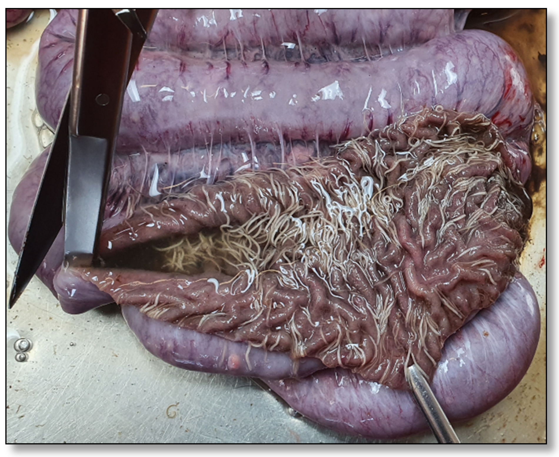
2.2. Section and Diagnostic Workup of Four Fattening Pigs

In August 2021, four fattening pigs (19.5, 23, 23.5 and 30 kg) were sent to the University Clinic for Swine, Vetmeduni Vienna, Austria, for diagnostic workup. All four animals had poor body conditions (Figure 1). Rectal temperatures of the animals were within the physiological range, except for one pig which suffered from hypothermia (35.3 °C). Two pigs also suffered from kyphosis, anaemia and dyspnoea. All four animals were euthanised for necropsy according to appropriate welfare procedures. During necropsy, high numbers of adult whipworms attached to the mucosa of the ileum, caecum and particularly the colon were observed in all animals (Figure 2).
In histopathologic examination, moderate infiltration with eosinophilic granulocytes in the lamina propria and hyperplasia of goblet cells were observed in all parts of the intestine affected by whipworms. The cranial parts of the whipworms embedded in the mucosa were detectable in the histopathologic investigation in multiple locations (Figure 3).
Additionally, hyperplasia of the bronchoalveolar lymphatic tissue was observed in the lungs of two animals. PCRs for detection of L. intracellularis, Brachyspira hyodysenteriae and B. pilosicoli were negative in scrapings from the ileal and colonic mucosa (Table 1, Supplementary File S1). Salmonella spp. were not detectable via enrichment with tetrathionate and consecutive microbiological examination of the tonsil, caecum and ileocaecal lymph node. While M. hyopneumoniae DNA was detectable in pneumonic lung tissue of two animals, PCR for the detection of Porcine Reproductive and Respiratory Syndrome Virus (PRRSV) RNA was negative in the lungs and lymphatic tissue of all animals. Upon faecal flotation with zinc sulphate, a high number of T. suis eggs was observed in faecal samples from all four animals. For details on the methods, refer to Table 1.

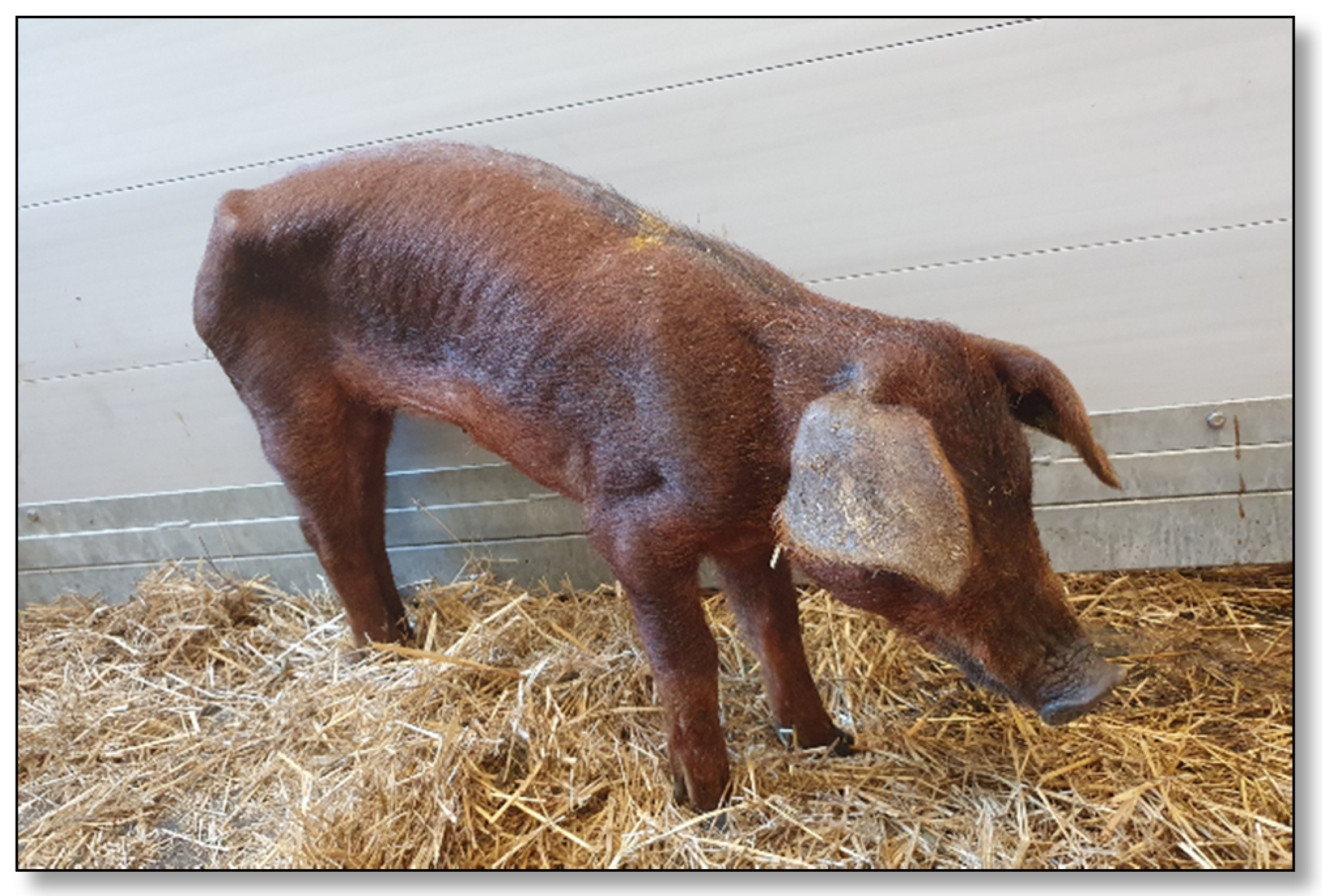
2.3. Farm Visit, Clinical Examination and Sampling
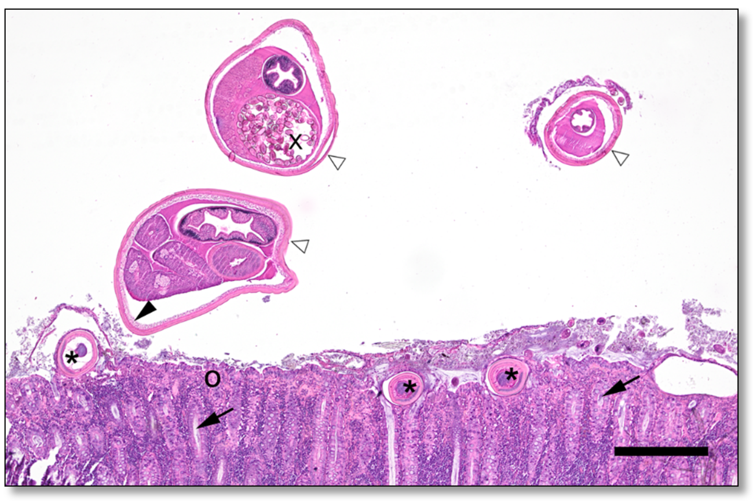
After the confirmation of trichurosis as the cause of colitis and poor body condition in the necropsied animals, a farm visit was conducted in order to evaluate the worm burden of the animals on the farm in more detail and to evaluate possible therapeutic and prophylactic measures for the farm. The body condition of 34 fattening pigs on pasture and 20 pre-fattening pigs in the semi-open pre-fattening stable was assessed (Table 2). Faeces (~10 g/animal) were sampled directly from the rectum, and faecal consistency was grossly evaluated for all animals. Faecal samples could be collected in sufficient amounts from all but two pigs, resulting in a total number of 52 faecal samples. Additionally, soil samples from the wallow, and the feeding area and a straw sample from the finisher unit were taken as well.
Faecal flotation for detection of parasitic stages (including determination of EPG in samples with a high grade of eggs upon flotation) and PCRs for the detection of L. intracellularis, B. hyodysenteriae and B. pilosicoli DNA were performed from 52 faecal samples and environmental samples, as described in Supplementary File S1.

2.4. Results

While all pre-fattening pigs had a good (n = 15) or moderate (n = 5) body condition, more than one-third of the fattening pigs kept on pasture had either a poor or bad body condition (Table 2). Out of all 20 pre-fattening pigs, the faecal consistency of 18 pigs was firm, while pasty faeces were recorded for the other two pigs (Table 2). Altogether, six fattening pigs had semiliquid diarrhoea, and eight animals had liquid diarrhoea (Table 2).
Adult whipworms were visible in faecal samples from five fattening pigs. While no T. suis eggs or stages of other helminth species were detected in the 20 faecal samples from the pre-fattening pigs, all but three samples of the 32 faecal samples from the fattening pigs kept on pasture were positive for Trichuris at flotation, 17 of them in a “high” grade (>10 eggs per coverslip). High numbers of T. suis eggs were detectable in faecal samples from twelve out of 14 fattening pigs with liquid or semiliquid diarrhoea. Nine out of twelve animals with a bad or poor body condition also had high numbers of detectable T. suis eggs in their faecal samples.
The median EPG determined in the 17 samples being highly positive for Trichuris was 23,750 (maximum: 778,000; Table 3). Of those 17 pigs, five had pasty faeces, five had semiliquid and seven had liquid diarrhoea. Firm faecal consistency was not observed in this group (Table 3).
Low numbers of oocysts from Eimeria spp. were detected in one faecal sample (ID 1; T. suis: 1,150 EPG). Another sample which was negative for T. suis contained a large amount (>10 cysts/coverslip) of Balantidium coli cysts. While DNA of neither B. hyodysenteriae nor B. pilosicoli was detectable by PCR, L. intracellularis DNA was detected in faecal samples from two animals which both had poor body conditions and diarrhoea.
Flotation of the straw sample from the finisher unit and the soil sample from the feeding area also revealed high numbers of T. suis eggs, whereas no eggs were detectable in the soil sample from the wallow.
All animals on pasture were immediately treated with a commercial formulation of fenbendazole for pigs at the recommended dose of 5 mg/kg body weight for seven days. Subsequently, the farmer reported a marked improvement in body conditions and an absence of further losses. The treatment was repeated after five weeks to further decrease the worm burden in the herd. Since eggs of T. suis were not detectable in faeces from pre-fattening piglets, no measures were taken for this age group. Regular treatment of all pigs every five weeks to prevent trichurosis was recommended.

3. Discussion

This case report demonstrates that infections with T. suis can cause severe clinical symptoms and economic losses if proper control measures, including hygiene and anthelmintic treatment, are neglected, especially on farms with free-range husbandry on pasture. Therefore, T. suis infections will probably regain importance in pig production due to the increasing demand for pork derived from extensive and free-range husbandry. Despite the fact that this branch of pig production is associated with a high standard of animal welfare, animal health and its impact on animal welfare are discussed far less frequently and sometimes even stand in contrast to common expectations of consumers due to the bad reputation of routine antibiotic therapy [15,16]. This case highlights the importance of animal health care (including proper anthelmintic treatment after correct diagnosis) for animal welfare as well as sustainable productivity.
On the presented farm, approximately 35% of the observed fattening pigs on pasture had a poor or bad body condition, whereas the body condition of the pre-fattening pigs was predominantly good. Since wasting and diarrhoea only occurred after several weeks on pasture, and anthelmintic treatment quickly relieved the pigs’ condition, T. suis was very likely the primary cause of the observed clinical symptoms on this farm. High excretion rates of T. suis eggs in three fattening pigs with good body conditions were presumably due to a recent infection, compared to pigs with poorer body conditions. The presence of diarrhoea in 24 out of 34 examined fattening pigs also emphasises that infections with T. suis should be included as a common differential diagnosis in cases of diarrhoea in pigs, especially in extensive husbandry systems.
Since eggs of T. suis were not detected in faeces from the pre-fattening pigs, which had been purchased five weeks prior to sampling, first contact with T. suis supposedly happened after animals had been moved on the pasture. This is not surprising since husbandry on pasture significantly increases the risk of infections with various helminths [17].
The detection of T. suis eggs in environmental samples further confirmed high contamination of the pasture, as previously described for T. suis-affected farms [18]. Interestingly, the soil sample from the wallow was negative in flotation. This might be due to a patchy distribution of eggs in this part of the pasture due to soil movements by the wallowing pigs.
Due to the high numbers and long persistence of T. suis eggs in the soil, decontamination of the pasture is not feasible [3] and was, therefore, not part of the recommended control strategy for this farm. In addition, yearly pasture rotation between two areas is probably not sufficient to interrupt transmission. To ensure proper decontamination over time, the areas would have to stay unused for several years, which is neither feasible nor economically sensible for most outdoor farming systems. When planning for extensive pig husbandry on pasture, sufficient space for multiple alternating pastures is highly recommended to decrease parasite density on pasture and to prevent a high worm burden. Another strategy could be to start outdoor farming with pigs which are either negative for helminths or are treated prior to husbandry on pasture. This would imply regular and repeated copromicroscopical examinations of pig faeces for the presence of parasitic stages in every batch. However, this is no guarantee for negativity, since several helminthic eggs are shed irregularly or (during prepatency) not at all. This underlines the impact of helminths on outdoor farming of fattening pigs, as it necessitates the development of a proper strategy for controlling parasites not only during but also prior to starting outdoor husbandry on pasture.
Although several case reports of clinical trichurosis in pigs provide data on the number of excreted EPG of infected pigs [3,8,19,20,21], an excretion rate of 778,000 EPG has not been reported so far. It was once estimated that one female T. suis is able to produce approximately 6500 to 7800 eggs per day, which would result in 6–8 EPG produced by each female [22]. Thus, we carefully assume that the fattening pig with 778,000 EPG was affected with over 100,000 female T. suis.
An increase in eosinophilic granulocytes in the large intestine is compatible with previous findings of experimental infections with T. suis [23]. Altogether, all histologic findings including multiple sections of adult whipworms further underpin the diagnosis of clinical trichurosis.
Since neither DNA of B. hyodysenteriae nor B. pilosicoli could be detected via PCR, and Salmonella spp. was not isolated via microbiological examination, some of the most common and relevant causative agents of diarrhoea during the fattening period could be ruled out [24]. While L. intracellularis DNA was detected in two animals with diarrhoea, there was no evidence for clinical manifestations, especially because L. intracellularis is ubiquitous in most European swine stocks [25]. Furthermore, since DNA was only detected in two out of 34 investigated samples, L. intracellularis could be neglected as a primary cause of clinical disease in this case herd. Nevertheless, accelerating effects between T. suis and L. intracellularis could not be ruled out, but they were evaluated as neglectable for this case herd since in case of cumulative pathogenicity of T. suis and L. intracellularis, higher in-herd prevalence rates may be expected and diagnosed. Balantidium coli was detected in the faecal sample of one pig. However, the impact of Balantidium coli on pigs has not been completely elucidated yet. While several authors assume that it is a non-pathogenic coloniser of porcine intestines, there is evidence that colonisation with Balantidium coli can lead to mild colitis [26,27,28].

4. Conclusions

Altogether, this case confirms that heavy T. suis infections can lead to severe clinical symptoms in pigs, even though trichurosis has become rarer with the introduction of closed stables and slatted floors. Therefore, farmers and veterinarians should not abandon regular anthelmintic treatment and routine parasitological examination, particularly on farms with extensive husbandry on pasture, to ensure animal health, animal welfare and farm productivity.

Supplementary Materials

The following are available online at https://www.mdpi.com/article/10.3390/pathogens11070775/s1, File S1: Methods of PCR for detection.

Author Contributions

Conceptualisation, L.S., M.B., R.R. and A.J.; methodology, R.B., C.W.-L., B.H., A.J., N.N. and J.M.; validation, A.J. and C.W.-L.; formal analysis, R.R., M.A.H. and M.B.; resources, A.J., L.S. and C.W.-L.; data curation, M.B., M.A.H., R.R. and L.S.; writing—original draft preparation, R.R. and M.B.; writing—review and editing, A.J.; visualisation, M.B., M.A.H. and R.B.; supervision, L.S. and A.J.; project administration, M.B., R.R. and L.S. All authors have read and agreed to the published version of the manuscript.

Funding

This research received no external funding.

Institutional Review Board Statement

The study was conducted according to the guidelines of good veterinary practice. The study was performed under conditions of routine diagnostic work-up in the sense of herd health management. Therefore, ethical approval was not required.

Informed Consent Statement

Not applicable.

Data Availability Statement

Not applicable.

Acknowledgments

The authors gratefully acknowledge the technical support of the laboratory staff of Vetmeduni Vienna. Open Access Funding by the University of Veterinary Medicine Vienna.

Conflicts of Interest

The authors declare no conflict of interest.

References

  1. Beer, R.J. Experimental Infection of Man with Pig Whipworm. BMJ 1971, 2, 44. [Google Scholar] [CrossRef] [PubMed] [Green Version]
  2. Kringel, H.; Roepstorff, A. Trichuris Suis Population Dynamics Following a Primary Experimental Infection. Vet. Parasitol. 2006, 139, 132–139. [Google Scholar] [CrossRef] [PubMed]
  3. Pittman, J.S.; Shepherd, G.; Thacker, B.J. Trichuris Suis in Finishing Pigs: Case Report and Review. J. Swine Health Prod. 2010, 18, 306–313. [Google Scholar]
  4. Beer, R.J. Studies on the Biology of the Life-Cycle of Trichuris Suis. Parasitology 1973, 67, 253–262. [Google Scholar] [CrossRef]
  5. Brewer, M.T.; Greve, J.H. Internal Parasites. In Diseases of Swine, 11th ed.; Zimmerman, J.J., Karriker, L.A., Ramirez, A., Schwartz, K.J., Stevenson, G.W., Jianqiang, Z., Eds.; Wiley-Blackwell/American Association of Swine Veterinarians: Hoboken, NJ, USA, 2019; pp. 1028–1040. ISBN 978-1-119-35090-3. [Google Scholar]
  6. Nicholls, J.; Obendorf, D.L. Application of a Composite Faecal Egg Count Procedure in Diagnostic Parasitology. Vet. Parasitol. 1994, 52, 337–342. [Google Scholar] [CrossRef]
  7. Marti, O.G.; Stewart, T.B.; Hale, O.M. Comparative Efficacy of Fenbendazole, Dichlorvos, and Levamisole HCl against Gastrointestinal Nematodes of Pigs. J Parasitol. 1978, 64, 1028–1031. [Google Scholar] [CrossRef] [PubMed]
  8. Caron, Y.; Delleur, V.; Cassart, D.; Losson, B.; Laitat, M. A Case of Trichurosis in Gilts and Fattening Pigs. JMM Case Rep. 2014, 1, e003582. [Google Scholar] [CrossRef] [Green Version]
  9. La, T.; Phillips, N.D.; Hampson, D.J. Development of a Duplex PCR Assay for Detection of Brachyspira hyodysenteriae and Brachyspira pilosicoli in Pig Feces. J. Clin. Microbiol. 2003, 41, 3372–3375. [Google Scholar]
  10. Jones, G.F.; Ward, G.E.; Murtaugh, M.P.; Lin, G.; Gebhart, C.J. Enhanced Detection of Intracellular Organism of Swine Proliferative Enteritis, Ileal Symbiont Intracellularis, in Feces by Polymerase Chain Reaction. J. Clin. Microbiol. 1993, 31, 2611–2615. [Google Scholar]
  11. Ellis, E.M.; Williams, J.E.; Mallinson, E.T.; Snoeyenbos, G.H.; Martin, W.J. Culture Methods for the Detection of Animal Salmonellosis and Arizonosis; Iowa State University Press: Ames, IA, USA, 1976. [Google Scholar]
  12. Kurth, K.T.; Hsu, T.; Snook, E.R.; Thacker, E.L.; Thacker, B.J.; Minion, F.C. Use of a Mycoplasma Hyopneumoniae Nested Polymerase Chain Reaction Test to Determine the Optimal Sampling Sites in Swine. J. Vet. Diagn. Investig. 2002, 14, 463–469. [Google Scholar] [CrossRef] [Green Version]
  13. Fetzer, C.; Pesch, S.; Ohlinger, V. High Risk of False Positive Results in a Widely Used Diagnostic Test for Detection of the Porcine Reproductive and Respiratory Syndrome Virus (PRRSV). Vet. Microbiol. 2006, 115, 21–31. [Google Scholar] [CrossRef]
  14. Deplazes, P.; von Samson-Himmelstjerna, G.; Zahner, H.; Joachim, A.; Mathis, A.; Taubert, A.; Strube, C. Parasitologie für die Tiermedizin, 4th ed.; Thieme (Verlag): Stuttgart, Germany, 2020; ISBN 978-3-13-242138-7. [Google Scholar]
  15. Mondéjar-Jiménez, J.A.; Sánchez-Cubo, F.; Mondéjar-Jiménez, J. Consumer Behaviour towards Pork Meat Products: A Literature Review and Data Analysis. Foods 2022, 11, 307. [Google Scholar] [CrossRef]
  16. Bradford, H.; McKernan, C.; Elliott, C.; Dean, M. Consumers’ Perceptions and Willingness to Purchase Pork Labelled ‘Raised without Antibiotics’. Appetite 2022, 171, 105900. [Google Scholar] [CrossRef] [PubMed]
  17. Thomsen, L.E.; Mejer, H.; Wendt, S.; Roepstorff, A.; Hindsbo, O. The Influence of Stocking Rate on Transmission of Helminth Parasites in Pigs on Permanent Pasture during Two Consecutive Summers. Vet. Parasitol. 2001, 99, 129–146. [Google Scholar] [CrossRef]
  18. Lindgren, K.; Gunnarsson, S.; Höglund, J.; Lindahl, C.; Roepstorff, A. Nematode Parasite Eggs in Pasture Soils and Pigs on Organic Farms in Sweden. Org. Agr. 2020, 10, 289–300. [Google Scholar] [CrossRef] [Green Version]
  19. Boes, J.; Johansen, M.V.; Eriksen, L.; Bøgh, H.O.; Nansen, P.; Stephenson, L.S. False-Positive Trichuris Suis Egg Counts in Pigs in Relation to Coprophagia. Parasite 1998, 5, 91–93. [Google Scholar] [CrossRef] [PubMed] [Green Version]
  20. Kagira, J.M.; Kanyari, P.N.; Githigia, S.M.; Maingi, N.; Ng’ang’a, J.C.; Gachohi, J.M. Risk Factors Associated with Occurrence of Nematodes in Free Range Pigs in Busia District, Kenya. Trop. Anim. Health Prod. 2012, 44, 657–664. [Google Scholar] [CrossRef]
  21. Lopes, W.D.Z.; Teixeira, W.F.P.; Felippelli, G.; Cruz, B.C.; Buzulini, C.; Maciel, W.G.; Fávero, F.C.; Gomes, L.V.C.; Prando, L.; Bichuette, M.A.; et al. Anthelmintic Efficacy of Ivermectin and Abamectin, Administered Orally for Seven Consecutive Days (100 Μg/Kg/Day), against Nematodes in Naturally Infected Pigs. Res. Vet. Sci. 2014, 97, 546–549. [Google Scholar] [CrossRef]
  22. Pedersen, S.; Saeed, I. Acquired Immunity to Trichuris Suis Infection in Pigs. Parasitology 2001, 123, 95–101. [Google Scholar] [CrossRef]
  23. Krag, L.; Thomsen, L.E.; Iburg, T. Pathology of Trichuris Suis Infection in Pigs Fed an Inulin- and a Non-Inulin-Containing Diet. J. Vet. Med. Ser. A 2006, 53, 405–409. [Google Scholar] [CrossRef]
  24. Jacobson, M.; Hård af Segerstad, C.; Gunnarsson, A.; Fellström, C.; de Verdier Klingenberg, K.; Wallgren, P.; Jensen-Waern, M. Diarrhoea in the Growing Pig—A Comparison of Clinical, Morphological and Microbial Findings between Animals from Good and Poor Performance Herds. Res. Vet. Sci. 2003, 74, 163–169. [Google Scholar] [CrossRef]
  25. Arnold, M.; Crienen, A.; Swam, H.; von Berg, S.; Jolie, R.; Nathues, H. Prevalence of Lawsonia Intracellularis in Pig Herds in Different European Countries. Porc. Health Manag. 2019, 5, 3–11. [Google Scholar] [CrossRef]
  26. Sharma, S.; Harding, G. Necrotizing Lung Infection Caused by the Protozoan Balantidium Coli. Can. J. Infect. Dis. 2003, 14, 163–166. [Google Scholar] [CrossRef] [PubMed] [Green Version]
  27. Ferry, T.; Bouhour, D.; De Monbrison, F.; Laurent, F.; Dumouchel-Champagne, H.; Picot, S.; Piens, M.A.; Granier, P. Severe Peritonitis Due to Balantidium Coli Acquired in France. Eur. J. Clin. Microbiol. Infect. Dis. 2004, 23, 393–395. [Google Scholar] [CrossRef] [PubMed]
  28. Symeonidou, I.; Tassis, P.; Gelasakis, A.Ι.; Tzika, E.D.; Papadopoulos, E. Prevalence and Risk Factors of Intestinal Parasite Infections in Greek Swine Farrow-To-Finish Farms. Pathogens 2020, 9, 556. [Google Scholar] [CrossRef] [PubMed]
Figure 1. One of four fattening pigs with a poor body condition sent for diagnostic workup at the University Clinic for Swine.
Pathogens 11 00775 g001
Figure 2. Large numbers of adult Trichuris suis attached to the intestinal mucosa of the colon of one of the necropsied pigs.
Pathogens 11 00775 g002
Figure 3. Histologic section (haematoxylin and eosin staining; bar = 400 µm) of colonic mucosa from one of the necropsied pigs. Asterisk: anterior end of a worm embedded in the intestinal epithelium, black arrowhead: bacillary band, white arrowheads: posterior end of a worm in the intestinal lumen, x: eggs, black arrow: crypts of Lieberkühn, o: Lamina propria mucosae.
Pathogens 11 00775 g003
Table 1. Laboratory diagnostic methods for pathogen detection applied in this study.
PathogenDetection MethodSpecimenReference
Lawsonia intracellularis, Brachyspira hyodysenteriae, Brachyspira pilosicoliPCR Ileal and Colonic Mucosal ScrapingsSupplementary File S1 [9,10]
Salmonella spp.Microbiological examinationTonsil, Caecum, Ileocaecal Lymph Node[11]
Mycoplasma hyopneumoniaePCRLung [12]
Porcine Reproductive and Respiratory Syndrome VirusqRT—PCRLung, Tonsil, Tracheobronchial Lymph Node [13]
Helminth eggsFlotation (saturated ZnSO4); modified McMaster techniqueFaeces[14]
Table 2. Number of pigs with different body conditions and faecal consistency.
Body ConditionFattening Pigs (n = 34)Pre-Fattening Pigs (n = 20)
Bad7 (21%)0 (0%)
Poor5 (15%)0 (0%)
Moderate13 (38%)5 (25%)
Good9 (26%)15 (75%)
Faecal ScoreFattening Pigs (n = 34)Pre-Fattening Pigs (n = 20)
Liquid8 (24%)0 (0%)
Semiliquid6 (18%)0 (0%)
Pasty10 (29%)2 (10%)
Normal10 (29%)18 (90%)
Table 3. EPG faeces of 17 fattening pigs with a high number of T. suis eggs in flotation and the respective faecal consistency and body condition scores.
IDFaecal ConsistencyBody ConditionTrichuris EPG (McMaster)IDFaecal ConsistencyBody ConditionTrichuris EPG (McMaster)
1LiquidBad115010LiquidGood26,700
2SemiliquidBad380011PastyGood32,560
3PastyBad620012LiquidPoor33,680
4SemiliquidPoor11,50013SemiliquidBad35,450
5SemiliquidGood13,25014PastyModerate35,800
6LiquidModerate14,25015PastyModerate44,500
7LiquidModerate20,55016LiquidModerate98,870
8LiquidBad22,45017SemiliquidBad778,000
9PastyPoor23,750
Publisher’s Note: MDPI stays neutral with regard to jurisdictional claims in published maps and institutional affiliations.

