Article

Terpinen-4-ol, the Main Bioactive Component of Tea Tree Oil, as an Innovative Antimicrobial Agent against Legionella pneumophila

by
Francesca Mondello
1,2,*,†,
Stefano Fontana
1,
Maria Scaturro
1,3,
Antonietta Girolamo
1,2,
Marisa Colone
4,
Annarita Stringaro
2,4,
Maura Di Vito
2,5,*,† and
Maria Luisa Ricci
1,3,*,†
1
Department of Infectious Diseases, Istituto Superiore di Sanità, Viale Regina Elena, 299, 00161 Rome, Italy
2
Società Italiana per la Ricerca sugli Oli Essenziali—SIROE (Italian Society for Research on Essential Oils), Viale Regina Elena, 299, 00161 Rome, Italy
3
ESCMID Study Group for Legionella Infections (ESGLI), 4001 Basel, Switzerland
4
National Center for Drug Research and Evaluation, Istituto Superiore di Sanità, Viale Regina Elena, 299, 00161 Rome, Italy
5
Dipartimento di Scienze Biotecnologiche di Base, Cliniche Intensivologiche e Perioperatorie, Università Cattolica del Sacro Cuore, Largo A. Gemelli 8, 00168 Rome, Italy
*
Authors to whom correspondence should be addressed.
The authors contributed equally to this work.
Pathogens 2022, 11(6), 682; https://doi.org/10.3390/pathogens11060682
Submission received: 11 May 2022 / Revised: 8 June 2022 / Accepted: 10 June 2022 / Published: 14 June 2022
(This article belongs to the Special Issue 10th Anniversary of Pathogens—Feature Papers)

Abstract

:
Legionella pneumophila (Lp), responsible for a severe pneumonia called Legionnaires’ disease, represents an important health burden in Europe. Prevention and control of Lp contamination in warm water systems is still a great challenge often due to the failure in disinfection procedures. The aim of this study was to evaluate the in vitro activity of Terpinen-4-ol (T-4-ol) as potential agent for Lp control, in comparison with the essential oil of Melaleuca alternifolia (tea tree) (TTO. Minimum Inhibitory Concentration (MIC) and Minimum Bactericidal Concentration (MBC) of T-4-ol were determined by broth micro-dilution and a micro-atmosphere diffusion method to investigate the anti-Lp effects of T-4-ol and TTO vapors. Scanning Electron Microscopy (SEM) was adopted to highlight the morphological changes and Lp damage following T-4-ol and TTO treatments. The greatest antimicrobial activity against Lp was shown by T-4-ol with a MIC range of 0.06–0.125% v/v and MBC range of 0.25–0.5% v/v. The TTO and T-4-ol MIC and MBC decreased with increasing temperature (36 °C to 45 ± 1 °C), and temperature also significantly influenced the efficacy of TTO and T-4-ol vapors. The time-killing assay showed an exponential trend of T-4-ol bactericidal activity at 0.5% v/v against Lp. SEM observations revealed a concentration- and temperature- dependent effect of T-4-ol and TTO on cell surface morphology with alterations. These findings suggest that T-4-ol is active against Lp and further studies may address the potential effectiveness of T-4-ol for control of water systems.

1. Introduction

Legionella, an opportunistic Gram-negative bacterium, ubiquitously found in freshwater environments and artificial water systems, represents a serious public health problem, mainly for elderly and immunocompromised people. The genus Legionella currently consists of 63 species, of which Legionella pneumophila (Lp) is responsible for most human infections [1]. Lp can be distinguished into 15 serogroups, of which Lp serogroup 1 is the most virulent and associated with over 80% of Legionnaires’ disease (LD) cases in Europe [1]. The infection occurs through inhalation or aspiration of Lp-contaminated aerosols and usually gives rise to two clinical syndromes: a life-threatening pneumonia called LD or Pontiac fever, a self-limited flu-like illness [2,3].
Since 2008, when LD was first reported to The European Surveillance System (Tessy), coordinated by the European Centre for Disease Prevention and Control (ECDC), an increasing trend (with the exception and unexplained decrease in 2011) has been observed, reaching a peak of 11,405 LD cases in 2018 (23% more cases than in 2017). This number remained unchanged in 2019 but decreased in 2020 following the Covid-19 pandemic [4,5]. The same trend has been observed in Italy, as incidence rates per million of population increased from 16.1 in 2011 to 52.9 in 2019, with a 35% notable decrease in 2020 [6,7,8]. Community and travel-associated outbreaks of LD have been mostly linked with cooling towers, building water-distribution systems, spas, fountains, and public baths. Water-distribution systems and aerosol-producing medical devices are main sources of health care-associated legionellosis [9]. Person-to-person transmission of LD is exceptional [10], so prevention control measures have focused on eliminating the pathogen from water systems. To date, the long-term control of Legionella contamination of water distribution and air conditioning systems still represents a great challenge, to both health and the structural complexity of the water systems [1]. Lp proliferate inside free-living protozoa [11] and colonize multispecies biofilms [12,13] that allow persistence in water systems even when high temperature, biocide treatment, scale, and corrosion are control measures. Therefore, the development of alternative or integrative methods that can counteract and prevent the colonization of Legionella in water distribution systems and minimize exposure to potentially hazardous chemicals is considered a high priority. Of all the pathogens present in water, Legionella is the one that causes the greatest health burden in the EU [14]. Recently, antimicrobial agents extracted from a variety of plants have been evaluated in particular essential oils (EOs) [15,16,17,18].
The biological activities of EOs are considered to have preventive and therapeutic benefits [19,20,21]. The antimicrobial activity of EOs and their components has been extensively investigated, both in aqueous and in vapor phases [22,23,24], against bacteria, fungi, viruses, parasites, and insects, resulting in the formulation of patents [25] and applications in the pharmaceutical technology [26]. In 2009, we pioneered in vitro bactericidal efficacy of the essential oil of Melaleuca alternifolia (tea tree) (TTO) against Lp strains by improving the susceptibility method with vapor control of TTO [27].
To our knowledge, studies on the in vitro activity of Terpinen-4-ol (T-4-ol), the main bioactive component of TTO, against Lp, are lacking. The aim of this study was to evaluate the in vitro activity of T-4-ol, both in the aqueous and the in vapor phase, against Lp sg1 and sg6 human and environmental isolates, proven to be among the most widespread in human infection in Italy and Europe [1,28].
This is the first report that highlights the in vitro susceptibility of Lp to T-4-ol in comparison to the anti-Lp activity of TTO, with the aim of developing new disinfectants for contamination control of Legionella in water distribution systems.

2. Results

2.1. Comparison of Anti-Legionella Activities of Terpinen-4-ol and Tea Tree Oil Analyzed by Broth Micro-Dilution Method

We compared the activities of T-4-ol and TTO against all the different strains of Lp sg1 and sg6 using the broth micro-dilution method with sterilized Transparent Microplate Sealer (TMS) at 36 ± 1 °C. This method was used to exclude vapor diffusion spreading over the wells, responsible for inter-experimental variation of Minimum Inhibitory Concentration (MIC) and Minimum Bactericidal Concentration (MBC), as described previously [27]. Lp was sensitive to T-4-ol, with MIC ranging from 0.06 to 0.125% v/v, and MBC at 0.5% v/v, while TTO MIC and MBC ranging from 0.125 to 0.5% v/v, and 0.5–1.0% v/v respectively (Table 1).
The MIC and MBC average values of T-4-ol and TTO against the two Lp serogroups were compared (Table 2). The uniformity of MIC values at 0.25% v/v for TTO and at 0.125% v/v for T-4-ol are noticeable, with rare exceptions, while the MBC values at 0.5% v/v was the same for both TTO and for the main component T-4-ol in both tables (Table 1 and Table 2). No difference either between the two serogroups or among the reference strains environmental and clinical isolates was seen, and for this reason the strains were grouped according to their serogroup in Table 2.
Figure 1 shows the comparison of T-4-ol and TTO inhibitory activities at temperatures of 36 °C, 40 °C, and 45 ± 1 °C against six strains of Lp sg1 and sg6 by broth micro-dilution method. MIC values of T-4-ol were always lower than the corresponding MICs obtained by testing the TTO. Furthermore, MICs of T-4-ol and TTO decreased with increasing temperature. This modulation was more evident for TTO, where the temperature significantly improved the MIC values (p < 0.0002). Under the same conditions, the MIC values obtained when testing T-4-ol at 36 °C and 40 ± 1 °C were always statistically more significant than those obtained with TTO (pT-4-ol < 0.0001 and pTTO < 0.0002, respectively).
The comparison of TTO and T-4-ol bactericidal activities (MBC) at temperatures of 36 °C, 40 °C and 45 ± 1 °C against six strains of Lp serogroups 1 and 6 with broth micro-dilution method was showed in Figure 2.
The temperature-dependent trend described for the MIC variation was also observed for MBCs. The MBC values with T-4-ol were lower than the correspondent values obtained with TTO. Furthermore, for both the natural products tested, the MBCs at 45 °C showed statistically significant differences to both 36 ± 1 °C (p < 0.002) and 40 °C (pT-4-ol < 0.002 and pTTO < 0.03, respectively).

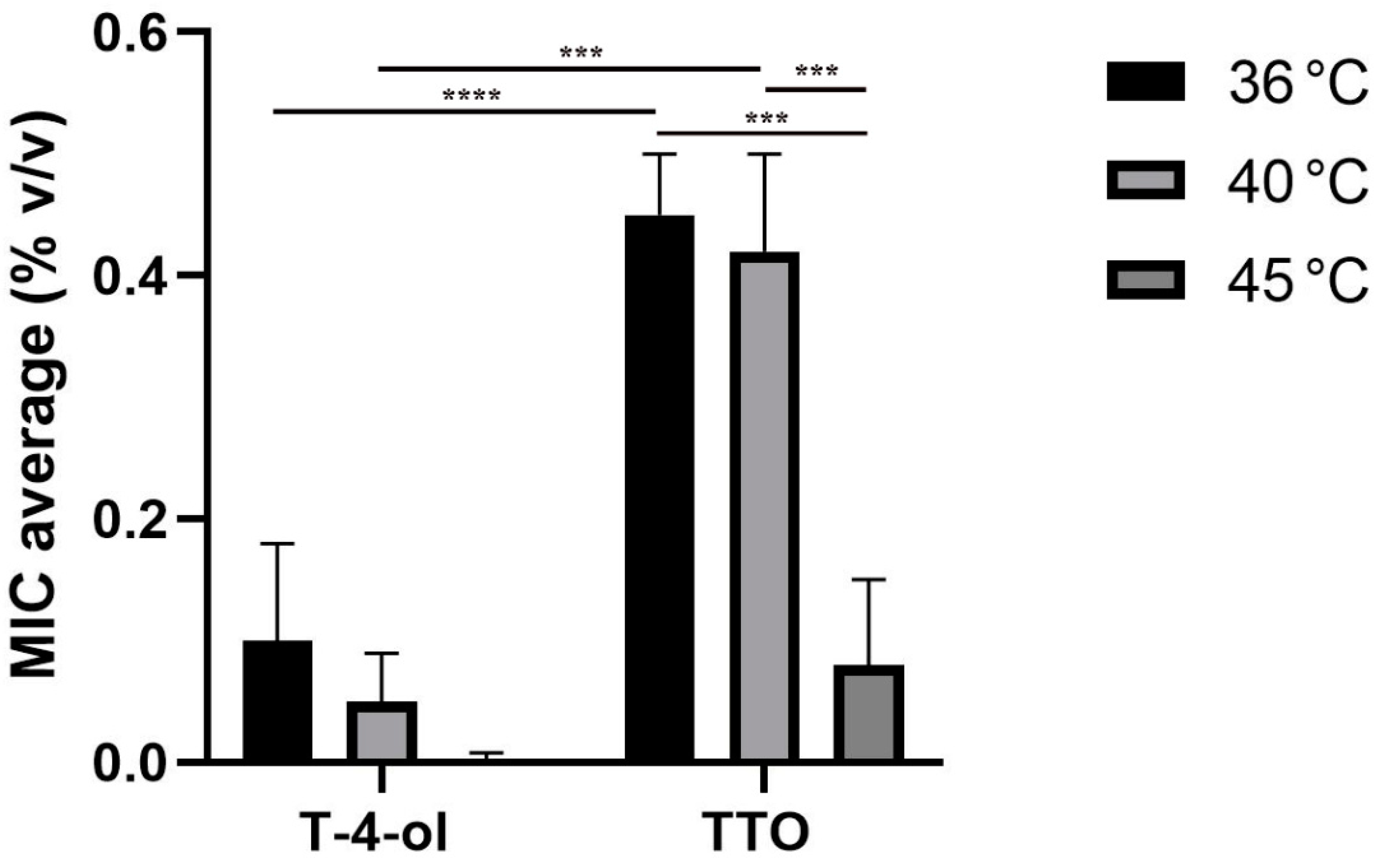
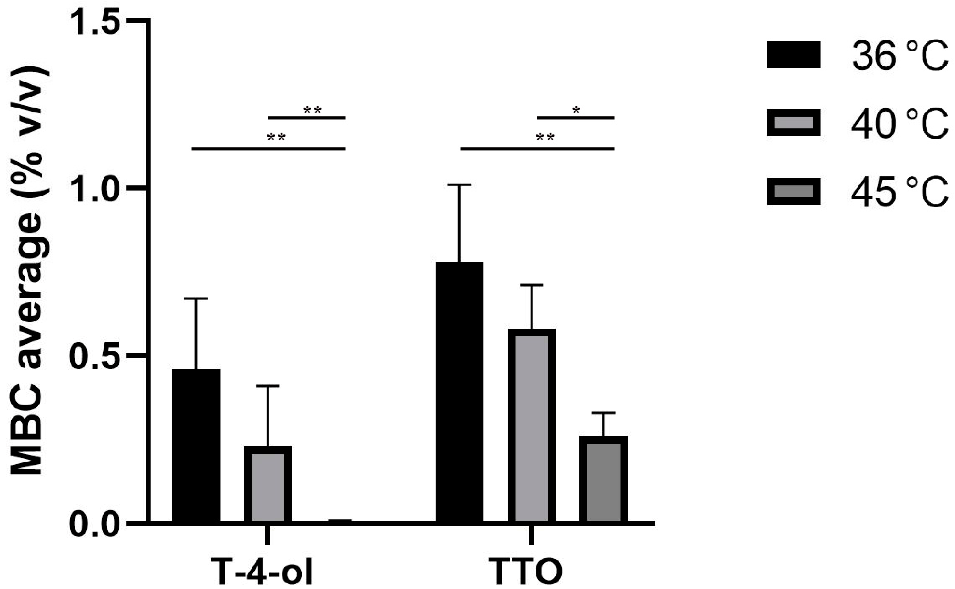
2.2. Comparison of Anti-Legionella Activities of Terpinen-4-ol and Tea Tree Oil Analyzed by the Micro-Atmosphere Diffusion Method

The anti-Lp activities of T-4-ol and TTO were also tested with a micro-atmosphere diffusion method, which allowed the contact of only T-4-ol or TTO vapors with bacterial cells at different temperatures. The same six selected Lp strains, examined by the broth micro-dilution method, were tested at 36 °C and 45 ± 1 °C to evaluate the antimicrobial effects of T-4-ol and TTO vapors. With this method, the vapor originating from T-4-ol caused total growth inhibition, as measured after 2 days of culture, with total killing after 7 days of culture. The results of the anti-Lp activities of the T-4-ol and TTO vapors are shown in Table 3 and Figure 3.
The comparison of the anti-Lp activities of T-4-ol and TTO vapors at temperatures of 36 °C and 45 ± 1 °C for the selected strains of Lp sg1 (one reference strain ATCC 33152, one clinical strain and one environmental strain) and Lp sg6 (one reference strain ATCC 33215, one clinical strain and one environmental strain) after 7 days of incubation are shown in Table 3. The diameter of the inhibition of the tested Lp strains was variable and was sometimes equal to the internal diameter of the Petri dish (90 mm). The Lp sg1 ATCC 33152 strain did not grow at 45 °C therefore it was not considered for experiments at that temperature [29]. The T-4-ol/TTO ratio was always >1, which mean that the average of the inhibition halos obtained with T-4-ol was always greater than that obtained with TTO at 36 ± 1 °C. The T-4-ol/TTO ratio was instead ≥1 at 45 ± 1 °C.

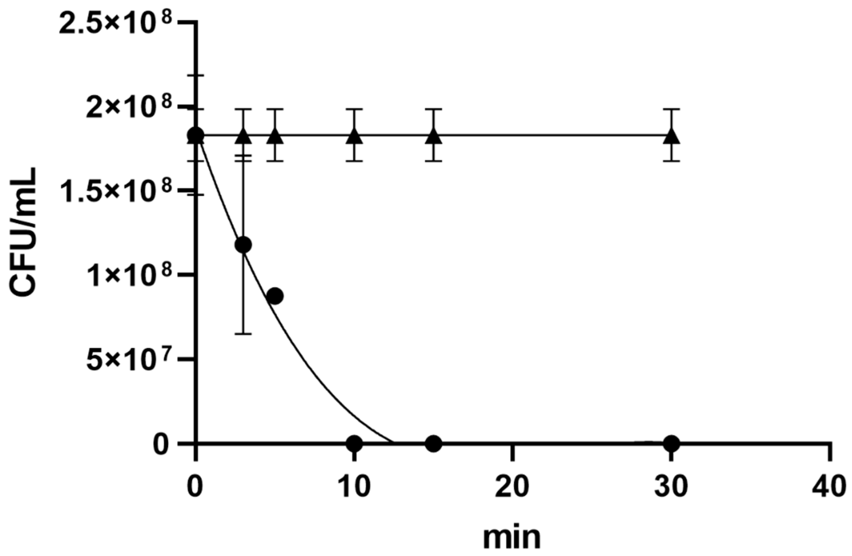
2.3. Anti-Legionella pneumophila Activity of Terpinen-4-ol Analyzed by Time-Killing Test

In the time-killing experiments, the in vitro survival of Legionella as a function of T-4-ol concentration and contact times was investigated (Figure 3). We highlighted the exponential trend of T-4-ol bactericidal activity against Lp. The vitality of Lp decreased over time in the presence of a concentration of 0.5% v/v of T-4-ol (corresponding to the MBC value). After 5 min, the mortality percentage was 54.3% ± 3.2%, while after 10 min, the mortality was 100%. The graph shows a polynomial decrease of Legionella vitality as a function of time (R = 0.921). The t-test indicated that the activity of T-4-ol at a concentration of 0.5% v/v was statistically significant (p = 0.0136).

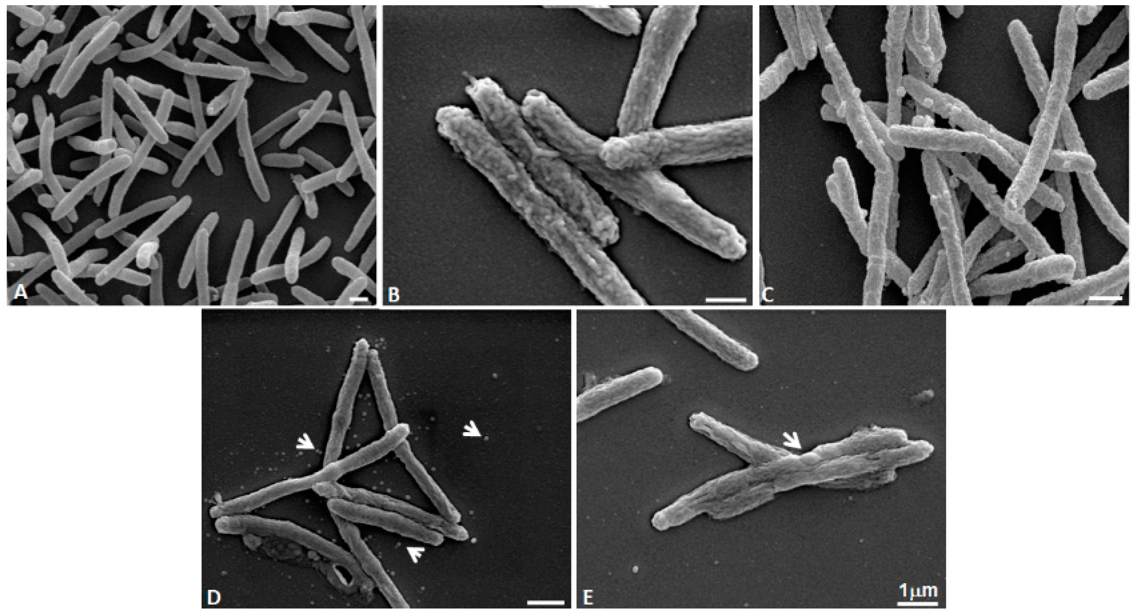
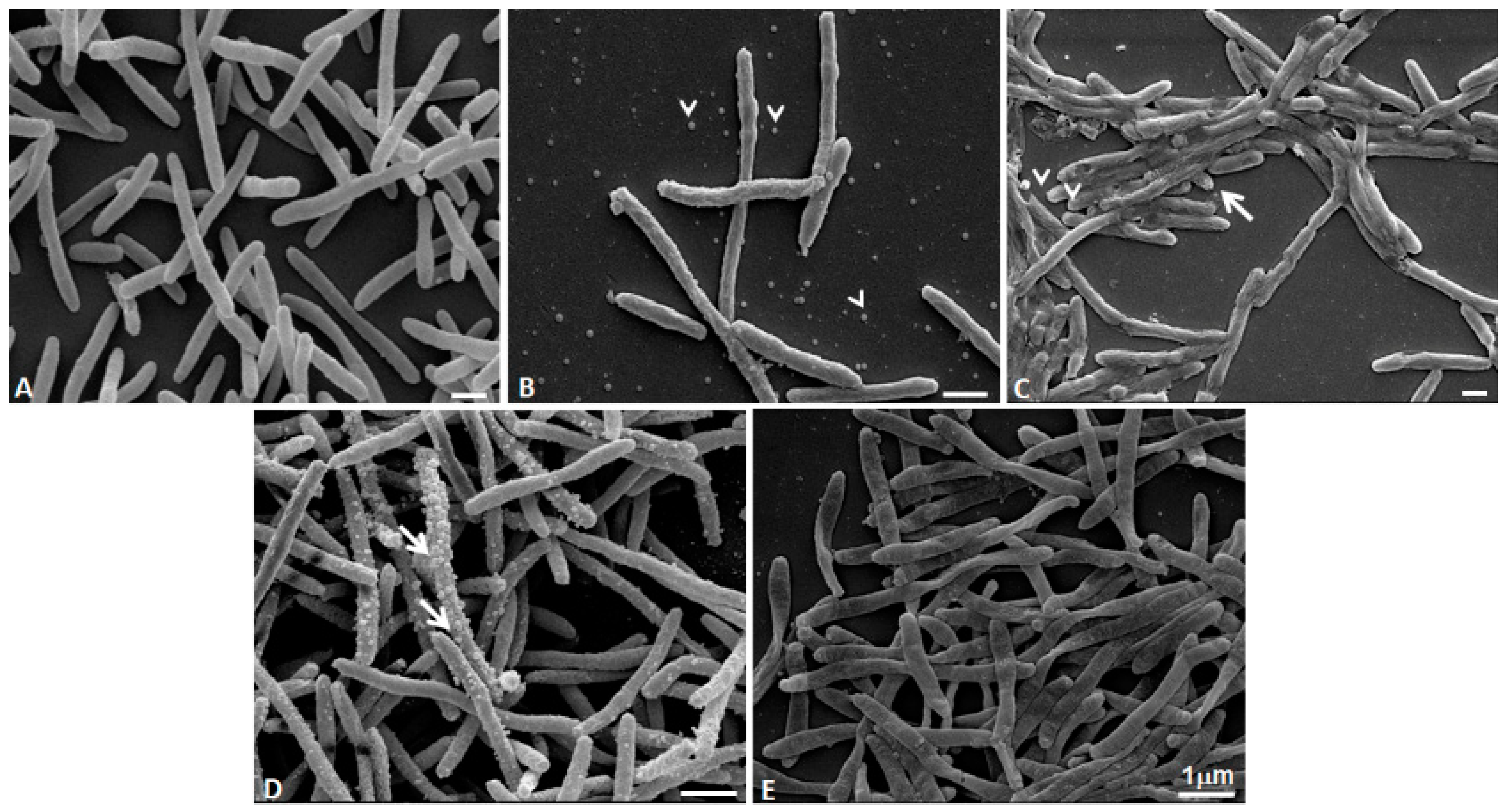
2.4. Morphological Effects of Terpinen-4-ol and Tea Tree Oil on Legionella pneumophila Analyzed by Scanning Electron Microscopy (SEM) at Different Temperatures

SEM analyses performed on untreated bacteria (controls) at 30 °C, 40 °C, and 45 °C revealed the typical rod shape and that temperature had a no effect on Lp morphology. Most cells were 2–10 µm long and the Lp surface appeared slightly wrinkled.
Figure 4 shows the effect of treatments (1%, 2.5% v/v of TTO and 1% v/v of T-4-ol) at 40 °C. Specifically, in Figure 4B (1% v/v TTO treatment) the surface morphology appears more wrinkled than control cells. Exposure to T-4-ol (0.42% v/v) induced numerous small bleb formations on the bacterial cell surface. When the highest (2.5% v/v TTO and 1% v/v T-4-ol) concentrations were used, Lp modifications were more evident. Figure 4D shows that 2.5% v/v TTO caused bleb formations and the cell surface was more rugose, with some cells appearing to swell at the center. Openings were present on surface, probably as a result of the inner membrane rupturing. Moreover, numerous blebs were present on the coverslips (Figure 4D). Figure 4E shows that treatment with 1% v/v T-4-ol induced the swelling and wrinkling of the cell surface. In addition, cells had a spheroplast-like collapsed structure.
Figure 5 shows the effect of the same treatments at 45 °C. In particular, Figure 5A shows bacteria at 45 °C temperature-exposed: cells appear with their typical rod shape without any ultrastructural modification due to the increase in temperature. In Figure 5B (1% v/v TTO) the cell surface appears more wrinkled with mid-cell swellings visible (arrowheads). Blebs are also present on the bacteria surface and on coverslips (arrowheads). Figure 5C shows multiple damages to the cell that have merged (arrow) with outer cell wall and inner membrane ruptures (arrowheads). TTO at a concentration of 2.5% v/v induced numerous blebs that are visible on numerous rugose cell surfaces (arrows) (Figure 5D). T-4-ol at a concentration of 1% v/v produced collapsed cells (Figure 5E). The cell surfaces appeared wrinkled, but their organization was different from the cell surface shown in Figure 5C.

3. Discussion

Investigations on the antimicrobial activity of EOs on Lp are limited in number and have only circumstantially examined Lp while they were in a context of EOs activity against multiple other bacteria [30,31,32,33,34,35,36,37,38].
Melaleuca alternifolia (Maiden & Betch) Cheel (tea tree) EO (TTO) has been studied for its antimicrobial activity, including in the vapor phase [39], against a large number of Gram-negative and -positive bacteria and fungi by in vitro, preclinical and clinical studies [22,40,41]. TTO contains more than 100 components, such as monoterpenes, sesquiterpenes, and phenolic compounds. The most abundant compound is T-4-ol, in an amount of at least 30% [40], but is also present in many EOs from other plant species. Various activities of T-4-ol have been described in the literature, encompassing antibacterial, antifungal, antiviral, and insecticidal effects, as well as antitumoral and anti-inflammatory activity [42,43,44,45,46,47]. Furthermore, it was highlighted that T-4-ol is the principal component responsible for the antimicrobial activity of TTO, as both possess similar antimicrobial effects [48,49]. Regarding the activity of T-4-ol against Lp, there has only been one study [33]. This study investigated the antibacterial activity of a fraction containing 53% of T-4-ol obtained from EO of Chamaecyparis obtusa. The authors showed high bacteriostatic activity against all Gram-positive and -negative tested strains, including a single Lp strain. However, the fraction was made up of many monoterpenes and sesquiterpenes that probably contributed to T-4-ol antibacterial activity. Pure synthetic T-4-ol has been tested against a number of bacterial strains but not Lp. Therefore, it was not possible to draw conclusions on its sole efficacy against Legionella in that study [33].
Our study focused attention on the main component of TTO, T-4-ol, highlighting the in vitro activity against Lp, in the aqueous and vapor phase, as we had previously shown for TTO [27]. In addition, the anti-Lp activity of T-4-ol was tested in comparison with TTO to verify to what extent the antibacterial phytocomplex activity could be influenced by its main component (42.35% of the mixture), at various temperatures.
Various types of non-standardized methods have already been reported in the literature for an assessment of the antimicrobial activity of EOs and their components, resulting in data variation [50]. Van de Vel and collaborators [50] pointed out that the different parameters used in these techniques (the incubation conditions, the culture media, and the use of emulsifiers/solvents) influenced the Minimum Inhibitory Concentration (MIC), causing this large variance.
EOs are partially hydrophobic substances with volatile components, therefore it is important to standardize a method to evaluate the antimicrobial activity of these oils, particularly considering the vapor emission effects seen in the aqueous phase [51,52]. There are a few publications in the literature [34,39,53,54], but no standardized method for testing EO vapors has been reported [20,55]. A meaningful comparison of the different studies remains, at best, problematic [56,57,58]. The parallel evaluation of the antimicrobial activity of EOs using standardized methods both in the aqueous phase, with evaporation control, and in the vapor phase, should provide more reliable and comparable results. It should be noted that some EOs may have peculiar characteristics in the liquid or vapor phase that could lead to the expression of different biological activities.
All the existing studies on the antimicrobial activity of EOs against Legionella have tested natural products with chemical compositions that often did not comply with the regulations or standardized methods. These all affect the anti-Legionella activity of the EOs and limit comparisons to determine the potential role of EOs in controlling the growth of the Legionella. Furthermore, only one or few Legionella strains have previously been tested against EOs [30,31,32,33,34,35].
The only study on the anti-Legionella activity of TTO, both in the liquid and in the vapor phases, dates back to our group [27]. To date, the in vitro activity of T-4-ol against quite a number of Lp strains, has never been tested, and this is the first report on the in vitro susceptibility of Lp to T-4-ol, both in the liquid and in the vapor phase, in comparison with anti-Lp activity of TTO.
To this aim, we tested the bacteriostatic and bactericidal activities of T-4-ol and TTO, compliant with international and European regulations, against 21 clinical and environmental Lp strains by an established standard broth micro-dilution (BMD), with slight modifications, and a micro-atmosphere diffusion method previously reported [27].
In the BMD method, microplate sealing was considered as previous work highlighted EO evaporation in the aqueous phase that resulted in considerable, irreproducible inhibitory effects. This method showed that all Lp strains from clinical and environmental sources are sensitive to T-4-ol at 36 ± 1 °C. It is important to underline that the T-4-ol and TTO MBCs were equal, highlighting that the main component of the mixture responsible for the antimicrobial activity was T-4-ol, confirming previous research [39,42,43,47,48].
According to some authors [47,59] the increased antimicrobial activity of T-4-ol depends on the simultaneous presence of both hydrophilic and hydrophobic characteristics sufficient to allow diffusion through the water present around the bacterial cytoplasmic membrane and through the phospholipid layer of the cytoplasmic membrane [59]. While the T-4-ol present in the TTO mixture would have a reduced bactericidal activity level due to the presence of non-oxygenated components, such as gamma-Terpinene, responsible for reducing T-4-ol aqueous solubility and consequently the concentration of molecules of T-4-ol on the bacterial surface [60].
Legionella are remarkably resistance to high temperatures, having been isolated from hot water systems up to 66 °C, with a variability of growth depending on the temperature [61,62]. Studies have shown that Lp naturally multiplies in water at temperatures between 25 °C and 45 °C, with an optimal temperature range of 32–42 °C [63]. This bacterium can grow in both thermal waters [64] and in the hot water systems of hospitals, hotels, and private apartments [65,66]. The effects of temperature on T-4-ol and TTO activity against Lp of at 40 °C and 45 °C showed a decrease in both MIC and MBC values with increasing temperature for both substances. Specifically, the MIC and MBC values obtained by testing the T-4-ol activity were always lower than with the TTO, highlighting the stronger antimicrobial effect of T-4-ol. A temperature of 45 °C gave statistically significant MBC values when compared to those obtained at 36 °C and 40 ± 1 °C.
To ascertain the contribution that vapor makes on the inhibitory and killing activities of T-4-ol and TTO, the micro-atmosphere diffusion method at 36 °C and 45 ± 1 °C showed that the temperature does significantly influence the effectiveness of the vapor of both the TTO and T-4-ol. As expected, an increase in the evaporation occurred at higher temperatures for both the phytocomplex and the isolated component, although there was clear evidence that the anti-Legionella activity was seen withT-4-ol at 36 ± 1 °C. It could be hypothesized that T-4-ol is more effective than TTO at 36 °C and not at 45 ± 1 °C because of the greater volatilization of the various components of the TTO that act synergistically in the phytocomplex. The anti-Lp activity of T-4-ol, both in liquid and vapor phase, clearly showed that this main single component can exert antimicrobial activity greater than or similar to that of the TTO.
In vitro survival of Lp treated with 0.5% v/v T-4-ol (corresponding to the MBC value) at various contact times highlighted the Lp killing in over 50% of bacteria in 5 min, reaching 100% killing in 10 min. As mentioned above, previous research would therefore be confirmed indicating that T-4-ol is mainly responsible for the antimicrobial activity of TTO. Further studies are needed to clearly define the kinetics of T-4-ol in comparison with TTO and determine the optimal bactericidal concentration against Lp.
The antibacterial activity of EOs has not been related to a specific mechanism but there are descriptions of damage to the cytoplasmic membrane and the cell wall, with consequent leak of cellular content [59,67,68,69,70,71]. The effects of EOs on the Lp cell morphology have not been extensively investigated and to date, there is only one study on the action of the EO of Thymus vulgaris, chemotype carvacrol [36], using SEM and Transmission Electron Microscopy (TEM). This research showed morphological modifications of the Legionella surface when treated with this EO. Lp possesses a higher proportion of branched-chain fatty acids in the cell wall compared to other Gram-negative bacteria [72]. This unique cell wall composition could allow for the expression of changing cell morphologies when the organism is treated with different concentrations of EO and its components.
In this study, for the first time, the activity of TTO and T-4-ol on Legionella was analyzed by SEM. After Lp cell treatment with T-4-ol at concentrations from 0.25 to 1.0% v/v, at 30 °C for 1 h, SEM observations showed a detrimental effect on the cell surface morphology as a function of the concentration of T-4-ol, (micrographs not shown). At the lowest concentration (0.25%), a slight difference could be revealed between the T-4-ol-treated cells and the control. However, 1% v/v T-4-ol altered the cell surface, creating marked cell shrinkage and randomly distributed cytoplasmic expulsions.
We also evaluated the influence of different incubation temperatures, 40 °C and 45 °C, following TTO and T-4-ol treatment, inducing the appearance of wrinkled and ruffled structures on the bacterial surface. Furthermore, 0.42% of T-4-ol, the concentration present in TTO at 1% v/v and corresponding to the sub-MBC, at 40 °C and 45 °C, caused the formation of small bubbles, probably due to the selective folding of the external cell wall, while leaving the inner membrane unchanged (micrograph not shown).
In particular, the 1 h treatment with T-4-ol 1%, at 45 °C induced even more marked alterations, generating numerous flattened cells differing from those found on cell surfaces treated with TTO (Figure 5E). These effects could be caused by the lipophilic nature of T-4-ol [73]. It has been hypothesized that T-4-ol can spread and damage cell membrane structures, causing greater fluidity or altering membrane organization and inhibition of membrane-bound enzymes [74]. SEM observations showed that TTO or T-4-ol treatment combined with increasing temperature induced severe morphological and structural alterations in Lp cells, including collapse of the spheroplast-like structure and opening of the cell membrane envelope.
This data has confirmed the synergistic effect of increased temperature (45 °C) with T-4-ol or TTO on the reduction of Lp growth and has identified methods to improve the effectiveness of these natural products.

4. Materials and Methods

4.1. Terpinen-4-ol and Melaleuca alternifolia Cheel (Tea Tree) Essential Oil

T-4-ol (CAS number 562-74-3) was extracted by vapor distillation of Australian Melaleuca alternifolia (Maiden & Betch) Cheel essential oil [Tea tree oil (TTO)-Pharma Grade], both supplied by Variati (Milan, Italy).
TTO was analyzed, prior to use, for the determination of single constituents by gas chromatography (GC-FID) and gas-chromatography-mass spectrometry (GC-MS), while complying with the International Standard ISO 4730 [75,76] and the European Pharmacopeia [77] as previously reported [27,42]. T-4-ol and 1,8 cineole, at a concentration of 42.35% and 3.57%, respectively, were the typical constituents that characterize the standard TTO composition (T-4-ol type).

4.2. Micro-Organisms and Culture Media

Twenty-one Lp clinical and environmental isolates from our frozen stock collection were tested. They included: American Type Culture Collection (ATCC) strains-Lp sg1 (Lp1) ATCC 33152 and Lp sg6 (Lp6) ATCC 33215, five Lp1 clinical isolates, five Lp1 environmental isolates, five Lp6 clinical isolates, and four Lp6 environmental isolates. Escherichia coli ATCC 25922 was used as quality control. The storage and culture conditions of all bacterial strains used to perform the micro-dilution and micro-atmosphere diffusion methods with T-4-ol were already described for TTO experiments [27]. Six selected Lp strains sg1 (one ATCC 33152 reference strain, one clinical, and one environmental strains) and sg6 (one ATCC 33215 reference strain, one clinical, and one environmental strains) were tested by both the broth micro-dilution and micro-atmosphere diffusion methods at various temperatures.

4.3. Determination of the Minimum Inhibitory (MIC) and Bactericidal (MBC) Concentrations by Broth Micro-Dilution Method

Susceptibility testing of T-4-ol and TTO were performed according to the Clinical and Laboratory Standards Institute (CLSI) broth micro-dilution method, with some modifications as previously reported [27]. T-4-ol and TTO were diluted using Buffered Yeast Extract Broth (BYEB) (Oxoid™, Thermo Fisher Diagnostics Limited, Cheshire, UK) in the presence of Tween™ 80 at 0.001% v/v. Experiments were performed to evaluate the minimal Tween 80 concentration necessary to solubilize T-4-ol and TTO and able to maintain Lp viability. These were carried out using BYEB at 36 ± 1 °C for 72 h, as previously described [27]. Aliquots of 50 µL of two-fold dilutions of T-4-ol or TTO solutions were dispensed in 96-well microtiter plates. The final concentration of T-4-ol and TTO ranged from 0.0078% to 4% v/v. For the inoculum, 72 h agar cultures of Lp were suspended in sterile distilled water to optical density (OD = 0.6) measured at 600 nm (≅109 CFU/mL), vortex-mixed and diluted to give ≅108 CFU/mL. Fifty microliters of each diluted suspension (≅108 CFU/mL) were added in duplicate to two-fold dilutions of TTO- or T-4-ol- solutions to give final concentration of ≅2.5 × 10⁶ cells/well. Microtiter plates were then incubated at 36 ± 1 °C with 2.5% CO2 for 72 h covered with a sterilized Transparent Microplate Sealer (TMS) (AMP Llseal, Greiner Bio-one, Frickenhausen, Germany). The control of bacterial growth was performed in T-4-ol and TTO free-medium added with 0.001% v/v Tween 80. Each tray included a sterility column (eight wells) without inoculum.
The MIC values were defined as the lowest T-4-ol or TTO concentrations, showing a growth inhibition of 99% compared with the oil-free Lp control growth determined either spectrophotometrically (450 nm) or visually. The MBC values were determined by plating 10 µL from each well with no apparent growth onto Buffered Charcoal Yeast Extract with 0.1% alpha-ketoglutarate (BCYE-α, Oxoid™, Thermo Fisher Diagnostics Limited, Cheshire, UK) and incubated at 36 ± 1 °C with 2.5% CO2. After 72 h, the viability was assessed. Each experiment was carried out in duplicate. MBC were defined as the lowest oil concentration resulting in the death of 99.9% of the initial inoculum.
To test the influence of temperature, on T-4-ol and TTO antimicrobial activities, six representative Lp strains sg1 and sg6 were tested at three different temperatures (36 °C, 40 °C, 45 ± 1 °C) (see Table 1).

4.4. Micro-Atmosphere Diffusion Method

The same six selected strains tested with the broth micro-dilution method at different temperatures were also tested with a slightly modified agar diffusion method to estimate the T-4-ol and TTO antimicrobial activities in vapor phase [78,79,80] as described in a previous report [27].
The BCYE-α agar medium was inoculated with ≅1 × 10⁸ CFU/mL Lp suspension and allowed to dry. A micro-coverglass (Prestige, Italy, 22 mm × 22 mm) was attached with a drop of commercial enamel on the upper lid of a Petri dish. Subsequently, a paper disc 6 mm in diameter (Oxoid, Unipath LTD, Basingstoke, England), moistened with 10 µL of pure T-4-ol or TTO, was put at the center of this slide. The surface of the disc was at a distance of about 4 mm from the growth surface of the test organism. The upper lid was then inverted, sealed with two sheets of parafilm (Pechiney Plastic Packaging, Chicago, Illinois, USA) to prevent vapor leakage. Plates were incubated at 36 °C and 45 ± 1 °C in 2.5% CO2 for 48 h and observed after 7 days to check the diameter of the resulting inhibition zone. BCYE-α agar with Tween 80 (0.01%) in absence of oil, was used as positive growth control. The space inside the sealed Petri dish was calculated to be ~60 cm3 of air. Each test was performed in duplicate and the mean values of the growth inhibition zone were determined after 7 days.

4.5. Time-Killing Assay

The in vitro survival of the reference strain Lp 1 ATCC 33152 as a function of T-4-ol concentration at 0.5% v/v and different contact times (3, 5, 10, 15, 30 min) was investigated at 30 °C, according to the EN 13623:2020 norm, slightly modified [81].
The strain stored at −80 °C in skimmed milk was thawed, cultured on BCYE-α agar, and incubated at 36 ± 1 °C in a humidified incubator with 2.5 % CO2 for 72 h. At the end of incubation, the strain was sub-cultured under the same conditions. The confluent grown subculture was the working culture. In order to prepare the bacterial suspension, the working culture was removed from the plate by suspending in Page’s saline to a concentration of ≅1 × 10⁹ CFU/ mL (OD600 = 0.6), ten-fold more concentrated as 1/10 dilution performed in the test.
Hard water (6 mL of solution A, consisting of 19.84 g of MgCl2 and 46.24 g of CaCl2 per liter and 8 mL of solution B, containing 35.02 g of NaHCO3 per liter, pH 7 ± 2) was used as the aqueous media in the test. Experiments were performed to a final inoculum concentration of ≅1 × 10⁸ CFU/mL at 30 °C (test suspension). The inoculum concentration at time zero was determined by CFU enumeration in BCYE-α agar medium. One mL of the bacterial test suspension was added to 8.9 mL of hard water plus 0.1 mL of yeast extract solution (as interfering substance) with the addition of Tween 80 (0.001% v/v) and T-4-ol at final concentration of 0.5% v/v. The concentration of 0.5% v/v T-4-ol was selected as it corresponds to the anti Lp MBC.
Briefly, the test solution (hard water plus the interfering substance) was added into a sterile screw cap glass tube and added to a shaker water bath at the 30 °C for 15 min. At the same time, the Lp test suspension was also maintained in a water bath at 30 °C. At the beginning of the contact time, the Lp test suspension was added to the test solution, mixed, and timing was immediately started for the chosen contact time. Shortly before the end of each contact time, the tube was mixed and 1 mL of the suspension taken and immediately diluted with 9 mL of hard water, then serially diluted ten-fold and 0.2 mL of each dilution, plated in duplicate on BCYE-α agar plates. Plates were incubated at 37 °C in 2.5% CO2 and colonies enumerated, after 7–10 days, to determine survival. Each test was carried out in duplicate.

4.6. Electron Microscope Analysis

4.6.1. Legionella pneumophila Inoculum

Lp ATCC 33152 sg1 was selected as the test organism. Storage and culture of the strain for electron microscope analysis are previously described [27]. The seeded plates with Lp ATCC 33152 were incubated for 48–72 h at in 2.5% CO2. A small quantity of bacterial growth (about 10 µL) was harvested from plates with a platinum loop, added to 50 mL of BYEB and incubated overnight, with gentle shaking at 36 ± 1 °C. The culture was transferred into 500 mL of BYEB, incubated again overnight until reaching an OD of 2.8 at 600 nm. The cell suspension was centrifuged (20 min at 3500× g) to obtain the bacterial pellet. A bacterial suspension of ≅1011 CFU/mL was prepared in sterile distilled water and transferred into 9 mL tubes with test conditions (with defined % v/v TTO or T-4-ol, (see Section 4.6.2) to a final concentration of ≅1010 cells/mL. Each experiment was performed in duplicate.

4.6.2. Scanning Electron Microscopy (SEM)

The cytological effects of T-4-ol and TTO on Lp ATCC 33152 sg1 (≅1010/mL) were observed by SEM analysis after one hour contact time at various temperatures.
The concentrations of T-4-ol (0.25%, 0.5%, 1% v/v) were tested at 30 °C, while only the concentrations of T-4-ol (0.42% v/v and 1% v/v = 2 MBC) and TTO (1% v/v = 2 MBC and 2.5% v/v) were tested at 40 °C and 45 °C. Lp suspension without TTO or T-4-ol at 0 min and 60 min at 30 °C, 40 °C, and 45 °C were used as controls to verify CFU/mL concentration by plating, after serial tenfold dilutions of the bacterial suspension onto BCYE and incubating at 36 ± 1 °C in 2.5% CO2 for 72 h (Figure S1 in Supplementary Materials).
Serial ten-fold dilutions of the initial (at time 0, control) and final (after 1 h) test conditions after T-4-ol or TTO addition, were performed to confirm the viability and number of CFU/mL by plating onto BCYE agar and incubating at 36 ± 1 °C in 2.5% CO2 for 72 h. At the end of the contact times, the mixtures with gentle shaking were transferred into 50 mL tubes, and centrifuged (20 min at 3500 g) to obtain the bacterial pellets discarding the supernatant. After two washes with sterile distilled water, the quantity of bacterial pellet required for microscopic examination was taken with a sterile loop and fixed for 30 min at room temperature in 2.5% (v/v) glutaraldehyde in 0.2 M cacodylate buffer (pH 7.4). After three washes in the same buffer, the bacterial cells were post-fixed with 1% (w/v) Osmium tetroxide (OsO4) for 1 h, dehydrated on an ethanol gradient, critical point dried in CO2, and gold coated by sputtering. The samples were examined with a Field Emission Gun Scanning Electron Microscope (FEG-SEM) (FEI, Eindhoven, the Netherlands).

4.7. Statistical Analysis

Normal distribution data were analyzed using mean and standard deviation parameters. Two-way ANOVA test was used to analyze data obtained from the broth micro-dilution method. Fisher’s LSD test was used to analyze data obtained from the micro-atmosphere diffusion test. One-way t-test was used to assess the effectiveness of 0.5% v/v of T-4-ol on cell viability. GraphPad PRISM version 8 3.0 XLM software (Dotmatics, Boston, MA, USA) as used to develop the statistical tests.

5. Conclusions

We have shown that Lp, irrespective of serogroup and source of isolation, were sensitive to T-4-ol, the main compound of the TTO mixture, and the effect was bactericidal. We also showed that the T-4-ol vapor and temperature exerted critical activity on the Lp cells. Moreover, our data confirmed previous hypotheses that T-4-ol was the main component responsible for the microbicidal activity of TTO. As TTO contains a lot of components, each with individual properties, it would be more practical to make a formulation containing a single active ingredient as a potential alternative or supplemental agent for the control of Legionella in water systems. T-4-ol could rightly be included in the list of potential biocides active against Legionella, used in combination with other chemical compounds or physical treatments for the control of Legionella contamination, especially in small waterlines or in particularly respiratory medical devices. Another important application could be spa pools, which represent an important risk of legionellosis due to aerosol inhalation, as documented by the legionellosis outbreaks associated with spa pools [82]. Further studies are needed to calculate the effective anti-Lp dose of the TTO main component on the spot and its possible toxicity. This study suggests that T-4-ol, both in liquid and vapor phase, could be considered a promising and innovative method for the control of Legionella in water systems.

6. Patents

There is a patent of Francesca Mondello and Maria Luisa Ricci (International application No. PCT/IT2011/000267; publication number WO/2012/014244A1) resulting from the work reported in this manuscript. It refers to the use of T-4-ol as an antimicrobial agent against bacteria of the genus Legionella, for the disinfection of distribution water systems, cooling towers, spas, small waterlines such as a dental units, and medical devices such as respirator medical devices [83].

Supplementary Materials

The following supporting information can be downloaded at: https://www.mdpi.com/article/10.3390/pathogens11060682/s1, Figure S1: Legionella pneumophila sg1 ATCC 33152 growth in T-4-ol treated and untreated samples at different temperatures for SEM experiments. (A) Untreated controls growth at T0; (B,C) Untreated controls growth after one hour at 40 °C and 45 °C; (D) Lp treated with T-4-ol (0.42% v/v) or TTO (1% v/v) after one hour at 40 °C and 45 °C (only a plate is represented).

Author Contributions

Conceptualization, F.M. and M.L.R.; Methodology, A.G., S.F., M.S., A.S. and M.C.; Statistical analysis, software, M.D.V.; Writing—Original Draft Preparation, F.M. and M.L.R.; data curation, F.M., M.L.R. and M.D.V.; Acquisition, analysis, or interpretation of data F.M., M.L.R. and M.D.V.; writing—review and editing, F.M., M.L.R. and M.D.V. All authors have read and agreed to the published version of the manuscript.

Funding

This research received no external funding.

Institutional Review Board Statement

Not applicable.

Informed Consent Statement

Not applicable.

Data Availability Statement

The data presented in this study are available on reasonable request addressed to the corresponding author.

Acknowledgments

The authors would like to thank Enrico Veschetti, from Department of Environment and health of Istituto Superiore di Sanità, Rome, Italy, for help in analyzing killing data and Diane Lindsay from Scottish Microbiology Reference Laboratories, Glasgow, United Kingdom, for English revisions.

Conflicts of Interest

The authors declare no conflict of interest.

References

  1. European Centre for Disease Prevention and Control (ECDC). Legionnaires’ Disease—Annual Epidemiological Report for 2019. Available online: https://www.ecdc.europa.eu/en/publications-data/legionnaires-disease-annual-epidemiological-report-2019 (accessed on 23 April 2022).
  2. Fraser, D.W.; Tsai, T.R.; Orenstein, W.; Parkin, W.E.; Beecham, H.J.; Sharrar, R.G.; Harris, J.; Mallison, G.F.; Martin, S.M.; McDade, J.E.; et al. Legionnaires’ disease: Description of an epidemic of pneumonia. N. Engl. J. Med. 1977, 297, 1189–1197. [Google Scholar] [CrossRef] [PubMed]
  3. Glick, T.H.; Gregg, M.B.; Berman, B.; Mallison, G.; Rhodes, W.W., Jr.; Kassanoff, I. Pontiac fever. An epidemic of unknown etiology in a health department: I. Clinical and epidemiological aspects. Am. J. Epidemiol. 1978, 107, 149–160. [Google Scholar] [CrossRef] [PubMed]
  4. European Centre for Disease Prevention and Control (ECDC). Legionnaires’ Disease—Annual Epidemiological Report for 2018. Available online: https://www.ecdc.europa.eu/en/publications-data/legionnaires-disease-annual-epidemiological-report-2018 (accessed on 23 April 2022).
  5. European Centre for Disease Prevention and Control (ECDC). Disease data from ECDC Surveillance Atlas—Legionnaires Disease. Available online: https://www.ecdc.europa.eu/en/legionnaires-disease/surveillance/atlas (accessed on 23 April 2022).
  6. Rota, M.C.; Caporali, M.G.; Napoli, C.; Bella, A.; Giannitelli, S.; Mandarino, G.; Scaturro, M.; Ricci, M.L. Sorveglianza della legionellosi in Italia nel 2011. Notiz. Ist. Super. Sanità 2012, 25, 17–23. [Google Scholar]
  7. Rota, M.C.; Caporali, M.G.; Bella, A.; Scaturro, M.; Giannitelli, S.; Ricci, M.L. Il sistema di sorveglianza della legionellosi in Italia: I risultati del 2019. Boll. Epidemiol. Naz. 2020, 1, 32–38. [Google Scholar]
  8. Rota, M.C.; Caporali, M.G.; Bella, A.; Scaturro, M.; Giannitelli, G.; Ricci, M.L. I risultati del sistema di sorveglianza della legionellosi in Italia nel 2020 durante la pandemia di COVID-19. Boll. Epidemiol. Naz. 2021, 2, 9–16. [Google Scholar]
  9. van Heijnsbergen, E.; Schalk, J.A.; Euser, S.M.; Brandsema, P.S.; den Boer, J.W.; de Roda Husman, A.M. Confirmed and Potential Sources of Legionella Reviewed. Environ. Sci. Technol. 2015, 49, 4797–4815. [Google Scholar] [CrossRef]
  10. Correia, A.M.; Ferreira, J.S.; Borges, V.; Nunes, A.; Gomes, B.; Capucho, R.; Gonçalves, J.; Antunes, D.M.; Almeida, S.; Mendes, A.; et al. Probable Person-to-Person Transmission of Legionnaires’ Disease. N. Engl. J. Med. 2016, 374, 497–498. [Google Scholar] [CrossRef] [Green Version]
  11. Albert-Weissenberger, C.; Cazalet, C.; Buchrieser, C. Legionella pneumophila—A human pathogen that co-evolved with freshwater protozoa. Cell. Mol. Life Sci. 2007, 64, 432–448. [Google Scholar] [CrossRef]
  12. Declerck, P. Biofilms: The environmental playground of Legionella pneumophila. Environ. Microbiol. 2010, 12, 557–566. [Google Scholar] [CrossRef]
  13. Abdel-Nour, M.; Duncan, C.; Low, D.E.; Guyard, C. Biofilms: The stronghold of Legionella pneumophila. Int. J. Mol. Sci. 2013, 14, 21660–21675. [Google Scholar] [CrossRef] [Green Version]
  14. WHO. Drinking Water Parameter Cooperation Project. Support to the revision of Annex I Council Directive 98/83/EC on the Quality of Water Intended for Human Consumption (Drinking Water Directive). Bonn, 11 September 2017. WHO Regional Office for Europe. Available online: https://ec.europa.eu/environment/water/water-drink/pdf/WHO_parameter_report.pdf (accessed on 23 April 2022).
  15. González-Lamothe, R.; Mitchell, G.; Gattuso, M.; Diarra, M.S.; Malouin, F.; Bouarab, K. Plant Antimicrobial Agents and Their Effects on Plant and Human Pathogens. Int. J. Mol. Sci. 2009, 10, 3400–3419. [Google Scholar] [CrossRef] [PubMed]
  16. Kenny, C.R.; Furey, A.; Lucey, B. A post-antibiotic era looms: Can plant natural product research fill the void? Br. J. Biomed. Sci. 2015, 72, 191–200. [Google Scholar] [CrossRef] [PubMed]
  17. Singh, P.A.; Desai, S.D.; Singh, J. A Review on Plant Antimicrobials of Past Decade. Curr. Top. Med. Chem. 2018, 18, 812–833. [Google Scholar] [CrossRef] [PubMed]
  18. Jain, C.; Khatana, S.; Vijayvergia, R. Bioactivity of secondary metabolites of various plants: A review. Int. J. Pharm. Sci. Res. 2019, 10, 494. [Google Scholar] [CrossRef]
  19. Bakkali, F.; Averbeck, S.; Averbeck, D.; Idaomar, M. Biological effects of essential oils-a review. Food Chem. Toxicol. 2008, 46, 446–475. [Google Scholar] [CrossRef]
  20. Lang, G.; Buchbauer, G. A review on recent research results (2008–2010) on essential oils as antimicrobials and antifungals. A review. Flavour Fragr. J. 2012, 27, 13–39. [Google Scholar] [CrossRef]
  21. Raut, J.S.; Karuppayil, S.M. A status review on the medicinal properties of essential oils. Ind. Crops Prod. 2014, 62, 250–264. [Google Scholar] [CrossRef]
  22. Carson, C.F.; Hammer, K.A.; Riley, T.V. Melaleuca alternifolia (Tea Tree) oil: A review of antimicrobial and other medicinal properties. Clin. Microbiol. Rev. 2006, 19, 50–62. Available online: https://www.ncbi.nlm.nih.gov/pmc/articles/PMC1360273/ (accessed on 23 April 2022). [CrossRef] [Green Version]
  23. Reichling, J.; Schnitzler, P.; Suschke, U.; Saller, R. Essential oils of aromatic plants with antibacterial, antifungal, antiviral, and cytotoxic properties—An overview. Complement. Med. Res. 2009, 16, 79–90. [Google Scholar] [CrossRef] [Green Version]
  24. Bueno, J. Models of evaluation of antimicrobial activity of essential oils in vapor phase: A promising use in healthcare decontamination. Nat. Volatiles Essent. Oils 2015, 2, 16–29. Available online: https://dergipark.org.tr/tr/download/article-file/185263 (accessed on 23 April 2022).
  25. Sienkiewicz, M.; Kowalczyk, E.; Wasiela, M. Recent patents regarding essential oils and the significance of their constituents in human health and treatment. Recent. Pat. Antiinfect. Drug Discov. 2012, 7, 13340. [Google Scholar] [CrossRef] [PubMed]
  26. Rai, M.; Paralikar, P.; Jogee, P.; Agarkar, G.; Ingle, A.P.; Derita, M.; Zacchino, S. Synergistic antimicrobial potential of essential oils in combination with nanoparticles: Emerging trends and future perspectives. Int. J. Pharm. 2017, 519, 67–78. [Google Scholar] [CrossRef] [PubMed]
  27. Mondello, F.; Girolamo, A.; Scaturro, M.; Ricci, M.L. Determination of Legionella pneumophila susceptibility to Melaleuca alternifolia Cheel (tea tree) oil by an improved broth micro-dilution method under vapor controlled conditions. J. Microbiol. Methods 2009, 77, 243–248. [Google Scholar] [CrossRef] [PubMed]
  28. Fontana, S.; Scaturro, M.; Rota, M.C.; Caporali, M.G.; Ricci, M.L. Molecular typing of Legionella pneumophila serogroup 1 clinical strains isolated in Italy. Int. J. Med. Microbiol. 2014, 304, 597–602. [Google Scholar] [CrossRef]
  29. Konishi, T.; Yamashiro, T.; Koide, M.; Nishizono, A. Influence of temperature on growth of Legionella pneumophila biofilm determined by precise temperature gradient incubator. J. Biosci. Bioeng. 2006, 101, 478–484. [Google Scholar] [CrossRef] [Green Version]
  30. Ishimatsu, S.; Ohga, Y.; Ishidao, T.; Hori, H. Antimicrobial activity of hinokitiol against Legionella pneumophila. J. UOEH 2003, 25, 435–439. [Google Scholar] [CrossRef] [Green Version]
  31. Chang, C.W.; Chang, W.L.; Chang, S.T.; Cheng, S.S. Antibacterial activities of plant essential oils against Legionella pneumophila. Water Res. 2008, 42, 278–286. [Google Scholar] [CrossRef]
  32. Chang, C.W.; Chang, W.L.; Chang, S.T. Influence of pH on bioactivity of cinnamon oil against Legionella pneumophila and its disinfection efficacy in hot springs. Water Res. 2008, 42, 5022–5030. [Google Scholar] [CrossRef]
  33. Park, M.J.; Choi, W.S.; Kang, H.Y.; Gwak, K.S.; Lee, G.S.; Jeung, E.B.; Choi, I.G. Inhibitory effect of the essential oil from Chamaecyparis obtusa on the growth of food-borne pathogens. J. Microbiol. 2010, 48, 496–501. [Google Scholar] [CrossRef]
  34. Laird, K.; Kurzbach, E.; Score, J.; Tejpal, J.; Chi Tangyie, G.; Phillips, C. Reduction of Legionella spp. in water and in soil by a citrus plant extract vapor. Appl. Environ. Microbiol. 2014, 80, 6031–6036. [Google Scholar] [CrossRef] [Green Version]
  35. Dunkić, V.; Mikrut, A.; Bezić, N. Anti-Legionella activity of essential oil of Satureja cuneifolia. Nat. Prod. Commun. 2014, 9, 713–714. [Google Scholar] [CrossRef] [PubMed] [Green Version]
  36. Chaftar, N.; Girardot, M.; Quellard, N.; Labanowski, J.; Ghrairi, T.; Hani, K.; Frère, J.; Imbert, C. Activity of six essential oils extracted from tunisian plants against Legionella pneumophila. Chem. Biodivers. 2015, 12, 1565–1574. [Google Scholar] [CrossRef]
  37. Chaftar, N.; Girardot, M.; Labanowski, J.; Ghrairi, T.; Hani, K.; Frere, J.; Imbert, C. Comparative evaluation of the antimicrobial activity of 19 essential oils. Adv. Exp. Med. Biol. 2016, 901, 1–15. [Google Scholar] [CrossRef] [PubMed]
  38. Ceylan, O.; Turasay, B. Removing Legionella pneumophila and biofilms from water supply systems using plant essential oils. J. Water Sanit. Hyg. Dev. 2017, 7, 67–73. [Google Scholar] [CrossRef] [Green Version]
  39. Andrade, B.F.; Murbach, T.; Barbosa, L.N.; Bérgamo Alves, F.C.; Albano, M.; Mores Rall, V.L.; Sforcin, J.M.; Fernandes, A.A.H.; Fernandes, A. The antibacterial effects of Melaleuca alternifolia, Pelargonium graveolens and Cymbopogon martinii essential oils and major compounds on liquid and vapor phase. JEOR 2016, 28, 227–233. [Google Scholar] [CrossRef] [Green Version]
  40. Mondello, F.; De Bernardis, F.; Girolamo, A.; Salvatore, G.; Cassone, A. In vitro and in vivo activity of tea tree oil against azole-susceptible and -resistant human pathogenic yeasts. J. Antimicrob. Chemother. 2003, 51, 1223–1229. [Google Scholar] [CrossRef]
  41. Sharifi-Rad, J.; Salehi, B.; Varoni, E.M.; Sharopov, F.; Yousaf, Z.; Ayatollahi, S.A.; Kobarfard, F.; Sharifi-Rad, M.; Afdjei, M.H.; Sharifi-Rad, M.; et al. Plants of the Melaleuca Genus as Antimicrobial Agents: From Farm to Pharmacy. Phytother. Res. 2017, 31, 1475–1494. [Google Scholar] [CrossRef]
  42. Zhang, Y.; Feng, R.; Li, L.; Zhou, X.; Li, Z.; Jia, R.; Song, X.; Zou, Y.; Yin, L.; He, C.; et al. The Antibacterial Mechanism of Terpinen-4-ol against Streptococcus agalactiae. Curr. Microbiol. 2018, 75, 1214–1220. [Google Scholar] [CrossRef]
  43. Mondello, F.; De Bernardis, F.; Girolamo, A.; Cassone, A.; Salvatore, G. In vivo activity of terpinen-4-ol, the main bioactive component of Melaleuca alternifolia Cheel (tea tree) oil against azole-susceptible and -resistant human pathogenic Candida species. BMC Infect. Dis. 2006, 6, 158. [Google Scholar] [CrossRef] [Green Version]
  44. Garozzo, A.; Timpanaro, R.; Bisignano, B.; Furneri, P.M.; Bisignano, G.; Castro, A. In vitro antiviral activity of Melaleuca alternifolia essential oil. Lett. Appl. Microbiol. 2009, 49, 806–808. [Google Scholar] [CrossRef]
  45. Tighe, S.; Gao, Y.-Y.; Tseng, S.C.G. Terpinen-4-ol is the most active ingredient of tea tree oil to kill Demodex mites. Transl. Vis. Sci. Technol. 2013, 2, 2. [Google Scholar] [CrossRef] [PubMed] [Green Version]
  46. Calcabrini, A.; Stringaro, A.; Toccacieli, L.; Meschini, S.; Marra, M.; Colone, M.; Salvatore, G.; Mondello, F.; Arancia, G.; Molinari, A. Terpinen-4-ol, the main component of Melaleuca alternifolia (tea tree) oil inhibits the in vitro growth of human melanoma cells. J. Investig. Dermatol. 2004, 122, 349–360. [Google Scholar] [CrossRef] [PubMed] [Green Version]
  47. Hart, P.H.; Brand, C.; Carson, C.F.; Riley, T.V.; Prager, R.H.; Finlay-Jones, J.J. Terpin-4-ol, the main component of the essential oil of Melaleuca alternifolia (tea tree oil), suppresses inflammatory mediator production by activated human monocytes. Inflamm. Res. 2000, 49, 619–626. [Google Scholar] [CrossRef] [PubMed]
  48. Loughlin, R.; Gilmore, B.F.; McCarron, P.A.; Tunney, M.M. Comparison of the cidal activity of tea tree oil and terpinen-4-ol against clinical bacterial skin isolates and human fibroblast cells. Lett. Appl. Microbiol. 2008, 46, 428–433. [Google Scholar] [CrossRef] [PubMed]
  49. Cordeiro, L.; Figueiredo, P.; Souza, H.; Sousa, A.; Andrade-Júnior, F.; Medeiros, D.; Nóbrega, J.; Silva, D.; Martins, E.; Barbosa-Filho, J.; et al. Terpinen-4-ol as an Antibacterial and Antibiofilm Agent against Staphylococcus aureus. Int. J. Mol. Sci. 2020, 21, 4531. [Google Scholar] [CrossRef]
  50. Van de Vel, E.; Sampers, I.; Raes, K. A review on influencing factors on the minimum inhibitory concentration of essential oils. Crit. Rev. Food Sci. Nutr. 2019, 59, 357–378. [Google Scholar] [CrossRef]
  51. Ács, K.; Balázs, V.L.; Kocsis, B.; Bencsik, T.; Böszörményi, A.; Horváth, G. Antibacterial activity evaluation of selected essential oils in liquid and vapor phase on respiratory tract pathogens. BMC Complement. Altern. Med. 2018, 18, 227. [Google Scholar] [CrossRef] [Green Version]
  52. Reyes-Jurado, F.; Navarro-Cruz, A.R.; Ochoa-Velasco, C.E.; Palou, E.; López-Malo, A.; Ávila-Sosa, R. Essential oils in vapor phase as alternative antimicrobials: A review. Crit. Rev. Food Sci. Nutr. 2020, 60, 1641–1650. [Google Scholar] [CrossRef]
  53. Doran, A.L.; Morden, W.E.; Dunn, K.; Edwards-Jones, V. Vapor-phase activities of essential oils against antibiotic sensitive and resistant bacteria including MRSA. Lett. Appl. Microbiol. 2009, 48, 387–392. [Google Scholar] [CrossRef]
  54. Abers, M.; Schroeder, S.; Goelz, L.; Sulser, A.; St Rose, T.; Puchalski, K.; Langland, J. Antimicrobial activity of the volatile substances from essential oils. BMC Complement. Med. Ther. 2021, 21, 124. [Google Scholar] [CrossRef]
  55. Pauli, A.; Schilcher, H. In vitro antimicrobial activities of essential oils monographed in the European Pharmacopoeia. In Handbook of Essential Oils: Science, Technology, and Applications, 6th ed.; Baser, K.H.C., Buchbauer, G., Eds.; Taylor & Francis Group: Boca Raton, FL, USA, 2010; pp. 353–547. [Google Scholar]
  56. Inouye, S.; Nishiyama, Y.; Yamagughi, H. Antibacterial activity of essential oils and their major constituents against respiratory tract pathogens by gaseous contact. J. Antimicrob. Chemother. 2001, 47, 565–573. [Google Scholar] [CrossRef] [PubMed] [Green Version]
  57. Nedorostova, L.; Kloucek, P.; Kokoska, L.; Stolcova, M.; Pulkrabek, J. Antimicrobial properties of selected essential oils in vapor phase against foodborne bacteria. Food Control 2009, 20, 157–160. [Google Scholar] [CrossRef]
  58. Yousef, S.A.A. Essential oils: Their antimicrobial activity and potential application against pathogens by gaseous contact—A review. Egypt. Acad. J. Biol. Sci. 2014, 6, 37–54. [Google Scholar] [CrossRef]
  59. Carson, C.F.; Mee, B.J.; Riley, T.V. Mechanism of action of Melaleuca alternifolia (tea tree) oil on Staphylococcus aureus determined by time-kill, lysis, leakage, and salt tolerance assays and electron microscopy. Antimicrob. Agents Chemother. 2002, 46, 1914–1920. [Google Scholar] [CrossRef] [PubMed] [Green Version]
  60. Cox, S.D.; Mann, C.M.; Markham, J.L. Interactions between components of the essential oil of Melaleuca alternifolia. J. Appl. Microbiol. 2001, 91, 492–497. [Google Scholar] [CrossRef]
  61. Kusnetsov, J.M.; Ottoila, E.; Martikainen, P.J. Growth, respiration and survival of Legionella pneumophila at high temperatures. J. Appl. Bacteriol. 1996, 81, 341–347. [Google Scholar] [CrossRef]
  62. Sharaby, Y.; Rodríguez-Martínez, S.; Oks, O.; Pecellin, M.; Mizrahi, H.; Peretz, A.; Brettar, I.; Höfle, M.G.; Halpern, M. Temperature-Dependent Growth Modeling of Environmental and Clinical Legionella pneumophila Multilocus Variable-Number Tandem-Repeat Analysis (MLVA) Genotypes. Appl. Environ. Microbiol. 2017, 83, e03295-16. [Google Scholar] [CrossRef] [Green Version]
  63. Schulze-Robbecke, R.; Rodder, M.; Exner, M. Propagation and killing temperatures for Legionella. Schriftenr. Ver. Wasser Boden Lufthyg. 1987, 72, 83–89. [Google Scholar]
  64. Furuhata, K.; Hara, M.; Yoshida, S.; Fukuyama, M. Distribution of Legionella spp. in Hot Spring Baths in Japan. Kansenshogaku Zasshi 2004, 78, 710–716. [Google Scholar] [CrossRef]
  65. Ricci, M.L.; Rota, M.C.; Caporali, M.G.; Girolamo, A.; Scaturro, M. A Legionnaires’ Disease Cluster in a Private Building in Italy. Int. J. Environ. Res. Public Health 2021, 18, 6922. [Google Scholar] [CrossRef]
  66. Mazzotta, M.; Salaris, S.; Pascale, M.R.; Girolamini, L.; Cristino, S. Occurrence of Legionella spp. in Man-Made Water Sources: Isolates Distribution and Phylogenetic Characterization in the Emilia-Romagna Region. Pathogens 2021, 10, 552. [Google Scholar] [CrossRef] [PubMed]
  67. Helander, I.M.; Latva-Kala, K.; Lounatmaa, K. Permeabilizing action of polyethyleneimine on Salmonella typhimurium involves disruption of the outer membrane and interactions with lipopolysaccharide. Microbiology 1998, 144, 385–390. [Google Scholar] [CrossRef] [PubMed] [Green Version]
  68. Ultee, A.; Kets, E.P.; Smid, E.J. Mechanisms of action of carvacrol on the food-borne pathogen Bacillus cereus. Appl. Environ. Microbiol. 1999, 65, 4606–4610. [Google Scholar] [CrossRef] [PubMed] [Green Version]
  69. Lambert, R.J.; Skandamis, P.N.; Coote, P.J.; Nychas, G.J. A study of the minimum inhibitory concentration and mode of action of oregano essential oil, thymol and carvacrol. J. Appl. Microbiol. 2001, 91, 453–462. [Google Scholar] [CrossRef] [Green Version]
  70. Ultee, A.; Bennik, M.H.; Moezelaar, R. The phenolic hydroxyl group of carvacrol is essential for action against the food-borne pathogen Bacillus cereus. Appl. Environ. Microbiol. 2002, 68, 1561–1568. [Google Scholar] [CrossRef] [Green Version]
  71. Nazzaro, F.; Fratianni, F.; De Martino, L.; Coppola, R.; De Feo, V. Effect of essential oils on pathogenic bacteria. Pharmaceuticals 2013, 6, 1451–1474. [Google Scholar] [CrossRef]
  72. Geiger, O. Lipids and Legionella Virulence. In Handbook of Hydrocarbon and Lipid Microbiology, 1st ed.; Timmis, K.N., Ed.; Springer: Berlin/Heidelberg, Germany, 2010; p. 3195. [Google Scholar]
  73. Stringaro, A.; Vavala, E.; Colone, M.; Pepi, F.; Mignogna, G.; Garzoli, S.; Cecchetti, S.; Ragno, R.; Angiolella, L. Effects of Mentha suaveolens Essential Oil Alone or in Combination with Other Drugs in Candida albicans. Evid. Based Complement. Alternat. Med. 2014, 2014, 125904. [Google Scholar] [CrossRef] [Green Version]
  74. Filoche, S.K.; Soma, K.; Sissons, C.H. Antimicrobial effects of essential oils in combination with chlorhexidine digluconate. Oral Microbiol. Immunol. 2005, 20, 221–225. [Google Scholar] [CrossRef]
  75. ISO 4730:2004; Oil of Melaleuca, Terpinen-4-ol Type (Tea Tree Oil). International Organization for Standardization: Geneva, Switzerland, 2004.
  76. ISO 4730:2017; Oil of Melaleuca, Terpinen-4-ol Type (Tea Tree Oil). International Organization for Standardization: Geneva, Switzerland, 2017.
  77. European Pharmacopoeia (EP 7.0). European Directorate for the Quality of Medicines and Health Care: Strasbourg, France, 2008. Melaleuca Aetheroleum. Available online: https://www.edqm.eu/en/european-pharmacopoeia-ph-eur-10th-edition- (accessed on 2 May 2022).
  78. Maruzzella, J.C.; Sicurella, N.A. Antibacterial activity of essential oil vapors. J. Am. Pharm. Assoc. 1960, 49, 692–694. [Google Scholar] [CrossRef]
  79. López, P.; Sánchez, C.; Batlle, R.; Nerin, C. Solid- and vapor-phase antimicrobial activities of six essential oils: Susceptibility of selected foodborne bacterial and fungal strains. J. Agric. Food Chem. 2005, 53, 6939–6946. [Google Scholar] [CrossRef]
  80. Tyagi, A.K.; Malik, A. Antimicrobial action of essential oil vapors and negative air ions against Pseudomonas fluorescens. Int. J. Food Microbiol. 2010, 143, 205–210. [Google Scholar] [CrossRef] [PubMed]
  81. EN 13623; Chemical Disinfectants and Antiseptics. Quantitative Suspension Test for the Evaluation of Bactericidal Activity against Legionella pneumophila of Chemical Disinfectants for Aqueous System: Test Method and Requirements (Phase2, Step1). European Committee for Standardization, CEN-CENELEC Management Centre: Brussels, Belgium, 2020.
  82. ECDC. European Technical Guidelines for the Prevention, Control and Investigation of Infections Caused by Legionella Species. Available online: https://www.ecdc.europa.eu/en/publications-data/european-technical-guidelines-prevention-control-and-investigation-infections (accessed on 29 April 2022).
  83. Mondello, F.; Ricci, M.L. International Application PCT/IT2011/000267 Patent WO/2012/014, 244, 2012. Use of Terpinen-4-ol as Antimicrobial Agent against Bacteria of Legionella Genus. Available online: https://patentscope.wipo.int/search/en/detail.jsf?docId=WO2012014244 (accessed on 2 May 2022).
Figure 1. Comparison of inhibitory activities of terpinen-4-ol and tea tree oil against six Legionella pneumophila serotypes 1 and 6 with broth micro-dilution method and sealing of microtiter wells at various temperatures, (*** p < 0.0002, **** p < 0.0001).
Pathogens 11 00682 g001
Figure 2. Comparison of cytocidal activities of terpinen-4-ol and tea tree oil against six Legionella pneumophila serogroups 1 and 6 with broth micro-dilution method and sealing of microtiter wells at various temperatures, (* p < 0.03, ** p < 0.002).
Pathogens 11 00682 g002
Figure 3. Legionella pneumophila CFU/mL as a function of the terpinen-4-ol concentration (0.5% v/v) and contact times (dots). L. pneumophila CFU/mL without terpinen-4-ol (triangles).
Pathogens 11 00682 g003
Figure 4. SEM observations of Legionella pneumophila exposed at 40 °C untreated (A) and treated with different concentrations of terpinen-4-ol (T-4-ol) and tea tree oil (TTO). (B) 1% v/v TTO; (C,D) 2.5% v/v TTO; (E) 1% v/v T-4-ol. Arrows indicate blebs.
Pathogens 11 00682 g004
Figure 5. SEM observations of Legionella pneumophila exposed at 45°C untreated (A) and treated with different concentrations of terpinen-4-ol (T-4-ol) and tea tree oil (TTO). (B,C) 1% v/v TTO; (D) 2.5% v/v TTO; (E) 1% v/v T-4-ol. Arrows indicate blebs and damage on cell surface as well as collapsed bacteria.
Pathogens 11 00682 g005
Table 1. Activity of terpinen-4-ol and tea tree oil against individual Legionella pneumophila (Lp) strains with micro-dilution method and sealing of microtiter wells at 36 ± 1 °C.
Tested OrganismsTerpinen-4-olTea Tree Oil
MIC % (v/v)MBC % (v/v)MIC % (v/v)MBC % (v/v)
Lp1 ATCC 33152 *0.1250.50.250.5
Lp1 1/3224 1,*0.1250.50.251.0
Lp1 2510 10.060.50.250.5
Lp1 41/2883 10.1250.50.250.5
Lp1 3260 10.1250.50.1250.5
Lp1 4357 10.1250.50.250.5
Lp1 73 20.1250.50.250.5
Lp1 66 2,*0.1250.50.250.5
Lp1 55/3646 20.1250.50.251.0
Lp1 2258 20.1250.50.250.5
Lp1 3261 20.1250.50.250.5
Lp6 ATCC 33215 *0.1250.50.250.5
Lp6 11/2378 1,*0.1250.50.50.5
Lp6 51/3380 10.1250.50.250.5
Lp6 3265 10.1250.50.250.5
Lp6 3374 10.1250.50.250.5
Lp6 3303 10.250.50.250.5
Lp6 44/2944 20.1250.50.250.5
Lp6 54/3645 20.1250.50.50.5
Lp6 2868 20.1250.50.250.5
Lp6 4405 2,*0.1250.250.250.25
1 Clinical isolate; 2 Environmental isolate; ATCC = Reference strain from American Type Culture Collection (ATCC); * Strains used for broth micro-dilution and micro-atmosphere diffusion methods at various temperatures.
Table 2. Comparison of the activities of terpinen-4-ol and tea tree oil against Legionella pneumophila (Lp) isolates with micro-dilution method and sealing of microtiter wells.
Tested Strains (n)Test AgentMIC % v/vMBC % v/v
RangeMIC90RangeMBC90
Lp1 § (11)T-4-ol0.06–0.1250.1250.50.5
TTO0.125–0.250.250.5–1.00.5
Lp6 §§ (10)T-4-ol0.125–0.250.1250.25–0.50.5
TTO0.25–0.50.250.5–1.00.5
§ (1 ATCC 33152 Lp 1 reference strain + 5 clinical and 5 environmental strains); §§ (1 ATCC 33215 Lp 6 reference strain + 5 clinical and 4 environmental strains); n, number; MIC90 = MIC which inhibits 90% of isolates.
Table 3. Micro-atmosphere diffusion method: terpinen-4-ol and tea tree oil vapors effects on Legionella pneumophila (Lp) after 7 days contact at different temperatures.
Strains36 ± 1 °C45 ± 1 °C
T-4-ol 1TTO 1T-4-ol/TTOT-4-ol 1TTO 1T-4-ol/TTO
Lp1 ATCC 3315290 20-NGNG-
Lp1 66248172.8277401.93
Lp1 3224330152.0090901.00
Lp6 ATCC 3321520151.3390901.00
Lp6 4405248202.4090382.37
Lp6 11/2378390253.6090901.00
AV ± SD47.20 ± 24.018.40 ± 3.82.43 ± 0.887.4 ± 5.269.60 ± 251.46 ± 0.6
1 Inhibition area in mm at different temperatures; 2 Environmental isolate; 3 Clinical isolate. NG= no growth of this Legionella strain at 45 °C. Data are the average of three experiments. AV ± SD = Average + Standard Deviation.
Publisher’s Note: MDPI stays neutral with regard to jurisdictional claims in published maps and institutional affiliations.

